Syncope 2015 update Case Study n° 2 - Venice Arrhythmias
26
Venicearrhythmias 2015 16 – 18 October 2015 Venice Syncope 2015 update Case Study n° 2 Franco Giada, MD Cardiovascular Department CV Rehabilitation and Sports Medicine Center PF Calvi Hospital , Noale-Venice, Italy
Transcript of Syncope 2015 update Case Study n° 2 - Venice Arrhythmias

Venicearrhythmias 2015 16 – 18 October 2015 Venice
Syncope 2015 update
Case Study n° 2
Franco Giada, MD
Cardiovascular Department CV Rehabilitation and Sports Medicine Center
PF Calvi Hospital , Noale-Venice, Italy

• 18-years-old girl was referred because of frequently recurrent syncopal episodes in the last 12 months (mean 1 episode/ week). Syncope occurred at rest and were always associated with nausea and pallor
• The girl was also complaining of fatigue, reduction of physical performance, sleeping disorders and oligomennorhea
• The girl was a student, and also a competitive athlete (endurance swimming 3 hours per day, 6 days a week), and she had severe psychological stress (family, school, and sports) in the last months
CASE STUDY n° 2: history

• Physical examination was normal (very “fitted” girl: body composition with bioimpedance analysis revealed a 10% of fat mass; body weight 60 Kg; height 170 cm)
• ECG resulted completely negative.
CASE STUDY n° 2: Initial evaluation


1. None
2. Holter monitoring
3. Tilt Table Test
4. Echocardiogram
CASE STUDY n° 2: Further Investigations

4 days Holter Monitoring
Holter revealed sinus slowing during syncope, and bradycardia during resting conditions

Tilt Table Test
45 68 90
HR 112
BP
44 67 89
111 133 156 178
TTT resulted positive for syncope after s.l. TNT administration, with relative
bradycardia and marked hypotension

Concern regarding low test’s
specificity (risk of false positive
response):
• t raining related orthostat ic
intollerance
Diagnostic value of TTT in Athletes

TTT: Positive Rate in Athletes without Syncope
Specificity: 50 - 100 % Giada et al. Sports Med 2004

• Training-related factors: ↑ vagal tone; left ventricle hypertrophy with ↑ wall stress; ↓ peripheric vasoconstriction • Other factors : doping, ect.
Increased of orthostatic intollerance and VVS in athletes

J Cardiovasc Med 2013

Figure 1 legend.
The figure shows the diagnostic flow-chart of athletes with syncope (modified by ESC guidelines
on management of syncope. Eur Heart J 2004).
BP = blood pressure; ECG = electrocardiogram; ECHO = echocardiogram;
ET = exercise test; HM = Holter monitoring; EPS = electrophysiological study;
HUT = head-up tilt testing; CSM = carotid sinus massage
History,physical examination, supine andupright BP, ECG, ECHO
Certain or suspecteddiagnosis
Evaluation and confirmation
Figure 1. Athlete with Syncope
Diagnosis
TreatmentTreatment
Unexplained syncope
Structural heart diseaseor abnormal ECG
No structural heartdisease and normal ECG
Cardiac evaluation: HM, ET, EPS
single / rareFrequentor severe
StopHUT, CSM,ET+
-Re-appraisal
No
+
Treatment
-
Initial evaluation
Athletes with Syncope: COCIS Flow-Chart Because of low specificity of HUT and prognositic impact of
CVD, ECHO should be included in the initial evaluation

Echocardiogram
! ECHO resulted completely negative
! “athlete’s heart”

Correct answers: n° 2, 3, 4
CASE STUDY n° 2: Further Investigations
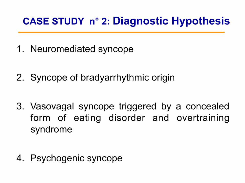
1. Neuromediated syncope
2. Syncope of bradyarrhythmic origin
3. Vasovagal syncope triggered by a concealed form of eating disorder and overtraining syndrome
4. Psychogenic syncope
CASE STUDY n° 2: Diagnostic Hypothesis
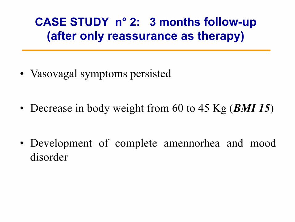
• Vasovagal symptoms persisted
• Decrease in body weight from 60 to 45 Kg (BMI 15)
• Development of complete amennorhea and mood disorder
CASE STUDY n° 2: 3 months follow-up (after only reassurance as therapy)


Med Sci Sports Exerc 2012


Correct answer: vasovagal syncope triggered by a concealed form of eating disorder and overtraining syndrome
CASE STUDY n° 2: Diagnostic Hypothesis

• Complete training interruption • Psychotherapy • Nutritional support
After other 3 months: increase in body weight (from 45 to 50 Kg), reduction
of bradycardia, and no more syncopal spells
CASE STUDY n° 2: Therapy

Thanks for your kind attention !