Symposium on Harmful Marine Algae in the U.S....Symposium on Harmful Marine Algae in the U.S....
224
Symposium on Harmful Marine Algae in the U.S. December 4-9, 2000 Symposium Agenda, Abstracts and Participants Marine Biological Laboratory Woods Hole, Massachusetts
Transcript of Symposium on Harmful Marine Algae in the U.S....Symposium on Harmful Marine Algae in the U.S....

Symposium on Harmful Marine Algae in the U.S.
December 4-9, 2000
Symposium Agenda, Abstracts and Participants
Marine Biological Laboratory
Woods Hole, Massachusetts

Symposium Director:Donald M. Anderson
Symposium Coordinator:Judy Kleindinst
Steering Committee:
Don Anderson Woods Hole Oceanographic InstitutionDan Baden University of North Carolina, WilmingtonSue Banahan NOAA, National Ocean Service, Silver SpringJoAnn Burkholder North Carolina State UniversityPat Glibert University of Maryland Center for Environmental ScienceJohn Heisler EPA, Oceans and Coastal Protection DivisionDennis McGillicuddy Woods Hole Oceanographic InstitutionChris Scholin Monterey Bay Aquarium Research InstituteKevin Sellner NOAA, Coastal Ocean Program, Silver SpringRick Stumpf NOAA, National Ocean Service, Silver SpringPat Tester NOAA, National Ocean Service, BeaufortFran VanDolah NOAA, National Ocean Service, CharlestonTracy Villareal The University of Texas at Austin
Session Coordinators/Chairs:(Note: Names of Session Chairs are underlined)
ECOHAB – Florida: Karen Steidinger, Pat Tester, Fran VanDolahGulf of Mexico HABs Quay Dortch, Tracy VillarealPfiesteria – NC, SC, FL JoAnn Burkholder, Jan Landsberg, Alan Lewitus, Wayne
LitakerPfiesteria – DE, MD, VA Pat Glibert, Dave Oldach, Jeff ShieldsWest Coast HABs Rita Horner, Chris Scholin, Vera TrainerNon-regional HABs Kevin Sellner, Tracy VillarealECOHAB – GOM Don Anderson, Dave TownsendBrown Tides Sue Banahan, Greg Boyer, Cornelia Schlenk
Sponsors:
U.S. National Office for Marine Biotoxins and Harmful AlgaeCalifornia Sea Grant CollegeMaryland Sea Grant College
Monterey Bay Aquarium Research InstituteNational Institute of Environmental Health Sciences
NOAA / Coastal Ocean ProgramNOAA / Center for Coastal Monitoring and Assessment
National Sea Grant OfficeNew York Sea Grant College
South Carolina Sea Grant ConsortiumVirginia Sea Grant College
Symposium on Harmful Marine Algae in the U.S.
December 4-9, 2000
Marine Biological LaboratoryWoods Hole, Massachusetts
Symposium Agenda, Abstracts
and Participants

Symposium on Harmful Marine Algae in the U.S.Program*
December 5-9, 2000
Schedule Tues., Dec. 5 Wed., Dec. 6 Thurs., Dec. 7 Fri., Dec. 8 Sat., Dec. 98:30 – 8:50 Welcome and
Introduction8:30 – 12:15 ECOHAB – Florida Pfiesteria research
– NC, SC & FLWest Coast HABresearch
ECOHAB – GOMresearch
Brown Tides (NY,NJ, DE, MD, TX)
12:15 – 1:30 Lunch break,Swope Dining Hall
Lunch break,Swope Dining Hall
Lunch break,Swope Dining Hall
Lunch break,Swope Dining Hall
Lunch break,Swope Dining Hall
1:30 – 5:15 Gulf of Mexicoregional HABresearch
Pfiesteria research– DE, MD & VA
Non-regional HABresearch
Technicalworkshops/ demos(TBA)
Plenary discussion
Break Break Break Break Break
6:00 – 7:30 Dinner,Swope Dining Hall
Dinner,Swope Dining Hall
Dinner,Swope Dining Hall
Dinner,Swope Dining Hall
Lobster dinner,Swope Dining Hall
7:30 - Discussion Sessions(TBA)
Poster Session (forMon. & Tues.themes)Swope upstairs
Social Hour,Swope upstairs
Poster Session (forThurs., Fri. & Sat.themes)Swope upstairs
Reception,Swope MeigsRoom
*NOTE: All science presentations will be held in the Lillie Auditorium, MBL, MBL Street, Woods Hole.Meals will be held in the Swope Dining Hall, MBL.

3
Symposium on Harmful Marine Algae in the U.S.
December 4-9, 2000Marine Biological LaboratoryWoods Hole, Massachusetts
Program Schedule:
Monday, December 4, 2000
3:00 – 6:00 p.m. Registration, Swope Lobby
6:00 – 7:30 p.m. Dinner, Swope Dining Hall
7:00 – 9:30 p.m. Reception, Swope upstairs lobby
Tuesday, December 5, 2000
8:30 – 8:50 a.m. Welcome and IntroductionLillie Auditorium Don Anderson, Woods Hole Oceanographic Institution
Session I – ECOHAB – Florida(K. Steidinger, F. Van Dolah, P. Tester – Session Coordinators)(P. Tester – Session Chair)
8:50 – 9:10 ECOHAB: Florida overview – the environmentKaren Steidinger, John Walsh, and Gary Kirkpatrick
9:10 – 9:30 Roles of endogenous cellular rhythms and life cycle stage recruitment inGymnodinium breve bloom developmentFrances M. Van Dolah, Michele Barbier, Tod A. Leighfield, Karen A.Steidinger, Bill Richardson and Peter M. McGuire
9:30 – 9:50 ECOHAB Florida: fate and effects of brevetoxins in selected biota,water, and sediments along the west Florida shelf, USAJan Landsberg, Pat Tester, Richard Pierce, Damian Shea, Fran VanDolah, Emilio Sosa, Mike Henry, Jack Fournie, Leanne Flewelling,Sabrina Varnam, and Tod Leighfield
9:50 – 10:10 ECOHAB – Florida: bio-optics and physiologyGary Kirkpatrick, David Millie, Steve Lohrenz, Oscar Schofield, GaryFahnenstiel, Donald Redalje and Terrance Evens
10:10 – 10:30 BREAK
4
10:30 – 10:50 Hydrography and nutrient characteristics within the ECOHAB: Floridacontrol volume on the west Florida shelfGabriel A. Vargo, Cynthia A. Heil, John J. Walsh, Kent Fanning,Carmelo R. Tomas, Karen A. Steidinger, Danylle Ault, Merrie BethNeely, Kristen Lester, and Rachel Merkt
10:50 – 11:10 ECOHAB Florida, physical oceanographyRobert H. Weisberg, Ruoying He, William Hemme, Zhenjiang Li, andHuijun Yang
11:10 – 11:30 The role of behavior in Gymnodinium breve bloom formationDaniel Kamykowski, Gerald S. Janowitz, Gang Liu, Edward J.Milligan, and Robert E. Reed
11:30 – 12:00 Coupled numerical models of Florida red tides of Gymnodinium breveJohn J. Walsh, W. Paul Bissett, Bradley Penta, Dwight A. Dieterle,Robert H. Weisberg, Zhenjiang Li, and Huijun Yang
12:00 – 12:15 Discussion session (if time allows)
12:15 – 1:30 LUNCH BREAK, Swope Dining Hall
Lillie Auditorium Session II – Gulf of Mexico Regional HAB Research(T. Villareal, Q. Dortch, Session Coordinators)(Q. Dortch, Session Chair)
1:30 – 1:50 Pseudo-nitzschia spp. in the northern Gulf of Mexico: overview andresponse to increasing eutrophicationQ. Dortch, M.L. Parsons, G.J. Doucette, G. A. Fryxell, A. Maier, A.Thessen and C.L. Powell
1:50 – 2:10 Algicidal bacteria active against Gymnodinium breve: use of moleculartechniques to assess changes in microbial communities following theintroduction of bacteriaXavier Mayali and Gregory J. Doucette
2:10 – 2:30 Variable brevetoxin production in Gymnodinium breve attributable togrowth conditions and strain differencesRichard M. Greene, Janis C. Kurtz, Roman S. Stanley, Cynthia A.Chancy, Michael C. Murrell, Fred J. Genthner, John E. Rogers, andCalvin C. Walker

5
2:30 – 2:50 Gymnodinium breve red tides in the Gulf of Mexico: analysis ofbrevetoxins and metabolites in shellfishR.W. Dickey and S.M. Plakas
2:50 – 3:10 Gymnodinium breve in the western Gulf of Mexico: two years of coastalsampling off the Texas coastTracy A. Villareal, Mary A. Brainard, and H. Magaña and LawrenceW. McEachron
3:10 – 3:40 The effect of nitrogen source on the growth and toxicity of speices of thegenus ProrocentrumCary L. Burns and Jonathan R. Pennock
3:40 – 4:00 Discussion session (if time allows)
4:00 – 5:00 Informal discussion groups or free time – put up posters
5:00 – 6:00 INFORMAL SOCIAL HOUR, Swope upstairs Lobby
6:00 – 7:30 - DINNER, Swope Dining Hall
7:30 – DISCUSSION SESSIONS (TBA)
Wednesday, December 6, 2000
Lillie Auditorium Session III – Pfiesteria research – NC, SC, & FL(J. Burkholder, J. Landsberg, W.Litaker, A. Lewitus – SessionCoordinators)(J. Burkholder, Session Chair)
8:30 – 8:50 Pfiesteria, Pfiesteria-like species, and fish health in Florida: an updateJan Landsberg, Karen Steidinger, Susan Cook, Elizabeth Singh, EmilioSosa, Ann Forstchen, Robin Wood, Parke Rublee, Paula Scott, JenniferWolny, and Brian Bendis
8:50 – 9:10 Preliminary characterization of “Cryptoperidiniopsoid” cultures isolatedfrom FloridaSteve L. Morton, Tina Mikulski, Elizabeth R. Fairey, Brad Mitchell,Peter D.R. Moeller, Bill Richardson, Karen Steidinger, and JohnRamsdell
9:10 – 9:30 Pfiesteria spp. and “Pfiesteria-like organisms” in South CarolinaestuariesAlan J. Lewitus, J.M. Burkholder, C. Cary, H.B. Glasgow Jr., K.C.Hayes, A.F. Holland, J.M. Law, and P.A. Rublee
6
9:30 – 9:50 Nutrient enrichment and the toxic Pfiesteria complex: comparativestimulation by swine effluent, poultry manure leachate, human sewage,and other sourcesJ. Burkholder, C. Zheng, H. Glasgow, N. Deamer-Melia and M.Parrow
9:50 – 10:10 Distribution of Pfiesteria species: comparison of results from water andsediment samples across multiple scales, 1998-2000Parke A. Rublee, Eric F. Schaefer, Coy Allen, Janera Harris, HollyBowers, Torstein Tengs, and D.W. Oldach
10:10 – 10:30 BREAK
10:30 – 10:50 Pfiesteria field ecology and toxic activity: trends from a decade ofintensive study in North Carolina estuariesR. Reed, H. Glasgow, J. Burkholder, N. Deamer-Melia and M. Mallin
10:50 – 11:10 Toxic Pfiesteria promotes acute and chronic lesions in finfish, incontrolled experimental trialsH. Glasgow, Jr., R. Smolowitz, N. Deamer-Melia and J. Burkholder
11:10 – 11:30 Interactions between Pfiesteria and representative species ofcommercially valuable shellfishS. Shumway, J. Springer, J. Burkholder and H. Glasgow
11:30 – 11:50 Characterization of a putative toxin produced by Pfiesteria piscicidaJ.S. Ramsdell, P.D.R. Moeller, E.R. Fairey, A.C. Melo, K.L. Kimm-Brinson, B. Mitchell, S.A. Morton, N. Deamer-Melia , H.B. Glasgow,and J.M. Burkholder
11:50 – 12:15 Discussion session (if time allows)
12:15 – 1:30 LUNCH BREAK, Swope Dining Hall
Lillie Auditorium Session IV – Pfiesteria research – DE, MD & VA(P. Glibert, D. Oldach, J. Shields - Session Coordinators)(P. Glibert, Session Chair)
1:30 – 1:45 Approaches to the investigation and interpretation of possible Pfiesteria-related events in MarylandRobert E. Magnien, David M. Goshorn, David W. Oldach, Holly A.Bowers, and Torstein Tengs
1:45 – 2:00 Intensive monitoring for Pfiesteria and related HAB events

7
B. Boicourt, L. Codispoti, M. Roman, V. Holliday, H. MacIntyre, P.Glibert, R. Magnien, and B. Michael
2:00 – 2:15 Assessing temporal and spatial variability in Pfiesteria piscicidadistributions using molecular probing techniquesKathryn J. Coyne, David A. Hutchins, Clinton E. Hare and S. CraigCary
2:15 – 2:30 Functional type (toxicity status) controls Pfiesteria response to nutrientsand algal versus fish preyJ. Burkholder, H. Glasgow, P. Glibert, A. Lewitus, M. Parrow, C.Zheng, P. Cancellieri and N. Deamer-Melia
2:30 – 2:45 Nitrogen uptake and nutrient relationships in laboratory cultures andfield assemblages of PfiesteriaP.M. Glibert, A. Lewitus, J. Burkholder, H. Glasgow, M. Mulholland,and C. Lee
2:45 – 3:00 Potential grazing on Pfiesteria piscicida by microzooplankton in thePocomoke RiverDiane K. Stoecker and Daniel E. Gustafson, Jr.
3:00 – 3:20 BREAK
3:20 – 3:35 Trophic relationships of phytoplankton and microzooplankton withPfiesteria-like heterotrophic dinoflagellates in Pocomoke River andTransquaking/Chicamacomico Rivers, MD, USARichard V. Lacouture, Jennifer Gronefeld, Ann Marie Hartsig, StellaSellner and Amy Imirie
3:35 – 3:50 Results of a series of fish bioassays with the toxic dinoflagellatePfiesteria piscicidaHarold G. Marshall, Andrew S. Gordon, David W. Seaborn, BrianDyer, William M. Dunstan, and A. Michelle Seaborn
3:50 – 4:05 Skin lesions in estuarine fishes: a comparative pathological evaluation ofwild and laboratory-exposed fishWolfgang K. Vogelbein, Kimberly S. Reece, Jeffrey D. Shields, DavidE. Zwerner, Patrice L. Mason, Larry W. Haas and Vicki Blazer
4:05 – 4:20 Development and testing of molecular diagnostics for Pfiesteria-likeorganisms in laboratory and environmental samplesKimberly S. Reece, Nancy A. Stokes, Wolfgang K. Vogelbein, WayneL. Litaker, Jeffrey D. Shields, Larry W. Haas, Patrice L. Mason, VictoriaM. Foster and Eugene M. Burreson

8
4:20 – 4:35 Pfiesteria or fungus? Induction of skin ulcers in menhaden withzoospores of Aphanomyces spp.Yasunari Kiryu, Jeffrey D. Shields, Wolfgang K. Vogelbein, David E.Zwerner, Howard Kator, and Vicki S. Blazer
4:35 – 4:50 The ambush predator Pfiesteria piscicida: fad or fallacyPatrick Gillevet, Thomas Nerad, Michael T. Peglar, Greg C. Garman,Stanley Webb, Bonnie Brown, and Charles J. O'Kelly
4:50 – 5:15 Discussion session (if time allows)
6:00 – 7:30 DINNER, Swope Dining Hall
7:30 – POSTER SESSION, Swope upstairs and Meigs Room
Thursday, December 7, 2000
Lillie Auditorium Session V – West Coast HAB research(C. Scholin, R.Horner, V. Trainer – Session Coordinators)(C. Scholin, Session Chair)
8:30 – 9:00 HABs-related physical oceanography off the U.S. west coastBarbara M. Hickey
9:00 – 9:30 The challenges of forecasting and managing toxic Pseudo-nitzschiablooms on the U.S. west coastVera L. Trainer
9:30 – 10:00 Field studies of toxic phytoplankton in central California: 1999-2000Mary Silver, Susan Coale, Shonna Dovel, Kathi Lefebvre, GregDoucette, Ron Tjeerdema, and Rikk Kvitek
10:00 – 10:20 BREAK
10:20 – 10:50 Approaches to the detection of domoic acid in marine food websGregory J. Doucette
10:50 – 11:20 Trace metals and Pseudo-nitzschia blooms: a possible role for the toxindomoic acidEden Rue, Maria Maldonado, Ken Bruland and Mark Wells
11:20 – 11:50 SummaryRita A. Horner
11:50 – 12:15 Discussion session (if time allows)

9
12:15 – 1:30 LUNCH BREAK, Swope Dining Hall
Lillie Auditorium Session IV – Non-regional HAB Research(T. Villareal, K. Sellner, Session Coordinators and Chairs)
1:30 – 1:50 Long term occurrence patterns and dynamics of ichthyotoxicHeterosigma akashiwo in Narragansett BayYaqin Li and Theodore J. Smayda
1:50 – 2:10 An expansion of harmful raphidophyte blooms in U.S. coastal watersCarmelo R. Tomas
2:10 – 2:30 The potential for sediment-water column interactions to stimulategrowth of the bloom-forming dinoflagellate Prorocentrum minimumHugh MacIntyre, Jason Adolf and Angela Dubois
2:30 – 2:50 Use of cell specific PAM -fluorometry to characterize light acclimationresponses of Gambierdiscus toxicus (Dinophyceae)Tracy A. Villareal and Steve Morton
2:50 – 3:10 Evidence for adaptation to toxins in populations of the softshell clam,Mya arenaria, subjected to recurrent toxic bloomsV. Monica Bricelj, Scott P. MacQuarrie, Betty M. Twarog, and Vera L.Trainer
3:10 – 3:30 BREAK
3:30 – 3:50 Rapid testing using the MIST alert™ for paralytic shellfish poisoning(PSP), for trials within the United StatesJoanne F. Jellett
3:50 – 4:10 Antillatoxin, a novel neurotoxin from the marine cyanobacteria Lyngbyamajuscala, is a potent activator of voltage-gated Na+ channelsThomas F. Murray, W.I. Li, F.W. Berman, T. Okino and W.H.Gerwick
4:10 – 4:30 Unusual sterols from harmful algae: more than biomarkers?José-L. Giner, Gregory Boyer, Juan Faraldos, Xiaoyong Li and HuiZhao
4:30 – 4:50 High evolutionary rates in Gymnodinium galatheanum chloroplast DNAsequences and development of a molecular detection assayTorstein Tengs, Holly A. Bowers, Andrew P. Ziman, Diane K. Stoeckerand David W. Oldach

10
4:50 – 5:10 Harmful algal blooms in the United States: estimates of economicimpacts and policy responsesPorter Hoagland, Di Jin, Hauke Kite-Powell and Tracey Morin
5:10 – 5:40 Discussion session (if time allows)
6:00 – 7:00 DINNER, Swope Dining Hall
7:00 – 8:00 INFORMAL SOCIAL HOUR, Swope Upstairs Lobby
December 8, 2000
Lillie Auditorium Session VII – ECOHAB – Gulf of Maine(D.Anderson, D. Townsend, D. McGillicuddy – Session Coordinators)(D. Townsend, Session Chair)
8:30 – 8:45 ECOHAB-GOM: the ecology and oceanography of toxic Alexandriumblooms in the Gulf of MaineDonald M. Anderson
8:45 – 9:30 Offshore blooms of the red tide dinoflagellate, Alexandrium sp., in theGulf of MaineDavid W. Townsend, Neal R. Pettigrew and Andrew C. Thomas
9:30 – 10:00 Alexandrium fundyense blooms in the western Gulf of MaineDonald M. Anderson, Bruce A. Keafer, James H. Churchill, Richard P.Signell and Wayne R. Geyer
10:00 – 10:20 BREAK
10:20 – 10:35 Physiological diagnostics and behavior of the toxic dinoflagellateAlexandrium fundyense, in Casco Bay, Maine – evidence of nitrogenlimitationNicole J. Poulton, J. Geoff MacIntyre, John J. Cullen, and Donald M.Anderson
10:35 – 10:50 Accumulation of PSP toxins in zooplankton assemblages in the Gulf ofMaineJefferson T. Turner, Christine L. Powell, David M. Kulis, Bruce A.Keafer, Donald M. Anderson, and Gregory J. Doucette
10:50 – 11:05 An overview of interactions between zooplankton grazers andAlexandrium sp., and effects of grazing on bloom dynamics in the near-shore environment of the Gulf of MaineGregory J. Teegarden, Robert G. Campbell, Allan D. Cembella, andEdward G. Durbin

11
11:05 – 11:20 Alexandrium cyst dynamics in the Gulf of MaineDonald M. Anderson, Bruce A. Keafer, Patricia A. Matrai, MaureenKeller
11:20 – 11:50 Modeling Alexandrium spp. blooms in the Gulf of MaineDennis J. McGillicuddy, Jr. , Richard Signell, Charles Stock, Daniel R.Lynch and Andrew Thompson
11:50 – 12:15 Discussion session (if time allows)
12:15 – 1:30 LUNCH BREAK, Swope Dining Hall
1:30 – 5:00 p.m. Technical Workshops/Demonstrations
6:00 – 7:30 p.m. - DINNER, Swope Dining Hall
7:30 - POSTER SESSION, Swope upstairs & Meigs Room
Saturday, December 9, 2000
Lillie Auditorium Session VIII – Brown Tides(S. Banahan, C. Schlenk, G. Boyer – Session Coordinators)(S. Banahan, Session Chair)
8:30 – 9:00 An overview of brown tide in the northeast U.S.Gregory Boyer
9:00 – 9:20 The impact of bottom-up and top-down processes on the abundance ofAureococcus anophagefferens during the 1999-2000 brown tide bloomin Great South Bay, NY, USAChristopher J. Gobler
9:20 – 9:40 Causes and prevention of brown tides in the northeastern United States:the importance of trophic links in the plankton and benthosDavid A. Caron, Darcy J. Lonsdale, Rebecca Schaffner, Robert Cerratoand Julie Rose
9:40 – 10:00 Benthic-pelagic coupling and LI brown tideMichael W. Lomas, Hugh L. MacIntyre, Jeffrey C. Cornwell and ToddM. Kana
10:00 – 10:20 BREAK
10:20 – 10:40 Growth of Aureococcus anophagefferens on complex sources ofdissolved organic nitrogen in cultureGry Mine Berg, Julie LaRoche, and Dan Repeta

12
10:40 – 11:00 Amino acid oxidation and peptide hydrolysis in populations seasonallydominated by Aureococcus anophagefferensMargaret R. Mulholland, Christopher Gobler and Cindy Lee
11:00 – 11:20 Brown tide assessment project in Barnegat Bay, NJ and the presence ofviral-like particles in natural populations of AureococcusanoohagefferensMary Downes Gastrich, O.R. Anderson, and Elizabeth M. Cosper
11:20 – 11:40 Role of long-term variation in freshwater input and dissolved organicnitrogen delivery in the initiation and maintenance of the 1985Narragansett Bay brown tideDavid Borkman and Theodore J. Smayda
11:40 – 12:00 Physical, chemical and biological conditions associated with theNarragansett Bay brown tideTheodore J. Smayda and David Borkman
12:00 – 12:20 Discussion session (if time allows)
12:20 – 1:30 p.m. - LUNCH BREAK, Swope Dining Hall
1:30 – 5:00 p.m. Plenary Discussion, Lillie Auditorium
Update on U.S. – European Union interactions on HABs (E. Lipiatou)
Summaries of discussion sessions
HABs in the U.S. – comparative discussion (common impediments,common mechanisms, identified needs for methods development, etc.)
Status of National HAB program (congressional activities, futureinitiatives, workshops, etc.)
5:00 – 6:00 p.m. - BREAK
6:00 – 7:30 p.m. LOBSTER DINNER, Swope Dining Hall
7:30 – 11:00 p.m. RECEPTION, Meigs Room, Swope

13
__
ABSTRACTS OF ORAL PRESENTATIONS

14
15
ECOHAB-GOM: THE ECOLOGY AND OCEANOGRAPHY OF TOXIC ALEXANDRIUMBLOOMS IN THE GULF OF MAINE
Donald M. AndersonWoods Hole Oceanographic Institution, Woods Hole MA 02543
A five-year program called ECOHAB-GOM was initiated to address several fundamental issuesregarding Alexandrium blooms in the Gulf of Maine: 1) the source of the Alexandrium cellsthat appear in the fresh water plumes in the western Maine coastal current (WMCC); 2) Alexandriumcell distribution and dynamics in the eastern Maine coastal current (EMCC); and 3) linkages amongblooms in the WMCC and the EMCC. Utilizing a combination of numerical modeling, hydrographic,chemical, and biological measurements, moored and drifting current measurements, and satelliteimagery, the project will characterize the structure, variability and autoecology of the majorAlexandrium habitats in the Gulf of Maine.
In the western Gulf, Alexandrium blooms and patterns of PSP have been linked to a coastal current orplume of low salinity river outflow (the WMCC). One major project goal is to investigate an area nearCasco Bay implicated as the major "source region" for the toxic cells that populate that coastal current.Field surveys will elucidate the biological, chemical, and physical processes that control bloominitiation and development, the delivery of cells from that source region into the WMCC, and themanner in which late-season, localized blooms are retained there to re-seed future blooms with cysts.The second major set of objectives is to characterize the linkage between toxic blooms and the EMCC,to investigate the role of tidal mixing, frontal systems, and upwelling/downwelling in Alexandriumdynamics, and to define the linkage between EMCC Alexandrium populations and those in both theWMCC (downstream) and the Bay of Fundy (upstream). A series of "process" studies will focus ondiscrete blooms or patches of cells and quantify such parameters as in situ growth rates and grazinglosses of Alexandrium, the nutritional physiology, vertical migration behavior and transport of thisspecies, and the partitioning of toxins within the food web. A hierarchy of coupled physical-biologicalmodels are being used together with ECOHAB-GOM data for investigation of: 1) detailed structurewithin each habitat; 2) interconnections among habitats; and 3) the role of the larger Gulf-scalecirculation in the long-term maintenance of Alexandrium populations in the region. ECOHAB-GOM isthus a combined modeling/observational program, utilizing the most current and innovativetechnologies in an approach commensurate with the multiple scales and oceanographic complexity ofPSP phenomena in the Gulf of Maine. More details on this project, including an update of cruiseactivities and results can be found at the project web page at: http://crusty.er.usgs.gov/ecohab/
ECOHAB-GOM Principal Investigators and their affiliations are: David W. Townsend (University ofMaine), James H. Churchill (Woods Hole Oceanographic Institution), John J. Cullen (DalhousieUniversity), Gregory J. Doucette (Medical Univ. of South Carolina), Wayne R. Geyer (Woods HoleOceanographic Institution), John Hurst (State of Maine Dept. of Marine Resources), Maureen D.Keller (Bigelow Laboratory for Ocean Sciences), Theodore C. Loder III (University of NewHampshire), Daniel R. Lynch (Dartmouth College), Jennifer L. Martin (Canadian Dept. of Fisheriesand Oceans), Dennis J. McGillicuddy (Woods Hole Oceanographic Institution), Neal R.Pettigrew (University of Maine), Richard P. Signell (U.S. Geological Survey), Andrew C. Thomas(University of Maine), Jefferson T. Turner (University of Massachusetts Dartmouth).
16
ALEXANDRIUM FUNDYENSE BLOOMS IN THE WESTERN GULF OF MAINE
Donald M. Anderson1, Bruce A. Keafer1, James H. Churchill1, Wayne R. Geyer1 and Richard P.Signell2
1Woods Hole Oceanographic Institution, Woods Hole, MA 025432US Geological Survey, Woods Hole, MA 02543
The first outbreaks of PSP in the Gulf of Maine are reported nearly every year along the WesternMaine coastline, especially in the Casco Bay region. Previous research has shown that this region isstrongly influenced by the Kennebec River, one of the major freshwater inputs into the western Gulf,and that surface populations of Alexandrium fundyense are generally associated with the river plume.An “initiation zone” has also been hypothesized in the Casco Bay area, though the specific distributionand source of those cells are not known. During the spring of 1998 and 2000, weekly hydrographicsurveys in Casco Bay and the adjacent Kennebec River plume area collected A. fundyense samples atvarious depths to 20m to observe physical water properties associated with the blooms. Hydrographicmoorings with current meters were deployed at several sites along with mussel bags hung near thesurface. Further information on the current field was obtained from satellite-tracked drifters.
Prior to detection of shellfish toxicity (i.e., late April), low abundances (<100 cells liter-1) of A.fundyense cells were observed within the inshore waters of Casco Bay as well as in offshore watersbeyond the influence of the Kennebec River plume. During the initial onset of toxicity, cellabundances increased to ca. 200-300 cells liter-1 with populations found at both inshore and offshorestations. In 1998, the toxicity was first detected at an offshore mooring suggestive of an offshore originof the bloom. However, in 2000, toxicity was first reported inshore. These observations are consistentwith the view that there are two populations of A. fundyense; one located in Casco Bay and another inthe adjacent offshore waters and to the east (i.e., "upstream”).
During both years, the rapid rise in shellfish toxicity in mid-May was recorded at the inshoremonitoring stations and at the mussel moorings following downwelling favorable conditions. Surfaceconcentrations of toxic cells ranged from <100 to ca. 1000 cells liter-1 with no evidence that the higherconcentrations were associated with any particular water mass. Within the plume waters, a 24 hourstudy indicated that the toxic cells predominantly occupied the stratified, surface waters (<10m) evenduring the night, but subsurface populations were occasionally observed below the pycnocline thatwere linked to advection from the "upstream" waters. Higher abundances of cells were sometimesfound near a frontal boundary where the offshore waters converge with the Kennebec and Penobscotriver plumes. Evidence from Acoustic Doppler Current Profiler (ADCP) and drifters suggested thattoxic cells found near the convergence were transported in a coastal "jet" which drives the cells rapidlyalongshore with the Western Maine Coastal Current.
These results indicate a wide distribution of A. fundyense in this highly dynamic region. Cells arepresent at approximately the same times in both inshore waters, offshore waters, and at convergencesassociated with river plumes. Therefore, it is not yet possible to determine whether there is an inshore“source”, offshore "source", or multiple "sources" for A. fundyense cells in the western portion of theGulf of Maine, versus a more broadly dispersed initial population. The development of the offshorebloom appears dependant on complex physical-biological processes (accumulation vs. growth)associated with the different water masses. Meanwhile, inshore populations within Casco Bay aregenerally retained in a slower flow leading to the higher toxicities observed in the bay. The Casco Baypopulations may derive from the offshore populations, delivered to the bay with downwelling winds,or they could arise independently from cyst populations in the shallow nearshore waters. In either case,there is still an identifiable “initiation zone” in the general region where the relative abundance of A.fundyense and toxicity is consistently higher earlier in the year compared to any area along the Gulf of
17
Maine coastline. The study is ongoing with another field year remaining to elucidate the dominantprocesses responsible for initiation and development of A. fundyense blooms in the region.
ALEXANDRIUM CYST DYNAMICS IN THE GULF OF MAINE
Donald M. Anderson1, Bruce A. Keafer1, Patricia A. Matrai2, Maureen Keller2*
1Woods Hole Oceanographic Institution, Woods Hole, MA 025432Bigelow Laboratory for Ocean Sciences, W. Boothbay Harbor, ME 04575
Alexandrium fundyense, the dinoflagellate responsible for Paralytic Shellfish Poisoning (PSP) in theGulf of Maine (GOM), has a dormant cyst stage in its life history that plays a critical role in bloomdynamics. Given the highly seasonal nature of PSP outbreaks in the region, the cold winters, and thetemperature requirements for A. fundyense growth (5-21 ˚C), blooms of this species presumably beginfrom germinated cysts. Surveys of cyst abundance in GOM sediments document high cystconcentrations in deeper, offshore basins, grading to much lower concentrations in shallow waters. Itwas hypothesized that the most significant input of A. fundyense cells would be from cysts in shallowwaters that would be exposed to high light levels and more rapid temperature increases than the cystsin the darker and colder offshore waters, even though cyst concentrations are 1-2 orders of magnitudehigher offshore. During the ECOHAB-GOM project, several parallel efforts were made to estimatethe magnitude and timing of cell input via cyst germination. These efforts, and their present status, aresummarized as follows:
1) Cyst germination traps: Several benthic chambers designed to capture germinated cellswere collected. Thus far, none of these have provided reliable data.
2) Cyst fluorescence: Surface sediments were collected at a number of stations and A.fundyense cysts examined for signs of chlorophyll fluorescence, thought to be a sign of impendinggermination. Data thus far show no meaningful patterns or trends.
3) Modeled cyst germination: Laboratory experiments were conducted to characterize A.fundyense cyst germination rates as functions of temperature, light and time of year (the latterreflecting control of germination by an internal clock). In conjunction with a large-scale cyst map ofthe region, these data are being used in model simulations driven by environmental and hydrographicconditions observed and modeled in 1993. Results thus far provide a cell inoculum to the watercolumn that varies temporally and spatially. In particular, it appears that the deep-water cyst seedbeddominates the inoculum process, but that seedbed is not in the proper location to account for observedblooms in the Casco Bay area. An eastward extension of this seedbed, lying outside the modeleddomain could be the source of A. fundyense cells to the Casco Bay region. Model simulations havesuggested a mechanism whereby upwelling-favorable winds move nearshore waters over the deep-water cyst seedbeds, capturing germinated cells that are then carried to shore when winds shift todownwelling-favorable. Details of this modeling effort will be provided by McGillicuddy et al. in aseparate presentation. Armstrong et al. and Thompson et al. present posters with details of similar cystgermination rate experiments for eastern Gulf of Maine populations which will allow an extension ofthe modeling effort.
These observations on cyst distribution and dynamics will be discussed in the context of observedAlexandrium motile cell distribution in 1998 and 2000.
*Portions of this work were started under the leadership of the late Dr. Maureen D. Keller.

18
GROWTH OF AUREOCOCCUS ANOPHAGEFFERENS ON COMPLEX SOURCES OFDISSOLVED ORGANIC NITROGEN IN CULTURE
Gry Mine Berg1, Julie LaRoche1, Dan Repeta2
1Institut für Meereskunde, Kiel University, 24105 Kiel, Germany2Woods Hole Oceanographic Institution, Woods Hole, MA 02540, USA
Aureococcus anophagefferens repeatedly blooms in several Long Island (New York, USA)embayments, forming "brown tides" that discolor the water. Surveys of the northeast coast of the USAhave shown that A. anophagefferens exits in several places that have no records of brown tide.Therefore, the recurrence of the brown tide in Long Island is somewhat unusual. The coastal bays inLong Island are strongly influenced by groundwater, contributing the largest input of fixed nitrogen.In years of draught and low groundwater flow, the supply of NO3
- is sharply reduced leaving dissolvedorganic nitrogen (DON) as the largest source of nitrogen available to the phytoplankton. The ability ofA. anophagefferens to grow on DON has been hypothesized to be an important factor in sustaining thebrown tide during periods of dissolved inorganic nitrogen depletion. In order to test this hypothesis weprepared an axenic culture of A. anophagefferens and followed growth with a number of DONsubstrates as the sole source of nitrogen in culture. In addition to commercially available substrates weused >1 kDa ultrafiltered DON isolated from West Neck Bay (WNB) pore waters, Long Island.Efforts to characterize components of the bulk DON pool were conducted in parallel withinvestigations of the bioavailability of these components.
For the preparation of an axenic, artificial seawater culture of strain CCMP 1784 we modified aprotocol published by Cottrell and Suttle (1995 J. Phycol. 29: 385-387). Exponentially growingcultures in F/2 media were exposed sequentially to Penicillin G, Neomycin, Streptomycin, andPenicillin G. Of the antibiotics tested, Penicillin G was the most effective in eliminating bacterialcontaminants. Bacterial strains isolated from the culture medium were identified throughamplification of bacterial 16S rRNA gene sequences using PCR. Two bacterial strains isolated fromthe culture media, belonging to the Gamma Proteobacteria and to the Cytophaga-Flavobacteria, wereof marine origin.
A. anophagefferens showed good growth on > IkDa WNB DON. To date, this is the first studydemonstrating that an autotrophic phytoplankton can grow on bulk DON as the sole source ofnitrogen, suggesting that autrotrophs have the capability to enzymatically degrade complex DON.Future research will investigate enzyme pathways involved with DON degradation, and on interactionsbetween A. anophagefferens and heterotrophic bacteria.
19
INTENSIVE MONITORING FOR PFIESTERIA AND RELATED HAB EVENTS
B. Boicourt1, L. Codispoti1, M. Roman1, V. Holliday2, H. MacIntyre1, P. Glibert1, R. Magnien3, and B.Michael3
1Horn Point Laboratory, University of Maryland Center for Environmental Science, PO Box 775,Cambridge, MD 216132 BAE SYSTEMS, Integrated Defense Solutions, 6500 Tracor Lane, Austin, TX 787253Maryland Department of Natural Resources, Resource Assessment Service, 580 Taylor Ave., D2,Annapolis, MD 21401
Intensive monitoring efforts, a collaboration between the University of Maryland Center forEnvironmental Science, Maryland Department of Natural Resources, and NOAA, are ongoing fordetermining environmental conditions leading to the outbreaks of Pfiesteria and related HAB species.Our intent is to develop an adaptable, autonomous system measuring a key suite of variables andreporting in real time. Initial efforts have been brute-force, deploying a wide range of new andconventional sensors from both fixed and floating platforms, in an attempt to assemble a tractable andsensitive detection system. In addition to monitoring meteorological and physical variables, sensorsare deployed to monitor chlorophyll, zooplankton, optics, oxygen, nutrients and presence of fish.Acoustic telemetry is employed for underwater communications, and spread-spectrum radio relays thedata to shore stations where the data are placed on the internet. A modified Chesapeake BayObserving System (CBOS) buoy and shallow water platforms were employed to mount the varioussensors in the lower Pocomoke River, a tributary of Chesapeake Bay, during late summer/early fall2000.
Nutrient enrichment has been shown to stimulate Pfiesteria, but the relationships between nutrientloading and Pfiesteria growth are not clear. For nutrient monitoring, WS Ocean System nutrientmonitors are employed to autonomously determine nitrate+nitrite and phosphate concentrationsapproximately every two hours. During instrument deployments, discrete samples are collected at thesame location and time every few days for calibration of nutrients and water column chemistry. Weare working on developing a system that can autonomously collect ammonium data during futuredeployments.
A suite of bio-optical instruments (fluorometer, turbidimeter and AC-9) are used for detectingvariations on the optical characteristics of the water. Absorption spectra are resolved into thecontribution of CDOM, detritus, and plant pigments at 8 wavelengths, using empirically-derivedrelationships based on fluorescence and back-scatter. The approach may permit the development ofspecies-specific spectral signatures, depending on the robustness of the algorithms for correcting thevery high concentrations of CDOM and suspended sediment at the site. Although Pfiesteria cannot bedetected with this approach directly, various spectral signatures may be correlated with its presence.
Two biological components which have the potential to affect Pfiesteria are zooplankton and fish.Zooplankton consume Pfiesteria and thus are a biological control of their abundance and potential tobloom. High concentrations of fish can potentially trigger the metamorphosis of toxic stages ofPfiesteria. An in situ acoustic sensor capable of monitoring zooplankton and fish is being used todetect zooplankton and fish. The instrument is moored on the sediment surface, and includes a side-looking low-frequency acoustic transducer for detection of fish schools, and a high-frequency sensoraimed at the surface for monitoring the zooplankton in the water column above the mooring.

20
ROLE OF LONG-TERM VARIATION IN FRESHWATER INPUT AND DISSOLVEDORGANIC NITROGEN DELIVERY IN THE INITIATION AND MAINTENANCE OF THE1985 NARRAGANSETT BAY BROWN TIDE
David Borkman and Theodore J. SmaydaGraduate School of Oceanography, University of Rhode Island, Kingston, RI 02881
The 1985 brown tide bloom of Aureococcus anophagefferens in Narragansett Bay was the dominantHAB event in a nearly 40-year time series of weekly observations of Narragansett Bay phytoplankton.Mechanisms responsible for this summer-long bloom, which occurred simultaneously in severalestuaries along the Northeastern US coast, are not fully known. Several features of A. anophagefferensphysiology and ecology indicate that freshwater input patterns with accompanying patterns in deliveryof organic nutrients may play an important role in bloom initiation and maintenance. We present atime-series of estimated dissolved organic nitrogen (DON) loading and related physical data forNarragansett Bay, Rhode Island. Trends in Narragansett Bay riverine and groundwater DON input areanalyzed, indicating a relative peak in riverine DON concentration accompanied by a decrease in theriverine nitrate:DON ratio in 1985. Levels of A. anophagefferens that may have been supported by thisDON delivery are estimated and compared to observed abundance in Narragansett Bay. The spring of1985 was marked by a departure from the usual relation between groundwater levels and salinity inNarragansett Bay, indicative of a change in Narragansett Bay estuarine circulation patterns. Changes infreshwater delivery into Narragansett Bay in 1985 and accompanying relative increases in DONdelivery are implicated in the initiation and maintenance of the 1985 brown tide.
21
AN OVERVIEW OF BROWN TIDE IN THE NORTHEAST U.S.
Gregory L. BoyerCollege of Environmental Science and Forestry, SUNY Syracuse, NY 13210
Brown tide blooms are regional, episodic phenomena. The first major occurrences of brown tide(Aureococcus anophagefferens) were reported in 1985 in the eastern and southern bays of Long Island(NY), Narragansett Bay (RI), and Barnegat Bay (NJ). Since then blooms of varying severity andduration continue to occur in Long Island waters and Barnegat Bay, and as of summer 1998, browntide cells were reported in the eastern bays of Maryland and Delaware.
Brown tide blooms can have had serious impacts on shellfish fisheries. The massive bloom of 1985resulted in the recruitment failure of scallops in the Peconic Bay (Long Island) system. While therehave been some modest harvests since that failure, bay scallop populations have not recovered to theirpre-1985 levels. In recognition of a need to focus more expertise into understanding this phenomenon,a research program to understand the causes of these blooms was developed. The Brown TideResearch Initiative (BTRI) began in 1996 with two objectives: 1) to isolate, develop, and maintainaxenic cultures of Aureococcus, and 2) identify the environmental factors that contribute to theinitiation, duration, and cessation of brown tide blooms.
Multiple isolates of Aureococcus have been established and are maintained in culture at CCMP,however problems with maintaining axenic cultures persist. To address the broader 2nd objective,investigators have been evaluating the relative importance of factors such as DIN, DON, dissolvediron, groundwater loading, and light in Aureococcus growth physiology. The environmental andecological factors examined are water column stability and residence times, changes in the speciescomposition of the microbial plankton community, microbial and bivalve grazing, and elucidating bio-geochemical processes at the sediment-water interface.
Initial assumptions were that Aureococcus blooms were, in part, the result of unique growthcharacteristics. However, research to date suggests that Aureococcus shares similar growthcharacteristics with other picoplankton. Blooms are more likely the result of a combination ofecological and environmental factors.
22
EVIDENCE FOR ADAPTATION TO TOXINS IN POPULATIONS OF THE SOFTSHELLCLAM, MYA ARENARIA, SUBJECTED TO RECURRENT TOXIC BLOOMS
V. Monica Bricelj1, Scott P. MacQuarrie1, Betty M. Twarog2, and Vera L. Trainer3
1Inst. for Marine Biosciences, National Research Council, 1411 Oxford St., NS B3H 3Z1, Canada2Darling Marine Center, University of Maine, Walpole, ME 045733Northwest Fisheries Science Center, Seattle, WA 98112-2097
Our ECOHAB study demonstrates that softshell clam, Mya arenaria, populations differ markedly intheir sensitivity to PSP toxins depending on their history of exposure to paralytic shellfish poisoning(PSP) episodes in Atlantic coastal waters. Significant differences were found in burrowing capacity,feeding (clearance) rates, nerve resistance to saxitoxin (STX), metabolic rates, toxin accumulation andsurvival of juvenile clams during laboratory exposure to a highly toxic Alexandrium tamarense isolate.Marked phenotypic differences in these parameters were also documented among individual clamswithin a population. Sensitive clams predominate in populations not previously exposed to PSPtoxins, whereas resistant phenotypes prevail in historically exposed populations. Toxicity of visceracould vary up to 52x among contrasting phenotypes exposed to common conditions of toxification.Our results strongly support the hypothesis that softshell clam populations recurrently exposed to toxicblooms undergo genetic or epigenetic adaptation to PSP toxins, via selective mortality or reducedfitness of sensitive individuals.
Repeated testing of the same individuals showed that clams with no history of exposure to PSPexperienced significant (up to 4-fold) reduction in metabolic rate (rate of oxygen consumption, VO2)during laboratory exposure to bloom levels of toxic A. tamarense cells, relative to controls exposed tonon-toxic algae. In contrast, clams from a population recurrently affected by PSP showed onlytransient reduction of VO2 after 9 days of toxification, and recovery of VO2 to normal levels at 18 d oftoxification. Laboratory exposure to PSP toxins also induced significant clam mortalities relative tonon-toxic controls, starting within 7-11 days of toxification. Mortality rates were population-specific(higher in an unexposed population than in one historically affected by PSP), and phenotype-specific.Thus sensitive individuals within each population (characterized by loss of burrowing capacity duringtoxification) consistently suffered higher mortalities than resistant individuals. Considerableunexplained variation was observed, however, in cumulative mortality rates among three independentexperiments. Neither starvation nor impaired ability to maintain a normal position in sediment due toparalysis, were responsible for mortalities in sensitive clams. However, our observations suggest thatO2 exchange is severely limited in sensitive clams exposed to toxins, and could eventually causehypoxia/anoxia within the pallial cavity and in tissues, a potential cause of toxin-induced mortalities.
In vitro exposure of isolated cerebro-visceral nerve trunks to increasing concentrations of STX showedmarked differences in sensitivity of individual clams to PSP toxin, both among and within clam testpopulations. Toxin concentrations required to block the nerve action potential ranged from 5x10⁻⁶ to2x10-4 g STX ml-1, with most clams from the historically exposed population at least 10-fold moreresistant than the unexposed. Inter-population differences in nerve response were detectable prior tolaboratory toxin exposure, and were thus not induced by toxin accumulation in tissues, but rather anintrinsic property of individual nerves. Gene sequencing of the sodium channel of resistant andsensitive clams has begun (L. Connell et al. poster, this conference) as part of future studies designedto elucidate the mechanism responsible for differences in toxin sensitivity, and thus the capacity fortoxin accumulation, at the molecular level.

23
FUNCTIONAL TYPE (TOXICITY STATUS) CONTROLS PFIESTERIA RESPONSE TONUTRIENTS AND ALGAL VERSUS FISH PREY
J. Burkholder,1 H. Glasgow,1 P. Glibert,2 A. Lewitus,3 M. Parrow,1 C. Zheng,1 P. Cancellieri1 and N.Deamer-Melia1
1Center for Applied Aquatic Ecology, North Carolina State University, Raleigh, NC 276062Horn Point Environmental Laboratory, University of Maryland, Cambridge, MD 216133Belle Baruch Marine Field Laboratory, University of South Carolina, Georgetown, SC 29442
Many “toxic algae” (here, including heterotrophic dinoflagellates and blue-green algae orcyanobacteria, as well as photosynthetic taxa such as toxic Alexandrium spp.) have naturally occurringtoxic as well as apparently benign (non-inducible) strains with undetectable toxin production. About60% of 470 Pfiesteria clones isolated by JB/HG over the past decade from estuaries known for toxicPfiesteria outbreaks have tested as ichthyotoxic. Pfiesteria zoospores may be 1of 3 functional types asactively toxic (TOX-A, in fish-killing mode), temporarily nontoxic (TOX-B, capable of toxinproduction but without recent access to live fish), or non-inducible (NON-IND, apparently unable tocause fish stress, disease, and death). As a separate phenomenon and an apparent artifact of cultureconditions, toxic Pfiesteria strains (like many other toxic algae) gradually lose toxin-producingcapability in culture. About 98% of all cultured toxic Pfiesteria strains have become NON-IND after6-8 months even when maintained with live fish, and after a shorter duration when maintained withother prey).
We tested the hypothesis that the three functional types of Pfiesteria differ significantly in response toN and P enrichments, algal prey, fish prey, and other factors. The cultures of Pfiesteria piscicida andP. shumwayae used in this research were isolated from the Neuse Estuary. One isolate each of eachspecies was confirmed by JB/HG as toxic to fish (toxicity cross-confirmed by H. Marshall, ODU; uni-dinoflagellate clonal quality – Heteroduplex mobility assay, D. Oldach, U.MD; species identificationsfrom suture-swollen cells with SEM, cross-confirmed by PCR probes from HG/JB and FISH probes ofP. Rublee, UNC-G). A second isolate of each species was cloned and tested as NON-IND, with cross-confirmations as above. TOX-A Pfiesteria spp. were grown with live fish for 3-4 months prior to theexperiments; TOX-B cultures were grown with live fish for 2-3 months, then were switched to a dietof cryptomonad prey for 3 weeks; and NON-IND cultures were similarly grown with cryptomonadprey. In the 1st experimental series, we tested Pfiesteria zoospore production in response to Ni or Pi
enrichment + cryptomonad prey (500 µg NO3- or PO4
-3/L). A 2nd experiment tested Pfiesteria responseto crypto-monad prey (batch mode, 6 d, n=4). In a 3rd experiment, we examined Pfiesteria response tofresh fish mucus (minutes, microcapillary tube assay).
NON-IND Pfiesteria attained highest zoospore production on both N and P enrichments, and oncryptomonad algal prey. This functional type retained kleptochloroplasts for longer periods andexerted the lowest grazing pressure on algal prey, suggesting increased reliance on photosynthesis in amore ‘plant-like’ mode. TOX-B Pfiesteria was intermediate in stimulation by N and P,kleptochloroplast retention, and cell production with cryptomonad prey. TOX-A zoospores had lowestcell production on N and P enrichment, and on algal prey; and their kleptochloroplast retention wasnegligible. Although this functional type had the highest algal grazing rates and most tightly coupledLotka-Volterra fluctuations with cryptomonad prey, the consumed prey supported negligible zoosporeproduction relative to that of unfed controls. Trials with fresh fish mucus indicated strongest attractionby TOX-A Pfiesteria, with intermediate to low attraction by TOX-B zoospores and negligibleattraction by NON-IND Pfiesteria. These data demonstrate that functional types of Pfiesteria spp. arestrikingly different in response to nutrient enrichment, algal prey, and fish prey. The data raise a ‘redflag’ in warning against use of NON-IND strains in research to gain insights about environmentalcontrols on toxic Pfiesteria.
24
NUTRIENT ENRICHMENT AND THE TOXIC PFIESTERIA COMPLEX: COMPARATIVESTIMULATION BY SWINE EFFLUENT, POULTRY MANURE LEACHATE, HUMANSEWAGE, AND OTHER SOURCES
J. Burkholder, C. Zheng, H. Glasgow, N. Deamer-Melia and M. ParrowCenter for Applied Aquatic Ecology, North Carolina State University, Raleigh, NC 27606
Toxic Pfiesteria outbreaks have been documented in poorly flushed eutrophic estuaries impacted byanthropogenic nutrient loading from poorly treated animal wastes, human sewage, and other sources.A frequently asked question in efforts to develop management strategies to reduce Pfiesteria activity isthe relative importance of various nutrient forms and sources in stimulating Pfiesteria populations.Here we report a series of short-term (5-day), semi-continuous experiments designed to separately testthe response of Pfiesteria piscicida and P. shumwayae (sp. nov.) zoospores to N versus P enrichment;and to nutrient sources including swine effluent, poultry wastes, and human sewage.
Pfiesteria piscicida and P. shumwayae were isolated from the Neuse Estuary, cloned, and confirmed astoxic to fish (uni-dinoflagellate clones confirmed by the Heteroduplex mobility assay of D. Oldach,U.MD; species identifications from suture-swollen cells with SEM, cross-confirmed by molecularprobes from our lab. and the FISH probe of P. Rublee, UNC-G; toxicity cross-confirmed by H.Marshall, ODU). To eliminate the confounding influence of nutrient-rich fish excreta, we tested eachPfiesteria species in the absence of fish (TOX-B functional type), with/without nutrient-depletecryptomonads (N,P-limited; cloned from multi-species material of the CCMP listed as Rhodomonas757) as a prey source. Swine effluent was collected from a depth 0.5 m in a lagoon at a swineoperation near NCSU; poultry waste leachate was collected from a waste pit at an NCSU researchfacility; and raw human sewage was supplied from the Raleigh municipal WWTP. These nutrientsources were sterile-filtered and then diluted in series (1:10, 1:50, 1:100, 1:1000) using 15-psu filteredInstant Ocean water. The nutrient content (N,P,C – organic, inorganic forms) of each source wascharacterized. Controls consisted of cryptomonad prey, P. piscicida, P. shumwayae. Treatmentsincluded [cryptomonads + nutrient source]; [each Pfiesteria species + nutrient source]; and [eachPfiesteria species + nutrient source + cryptomonads]. To gain additional insights about the stimulatoryeffects of nutrients, a similar experimental design was used to separately test effects of enrichmentwith P versus N on zoospore production of each Pfiesteria spp.
We documented significantly higher Pfiesteria zoospore abundance in all treatments with each nutrientsource, relative to abundance in controls without nutrient additions. Cell production increased withincreasing nutrient source concentration except at the lowest dilution (10:1) where there was a longerlag effect before zoospore production increased. P. piscicida showed higher stimulation by P than byN enrichment, and maximal cell production with swine wastes. In contrast, P. shumwayae cellproduction was higher with N than with P enrichments, and maximal with poultry wastes. Usingfluorescent markers for each species (P. piscicida – Alexa Fluor 488; P. shumwayae – Alexa Flour350), we are continuing this effort by examining the comparative response of P. piscicida and P.shumwayae to nutrient sources in mixed-species trials. These data have provided insights to explainobservations about Pfiesteria abundance and toxic activity over the past decade in estuaries drainingurbanized versus agricultural watersheds.

25
THE EFFECT OF NITROGEN SOURCE ON THE GROWTH AND TOXICITY OF SPECIESOF THE GENUS PROROCENTRUM
Cary L. Burns1 and Jonathan R. Pennock2
1University of South Alabama and Dauphin Island Sea Lab, 101 Bienville Boulevard, Dauphin Island,Alabama 365282Dauphin Island Sea Lab and The University of Alabama, 101 Bienville Boulevard, Dauphin Island,Alabama 36528
The environmental factors that select for growth of harmful algal species is not well known, but somestudies suggest that utilization of organic nutrients may be a way that harmful algae can out competefaster growing species. This study investigated the effect of nitrogen source on the growth and toxinproduction of four potentially harmful dinoflagellates of the genus Prorocentrum: two planktonicspecies, P. minimum and P. cf. scutellum, one semi-planktoninc/epibenthic species, P. mexicanum andone benthic species, P. hoffimannianum.
Experiments were carried out in triplicate batch cultures in modified L1-Si media (Guillard &Hargraves 1993) using five different sources of nitrogen (N) at an initial concentration of 50 µM.Nitrogen was delivered as: (1) nitrate, (2) ammonium, (3) urea, (4) l-glutamic acid, and (5) highmolecular weight dissolved organic nitrogen (DON, concentrated from Mobile Bay waters usingtangential flow ultra-filtration). Growth rates were monitored as cell numbers and in vivo fluorescenceover periods of 10–28 days, depending on the growth rates of the individual species.
Results indicate that none of these species were capable of growing on the natural DON as a solesource of nitrogen. Although there were slight variations in some replicates, all species achieved equalgrowth on nitrate, ammonium and urea; growth on l-glutamic acid was generally lower. All specieswere also tested for okadaic acid (OA) production. Prorocentrum hoffmannianum was the onlyspecies to show significant OA production. OA production in this species was found to vary withnitrogen source and with growth stage/rate. Preliminary results (to be completed by the Symposium)suggest that OA production by P. hoffnannianum is greater in late log and stationary phase whengrown on inorganic nutrients, and greater in log phase when grown on organic nutrients as the sole Nsource.
26
CAUSES AND PREVENTION OF BROWN TIDES IN THE NORTHEASTERN UNITEDSTATES: THE IMPORTANCE OF TROPHIC LINKS IN THE PLANKTON AND BENTHOS
David A. Caron1, Darcy J. Lonsdale2, Rebecca Schaffner1, Robert Cerrato2 & Julie Rose1
1Department of Biological Sciences, University of Southern California, 3616 Trousdale Parkway AHF301, Los Angeles, CA 90089-0371 ([email protected])2Marine Sciences Research Center, State University of New York at Stony Brook, Stony Brook, NY11794-5000
Numerous factors have been implicated in the outbreak of harmful algal blooms of the pelagophyteAureococcus anophagefferens, including specific meteorological, chemical, physical and biologicalconditions. Few of these factors have been examined experimentally using natural assemblages. Ourgroup has been performing studies in 300 liter mesocosms in an effort to test specific factors thatmight be involved in the initiation of brown tides, and to identify means of preventing or mitigatingthese events. We have devised an experimental system in which we have repeatedly induced browntides, a situation which has allowed us to investigate some of the parameters that have been proposedas factors promoting (and preventing) HABs by A. anophagefferens. Our work in prior yearsdemonstrated that additions of specific inorganic (NO3
-, NH4-, PO4
-3) and organic (urea) nutrients, ormicronutrients (Fe), were not sufficient to stimulate significant net population growth of the alga,although other phytoplankton species were definitely stimulated. In contrast, physical disturbance tothe microbial food web (via submersible pumps) resulted in increases in the absolute and relativeabundances of A. anophagefferens. Experiments carried out this past summer were aimed (in part) atdetermining whether or not selective grazing by microbial consumers could explain the success of A.anophagefferens in natural, mixed phytoplankton assemblages. The brown tide alga in theseexperiments reached maximal abundances of >300,000 cells ml-1. Dilution experiments wereperformed to examine grazing on A. anophagefferens (via an antibody assay) and on the totalphytoplankton assemblage (via chlorophyll analysis). Interestingly, the results of these studiesindicated that rates of mortality for A. anophagefferens were generally similar to rates for the wholephytoplankton assemblage. That is, we could not demonstrate that the rejection of the brown tide cellsby microbial consumers was a major factor explaining increases in its population abundance. Weconclude that both growth stimulation (studies in 2000) and reduced predation (previous work) remainviable explanations for blooms of A. anophagefferens. Alternatively, some factor(s) unrelated tograzing that were induced by physical agitation (e.g. altered nutrient availability resulting from theaction of the submersible pumps) may explain our results of previous years.
In addition to demonstrating factors involved with bloom initiation, we have repeatedly demonstratedthat the presence of hard clams, Mercenaria mercenaria, has a dramatic effect on the absolute andrelative abundance of the brown tide alga within natural phytoplankton assemblages. Populationgrowth of A. anophagefferens in the presence of clams was dramatically constrained under conditionsthat otherwise resulted in high abundances of the alga. In addition, the presence of hard clamsprevented a shift in the phytoplankton assemblage to dominance by brown tide cells. An overview ofour experimental results to date will be provided.

27
ASSESSING TEMPORAL AND SPATIAL VARIABILITY IN PFIESTERIA PISCICIDADISTRIBUTIONS USING MOLECULAR PROBING TECHNIQUES
Kathryn J. Coyne, David A. Hutchins, Clinton E. Hare and S. Craig CaryUniversity of Delaware College of Marine Studies, 700 Pilottown Rd., Lewes, DE 19958
The toxic dinoflagellate, Pfiesteria piscicida, poses a significant threat to natural resources and topublic health. Although often linked to increases in pollution and nutrient loading, the environmentalfactors that contribute to toxic Pfiesteria blooms are unclear. An understanding of these relationshipsis critical to the development of monitoring strategies for high risk areas and requires that the presenceof P. piscicida be rapidly and accurately assessed. Routine monitoring by light microscopy lacks boththe sensitivity and accuracy required for species-specific detection of low levels of Pfiesteria. In thisstudy, we used molecular probes to identify and enumerate Pfiesteria piscicida and several closelyrelated members of the Pfiesteria species complex in the Delaware Inland Bays and the PocomokeRiver, Maryland. Low levels of Pfiesteria were detected in water and sediment samples confirmingthe presence of Pfiesteria as a minor but prevalent member of the phytoplankton community in mid-Atlantic estuaries. We also describe a novel technique, Polymerase Chain Reaction-FluorescentFragment Detection (PCR-FFD), for specific and quantitative detection of low levels of Pfiesteria inenvironmental samples. Using PCR-FFD, we conducted a diel study of Pfiesteria in the BroadkillRiver, Delaware. Preliminary data generated by this study indicates a possible tidal influence on thepresence and concentration of Pfiesteria as well as possible correlations to biological variables andphosphate.
GYMNODINIUM BREVE RED TIDES IN THE GULF OF MEXICO: ANALYSIS OFBREVETOXINS AND METABOLITES IN SHELLFISH
R.W. Dickey and S.M. PlakasFDA, Gulf Coast Seafood Laboratory, Dauphin Island, AL 36528
Red tides of the marine dinoflagellate Gymnodinium breve occur regularly in the lower latitudes of theGulf of Mexico (southwest Florida and south Texas) and infrequently in northern Gulf waters. G.breve produces lipophilic neurotoxins (brevetoxins) which are responsible for marine animalmortalities, non-fatal human health effects, and localized economic recession. Human health effectsinclude respiratory distress from exposure to seawater aerosols containing brevetoxins, and foodpoisoning (neurotoxic shellfish poisoning, NSP) from consumption of brevetoxin-contaminatedshellfish. Analyses of shellfish from red tide events and from experimental exposures show thatbrevetoxins are readily accumulated and metabolized by shellfish. The metabolites identified by liquidchromatography/mass spectrometry (LC/MS) include diastereomers of cysteine-conjugatedbrevetoxin-3, their oxidation products and a thio-glycerol adduct. These molluscan metabolites ofbrevetoxins retain toxicity to mammalian test systems and are slowly eliminated from shellfish tissuesin situ. The only officially recognized method for NSP shellfish analysis, mouse bioassay,underestimates the toxicity of brevetoxin-contaminated shellfish because the toxin metabolites are notrecovered using the official protocol. More efficient extraction techniques and in vitro or instrumentalmethods (e.g. cytotoxicity, sodium channel competitive binding, radioimmunoassay, LC/MS) showppb level sensitivity for the brevetoxins and their metabolites. These methods are currently used for amore complete characterization of brevetoxin metabolism in shellfish, and will be further refined forapplication in NSP HAB events.

28
PSEUDO-NITZSCHIA SPP IN THE NORTHERN GULF OF MEXICO: OVERVIEW ANDRESPONSE TO INCREASING EUTROPHICATION
Q. Dortch1, M. L. Parsons2, G. J. Doucette3, G. A. Fryxell4, A. Maier1, A. Thessen1 and C. L. Powell3
1Louisiana Universities Marine Consortium, Chauvin, LA 703442Natural Sciences Division, Marine Science Department, University of Hawaii-Hilo, 200 W. KawiliStreet, Hilo, HI 967203Marine Biotoxins Program, Center for Coastal Environmental Health & Biomolecular Research,National Ocean Service, 219 Fort Johnson Road, Charleston, SC 294124Integrative Biology A6700, University of Texas, Austin, TX 78712
Pseudo-nitzschia spp. are extremely abundant on the Louisiana shelf in the extended plume of theMississippi River. Abundances often exceed 106 cells/liter and were sometimes greater than107
cells/liter, especially in the spring when river flow is high. In fact, it is frequently the most abundantdiatom. Pseudo-nitzschia spp. do not occur as often or at such high abundances in lower salinityestuaries, but peak abundances can still exceed 106 cells/liter. Pseudo-nitzschia spp. have beenobserved at salinities from 0.5-36 ppt and growth can be sustained in cultures of some clones down to6 ppt.
At least 6 species have been observed in this region, including P. pseudodelicatissima, P.delicatissima, P. multiseries, P. pungens (2 varieties), P. cf. americana, and P subfraudulenta, andthere may be one or more unknown species. Five of the species observed in Louisiana waters havebeen shown to produce domoic acid (DA) in field samples or laboratory cultures elsewhere. Clones ofP. pseudodelicatissima, but not clones of P. delicatissima, isolated from the Louisiana shelf, produceDA. In this area DA has been measured in plankton samples from the field, including samples takendirectly over oyster beds. Despite the presence of abundant and toxic Pseudo-nitzschia, there havebeen no known incidents of Domoic Acid Poisoning (DAP) reported from the northern Gulf ofMexico.
Nonetheless, several lines of evidence provide strong proof that Pseudo-nitzschia spp. are stimulatedby high nutrient inputs, thereby increasing the potential for DAP incidents in areas whereeutrophication is increasing.
1. Pseudo-nitzschia spp. are more abundant in the spring when Mississippi River flow andnutrient inputs are highest and in the areas directly influenced by the Mississippi River. Univariateand multivariate statistical analyses of species, nutrient availability, and other environmentalconditions show that individual Pseudo-nitzschia species are stimulated by different conditions.
2. There has been a large documented increase in nutrient inputs from the Mississippi Riversince the 1950’s. Historical phytoplankton data (1950’s, 1970’s, 1990’s) show a large increase inPseudo-nitzschia abundance over that time. Further, the abundance of Pseudo-nitzschia in sedimentcores taken from the Louisiana shelf has increased up core even more rapidly than other indicators ofeutrophication.
3. Nutrient additions were made to microcosms (N, P, or Si and all combinations) of naturalpopulations taken from the shelf at different seasons. In those microcosms where a response wasobserved, Pseudo-nitzschia spp. were either the only species to respond by rapid growth within 12-24hours of nutrient addition (2 out of five microcosm experiments) or one of several species responding(2 out of five microcosms). The nutrient or nutrient combination stimulating growth was highlyvariable, including P alone (1 microcosm), N, P, and Si in combination (2 microcosms), or primarily Nwith P secondary (1 microcosm).

29
APPROACHES TO THE DETECTION OF DOMOIC ACID IN MARINE FOOD WEBS
Gregory J. DoucetteMarine Biotoxins Program, Center for Coastal Environmental Health & Biomolecular Research,National Ocean Service, 219 Fort Johnson Rd., Charleston, SC 29412
Just over a decade ago in eastern Canada, the neurotoxin domoic acid was identified as the causativeagent of a human intoxication syndrome known as amnesic shellfish poisoning. Since that event in1987, domoic acid and the diatoms that produce it (i.e., Pseudo-nitzschia spp.) have been reportedfrom many U.S. coastal regions, and are now recognized as a public health concern through theircontamination of seafood resources. However, it has become increasingly clear that toxic Pseudo-nitzschia species also pose a wider threat to coastal ecosystems based on their association with unusualmortality events involving marine birds and mammals. In order to describe the process by whichdomoic acid is moved through marine food webs, it is imperative that robust, reliable methods of toxindetection be established. We have adopted a tiered approach to domoic acid detection in diversesample types (e.g., plankton, seawater, invertebrates, fish, mammals) involving a high throughputreceptor binding assay and tandem mass spectrometry, which provides information on toxic activity aswell as the unambiguous confirmation of toxin presence. Moreover, innovative techniques for samplecollection and extraction of domoic acid from specific matrices have been established that yield highquality samples and optimize our toxin detection capabilities. Finally, methods for the automated, in-situ collection of plankton samples for toxin analysis are under development, with the ultimate aimbeing remote detection of domoic acid. These various approaches to toxin detection will be discussedin the context of their application to studies of domoic acid in marine food webs.
BROWN TIDE ASSESSMENT PROJECT IN BARNEGAT BAY, NJ AND THE PRESENCEOF VIRAL-LIKE PARTICLES IN NATURAL POPULATIONS OF AUREOCOCCUSANOPHAGEFFERENS
Mary Downes Gastrich1,2 , O.R. Anderson 2, and Elizabeth M. Cosper3
1New Jersey Department of Environmental Protection, Division of Science, Research and Technology,P.O. Box 409, Trenton, NJ 08625 2Lamont Doherty Earth Observatory of Columbia University, Palisades, NY 109643Coastal Environmental Studies, Inc., 83 Carlough Road, Bohemia, NY 11716
Brown tide blooms, caused by Aureococcus anophagefferens, were documented in Barnegat Bay in1995 and were associated with a reduction in growth of juvenile hard clams at a commercialaquaculture facility. In 1999, a significant and extensive bloom was reported in Little Egg Harbor.There are environmental factors present in Barnegat Bay which appear to be similar to other bays (e.g.,south shore bays of NY) that have experienced blooms (e.g., shallow bay, elevated salinity, poorflushing time and long residence times). Because of limited data, particularly related to the 1999brown tide bloom, the New Jersey Dept. of Environmental Protection, in cooperation from the U.S.EPA, established a Brown Tide Assessment Project in 2000 to determine the spatial and temporalextent of these blooms and ultimately to develop a predictive model leading to control strategies.Water samples were collected from up to 44 stations from Raritan Bay to areas south of Barnegat Bayand Great South Bay from April through November 2000. The brown tide organism was enumeratedusing a newly developed monoclonal antibody (ELIZA) technique. Selected water quality parameterswere also measured (e.g., salinity, temperature, nutrients). Water samples from 1999 and 2000 werealso collected and viewed, using transmission electron microscopy, to quantify the presence of viral-like particles (VLPs) in natural populations of A. anophagefferens.

30
The results of monoclonal analysis confirmed that several sites in Little Egg Harbor, NJ including ShipBottom and Tuckerton, had a substantial brown tide bloom with the highest concentrations of A.anophagefferens over a million cells per mL representing full bloom conditions in early June. Thehighest cell counts were observed in the vicinity of Little Egg Harbor, below the Barnegat Inlet, withcell counts up to 2.2 X 10⁶ cells per mL on June 8 which decreased to 3.0 X 10⁴ cells per mL in earlyJuly. At Tuckerton, the counts reached two million per mL on June 15 and decreased to a low of 3.5 X10 4 cells per mL on July 12. At Ship Bottom, the cell numbers reached 1.8 X 10 6 cells per mL onJune 23 and decreased to 4.1 X 10 5 cells per mL on July 12. While concentrations of A .anophagefferens exceeded 105 cells/mL (representing smaller blooms) in areas near and just north ofthe Barnegat Inlet and south of Little Egg Harbor in Great Bay, representing an extended geographicoccurrence of these blooms, full bloom concentrations were not observed in these areas. The severebrown tide bloom appeared to be concentrated in Little Egg Harbor and the southern part of BarnegatBay between Barnegat Inlet and Little Egg Inlet.
For the first time, intracellular viral-like particles (VLPs) were quantified in the brown tide organism,Aureococcus anophagefferens during the 1999 brown tide bloom in Barnegat Bay and Little EggHarbor, NJ. Up to 8% of the total individual A. anophagefferens cells examined (Total = 4,380) fromnatural populations contained VLPs (ca. 140 nm in diameter). The intracellular VLPs were similar insize and morphology to viruses reported in natural populations of A. anophagefferens fromNarragansett Bay over a decade earlier and were also similar to observations of intracellular virusesthat were inoculated previously into laboratory cultures of A. anophagefferens. Preliminary data alsoconfirms the presence of VLPs in natural populations of A. anophagefferens sampled during the browntide bloom in 2000 in Barnegat Bay. The presence of VLPs in natural populations of A .anophagefferens is significant because they have not been previously quantified in field blooms. Therole of viral infection needs further study in relation to the bloom dynamics. Further sampling isneeded in 2001 to continue the spatial and temporal analysis including an assessment of environmentalfactors that may be associated with the promotion and sustenance of brown tide blooms in BarnegatBay.
31
THE AMBUSH PREDATOR PFIESTERIA PISCICIDA: FAD OR FALLACY
Patrick Gillevet1, Thomas Nerad2, Michael T. Peglar2, Greg C. Garman3, Stanley Webb3, BonnieBrown3, and Charles J. O'Kelly4
1George Mason University, Manassas, VA 20110 USA2American Type Culture Collection, Manassas, VA 20110 USA3Virginia Commonwealth University, Richmond, VA USA4 Bigelow Laboratory for Ocean Sciences, West Boothbay Harbor, ME 04575
The ambush predator hypothesis is based on three principal tenets: (1) in the presence of fish,Pfiesteria piscicida undergoes sexual reproduction and transforms into an amoeba that feeds onlesioned fish, (2) the dinoflagellate releases exotoxins that narcotize and cause lesions in fish, and (3)the toxic ambush predator behavior is induced in aquaria if fish are present during an extendedincubation period.
We have isolated and characterized the most prevalent amoebae with stellate floating forms as well asPfiesteria complex (PCOs) dinoflagellates from rivers in Maryland and VA where epizootic eventsinvolving large numbers of dead or moribund fish have occurred. Based upon morphological andmolecular data the amoebae were shown not to be life stages of the co-isolated dinoflagellates. Thesefindings suggest that the reported amoeboid life stages of Pfiesteria piscicida and relateddinoflagellates are likely phylogenetically distinct taxa. After nearly two years of continualobservation, clonal strains of Pfiesteria-like dinoflagellates and P. piscicida reference strains (CCMP1830,1831,1834) have never yielded an amoeboid form. Further, clonal cultures of P. piscicida havebeen observed to form planozygotes indicating that P. piscicida is capable of undergoing sexualreproduction in the absence of fish or fish derived materials without converting to amoeboid stages.
During a fish survey (Oct 1999) in the lower James River near Richmond, VA, penetrating lesionssimilar to those reported for P. piscicida were found in approximately 75% of Atlantic menhadensampled. Kudoa clupeidae was found in histological sections from fish without evident lesions andfrom fish with pre-lesions (raised unruptured regions of the epidermis) as well as from fish with openlesions. No evidence of fungal, bacterial, or amoeba involvement were found in fish without lesions orin fish with pre-lesions. This study indicates that Kudoa clupeidae may be a significant agent ininitiating lesion development in recent outbreaks of fish mortality in eastern Atlantic coastal watersand calls into doubt the role of P. piscicida and related dinoflagellates (PCOs) as the causative agentsof these epizootics.
We have characterized the complex microbial consortia from fish culture experiments using AmpliconLength Heterogeneity Fingerprinting. We observed the presence of several protist and bacterial taxathat may be correlated with fish mortality. The results indicate the existence of other putativemicrobial organisms besides Pfiesteria that may be the causative agent of fish mortality in these fishculture experiments.
Thus, the three basic tenets of the ambush-predator hypothesis are called into question by our studiescasting further doubt that Pfiesteria piscicida plays any role as the causative agent of epizootic eventsinvolving penetrating fish lesions in the Chesapeake Bay and its tributaries.

32
UNUSUAL STEROLS FROM HARMFUL ALGAE: MORE THAN BIOMARKERS?
José-L. Giner, Gregory Boyer, Juan Faraldos, Xiaoyong Li and Hui ZhaoDepartment of Chemistry, SUNY-ESF, Syracuse, NY 13210, USA.
Unusual sterols found in marine algae are useful biomarkers. Dinosterol is found only indinoflagellates and 24-propylidenecholesterol is found only in pelagophyte algae. We have founduseful sterol biomarkers for Aureococcus anophagefferens, Aureoumbra lagunensis, andGymnodinium breve. The biomarker for Aureoumbra lagunensis is an extremely rare sterol, while thesterols characteristic of Aureococcus anophagefferens and Gymnodinium breve are unique to theseorganisms. These biomarkers allow us to probe the sediment record for evidence of past harmful algalblooms and offer an alternative method of detection for harmful algae.
HOFrom Aureococcus anophagefferens
HOFrom Aureoumbra lagunensis
HO
From dinoflagellates
HO
From Gymnodinium breve
Why do these algae produce these unusual sterols? What benefit is it to them? Many importantgrazing organisms rely on dietary sterols. Arthropods and molluscs lack the capability of de novosterol biosynthesis and fulfill their sterol requirement by modifying dietary sterols. It is likely thatunusual algal sterols interfere with this process. The inability for grazers to meet their sterolrequirements may be important to bloom formation and maintenance.

33
TOXIC PFIESTERIA PROMOTES ACUTE AND CHRONIC LESIONS IN FINFISH, INCONTROLLED EXPERIMENTAL TRIALS
H. Glasgow, Jr., 1 R. Smolowitz,2 N. Deamer-Melia1 and J. Burkholder1
1 Center for Applied Aquatic Ecology, North Carolina State University, Raleigh, NC 276062 Marine Biological Laboratory, Woods Hole, MA 02543
Bioactive substances from the toxic Pfiesteria complex have been shown to destroy fish epidermis,and to render fish susceptible to opportunistic bacterial and fungal pathogens in lesion formation.Here we report the findings from repeat trial experiments (n=12) in which we (i) characterized acutelesion development and other pathology in tilapia (juveniles, t.l. 5-8 cm) ex-posed to toxic clonalPfiesteria piscicida (TOX-A functional type); and (ii) tracked chronic lesion development in tilapiafollowing ‘recovery’ from exposure to toxic clonal P. piscicida.
Pfiesteria piscicida was isolated from the Neuse Estuary (isolate ND-PP990708), cloned, andconfirmed as toxic to fish (uni-dinoflagellate clones -- Heteroduplex mobility assay of D. Oldach,U.MD; species identifications from suture-swollen cells with SEM, cross-confirmed by molecularprobes from our lab. and the fluorescent in situ hybridization probe of P. Rublee, UNC-G; fishbioassay process, following Koch’s postulates for modified for toxic rather than infectious agents;toxicity cross-confirmed by H. Marshall, ODU).
Toxic zoospore densities > 100 cells/mL induced epithelial destruction and lesions. In repeat trials,acute lesions formed within < 12 hr (sometimes in < 2 hr, typically in < 8 hr), generally withhemorrhaging (sometimes within minutes) and often culminating in rupture of the peritoneal sack withexposure of the viscera. Dermatological lesion formation involved intra- and extracellular edema andnecrosis of epithelium (with pyknotic and eosinophilic cytoplasm), progressing to erosions thatextended through the basement membrane (50-80% loss of epidermis, depending on exposureduration). Epidermal and skeletal muscle tissues were characterized with mild to severe multifocalgranulocytic and lymphocytic epdermatitis; moderate dermal edema; marked diffuse lymphocyticepidermatitis; and/or mild to marked necrotizing lymphocytic epidermatitis. Other pathology wasdocumented in the gill (cytomegalic bacterial inclusions and mild to severe edema); cornea (mild tosevere erosion); pharynx (mild to severe edema); hepatopancreas (mild multifocal lymphoplasmacytic,granulcytic, hepatopancreatitis [sometimes necrotizing]); kidney (mild multifocal tubularmineralization [+ granuloma formation] and minimal multi-focal lymphohematopoietic necrosis); andbrain (moderate subacute to chronic multifocal meningitis, mild to acute granulocytic optic neuritis,and encephalitis). Control fish, maintained similarly except without exposure to toxic Pfiesteria,remained healthy, and did not show pathologies.
Recovery from sublethal exposure to toxic Pfiesteria was tested by removing fish with mild tomoderate lesion development from additional exposure to toxic Pfiesteria (basis: no detection ofPfiesteria from daily analysis of water samples in culture vessels, using light microscopy at 600x) andtracking fish health for 6 wk. Control fish were treated similarly except for no prior exposure to toxicPfiesteria, the test fish sustained ‘easy infections’ from bacteria and fungi. About 80% of the fishdeveloped ulcers with moderate to severe, acute myonecrosis and mixed gram-negative bacterialinfections (predominantly Aeromonas hydrophila). Ulcers were not observed in the control fish.These findings are consistent with Noga et al. (1996, Marine Pollution Bulletin vol. 32), and indicatethat exposure to toxic Pfiesteria can promote chronic as well as acute lesion development in finfish.

34
NITROGEN UPTAKE AND NUTRIENT RELATIONSHIPS IN LABORATORY CULTURESAND FIELD ASSEMBLAGES OF PFIESTERIA
P.M. Glibert1, A. Lewitus2, J. Burkholder3, H. Glasgow3, M. Mulholland4, and C. Lee4
1 Horn Point Laboratory, University of Maryland Center for Environmental Science, PO Box 775,Cambridge, MD 216132 Belle Baruch Marine Field Laboratory, University of South Carolina, Georgetown, SC 294423 Center for Applied Aquatic Ecology, North Carolina State University, Raleigh, NC 276064 Marine Sciences Research Center, SUNY, Stony Brook, NY 11794
Due to the largely heterotrophic nutrition of Pfiesteria, the degree to which nutrients are directly orindirectly taken up by this organism is of considerable interest. Pfiesteria is typically associated withnutrient rich conditions in the field, but the extent to which eutrophication may stimulate this organismis yet unclear. Laboratory studies have demonstrated the direct uptake of both inorganic and organicnutrients by Pfiesteria. Rates of nitrogen uptake, as determined using 15N tracer techniques, comparewith rates of nitrogen acquisition by phagotrophy under certain growth conditions. Directcomparisons of patterns in nitrogen uptake were made using actively toxic (“TOX A”), previouslytoxic (“TOX B”) and non-toxic (algal-fed, “NON –IND”) Pfiesteria piscicida, and rates varied withfeeding history and culture incubation conditions.
Direct uptake of nitrogen may not be the only pathway by which this organism obtains dissolvednitrogen. Recently toxic cultures of Pfiesteria have been shown to have mechanisms for obtainingnitrogen in the form of amino acids via extracellular oxidation. High rates of extracellular amino acidoxidation have also been observed in the field when Pfiesteria has been confirmed to be present. Thispathway of nitrogen acquisition has previously been observed for some other phytoplankton species,and in particular, dinoflagellates.
In Maryland’s tributaries, outbreaks of toxic Pfiesteria have consistently been associated with ratios oforganic carbon: organic nitrogen that are elevated above levels that are observed during periods ofnon-Pfiesteria activity. With the ability of Pfiesteria to access nutrients by both heterotrophic orautotrophic pathways, it may be able to grow successfully on available particulate or dissolved,inorganic or organic, nutrients when other necessary conditions, such as reduced grazing pressure andappropriate temperatures are present.
35
THE IMPACT OF BOTTOM-UP AND TOP-DOWN PROCESSESS ON THE ABUNDANCEOF AUREOCOCCUS ANOPHAGEFFERENS DURING THE 1999-2000 BROWN TIDEBLOOM IN GREAT SOUTH BAY, NY, USA
Christopher J. Gobler, Natural Science Division, Southampton College of Long Island University,Southampton, NY 11968
Beginning in the fall of 1999, the most intense Brown Tide (Aureococcus anophagefferens) bloom inNY waters since the 1980’s occurred throughout Great South Bay (GSB). The bloom persistedthrough the summer of 2000, with peak, monospecific cell densities exceeding 1 x 10⁶ cells per mL.To identify factors which contributed to the initiation and persistence of this bloom, a 1-yrobservational and experimental field campaign was established in October 1999 at stations in theeastern (Patchogue Bay) and western (Bay Shore Cove) portions of GSB. Nutrient bioassays wereconducted in parallel with dilution-style microzooplankton grazing experiments to allow theimportance of bottom-up and top-down factors to be simultaneously evaluated. During the study,dissolved organic nitrogen (DON) concentrations present in GSB were high (30 – 40 µM), whiledissolved inorganic nitrogen (DIN) levels were relatively low (1 – 4 µM). Although the addition ofnitrogen (nitrate or urea) during short-term (24 - 48 h) nutrient bioassays typically enhanced thegrowth rates of the total phytoplankton community, such additions often had no impact on or caused adecrease in growth rates of Aureococcus relative to unamended control treatments. These observationssuggest Aureococcus was able to subsist on the copious DON pool in GSB, while growth of non-Brown Tide phytoplankton depended on ambient N supply rates. Dilution experiments indicated thatgrazing rates on Aureococcus were significantly lower (P<0.05) than those on the total phytoplanktoncommunity, suggesting that microzooplankton selectively avoided Aureococcus during this BrownTide event. Significantly higher microzooplankton grazing rates (P<0.05) on the picoplankter,Synechococcus sp. compared to Aureococcus during this bloom event indicated that reduced grazingon the Brown Tide was likely not a function of cell size. The sum of these results demonstrates, forthe first time, that both top-down (low grazing rates) and bottom-up (a high DON, low DIN nutrientregime) factors can contribute to the proliferation of Brown Tide blooms on Long Island.
36
VARIABLE BREVETOXIN PRODUCTION IN GYMNODINIUM BREVE ATTRIBUTABLETO GROWTH CONDITIONS AND STRAIN DIFFERENCES
Richard M. Greene, Janis C. Kurtz, Roman S. Stanley, Cynthia A. Chancy, Michael C. Murrell, Fred J.Genthner, John E. Rogers, and Calvin C. WalkerU.S. EPA, National Health and Environmental Effects Research Laboratory, Gulf Ecology Division,Gulf Breeze, FL 32561
Blooms of the dinoflagellate Gymnodinium breve can produce sufficient concentrations of brevetoxinto negatively impact coastal ecosystems, human health, and local economies. Assessment of risksassociated with G. breve blooms and subsequent brevetoxin exposure requires better understanding offactors governing bloom development and toxin production. Although the complete biosyntheticpathway for brevetoxin has not been elucidated, reports in the literature suggest some degree ofgeographic or strain-specific variability in total cellular brevetoxin concentration, ranging from 7 to 17pg cell-1 (Baden and Tomas, 1988; Hua et al., 1996).
During Fall 1999, a G. breve bloom impacted a 150 mile stretch of the Florida panhandle shoreline. Insamples collected from16 locations on 3 fall sampling dates, G. breve abundance ranged from 0.1 - 20x 106 L-1. Using HPLC methods, we measured total brevetoxin (PbTx 1, 2, 3, 6) concentrations thatgreatly exceeded those reported in the literature. In shelf waters off Pensacola and Navarre beachesand in estuarine waters of Cinco Bayou and Santa Rosa Sound, total brevetoxin ranged from 47 to 67pg cell-1 (n=5), 59 to 126 pg cell-1 (n=3), and 12 to 63 pg cell-1 (n=8), respectively.
Brevetoxin production and accumulation in response to nitrogen, phosphorus, and light availabilitywere examined in laboratory cultures using three G breve strains; the Piney Island (courtesy of FMRI)and Charlotte Harbor (courtesy of NOAA) strains isolated from the central west Florida coast, and thePensacola Beach strain (EPAJR1) isolated from the Fall 1999 panhandle bloom. When grown understandard conditions with f/2 nutrient concentrations, total brevetoxin concentrations in Piney Islandand Charlotte Harbor strains ranged from 10 to 20 pg cell-1, whereas the Pensacola Beach strainproduced about 30 pg cell-1 five months following isolation. However, by reducing nitrate (<10 µM) orphosphate (<0.6 µM), by substituting urea (<5 µM), or by lowering irradiance (30 µmol quanta m-2 s-
1), total brevetoxin content increased to >100 pg cell-1 in the Piney Island and Charlotte Harbor strains.
These results suggest that physiological responses to growth limiting conditions may enhancebrevetoxin production, and that there may be genetic differences between strains that result in variabletoxin production. Understanding and modeling stressor-response relationships, such as the effects ofenvironmental conditions on growth and toxin production, will be important to predicting bloom toxinconcentrations, toxicity, and ecological effects.
References
Baden, D.G. and C.R. Tomas, 1988. Variations in major toxin composition for six clones ofPtychodiscus brevis. Toxicon 26: 961-963.
Hua, Y., W. Lu, M.S. Henry, R.H. Pierce, and R.B. Cole, 1996. On-line liquid chromatography-electrospray ionization mass spectrometry for determination of the brevetoxin profile in natural “redtide” algae blooms. J. Chromatography 750: 115-125.
37
HABS-RELATED PHYSICAL OCEANOGRAPHY OF THE U.S. WEST COAST
Barbara M. HickeySchool of Oceanography, University of Washington, Seattle, WA 98195
The oceanography of the U.S. West coast is dominated by the California Current System, a system ofcurrents with strong interannual, seasonal and several day scale variability. In contrast to most eastcoast environments, the shelf is relatively narrow, so that nutrient-rich deeper water can be effectivelybrought to the surface by the upwelling that occurs in the growing season along the entire coastalboundary. It is the wind-driven upwelling of nutrients from deeper layers that fuels coastalproductivity, resulting in both a strong seasonal cycle and several day fluctuations that mimic changesin the wind direction and, hence, upwelling. During an upwelling event, phytoplankton respond to theinfusion of nutrients near the coast and this "bloom" is moved offshore, continuing to grow whiledepleting the nutrient supply. When winds reverse, the bloom moves back toward shore where it cancontact the coast or enter coastal estuaries. In the mean, coastal currents in near surface layers at mostlatitudes are northward in winter and southward in spring and summer, although direction reversalsoccur frequently in every season.
The majority of current and nutrient fluctuations over the shelves along the coast are determined bywind forcing--sometimes by the local wind stress, but also by "remote" wind stress, which sendssignals northward along the coast in the form of propagating waves. In contrast to the east coast,alongshore topography of the coastline is relatively straight and wind systems are large scale. Thus,currents and water properties (e.g., temperature, stratification etc.) are similar over relatively large (>500 km) distances along the coast. This similarity in ocean conditions may have strong implicationswith respect to the generation and movement of HABs.
In the several regions where large coastal promontories occur, phytoplankton are swept offshore andsouthward by the meandering jets and/or eddies that form where the coastal jets detach from the shelf.At both the north and south extremities of the U.S. west coast, topography is more complex andplanktonic retention areas are more likely to occur. For example, off the Washington coast a semi-permanent eddy develops seaward of the Strait of Juan de Fuca and this eddy has been shown tocontain high levels of domoic acid. During some years domoic acid from this eddy appears to moveonshore and also into coastal estuaries during the first major storm of the fall season.
38
HARMFUL ALGAL BLOOMS IN THE UNITED STATES: ESTIMATES OF ECONOMICIMPACTS AND POLICY RESPONSES
Porter Hoagland, Di Jin, Hauke Kite-Powell and Tracey MorinWoods Hole Oceanographic Institution, Woods Hole, MA 02543
During the last several decades, harmful algal bloom (HAB) events have occurred in more locationsthan ever before throughout the United States. The number of algal species involved in such eventshas increased, there are more known toxins, and more fisheries resources are affected. Anderson et al.(2000)† estimate the economic impacts of HAB events in the United States where such impacts weremeasurable with a fair degree of confidence during 1987-92. They examine impacts of four basictypes: (1) public health; (2) commercial fisheries; (3) recreation and tourism; and (4) monitoring andmanagement. Their conservative estimate of total economic impacts in the United States due to HABsis on the order of $50 million per year. They define “economic impacts” broadly to mean either lostgross revenues in the relevant product or factor markets, expenditures for medical treatments,environmental monitoring and management, or other costs that would not have been incurred in theabsence of HABs. Although estimates of lost economic surpluses are more useful for normativeanalyses, economic impact analysis still may help in the identification and implementation ofappropriate policy responses. We present the preliminary results of national level scenarios thatestimate the direct, indirect, and induced economic impacts of HABs in the United States. Thesescenarios consider annual average impacts at the state level during 1987-98 and hypothetical impactsassociated with historic extreme HAB events. We relate these estimates to the results of the Andersonet al. study. We discuss the usefulness of these estimates for developing appropriate policy responses.
Anderson, D.L., P. Hoagland, Y. Kaoru and A. White. 2000. Estimated annual economic impactsresulting from harmful algal blooms (HABs) in the United States. Woods Hole Oceanographic Inst.Tech. Rept., WHOI 2000-11. (99 pp)

39
RAPID TESTING USING THE MIST ALERT™ FOR PARALYTIC SHELLFISHPOISONING (PSP), FOR TRIALS WITHIN THE UNITED STATES
Joanne F. JellettJellett Biotek Limited, PO Box 790, Dartmouth, Nova Scotia, CANADA, B2Y 3Z7
Jellett Biotek Limited, a Canadian biotechnology company has developed, in collaboration with theNational Research Council of Canada, Institute of Marine Biosciences, a rapid test to screen forparalytic shellfish poisoning. The MIST Alert™ for PSP, is a simple, single sample test that provides aqualitative (yes/no) visual indication of the presence of toxicity in approximately 15 minutes. Thedevelopment of a single color line on the test indicates a positive sample, which is not safe to eat; twolines appearing means the sample is safe.
The MIST Alert™ can be used for various applications such as a quality control tool in a shellfishprocessing plant, in the field as a harvest management tool and in the regulatory lab to screen outnegative samples. The tests are currently being tested in parallel with the mouse bioassay in validationtrials using shellfish samples from two US states, Maine and Alaska.
The trial results to date show an excellent agreement with the mouse bioassay. The trial in Alaska withthe Department of Environmental Conservation (DEC) halfway through the trial period has anagreement level of 95.24% on 786 MIST Alert™ tests performed. Early results from Maine (89 testsperformed) indicate an agreement level around 90% when compared to the mouse bioassay. The mainPSP toxin analogues used in the development, and quality control of production of the MIST Alert™tests are saxitoxin (STX) and neosaxitoxin (NEO). A number of other analogues have also been testedon the MIST Alert™ in detail, including gonyautoxin (GTX) 2/3 and 1/4, B1 anddecarbamoylsaxitoxin (dcST).
The false positive and negative results of the trials will be discussed as well as the application of theMIST Alert™ for PSP to the regulatory environment and aquaculture industry.
Sensitivities of MIST Alert™ to PSP Toxins
0
2
4
6
8
10
0 200 400 600 800 1000
Toxin Concentration nM
Re
lati
ve
Lin
e I
nte
ns
ity
GTX1/4
NEOB1
C1/2
STX
dcST
GTX2/3

40
THE ROLE OF BEHAVIOR IN GYMNODINIUM BREVE BLOOM FORMATION
Daniel Kamykowski1, Gerald S. Janowitz1, Gang Liu2, Edward J. Milligan1, and Robert E. Reed1
1Department of Marine, Earth & Atmospheric Sciences, North Carolina State University,Raleigh, NC 276952NOAA/NESDIS/ORA/ORAD E/RA3, NOAA Science Center RM 711, 5200 Auth Road, CampSpring, MD 20746
The prediction of bloom formation by Gymnodinum breve, an autotrophic dinoflagellate capable ofswimming at a rate of 1 m/h, requires a consideration of cellular motion in the context of ambientwater motion. Unfortunately, the rules that G. breve follows in choosing its swimming direction arepoorly known. Field observations of the distribution patterns of G. breve demonstrate that cellaggregations can occur throughout the euphotic zone, but diel vertical migration generally is difficultto discern. Laboratory experiments (Kamykowski et al., 1998 [MEPS 167:105]) in 225 L columns (45x 150 cm) under nutrient replete conditions demonstrate that the diel vertical migration of G. breve ischaracterized by a surface aggregation during daylight that diffuses through the available watercolumn during the night. The strength of the surface aggregation depends on the time since lastdivision and the biochemical composition of the cells. Based on an experiment with a quantizedculture, parent cells apparently divide into unequal daughter cells. Daughter cells that contain smallerlipid reserves preferentially aggregate at the surface during the day, while those with larger lipidreserves tend to remain distributed throughout the available water column during the day. As bothdaughter cells age, the strength of the surface aggregation decreases until the sequence is re-initializedby another cell division (Kamykowski et al., 1998 [JPR 20:1781]). A numerical model of G. brevebiology tuned to its known physiology and behavior provides output that most closely resembles thepatterns of laboratory diel vertical migration when behavior is a based on positive phototaxis duringthe day and weakened negative geotaxis (that is, an increased tendency to descend compared to othertimes) at dusk, a cell division that yields unequal daughter cells, and a swimming orientation that isinfluenced by inhibitory light intensity (descent) and by the state of the cellular carbon and nitrogenpools (Liu et al., 2000 [MEPS, In Press]). This biological numerical model is modified for fieldapplication (deeper water columns, but with cell behavior as the sole component of vertical motion) byallowing cells to sense the nutrient gradient in the water column. When this modified model is appliedto different scenarios of vertical nutrient sources representative of the West Florida Shelf includingnear-surface Trichodesmium blooms, near bottom upwelling plumes, and/or near-surface outwellingfrom terrestrial sources, cell aggregations occur at different locations in the euphotic zone dependingon nutrient availability. The vertical distribution patterns are reminiscent of the field observations. Thetransition zones where different nutrient sources compete for cells generate model output characterizedby complex vertical distribution patterns of cell number and of cell quality in terms of carbon andnitrogen reserves.
These different studies support a working hypothesis that G. breve applies its motility in response to avariety of environmental and internal cellular cues that can interact in complex patterns. Underconditions where cell motility is effective in determining vertical location, field aggregations mayoccur where the balance of these conditions provide an optimal water column location for cell survivaland/or growth. Under conditions where vertical water motion is more significant, a gradient ofdisruption can be envisioned as cells strive for optimal locations that they may never attain. In bothinstances, the vertical location of the cells determines exposure to horizontal currents and subsequentmovement across the shelf and along the coast.

41
ECOHAB – FLORIDA: BIO-OPTICS AND PHYSIOLOGY
Gary Kirkpatrick1, David Millie2, Steve Lohrenz3, Oscar Schofield4, Gary Fahnenstiel5, DonaldRedalje3 and Terrance Evens2
1Mote Marine Laboratory, Sarasota, FL, 34236, USA2US Department of Agriculture, Agricultural Research Service, New Orleans, LA, 70179, USA3Center for Marine Sciences, University of Southern Mississippi, Stennis Space Center, MS, 39529,USA4Institute of Marine and Coastal Sciences, Rutgers University, New Brunswick, NJ, 08901, USA5National Oceanic and Atmospheric Administration, Great Lakes Ecological Research Laboratory,Muskegon, MI, 49441, USA
The Florida red tide organism, Gymnodinium breve, blooms in shallow continental shelf watersprincipally along the coast of the Gulf of Mexico. Dense blooms are often found at the water surfacewhere irradiance exposure, including ultraviolet, is very intense. Laboratory cultures of G. breve areslow to acclimated to high irradiance, seeming to do best at low irradiance. The acclimation capabilityof this dinoflagellate is critical to modeling its presence at high cell concentrations in surface waterswhere it is transported by wind forcing.
A field study with laboratory culture, acclimated to moderate irradiance levels, showed highabsorption cross sections in the early part of the diurnal cycle followed by a dramatic reduction atmidday. Quantum yield was strongly depressed at midday and recovery was not evident until nearsunset. A subsequent field experiment with laboratory culture that had been more extensivelyacclimated to moderately high irradiance exhibited similar diurnal patterns to those just described. Theextent of midday depression of the quantum yield was directly correlated to peak irradiance for each ofthe three experimental days. Elimination of ultraviolet from the irradiance spectrum did not produce adetectable change in response. Photoprotective pigments were elevated in the higher-light acclimatedcultures. Additionally, on days of high peak irradiance the ratios of the photoprotective pigmentsdiadinoxanthin and diatoxanthin indicated strong protective activity.
These findings will be detailed and available results from cruises currently underway will beincorporated as appropriate. This characterization of the photoacclimation capability of Gymnodiniumbreve will provide guidelines for the Florida red tide system modelers addressing this issue in theircomprehensive model.

42
PFIESTERIA OR FUNGUS? INDUCTION OF SKIN ULCERS IN MENHADEN WITHZOOSPORES OF APHANOMYCES SPP.
Yasunari Kiryu1, Jeffrey D. Shields1, Wolfgang K. Vogelbein1, David E. Zwerner1, Howard Kator1,and Vicki S. Blazer2
1Virginia Institute of Marine Science, Gloucester Point, Virginia 230622National Fish Health Research Laboratory, USGS, Kearneysville, West Virginia 25430
Menhaden, Brevoortia tyrannus, develop skin ulcers that have been attributed to exposure to Pfiesteriapiscicida toxins. However, the consistent presence of a fungal agent in the ulcers suggests a differentetiology. We have completed three challenge studies to determine the infectivity of Aphanomycesinvadans in relation to its role as a primary or secondary etiologic agent. (1) Hyphal injection:macerated hyphae of two strains of A. invadans (INV, an Asian strain, and WIC, an endemic strain inmenhaden from Maryland) were injected subcutaneously into 10-11 menhaden. Fish were kept instatic aquaria (6 ‰, 76 L, preconditioned whisper filters) at 23.0°C, and monitored daily for 26 d.Hyphal injections caused characteristic lesions. Small, incipient skin lesions developed after 4-5 d.Large necrotic lesions were present on 60% of fish injected with WIC after 26 d. No lesions weredetected in fish injected with INV or the saline control. (2) Zoospore injection: secondary zoospores ofWIC and INV and an additional strain, ATCC #62427 (isolated from menhaden from North Carolina)were injected subcutaneously into menhaden. Fish were inoculated with 1.9 x 10³ zoospores (WIC-low), 1.9 x 10⁴ (WIC-high), 6.5 x 10³ zoospores (INV strain), or 7.5 x 10³ zoospores (ATCC). Bothlow and high doses of WIC caused incipient, mildly granulomatous lesions in fish after 4-5 d. Fishinjected with the high dose WIC died within 7 d. After 10 d, all of the fish inoculated with the lowdose WIC had died. Fish treated with INV developed lesions after 9 d; i.e., later than those injectedwith WIC. Fish injected with ATCC or saline did not develop lesions after 21 d. (3) Zoospore bathchallenges: fish were exposed in baths containing zoospores of WIC. Treatments consisted of net-stressed fish (Net stress, exposed for 2 h to either 7.0 x 10² and 7.0 x 10³ zoospores/ml), fish with afew scales removed (Trauma, exposed for 1 h to 7.0 x 10³ zoospores/ml), fish with no handling (Notrauma, exposed for 5.5 h to 1.4 x 10³ zoospores/ml), and unexposed controls. Mortality andprevalence of the lesions were high (88-100%) for fish in stress treatments (net, trauma), while theywere lower (20-23%) for fish in the “No trauma” treatment, a more environmentally realistic exposure.
Lesions from fish inoculated with hyphae or zoospores or bath exposed with zoospores werehistologically identical to those observed in wild menhaden collected from several estuaries and riversalong the middle North Atlantic coast. The deeply penetrating ulcers were characterized by dermatitis,myofibrillar degeneration, and a deep necrotizing granulomatous myositis. Experimentally inducedlesions, however, exhibited more invasiveness, often involving the liver or kidney. Incipientgranulomas were apparent after 5 d with inoculation or bath exposure to zoospores, and became moreabundant with time. Infections developed into frank lesions over a relatively short time frame of 7-9 d.We demonstrated that "typical" skin ulcers attributed to exposure to Pfiesteria piscicida can beexperimentally induced following inoculation or bath exposure with fungal zoospores of an endemicstrain of A. invadans.

43
TROPHIC RELATIONSHIPS OF PHYTOPLANKTON AND MICROZOOPLANKTONWITH PFIESTERIA-LIKE HETEROTROPHIC DINOFLAGELLATES IN POCOMOKERIVER AND TRANSQUAKING/CHICAMACOMICO RIVERS, MD, USA
Richard V. Lacouture, Jennifer Gronefeld, Ann Marie Hartsig, Stella Sellner and Amy ImirieAcademy of Natural Sciences Estuarine Research Center, St. Leonard, MD, USA 20685
In laboratory experiments, reproduction and stage transformations of P. piscicida have been linked toabiotic (inorganic nutrients, temperature and salinity) and biotic factors (organic nutrients and algaland fish prey density) (Burkholder and Glasgow 1997). In these experiments, it has beendemonstrated that P. piscicida has specific algal prey preferences. Highest zoospore production tookplace in treatments that were fed Cryptomonas sp.. Treatments fed flagellates from other taxonomicgroups (Chlorophyceae and Prymnesiophyceae) were characterized by significantly lower abundancesof P. piscicida zoospores. A correlation between non-toxic vegetative cells and chlorophyll a andspecific algal prey has also been documented in field studies in the Pamlico and Neuse River estuaries,North Carolina (Fensin 1997). In this study, spring chlorophyll a and densities of the dinoflagellateProrocentrum minimum were correlated positively with zoospore abundance. This laboratory andfield data indicate links between nutrient enrichment, algal prey types and densities and P. piscicidazoospore densities.
The population dynamics of Pfiesteria-like heterotrophic dinoflagellates are likely controlled bybottom-up factors (food) as well as top-down forces (grazing). Oligotrichous ciliates and tintinnids >20 µm have been shown to prey on P. piscicida zoospores in laboratory experiments (Stoecker et al. inprep.). In contrast, other forms of metazoa and non-ciliate grazers were relatively ineffective inreducing the densities of zoospores. Grazing rates calculated in this laboratory study (0-0.46zoospores/h) indicate that protistan grazing may be able to regulate the densities of P. piscicidazoospores.
In the Pocomoke and Transquaking/Chicamacomico Rivers, the presumptive numbers of Pfiesteria-complex organisms closely tracks the density of cryptophytes. The few occasions when there is adisconnect between PCOs and this favorite prey group seem to occur as a result of fluctuating grazingpressure. For instance, during the 9/3/99 sampling in the Chicamacomico River (CCM0069) there wasa peak density of cryptophytes but a relatively low number of Pfiesteria-like heterotrophicdinoflagellates. This disconnect between food and grazer was likely the result of peak densities ofPCO consumers, oligotrichous ciliates and tintinnids > 20 µm. Bi-weekly field data from thePocomoke River and Transquaking/Chicamacomico Rivers will be analyzed for the summer of 2000and compared to the results generated from 1998 and 1999 in further attempts to establish trophiclinkages between the population dynamics of P. piscicida – like heterotrophic dinoflagellates andspecific algal prey and microzooplankton predators.
44
ECOHAB FLORIDA: FATE AND EFFECTS OF BREVETOXINS IN SELECTED BIOTA,WATER, AND SEDIMENTS ALONG THE WEST FLORIDA SHELF, USA
Jan Landsberg1, Pat Tester2, Richard Pierce3, Damian Shea4, Fran Van Dolah5, Emilio Sosa1, MikeHenry3, Jack Fournie6, Leanne Flewelling1, Sabrina Varnam2, and Tod Leighfield5
1Florida Marine Research Institute, Florida Fish and Wildlife Conservation Commission, St.Petersburg, FL 337012National Ocean Service, NOAA, Beaufort, NC 285163Mote Marine Laboratory, Sarasota, FL 342364 Department of Toxicology, College of Agriculture and Life Sciences, North Carolina StateUniversity, Raleigh, NC 276075National Ocean Service, NOAA, Charleston, SC 294126U.S. Environmental Protection Agency, National Health and Environmental Effects ResearchLaboratory, Gulf Ecology Division, Gulf Breeze, FL 32561
Gymnodinium breve red tides have been historically associated with mass mortalities of aquaticorganisms. Organisms are potentially exposed to brevetoxins either through ingestion of G. brevecells, toxin bioaccumulation, aerosolized transport, water-borne toxin after cell lysis, and sedimentsinks. In parallel with other components of the Florida: ECOHAB program, one of the objectives ofthis study was to determine the fate and effects of brevetoxins in water, sediments, and in selectedbiota during an intensive bloom.
Samples were collected off the Florida west coast during a non-bloom period and also during threebloom periods. Water and phytoplankton samples were collected at surface, mid, and bottom depths.Benthic pinfish Lagodon rhomboides, planktonic thread herring Opisthonema oglinum, zooplankton,sediment, and representative benthic invertebrate samples were collected at selected stations. Sampleswere processed for brevetoxins (PBTx-2 and PbTx-3) by several methods including MEKC-LIF,receptor-binding assay, and HPLC. Selected fish tissues were also evaluated by histopathology andimmunocytochemistry. Brevetoxins in the water column were processed by two different methods; oneto distinguish toxins associated with suspended particles from dissolved toxins and the other todistinguish intracellular from extracellular toxins. All sediment, water, and biota samples collectedduring the non-bloom period were negative for brevetoxins.
During a bloom, most of the brevetoxins in the water column were associated with particles (and cells)with very little in a true “dissolved” state. Early stages of the bloom indicated that most of the toxinswere intracellular. The extracellular toxins increased relative to intracellular toxins as the bloomprogressed. Low concentrations of PbTx2 (<0.5-11.8 ng/g) and PbTx-3 (<0.5-2.9 ng/g) were detectedin sediment samples. There was no positive correlation of brevetoxins in sediments and G. breve cellnumbers in the overlying water column. The persistence of a bloom may have a greater effect onsediment brevetoxin concentrations than transient high G. breve concentrations. From a range ofbenthic animals tested only shrimp, clams, and anemones were positive for brevetoxins. PbTx-2 (7.6-282 ng/g) and PbTx-3 (1.7-71ng/g) were detected in mixed zooplankton collected when G. breve cellnumbers ranged between 0.74-2.85 x 10⁶ cells/L. PbTx-2 (0.62-320 ng/g) and PbTx-3 (0.08-85 ng/g)were detected in both fish species, but only in specific tissues. Planktonic fish had higher brevetoxinconcentrations than benthic fish. Brevetoxin-induced pathology was not detected. PbTx-2 and PbTx-3concentrations in fish tissues and whole zooplankton samples were not correlated with G. brevecounts, but this does not take into account prior history of exposure or the presence of extracellulartoxins in water. Potential trophic linkages between G. breve, extracellular brevetoxins, zooplankton,benthic invertebrates, and fish are confirmed.

45
PFIESTERIA, PFIESTERIA-LIKE SPECIES, AND FISH HEALTH IN FLORIDA: ANUPDATE
Jan Landsberg1, Karen Steidinger1, Susan Cook1, Elizabeth Singh1, Emilio Sosa1, Ann Forstchen1,Robin Wood1, Parke Rublee2, Paula Scott1, Jennifer Wolny1, and Brian Bendis1
1Florida Marine Research Institute, Florida Fish and Wildlife Conservation Commission, St.Petersburg, FL 337012Department of Biology, University of North Carolina at Greensboro, Greensboro, NC 27402
Since the late 1970s, numerous fish species from several Florida freshwater and estuarine systems -primarily striped mullet (Mugil cephalus), silver mullet (Mugil curema), and sheepshead (Archosargusprobatocephalus) - have been affected by lesions, principally ulcerative mycosis (UM). The potentialrole of Pfiesteria as a causative agent in the development of UM has been discussed (e.g. Burkholderet al. 1998). In conjunction with studies on the distribution and etiology of lesions in fish from Florida,we are conducting an intensive statewide survey of the distribution and identification of Pfiesteria andPfiesteria-like species (PLS, also known as Pfiesteria-like organisms, PLOs). Our most recent surveyshave determined that fish affected by UM are found predominantly in low salinity or freshwaterhabitats where Pfiesteria does not usually occur in Florida. Thus far, we have confirmed the presenceof P. piscicida by molecular probe at only one site in southeast Florida in an area with no historicalrecords of fish kills or lesioned fish. However, repeated surveys (> 35 samples) in this area have failedto reconfirm the presence of P . piscicida. Pfiesteria shumwayae (proposed nov. sp. Glasgow andBurkholder) has been positively identified by molecular probe at two additional east coast sites inareas that have traditionally had very few or no lesioned fish. Repeated surveys (>40 samples) havealso failed to reconfirm the presence of P. shumwayae, although samples are still being analyzed.Additional temporal samples in hot spot areas are warranted. The PLS referred to by us as “Lucy” hasbeen confirmed by microalgal assay in the same area that was confirmed positive for P. piscicida andin one area confirmed positive for P. shumwayae. “Lucy” has been confirmed in the St. Lucie River,but appears to have a limited distribution thus far in Florida. Cryptoperidiniopsoids are the mostwidely distributed PLS around the state and occur in known fish lesion areas such as the St. Johns andSt. Lucie rivers. Water quality measurements, sediment profiles, and other environmental data havenot yet indicated any significant correlation with PLS distribution. An intensive sampling program hasbeen established in the St. Johns River at seven sites to evaluate environmental variables andoccurrence of PLS events. One site includes a floating, automated platform configured withcontinually recording sensors (NO3, PO4, relative fluorescence, salinity, temperature, DO, turbidity,currents, meteorological measurements, and other variables).
Although areas containing fish with UM do not appear to be correlated with areas where Pfiesteria hasbeen found, we are investigating possible links between lesioned fish and other potentially toxic PLS.The potential role of bioactive compounds produced by PLS in the initiation of fish lesions, andparticularly of UM, cannot yet be ruled out. However, the role of the fungus Aphanomyces invadans asa primary pathogen in the etiology of UM is almost conclusive (Kiryu et al. 2000). Although lesionedfish in Florida are often associated with a low incidence of myxosporean (primarily Myxobolus orKudoa) or microsporean infestations in deeper-lying musculature, these parasites are not considered aprimary cause of skin lesions. Studies are underway to determine if Aphanomyces, which areassociated with lesions in fish in other areas of the United States and in the Far East, are alsoassociated with UM in Florida's fish.

46
References:
Burkholder, J. M., H. B. Glasgow, Jr. and A. J. Lewitus. 1998. Physiological ecology of Pfiesteriapiscicida with general comments on “ambush-predator” dinoflagellates. In: Physiological Ecology ofHarmful Algal Blooms, pp. 175-191. (Anderson, D.M., A. D. Cembella, and G. M. Hallegraeff, Eds.).Springer-Verlag, Heidelberg.
Kiryu, Y., Shields, J.D., Vogelbein, W.K., Zwerner, D.E., Kator, H. and Blazer, V.S. 2000. Infectivityof Aphanomyces sp., the putative agent of skin ulcers in menhaden, Brevoortia tyrannus, fromChesapeake Bay. Abstr. P. 49, American Fisheries Society, Fish Health Section Annual Meeting,Pensacola Beach, Florida September 6-8, 2000.

47
PFIESTERIA SPP. AND “PFIESTERIA-LIKE ORGANISMS” IN SOUTH CAROLINAESTUARIES
Alan J. Lewitus1,2, J.M. Burkholder3, C. Cary4, H.B. Glasgow Jr.3, K.C. Hayes1, A.F. Holland2, J.M.Law5, and P.A. Rublee6
1Belle W. Baruch Institute, University of South Carolina, Georgetown, SC 294422Marine Resources Research Institute, SC DNR, Charleston, SC 29412-25593Center for Applied Aquatic Ecology, North Carolina State University, Raleigh, NC 276954College of Marine Studies, University of Delaware, Lewes, DE 19958-12985College of Veterinary Medicine, North Carolina State University, Raleigh, NC 276066Biology Department, University of North Carolina at Greensboro, Greensboro, NC 27402-6174.
Pfiesteria piscicida, P. shumwayae, and Cryptoperidiniopsis spp. are present and predictablywidespread in South Carolina estuaries. However, results from routine monitoring and fish kill orlesion event sampling have consistently indicated low abundances compared to estimates from similarprograms in North Carolina and Maryland that sample areas with a history of Pfiesteria toxic activity.One of the areas targeted in the SC event response efforts is Bushy Park (upper Cooper River,Charleston), a site of annually recurrent menhaden lesions (peak of 25-30% frequency on captured fishin September or October). The finding that “Pfiesteria-like organism” (PLO) abundances were alwayslow in samples collected during lesion events in Bushy Park suggested that other causative factorswere responsible for lesion development. However, the involvement of Pfiesteria spp. in at leastlesion initiation could not be discounted if a) the association between toxic Pfiesteria blooms and fishpopulations was short-lived, localized in space, or even intermittent, or b) toxic Pfiesteria amoebaewere involved. In summer 2000, we expanded our efforts in Bushy Park to include tests of thesehypotheses and other hypothetical causes of lesions (e.g. fungi, Kudoa). Results will be presentedfrom samples collected prior to and during the lesion event, and analyzed for PLO abundance byautomated sampling, and molecular probe identification and quantification of water column andsediment samples. These data will be correlated with findings from gross and histopathologicalexaminations of fish collected at this site.
Although, based on the above conservative interpretation of 1998-1999 results, there is uncertaintyregarding Pfiesteria’s potential involvement in SC fish events, no evidence supporting toxic activity ofthe organism in SC estuaries currently exists. Even if Pfiesteria-related problems in SC estuaries haveoccurred, it is clear that the dinoflagellate’s impact on fish in SC is historically extremely minorcompared to the situation in NC. We hypothesize that the reason why Pfiesteria is not abundant in SCestuaries is due to the low phytoplankton biomass that characterize these systems. For example, in astatewide assessment of SC estuaries, nearly 90% of chlorophyll a values in samples collected duringthe spring and summer were < 20 µg l-1, indicative of oligotrophic-to-mesotrophic conditions, and only2% exceeded 40 µg l-1. In comparison, the Neuse and Pamlico Rivers, areas most commonly linked toPfiesteria outbreaks, are characterized by annual chlorophyll a maxima typically > 40 µg l-1, andspring mean values > 20 µg l-1. Correspondingly, NO3 concentrations also were generally much lowerin SC estuaries. Based on the demonstrated positive relationship between PLO abundance, chlorophylla, and inorganic nutrient concentrations (in laboratory experiments and Neuse River fieldcorrelations), we hypothesize that relatively low phytoplankton prey abundance in SC estuariesrestricts PLO population growth.
Reports of HABs in SC estuaries are rare, contrasting strikingly with the situation in NC, wheredinoflagellate red tides, cyanobacterial blooms, and outbreaks of Pfiesteria, have been frequentlyreported over the last decade, and are thought to be linked to high nutrient loading. If the lack of HABproblems such as Pfiesteria in SC is related to the relatively low impact of anthropogenically-relatednutrient loading along the SC coast, then it follows that the threat of HABs to SC waters may increase

48
as nutrient inputs from coastal development continue to escalate (the SC coast is among the nation’sfastest growing areas). To date, our general assessment is that, whereas Pfiesteria spp. are present andpotentially widespread in SC estuaries, they typically are in low abundance. Although we cannotdiscount Pfiesteria as a cause of recurrent menhaden lesions in the Cooper River, there is no evidenceto suggest their involvement in this or other SC fish kill or lesion events. We therefore consider SCestuaries as reference sites for comparison with more anthropogenically impacted estuaries wherehigher Pfiesteria abundances are typically found, and/or where toxic events have been documented.
LONG TERM OCCURRENCE PATTERNS AND DYNAMICS OF ICHTHYOTOXICHETEROSIGMA AKASHIWO IN NARRAGANSETT BAY
Yaqin Li and Theodore J. SmaydaGraduate School of Oceanography, University of Rhode Island, Narragansett, RI 02882
Long term patterns and trends in abundance of Heterosigma akashiwo in Narragansett Bay based onweekly observations from 1959 to 1996 are described. Large interannual variations characterizedannual peaks and mean abundance levels. Since the high abundance years between 1959 to 1978, asignificant decrease in mean annual abundance has occurred during 1979 to 1996. Despite these longterm changes in abundance, its annual bloom cycle has remained characterized by two seasonal pulses:the first (usually larger) pulse usually lasts from late May to early July, and the second, lower pulsefrom October to November. Temperature appears to be a key factor in initiation of the first pulse.Seasonal and interannual variations in its abundance and bloom dynamics are not correlated with riverflow, unlike elsewhere within its distributional range. Correlations between local abundance and theNorth Atlantic Oscillation Index suggest that Heterosigma akashiwo bloom dynamics in NarragansettBay are influenced by large scale atmospheric patterns. This association and accompanying variationsin irradiance and nutrient concentrations and in abundance of the competing diatom species,Skeletonema costatum, are described.
49
BENTHIC-PELAGIC COUPLING AND LI BROWN TIDE
Michael W. Lomas1, Hugh L. MacIntyre1, Jeffrey C. Cornwell1 and Todd M. Kana1
1UMCES, Horn Point Laboratory, Cambridge, MD 21613, USA
Blooms of the Brown Tide organism Aureococcus anophagefferens have been intermittent in thecoastal bays of Long Island during the past 15 years. Several hypotheses have been proposed toexplain these bloom events, but no single unifying hypothesis has emerged that is widely supported.There are two general working hypotheses, one relating to ‘top-down’ control involving grazeravoidance and one relating to ‘bottom-up’ control involving regulation by nutrients. Our prior workon the Brown Tide phenomenon has been focused on bottom-up regulation of Aureococcus’photosynthetic physiology and its ability to utilize dissolved organic nitrogen (DON).
The standing stock of inorganic nutrients in the water column is low relative to the standing stock ofparticulate-bound phytoplankton nitrogen during Aureococcus bloom events. Nutrient inputs from theshallow sediments are likely to be important although little is known about sediment fluxes in browntide waters, particularly with regard to the organic nutrient fluxes. This research program focuses onbenthic-pelagic coupling in eastern Long Island bays. Specifically, we have hypothesized that therelease of DON from sediments is a significant factor in selecting for the growth and dominance ofAureococcus in Long Island Bays. We have developed a conceptual benthic-pelagic model in whichthe dominance of system level primary production can switch between benthic primary producers(microphytobenthos, macroalgae, or submerged aquatic vegetation) and pelagic primary producersdepending upon the distribution of energy (i.e. light) and nutrients in the water column and sediments.These two “states” are connected by feedback mechanisms that are driven by fluctuations in thephysical nature of the system.
This model is being studied in selected embayments in eastern Long Island (Quantuck and FlandersBays). Several sites within these ecosystems were compared in May, July, and September of 2000.Both Bays served as a nutrient trap as dissolved nitrogen concentrations increased 2-fold from May toJuly, driven solely by increases in organic nitrogen. In accordance with this observation, theplanktonic community in both bays shifted to a more heterotrophic state in July associated withincreased bacterial activity.
These bays differed substantially in terms of the underwater light environment. Quantuck Bay showedsubstantial increases in total underwater light attenuation from May to July, whereas in Flanders Bay,total light attenuation didn’t change with season although the importance of various components oflight attenuation varied. This disparity in seasonal water column light attenuation between Quantuckand Flanders Bays may well have a significant impact on the balance between water column andbenthic primary production.
Only Quantuck Bay in July was found to have significant populations of Aureococcus (>72,000cells/ml) coincident with a substantial increase in organic nitrogen and a shift to a pelagic dominatedproduction system. Although no conclusions can be drawn as yet, differences in ecosystemfunctioning between Long Island Bays are consistent with our conceptual model and the blooming ofAureococcus.

50
THE POTENTIAL FOR SEDIMENT-WATER COLUMN INTERACTIONS TO STIMULATEGROWTH OF THE BLOOM-FORMING DINOFLAGELLATE PROROCENTRUMMINIMUM
Hugh MacIntyre1, Jason Adolf1 and Angela Dubois2
1Horn Point Laboratory, University of Maryland, PO Box 775, Cambridge, MD 21613, USA2Bowdoin College, Brunswick, ME 04011, USA
Prorocentrum minimum is a dinoflagellate that regularly forms blooms in Chesapeake Bay. Thefactors that allow very dense blooms to last have not been fully elucidated. Observations of the 1998bloom in the Choptank River suggest that it persisted in spite of a daily demand for nitrogen that was30 - 795 times the size of the DIN pool and a demand for phosphorus that was 1 - 6 times the size ofthe DIP pool. A principal components analysis of the observations suggests that the environmentalparameters varied in response to three dominant factors. These were interpreted as meteorologically-driven forcing of river flow; the density of the bloom itself and its effect on the light environment; andbenthic resuspension. Corresponding physiological measurements indicated that diverse aspects of thephotosynthetic apparatus responded to each of these factors. The high degree of correlation betweenphysiological performance and presumed nutrient supply suggests that coupling between the watercolumn and benthos may be important in supplying nutrients during periods when inputs from theriver’s source waters are low. Time-series measurements of water column characteristics at the samesite showed a repeated pattern of resuspension associated with night-time shifts in wind direction andwere consistent with the idea that benthic coupling may play a role in supplying nutrients.
We also present direct evidence that sediment-derived nutrients can enhance the growth rate of P.minimum. Sediment pore-water collected from 18 sites on the Delmarva peninsula increased nutrient-replete growth rates in axenic cultures of P. minimum by 33 - 93% over a control in mineral growthmedium (f/10). The degree of enhancement varied with the concentrations of DOC and DON (whichshowed a high degree of covariation) and less strongly, with concentrations of NH4
+.
These observations cannot demonstrate directly the use of benthic-derived nutrients by P. minimum.However, when taken together, they demonstrate that transport of particles (and presumably solutes)across the sediment-water interface occurs regularly; that benthic porewater can strongly stimulategrowth of P. minimum; and that field observations of a bloom population’s physiological responses areconsistent with benthic coupling. Sediment-water column interactions may therefore play a role inbottom-up regulation of P. minimum blooms in shallow waters.

51
APPROACHES TO THE INVESTIGATION AND INTERPRETATION OF POSSIBLEPFIESTERIA-RELATED EVENTS IN MARYLAND
Robert E. Magnien1, David M. Goshorn1, David W. Oldach2, Holly A. Bowers2, and Torstein Tengs2
1Maryland Department of Natural Resources, 580 Taylor Ave., D-2, Annapolis, MD 214012University of Maryland School of Medicine, Baltimore, MD 21201
Pfiesteria was first documented in Maryland in 1992 (Lewitus et al., 1995. Estuaries 18: 373-378), butit was not until 1997 that it drew much public attention when several fish kills on Maryland’s LowerEastern Shore associated the dinoflagellate with fish health, fish mortality, and human illness (Grattenet al., 1998. Lancet 353: 532-539, MD DNR 1998). In consultation with external scientific panels andnumerous local, state and federal agencies, Maryland responded with a conservative public policy thatplaced a priority on protecting human health, while recognizing that a number of scientificuncertainties still existed. Using multiple lines of evidence, the State temporarily closed several milesof tidal rivers in 1997 until the suspect conditions were not evident for a period of two weeks. Thispublic policy continues and has remained flexible since the beginning to utilize the latest scientificunderstanding, measurement methods, and decision-making processes to assess the possibility that atoxic outbreak of Pfiesteria is occurring.
Since 1998, a system of intensive environmental and fish community monitoring has been establishedin Maryland. These “comprehensive assessments” are used as a surveillance system for possibleoutbreaks and to refine our understanding of the factors that may lead to the occurrences of toxic ornon toxic Pfiesteria in association with fish health/mortality. Additionally, a “rapid response”capability has been established to draw upon a pool of trained biologists that can be deployed withinhours to investigate suspicious findings. In most cases, these findings involve fish kills or the presenceof diseased fish, primarily Atlantic menhaden with ulcerative lesions. These two monitoring programsare being coordinated with a number of Pfiesteria-related research studies, including thoseinvestigating links to environmental conditions, fish health, and human health, and those pursuingmore precise and rapid techniques for the identification of Pfiesteria and its toxin(s).
Maryland has employed the presence of unexplained fish lesions as one of the indicators to pursuemore targeted investigations of Pfiesteria and associated environmental factors. Evidence of active,toxic Pfiesteria was documented at the sites of the 1997 fish lesion / mortality events, and the presenceof non toxic Pfiesteria has been documented at the site of almost every major menhaden lesionoutbreak that has been investigated from 1998 to 2000. Although the relationships between lesionedmenhaden and Pfiesteria remain areas of active research, in the absence of rapid and definitivescreening methodologies for potential toxic outbreaks, fish health remains a viable, albeit imperfect,indicator of possible Pfiesteria activity. Human health concerns can also be used to trigger aninvestigation.
The interpretation of data collected during a rapid response investigation is challenging because it iscurrently impossible to measure Pfiesteria-related toxin(s) directly, and river closure decisions must bemade quickly (24 -72 hrs.). Thus, multiple lines of evidence are used in a “weight of evidence”approach using measurements in the field and results from multiple laboratories. One of the mostvaluable additions to our measurement suite is the rapid (less than 24 hrs) molecular probe technologythat has recently been developed (Oldach et al., 2000. PNAS 97(8): 4303-4308) and has beenincorporated into our monitoring program starting in 1999. If no Pfiesteria genetic material is detectedwith these molecular techniques, we assume that the probability of toxicity due to Pfiesteria is verylow. An actively toxic event is also ruled out if there are no obvious signs of distressed behavior inresident fish or a significant fish kill in progress. Finally, an actively toxic event is ruled out if thecounts of Pfiesteria-like cells are below those reported in the literature to cause toxic impacts to fish.
52
Thus, all three conditions described above - presence of Pfiesteria genetic material, fish in distress orfish kill, and high cell counts - are necessary to consider any event as potentially toxic.
RESULTS OF A SERIES OF FISH BIOASSAYS WITH THE TOXIC DINOFLAGELLATEPFIESTERIA PISCICIDA
Harold G. Marshall1, Andrew S. Gordon1, David W. Seaborn1, Brian Dyer1, William M. Dunstan2, andA. Michelle Seaborn2
1Dept. Biological Sciences, 2Dept. of Ocean, Earth, and Atmospheric Science, Old DominionUniversity, Norfolk, VA 23529-0266
A series of fish bioassays were conducted to test the ichthyotoxic activity of a toxic strain of thedinoflagellate Pfiesteria piscicida using tilapia, with P. piscicida producing fish deaths in 9 of 10 ofthese experimental bioassays. Once toxicity was established, P. piscicida maintained this toxicitycausing fish deaths in the culture vessels as long as the dead fish were replaced with live fish. Inaddition, this toxicity was perpetuated following a series of inoculations to other culture vessels.Among these bioassays, there were differences in the period between inoculation of P. piscicida cellsand the onset of fish deaths. Using P. piscicida from a culture that had previously been maintained onalgal cells (Cryptomonas sp.) it took 16 days before fish deaths occurred. In contrast, fish deathsoccurred within hours when using a culture that had recently (previous day) killed fish. Initialinoculations of live P. piscicida into these bioassays was ca. 50-60 zoospores mL-1, withapproximately a 10-fold increase in numbers within the culture vessels before the first fish deathoccurred, with later concentrations at >5,000 zoospores mL-1 during periods of highly active toxicity.Throughout these bioassays the control fish remained healthy, with only one death occurring among 90fish. In contrast, 100’s of fish in the culture vessels of the bioassays that were exposed to the toxicPfiesteria died during the study.
There were no discernable differences in the microflora/fauna and bacterial populations in the controlversus the bioassay vessels, with protozoan ciliates rarely detected, and autopsies of moribund fishfrom the bioassay facility indicated a general lack of bacterial infection. Neither oxygen or ammonialevels were determined to be factors in the fish deaths.
Fish bioassays (using tilapia) were also conducted to determine toxicity of the Pfiesteria-likeorganisms Cryptoperidiniopsis sp. and Gyrodinium galatheanum. There were no fish deaths associatedwith these species over periods of 10-14 weeks, but at concentrations that did not exceed 800zoospores mL-1. These may have been non-toxic strains of these species, or higher cell concentrationsmay be needed to produce fish toxicity (e.g. 115,000 mL-1, Nielsen, 1993).
The toxic Pfiesteria piscicida cultures were provided by Dr. J. Burkholder and Dr. H. Glasgow(NCSU) for these studies. Clonal cultures of Cryptoperidiniopsis sp. and G. galatheanum weredeveloped from sediment samples taken in Virginia estuaries. Prior to and after this series ofbioassays took place, cells from these cultures were identified by SEM plate analysis and cross-confirmed by two outside laboratories using gene sequencing protocols.

53
ALGICIDAL BACTERIA ACTIVE AGAINST GYMNODINIUM BREVE: USE OFMOLECULAR TECHNIQUES TO ASSESS CHANGES IN MICROBIAL COMMUNITIESFOLLOWING THE INTRODUCTION OF BACTERIA
Xavier Mayali1,2 and Gregory J. Doucette2,3
1Grice Marine Laboratory, University of Charleston, Charleston, SC 294122Marine Biotoxins Program, NOAA/NOS/CCEHBR, Charleston, SC 294123Marine Biomedical and Environmental Sciences, Medical University of South Carolina, Charleston,SC 29412
A growing number of studies have suggested that algicidal bacteria may play a role in naturallyregulating the development and termination of harmful algal blooms (HABs). Interest in suchmicrobes has been further enhanced by their potential use as part of a HAB management strategy. Ourlaboratory has isolated two bacterial strains from the west Florida shelf that are lethal to Gymnodiniumbreve, a bloom-forming, toxic dinoflagellate responsible for severe economic losses in this regionthrough its impacts on fisheries and tourism.
Phylogenetic analysis of the entire 16S rDNA sequence for both algicidal bacteria indicates that onestrain (41-DBG2) is most closely related to Cytophaga latercula (92% similar) within the Cytophaga-Flavobacterium-Bacteroides (CFB) phylum, while the other strain (ANSW2-2) is most closely relatedto Alteromonas macleodii (99% similar) of the γ-Proteobacteria. We have analyzed available
sequences for algicidal bacteria isolated from different locations and found that most cluster togetherwithin these two taxa. Our results indicate that algicidal bacteria from each of these two phylogeneticgroups may be descendents of an ancestor that could have evolved the means to exploit algal-derivedorganic matter.
Fluorescent in-situ hybridization (FISH) with strain-specific oligonucleotide probes is currently beingused to enumerate algicidal bacteria in laboratory time-course experiments, as well as for theirdetection in field populations. In addition, denaturing gradient gel electrophoresis (DGGE) is beingused to assess changes in microbial communities associated with G. breve following the introductionof algicidal bacteria to algal cultures. Use of these two complementary techniques have indicated thefollowing in experiments employing algicidal bacterium strain 41-DBG2:
1. Following inoculation into bacteria-containing G. breve cultures at 103 cells/mL, 41-DBG2reaches densities of over 106 cells/mL within 2-3 days, after which its growth rate declines markedly.Little decrease in numbers occurs within 10 days of inoculation, suggesting that conditions remainfavorable for the subsistence of 41-DBG2 over the short-term.
2. Total bacterial numbers increase as G. breve cells die and lyse, due likely to the release oforganic nutrients from the algal cells into the medium.
3. The microbial assemblage associated with G. breve changes following the addition of 41-DBG2, with some phylotypes disappearing and others appearing throughout the killing event.
4. Algicidal activity of introduced bacteria seems to vary according to the target alga’sphysiological status, with resistance to attack decreasing with declining algal growth rate.
Our data suggest that algicidal bacterium 41-DBG2 may be able to fill a vacant niche (or displaceanother organism from its niche) within a G. breve culture, quickly reaching a high density andeventually causing death of the culture. Use of the above molecular approaches will aid indetermining whether a similar process occurs over the course of a natural G. breve bloom.
54
MODELING ALEXANDRIUM SPP. BLOOMS IN THE GULF OF MAINE
Dennis J. McGillicuddy, Jr.1 , Richard Signell2, Charles Stock1, Daniel R. Lynch3 and AndrewThompson3
1Woods Hole Oceanographic Institution, Woods Hole, MA 025432U.S. Geological Survey, Woods Hole, MA 025433Thayer School of Engineering, Dartmouth College, Hanover, NH 03755
Convolution of coastal circulation with the population dynamics of Alexandrium spp. createsenormously complex patterns in the abundance of this organism. Herein we attempt to diagnose thephysical-biological interactions that control Alexandrium blooms using coupled models together withobservations from ECOHAB-GOM and prior studies. One of the main gaps in knowledge identifiedin earlier work was the lack of information about the source function for the input of new cells into thewater column. Two new data sets relevant to this issue have emerged from early ECOHAB-GOMresults: (a) surveys of the cyst distribution, and (b) laboratory work which has documented thefunctional dependence of germination on environmental parameters such as light and temperature.Use of this information has led to an improvement in our ability to model the observed distributions ofAlexandrium spp. observed in 1993, as compared with earlier simulations based on a riverine input(Franks and Signell, 1997). In particular, cell concentrations in the vicinity of Casco Bay are muchmore realistic. Further downstream, the differences are less pronounced. From these simulations, itappears that the non-linear response of the river plume to wind forcing provides a mechanism forcross-isobath transport of Alexandrium cells. Under upwelling conditions, the plume thins and extendsfar offshore where it is inoculated by upward-swimming cells that germinated from the offshore cystbed. When the winds shift to favor downwelling, the plume thickens and moves onshore, therebyexposing the coast to high concentrations of Alexandrium.
Aspects of the large scale ECOHAB-GOM surveys have been examined using finite-element particletracking simulations based on the climatological circulation fields for the region. The synopticity ofthe 1998 surveys was evaluated by adjusting the station positions for advection by the mean flow andthen comparing distribution maps with those created using the original station locations. Althoughsome differences are visible, the main features of the observed Alexandrium distribution appear to berobust with respect to the mean advective transport. Additional particle tracking simulations examinethe potential source regions for the peaks in distribution observed in the interior of the Gulf of Maine.These suggest that Penobscot Bay and its estuaries to its west made little contribution to the observedpeaks. Areas surrounding Mt. Desert Island and Grand Manan Island were possible sources of cells,while the Bay of Fundy appears to provide a source to the western Gulf. Inflow from the Scotian Shelfis another potentially important pathway.
Work is underway to test and expand upon the pathways identified by the climatological modelingusing time dependent simulations for the 1998 and 2000 field years over the entire Gulf of Maine. Ofparticular interest is the linkage between Alexandrium populations in the eastern and western gulf.Initial simulations show clear transport events that bridge the two regions, and future simulations willtest the conditions under which the eastern population might seed the western gulf blooms.
References:
Franks, P.J.S and Signell, R.P, 1997, Coupled Physical-Biological Models for Study of Harmful AlgalBlooms, Open-File Report 97-498, U.S. Geological Survey. 21 p.
Thompson, A., 1999. Analysis of 1998 cruise data through finite element particle trackingsimulations. WHOI summer student fellow report.

55
Thompson, A., 2000. Analysis of Alexandrium distributions and hydrographic fronts in the Gulf ofMaine during the summer of 1998. Honors Thesis, Dartmouth College, Hanover, New Hampshire. 88pp.
PRELIMINARY CHARACTERIZATION OF “CRYPTOPERIDINIOPSOID” CULTURESISOLATED FROM FLORIDA
Steve L. Morton1, Tina Mikulski1, Elizabeth R. Fairey1, Brad Mitchell1, Peter D.R. Moeller1, BillRichardson2, Karen Steidinger2, and John Ramsdell1
1Marine Biotoxin Program, NOAA/NOS, Center for Coastal Environmental Health and BiomolecularResearch, 219 Ft. Johnson Rd. Charleston, SC 294122Florida Marine Research Institute, Florida Fish and Wildlife Conservation Commission, 100 EighthAve. SE, St. Petersburg, FL 33712
Cultures of different species of “Cryptoperidiniopsoid” dinoflagellates were grown under controlledconditions. Each culture was identified via scanning electron microscopy at the Florida MarineResearch Institute before shipment to the Marine Biotoxin Program. Each strain was re-identified aftermass culture and toxin analysis. Strains were grown in 100 L batches cultures and harvested at late-log growth phase. Production of biological active substances by each culture was examined from bothresulting cell mass and spent culture medium. Both cell mass and spent culture medium were passedthough a silica column and eluted with an elutropic solvent series. Totals of 5 samples were collectedfor both the cell mass extract and spent culture medium. Each of the 10 extracts was tested for thepossibility of bioactivity using both live assays and cell based assays. Live bioassays included brineshrimp and sheepshead minnows while cell based assay included the GH4C1 cytotoxicity assay.Solvent fractionation yielded several fractions that were active. A no n-p ol ar fra ct ion wa s act ive o n t hesh ri mp bio as say an d the sh ee psh ead minn ow bi oas say . Su bse qu ent st ru ctu ral a nal ysi s of thi s fra cti on sh owed thi s act ivi ty in pa rt wa s d ue to DEHP, a ma n- mad e p ht hal ate e ste r. This an d oth er fr act ion s are st il l u nde r pha rma co log ica l cha rac te riz ati on . A polar fraction was active on the brine shrimp bioassayand the cytotoxicity assay but was inactive on the sheepshead minnow assay. This data provides initialevidence of bioactive substances from cultures of Cryptoperidiniopsoid. Whether this organismproduces a toxic substance is presently unknown and will require future pharmacological and chemicalinvestigations.

56
AMINO ACID OXIDATION AND PEPTIDE HYDROLYSIS IN POPULATIONSSEASONALLY DOMINATED BY AUREOCOCCUS ANOPHAGEFFERENS
Margaret R. Mulholland1, Christopher Gobler2 and Cindy Lee1
1Marine Sciences Research Center, SUNY Stony Brook, Stony Brook, NY 11794-50002Southampton College, Long Island University, Southampton, NY 11968
Previous studies have demonstrated that the Brown Tide species Aureococcus anophagefferens can usedissolved organic nitrogen (DON) to meet its N demand when growing under bloom conditions.Further, elevated levels of DON relative to DIN may create conditions favorable for bloom initiation.Recent results suggest that dissolved organic material (DOM) can be used not only as an N source butas a C source by A. anophagefferens; cells can thereby augment autotrophic metabolism withheterotrophy. To evaluate the relative importance of organic and inorganic nutrients to the growth ofA. anophagefferens and associated picoplankton relative to co-occurring phytoplankton, we areconducted a seasonal study in which we measured inorganic and organic N uptake, organic C uptake,A. anophagefferens abundance, and rates of peptide hydrolysis and amino acid oxidation in size-fractionated samples from Quantuck Bay, Long Island. We found that rates of amino acid oxidationand peptide hydrolysis increased between April and June as Brown Tide populations developed andinorganic N sources were depleted. However rates decreased in July when Brown Tide populationscollapsed. Much of the amino acid oxidase activity in June, when brown tide was present at about350,000 cells ml-1, was in the bacterial size fraction (< 1.2 µm) while the bulk of the peptide hydrolysiswas in the < 5.0 µm size fraction. As seasonal Brown Tide populations developed, N uptake rates alsoincreased; the < 5.0 µm size fraction accounted for most of the N uptake in May and June.
When dissolved inorganic N (NH4+ or NO3
-) and organic compounds with different N contents (urea,glutamate and glucose) were added to incubations of natural populations, rates of extracellular enzymeactivity and N and C uptake were differentially affected among size-fractions, probably as a result ofrelative differences in the growth stimulation among bacteria, picoplankton, and larger phytoplankton.Virtually all of the peptide hydrolysis was always accounted for in the bacterial (< 1.2 µm) and BrownTide (< 5.0 µm) size fractions. The effect of N and C additions among size fractions shifted seasonallyas did population structure and the availability of combined N sources. Our results suggest thatseasonal changes in extracellular enzyme activity and N and C uptake in response to nutrient additionsmay reflect, 1) the degree to which C or N limits growth in various size-fractions and 2) competitionamong organisms for limiting nutrients.
We conclude that the relative availability of DIN, DON and DOC may be important in determining thedominant metabolism (autotrophy vs. heterotrophy) of A. anophagefferens. In addition, seasonal shiftsin population structure affect dominant pathways through which organic material is cycled.

57
ANTILLATOXIN, A NOVEL NEUROTOXIN FROM THE MARINE CYANOBACTERIALYNGBYA MAJUSCALA, IS A POTENT ACTIVATOR OF VOLTAGE-GATED Na+
CHANNELS
Thomas F. Murray1, W.I. Li1, F.W. Berman1, T. Okino2 and W.H. Gerwick2
1Department of Physiology and Pharmacology, College of Veterinary Medicine, The University ofGeorgia, Athens, GA 306022College of Pharmacy, Oregon State University, Corvallis, OR 97331
Lyngbya majuscala is a pantropical marine cyanobacteria whose blooms are associated with adverseimpacts on human health. These acute adverse effects include severe contact dermatitis, eye irritationand asthma-like respiratory distress. Antillatoxin is a novel lipopeptide derived from Lyngbyamajuscula that produces acute neuronal injury and death in primary cultures of rat cerebellar granuleneurons. This neurotoxic response is prevented by coapplication of either antagonists of the NMDAsubtype of glutamate receptor or the blocker of voltage-dependent Na+ channels, tetrodotoxin (TTX).Moreover, TTX also antagonized antillatoxin-induced Ca2+ influx in cerebellar granule neurons. Tofurther assess the interaction of antillatoxin we used [3H]batrachotoxin A 20-α-benzoate (BTX-B) as a
probe for neurotoxin site 2 on Na+ channel alpha subunits expressed in intact cerebellar granuleneurons. [3H]BTX-B specific binding to Na+ channels was enhanced by 100 nM brevetoxin (PbTx-1)and 10 µM deltamethrin. Similar to the influence of PbTx-1 and deltamethrin antillatoxin stimulated[3H]BTX-B binding to intact neurons. When combined with PbTx-1, antillatoxin produced a morerobust stimulation of [3H]BTX-B binding than did deltamethrin and PbTx-1. The [3H]BTX-B affinityfor Na+ channels in cerebellar granule neurons was 38.5 nM in the presence of PbTx-1 anddeltamethrin, while in the presence of PbTx-1 and antillatoxin the [3H]BTX-B affinity increased to aKd value of 8.9 nM. Antillatoxin caused a 4-fold increase in 22Na+ influx in intact neurons which wascompletely blocked by TTX. These data suggest that antillatoxin is a novel activator of voltage-gatedNa+ channels.

58
PHYSIOLOGICAL DIAGNOSTICS AND BEHAVIOR OF THE TOXIC DINOFLAGELLATEALEXANDRIUM FUNDYENSE, IN CASCO BAY, MAINE – EVIDENCE OF NITROGENLIMITATION
Nicole J. Poulton1, J. Geoff MacIntyre2, John J. Cullen2, and Donald M. Anderson1
1Woods Hole Oceanographic Institution, Woods Hole, MA 02540, USA2Department of Oceanography, Dalhousie University, Halifax, NS B3H 4JI, Canada
One challenge in phytoplankton ecology is to measure species-specific physiological responses tochanges in environmental conditions. Of particular importance are harmful algal bloom (HAB)species such as the toxic dinoflagellate Alexandrium fundyense which typically inhabit coastal regionswhere they are usually not dominant. Within the Gulf of Maine, environmental factors, specificallynitrogen, are likely to be a controlling factor for A. fundyense blooms. Therefore, the ability toascertain the nutritional status of this species in field assemblages in critical to understanding its bloomdynamics.
Since A. fundyense usually inhabits coastal areas that are frequently limited by nitrogen, behavioraladaptations and intracellular responses to nitrogen availability are a primary consideration. It wastherefore desirable to identify diagnostic indicators and behavioral adaptations of A. fundyense tonitrogen stress. Using laboratory water columns, nitrogen (N)-starved batch cultures, and N-limited,semi-continuous cultures, indicators of different N-nutritional states were identified. It wasdetermined that low N concentrations in the surface of a mesocosm did not induce a Casco Bay A.fundyense isolate to vertically migrate to deep nutrient pools. Prolonged N-stress caused dramaticchanges intracellular biochemistry, specifically chlorophyll a, carbohydrate, and protein content, aswell as C:N, toxin content and composition. Ratios of different toxin derivatives were identified thatincreased with increasing N-stress and appear to be sensitive and robust indicators of N-status.
Once indicators were developed for N-stress, variability in toxin content and composition wereexamined in the coastal waters of Casco Bay, Maine during an A. fundyense bloom in the spring of1998. Over the course of the field season, toxin compositional changes occurred that were generallyconsistent with increasing levels of N-stress as the bloom progressed and N levels decreased. Asshown in N-limited culture, large increases in some toxin ratios (e.g., GTX1,4:STX and NEO:STX)were observed during the latter portion of the field season, coinciding with low N:P ratios andundetectable levels of dissolved inorganic nitrogen in ambient waters. Overall, the toxincompositional trends were quite remarkable and suggest that this approach may provide valuablespecies-specific physiological information without the need for elaborate cell separation schemes suchas flow cytometry or immunomagnetic bead sorting. Further laboratory studies are underway to bettercharacterize the toxigenic response of A. fundyense isolates to environmental stresses before this suiteof toxin indicators can be considered robust.
59
CHARACTERIZATION OF A PUTATIVE TOXIN PRODUCED BY PFIESTERIAPISCICIDA
J.S. Ramsdell,1 P.D.R. Moeller,1 E.R. Fairey,1 A.C. Melo,1 K.L. Kimm-Brinson,1 B. Mitchell,1 S.A.Morton,1 N. Deamer-Melia2 , H.B. Glasgow2, and J.M. Burkholder2
1Marine Biotoxins Program, NOAA-National Ocean Service, Charleston, NC 294422Center for Applied Aquatic Ecology, North Carolina State University, Raleigh, NC 27606
The health hazards attributed to Pfiesteria piscicida point to the need to characterize bioactivesubstances capable of causing adverse effects produced by this organism. Extracts of Pfiesteria cellmass as well as filtered culture water have been chromatographically partitioned in reproduciblefashion, yielding fractions that demonstrate biological activity. These active fractions containsubstance(s) that induce cytotoxicity in GH4C1 rat pituitary cells and at non-toxic concentrationsinduce a c-fos luciferase reporter-gene. The pharmacologic activity of a putative toxin (pPfTx)produced by P. piscicida has been examined by characterization of the signaling pathways that inducethe c-fos luciferase construct in GH4C1 rat pituitary cells. A class of purinegic receptors mediates thisc-fos pathway with analog selectivity and functional ionic conductances including elevated cytosolicfree calcium and enhanced YOPRO-membrane permeability, consistent with a purinergic receptor ofthe P2X7 class. The irreversible P2X7 antagonist, adenosine 5'-triphosphate-2',3'-dialdehyde, wasused to demonstrate that the pPfTx requires this pathway for activation. P2X7 receptors are foundpredominantly on myeloic cells including mature macrophages, mast cells and microglial cells. A roleof P2X7 receptors in the action of pPfTx is of interest, in consideration of the fact that this toxicdinoflagellate has been reported to cause a range of health impacts in both finfish and humans. Theeffects linked to Pfiesteria toxicity may be related to an inflammatory response, either in macrophagesin the periphery or microglia in brain tissue. Implication of P2X7 receptors as a potential target for thebioactive substance produced by toxic P. piscicida provides a common basis for the investigation ofsymptoms that previously have been regarded as unrelated, such as ulcers in menhaden and cognitivedysfunction in humans.

60
DEVELOPMENT AND TESTING OF MOLECULAR DIAGNOSTICS FOR PFIESTERIA-LIKE ORGANISMS IN LABORATORY AND ENVIRONMENTAL SAMPLES
Kimberly S. Reece1, Nancy A. Stokes1, Wolfgang K. Vogelbein1, Wayne L. Litaker2, Jeffrey D.Shields1, Larry W. Haas1, Patrice L. Mason1, Victoria M. Foster1 and Eugene M. Burreson1
1Virginia Institute of Marine Science, The College of William and Mary, Gloucester Point, VA 230622Program in Molecular Biology and Biotechnology, University of North Carolina, Chapel Hill, NC27599
Currently the only reliable method to accurately identify and distinguish Pfiesteria piscicida and otherPfiesteria-like organisms (PLOs) is through 3-D reconstructions of the thecal plate structures byscanning electron microscopy (SEM). We are doing a comprehensive survey of DNA sequences forthe internal transcribed spacer (ITS) region and portions of both the small (SSU) and large (LSU)subunit genes of the ribosomal DNA complex for available PLO clonal cultures. Sequenceinformation is being used for phylogenetic studies and to develop DNA-based diagnostics. Sequencecomparisons among the PLO cultures and to other dinoflagellates and protozoans allowed design ofspecies–specific and genus–specific PCR primers and DNA probes. Clonal cultures of Pfiesteriapiscicida, Pfiesteria sp. “B”, Cryptoperidiniopsis spp., “Shepherd’s crook” and “Lucy-like” PLOswere first analyzed and identified by SEM and then obtained for molecular analysis. DNA wasisolated from a total of 18 clonal PLO cultures and 3 food source cultures. DNA sequences wereobtained from the food sources to assure that PLO, rather than food source DNA clones, were selectedfor analysis. SSU, ITS and LSU DNA sequences from the PLOs were aligned and subjected tophylogenetic analyses. The SSU gene was highly conserved while the ITS region (excluding the 5.8Sportion) and portions of the LSU gene fragment demonstrated considerable sequence variation.Comparison of DNA clone sequences obtained from clonal cultures demonstrated degenerate sites inthe ITS region and the LSU gene and even a few in the SSU gene. Overall the molecular data stronglysupported identifications made by SEM. Phylogenetic analyses grouped together all the culturesidentified as P. piscicida and placed Pfiesteria sp. “B” between the Pfiesteria piscicida clade andCryptoperidiniopsis spp. In addition, cultures identified by SEM as “Lucy” or “Lucy-like”, groupedtogether in the DNA-based phylogenies.
Sequence alignments and comparisons were used to design four sets of PCR primers to specificallyamplify P. piscicida, Pfiesteria sp. “B”, Cryptoperidiniopsis spp. or the “Lucy” group clonal cultureDNAs. PCR primers and reaction conditions were optimized for specificity with clonal culture DNAsand then tested for their ability to amplify these DNAs from water and sediment samples. In addition,specific DNA probes were designed for in situ hybridization. Samples from fish bioassay tanks andthe environment have been tested with the DNA probes and PCR primers. Results of moleculardiagnostic assays agreed with SEM identifications and PCR assays confirmed the presence of PLOspecies detected by in situ hybridization. For example, the Pfiesteria sp. “B” probe hybridized todinoflagellate cells found in the lateral line canal and alimentary tract of fish from an experimentaltank with high cell counts of Pfiesteria sp. “B”. DNA probe analyses indicated that dinoflagellatesfound in the alimentary tracts of menhaden collected from two sites in North Carolina were “Lucy-like” cells. PCR amplification of DNA from water samples collected at the same sites as thesemenhaden indicated the presence of “Lucy-like” DNA.

61
PFIESTERIA FIELD ECOLOGY AND TOXIC ACTIVITY: TRENDS FROM A DECADE OFINTENSIVE STUDY IN NORTH CAROLINA ESTUARIES
R. Reed,1 H. Glasgow1, J. Burkholder,1 N. Deamer-Melia1 and M. Mallin2
1Center for Applied Aquatic Ecology, North Carolina State University, Raleigh, NC 276062Center for Marine Sciences Research, UNC-Wilmington, Wilmington, NC 28409
Our research team has amassed a decade of data on toxic Pfiesteria outbreaks, extending back to thosefirst documented in 1991. During that time, we have tracked 88 toxic Pfiesteria outbreaks in NorthCarolina waters, most of which affected the Albemarle-Pamlico. This system is the second largestestuary on the U.S. mainland, and is regarded as the most important fish nursery ground on the U.S.Atlantic Coast. Toxic Pfiesteria outbreaks have been implicated as the primary cause in the death ofwell over 1 billion fish, 90% as Atlantic menhaden but also including southern flounder, spot, croaker,striped bass, American eel, and other species.
The ongoing, long-term study has included special focus on the mesohaline Neuse Estuary as the mostactive system for toxic Pfiesteria outbreaks. For the past eight years, the sampling program hasincluded 8 stations weekly and 16 biweekly, with additional sampling during major storm events.Through use of boat-mounted ADCP to obtain improved flow data, and high-frequency sampling toobtain improved water quality data, we have determined that P loading to the mesohaline Neuse hasdecreased while N loading has significantly increased (especially TNi, by ca. 40%). We also recentlyinstalled a series of seven automated platform stations in the mesohaline Neuse, with maintenance ofthe stations at < 3-day intervals. These stations can measure physical, chemical, and biologicalconditions hourly in depth profiles automated samplers, with real-time data transmitted to a freelyaccessible website. The stations have been strategically positioned in ‘hot spots’ for major fish kills(related to Pfiesteria, low oxygen stress, and other factors) so that we can strengthen acquisition of‘before’ and ‘during’ data needed to improve diagnosis of the causative factors leading to fish kills.
The extended period encompassed by this dataset has enabled us to construct a conceptual model ofPfiesteria seasonal dynamics in relation to various environmental factors, based on statisticallysignificant interactions from trend analysis. For example, in the Pamlico Estuaries where P loadinghas decreased by ca. 40%, toxic Pfiesteria outbreaks have significantly declined in frequency andduration. On the basis of archived sample analysis with molecular probes that recently have becomeavailable, P decline with concomitant N increase has coincided with an apparent shift at Pfiesteria-related fish kills from clear dominance by P. piscicida to occasional co-dominance by P. piscicida andP. shumwayae sp. nov. These data support laboratory experiments that have shown comparativelyhigher P stimulation of P. piscicida zoospores, and higher N stimulation of P. shumwayae. Theconceptual model is guiding collaborative research in progress to construct a quantitative, predictivemodel of Pfiesteria abundance and toxic activity.
This dataset has also enabled detection of a significant effect of high-intensity-storm years onsubsequent toxic Pfiesteria activity. For 1-2 years following a year with 2-3 hurricanes or severetropical storms that have passed through North Carolina, toxic Pfiesteria outbreaks affect relativelyfew fish, in comparison to the number of fish affected in the year preceding the high-intensity storms.This trend apparently is related to flooding displacement of resident Pfiesteria populations down-estuary to less conducive areas for toxic activity. In addition and with laboratory findings in support,the history of recent toxicity apparently is an important factor influencing subsequent toxicity.Populations that have been engaged in fish-killing activity in the previous season likely are more proneto become actively toxic in the next growing season than populations that have not been in fish-killingmode.

62
DISTRIBUTION OF PFIESTERIA SPECIES: COMPARISON OF RESULTS FROM WATERAND SEDIMENT SAMPLES ACROSS MULTIPLE SCALES, 1998-2000
Parke A. Rublee1, Eric F. Schaefer1, Coy Allen1, Janera Harris1, Holly Bowers2, Torstein Tengs2, andD.W. Oldach2
1Biology Department, Univ. North Carolina at Greensboro, Greensboro, NC, 274122Inst. Human Virology, Univ. Maryland, Baltimore, MD 21201
We have used PCR methods for the detection of Pfiesteria species from states along the US East andGulf coasts since 1988. During the first year we tested only for P. piscicida. In 1999, we began testingfor P. shumwayae as well as P. piscicida, and we began testing sediments as well as water columnsamples. During summer and fall of 2000, we tested water and sediment samples collectedsimultaneously from multiple East and Gulf coast sites. The distribution of P. piscicida detected inour studies ranged from New York to Texas. The distribution of P. shumwayae appears to be similar.During the summer and fall of 2000 we found a much higher incidence of P. shumwayae than P.piscicida.
On a fine scale, water and sediment samples are differential indicators of Pfiesteria spp. activity.Positive water samples indicate active, though not necessarily toxic, populations of zoospores and/oramoebae at a site. In contrast, sediment samples are likely a better indicator of endemic populationswhich can serve as the inoculum for planktonic populations, or they may represent residualpopulations from an earlier event. For example, in one case, a water sample collected from a reportedfish kill site, but collected after extensive rainfall, was found to be negative, while a sediment samplecollected within a week from the same site tested positive. In many cases we found positiveindications of Pfiesteria spp. in sediment samples when there was no signal detected in the overlyingwater. This was further confirmed when we studied fine scale distribution of Pfiesteria spp. at sites inthe Neuse River, NC where frequent fish kill and lesion events have occurred.
On a regional scale, a major factor affecting the distribution of Pfiesteria sp. appears to be tidalflushing of estuarine areas, consistent with previous observations that most fish kill or lesion eventsoccur in poorly mixed waters and that rainfall or storms can dissipate such events rapidly. Overall,results continue to suggest that Pfiesteria spp. are a widespread and probably common member ofestuarine benthic and planktonic communities.
63
TRACE METALS AND PSEUDO-NITZSCHIA BLOOMS: A POSSIBLE ROLE FOR THETOXIN DOMOIC ACID
Eden Rue1, Maria Maldonado2, Ken Bruland1 and Mark Wells2
1Institute of Marine Sciences, 1156 High Street, University of Ca, Santa Cruz, CA 950642School of Marine Sciences, U. of Maine, Orono, ME 04469
Toxigenic species of the pennate diatom Pseudo-nitzschia can produce domoic acid, an analog of theexcitatory neurotransmitter glutamate and a known causative agent of the human illness amnesicshellfish poisoning (ASP). Although the trophic transfer of this phycotoxin has resulted in massmarine bird and mammal mortality, the physiological role of domoic acid to the causative organism isstill unknown. The similarity in chemical structure of domoic acid to other phytosiderophoressuggests a role for domoic acid as a trace metal chelator. Using a highly sensitive adsorptive cathodicstripping voltammetric technique, we found that domoic acid forms strong chelates with iron andcopper, having conditional stability constants of KFeDA Fe III
cond, ( )′ = 108.7 + 0.5M-1 and KCuDA Cu II
cond, ( )©= 109.0 +
0.2M-1. Certain species may therefore produce domoic acid to selectively bind trace metals in order toeither increase the availability of an essential micronutrient such as in the case of iron, or to decreasethe availability of a potentially toxic trace metal, such as in the case of copper. The combination ofbinding strength and the potential production/release rates of domoic acid raises the possibility thatdomoic acid significantly alters the chemical speciation, and thus the availability, of both iron andcopper in seawater. In addition, domoic acid may be particularly important in solubilizing particulateiron suspended in these coastal waters, where Pseudo-nitzschia blooms tend to occur.
To further investigate why Pseudo-nitzschia species produce domoic acid and the possible role of tracemetals in this production, Pseudo- nitzschia sp. isolated from Monterey Bay were grown in achemically well defined artificial seawater media, under trace metal clean conditions. This allowed usto reproducibly induce iron-limitation (µ = 50% µmax) and copper-toxicity (µ = 30–50% µmax) in thecultures. We found that under iron and copper stressed conditions (but macronutrient repleteconditions) the rates of domoic acid production/release by the cells were significantly increased.However, the particulate (non-released domoic acid) production rate of domoic acid remainedessentially unchanged between these differing metal-containing culture conditions during log phasegrowth, resulting in the ratio of extracellular to intracellular domoic acid production rates to be 20 xhigher under iron-limited conditions! These data show that domoic acid is being released to thesurrounding medium under iron stress. To determine if domoic acid release affected metal acquisitionby the cells, we measured iron uptake rates with and without domoic acid in solution. Iron uptakerates for Pseudo-nizschia multiseries (domoic acid producer) and Pseudo-nizschia pungens (non-domoic acid producer) were consistently faster (3 x) when domoic acid was present in solution. Ourfindings indicate that the physiological role of domoic acid for toxigenic Pseudo-nizschia species isinvolved with bioactive metals, and that the domoic acid production is tied closely to the acquisition(iron) or detoxification (copper) of metals in coastal waters.

64
INTERACTIONS BETWEEN PFIESTERIA AND REPRESENTATIVE SPECIES OFCOMMERCIALLY VALUABLE SHELLFISH
S. Shumway,1 J. Springer,2 J. Burkholder2 and H. Glasgow2
1 Natural Sciences Division, Southampton College – LIU, Southampton, NY 119682 Center for Applied Aquatic Ecology, North Carolina State University, Raleigh, NC 27606
In response to substances in finfish excreta/secreta, species of the toxic Pfiesteria complex pro-ducebioactive substances that can cause finfish death and disease. As a unique trait among toxicdinoflagellates, they are also known to exhibit direct attack behavior toward live finfish, but inter-actions between Pfiesteria and shellfish have not been intensively examined. In a series of con-trolledlaboratory trials, we assessed the response of representative adult and pediveliger shellfish tozoospores of Pfiesteria piscicida. In addition, we examined attraction of Pfiesteria zoospores andamoebae to shellfish tissues, behavioral responses of Pfiesteria to larval shellfish, and survival ofPfiesteria zoospores consumed by adult eastern oysters. Pfiesteria piscicida was isolated from theNeuse Estuary, cloned, and confirmed as toxic to finfish (JB/HG laboratory; uni-dinoflagellate clones,Heteroduplex mobility assay of D. Oldach, U.MD; species identifications from suture-swollen cellswith SEM, cross-confirmed by PCR probes from HG/JB and FISH probes of P. Rublee, UNC-G; fishbioassay process, toxicity cross-confirmed by H. Marshall, ODU).
Acute challenges of toxic P. piscicida (actively toxic or TOX-A zoospores, 2-5 x 10³/mL) werecompleted with adult shellfish including bay scallops (Argopecten irradians, shell width 5 cm,3/replicate, n=3), northern quahogs (Mercenaria mercenaria, shell width 6-8 cm, n=3), and easternoysters (Crassostrea virginica, shell width 10-12 cm, n=5). Control animals were maintained similarlywith benign algae (diatom Thalassiosira). Bay scallops showed an extreme escape response (seconds)followed by shell gaping and death (minutes to hours). Quahogs were intermediate in response (shellclosure in minutes to hours, death in 1-2 days), whereas adult oysters were narcotized with reducedfiltering but were alive at 21 days. P. piscicida zoospores and amoebae showed strong attraction tomost scallop tissues tested with exception of gonad; strong attraction to quahog siphon (zoospores) andstomach tissues (zoospores, amoebae); and low response to oysters except for gill tissues.
Two other P. piscicida clones from the Neuse Estuary were used for other experiments on C. virginicaand A. irradians (1st isolate yielding two functional types as TOX-A and TOX-B [previously tested ascapable of toxin production in the presence of live fish, but maintained for 6 wk without fish]; 2nd
isolate for NON-IND functional type, benign, i.e., previously tested as incapable of toxicity in thepresence of live fish). Both TOX-A and TOX-B zoospores sometimes attacked and consumed oysterand scallop pediveliger larvae that had discarded their vela (only adductor muscle tissue remained;minutes). During 1-hr trials when P. piscicida was maintained within dialysis membrane to preventdirect contact with pediveligers, there was high (TOX-A, 90-100%) to moderate (TOX-B, 40-50%larval mortality, but negligible mortality when pediveligers were exposed to benign prey. In anotherexperiment, oyster pediveligers appeared to detect residual toxicity from TOX-B zoospores; theirgrazing activity was highest on NON-IND zoo-spores, with intermediate and lowest grazing on TOX-B and TOX-A zoospores, respectively. In contrast, adult oysters grazed significantly less TOX-APfiesteria, but grazing was comparable on TOX-B and NON-IND zoospores. Examination of adultoyster faeces indicated that zoospores had formed temporary cysts in the digestive tract. Within 20 hrafter gut tract passage, 90% of the previously TOX-A zoospores had excysted and regained motility,with lower (ca. 40-70%) survival shown by TOX-B and NON-IND zoospores. These data indicatethat toxic Pfiesteria zoospores could potentially affect recruitment and survival of commerciallyimportant shellfish species. The demonstrated ability of adult oysters to remove toxic zoospores fromthe water column indicates a potential, as well, for trophic mitigation/control of toxic Pfiesteriaoutbreaks.

65
FIELD STUDIES OF TOXIC PHYTOPLANKTON IN CENTRAL CALIFORNIA: 1999-2000
Mary Silver1,2, Susan Coale1, Shonna Dovel1, Kathi Lefebvre1, Greg Doucette3, Ron Tjeerdema4, andRikk Kvitek5
1University of California, Santa Cruz, CA 950602Monterey Bay Aquarium Research Institute, Moss Landing, CA 950393Marine Biotoxins Lab, NOAA/NOS Charleston, SC 294124University of California, Davis, CA 956165California State University at Monterey Bay, Seaside, CA 93955
Since 1991, toxic species of Pseudo-nitzschia have been known to be sources of domoic acid on theU.S. west coast, responsible for mortality events involving marine birds and mammals. Longer termrecords in the Central California region, the site of most of the mortality events, show a typicalseasonal pattern of occurrence of Nitzschia-like species, the taxon formerly used to enumerate themorphologically similar species that include Pseudo-nitzschia. However, a full annual record for thetoxic species has not yet been presented, nor has the spread of the toxin through the food chains beenfollowed through the cycle. The purpose of our ECOHAB-funded research is to document the cycle ina coastal site near the epicenter of past toxic events, determining simultaneously the pattern of toxicPseudo-nitzschia species abundance, domoic acid (DA) concentrations in the phytoplankton, and thespread of the toxin into benthic and pelagic populations over the cycle.
This presentation will review our findings on the occurrence of toxic Pseudo-nitzschia species and DAlevels at a coastal station in northern Monterey Bay, a concentration site for phytoplankton in theregion. A 14 month sequence of Pseudo-nitzschia australis and P. multiseries abundance will bepresented, along with associated DA concentrations, and the pattern of DA levels in schoolingplanktivorous fish (sardines and anchovies) from the Bay. Additional data for an approximately 5month period will show the co-occurring abundance cycles of toxic Alexandrium and Dinophysisspecies at the same site. The results suggest that episodic blooms of toxic microalgal species are notuncommon in this relatively “pristine” coastal system, that a wider array of species than previouslysuspected may be sequentially present, and that at least one of the toxins (DA) is transmitted intopelagic foodwebs by schooling fish abundantly consumed by marine birds and mammals in the region.

66
PHYSICAL, CHEMICAL AND BIOLOGICAL CONDITIONS ASSOCIATED WITH THENARRAGANSETT BAY BROWN TIDE
Theodore J. Smayda and David BorkmanGraduate School of Oceanography, University of Rhode Island, Kingston, RI 02881
A retrospective analysis of the 1985-1986 Aureococcus anophagefferens brown tide in NarragansettBay was carried out under the auspices of the Brown Tide Research Initiative. Regional climaticevents appear to have been important in triggering this event. Evidence for this includes: regionalsynchroneity and correlations with the North Atlantic Oscillation Index (NAO) and proxies foratmospheric/weather parameters, including wind direction, strength, rainfall, cloudiness, temperatureand groundwater levels. Correlations occurred between the NAO and Groundwater Index (GW),similar to that reported for Long Island brown tide bloom sites. There is no strong evidence to suggestreduced flushing was the basic cause of the 1985 brown tide outbreak, contrary to previous views andunlike that proposed for Long Island embayments. The issue of wether Narragansett Bay wasenvironmentally different in 1985 relative to long term patterns was addressed applying PrincipalComponents Analysis, and revealed that 1985 was a unique year within the 32-year time seriesanalyzed: it clusters with drought years 1956, 1966 and is among the three years of highest irradianceand lowest river flow. The role of nutrients and grazing control in this bloom event, and thecommonalities and divergences in brown tide dynamics in Narragansett Bay, Long Island embaymentsand Laguna madre are also considered. Analysis of the 38-year time series for Narragansett Baysuggests brown tide events there will occur twice per century.

67
ECOHAB: FLORIDA OVERVIEW – THE ENVIRONMENT
Karen Steidinger1, John Walsh2, and Gary Kirkpatrick3
1Florida Marine Research Institute, Florida Fish and Wildlife Conservation Commission, St.Petersburg, FL 337012College of Marine Science, University of South Florida, St. Petersburg, Florida 337013 Mote Marine Laboratory, Sarasota, FL 34236
ECOHAB: Florida is a five year federally funded (NOAA and EPA jointly) and state-supplementedprogram to study Gymnodinium breve blooms on the west Florida shelf (WFS). The goal is to developa coupled physical-biological model to predict the development and landfall of this type of harmfulalgal bloom or red tide. Marine animal mortality, human respiratory irritation, and toxic shellfish(Neurotoxic Shellfish Poisoning) can occur in coastal and nearshore waters once the bloom hasdeveloped and intensified as it moves shoreward. Not all blooms are transported inshore. Thecirculation patterns that cause upwelling can move a bloom further offshore whereas coastaldownwelling can drive it onshore. Blooms typically last several months but can last up to 18 monthswith offshore waters reinoculating inshore areas. A large bloom can occupy thousands of km2. In 1999,there were three simultaneous G. breve blooms between Pensacola and Jacksonville. Thesephytoplankton blooms start offshore on the mid-shelf and can develop to fish-killing proportions inabout two to four weeks. Seventy percent of historical red tides have initiated between September andDecember. There are four sequential phases of red tide development on the WFS, 1) initiation inoligotrophic waters from resting or vegetative cells, 2) growth (vegetative growth exceeds losses), 3)concentration (physical mechanisms), and 4) dissipation or termination (where red tide can beentrained and advected by a current or a prevailing wind system). In other areas of the Gulf of Mexicowhere G. breve blooms occur, the development and progression of blooms may not follow the samepattern.
There are 23 ECOHAB: Florida investigators representing 13 institutions. The goal is being pursuedby 1) characterizing and monitoring (monthly) a control volume (11,000 km2) of water on the WFSbetween Tampa Bay and Charlotte Harbor using three cross shelf, two along shelf, and one diagonaltransect, 2) repeating a portion of the central cross shelf transect monthly, and 3) conducting an annualthree week process cruise in and outside of G. breve blooms for physiological, behavioral, and toxindistribution studies. The objectives are to 1) model each phase of the bloom and forecast landfall, 2)describe the physical environment, cross shelf transport, and concentrating mechanisms, 3) determinethe interactions of cellular, behavioral, life cycle, and community regulation processes withenvironmental forcing factors during stages of bloom development, 4) determine the source ofnutrients that provide growth inshore and offshore and allow persistence, and 5) determine the fate andeffect of G. breve toxins in the marine environment (including the food web).
Other micro-, meso- and macroscale studies (e.g., HyCODE) on the WFS (funded by the state ofFlorida, NOAA, NASA, MMS, ONR, USGS, and EPA) are able to use data from the ECOHAB:Florida cruises and moored buoy arrays, and federally available satellite imagery to characterize thecontrol volume area, as well as areas that may influence it. Some of these data are real-time, or nearreal-time, and will be used for interpretation of remotely sensed data and for modeling shelf processes.Hopefully remote sensing (water and/or satellite sensors) will be used in the future to monitor systemsand subsystems to feed biophysical prediction models for harmful algal blooms and their effects.Long-term monitoring commitments for calibration and validation of prediction models or for statusand trend analyses require a dedicated regional or system data and metadata management network andcore group, particularly for common use data such as basic environmental data.
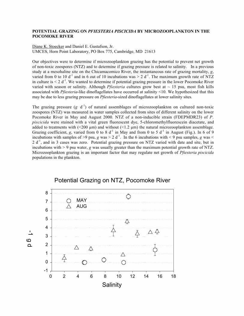
68
POTENTIAL GRAZING ON PFIESTERIA PISCICIDA BY MICROZOOPLANKTON IN THEPOCOMOKE RIVER
Diane K. Stoecker and Daniel E. Gustafson, Jr.UMCES, Horn Point Laboratory, PO Box 775, Cambridge, MD 21613
Our objectives were to determine if microzooplankton grazing has the potential to prevent net growthof non-toxic zoospores (NTZ) and to determine if grazing pressure is related to salinity. In a previousstudy at a mesohaline site on the Chicamacomico River, the instantaneous rate of grazing mortality, g,varied from 0 to 10 d-1 and in 6 out of 10 incubations was > 2 d-1. The maximum growth rate of NTZin culture is < 2 d-1. We wanted to determine if potential grazing pressure in the lower Pocomoke Rivervaried with season or salinity. Although Pfiesteria cultures grow best at ~ 15 psu, most fish killsassociated with Pfiesteria-like dinoflagellates have occurred at salinity <10. We hypothesized that thismay be due to less grazing pressure on Pfiesteria-sized dinoflagellates at lower salinity sites.
The grazing pressure (g d-1) of natural assemblages of microzooplankton on cultured non-toxiczoospores (NTZ) was measured in water samples collected from sites of different salinity on the lowerPocomoke River in May and August 2000. NTZ of a non-inducible strain (FDEPMDR23) of P.piscicida were stained with a vital green fluorescent dye, 5-chloromethylfluorescein diacetate, andadded to treatments with (<200 µm) and without (<1.2 µm) the natural microzooplankton assemblage.Grazing coefficient, g, varied from 0 to 8 d-1 in May and from 0 to 5 d-1 in August (Fig.). In 6 of 9incubations with samples of >9 psu, g was > 2 d-1. In the 6 incubations with < 9 psu samples, g was <2 d-1, and in 3 cases was zero. Potential grazing pressure on NTZ varied with date and site, but inincubations with > 9 psu water, g was usually greater than the maximum potential growth rate of NTZ.Microzooplankton grazing is an important factor that may regulate net growth of Pfiesteria piscicidapopulations in the plankton.
Potential Grazing on NTZ, Pocomoke River
Salinity
0 2 4 6 8 10 12 14 16 18
g d-1
-1
0
1
2
3
4
5
6
7
8
MAYAUG

69
AN OVERVIEW OF INTERACTIONS BETWEEN ZOOPLANKTON GRAZERS ANDALEXANDRIUM SP., AND EFFECTS OF GRAZING ON BLOOM DYNAMICS IN THENEAR-SHORE ENVIRONMENT OF THE GULF OF MAINE
Gregory J. Teegarden1, Robert G. Campbell3, Allan D. Cembella2, Edward G. Durbin3
1Bowdoin College, 6500 College Station, Brunswick, Maine 040112Institute for Marine Biosciences, NRC, 1411 Oxford St., Halifax, Nova Scotia, Canada3Graduate School of Oceanography, University of Rhode Island, Narragansett, Rhode Island
Laboratory and field studies were conducted during an ECOHAB targeted individual project, toinvestigate the mechanisms and biology of zooplankton feeding and potential impacts of feedingduring blooms of Alexandrium fundyense in the near-shore Gulf of Maine. Laboratory experimentsestablished that copepods are capable of discriminating toxic cells from non-toxic cells, and that non-toxic Alexandrium sp. cells are preferred to many similar algal types while toxic cells are frequentlyavoided. Selective feeding was not however universal, and also appeared to depend on theconcentration of toxic Alexandrium sp. cells as well as the species composition of the prey fieldexperienced by zooplankton. Selective feeding by zooplankton in Alexandrium sp. blooms is possibleor likely, but not easily predicted, since dominant grazers, prey species composition, and Alexandriumsp. concentration must be known to predict response with any confidence. When zooplankton consumetoxic Alexandrium sp. cells, some toxins are accumulated, but laboratory experiments demonstrate thatretention of toxins in body tissues is very inefficient (<5% of ingested toxin). Zooplankton may act asvectors of toxin to higher trophic levels, but are not efficient vectors (see poster by Teegarden et al.).
The impact of zooplankton grazing on Alexandrium sp. was investigated in field studies during thespring of 1998 and 1999 in coastal waters of the Gulf of Maine. Samples were collected at weeklyintervals from several stations for zooplankton and phytoplankton abundance, biomass, speciescomposition, and toxin content. Grazing rates of the dominant zooplankton species were determinedusing natural water samples. Dominant zooplankton included copepods (primarily Acartia hudsonicaand Calanus finmarchicus) and barnacle nauplii (Semibalanus sp.). The feeding behavior of thezooplankton on Alexandrium sp. was species specific: Acartia was non-selective, Calanus wassomewhat selective, and Semibalanus avoided ingesting Alexandrium sp. During 1998, there was amoderate bloom of Alexandrium with peak concentrations reaching 3000 cells/L in late May at one ofthe inshore stations. In contrast, in 1999 concentrations were very low throughout the study period.During both years zooplankton biomass was low during the early spring but increased exponentiallyover the study period. In 1998, grazing impacts increased from 0 to 80% day-1 in concert with theincrease in zooplankton biomass and appeared to contribute to the bloom’s demise. Our findingssuggest that grazing can be an important source of mortality and will depend on zooplankton filtrationrates, degree of selective feeding, and the biomass and species composition of both the phytoplanktonand zooplankton communities (see poster by Campbell et al.).

70
HIGH EVOLUTIONARY RATES IN GYMNODINIUM GALATHEANUM CHLOROPLASTDNA SEQUENCES AND DEVELOPMENT OF A MOLECULAR DETECTION ASSAY
Torstein Tengs1, Holly A. Bowers1, Andrew P. Ziman1, Diane K. Stoecker2 and David W. Oldach1
1Institute of Human Virology, University of Maryland at Baltimore, 725 West Lombard Street,Baltimore, MD 212012University of Maryland Center for Environmental Science, Horn Point Laboratory, PO Box 775,Cambridge, MD 21613
Nuclear and chloroplast-encoded small subunit ribosmal RNA sequences were obtained from severalstrains of the toxic dinoflagellate Gymnodinium galatheanum. Phylogenetic analyses and comparisonof sequences indicate that the chloroplast sequences have a higher evolutionary rate than the nuclearhomolog. The chloroplast sequences were chosen as targets for the development of a 5'-3' exonucleaseassay for detection of the organism. The assay has a very high degree of specificity and has been usedto screen environmental water samples from a fish farm where the presence of this dinoflagellatespecies has previously been associated with fish kills. Various hypotheses for the derived nature of thechloroplast sequences are discussed, as well as what is known about the toxicity of the species.
71
AN EXPANSION OF HARMFUL RAPHIDOPHYTE BLOOMS IN U.S. COASTAL WATERS
Carmelo R. TomasCenter for Marine Science, University of North Carolina at Wilmington, 1 Marvin K. Moss Lane,Wilmington, NC 28409
Historically Raphidophyte blooms have been a common feature of some coastal areas of the U.S.Most notable were the recurrent blooms of Heterosigma akashiwo (formerly called Olisthodiscusluteus) in Narrangasett Bay and Long Island Sound. Data spanning over 20 years indicated that theseblooms were seasonal, periodic but varied in intensity from year to year. While causing massivediscoloration H. akashiwo blooms were rarely accompanied by mortality of finfish and shellfish. Inrecent years, blooms of H. akashiwo have become common in the Pacific Northwest region wheresalmon, shellfish and crabs were found dead after exposure to waters containing large concentrationsof this species. These reports agree with the observations from nearby British Columbian waterswhere H. akashiwo blooms have commonly killed salmonoid fish since 1986 to the present. Duringthe last two years, Texas has experienced frequent blooms of H. akashiwo in the Corpus Christi/PadreIsland area where blooms were found accompanying mortality of finfish.
Another genus of the Raphidophytes, Chattonella has also become a common bloom species withinthe last 10 years. Chattonella subsalsa was noted as a seasonal bloom forming species in BayboroHarbor and greater Tampa Bay and Florida Bay. While initially co-occurring with H. akashiwo, C.subsalsa normally dominated in the later phases of the bloom during the seasonal warmer periods ofmid-late summer. During the 2000 summer season, (June-September) blooms of Chattonella wereidentified from Corpus Christi, Texas, Perdido Bay, Bayboro Harbor and St. Johns River Estuary,Florida, New and Neuse Rivers, North Carolina, Ayre Creek, Maryland and Bald Eagle Creek,Delaware. For the past five years, blooms of Chattonella have also been reported for the Salton Sea(California) where extensive fish and bird deaths were observed. While the exact species ofChattonella is not clearly defined for all cases, with the exception of Tampa Bay, Chattonella bloomsoccurred in regions where fish kills were reported. The blooms often accompanied or followed thefish mortalities and rarely were noted as being associated as a causative agent.
Japanese investigators have reported toxins from three of the four Raphidophyte genera, namely,Heterosigma, Chattonella and Fibrocapsa. Each of these species was demonstrated to be associatedwith fish kills in the Japanese Inland Sea and more recently in New Zealand. Chattonella marina wasidentified as killing cultured fish in Tasmania. Blooms of Chattonella verruculosa have for the secondyear in a row caused extensive blooms in the North Sea visible by satellite imagery. Mortality of fishwas associated with the blooms. Considering these reports and increase in occurrence, closer scrutinyis required to define the significance of the Raphidophyte blooms. Do they represent a new threat? Isthe increased frequency a function of evolving environments? Do they represent a viable vector fornew toxins in coastal waters? Are we to expect a continuation of more frequent blooms in the nearfuture? Are there new Raphidophyte species involved in this apparent increase?
A greater effort in the ability to identify these species is required in routine monitoring efforts. Fishkill events in lower salinity estuarine waters need to be considered in light of the presence of toxicRaphidophytes. Greater efforts are needed towards the detection of their toxins by a rapid, accurateand easy field method. And an understanding of not only the acute toxicity of large blooms but therole of chronic low levels of biotoxins in estuarine waters critically needs to be better understood tofully appreciate the risks associated with inshore Raphidophyte blooms.

72
OFFSHORE BLOOMS OF THE RED TIDE DINOFLAGELLATE, ALEXANDRIUM SP., INTHE GULF OF MAINE
David W. Townsend, Neal R. Pettigrew and Andrew C. Thomas5741 Libby Hall, School of Marine Science, University of Maine, Orono, ME 04469
We conducted three large-scale surveys of the coastal and offshore waters of Gulf of Maine during thesummer of 1998, and two similar surveys in spring and summer of 2000. Hydrographic data werecollected and concentrations of phytoplankton chlorophyll, inorganic nutrients and densities ofAlexandrium cells were measured in discrete water samples. Moorings, measuring Doppler currentprofiles and temperature and conductivity at several depths, were deployed within the Eastern MaineCoastal Current (EMCC) system during each of the field seasons. During 1998 the moored programfocused on the alongshelf coherence within the EMCC, while in 2000 the emphasis was on flowstructures in the frontal region near Penobscot Bay and the linkages to the Western Maine CoastalCurrent (WMCC). Daily NOAA AVHRR and NASA SeaWiFS satellite data were downloaded andprocessed to provide coincident spatial patterns of sea surface temperature and chlorophyll over eachcruise period.
Our specific objectives were to:
(1) Investigate the physical oceanography, nutrient chemistry, and abundances and distributions ofAlexandrium in the coastal and offshore waters of the northern Gulf of Maine and to identify thefactors that regulate Alexandrium population dynamics.
(2) Determine the linkages between the major Gulf-wide current systems, in particular the EasternMaine Coastal Current and western Maine coastal waters, and between the Gulf of Maine and the Bayof Fundy with respect to freshwater, nutrients, and Alexandrium cells.
(3) Identify environmental factors that result in the initiation of Alexandrium blooms in Gulf of Mainewaters, and how those blooms are controlled with respect to their spatial and temporal distributions.
Our most complete results, as of this writing, are for the 1998 field season. Both surface andsubsurface concentrations of Alexandrium displayed maximum cell densities in the offshore waters ofthe Gulf on all cruises. Highest cell densities in surface waters (ca. 5.5 x 10³ cells L-1) were observedin two broad patches: one in the Bay of Fundy and another in shelf and offshore waters of the centraland eastern Gulf of Maine in association with the Eastern Maine Coastal Current. Highest subsurfacedensities of cells appeared to be associated with the frontal edges beyond the cold surface watersassociated with the Eastern Maine Coastal Current. As the summer progressed, the highest surfacedensities of Alexandrium receded toward the eastern portions of the Gulf and the Bay of Fundy.Locations of high cell densities were described and interpreted using a non-dimensional light-nutrientparameter, computed as the ratio of the depth of the 10% surface irradiance to the depth of 4 µM NO3
concentration.
We suggest that the offshore distributions of relatively high densities of Alexandrium are naturally-occurring and can be related to seasonal and vertical patterns of inorganic nutrientconcentrations/ratios and the ambient light field. Possible mechanisms responsible for periodicdevelopment of PSP outbreaks in nearshore shellfish beds are discussed in light of our recent findings,and placed in the context of an historical overview of past observations in the Gulf of Maine region.
73
THE CHALLENGES OF FORECASTING AND MANAGING TOXIC PSEUDO-NITZSCHIABLOOMS ON THE U.S. WEST COAST
Vera L. TrainerNOAA/National Marine Fisheries Service/Northwest Fisheries Science Center, 2725 Montlake Blvd.E., Seattle, WA 98112
The food web transfer of domoic acid to shellfish, crustaceans, seabirds, finfish, and marine mammalshas been recently documented on the U.S. West coast. Sub-regions of the U.S. West Coast faceunique challenges during toxic episodes, including choosing the appropriate sentinel organism andunderstanding which Pseudo-nitzschia species are responsible for toxicity. Data collected during WestCoast cruises in the years 1997-2000 indicate that often the highest toxin levels and greatest numbersof toxic cells are positioned in water masses associated with offshore eddies or in upwelling zones nearcoastal promontories. Such cruise data are essential in the characterization of offshore initiation sitesthat will lead to the effective placement of automated sensors, such as moored arrays. In addition,beach monitoring is a necessary component of regional species characterization, resulting in thedevelopment of specific molecular and biochemical tools needed to assist managers in each coastalarea. Indeed, beach samples collected in 1998 indicated that a P. pseudodelicatissima bloom wasresponsible for razor clam toxicity on the Washington coast, whereas toxin produced by P. australisresulted in sea lion mortalities in central California. The challenges faced on the West Coast due toHAB-related mammal mortalities, widespread closures of shellfish harvest, and human illness can onlybe met by sustained beach monitoring programs such as the Olympic Region Harmful Algal Bloom(ORHAB) project and dedicated research cruises. These onshore and offshore efforts will give us acomprehensive picture of the oceanography influencing the location and intensity of domoic acid-producing HABs. Complete characterization of physical, biological and chemical conditions thatfavor harmful Pseudo-nitzschia blooms, only possible through large-scale, synergistic collaboration, isa prerequisite for forecasting of these events. A forecasting capability will substantially improve themanagement of valuable coastal resources and the protection of human health, both of which areaffected by these toxins.

74
ACCUMULATION OF PSP TOXINS IN ZOOPLANKTON ASSEMBLAGES IN THE GULFOF MAINE
Jefferson T. Turner1, Christine L. Powell2, David M. Kulis3, Bruce A. Keafer3, Donald M. Anderson3,and Gregory J. Doucette2,4
1Biology Dept., U Mass Dartmouth, North Dartmouth, MA 02747-23002Marine Biotoxins Program, NOAA/National Ocean Service, Charleston, SC 294123Biology Dept., Woods Hole Oceanographic Institution, Woods Hole, MA 025434Marine Biomedical & Environmental Sciences, Medical Univ. SC, Charleston, SC 29412
The transfer of algal toxins through marine food webs can affect the health of both humans andwildlife, as well as negatively impact the trophic structure of ecosystems. In the case of PSP toxinsproduced by dinoflagellates, the most common route of entry into the food web is via directconsumption of toxic algae by filter-feeding bivalves as a component of their natural diet. Anotherpotentially important, although poorly understood, vector for the transfer of these toxins into highertrophic levels is the zooplankton grazer community, which represents the prey of certain marinemammals as well as various planktivorous fish species. Such fish can, in turn, experience reductionsin fecundity and recruitment with exposure to PSP toxins, and may also transfer toxins to theirpredators. PSP toxins contained in mackerel were, in fact, implicated in an unusual mortality eventseveral years ago involving humpback whales. Moreover, a recent study by our group inMassachusetts Bay demonstrated not only the accumulation of PSP toxins in zooplankton, but also thepreferential movement of toxin into larger size fractions although these animals did not numericallydominate the grazer assemblage. Some larger zooplankton species comprise a majority of the diet forthe North Atlantic right whale and thus pose a potential health threat to these endangered marinemammals.
Given the paucity of detailed information on the role of zooplankton grazers as vectors for PSP toxins,we initiated a study of zooplankton mediated trophic transfer of these toxins as a component of theECOHAB Gulf of Maine regional program. We will be reporting the results of detailed survey cruisesin the Casco Bay region of the Gulf of Maine conducted in 1998, which describe the PSP toxindistribution among several plankton size fractions (20-64, 64-100, 100-200, 200-500, >500 µm) aswell as the taxonomic composition of these size classes. As expected, the predominant PSP toxinsignal was detected in the 20-64 µm size class containing toxic Alexandrium spp., and changedaccording to fluctuations in the cell concentrations of these dinoflagellates. Nonetheless, toxicity wasalso associated with all other size classes examined at some point during the study, with thedistribution pattern varying markedly among stations within a cruise and also between cruises. Unlikeour previous work in Massachusetts Bay, toxin signals were occasionally detected in the 64-100 µmsize class. Preliminary analyses indicated that these samples did not contain Alexandrium cells, butrather were dominated by tintinnid ciliates as well as larger, non-toxic dinoflagellates. The overallpattern of toxin accumulation in the remaining three size fractions was similar to that observedpreviously, with toxicity occurring consistently in the 200-500 and/or >500 µm size fractions, whichwere generally dominated by copepods of the genera Calanus, Centropages, and Pseudocalanus - allpotential grazers of Alexandrium cells. Although toxicity was not associated with the 100-200 µmfraction as frequently, there were several cases in which this fraction, comprised mostly of copepodnauplii, adults of small copepods (e.g., Oithona similis), and rotifers, accumulated as much or moretoxin than the larger size classes. More detailed analyses of zooplankton taxonomic composition,Alexandrium cellular toxicity, and implications for PSP toxin transfer to higher trophic levels will bediscussed.
75
ROLES OF ENDOGENOUS CELLULAR RHYTHMS AND LIFE CYCLE STAGERECRUITMENT IN GYMNODINIUM BREVE BLOOM DEVELOPMENT
Frances M. Van Dolah1, Michele Barbier1, Tod A. Leighfield1, Karen A. Steidinger2,Bill Richardson2 and Peter M. McGuire3
1NOAA/NOS Center for Coastal Environmental Health and Biomolecular Research, 219 Ft. JohnsonRd., Charleston, SC 294122Florida Marine Research Institute, Florida Fish and Wildlife Conservation Commission, 100 EighthAve, SE, St. Petersburg, FL 337123Department of Biochemistry and Molecular Biology, University of Florida, Gainesville, FL 32610
Blooms of the Florida red tide dinoflagellate, Gymnodinium breve, occur almost every year off thewest coast of Florida. As a component of the Florida ECHOAB program, this work investigatescellular level controls involved in G. breve bloom dynamics. Gymnodinium breve has both asexualand sexual life cycle phases, both of which play roles in the formation of Florida red tides. The sexualcycle has been only partially elucidated through the planozygote stage. Current laboratory studiesinclude experiments to evaluate planozygote longevity and hypnozygote (resting cyst) formation. Therepeated occurrence of early stage blooms (e.g., 5000 - 20,000 cells per liter) in isolated spots in themid-shelf region suggests that bloom initiation may result from a benthic life cycle stage. Theexistence of resting cysts of G. breve has not yet been confirmed; however, laboratory observationshave identified the existence of a benthic palmelloid stage that may alternatively serve as a seedpopulation for G. breve blooms. Confirmation of the presence of cysts, palmelloid cells, or otherbenthic stages of G. breve in bloom initiation zones is the focus of current studies. Identification ofunusual life cycle stages is problematic in field samples; therefore, we have developed an antibodyprobe to cell surface proteins of G. breve to assist in these identifications.
Once initiated, the development of a G. breve bloom proceeds through asexual cell division. In bothlaboratory and field populations, G. breve growth rates are consistently found to average 0.2-0.5divisions per day. Like most phototrophic dinoflagellates, the cell cycle in G. breve appears to beunder the control of a circadian rhythm. Consequently, the cell cycle is phased to the diel cycle, suchthat the portion of the population destined to divide on a given day enters S- phase (DNA synthesis) 6-8 h into the light phase, and enters mitosis 12-15 h later, during the dark. Naturally occurring blooms,observed over four years of ECOHAB cruises, exhibit similar diel cell cycle phasing. In laboratorycultures, the dark/light or "dawn" transition was shown to provide the diel cue that serves to entrain theG. breve cell cycle: a forward or backward shift in the timing of this cue results in a concomitant shiftin the timing of S-phase entry. We have identified by western blotting and immunolocalization twokey components of the cell cycle regulatory complex that are known in higher eukaryotes to controlentry into both S-phase and mitosis: cyclin and cyclin dependent kinase (CDK). Inhibition of theactivity of this complex with a specific CDK inhibitor, olomoucine, blocks the G. breve cell cycle bothprior to S-phase and prior to mitosis, and demonstrates a functional role for this complex in cell cycleregulation. Our current work focuses on the mechanisms by which the diel entraining signal acts onthese cell cycle regulators to control cell cycle progression.

76
HYDROGRAPHY AND NUTRIENT CHARACTERISTICS WITHIN THE ECOHAB:FLORIDA CONTROL VOLUME ON THE WEST FLORIDA SHELF
Gabriel A. Vargo1, Cynthia A. Heil1, John J. Walsh1, Kent Fanning1, Carmelo R. Tomas2, Karen A.Steidinger3, Danylle Ault1, Merrie Beth Neely1, Kristen Lester1, and Rachel Merkt1
1College of Marine Science, Univ. of South Florida, St. Petersburg, FL 337012Center for Marine Science, Univ. of North Carolina at Wilmington, Wilmington, NC 284033Florida Marine Research Institute, Florida Wildlife Conservation Commission, St. Petersburg, FL33701
Blooms of Gymnodinium breve may re-occur annually in coastal waters of the West Florida Shelfprimarily within the area bounded by Tampa Bay on the north and Charlotte Harbor on the south (i.e.the Ecohab: Florida control volume). This region is oligotrophic with typical inorganic nitrate andphosphate concentrations of <0.5 and 0.2 µM, respectively, within 2 to 4 km of the shoreline. G. breve
is well adapted for life in this oligotrophic environment with a Ks value for nitrate uptake of 0.42, andfor growth utilizing phosphate, ammonia and urea on the order of 0.18, 0.47 and 1.07, respectively.However, when blooms with chlorophyll concentrations >10 µg/l persist within limited geographic
areas for 2 to 4 months, additional nutrient sources are required for long-term maintenance.
Ongoing monthly quasi-synoptic cruises collect standard hydrographic measurements at approximately65 locations along three cross-shelf and two along shelf transects to determine the hydrographicfeatures and potential nutrient sources for bloom inception, growth, and maintenance. Severalhydrographic features can be related to bloom formation and persistence. Steep cross shelf thermaland salinity gradients occur in nearshore waters during the winter and rainy season, respectively.Rainy season outwelling of estuarine waters have a characteristic signal of elevated inorganic andorganic nutrients with inorganic molar N:P ratios of ~1.0, and leads to the formation of fronts incoastal waters and chlorophyll signatures at the mouth of each estuary. Vertical thermal stratificationof shelf waters from May through October leads to the formation of near bottom chlorophyll maxima,dominated by diatom populations, which are potentially fueled by nitrate originating from offshoreintrusions of the Loop Current. Fall and winter months are typified by vertically well-mixed watercolumns which resuspend PON and DON from decaying summer populations.
Phosphate flux from estuarine and/or sediment sources is sufficient to meet bloom requirements.Although inorganic nitrogen levels are low, DON concentrations within the estuaries and in nearshorewaters range from 5.0 to 15.0 µM and may provide support for nearshore blooms. Mid to late summer
blooms of the diazotrophic cyanobacterium Trichodesmium erythraeum , which often precede G. breveblooms, appear to be stimulated by iron input from Saharan dust events, and may also act as a sourceof organic N via excretion of amino acids or inorganic N via bacterial breakdown of excretory materialand particulate matter. Elevated DON levels coincide with Trichodesmium cell counts in offshore andmid-shelf areas. Other potential DON sources include the degradation of POM such as sea grassblades from near-shore and estuarine sources.
Measurements of particulate δ15PON from various locations within the control volume strongly
suggest that DON from several potential sources is used by G. breve. The δ15PON values for surface
phytoplankton populations during May-June, 1998 ranged from -1.6 to +2.8 0/oo with a mean of 0.30/oo suggesting Trichodesmium accounted for most of the phytoplankton biomass. The δ15PON of
estuarine derived and sea grass derived material has a similar but slightly heavier range; +1.1 to +2.90/oo while the diatom dominated near bottom chlorophyll maxima found during September, 1998 afternitrate depletion had the δ15N signature of Gulf of Mexico sub-thermocline nitrate; e.g. +6.7 to 8.30/oo.

77
The December, 1998 red tide with approximately 5 µg chl/l in nearshore waters had a δ15PON of
+4.80/oo which reflects use of a 15N-enriched DON substrate which has been modified by bacterial orherbivore fractionation or mixing with DON derived from near bottom nitrate enriched diatom stocks.Several potential N sources are therefore available in support of G. breve blooms on the West FloridaShelf. Ongoing studies are aimed at distinguishing between offshore and coastal sources of utilizableDON.

78
USE OF CELL SPECIFIC PAM-FLUOROMETRY TO CHARACTERIZE LIGHTACCLIMATION RESPONSES OF GAMBIERDISCUS TOXICUS (DINOPHYCEAE)
Tracy A. Villareal1 and Steve Morton2
1Marine Science Institute, The University of Texas at Austin, 750 Channel View Dr., Pt. Aransas,Texas 783732NOS/NOAA; 219 Fort Johnson Rd.; Charleston, SC 29412
The fluorescence parameter Fv:Fm was determined on single Gambierdiscus toxicus over a diurnalcycle using a microscopy-PAM system. Cells collected from foliose red algae growing in the subtidalmargin of Southwater Cay, Belize were examined at intervals during the day (May, 2000). Anexperimental treatment of rinsed cells free of the host algae was incubated in-situ under both sunnyand cloudy conditions, and the results were compared to natural samples.
The dark adapted yield parameter Fv:Fm reached 0.7-0.8 in pre-dawn and early morning naturalsamples (maximum = 0.81). Pre-dawn samples were dominated by high yield cells, but a significantnumber of cells exhibited lower yields (Fig. 1). High yield cells were present throughout the day, butthe proportion of low yield cells increased over the course of the day resulting in a decrease in theaverage Fv:Fm. Yield decreased during the day at a similar rate under both cloudy and sunnyconditions to a daily average minimum of 0.4-0.5. However, the maximum values for individual cellsstill exceeded 0.7. Incubated samples on cloudy days showed a similar response to naturalpopulations; however, incubated samples under sunny conditions showed a significantly greaterdepression of Fv:Fm and reached a minimum of 0.15 (Fig. 2). Cells in this treatment started to recoverin late afternoon, and fully recovered after one dark period to near maximum yields (Fig. 2).
The maximum dark adapted yield in G. toxicus approaches the theoretical maximum for PAMfluorometry and does not suggest physiological limitation of photosynthesis by either N or Fe. Thedown regulation of Fv:Fm appears to be a response to solar radiation, and indicates the existence ofactive non-photochemical quenching to protect the photosystem. However, the depression of the yieldin host-free incubation bottles under sunny conditions as well as the persistence of high yields at mid-day suggests that behavior mechanisms may be at work as well. G. toxicus may be actively moving toshaded regions of the algae to reduce its exposure to solar radiation.
Fig. 1. Frequency histogram of yield in G. toxicus. Results are binned into 2 hour time periods.

79
Fig. 2. Diurnal variability inaverage yield in natural andexperimental samples of G.toxicus.

80
GYMNODINIUM BREVE IN THE WESTERN GULF OF MEXICO: TWO YEARS OFCOASTAL SAMPLING OFF THE TEXAS COAST
Tracy A. Villareal1, Mary A. Brainard1, and H. Magaña1 and Lawrence W. McEachron2
1Marine Science Institute, The University of Texas at Austin, 750 Channel View Dr., Port Aransas,Texas 783732Coastal Fisheries Division, Texas Parks and Wildlife Department, 702 Navigation Circle, Rockport,Texas 78382
We established a sampling program in Texas coastal waters to examine near shore waters (<15 km) forthe presence of G. breve and related species. The first two years of sampling noted G. breve from thewaters inside 15 km in <10% of the samples. During this period, significant red tide events occurredalong the Texas coast near Brownsville and the Louisiana border. In all cases, the routine sampling(twice per month) did not provide advance warning of the bloom. The first event in Oct. 1999 off ofBrownsville, TX may have been related to introduction of offshore populations (>15 km) during strongnortheasterly winds. This bloom advected to the south over a two week period. An offshore transectthat found nutrient-rich water advecting onto the shelf in association with an anticyclonic feature. Thismesoscale feature resembled Loop Current intrusions and could have provided the offshore sourcewaters for the bloom. There was no surface manifestation of this intrusion on the shelf.
A July, 2000 bloom moved north up the coast from Brownsville, TX and may have been transported ina coastal upwelling plume that extended from Tamulipas, Mexico to near Corpus Christi, Texas.However, this upwelling event was associated only with temperature reduction at the surface (T>26 °C) and had no apparent nutrient enrichment (Fig. 1). A subsequent red tide off Sabine, Texas occurrednear the Louisiana border (Aug-Sept. 2000). This bloom was primarily offshore and impacted thecoastal zone only for a brief period in late August south of Galveston. A brief analysis of the historicalcirculation data suggests the seasonal migration of the coastal current convergence could beresponsible for maintaining this bloom in a gyre like feature in the northern shelf. The historicalrecord of G. breve blooms notes similar complex dynamics and suggests the seasonal coastal currentpatterns may play a fundamental role on determining whether blooms propagate onshore and in whichdirection they are transported.
Initial culture studies on G. breve isolated in Oct. 99 showed a lower salinity tolerance between 23 and25 PSU with a maximum growth rate of 0.3 –0.4 div d-1. Ongoing experiments are evaluating thegrowth response over a wider range of light, salinity and temperature conditions.
Fig. 1. Sea surfacetemperature image showingcoastal upwelling alongnorthern Mexico and
81
SKIN LESIONS IN ESTUARINE FISHES: A COMPARATIVE PATHOLOGICALEVALUATION OF WILD AND LABORATORY-EXPOSED FISH
Wolfgang K. Vogelbein1, Kimberly S. Reece1, Jeffrey D. Shields1, David E. Zwerner1, Patrice L.Mason1, Larry W. Haas1 and Vicki Blazer2
1Department of Environmental Science, Virginia Institute of Marine Science, The College of Williamand Mary, Gloucester Point, VA 230622National Fish Health Research Laboratory, Leetown Science Center, USGS, Kearneysville, WV25430
The toxic dinoflagellate, Pfiesteria piscicida, is widely blamed for acute mortalities and skin ulcers inmenhaden, Brevoortia tyrannus, from Mid-Atlantic U.S. estuaries. However, the underlying causes ofthese acute fish kills and the so-called "Pfiesteria-specific" skin ulcers of menhaden remain poorlyunderstood and controversial. We initiated field and laboratory studies to clarify the role, if any, ofPfiesteria spp. and other environmental factors in menhaden lesion development. Histopathologically,skin ulcers in > 250 wild menhaden from Chesapeake Bay and the Pamlico Estuary, NC all harbored adeeply invasive, pathogenic fungus eliciting severe tissue necrosis and intense granulomatousinflammation. One of us (VB) recently determined this fungus to be Aphanomyces invadans, thecausative agent of epizootic ulcerative syndrome (EUS) in fishes from the Indo-Pacific Region. Theconsistent presence of this fungus and the granulomatous nature and severity of the resultinginflammatory response suggest these lesions are chronic (age > one week). Their use as an indicatorof current, localized Pfiesteria activity may therefore not be valid. However, these field data do notallow us to rule out a possible early role for Pfiesteria spp. as initiator of lesion development. It istherefore important to better understand the pathology of Pfiesteria exposure and to investigatepotential environmental factors that might modulate lesion pathogenesis in exposed fishes. To this end,we conducted a series of preliminary laboratory challenges of tilapia (Oreochromis niloticus) andmummichog (Fundulus heteroclitus) with high concentrations (5000 - 30,000 cells/ml) of Pfiesteriasp. "B" (characterization based on SEM and molecular analyses). Results indicate Pfiesteria-associated pathology is consistent with acute toxin exposure and differs significantly from pathologyobserved in wild menhaden with ulcerative mycosis caused by A. invadans. Fish mortalities in theassays were directly associated with abundance of Pfiesteria "B" cells. Grossly, fish exhibited loss ofscales, mucus coat and occasional mild petechial hemorrhage. Histologically, fish exhibitedwidespread loss of epidermis with bacterial colonization but minimal associated inflammation.Epidermal erosion extended into the oral and branchial cavities. Gills exhibited epithelial lifting, lossof secondary lamellar structure and infiltration by lymphoid cells. Interestingly, dinoflagellates wereobserved within skin folds, scale pockets and the lateral line canal system of the head region.Epithelial lining of the lateral line canal and associated sensory structures exhibited degeneration andnecrosis. However, bacterial colonization of tissues harboring dinoflagellates was a consistentobservation. In situ hybridization analyses using specific DNA probes to P. piscicida, Pfiesteria sp."B" and the "Lucy" group confirmed that dinoflagellates associated with fish tissues were Pfiesteriasp. "B". We consider this association to be an artifact of the very high dinoflagellate concentrations inour exposures. However, in situ hybridization analyses of wild menhaden intestine with these probesmay serve as a sensitive, specific and quantifiable bioindicator of local Pfiesteria activity in east coastestuaries.

82
COUPLED NUMERICAL MODELS OF FLORIDA RED TIDES OF GYMNODINIUM BREVE
John J. Walsh1 , W. Paul Bissett2 , Bradley Penta1, Dwight A. Dieterle1, Robert H. Weisberg1,Zhenjiang Li1, and Huijun Yang1
1College of Marine Science, University of South Florida, St. Petersburg, FL 337012Florida Environmental Research Institute, 4807 Bayshore Boulevard, Tampa, FL 33611
Successful ecological models are data-driven, distilling qualitative hypotheses and aliased fieldobservations into simple analogues of the real world in a continuing cycle of model testing andrevision. Prediction of the origin, transport and fate of Gymnodinium breve blooms on the WestFlorida shelf is the goal of the ECOHAB: Florida project - based on 1) shipboard and remote sensingsurveys of hydrography, nutrients, DOM, species composition, pigments, and zooplankton 2)experimental cruises and laboratory studies describing their physiology, life cycles, optical properties,and toxin transfer, 3) arrays of current meters, 4) circulation submodels, 5) cell metabolism andmigration submodels, and 6) coupled bio-optical models. The last component utilizes these submodelsand assimilated observations in a complex, numerical food web to describe the consequences ofphytoplankton competition in terms of signals seen by satellite, aircraft, and moored sensors. We haveused one-dimensional models to specify the rules of engagement between G. breve and otherfunctional groups of phytoplankton, two-dimensional models to explore the consequences of theirinteraction with the microbial food web, and three-dimensional models to predict their transport,landfall, and residence time at the surface of the sea.
Following competition theory, our present models of the limiting resources of light, nitrate,ammonium, DON, phosphate, DOP, iron, and silicate should allow the coexistence of eight functionalgroups of phytoplankton, without differential grazing pressure on chromatically-adaptedphytoplankton. In our analogue of the West Florida shelf, CO2 and N2 are state variables, but they areconsidered to be in excess of algal needs. From our simulation analyses thus far, we find that 1)diatoms win when estuarine and shelf-break supplies of nitrate are made available to a modelcommunity of small and large diatoms, coccoid cyanophytes and Trichodesmium, non-toxic and red-tide dinoflagellates, microflagellates, and coccolithophores, 2) a numerical recipe for large red tides ofG. breve instead requires DON supplies, mediated by iron-starved, nitrogen-fixers in response toSaharan dust events, while their small blooms may persist on sediment sources of DON, 3) selectivegrazing must still be exerted on the other non-toxic dinoflagellates by copepods, 4) bacteria drive theouter shelf food web into P-limitation, until coastal supplies of low N/P ratio of ~1 favor nitrogen-fixers, 5) light-cued vertical migration of G. breve in relation to seasonal changes of summerdownwelling and fall/winter upwelling flow fields determines both their duration within the firstoptical depth as a remotely-sensed signal and the intensity of red tide landfalls along the barrier islandsand beaches of West Florida, and 6) termination of G. breve blooms is likely to result fromcumulative, biomass-dependent losses in the form of UV-B irradiation, microbial-induced lysis, andunselective grazing pressure from protozoans and heterotrophic dinoflagellates.

83
ECOHAB FLORIDA, PHYSICAL OCEANOGRAPHY
Robert H. Weisberg, Ruoying He, William Hemme, Zhenjiang Li, and Huijun YangCollege of Marine Science, University of South Florida, St. Petersburg, FL. 33701
The circulation of west Florida continental shelf exhibits seasonal variability in both its backgroundcurrents and the responses of these currents to synoptic scale weather forcing. The seasonalcirculation is primarily forced by local momentum and buoyancy inputs. However, it is alsomodulated by mass and heat exchanges with the adjacent Gulf of Mexico. In situ measurements showsummer and winter seasons of predominantly downwelling and upwelling circulations, respectively.This occurs regardless of adjacent offshore Loop Current influence, although such influence doesaffect the seasonal behavior by modifying the across-shelf density field. The seasonally varyingdensity field, in turn, presents a critical control on the response of the shelf to local weather forcing.Under stratified conditions we find that the inner-shelf responses to upwelling and downwellingfavorable winds are rectified such that the upwelling responses are disproportionately larger than thedownwelling responses. This behavior is attributed to the relative slopes of the isopycnals and thebottom. Buoyancy torque adds constructively with planetary vorticity tilting by the sheared coastal jetunder upwelling favorable winds, whereas it adds destructively under downwelling favorable winds.The end result is that the bottom Ekman layer response is enhanced (suppressed) for upwelling(downwelling), and this bottom Ekman layer asymmetry causes the asymmetry in the inner-shelfresponses. Since the biology of the west Florida shelf is strongly influenced by bottom Ekman layerprocesses, these physical oceanographic findings are potentially very important for ECOHAB.
Along with its in-situ measurements, ECOHAB Florida has a numerical circulation modelingcomponent aimed at simulating the seasonal and synoptic scale variability and supplying three-dimensional circulation fields for use in ecological models. For reasons given above the numericalmodel results are critically tied to the in-situ density field and to the fluxes of momentum andbuoyancy, both locally and at the shelf break. Model simulations are therefore limited by modelinitializations and forcing functions. Without adequate in-situ information, modeling is of limited use.Nowcast or hindcast model runs tends to be useful for integration times of order one month.Improving upon this requires improving upon both the surface and the offshore boundary conditiondata and providing adequate interior density data for model assimilation. Initial attempts at combiningthe physical and ecological models are encouraging.

84

85
ABSTRACTS OF POSTERS

86
87
DEVELOPMENT OF THE VOLUNTEER OFFSHORE RED TIDE MONITORINGPROGRAM FOR THE GULF COAST OF FLORIDA
Jay P. Abbott, Earnest W. Truby, and Karen A. SteidingerFlorida Marine Research Institute, Florida Fish and Wildlife Conservation Commission, 100 EighthAvenue S.E., St. Petersburg, FL 33701
Gymnodinium breve red tides originate offshore on the west Florida shelf and then can be transportedto inshore areas by currents and winds. In the past, detection of blooms before they reached inshorewaters was mostly by research cruises or the occasional report of discolored water or dead fish byboaters, commercial fishermen or airplane pilots. The rationale for this program was to have regularsampling in areas offshore of areas where red tides have traditionally come inshore, Tampa Bay toCharlotte Harbor, and where shellfish harvesting is economically important.
Developing a plan for the Volunteer Offshore Monitoring Program involved determining where, whenand how the sampling should take place as well as who should be targeted for involvement.Since red tides may affect any portion of Florida’s Gulf coast, a few key areas were selected forvolunteer recruitment. These areas included Pensacola, Panama City, Apalachicola, Cedar Key, St.Petersburg, Charlotte Harbor, Naples and the Lower Keys. The coastal nature of these areas, with easyaccess to the Gulf of Mexico, make them ideal commercial charter fishing operations.
Utilizing FMRI contacts, Internet web-sites, and general networking, charter captains in these areaswere contacted about participating in the program. Most commercial charter captains understand thatmonitoring for red tide is in their own best interest, however, sampling may differ from scheduledtimes based on weather, business, and other reasons. It was important for us to understand that thesevolunteer captains were sampling on their paying customer’s time. The sampling procedure and returnshipping process needed to be quick and user friendly. All the sampling bottles and shipping materialswere pre-labeled and any instructions were made as concise and unambiguous as possible. Thevolunteer captains were asked to sample at offshore distances of 1,5,10,20, and 30 milesapproximately twice a month. When possible, additional volunteers in the same area were recruited.This reduced the possibility of sampler “burnout” by any one captain as well as providing backupcoverage during mechanical troubles, vacations, etc.
Possibly the most important key to obtaining sampling stability is regular contact with the volunteercaptains and providing them with some type of visual feedback on their work. Maps displaying thered tide cell counts from each sample are relayed to the volunteers, allowing them to serve as monitorsof their local waters. It is crucial that the volunteers feel as though they are a part of a meaningfulprogram and that their efforts are recognized. Current activities involve developing appropriaterewards and enlisting captains to collect bottom water samples. At present only surface water samplesare collected.

88
ENVIRONMENTAL CONDITIONS ASSOCIATED WITH DOMOIC ACID IN RAZORCLAMS ON THE WASHINGTON COAST
Nicolaus G. Adams1, Mitch Lesoing2, and Vera L. Trainer1
1National Marine Fisheries Service, Northwest Fisheries Science Center, Environmental ConservationDivision, Seattle, Washington 981122Quileute Natural Resources, Quileute Indian Tribe, La Push, Washington 98350
In October 1998, record levels of the neurotoxin domoic acid were detected in razor clams (Siliquapatula, Dixon) resulting in the closure of shellfish harvesting areas along the Washington coast. Thistoxin was measured in seawater samples collected at Kalaloch Beach and Second Beach on the centralWashington coast using a receptor binding assay and liquid chromatography-tandem massspectroscopy. Domoic acid levels ranging from 0-2700 ng/L were measured in seawater samplescontaining from 70-100% Pseudo-nitzschia pseudodelicatissima (Hasle) Hasle at concentrations of1.0-15 x 106 cells/L, resulting in maximum levels of cellular toxin of approximately 500 fg/cell. Acultured isolate of this species collected from Kalaloch Beach also produced DA, as determined by thereceptor binding assay, during late exponential and stationary stages of growth. The toxic P.pseudodelicatissima bloom in the late summer and autumn of 1998 occurred 2-3 weeks after strongcoastal upwelling during a period of anomalously low rainfall, typical in post-El Niño years. Highertoxin levels in seawater at Kalaloch Beach compared to Second Beach were attributed to the periodicnature of upwelling at Kalaloch Beach, demonstrated by a 175-fold increase in nitrate in seawatercoincident with a 5oC decrease in sea surface temperature on September 1. The upwelling event inSeptember was followed by wind relaxation and reversal at the end of that month, resulting in thetransport of toxic cells toward the coast where nutrients were already present to fuel the algal bloom.A pulse of nutrients, either from rainfall or upwelling, to coastal regions that have experienced severalweeks of low nutrients, followed by wind relaxation or reversal events that transport cells to inshoreregions, are suggested to be important factors in the initiation of the most toxic Pseudo-nitzschia spp.blooms on the Washington coast.

89
IMPACT OF DINOFLAGELLATE-COEXISTING BACTERIA ON THE PHYSIOLOGY OFPFIESTERIA-LIKE DINOFLAGELLATES
Mohammad R. Alavi and Robert M. BelasCenter of Marine Biotechnology, University of Maryland Biotechnology Institute, 701 East PrattStreet, Baltimore MD 21202
In nature, dinoflagellates exist alongside a myriad of other microorganisms. In numbers alone, thelargest percentage of this microbial population is made up of the bacteria. Some of these bacteria maydirectly and indirectly influence the dinoflagellates physiology and metabolism. For this reason, we areinterested in understanding how the bacterial community specifically affects the physiology and toxinproduction of Pfiesteria-like dinoflagellates. We have taxonomically classified the bacterial speciesco-existing in four laboratory cultures of Pfiesteria and Cryptoperidiniopsis spp. originally obtainedfrom sites of Chesapeake Bay fish kills. Our approach was based on the use of oligonucleotide primersand polymerase chain reaction to amplify a portion of the small ribosomal subunit gene (16S rDNA)from both the culturable and nonculturable bacteria in these cultures. The nucleotide sequence of eachunique clone was determined and the homology between the dinoflagellate bacteria and known specieswas used to construct phylogenetic trees of taxonomic relatedness. We then produced axenicdinoflagellate cultures and studied the influence of culturable bacterial isolates on the growth andfeeding behavior of these cultures by add-back experiments. Our results indicate that there aresubstantial differences between axenic dinoflagellate cultures and their bacterized counterparts. Thesedifferences include, but may not be limited to, reduction in growth rate and lowered cell density, aswell as changes in feeding behavior.

90
SALINITY TOLERANCE FOR 62 STRAINS OF PFIESTERIA AND PFIESTERIA-LIKESTRAINS
Robert A. Andersen and Barbara E. SullivanBigelow Laboratory for Ocean Sciences, West Boothbay Harbor, ME 04575 USA
Based upon preliminary studies, experimental cultures were prepared at salinities of 0.5, 1, 2, 3, 4, 5and 6 ppt and 35, 40, 45, 50, 55 and 60 ppt for lower upper tolerance limits, respectively. Cells weregrown at 22o C under a 12 hr light/dark cycle. Cultures (at 12 ppt salinity) were fed immediately beforebeginning the experiment with Rhodomonas (CCMP768); no food was added to the cultures during theexperiment. For each salinity treatment, the water was prepared as a batch and then dispensed. After5 days, each test tube was examined with a dissecting microscope to determine the presence ofswimming dinoflagellate cells. After this examination, the salinity of each test tube was measuredagain using a refractometer to verify that the experimental salinity had not changed due to evaporation.If no swimming cells were observed in all three tubes, the contents of two tubes were transferred into aclean Petri dish so that a more thorough examination was possible. If no swimming cells wereobserved in the Petri dish, then the third tube was adjusted to 12 ppt and the culture was maintained fortwo additional days. At the end of the two-day period, the test tube was examined again for swimmingcells as described above. When swimming cells were observed at the end of the two-day period, weassumed that viable cysts were the source of the swimming cells; when no swimming cells wereobserved at the end of the two day period, we assumed that all cells and cysts died. After five days,most Chesapeake Bay and Neuse River system cells were still swimming at 1 ppt and 40 ppt; onestrain had swimming cells at 0.5 ppt and six strains had swimming cells at 50 ppt. After 5 days, mostWilmington River cells were still swimming at 4 ppt and 50 ppt; several strains had swimming cells at3 ppt and two had swimming cells at 60 ppt. Putative cysts allowed for even greater salinity tolerancebecause many cultures without swimming cells at extremely low or high salinities produced swimmingcells when the salinity was adjusted back to 12 ppt. There was a statistically significant (p < 0.001)difference between the mean lower and mean upper salinity tolerances of the Wilmington Riversystem and the Chesapeake Bay system or Neuse River system. The original Wilmington River watersamples were more saline, suggesting that salinity tolerance is under genetic control.

91
THE EFFECTS OF CLAY, USED IN THE CONTROL OF HARMFUL ALGAL BLOOMS, ONTHE GROWTH RATE OF JUVENILE HARD CLAMS, MERCENARIA MERCENARIA
Marie-Claude Archambault1, Monica Bricelj1,2, Jon Grant1 and Donald M. Anderson3
1 Department of Oceanography, Dalhousie University, 1411 Oxford Street, Halifax, NS B3H 4J1,Canada2Institute for Marine Biosciences, National Research Council, Halifax, NS B3H 3Z1, Canada3Biology Department, Woods Hole Oceanographic Institution, Woods Hole, MA 02543-1049
In recent years there has been increasing interest in using clays as a means of mitigating toxic orHarmful Algal Blooms (HABs) at relatively shallow, enclosed mariculture sites. This control method,which causes flocculation and accelerated sedimentation of large algal cells (dinoflagellates), has beenused at fish aquaculture sites in Korea and Japan. It is presently being evaluated as a potential methodfor controlling HABs in the United States (ECOHAB, Woods Hole Oceanographic Institution).
The sedimenting flux of flocculated clay-algal particles stripped from the water column may haveimplications for the health of benthic suspension feeders, e.g. commercially important bivalves. Thisis the first of a series of experiments to evaluate these effects by re-creating the field application of aPhosphatic Clay (IMCP, 2 µm mean equivalent spherical diameter, ESD) to a HAB condition using alaboratory re-circulating flume, and Prorocentrum micans, as a test, non-toxic dinoflagellate species.It was hypothesized that survival and growth rate of juvenile Mercenaria mercenaria would beadversely affected by the bottom deposition of a relatively thick clay and dinoflagellate layer (~ 7 mmin depth, at a loading rate of 155 g dry weight m-2) in a low flow environment. Initial results indicatethat clams subject to the deposition of clay and dinoflagellates over a coarse sand bottom, and fedIsochrysis galbana for two weeks, suffered a 50% decrease in shell growth rate compared to clams in azero clay/dinoflagellate control. No significant mortality occurred in either treatment. Ongoinganalysis of these experiments will consider clam response in terms of tissue growth and conditionmeasures. Particle size and clay-algal interactions in different flow conditions were quantified using aSmall Volume Particle Microsampler (SVPM), which captures intact flocculated particles on a filter,and is subject to image analysis. Floc characteristics were quantified in order to determine theirpotential effects on clam feeding behavior. Results indicate that the mean ESD of flocs ranges from 5to 15 µm regardless of the tested flow conditions. This floc size range is maintained after the
resuspension of clay-dinoflagellate deposits allowed to settle over 24 hrs. Dinoflagellates werestripped from the flume water column at a removal efficiency of 90% in 4.5 hours by sinking clayaggregates. The flume water column was completely expunged of particles within 48 hours.Continuing experiments involve varying flow/deposition conditions, e.g. near-bottom clayresuspension at higher flows, and further particle imaging as a measure of floc dynamics.

92
TEMPERATURE-CONTROLLED GERMINATION OF CYSTS OF THE TOXICDINOFLAGELLATE ALEXANDRIUM TAMARENSE, FROM BAY OF FUNDY, CANADA
Tamieka Armstrong1, Matthew Harris1, Brian Thompson2, Patricia Matrai2, and Maureen Keller2*
1University of New England, Biddeford, ME 04005, USA2Bigelow Laboratory for Ocean Sciences, W. Boothbay Harbor, ME 04575, USA
The physiology of the eastern Gulf of Maine Alexandrium populations is unknown and laboratorywork is required to develop an understanding of Alexandrium dynamics in this area. To this effect, anexperiment was conducted to determine the rates of cyst germination as a function of temperature (2,4, 6, 8 and 15°C) and light (light vs. dark), following the experimental protocol used previously byBronzino et al. for cysts of the Casco Bay region. Preliminary observations of the percentage of cystgermination in individual sediment samples kept at 8°C over time show that the germination rateincreased exponentially after 12 days. On the other hand, excystment at this temperature under darkconditions shows the same pattern as under light conditions but with lower abundance. Results fromthe various incubation temperatures will be compared. Germination functions will be developed andcompared with those derived for the Casco Bay Alexandrium isolates.
*This work was started under the leadership of the late Dr. Maureen D. Keller.

93
IDENTIFICATION OF CELL CYCLE REGULATORS IN THE FLORIDA RED TIDEDINOFLAGELLATE, GYMNODINIUM BREVE
Michele Barbier, Tod Leighfield and Frances Van DolahMarine Biotoxins Program, Center for Coastal Environmental Health and Biomolecular Research,National Ocean Service, Charleston, SC 29412
The diel cycle is a key regulator of the cell cycle in the Florida red tide dinoflagellate, Gymnodiniumbreve, and may play a rate limiting role in bloom formation. Both laboratory cultures and fieldpopulations of G. breve exhibit phased cell division in which approximately one-third of thepopulation divides each day. The dark/light "dawn" transition provides the diel cue that serves toentrain the G. breve cell cycle, with S-phase beginning 6-8 h into the light phase, and mitosisfollowing 12-14 h later. This research is aimed at identifying the molecular targets of this diel cue.Here we identify in G. breve the two components of cell cycle regulatory complex, cyclin and cyclindependent kinase (CDK), which are the molecular basis of cell cycle regulation in eukaryotes. Thepresence of CDK was identified by western blotting and by cell cycle inhibition with the specific CDKinhibitor, olomoucine. Cyclin was identified in G. breve by western blotting using two antibodies: thefirst one raised against cyclin B of the yeast Schizosaccharomyces pombe, the second one raisedagainst the cyclin box of the sea-urchin cyclin. Both antibodies recognize a ~64 kD antigen in G.breve, and cross- react specifically with the control peptide cyclin B1, but not with cyclin A. Thissuggests that the G. breve cyclin identified is a cyclin B homologue.
The expression of the cyclin B homologue in G. breve was followed by western blotting at differenttime points during cell cycle. Unlike cyclin B in higher eukaryotes, which is expressed only late in thecell cycle, the cyclin B homologue in G. breve was expressed at similar levels throughout cell cycle.This unusual behavior was confirmed by immunohistochemistry. Preliminary results show that cyclinB was present in the cytoplasm throughout the cell cycle, where it appeared to be localized to thecentrosomes. Unlike higher eukaryotes, cyclin B did not appear to be translocated to the nucleus priorto mitosis; this may reflect the fact that the nucleus remains intact during mitosis in dinoflagellates.However, it was localized to the mitotic spindles in mitotic telophase, similar to observations inmammalian cells. The unusual constitutive expression of dinoflagellate cyclin has been previouslydemonstrated in Crypthecodinium cohnii (Barbier et al., 1995), and has also been shown in themyxomycete, Physarium polycephalum.
94
CAN KRILL TRANSFER DOMOIC ACID TO HIGHER TROPHIC LEVELS SUCH ASSQUID AND WHALES?
Sibel Bargu1, Christine L. Powell2, Gregory J. Doucette2 and Mary Silver1
1Institute of Marine Sciences, University of California, Santa Cruz, CA 950642Marine Biotoxins Program, NOAA/National Ocean Service, Charleston, SC 29412
Euphausiids (krill) are a ubiquitous prey for many consumers. The principal predators of Pacificeuphausiids are squid, baleen whales, and certain seabirds (e.g., ashy storm petrel). Krill are potentialvectors for domoic acid (DA) transfer from toxic Pseudo-nitzschia to higher trophic levels in theMonterey Bay food web. An understanding of the quantitative trophic interactions and body burden ofDA in krill is required to predict whether krill can effectively vector the toxin. Nonetheless, laboratoryexperiments on euphausiids together with their larger predators are not currently feasible.
This poster presentation will describe the Pseudo-nitzschia link to whale and squid diets in MontereyBay based on results from the examination of blue and humpback whale fecal material and squidstomach contents collected from 1993 to 2000. Scanning electron microscopy was used to identifyPseudo-nitzschia frustules in fecal material or stomach contents of whale and squid. Our aim was todetermine whether these diatoms were transmitted to the predators through their food source, as wasreported previously for a sea lion mortality event in 1998. Samples are also being tested for domoicacid activity using a receptor binding assay. Detection of Pseudo-nitzschia frustules in feces andstomach contents demonstrated that both baleen whales and squid obtained this diatom as part of theirdiet. Results of toxin analyses currently underway will be discussed in the context of potential transferof DA via krill vectors from Pseudo-nitzschia to higher trophic levels.

95
VERTICAL DISTRIBUTION OF ALEXANDRIUM SP. IN THE WATER COLUMN
Stephanie L. Bennett1, David W. Townsend1
15741 Libby Hall, School of Marine Sciences, University of Maine, Orono, ME 04469
Much research has been done on the species of Alexandrium that are the cause of PSP in the Gulf ofMaine, trying to establish under what conditions dinoflagellate blooms are likely to occur. Pastsurveys of Alexandrium in the GOM have focussed on the phytoplankton populations in the surfacelayers. With evidence published by McIntyre et al in 1997 that Alexandrium were capable of dielvertical migration under laboratory conditions of nutrient stress, the question of whether this occurs innatural populations arises. My research is an investigation into the vertical distribution ofAlexandrium in the Gulf of Maine, and how that distribution changes over time.
In June of 2000, two sites in the Gulf of Maine were chosen for 24-hour sampling of the verticalprofile. Both sites were well offshore and chosen because of the presence of Alexandrium in the nettow and the absence of the dense diatom population that we saw near shore. Water samples weretaken at 5-meter intervals between the surface and 55 meters, a total of 12 depths collected each hourfor 25 hours. All seawater was collected in 5 L Niskin bottles attached to the CTD rosette. The shipfollowed a 25 m drogue during each experiment. Hydrographic data was collected and water wasdrawn from each sample for measurements of chlorophyll, inorganic and organic nutrients andAlexandrium counts. Cell counts were done using a stain comprised of an antibody, specific to thegenus Alexandrium, attached to a fluorescent tag. All counts were done using an epifluorescencemicroscope.
Results will be presented, showing the depth distribution of Alexandrium populations in relation tototal chlorophyll, light and nutrients.

96
DEVELOPMENT OF REAL-TIME PCR ASSAYS FOR RAPID DETECTION OFPFIESTERIA PISCICIDA AND RELATED DINOFLAGELLATES
Holly A. Bowers1, Torstein Tengs1, Howard B. Glasgow, Jr.2, JoAnn M. Burkholder2, Parke A.Rublee3 and David W. Oldach1
1Institute of Human Virology and University of Maryland School of Medicine, Baltimore, MD 21201;2Department of Botany, North Carolina State University, Raleigh, NC 276953Biology Department, University of North Carolina at Greensboro, Greensboro, NC 27402
Pfiesteria complex species are heterotrophic and mixotrophic dinoflagellates that have beenrecognized as harmful algal bloom species (HAB) associated with adverse fish and human healtheffects along the East Coast of North America, particularly in its’ largest (Chesapeake Bay, Maryland)and second largest (Albemarle-Pamlico, North Carolina) estuaries. In response to impacts on humanhealth and the economy, monitoring programs to detect the organism have been implemented inaffected areas. However, until recently, specific identification of the two toxic species known thus far,Pfiesteria piscicida and Pfiesteria shumwayae (sp. nov.), required scanning electron microscopy(SEM). SEM is a labor-intensive process in which a small number of cells can be analyzed, posinglimitations when the method is applied to environmental estuarine water samples. To overcome theseproblems, we developed a real-time PCR-based assay that permits rapid and specific identification ofthese organisms in culture and heterogeneous environmental water samples. Various factors likely tobe encountered when assessing environmental samples were addressed and assay specificity wasvalidated through screening of a comprehensive panel of cultures, including the two recognizedPfiesteria species, morphologically similar species, and a wide range of other estuarine dinoflagellates.Assay sensitivity and sample stability were established for both unpreserved and fixative-preserved(acidic Lugol’s solution) samples. The effects of background DNA on organism detection andenumeration were also explored and, based on these results we conclude that the assay may be utilizedto derive quantitative data. This real-time PCR-based method will be useful for many otherapplications, including adaptation for field-based technology.
97
TEMPORAL AND SPATIAL DISTRIBUTION OF PFIESTERIA PISCICIDA INCHESAPEAKE BAY WATERS, 1999-2000
Holly Bowers1, Torstein Tengs1, Andrew Ziman1, Peter Tango2, David Goshorn2, Robert Magnien2,Renee Karrh2 and David Oldach1
1Institute of Human Virology and University of Maryland School of Medicine, Baltimore, MD 21201;2Maryland Department of Natural Resources, Annapolis, MD 21401.
Pfiesteria piscicida, a heterotrophic dinoflagellate, has been associated with adverse fish and humanhealth effects in estuarine environments extending from Delaware to Florida. Due to public concernover the risk of exposure to putative toxins and the economic loss related to fish kills in theChesapeake Bay in Maryland, the Maryland Department of Natural Resources and collaboratinglaboratories began monitoring various tributaries of the Bay in 1997 and developed those efforts intoan extensive and comprehensive program for 1998 - 2000. Targeted rivers include those in which P.piscicida has been previously detected (through molecular methods, scanning electron microscopy andfish kill bioassays), rivers having a possible predisposition to Pfiesteria activity (chemical and physicalcharacteristics similar to affected systems), and rivers with no history or apparent tendency forPfiesteria-related problems. A real-time PCR-based assay for detection of the organism developed byour laboratory was utilized in the comprehensive monitoring program. In 1999, P. piscicida was notdetectable in surface waters through the spring and early summer, with the organism being detected atonly one station by mid-July. More intensive sampling during 2000 has detected P. piscicida in Aprilat one site and at seven other sites by mid-July. In general, nutrient concentrations and phytoplanktonbiomass were higher at stations where P. piscicida was detectable in the water column, in comparisonto sites repeatedly testing negative. Detection of P. piscicida in the water column has been repeatedlyassociated with the occurrence of summer/fall populations of Atlantic menhaden with ulcerativelesions that have been found to harbor a number of pathogens (e.g. fungi, bacteria, parasites). Furtheranalysis of biological and physicochemical parameters associated with the detection of P. piscicidaand assessment of those parameters in waters consistently negative for the organism may enableidentification of conditions conducive to Pfiesteria blooms. Hypotheses generated with these data willbe tested through ongoing longitudinal analyses of samples collected at multiple stations throughoutthe Chesapeake Bay region.
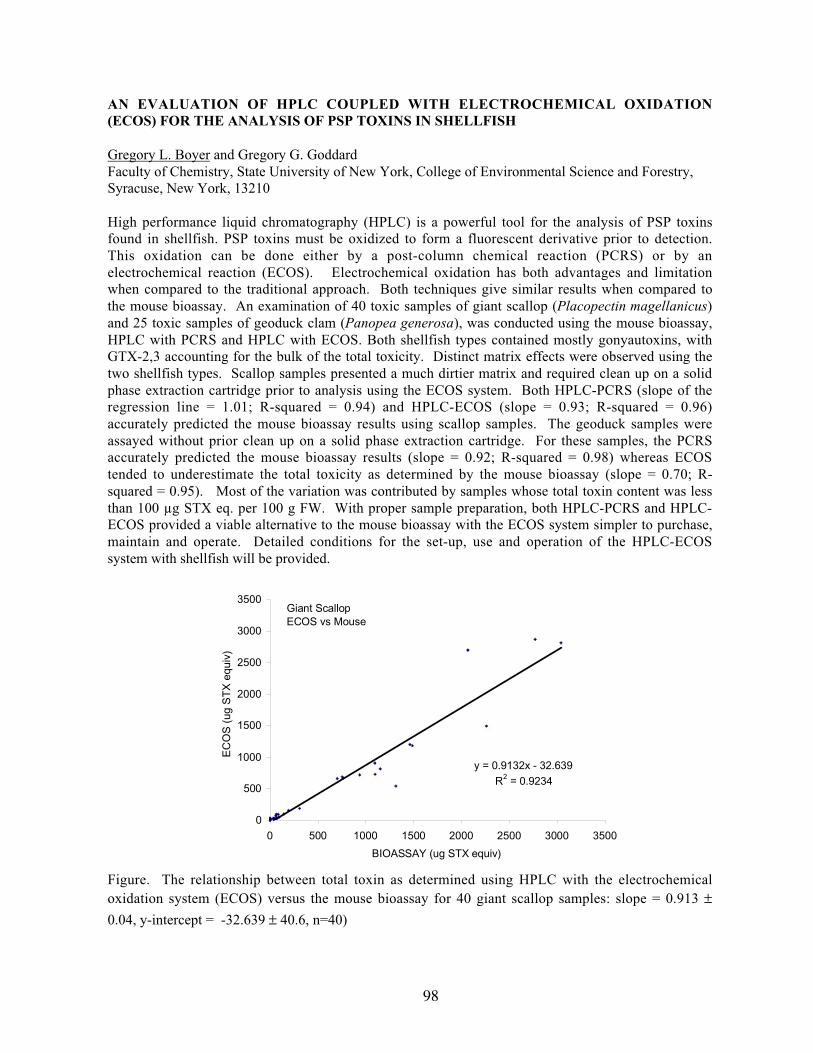
98
AN EVALUATION OF HPLC COUPLED WITH ELECTROCHEMICAL OXIDATION(ECOS) FOR THE ANALYSIS OF PSP TOXINS IN SHELLFISH
Gregory L. Boyer and Gregory G. GoddardFaculty of Chemistry, State University of New York, College of Environmental Science and Forestry,Syracuse, New York, 13210
High performance liquid chromatography (HPLC) is a powerful tool for the analysis of PSP toxinsfound in shellfish. PSP toxins must be oxidized to form a fluorescent derivative prior to detection.This oxidation can be done either by a post-column chemical reaction (PCRS) or by anelectrochemical reaction (ECOS). Electrochemical oxidation has both advantages and limitationwhen compared to the traditional approach. Both techniques give similar results when compared tothe mouse bioassay. An examination of 40 toxic samples of giant scallop (Placopectin magellanicus)and 25 toxic samples of geoduck clam (Panopea generosa), was conducted using the mouse bioassay,HPLC with PCRS and HPLC with ECOS. Both shellfish types contained mostly gonyautoxins, withGTX-2,3 accounting for the bulk of the total toxicity. Distinct matrix effects were observed using thetwo shellfish types. Scallop samples presented a much dirtier matrix and required clean up on a solidphase extraction cartridge prior to analysis using the ECOS system. Both HPLC-PCRS (slope of theregression line = 1.01; R-squared = 0.94) and HPLC-ECOS (slope = 0.93; R-squared = 0.96)accurately predicted the mouse bioassay results using scallop samples. The geoduck samples wereassayed without prior clean up on a solid phase extraction cartridge. For these samples, the PCRSaccurately predicted the mouse bioassay results (slope = 0.92; R-squared = 0.98) whereas ECOStended to underestimate the total toxicity as determined by the mouse bioassay (slope = 0.70; R-squared = 0.95). Most of the variation was contributed by samples whose total toxin content was lessthan 100 µg STX eq. per 100 g FW. With proper sample preparation, both HPLC-PCRS and HPLC-ECOS provided a viable alternative to the mouse bioassay with the ECOS system simpler to purchase,maintain and operate. Detailed conditions for the set-up, use and operation of the HPLC-ECOSsystem with shellfish will be provided.
Figure. The relationship between total toxin as determined using HPLC with the electrochemicaloxidation system (ECOS) versus the mouse bioassay for 40 giant scallop samples: slope = 0.913 ±0.04, y-intercept = -32.639 ± 40.6, n=40)
y = 0.9132x - 32.639
R2 = 0.9234
0
500
1000
1500
2000
2500
3000
3500
0 500 1000 1500 2000 2500 3000 3500
BIOASSAY (ug STX equiv)
EC
OS
(ug
ST
X e
quiv
)
Giant ScallopECOS vs Mouse

99
THE ROLE OF ZOOPLANKTON GRAZERS IN ALEXANDRIUM SP. BLOOM DYNAMICSIN THE NEAR-SHORE ENVIRONMENT OF THE GULF OF MAINE
Robert G. Campbell, 1 Gregory J. Teegarden,1,2 Allan D. Cembella,3 Edward G. Durbin1
1Graduate School of Oceanography, University of Rhode Island, Narragansett, RI 02882 USA2Environmental Studies, Bowdoin College, 6500 College Station, Brunswick, Maine 04011 USA3Institute for Marine Biosciences, NRC, 1411 Oxford St., Halifax, Nova Scotia B3H 3Z1 CANADA
The role of zooplankton grazers in Alexandrium sp. bloom dynamics was investigated in coastal watersof the Gulf of Maine during the spring of 1998 and 1999. We collected samples at weekly intervalsfrom several stations for zooplankton and phytoplankton abundance, biomass, species composition,and toxin content. In addition, grazing rates of zooplankton were determined using natural watersamples. Dominant zooplankton included copepods (primarily Acartia hudsonica and Calanusfinmarchicus) and barnacle nauplii (Semibalanus sp.). The feeding behavior of the zooplankton onAlexandrium sp. in moderate bloom conditions (≤3000 cells/L) was species specific; Acartia was non-selective, Calanus was somewhat selective, and Semibalanus avoided them. Toxin was measured inzooplankton tissues collected from the field stations and was present for several weeks after thedecline of the bloom. Despite our laboratory findings that toxin retention was inefficient (see abstractby Teegarden), it appears that retention is sufficient to pose risks to higher trophic levels. Acartia wasby far the most abundant zooplankton species at the near-shore stations and its filtration rates onAlexandrium sp. was positively related to temperature but independent of ambient chlorophyllconcentrations. The filtration rates did not saturate over the range of food concentrations encounteredduring the study, and consequently, measured grazing impacts were in good agreement with estimatesfrom temperature and biomass measurements alone. During 1998, there was a moderate bloom ofAlexandrium; peak concentrations reached 3000 cells/L in late May at one of the inshore stations. Incontrast, in 1999 concentrations were very low throughout the study period. During both yearszooplankton biomass was low during the early spring but increased exponentially over the studyperiod. In 1998, grazing impacts during the peak and subsequent decline of the bloom increased from25 to 80% day-1 in concert with the increase in zooplankton biomass and appeared to contribute to thebloom’s demise. Our findings suggest that grazing can be an important source of mortality and willdepend on zooplankton filtration rates, degree of selective feeding, and the biomass and speciescomposition of both the phytoplankton and zooplankton communities.
100
INVOLVEMENT OF DOMOIC ACID IN MARINE MAMMAL MORBIDITIES ANDMORTALITIES ON THE WEST COAST OF THE U.S. DURING THE YEAR 2000
Mei Mei Ch’ng1,2, Tod A. Leighfield1, Mark A. Busman1, Frances Gulland3, Keith Matassa4, MelissaChechowitz5, Teri Rowles6 and Frances M. Van Dolah1
1NOAA, NOS, Center for Coastal Environmental Health and Biomolecular Research, Charleston, SC2National University of Malaysia (UKM), Selangor, Malaysi3The Marine Mammal Center, Marin Headlands, Sausalito, CA4Marine Mammal Care Center, Fort MacArthur, San Pedro, CA5California Department of Fish and Game, Santa Cruz, CA6NOAA, NMFS, Office of Protected Species, Silver Spring, MD
Domoic acid (DA) is an neuroexcitatory amino acid that has been the causative agent for amnesicshellfish poisoning in humans (Prince Edward Is., 1987), seabird mortalities (Monterey Bay, CA,1991), and sea lion mortalities (Monterey Bay, 1998). This study summarizes DA analyses of bodyfluids from marine mammal stranding events that occurred along the California coast in the year 2000.For the first time, DA poisoning has had widespread impacts on several different marine mammalspecies with diverse feeding habits and geographic distributions. Year 2000 west coast marinemammal mortality events began early in the spring, with an abnormally high number of gray whalemortalities occurring during their northward migration from the Baja calving grounds to the Alaskafeeding grounds. Over 350 gray whale mortalities were documented on the west coast this year,compared with 273 in 1999 and less than 50 in previous years. Of these, 25 mortalities were reportedin the San Francisco Bay area (San Mateo to Marin counties). Unlike many of the stranded graywhales elsewhere on the west coast, most of the animals that stranded in the Bay area in during Apriland May were in good body condition. Their stranding followed, by approximately two weeks, abloom of the DA producing diatom Pseudonitzschia australis in Monterey Bay, an area the whaleswould have migrated through. Body fluid samples (urine, feces) were positive for DA in two of the 11animals tested (for the other 9 animals, only blood was available for analysis). Gray whales feed onbenthic invertebrates by disturbing the sediment and then sieving disrupted sediments through theirbaleen. Therefore, it was reasoned that toxin exposure might be through a benthic food web thatbecame exposed to DA as the planktonic diatom bloom declined. Urine from 4 of 7 benthic-feedingsea otters that died during the same time frame (February – April) in this region also tested positive forDA.
In June - July, more than 90 sea lions stranded on the central coast of California, primarily in San LuisObispo County. Most of the stranded animals were in good body condition, but displayed seizure andscratching activities documented previously in the 1998 sea lion mortality event. All animalsdisplaying DA symptoms tested positive for DA in urine or feces. Blood samples from the sameanimals rarely had measurable levels of DA; thus, analysis of blood in future mortality events may beregarded as non-informative. Concurrent Pseudo-nitzschia bloom activity was documented in thisregion. Subsequent to sea lion strandings in the San Luis Obispo area, seizuring sea lions began tooccur approximately 100 miles to the south, in Ventura and Los Angeles counties (mid-late July),where all symptomatic animals tested were positive for DA in urine. Sea lions most likely acquiredDA toxicity through the planktonic food web, as previously described (Scholin et al. 2000 Nature 403:80-83).
In addition to these species, two humpback whale strandings occurred during this time frame, the firstin May and the second in late July. The second stranding co-occurred with a transition from Pseudo-nitzschia dominated algal blooms to Alexandrium dominated blooms. Because Alexandrium is asaxitoxin (STX) producer (paralytic shellfish poisoning), serum and stomach contents were analyzedfor both DA and STX. Humpback whale samples were negative for both algal toxins.

101
ANNUAL BLOOM CYCLES OF TOXIC PSEUDO-NITZSCHIA, CELLULAR DOMOIC ACIDAND MACRONUTRIENT DYNAMICS IN MONTEREY BAY, CALIFORNIA
Susan Coale1, Mary Silver1,2, Shonna Dovel1, Raphael Kudela1,2 and Ron Tjeerdema3
1 University of California, Santa Cruz, CA 950642 Monterey Bay Aquarium Research Institute, Moss Landing, CA 950393University of California, Davis, CA 95616
Much of our understanding of the physiology and toxin production in species of Pseudo-nitzschia isinferred from laboratory based studies conducted on cultures of P. multiseries. These studies havedone much to advance our knowledge but are limited since they were conducted on clonal isolatesinstead of natural populations.
Here we present, for the first time, a 12 month sequence of field observations documenting severalbloom cycles of Pseudo-nitzschia australis and P. multiseries. Abundances of toxic Pseudo-nitzschiaspecies, together with cellular domoic acid (DA) concentrations and corresponding levels of watercolumn macronutrients were measured. We compare these data with previously reported HABs andresults of current laboratory research on DA production in cultures of Pseudo-nitzschia sp. to betterunderstand bloom dynamics and DA production in natural populations. Preliminary results indicateparticulate DA concentrations in the water column are strongly correlated with the abundances of theknown toxin producers Pseudo-nitzschia australis and P. multiseries. In addition, cellularconcentrations of DA from these field samples corroborate published values for Pseudo-nitzschiacultures. Peak abundances of Pseudo-nitzschia do not necessarily correspond to maxima inchlorophyll a concentrations but likely reflect rapid response to changing nutrient levels or other mesoscale features. Likewise, cellular concentrations of DA in P. australis may vary due to changing ratiosof macronutrients. The results and preliminary interpretations from this ongoing study will bepresented.

102
DIFFERENTIAL EFFECTS OF TOXIC ALEXANDRIUM SP. ON NORTHERN VERSUSSOUTHERN POPULATIONS OF THE COPEPOD ACARTIA HUDSONICA
Sean P. Colin1 and Hans G. Dam1
1Department of Marine Sciences, University of Connecticut, Groton, CT
The frequency and duration of harmful algal blooms have been increasing worldwide with detrimentaleffects including fish kills and human illness. Locally, along the east coast of North America, thetoxic dinoflagellate Alexandrium sp. appears to have spread in its geographical distribution. Theconsequences of this spreading to grazers are not understood. Our goal is to examine the effects ofAlexandrium on the copepod Acartia hudsonica (=clausi) from five different regions along the coast ofNorth America (New Brunswick, Maine, Cape Cod, Connecticut, and New Jersey) which havedifferences in the frequency and toxicity of Alexandrium blooms. We hypothesize that, in the presenceof local adaptation, copepod populations from regions which experience regular and highly toxicAlexandrium blooms will exhibit enhanced fitness when feeding on toxic Alexandrium compared tocopepod populations from regions where the blooms rarely occur and are less toxic. To test thishypothesis, we performed laboratory experiments examining the ingestion, egg production andhatching rates of A. hudsonica feeding on a toxic strain of Alexandrium. For all of the copepodpopulations, ingestion and egg production rates increased with concentration of Alexandrium (rangingfrom 25-500 µgC/l). However, consistent with our hypothesis, the northern populations (Cape Cod,Maine and New Brunswick) exhibited significantly higher ingestion and egg production rates than thesouthern populations. In another experiment, the five copepod populations were given toxic and non-toxic Alexandrium, and non-toxic Tetraselmis sp. (a green flagellate) at a high (500 µgC/l) and lowconcentration (100 µgC/l). Again, we found that the populations from Connecticut and New Jerseyexhibited significantly lower ingestion and egg production rates when fed the toxic Alexandrium strainthan the other two diets. Thus is appears that local adaptation has occurred in northern A. hudsonicapopulations enabling them to resist the toxic effects of Alexandrium better than the southernpopulations. Possible causes for the observed decrease in rate processes of the southern copepodpopulations will be discussed in light of experiments examining the possibility of physiologicalincapacitation.
103
CLONING AND IDENTIFICATION OF A SODIUM CHANNEL GENE FROM THE CLAMMYA ARENARIA
Laurie Connell1, V. Monica Bricelj2, Betty M. Twarog3, Scott P. MacQuarrie2, William T. Roubal1,Vera L. Trainer1
1Northwest Fisheries Science Center, National Marine Fisheries Service, Seattle, WA, 98112Institute for Marine Biosciences, National Research Council of Canada, Halifax, Nova Scotia, B3H3Z1 Canada3University of Maine, Darling Marine Center, Walpole, ME, 04573
Paralytic shellfish poisoning (PSP), caused by consumption of bivalve molluscs that have beensuspension-feeding on toxigenic dinoflagellates (species of Alexandrium in North America),constitutes a public health hazard and causes severe economic losses on both coasts of the U.S.Paralytic shellfish toxins (PST) inhibit nerve impulse action potential via specific blocks of thevoltage-gated sodium channel. It is known that bivalve species vary greatly in their sensitivity to PSTsand thus in their capacity for toxin accumulation. However, the molecular basis of this variability isunknown. Specific mutations in the pore region of the sodium channel can reduce or eliminate bindingto the nerve fiber of the potent PST, saxitoxin (STX) (Noda et al. 1989; Terlau et al. 1991). It ishypothesized that mutations in the STX-binding regions of the sodium channel protein are able toconfer resistance to shellfish exposed to PSTs, analogous mutations are found in insects withpyrethroid resistance (Martinez-Torres et al. 1998; He et al. 1999).
Previous work has established that the clam, Mya arenaria, can indeed be physiologically impairedand lose their burrowing capacity by exposure to Alexandrium and that their nerve action potential isblocked by STX (Bricelj et al. this meeting). However, these responses were shown to vary amongindividuals. For example, most clams from populations previously exposed to toxic Alexandriumblooms are able to burrow during toxin exposure, yet those from previously unexposed populationswere not. Less is known about what mechanism(s) clams utilize to resist the effects of PST exposure.In order to characterize a potential mechanism for clam resistance to STX the sodium channel genewill be sequenced and compared among populations with varying resistance to Alexandrium exposure.
Because STX is known to bind specifically to sequences at the sodium channel pore, a first step in thisstudy involves the identification of the sodium channel gene from the target clam species and isfollowed by a comparison of the pore regions among populations. Mya arenaria nerve and gangliontissues were dissected from populations with no history of exposure to PSTs for use in bothbiochemical and molecular biological assays. Using western blot techniques, nerve tissue was probedwith a polyclonal sodium channel antibody resulting in the detection of a ~250Kd protein, a sizeconsistent with sodium channels from other organisms. Total RNA was extracted from the pooledtissues for cDNA synthesis. Degenerate sodium channel PCR primers were designed by consensussequence comparisons compiled from mammalian, insect, and molluscan genera. The DNA sequenceof PCR amplicons derived from cDNA using degenerate primers was determined. The resultingpredicted amino acid sequence revealed a high degree of homology with voltage-gated para-typesodium channel genes. As expected, the putative amino acid sequence of the sodium channel domainIV pore regions SS1 and SS2 from STX-sensitive clam populations did not have a mutation at aminoacid D1717. This M. arenaria sodium channel gene sequence was then used as a probe to retrievesodium channel genes from M. arenaria nerve tissue cDNA libraries. cDNA library derived sodiumchannel gene isolation and sequencing is currently underway. Future work will involve comparison ofthe pore regions between individual clams previously tested for STX susceptibility/resistance anddetermination of the relative regulation of those genes.
104
References:
He, H., et al. (1999). Identification of a point mutation in the para-type sodium channel gene from apyrethroid-resistant cattle tick. Biochemical and Biophysical Research Communications 261: 558-561.Martinez-Torres, D., et al. (1998). Molecular characterization of pyrethroid knockdown resistance(kdr) in the major malaria vector Anopheles gambiae s.s. Insect Molecular Biology 7(2): 179-184.
Noda, M., et al. (1989). A single point mutation confers tetrodotoxin and saxitoxin insensitivity on thesodium channel II. FEBA Letters 259(1): 213-216.Terlau, H., et al. (1991). Mapping the site of block by tetrodotoxin and saxitoxin of sodium channel II.FEBS Letters 293(1,2): 93-96.

105
RELATIONSHIP BETWEEN OCTADECAPENTAENOIC ACID, ALGICIDALTREATMENTS, AND TOXICITY IN TWO BLOOM FORMING DINOFLAGELLATES,GYRODINIUM GALATHEANUM AND PROROCENTRUM MINIMUM, FOUND IN THECHESAPEAKE BAY, MD.
Jonathan R. Deeds1, Daniel E. Terlizzi2,1, Diane Stoecker3, Robin Way1, and Allen R. Place1
1University of Maryland Biotechnology Institute, Center of Marine Biotechnology, Baltimore, MD21202 2University of Maryland Sea Grant Program, College Park, MD, 207423University of Maryland Center for Environmental Science, Horn Point Laboratory, Cambridge, MD,21613
Since 1996, fish kill events associated with blooms of Gyrodinium galatheanum, a small (10-15 µm)
mixotrophic, non-thecate dinoflagellate, have occurred periodically at an estuarine pond productionfacility, raising hybrid striped bass on the Chesapeake Bay. It has been observed at this facility that G.galatheanum blooms treated with potassium permanganate (KMnO4) result in fewer, or no, fishmortalities, compared with treatments of copper sulfate (CuSO4). One bloom in 1996, treated withCuSO4, resulted in the loss of 20,000 fish. We have shown Chesapeake Bay isolates of bothGyrodinium galatheanum and Prorocentrum minimum to contain the hemolytic endotoxinoctadecapentaenoic acid (18:5n3). The fatty acid 18:5n3 has been suggested as the toxic agentresponsible for aquaculture related fish kills due to the dinoflagellates Gyrodinium aureolum andGymnodinium cf. mikimotoi. This study was undertaken to determine if differences exist betweenalgicidal treatments of CuSO4 and KMnO4 on two bloom forming dinoflagellates, G. galatheanum andP. minimum, and if these differences influence ichthyotoxicity.
We have shown that, depending upon the addition or deletion of soil extract and/or chicken manureextract to culture media, G. galatheanum and P. minimum cells can contain from 4.88% - 15.43% andfrom 5.81% -19.44%, respectively, of their total fatty acids as 18:5n3. The addition of 1 nM seleniumto selenium replete culture media was shown to stimulate a similar effect in two different isolates of P.minimum. Further, we have shown that for G. galatheanum cells, the major pools for 18:5n3 are themonogalactosyl diacylgylcerols (MGDG) and diagalactosyl diacylglycerols (DGDG). This is also inaccordance with other toxic dinoflagellates containing 18:5n3. Pfiesteria piscicida, anotherdinoflagellate implicated in MD fish kills, was shown to be present at the facility and its role in thekills cannot be ruled out. Several isolates of P. piscicida, cultured on cryptomonads, were found tocontain no 18:5n3. This study was part of a continuing research effort to determine if poly-unsaturatedfatty acids (PUFAs) contribute to ichthyotoxicity in selected Chesapeake Bay dinoflagellates.
Cell lysis (70-90% max.) occurred in both clonal cultures tested uponexposure to varying levels of KMnO4, while cell lysis due to CuSO4
only occurred in G. galatheanum cultures (approx. 75% max.). In arelated experiment at the above mentioned facility, samples of a bloomof Katodinium rotandatum treated with either CuSO4 or KMnO4,showed increased aqueous inorganic N and P levels in both treatments,suggesting cell lysis or leakage. However, chlorophyll aconcentrations dropped only in KMnO4 treated samples suggestingoxidation of certain cellular components. The results obtained thus farsuggest that: 1. Both G. galatheanum and P. minimum contain apotentially toxic hemolytic lipid fraction, and 2. Although bothCuSO4 and KMnO4 treatments were algicidal, CuSO4 treatments mayallow the intact release of cellular contents while KMnO4 treatmentsmay not. Further experimentation will be required to validate these
2
6
10
14
0 1 10 100
2
6
10
14
Selenium (nM)
Per
cent
Fat
ty A
cids
as1
8:5n
3 North CarolinaIsolate
Chesapeake BayIsolate
Prorocentrum minimum

106
hypotheses and to determine their respective involvement in ichthyotoxicity. Results will be discussedin relation to algicidal treatment effects on PUFA content and release.
OCCURRENCE OF ALEXANDRIUM OSTENFELDII IN THE GULF OF MAINE
Abigail Deitz1 and David W. Townsend1
15741 Libby Hall, School of Marine Sciences, University of Maine, Orono, ME 04469
Phytoplankton samples were collected in the Gulf of Maine during April-May and June of 2000 as partof the broadscale survey cruises of the GOM-ECOHAB Program. Samples were examined for thepresence of Alexandrium sp. based on an immunofluorescent technique that involves a genus-specificantibody-antigen assay. In this procedure, a secondary antibody is conjugated to a fluorescentcompound, fluorescein isothiocyanate (FITC), which allows visual detection using an epifluorescencemicroscope. Enumeration of the samples revealed two distinct categories of size ranges, those cells ofdiameter 30-35µm, indicative of A. tamarense and/or A. fundyense, and larger cells, greater than 40µmdiameter, with some reaching a maximum of 55µm. These larger cells exhibited unique intracellularcharacteristics in the form of golden-colored inclusions when viewed under epifluorescence.Inclusions found in these large cells numbered from one to three per cell and usually appeared round toelongate in shape and lacked internal structure. The presence of these inclusions may indicatemixotrophy. We suspect that these larger cells are Alexandrium ostenfeldii (Jacobson and Anderson,1996; Balech and Tangen, 1985).
Highest surface (2m depth) densities of A. ostenfeldii were observed in the June 2000 samples,although maximum densities were <100 cells L-1; cell densities were <50 cells L-1 in April-May. Thedistributions of A. ostenfeldii in April-May showed no single center of abundance but were generallywidespread. Concentrations on the order of 50-100 cells L-1 occurred offshore of Penobscot Bay, inthe Casco Bay region and at the mouth of the Bay of Fundy. In June, the greatest concentration ofcells occurred at the mouth of the Bay of Fundy and along the axis of the Eastern Maine CoastalCurrent. Distributions of A. ostenfeldii were similar to those of total Alexandrium sp., except that A.ostenfeldii was absent from Jordan Basin in June, while other Alexandrium spp. were abundant.Possible correlations with nutrients and hydrography will be presented.
107
ASSESSMENT OF HEAT TREATMENT TO REDUCE RISK OF MICROBIALINTRODUCTIONS VIA BALLAST WATER
Elif Demir, Martina Doblin, Lisa Drake, Fred DobbsOcean Earth and Atmospheric Sciences, Old Dominion University, 4600 Elkhorn Avenue, Norfolk,VA 23529-0276
International shipping has great potential to transport harmful organisms via ballast water. Theinvading capacities of these non-indigenous organisms were recognized after several prominentinvasions in different locations around the world (e.g., zebra mussels in the North American GreatLakes and toxic dinoflagellates in Australia). Scientists are now evaluating different methods fortreating ballast water to reduce the risks of introducing harmful and non-harmful organisms to coastalenvironments.
A recent study (Rigby et al., 1999) tested the effects of heating ballast water of tropical origin, usingheat diverted from the ship's engine during a 10 day transoceanic voyage. A temperature increase of10oC over 2 days killed most of the phytoplankton cells and dinoflagellate cysts, but the fate of othermicrobes was not determined. We conducted a similar study in the laboratory, using water from thelower Chesapeake Bay to determine the effectiveness of heat treatment in temperate climes. Waterwas collected, filtered (53 µm), dispensed into dark bottles to simulate conditions in ballast tanks, andseeded with Gymnodinium catenatum cysts (produced from strains HU11 and DE08). The water washeated from 25°C, its temperature on collection, to 35°C over 2 days, mimicking the thermal regimeused by Rigby et al. (1999). Controls were not heated but otherwise treated the same. Throughout the10-day incubation, samples were collected for chlorophyll (fluorescence), bacteria (direct counts), andvirus-like particles (direct counts). After 10 days, cysts of G. catenatum were enumerated. Findingsfrom this study will help assess the efficacy of heat treatment of ballast water from temperate sources.

108
GROWTH CHARACTERISTICS OF CLONAL PFIESTERIA PISCICIDA IN CULTURE
Tomas Drgon, Danara Krupatkina, Jana Drgonova, and Gerardo R. VastaCenter of Marine Biotechnology, University of Maryland Biotechnology Institute701 East Pratt Street, Baltimore MD 21202
Pfiesteria piscicida is a heterotrophic dinoflagellate that exhibits predatory behavior towards live algaeand other marine microorganisms. Due to its association with fish kills along the mid-Atlantic coast ofNorth America and possible impact on human health it has become the subject of intense research onits biological and molecular aspects. This required the development and optimization of reliableculture methodologies that would yield a reproducible biomass. Published reports on the culture andlife cycle of P. piscicida have described multiple life stages present in aquaria containing fish andenvironmental water or sediments. We examined selected growth characteristics of clonal P. piscicidacultured in the presence of cryptomonads, and attempted to produce high density cultures to be used asa source for isolation of DNA, RNA, proteins or other molecules of interest. Cultures of Rhodomonassp. (50 ml; 3x10⁵ cells/ml) in 75 ml flasks were inoculated with 5 ml of a fast-growing culture ofclonal P. piscicida to a final density of 103 cells/ml, and grown under standard conditions (85 µM.m-
2sec-1 light intensity; 14h/10h light/dark; 23° C temp). Samples were collected daily, and relative cellnumbers of P. piscicida and Rhodomonas sp., P. piscicida cells containing two nuclei, and cells withingested Rhodomonas sp. were determined by microscopy. Rhodomonas sp. actin by assessed by PCR.Size distribution of P. piscicida cells was determined microscopically by measuring the cells' long axis(n= 50-100) with a micrometer eyepiece. Reproducible high-density P. piscicida cultures (2x10⁵
cells/ml) were obtained by the optimized protocol described above. One day after inoculation therewas a substantial increase in numbers of round-shaped binucleated P. piscicida cells. This wasfollowed by increases in P. piscicida cell numbers and median cell size, and a sharp decline inRhodomonas sp. numbers, which was followed by a decrease in P. piscicida cells with visible ingestedRhodomonas sp. This was correlated with the disappearance of the actin signal for Rhodomonas sp.After the disappearance of Rhodomonas sp. cells from the culture, P. piscicida cells with visibleingested Rhodomonas sp. were only observed during the following 1 - 2 days. After the culture wasdepleted of Rhodomonas sp., there was a sharp drop in P. piscicida cell numbers, with a concomitantdecrease in median cell size. An increase in the proportion of P. piscicida cells with two or morenuclei was observed at the end of the 10-day culture period. These cells, however, differedmorphologically from those observed immediately after inoculation.
109
CHARACTERIZATION OF ACTINS AND TUBULINS FROM THE HETEROTROPHICDINOFLAGELLATE PFIESTERIA PISCICIDA. SUBCELLULAR LOCALIZATION BYIMMUNOFLUORESCENCE
Jana Drgonova, Eric J. Schott, Cathleen A. Coss, Danara Krupatkina, Tomas Drgon and Gerardo R.VastaCenter of Marine Biotechnology, University of Maryland Biotechnology Institute, 701 E. Pratt St.Baltimore, MD 21202
The phagotrophic dinoflagellate Pfiesteria piscicida, a species associated with fish kills in mid-Atlantic estuaries, requires algal prey as a source of nutrition. P. piscicida biflagellated zoosporesactively swim towards their prey, attach to it, and feed on its contents in a process known asmyzocytosis. When the prey is abundant, and ambient conditions favorable, P. piscicida multipliesvegetatively by cell division. Mitosis in dinoflagellates differs from nuclear division in othereukaryotes by the permanent presence of a nuclear envelope and an extranuclearly localized spindle,the apparatus responsible for separation of chromosomes. Swimming, feeding and cell division rely onfunctional actin and tubulin cytoskeletons, and their characterization is critical for understanding themolecular aspects of these processes. Furthermore, actins and tubulins are well conserved throughoutthe eukaryotic lineages, and their primary structures have proven useful tools for phylogeneticanalysis. Thus, P. piscicida actins and tubulins may provide valuable information, in addition to thatfrom rRNA genes, for revealing its taxonomic relationships to other Dinozoa. DNA from P. piscicidawas isolated and used as a template for PCR amplification with "universal" actin primers (kindlyprovided by Dr. G.W. Warr, Medical University of SC, USA). Two PCR products, 0.7 kb and 1.9 kbwere cloned and sequenced. A new set of primers designed on the 0.7 kb amplicon was used toconfirm its origin in P. piscicida DNA. This P. piscicida actin fragment had no introns and was about70 % identical to the corresponding actin fragments from the dinoflagellates Prorocentrum minimum,Gyrodinium galatheanum, and Amphidinium carterae. Three partial β-tubulin sequences from P.
piscicida were obtained using degenerate primers designed by Keeling et al. (J. Euk. Microbiol. 45:561-570, 1998). Two of the sequences were 97 % identical to each other, 70 % identical to the thirdone, and all three contained introns. Alpha-tubulin was localized by indirect immunofluorescence tothe longitudinal flagellum.
110
MOLECULAR IDENTIFICATION AND DETECTION OF CYLINDROSPERMOPSISRACIBORSKII (CYANOBACTERIA) USING NIFH AND CPCBA-IGS
Julianne Dyble, Pia H. Moisander and Hans W. PaerlInstitute of Marine Sciences, University of North Carolina at Chapel Hill, 3431 Arendell St, MoreheadCity, NC, 28557, USA
Cylindrospermopsis raciborskii is a cosmopolitan cyanobacterial species, found worldwide intemperate and tropical habitats. It is an invasive species that is increasingly common in eutrophyingfreshwater and estuarine systems and has been a significant threat to water quality for many years in L.Balaton (Hungary), Paranoá Reservoir (Brazil), and the Murray-Darling River System (Australia).Since it forms toxic blooms and is often found in water used for drinking and recreation, sickness anddeath in both animals and humans has been attributed to C. raciborskii toxins. C. raciborskii is adominant member of the phytoplankton community in the St. Johns River System, FL and Falls Lake,NC at certain times of the year. Not only are these water bodies used as drinking water sources, butthey are also in the drainage basins of larger estuarine systems. Establishment of C. raciborskii inthese systems, and other US estuaries, could be detrimental to human health as well as provide amechanism of transport for expansion.
Molecular approaches are particularly useful in characterizing differences in populations, especiallythose that are morphologically identical, to track their sources and dispersion. C. raciborskiipopulations from NC and FL were characterized based on two environmentally relevant genes: nifHand cpcBA-IGS. NifH is a highly conserved gene that encodes one of the protein subunits ofnitrogenase, the enzyme involved in N2 fixation. CpcBA-IGS is the highly variable phycocyaninintergenic spacer region, which is part of the phycocyanin operon that gives cyanobacteria theircharacteristic blue-green color. We developed primers specific to C. raciborskii for both nifH andcpcBA-IGS based upon DNA sequences from these gene fragments obtained from multiple C.raciborskii cultures. These primer sets have been used to detect C. raciborskii in environmentalsamples, even at low cell densities. Differentiating populations is critical to determining populationsources and predicting expansion. Molecular tools are useful to obtain a quantitative measure ofpopulation similarity as well as detect those target strains in a mixed phytoplankton community.Molecular approaches are also valuable in determining how many different strains exist and need to beconsidered by managers.

111
TIME SERIES OF OPTICAL PROPERTIES AND BLOOM ECOLOGY FROM A BROWNTIDE AND AN ADJACENT CONTROL SITE IN LONG ISLAND
Stacey M. Etheridge1,2 and Collin S. Roesler1,2
1Univ. of Connecticut, Dept. of Marine Sciences, Groton, CT2Bigelow Laboratory for Ocean Sciences, W. Boothbay Harbor, ME
Since 1985 Long Island, New York embayments have been plagued with recurrent blooms of the 2.5µm chrysophyte Aureococcus anophagefferens. Several hypotheses abound regarding the ecological
controls on these blooms, including the ability of this species to out compete others due to its uniquecapacity to utilize organic nitrogen and carbon. These blooms, referred to as brown tides due to thecolor they impart to the water, were the focus of this study. From 17 May-8 June 2000, a time series ofocean color, particulate and dissolved absorption, dissolved fluorescence, particulate scattering,phytoplankton pigments, and particle size distributions were collected from two Long Islandembayments. A brown tide developed in Quantuck Bay, whereas in West Neck Bay A .anophagefferens cells were in low concentrations and represented an insignificant contribution to thealgal community.
During the brown tide in Quantuck Bay, spectral radiance reflectance changed in both magnitude(brightness) and shape (Fig. 1a), while phytoplankton and colored particulate organic material (CPOM,non-phytoplankton) contributed significantly to total absorption of blue photons (Fig. 1b,c).Phytoplankton size-fractionated absorption demonstrated that most of the cells were between 1-3 µm,
consistent with A. anophagefferens. Spectral shape indicated that after the first day of the study, algalcommunity structure remained constant. Absorption by the <0.2 µm CDOM in Quantuck Bay
approximately equaled that by phytoplankton (Fig. 1d). Near the end of the time series the contributionby the 0.2-0.7 µm size fraction increased, suggesting new CDOM release or colloidal aggregation.
The control site exhibited a different suite of optical properties and size contributions. The relativelyconstant shape and slight magnitude fluctuations detected in radiance reflectance in West Neck Baysuggested minor community structure alterations (Fig. 1e). The <0.2 µm CDOM dominated theabsorption coefficient (Fig. 1h). The concentration and spectral shape of this component remainedinvariant during the study. Phytoplankton and CPOM absorption were comparable (Fig. 1f,g). CPOMdisplayed no variation in shape; however, phytoplankton absorption, due mainly to cells between 1-3µm and some <1 µm, changed in spectral shape indicating that the algal community varied slightly.
Further investigation of the optical properties separated into size-fractionated components providescharacteristics of bloom ecophysiology. It is feasible that modeling these parameters from remotelysensed ocean color will provide a breadth of knowledge about bloom dynamics.
112
Figure 1 a) Spectral radiance reflectance, b) phytoplankton, c) CPOM, and d) CDOM absorption at440 nm at Quantuck Bay. Plots e-h display those measurements for West Neck Bay.
a b c d
ef g h

113
PHOTOPHYSIOLOGICAL RESPONSES OF THE RED-TIDE DINOFLAGELLATEGYMNODINIUM BREVE (DINOPHYCEAE) UNDER NATURAL SUNLIGHT
Terence J. Evens1, Gary J. Kirkpatrick2, David F. Millie1,2 and David J. Chapman3
1Southern Regional Research Center, Agricultural Research Service, U.S. Department of Agriculture,New Orleans, Louisiana 701792Mote Marine Laboratory, 1600 Ken Thompson Parkway, Sarasota, Florida 342363Department of Ecology, Evolution and Marine Biology, and the Marine Science Institute, Universityof California at Santa Barbara, Santa Barbara, California 93106
Little is known concerning the physiological mechanisms by which microalgae, particularly bloom-forming taxa, tolerate large and variable amounts of photosynthetically available radiation (400-700nm) and ultraviolet radiation (295-400 nm). Because Gymnodinium breve Davis often accumulates ator near the air-water interface, the diurnal, photophysiological responses of this red-tide dinoflagellatewere investigated. Laboratory cultures of G. breve were incubated outdoors, and allowed to acclimateto attenuated natural irradiance. Aliquots of photoacclimated cultures were exposed to PAR+UV orPAR-only irradiances, incubated within Sarasota Bay, Florida (USA), and assessed for diurnalresponses of in vivo fluorescence and in vitro pigmentation, lipid, carbohydrate, and protein contentsover three distinct photoperiods varying from overcast to partly cloudy to extremely sunny conditions.The maximum quantum yield for stable charge separation at photosystem II exhibited middaydepressions (roughly) symmetric about solar noon on the overcast and partly cloudy days, butexhibited a pronounced hysteresis on the sunny day. The induction and relaxation of the xanthophyllcycle over the course of the photoperiod during the partly cloudy and sunny days resulted instoichiometrically inverse cellular accumulation of the photoprotective pigments, diadinoxanthin anddiatoxanthin. Only minor adjustments in the cellular chlorophyll a and fucoxanthin contents occurredduring any photoperiod. No differences in the epoxidation state of the xanthophyll-cycle pigments orin the maximum quantum yields occurred between cultures exposed to PAR-only or PAR+UVtreatments. However, the observed differences in the oxygen production rates and other biochemicalparameters between cultures exposed to PAR-only or PAR+UV treatments were not directlyattributable to UV-induced effects. These findings indicate that G. breve possesses an inherent UVresistance and a robust photosynthetic capability, thereby allowing cells to acclimate to variableirradiance regimes over relatively short time scales.

114
EFFECT OF CLAY SUSPENSIONS ON CLEARANCE RATE IN SIX SPECIES OF BENTHICINVERTEBRATES
Lisa Ewert1, Dane Frank2, Sandra E. Shumway1, J. Evan Ward2
1Southampton College, Long Island University, Southampton, NY 119682Department of Marine Sciences, University of Connecticut, Groton, CT 06340
Harmful algal blooms pose a threat to areas where fisheries and aquaculture products are a vital part ofthe economy. Recent attempts have been made, especially in Asia, to displace harmful algal bloomsby spraying fine particulate mineral suspensions ( e.g. “china clay”) over the surface of affectedcoastal waters. In practice, the particles adsorb onto the surface of the algal cells, promotingcoagulation and displacement to the bottom. Very little is known, however, about the impact of thistechnique on benthic communities and processes.
To examine the effects of china clay on filter feeding invertebrates, short-term feeding experimentswere performed in the laboratory on six benthic species: the bay scallop (Argopecten irradians),Eastern and Pacific oysters, Crassostrea virginica and C. gigas, blue and foolish mussels, Mytilusedulis and M. trossulus, respectively. The percent time that hydroids, Obelia sp. remained open in thepresence of clay was also analyzed at different concentrations. China clay used as a means of harmfulalgal bloom mitigation in Korea was supplied by colleagues. Measurements of depletion rates of theinvertebrates were performed using solutions of 10, 100, 1000, or 10,000 mg·L-1 of clay suspendedwith the unicellular alga Rhodomonas lens (2.5x10⁴ cells·ml-1) in 0.4µm filtered seawater. Sizedistribution and concentration of particles were determined before and after experiments using aCoulter Multisizer. The effects of clay on clearance rates and behavior of the invertebrates werespecies specific. The clearance rate of C. virginica was not impacted until clay concentrations reached10,000 mg·L-1. Argopecten irradians showed a statistically significant difference between clearancerates of Rhodomonas and all clay concentrations. Mytilus edulis showed a decrease in clearance ratesat the 1000 and 10,000 mg·L-1 concentrations. Clearance rates of M. trossulus and C. gigas wereaffected at clay concentrations of 100, 1000, and 10,000 mg·L-1. The percent-time-open of hydroidswas statistically lower than the control at the 10, 100, and 10,000 mg·L-1 concentrations.
These results clearly demonstrate that clay has a significant and negative impact on filter feedinginvertebrates and that some species are more sensitive than others to the suspended particulates. Theuse of clay as a strategy for the mitigation of harmful algal blooms should be approached with extremecaution.

115
DEVELOPMENT AND APPLICATION OF NEW CELL-SURFACE ANTIBODIES IN THESTUDY OF PFIESTERIA PISCICIDA
Tim Feinstein, Keri Costa and Senjie LinUniversity of Connecticut Department of Marine Sciences, Groton, CT 06340, USA
We developed cell-surface antibodies against Pfiesteria piscicida and a major prey species,Rhodomonas sp., in an attempt to develop specific new tools in the study of this toxic dinoflagellate.Well-designed antibodies allow the quantitative measurement of field concentrations and haveadditional uses when coupled with immunomagnetic beads. We tested the antibodies’ affinity andspecificity on cultures Pfiesteria and related species and field samples. Some nonspecific binding bycrude anti-Pfiesteria sera was encountered and was significantly mitigated through pre-adsorption ofthe serum onto other species and through blocking procedures. We also tested immunomagneticprotocols using mixtures of Pfiesteria with Rhodomonas as well as field samples, and negativeseparation. Direct and indirect methods with different sample fixation and blocking protocols werecompared. We achieved a good negative separation (use of anti-Rhodomonas-coated beads to removeRhodomonas and leave behind Pfiesteria) using anti-Rhodomonas but efforts continue to improve thespecificity of the positive separation procedure (use of anti-Pfiesteria-coated beads to isolatePfiesteria) that may be more useful for in situ applications.
EMERITA ANALOGA (STIMPSON)- POSSIBLE NEW INDICATOR SPECIES FOR THEPHYCOTOXIN DOMOIC ACID IN CALIFORNIA COASTAL WATERS
M. E. Ferdin1, Rikk G. Kvitek1, Carolyn Bretz1, Mary W. Silver2, Gregory J. Doucette3, Christine L.Powell3, Christopher A. Scholin4
1California State University, Monterey Bay (CSUMB), CA 939552University of California, Santa Cruz (UCSC), CA 950643Marine Biotoxin Program, NOAA/National Ocean Service, Charleston, SC 294124Monterey Bay Aquarium Research Institute, Moss Landing, CA 95039
Documented occurrences of harmful algal blooms (HAB's) producing the neurotoxin, domoic acid(DA), has increased dramatically along the west coast of North America over the last decade. Bloomsof DA synthesizing diatoms (Pseudo-nitzschia spp.) have been associated with the death and injury ofhundreds of marine birds and mammals, posed serious health risks to humans, and threatened tosignificantly impact coastal fisheries and economies dependent on marine resources. Unlike theparalytic shellfish poisoning (PSP) toxins, also common on the west coast, a reliable intertidalindicator species for monitoring the presence of DA in the environment remains to be identified. Herewe evaluate and confirm the utility of the common sand crab (Emerita analoga) as a DA indicatorspecies in comparison with sea mussels (Mytilus californianus), the species currently used by theCalifornia Department of Health Services Shellfish Program for HAB toxin monitoring. Mussels andEmerita sampled from natural populations at two state beaches in Santa Cruz, California (Apr. 1999 -Feb. 2000) were tested for DA by the HPLC-UV method. Determinations of DA in mussels werebelow instrument detection. However, detectable levels of the toxin in Emerita ranged from 0.07 to10.4 ug DA g-1 and coincided with observed trends in densities of DA producing Pseudo-nitzschiaspecies nearshore. The rise and fall of DA in Emerita in synchrony with Pseudo-nitzschia abundance,combined with this common intertidal species’ accessibility and ease of DA extraction, recommendsEmerita as a reliable, cost effective monitoring tool for DA.

116
CANNIBALISM IN PFIESTERIA PISCICIDA, PFIESTERIA “B” AND PFIESTERIA-LIKEDINOFLAGELLATES
V. Foster, L. W. Haas, L. A. OttVirginia Institute of Marine Science, School of Marine Science, College of William and Mary,Gloucester Point, Virginia, P.O. Box 1346, 23062
Under laboratory culture conditions, the fate of heterotrophic dinoflagellates, including Pfiesteriapiscicida, Pfiesteria “B”, “Lucy”, Cryptoperidiniopsis sp., and “Shepherd’s crook” is directly relatedto the abundance of available cryptophyte prey. We observed that as prey abundance decreased,heterotrophic dinoflagellate cultures cannibalized each other resulting in decreased cell concentrations.All observations were made from clonal, aseptically maintained stock cultures of heterotrophicdinoflagellates. Cannibalism is evident by the appearance of two nuclei visible inside a singledinoflagellate cell using epifluorescent microscopy. Depending on the time elapsed since the preydinoflagellate was ingested, various-sized remnants of ingested nuclei are visible within a foodvacuole inside the cannibalizing dinoflagellate. In the presence of abundant cryptophyte prey,cannibalism was observed at a low rate in all of the heterotrophic dinoflagellate cultures. In cultureswhere cryptophyte prey availability was greatly reduced, an increased rate of cannibalism wasobserved (ten-fold increase of dinoflagellates containing two nuclei). Our observations provide directvisual evidence of heterotrophic dinoflagellate cannibalism. These results suggest that in experimentswhere heterotrophic dinoflagellate concentration plays a role, cannibalism must be considered as afactor affecting experimental population concentrations as well as the overall experimental dynamics.
FINE-SCALE VERTICAL DISTRIBUTIONS AND POTENTIAL MICROZOOPLANKTONGRAZING IMPACT ON TOXIC DINOFLAGELLATES IN EAST SOUND, WA
Dian J. GiffordGraduate School of Oceanography, University of Rhode Island
Many microplankton taxa occur in <0.5 m thick layers in the water column of East Sound, WA.Vertical profiles of this highly structured water column were collected during June in 1997 and 1998using a high resolution profiling package. In both years the toxic dinoflagellate Dinophysis acuminatawas located in a layer near the surface. The toxic dinoflagellate Alexandrium catenella was located ina layer centered at ~5 m depth. A. catenella cells were plainly visible in the food vacuoles of a varietyof heterotrophic protist taxa distributed within and around the A. catanella layer, including Lacrymariasp., Polykrikos schwartzii, and various aloricate choreotrich ciliates. These protozoan predatorsexhibited maxima around, but not within, the A. catenella layer. The role of predation byprotozooplankton in cropping and termination of HABs is not well understood. These preliminaryobservations demonstrate (1) that the spatial distributions of HAB organisms can be highly localized inthe water column and (2) that protozooplankton are potentially important predators on HABorganisms.

117
GEOHAB: GLOBAL ECOLOGY AND OCEANOGRAPHY OF HARMFUL ALGALBLOOMS
The GEOHAB Scientific Steering Committee. P. Gentien, Chair. The U.S. representatives are P.M.Glibert1 and D. Anderson2
1Horn Point Laboratory, University of Maryland Center for Environmental Science, PO Box 775,Cambridge, MD 216132Woods Hole Oceanographic Institution, Woods Hole, MA
GEOHAB is a new international programme sponsored by The Scientific Committee on OceanicResearch (SCOR) and the Intergovernmental Oceanographic Commission (IOC) that will fosterinternational collaborative research on harmful algal blooms (HABs). The aim is to compareecosystem types sharing common features, key species involved and the oceanographic processes thatinfluence their population dynamics. The overall goal of GEOHAB is to improve the prediction ofHABs by determining the ecological and oceanographic mechanisms underlying the populationdynamics of HABs by integrating biological, chemical, and physical studies supported by enhancedobservation and modeling systems. The approach of GEOHAB is comparative, from the cellular to theecosystem level. GEOHAB will address such research questions as:
1. What are the environmental factors that determine the changing distribution of HABspecies, their genetic variability and the biodiversity of the associated communities?
2. What are the effects of increasing human activities (such as eutrophication and thetranslocation of species) on the occurrence of HABs?
3. What are the unique adaptations of HAB species that determine when and where theyoccur and the extent to which they produce harmful effects?
4. How do HAB species, their population dynamics and community interactions respond tochanges in their environment?
GEOHAB projects will be funded by a variety of national and international sources; the programmetherefore is a coalescence of projects at many levels – from large, multi-investigator, multi-nationalfield investigations, to co-ordinated laboratory studies on specific processes or methods. TheGEOHAB Programme will assist in bringing together investigators from different disciplines andcountries to exchange technologies, concepts, and findings. This may take the form of formal orinformal workshops, working groups, or collaborating teams of investigators.

118
NUTOX: an EUROHAB projectEFFECT OF NUTRIENT RATIOS ON HARMFUL MARINE PHYTOPLANKTON ANDTHEIR TOXIN PRODUCTION
Coordinator : Edna Granéli1, Partners Egil Sakshaug2, Jan Pallon3, Rosa Martinez4, Bernd Luckas5,and Serge Maestrini6
1Marine Sciences Dept., University of Kalmar, S-391 82 Kalmar, Sweden2Biological Station, Trondheim, Norway3Dept. Nuclear Physics, Lund, Sweden4Dept. of Aquatic and Environmental Sciences & Techniques, Santander. Spain, 5 Inst. of Nutrition &Environment, Jena, Germany, 6CREMA, L’Houmeau, France
The NUTOX project initiates long-term research to understand the interactions between HAB and thechanges of environmental conditions in the European coastal ecosystems related to human activities(EUROHAB initiative). Since 1998, the NUTOX team is focusing its research to determine if theincrease of N and P in relation to Si, in the Baltic Sea and the Norwegian Sea, has favoured theoccurrence of blooms of non-siliceous HAB species (i.e. dinoflagellates, haptophytes, cyanobacteria),and to understand how the ratios between N and P affect toxin production in some of these species.Making general recommendations for minimizing HAB toxin production is the ultimate goal ofNUTOX, bringing together biologists, chemists and physicists of Northern, Western and SouthernEurope. More specifically, the project addresses the following questions: Are potentially toxicflagellates and cyanobacteria outcompeting diatoms at high ratios and concentrations of N. P inrelation to Si? Is toxin production directly connected to external nutrient (N, P) conditions? Is toxinproduction connected to cellular nutrient status? Is toxin production regulated at the gene level? Cantoxin production be reduced by nutrient manipulation?
The NUTOX approach was to conduct studies with natural phytoplankton communities in mesocosms,and with strains isolated from European waters in semi-continuous and batch cultures. These studieswere combined with algal ecophysiology investigations, the creation of molecular probes, the use of anuclear microprobe for C, N and P analyses in single cells (elemental mapping) and bio-optical tools,the development of new analytical methods for algal toxins.
Among the achievements already made by NUTOX the following can be mentioned:
5. Potentially toxic flagellates and cyanobacteria can outcompete diatoms at high ratios andconcentrations of N: P in relation to Si. However, silicon is not controlling the algalsuccession in summer phytoplankton communities in the Baltic Sea. Lowest algalbiomass was obtained under N-deficient conditions compared to P-deficient or N, P, Sisufficient conditions. H. triquetra was most abundant in N-deficient compared tonutrient sufficient conditions. This result confirms the dominance of this species in theBaltic Sea from July to September where inorganic N concentrations are belowdetection
6. Toxin production is directly connected to cell nutrient quotas and therefore to externalnutrient (N, P) concentrations. Toxin content is usually high when the algae are undernutrient stress for the ichthyotoxic species (Chrysochromulina polylepis, Prymnesiumspp., , Gymnodinium mikimotoi,. However, for the species producing N-rich toxins suchas saxitoxin and nodularin (Alexandrium tamarense, A. minutum, Nodularia spumigena)production decreased in N-limited cultures.
7. Toxin production can be reduced on a short time scale by nutrient manipulation incontrolled conditions, and this is achieved by restoring the nutrient balance between

119
nitrogen and phosphorus. However, this approach implies the supply of the limitingnutrient in excess which would lead to increasing eutrophication. Therefore, decreasingboth nitrogen and phosphorus loading to coastal systems is the ultimate solution toreduce algal toxin production.
8. Analyses of D. norvegica single cells chemical composition (originated from the BalticSea), showed that the C, N, P quotas showed a large diversity, indicating that all thecells were lightly to N- deficient (C:N = 20 to 55).
9. Probes for detecting the presence of stress proteins in A. tamarense cells have beendesigned.
THE ECONOMIC EFFECTS OF PFIESTERIA IN THE MID-ATLANTIC REGION
Timothy Haab1, John Whitehead2, Douglas Lipton3, James Kirkley4, George Parsons5
1Department of Agricultural, Environmental and Development Economics, Ohio State University,Columbus, OH 432102Department of Economics, East Carolina University, Greenville, NC 278583Department of Agricultural and Resource Economics, University of Maryland, College Park, MD207424Virginia Institute of Marine Science, School of Marine Science, College of William and Mary,Gloucester Point, Virginia 23062-13465Departments of Economics and Marine Studies, University of Delaware, Newark, DE 19716
While significant amounts of research are currently being conducted to assess the biological,ecological and environmental effects of Pfiesteria piscicida and other harmful algal blooms (HABs),very little work has been conducted to look at the economic impacts or lost benefits due to Pfiesteriaoutbreaks or HABs. We report on the results from a 2-year Mid-Atlantic (North Carolina, Virginia,Maryland and Delaware) study of the economic impacts of Pfiesteria and HABs, on seafoodconsumption. The study consisted of a phone-mail survey of 2,000 Mid-Atlantic residents focusing oncurrent seafood consumption patterns and reactions to harmful algal blooms, Pfiesteria outbreaks, andvarious education materials. Questions addressed include: What are the current seafood consumptionpatterns in the Mid-Atlantic region? What is the effect of a HAB/Pfiesteria outbreak on seafoodconsumption in the Mid-Atlantic region? What are the potential economic impacts of an outbreak (asopposed to welfare losses)? What are the regional similarities or differences in response to outbreaks(Albemarle/Pamlico Sound estuary system versus Chesapeake system)?
Preliminary results suggest that localized Pfiesteria associated fish kills significantly decrease thedemand for seafood over large geographic regions creating large short-term economic losses.Reducing consumer uncertainty in relation to the risks from a Pfiesteria outbreak significantlymitigates the negative consumption effects of an outbreak. Demand models of seafood consumptiondemonstrate that perceived reductions in risk associated with an outbreak can significantly lessen theeconomic losses during and after an outbreak. Public distribution of scientific findings regarding therisks associated with Pfiesteria can reduce demand uncertainty and consequently reduce the economiclosses.

120
GYRODINIUM GALATHEANUM GRAZING ON PFIESTERIA PISCICIDA ANDPFIESTERIA-LIKE DINOFLAGELLATES
L. W. Haas, V. Foster, L. Ott and A. RochaVirginia Institute of Marine Science, School of Marine Science, College of William and Mary,Gloucester Point, Virginia, P.O. Box 1346, 23062
Gyrodinium galatheanum is a mixotrophic dinoflagellate commonly observed in mid-Atlanticestuarine waters. Past laboratory and field observations revealed a range of G. galatheanum algal preyincluding flagellates and cyanobacteria (Li et al., 1999, 2000). We report here the results of grazingstudies of G. galatheanum on Pfiesteria piscicida and other Pfiesteria-like dinoflagellates includingPfiesteria “B”, “Lucy” and Cryptoperidiniopsis sp. All grazing experiments were conducted usingclonal cultures. Under laboratory conditions (24 C and 12:12 light:dark cycle), G. galatheanum wascapable of grazing P. piscicida and other similar dinoflagellates to near extinction within several days.The effect of the nutritional status of both predator and prey on grazing dynamics was examined. Pre-conditioning of G. galatheanum at various levels of inorganic nutrients appeared to have no effect onits grazing rate. The nutritional status of the prey appears to influence their susceptibility to beinggrazed, as grazing rates increased as food scarcity induced a reduction in prey cell size presumablyrelated to starvation. Our results suggest that the mixotrophic nature of G. galatheanum allows thisorganism to persist under a wide range of environmental conditions and effectively graze otherdinoflagellates as the food supply is reduced.
References:
Li. A., D.K. Stoecker, J.E. Adolf. 1999. Aquat Microb Ecol 19:163-176.
Li, A., D.K. Stoecker, D. W. Coats, 2000. J. Phycol. 36:33-45.

121
SCRIPPSIELLA CAROLINIUM, A NEWLY IDENTIFIED DINOFLAGELLATE CAUSINGWIDESPREAD RED TIDES IN SOUTH CAROLINA ESTUARIES
Kenneth C. Hayes1,3 and Alan J. Lewitus1,2
1Belle W. Baruch Institute, University of South Carolina, Georgetown, SC 294422Marine Resources Research Institute, SC DNR, Charleston, SC 29412-25593Aquatic Biology Section, Bureau of Water, SC DHEC, Columbia, SC 29201
Reports of harmful algal blooms (HABs) in South Carolina estuaries or coastal waters are rare. Priorto 1998, the only published record of a HAB in South Carolina marine waters was a 1988Gymnodinium breve red tide that originated in the Gulf of Mexico and was transported with the GulfStream to continental shelf waters off North Carolina and then southward to South Carolina nearshorewaters. It is unknown whether the lack of HAB reports reflects the character of the state’s estuaries(e.g., generally shallow and well-flushed, low-to-moderate nutrient levels) or a historical lack of effortin detecting HABs. In the spring of 1998, a new species of dinoflagellate, Scrippsiella carolinium(Fig. 1, formal description in prep) formed a red tide in Bulls Bay near McClellanville, South Carolina,the first documentationof a red tide localized to South Carolina estuarine waters.In the spring of 1999, the dinoflagellate formed red tidesat several sites in South Carolina estuaries ranging > 100miles apart (Georgetown to Hilton Head), commonlycomprising > 99% of the total phytoplankton biomass,and at times reaching > 105 cell ml-1. Anecdotal informa-tion from fishermen and scientists involved in long-termmonitoring projects suggested that these red tides were anew phenomenon in SC estuaries. Studies in one of theaffected estuaries (North Inlet near Georgetown) revealedthat bloom initiation followed rain events, which resulted inlow salinity water containing high concentrations of forest-derived dissolved organic material; e.g. dissolved organiccarbon, DOC. Over the course of thebloom periods, S. carolinium abundance variedinversely with DOC, DON, and DOP concentra-tions, and positively with dissolved inorganiccarbon. These patterns likely reflect high respiratory rates during the blooms, and may indicate that S.carolinium has a high ability to use organic nutrients loaded into estuaries. The causal link of organiccarbon-rich loads to blooms of this species, Prorocentrum minimum, and some other red tidedinoflagellates suggests commonality in nutrient patterns that select for some red tides. Therefore, thepotential influence of nutrient loading patterns on HAB stimulation should include consideration ofboth organic and inorganic nutrient types.
Fig. 1. Scrippsiella carolinium. SEMcourtesy of Howard Glasgow, Jr., NCSU’sCenter for Applied Aquatic Ecology.
122
EFFECTS OF CLAY FLOCCULATION ON BREVETOXIN IN SEAWATER
Michael S. Henry, Richard H. Pierce, and Patricia C. BlumMote Marine Laboratory, Sarasota, Florida
The Florida red tide has been a recurrent and serious problem along the West Coast of Florida. It iscaused by the dinoflagellate, Gymnodinium breve, a single celled alga that can grow to extremely highcell densities sufficient to discolor the water. G. breve produces an array of highly potent neurotoxinscommonly called brevetoxins. Episodes of red tide cause shellfish toxicity, mortalities of fish andother marine organisms, as well as respiratory irritation in humans and marine mammals. Studies atWoods Hole Oceanographic Institution (WHOI) have shown clay to be a promising approach tocontrol red tide by the use of clay as a flocculent to aggregate cells and other particles into a floc thatsettles to the bottom. Poly-aluminum chloride (PAC) is a compound commonly added with clay tofacilitate the production of a floc. Studies were conducted at Mote Marine Laboratory to determine theeffects of clay flocculation on brevetoxin in seawater. Two concentrations of clay were used with andwithout the addition of PAC. Two concentrations of G. breve were also used, 5 and 10 million cellsper Liter. These red tide levels represent naturally occurring blooms. The higher loading of clay testedrepresented the amount used in the field pilot study (500 g/M2) conducted in cooperation with WHOIin March 1999. The lower loading reflected the amount of IMCP clay currently being used in studiesconducted at WHOI (0.25 mg/L).
These experiments were conducted in 4-L beakers with 3-L of 5 x 10⁶ cells/L or 10 x 106 cells/L ofdiluted G. breve culture. The clay was added to each beaker as a wet slurry at the rate of 10g (highlevel; dry weight) or 0.75 g (low level; dry weight) IMCP clay in 100ml salt water. Each treatmentwas conducted in triplicate. For the experiments using PAC, it was added immediately after theaddition of the clay in 10 ml of deionized water. After settling for 2.5 hours, a 10 ml sub-sample wascollected for post flocculation cell counts and the rest of the samples was processed as described belowfor brevetoxin analysis.
The clarified culture above the settled clay (ca 2.8-L) was collected by siphon. The settled clay wasthen transferred to a 500 ml beaker. The G breve controls (no clay) were processed by extraction ofthe whole 3-L. The culture was extracted by using a C-18 disc (Ansys Diagnostics) and the toxin waseluted from the disc with methanol.
After allowing the clay to settle for 24 hours, the clay was transferred to centrifuge tubes anddewatered by centrifugation at 2800 rpms for 4 minutes. The clay pellet was extracted with anultrasonic probe in acetone. After sonication the clay was centrifuged and the acetone collected. Theacetone was evaporated to dryness and transferred to a vial in 3 ml of methanol for HPLC analysis.
Seven different treatments are presented. Clay, with and without PAC, at a high (Hi) and Low (Lo)loading as well as 5 and 10 million cells per liter cultures were tested. The treatments were asfollows: 1) High clay loading with G. breve at 5 mc/L, 2) Same as 1 with the addition of PAC, 3) Lowclay loading with G. breve at 5mc/L, 4) Same as 3 with the addition of PAC, 5) Low clay loading withlysed G. breve cells at 5mc/L, 6) Low clay loading with G. breve at 10 mc/L, and 7) Same as 6 withPAC. Figures 1 and 2 show cultures immediately after the addition of clay, and two and a half hourslater, respectively.
For all treatments of live G. breve cells, the percent reduction of brevetoxin was greater than 94%.When used with lysed cells, the clay was only able to remove 80 % of the toxin from the lysed culture.In the absence of cells, this represents a situation where the clay is binding extracellular toxin,

123
suggesting that both the physical flocculation of cells as well as the binding of toxin occurs during theclay settling process.
Low loading of clay worked as well, if not better, than high loading. The addition of PAC was notsignificant. These data show that clay is a highly effective method for removal of brevetoxin fromculture in the laboratory. Future studies will involve natural blooms in enclosed mesocosms.

124
OLYMPIC REGION HARMFUL ALGAL BLOOMS (ORHAB): WASHINGTON STATEHARMFUL ALGAL BLOOM MONITORING PROJECT
Rita A. Horner1 and Vera L. Trainer2
1School of Oceanography, University of Washington, Seattle, WA 98195-79402National Marine Fisheries Service, Northwest Fisheries Science Center, Seattle, WA 98112
Harmful algal blooms (HABs) are a common occurrence on the Washington coast, but our knowledgeof the processes that govern their timing and spatial distributions, subsequent advection to the coast,and dispersal are not sufficient to predict the possibility of shellfish contamination in the area. Inresponse to this need, the Olympic Region Harmful Algal Bloom (ORHAB) group was organized as aforum for collaboration and cooperation among federal, state, and local government agencies,academic institutions, coastal Native tribes, marine resource-based businesses, and public interestgroups. Its mission is to support applied and basic research on HABs and to build a greater localcapacity to monitor and mitigate the effects of such events. The ultimate goal is to sustain a long-termmonitoring program into the future without reliance on federal support.
A multi-agency, multi-disciplinary project funded by NOAA's Coastal Ocean Program is investigatingthe origins of open-coast toxic blooms, monitoring where and when the toxic species are present on thecoast, assessing the environmental conditions under which blooms occur and are transported tointertidal shellfish populations, and exploring methods that can be used to forecast HABs. Theprimary focus will be on the presence of Pseudo-nitzschia spp. and domoic acid in razor clams (Siliquapatula Dixon). The monitoring sites were chosen because they have harvestable razor clampopulations and several have historical phytoplankton data.
The working hypothesis is thatphytoplankton blooms in water over theWashington continental shelf are thesource of toxins in razor clams on coastalbeaches. The precise timing of physicalprocesses (currents, winds, upwelling,and downwelling) off the coastdetermines whether a toxic bloom will beadvected into the nearshore region and besustained there long enough for razorclams to become toxic. The project willdetermine the temporal and spatialdistributions of Pseudo-nitzschia spp.and relate these to hydrological andmeteorological parameters using standardoceanographic and biological methods. New techniques for the rapid detection of toxins and toxigenicspecies will be tested in the field as they become available.
The major partners/collaborators and contact persons are: Northwest Fisheries Science Center (VeraTrainer, project PI); Battelle Marine Laboratory (Dana Woodruff); Olympic Coast National MarineSanctuary (Ed Bowlby); Pacific Shellfish Institute (Dan Cheney, Ralph Elston); Quinault IndianNation (Joe Schumacker); Saigene Corporation (Paul Haydock); University of Washington OlympicNatural Resources Center (Miranda Wecker); University of Washington School of Oceanography(Barbara Hickey, Rita Horner); Washington Department of Ecology (Jan Newton); WashingtonDepartment of Health (Judy Dowell); Washington Department of Fish and Wildlife (Doug Simon, DanAyres).
Seattle˜Kalaloch
Copalis
Grayland
Long Beach
Beach Sampling Sites
Columbia River
Vancouver Island
PACIFICOCEAN
Pt. Grenville
4
3
2
1

125
RELATIONSHIPS BETWEEN SEDIMENT TYPE AND ALEXANDRIUM TAMARENSECYST MEASUREMENTS IN THE WESTERN GULF OF MAINE AND CASCO BAYREGIONS
Jason Hyatt1,2, Richard P. Signell2, Bruce Keafer1, and Donald M. Anderson1
1Woods Hole Oceanographic Institution, Woods Hole, MA 025432United States Geological Survey, Woods Hole, MA 02543
To date, the source of the harmful algae Alexandrium tamarense in the Western Gulf of Maine andCasco Bay has not been well quantified and is a topic of debate. One possible source of cells lies inthe sediment in the form of dormant cysts. Sediment samples analyzed in the Anderson Laboratory atthe Woods Hole Oceanographic Institution (WHOI) show a patchy distribution, with neighboringstations having very different values. As a result, the cyst map is very sensitive to interpolationtechniques. In an effort to improve the map, we analyzed the sediment samples for sediment type,with the goal being to use well established sediment type (Poppe et al.) and sediment environmentmaps (Kelley et al.) to improve the cyst map. These sediment maps express the patchy nature ofsediment in this energetic and bathymetrically complex region. The cyst map is an important inputinto a physical biological model of Alexandrium tamarense, which will yield insights towardpredicting blooms.
If Alexandrium cells are forming cysts due to environmental stresses (e.g. seasonal changes in watertemperature), these cysts might settle in a fashion similar to some sediment type, and even betransported along with the sediment. Cyst counts were mapped onto a ternary diagram to show thatmost of the high cyst counts were found in silty/clayey regions, but not all such regions containedcysts. Then the sediment types were compared with those predicted by the sediment type map. Aspatially higher resolved map of sedimentary environment (accumulation, erosion, or reworking) wasalso used for comparison.
The analyses did not yield a great improvement of the original cyst map due to the relatively largerspatial scales of the current model grid, potential aliasing of the cyst data due to selectively coring in'soft' areas (where coring is possible), and a lack of agreement with the sediment type map itself.However, the future possibility of a higher resolution model grid and good agreement with the highlyresolved sedimentary environment map keep the analysis viably useful.
References:
Poppe et al. Map Showing Distribution of Surficial Sediment, Gulf of Maine and Georges Bank.USGS MAPI-1986-A. Published 1989.
Kelley et al. Surficial Geology of the Maine Inner Continental Shelf. Maine Geological SurveyGeologic Map No. 96-9. 1996.

126
PHYSIOLOGY AND CELL CYCLE PROGRESSION OF GYMNODINIUM BREVE UNDERLIGHT AND NITRATE LIMITATION
Christopher S. Johnson1 and Richard M. Greene2
1Florida State University, Department of Oceanography, Tallahassee, FL 323062US EPA, Gulf Ecology Division, Gulf Breeze, FL 32561
Reports of blooms of Gymnodinium breve, a brevetoxin-producing dinoflagellate, date back centuries,yet information on the physiology of this organism is lacking in the published literature. The primaryobjective of this study is to describe the physiological properties of laboratory cultures of G. breve inresponse to conditions of light and nitrate limitation. Additionally, the intraspecific variability in thesephysiological responses is addressed using several geographic isolates of this species.
Laboratory cultures of G. breve (Pensacola, Piney Island and Charlotte Harbor isolates) are grownover a range of irradiances from 16 - 360 µE m-2 s-1 and two nitrate concentrations, ~5 and ~50µM.Measured physiological parameters include population growth rates, photosynthesis-irradiance (P-I)curves, cellular carbon and nitrogen, and photopigment content and composition.
We are also investigating the regulation of cell cycle progression by growth-limiting factors in culturesof G. breve and its relationship to toxin production. Olson and Chisholm (1986) have demonstratedthat both nitrogen and light limitation resulted in an extension of the G1 phase of the cell cycle in thedinoflagellate Amphidinium carteri. Toxin production in two other toxic dinoflagellates has beenshown to be coupled directly to specific phases of the cell cycle (Taroncher-Oldenburg et al. 1997; Panet al. 1999). However, to our knowledge, the relationship between limiting conditions, cell cycleprogression, and toxin production has not been previously addressed for any brevetoxin-producingmicroalgae.
References:
Olson RJ and Chisholm SW. 1986. Effects of light and nitrogen limitation on the cell cycle of thedinoflagellate Amphidinium carteri. J Plank Res. 8:785-793.
Pan Y, Cembella AD, and Quilliam MA. 1999. Cell cycle and toxin production in the benthicdinoflagellate Prorocentrum lima. Mar Biol. 134:541-549.
Taroncher-Oldenburg G, Kulis DM, and Anderson DM. 1997. Toxin variability during the cell cycleof the dinoflagellate Alexandrium fundyense. Limnol Oceanogr. 42:1178-1188.
127
BIOMONITORING FOR TOXICITY CAUSED BY HARMFUL ALGAL BLOOMS ANDOTHER WATER QUALITY PERTURBATIONS
Andrew S. Kane1, Tommy R. Shedd2, Geoffrey T. Gipson1, Mark W. Widder2, Jennifer Choich3, EllenK. Silbergeld3, Nora J. Deamer4, JoAnn M. Burkholder4, Mark Poli5, Renate Reimschuessel6 andWilliam H. van der Schalie7
1University of Maryland Aquatic Pathobiology Center, Department of Veterinary Medicine, 8075Greenmead Drive, College Park, MD 20742, USA2U.S. Army Center for Environmental Health Research, 568 Doughten Drive, Fort Detrick,Frederick, Maryland 21702-5010, USA3University of Maryland Program on Human Health and the Environment, 10 South Pine Street,Baltimore, Maryland, 21201-1192, USA4North Carolina State University, Center for Applied Aquatic Ecology, 620 Hutton Street, Suite 104,Raleigh, North Carolina 27606, USA5U.S. Army Medical Research Institute for Infectious Disease, 1425 Porter Street, Fort Detrick, MD21702-5010, USA6U.S. Food and Drug Administration, Center for Veterinary Medicine, Office of Research, 8401Muirkirk Road, Laurel, MD 20708, USA7U.S. Environmental Protection Agency, National Center for Environmental Assessment, 401 M StreetSW, Washington, DC, 20460, USA
Harmful algal blooms, including those associated with toxicity, hypoxia events and other water qualityperturbations, have increased in frequency and severity worldwide. An automated, remotebiomonitoring system is being developed to continuously monitor waterways that are susceptible tothese water quality perturbations. The system consists of a series of flow-through chambers that exposesentinel fish. Changes in ventilatory and movement patterns, as they relate to altered or toxic waterquality, were monitored. Paired electrodes in each chamber non-invasively transmit an amplifiedelectrical signal from each fish to a computer. These signals and water quality data can be transmittedto remote locations for further analysis and potential management response. Freshwater laboratorytrials with bluegill indicated that exposure to sublethal hypoxia produced an increase in ventilatory ratewith minor associated depressions in ventilatory depth. For the purpose of validating the system withHAB toxins, bluegill were also exposed to brevetoxin (PbTx2) and Pfiesteria culture water. One hourexposure to 40 ppb PbTx2 at 25˚C, but not 19˚C, produced minor elevations in ventilatory rate withstrong cough and movement responses. Brackish (15 ppt) Pfiesteria culture water exposures caused adifferent response pattern: strong elevations in cough rate and % movement. Initial efforts to linkbiomonitoring signals with stress mechanisms were initiated with PbTx2-exposed bluegill; theseanimals showed altered patterns of glucose metabolism in their brains based on incorporation of 2-deoxyglucose (2-DG) in vivo. These laboratory biomonitoring studies indicate different responsepatterns under different stress conditions, and demonstrate utility of the biomonitoring system and the2-DG method.

128
PFIESTERIA AND OTHER STRESS FACTORS ASSOCIATED WITH KILLS ANDULCERATIVE LESIONS OF ESTUARINE FINFISH
Andrew S. Kane1, Ana M. Baya1, Cindy P. Driscoll2 and Renate Reimschuessel3
1University of Maryland Aquatic Pathobiology Center, Department of Veterinary Medicine, 8075Greenmead Drive, College Park, MD 20742, USA2Maryland Department of Natural Resources, Cooperative Oxford Laboratory, 904 South Morris Street,Oxford, MD 21654, USA3U.S. Food and Drug Administration, Center for Veterinary Medicine, Office of Research, 8401Muirkirk Road, Laurel, MD 20708, USA
Ulcerative lesions in mid-Atlantic estuarine finfish are associated with bacterial, fungal, viral andparasitic infectious agents, as well as suboptimal water quality. Atlantic menhaden are a relativelysusceptible species to such stressors. In recent years, ulcerative lesions on field collected menhadenhave been associated with exposure to Pfiesteria-like dinoflagellates and their toxins. These lesions aretypically solitary, focal, deep, often perianal, and granulomatous with oomycete hyphae. AlthoughPfiesteria-like dinoflagellates have not been recovered from fish ulcers, and there is little evidence thatsupports a single causal relationship between toxic Pfiesteria and ulcerative lesions, much emphasis hasbeen placed on Pfiesteria and fish lesions in the scientific and news media. A group of fish healthexperts examined ulcerated fish from estuarine waters in Maryland and North Carolina. Ulcerativemenhaden lesions histologically demonstrated a marked chronic inflammatory infiltrate in large areas ofexposed necrotic muscle. The ulcers contained granulomas with fungal hyphae in the necrotic tissue.Gram negative rod-shaped bacteria were also observed in the lesions, a common finding in ulcers ofaquatic organisms. Other menhaden specimens contained Kudoa-like protozoan parasites, but lackedgranulomas. Menhaden with solitary ulcers sampled from Delaware also lacked granulomas but wereassociated with Pennellid copepods. Bluegill, channel catfish, yellow perch, striped bass and carpsampled from Maryland waters with Pfiesteria were externally non-remarkable. These findings suggestthat "typical" ulcerative lesions observed on menhaden from areas with Pfiesteria-like dinoflagellateblooms are reflective of dermatosis which may be related to a variety of individual or combinedenvironmental stressors. Exposure to dinoflagellate toxin(s) potentially represents one such stressor.This presentation reviews the multifactorial nature of ulcerative lesions on finfish, and the role ofAtlantic menhaden in monitoring the health of estuarine systems.
129
AN INTEGRATED FIELD STUDY TO IDENTIFY FACTORS RESPONSIBLE FORULCERATIVE LESIONS IN JUVENILE MENHADEN IN THE GREAT WICOMICORIVER, VIRGINIA
Kator, H., L.W. Haas, W. Vogelbein, D. Zwerner, D. Hata, and J. ShieldsAddresses: Virginia Institute of Marine Science, School of Marine Science, College of William andMary, Gloucester Point, VA 23062.
The occurrence of deeply-penetrating ulcerative lesions in young-of-the-year (YOY) Atlanticmenhaden (Brevoortia tyrannus) was investigated in the Great Wicomico River (GWR) estuary, VA,USA in 1998-2000. In 1998, 6,874 caught YOY menhaden exhibited 211 ulcerative lesions (3%lesion rate). Lesion prevalence increased substantially in menhaden caught at salinities below 12-14psu with the highest prevalence (11.5 %, n=823) at 10-12 psu. A similar relationship was observed in13 separate estuaries throughout Virginia in September-October, 1998, where YOY menhadencaptured (n= 5,077, lesion rate 5.6%) exhibited a marked increase in lesion rate at salinities of 12-14psu and lower. In 1999 (through 9/9/99), a total of 4,005 YOY menhaden caught in the GWRexhibited a total of only 7 lesions (0.2% lesion rate). In 1998 a wet spring resulted in water withsalinities associated with high lesion-prevalence (i. e., ≤12 psu) occupying the entire upper half of the
estuary. Dry conditions in the remainder of 1998 and in the warmer months of 1999 through Augustreduced the area of salinities of 6-12 psu to only a short segment (ca. 100 m) in the shallow, narrowheadwaters of the estuary. The greatly reduced area of ≤12 psu water in the GWR in 1999 ispostulated to be the primary cause of reduced lesion prevalence in this year. Comprehensive data setscollected to examine aspects of the biological, chemical and physical characteristics of the GWR didnot reveal obvious differences between 1998 and 1999 that could account for the low lesion prevalencein 1999. Low dissolved oxygen, a predisposing factor to fish disease, occurred in bottom waters of theupper reaches of the estuary in all years. Comparative analyses of heterotrophic dinoflagellates, othercomponents of the microbial community, and chlorophyll a for both years were similarlyunremarkable. Menhaden with typical ulcerative lesions examined in 1998-9 and 2000 confirmed thepresence of an aseptate fungus (water mold) belonging to the genus Aphanomyces. Studies in 1999 todetect and quantify water molds as an index of Aphanomyces presence, suggested runoff was animportant factor controlling its occurrence. Although field work and data analysis in 2000 are not yetcomplete, ulcerative lesion prevalences in GWR YOY menhaden increased from none in May to amaximum of 14% in late August. July, August and early September experienced periods of intense,episodic thunderstorms which produced large reductions in surface salinities in the upper reaches ofthe GWR. These observations and other data imply lesion occurrence is related to the presence offreshwater runoff which favors the introduction of infective Aphanomyces spp. and/or provides verylow salinity (ca. 1 psu) conditions required for zoospore formation and release.
130
COMPARISON OF MOLECULAR PROBES FOR THE IDENTIFICATION ANDENUMERATION OF ALEXANDRIUM SP. FROM THE GULF OF MAINE
Bruce A. Keafer1, David M. Kulis1, Kristin Gribble1, Chris Scholin2, Roman Marin2 and Donald M.Anderson1
1Woods Hole Oceanographic Institution, Woods Hole, MA 025432Monterey Bay Aquarium Research Institute, Moss Landing CA 95039
Over the last several years, molecular probes for the identification and enumeration of HAB specieshave become more readily available, but a comparative evaluation of natural samples has not been welldocumented. Three different methods that target Alexandrium sp. were tested on samples collectedduring the ECOHAB-Gulf of Maine (GOM) cruises in the spring of 1998 and 2000. Two of these, anoligonucleotide probe (NA-1) that binds to large-subunit ribosomal RNA (LSU rRNA) of the NorthAmerican Alexandrium ribotype and a monoclonal antibody probe (M8751-1) that recognizes the outercell surface antigens of several Alexandrium species were used in fluorescent whole cell microscopicassays. In addition, analyses were performed using the NA-1 probe on sample lysates empolying asemi-quantitative sandwich hybridization (SH) assay where the resulting color intensity is proportionalto the Alexandrium cell density.
During 1998, about 70 samples were analyzed using the 3 methods. Comparison of the two whole cellassays indicated that estimates of Alexandr ium sp. were consistently higher with theimmunofluorescent method vs. the oligonucleotide probe method. Re-examination of the samplesrevealed that the NA-1 probe recognized only the smaller Alexandrium cells that did not containautofluorescent inclusion bodies, while the M8751-1 probe labeled both larger and smallerAlexandrium sp., some of which contained inclusion bodies. The SH method detected cells whendensities were high enough to cause toxicity (~200 cells liter-1), but did not detect very low abundancesof Alexandrium prior to the bloom.
Based on the observed differences in the 1998 counts, the 2000 immunofluorescent counts wereimproved by discriminating the counts into 4 categories: <50 µm cells with inclusion bodies, <50 µmcells without inclusion bodies, > 50 µm cells with inclusion bodies and >50 m cells without inclusionbodies. The cells <50µm without inclusion bodies were identified as the Alexandrium tamarensiscomplex (i.e., fundyense, tamarense), while the cells >50um with inclusion bodies were identified asA. ostenfeldii. Sensitivity of the SH assay was improved by collecting more volume per sample and byenhancing the detection of the target LSU rRNA. As a result of these modifications, better agreementof the 3 methods was achieved. These data suggest that different probes recognize Alexandriumspecies differently, and that at least two Alexandrium species co-occur in GOM waters during thespring months. Thus, the choice of probe is not always clear. A researcher must consider these issuesalong with other factors such as ease of preservation and long-term sample integrity when choosing anenumeration tool. Ultimately, a suite of probes must be developed to accurately distinguish all thepotential toxic species of Alexandrium that are present in the sample.
131
THE RELATIONSHIP BETWEEN THE OCCUPATIONAL EXPOSURE TO GYMNODINIUMBREVE (DINOPHYCEAE) TOXIN AND PULMONARY FUNCTION
Barbara Kirkpatrick1, Terrance Kane2, and Michael Henry1
1Mote Marine Laboratory, 1600 Ken Thompson Parkway, Sarasota, FL, U.S, 34236 and 2LungAssociates of Sarasota,1895 Floyd Street, Sarasota, FL, U.S. 34239.
The Western Coast of Florida frequently experiences a harmful algal bloom caused by thedinoflagellate, Gymnodinium breve, Davis (G. breve). G. breve releases a toxin when the cells lyse,and this toxin becomes part of the marine aerosol. When humans are exposed to G. breve toxin inmarine aerosol, upper respiratory symptoms such as runny nose, nasal congestion, cough, and sorethroat are commonly reported. Since the estimated size of the particle is 7 - 10 µm most toxin should
be filtered by the upper airway. If the red tide toxin impacts the lower airway, bronchoconstriction ofthe smooth muscle may occur. The common method to detect bronchoconstriction is the measurementof the Forced Vital Capacity (FVC) and the forced vital capacity exhaled in 1 second (FeV1). A pilotstudy in 1999, conducted on the Florida ECOHAB cruise revealed that most scientists studied did nothave a significant change in pulmonary function when conducting research in a G. breve bloom.However, 2 of the 17 did have a change and these two scientists were nonsmokers, young, with noknown pulmonary history. Further investigation was indicated.
For the September 2000 Florida ECOHAB cruise, similar procedures from 1999 were followed. Theprimary purpose of this cruise was to conduct experiments in a red tide bloom in the Gulf of Mexico.Volunteer scientists were instructed on the correct method to perform the FVC maneuver. They werealso asked to complete a Health History Questionnaire (Hollister Inc, 1980). The volunteers then
performed the forced vital capacity maneuver at varying times of the cruises and also documented anyrespiratory symptoms. Variation in FVC and FeV1 in correlation to surface cell counts, wind speed,and scientist subjective symptoms were analyzed. Findings from the cruise will be reported.
132
ALEXANDRIUM SPP. HYPNOZYGOTE CYSTS IN THE WATER COLUMN IN THE GULFOF MAINE
Sarah L. Kirn and David W. Townsend5741 Libby Hall, School of Marine Sciences, University of Maine, Orono, ME 04469
Alexandrium spp. in the Gulf of Maine are known to produce hypnozygote resting cysts as part of theirlife cycle. These cysts are thought to form near the end of the growing season and fall to the bottomwhere they undergo mandatory quiescence on the order of a few months. Excystment of these benthiccysts in response to some environmental cue is thought to initiate the annual spring and summerblooms of Alexandrium. Light and temperature have been determined to affect excystment in thelaboratory, but photoperiod, physical, or chemical cues have not been eliminated as possiblemechanisms. After excystment, the newly germinated planomeiocite cells undergo division,establishing the spring Alexandrium vegetative cell population.
Assuming that the excystment cue is light or temperature related, this conceptual model is adequate forshallow water environments where light and vernal warming reach the benthos where the cysts lie. Indeeper waters such as near coastal Gulf of Maine and moderately deep waters with extensive tidalmixing and subsequent turbidity such as in the mouth of the Bay of Fundy, the conceptual model fallsshort. Evidence exists, however, for offshore bloom initiation (see Townsend et al. abstract, thisvolume), which suggests some other source of cysts.
One possible source is the water column, and especially the bottom nepheloid layer. Physicalprocesses (winter convection, tidal mixing) may resuspend cysts or slow the descent of newly formedcysts such that the entire dormancy period passes while the cysts remain in the water column. Eitherscenario would result in cysts near enough to the surface to receive seasonal environmental cues totrigger excystment.
To begin researching this possibility, samples were collected during a cruise in the Gulf of Maine inFebruary of 2000. Samples were taken in 30 L Niskin bottles from three depths: 5 m above thebottom, the top of the nepheloid layer as located by transmissometer, and 2 m below the surface. Inthe lab, samples were sonified and stained with the fluorochrome stain primulin, then examined forcysts using an epiflourescence microscope.
Cysts are indeed present in the water in the Gulf of Maine in February. Fourteen of fifteen samplestaken from 5 sample sites around Grand Manan Island and the mouth of the Bay of Fundy containedcysts. Concentrations in those fourteen samples ranged from 90 to >2500 cysts per cubic meter. Thenear bottom sample from southern Jordan Basin had the highest concentration of cysts recorded, ataround 9000 cysts per cubic meter.
The presence of cysts in the water column makes offshore bloom initiation a possibility. Evaluatingthe efficacy of cysts in the water column initiating blooms hinges on knowing how many cysts areneeded to inoculate a bloom, which in turn depends on vegetative growth rate which is at present notknown with any certainty. These early results also raise questions regarding previous assumptionsconcerning bloom dynamics, such as the importance of benthic cyst beds and the physical origin ofbloom initiation.
133
NUTRIENT REGULATION OF PHOTOSYNTHETIC PERFORMANCE AND VARIABLEFLUORESCENCE IN PSEUDO-NITZSCHIA MULTISERIES
Raphael Kudela1 , Alice Roberts1, Peter Miller2, Joel Goldman1, and Chris Scholin2
1 Ocean Sciences Department, University of California, Santa Cruz, CA 950642Monterey Bay Aquarium Research Institute, Moss Landing, CA 95039
Pseudo-nitzschia spp. are known to vary domoic acid production as a function of multiple nutrients,including nitrogen, phosphorous, silicate, iron, copper, and possibly lithium. Although there has been agreat deal of work done on several of these nutrients (in particular N, P, and Si), the interactionsbetween macro- and micronutrients are further complicated by the role of light in controlling diatomproduction. Silicic acid transport requires ATP, a byproduct of photosynthesis. Previous studies,however, have reported that it takes > 6h for transport to degrade, and that it does so fairly slowly. Theimplication is that silicic acid utilization is only weakly controlled by the availability of photosynthateand is essentially uncoupled from ambient irradiance, unlike C and N assimilation, which are stronglydependent on light and can become uncoupled from one another under non-steady-state growth.Recent evidence from Monterey Bay, California demonstrates that contrary to expectations, Siassimilation demonstrates a positive uptake vs. irradiance response. Work done with non-Pseudo-nitzschia diatom cultures also demonstrates that Si limitation can directly affect photosyntheticperformance, and acts similarly to iron and nitrogen (nitrate) limitation. This response includesvariability in photosynthesis versus irradiance parameters, and more interestingly, in reversiblechanges in the quantum efficiency of Photosystem II as measured by variable fluorescence.
Here we present results from continuous culture experiments using Pseudo-nitzschia multiseries(Hasle) grown under constant temperature and irradiance with either silicic acid or nitrate as thelimiting nutrient. At the termination of the experiments photosynthetic performance was assessedusing 14C photosynthesis versus irradiance (P vs E) curves and by off-line assessment of PhotosystemII efficiency using a Walz Pulsed Amplitude Modulation (PAM) fluorometer. As expected, DAproduction was significantly enhanced under Si limitation. We also observed a significant decrease inphotosynthetic performance under both nitrate and silicate limitation, suggesting that silicic acidavailability directly affects photosynthesis in this diatom with diagnostic symptoms similar to iron ornitrogen limitation. Results and preliminary analyses of these data will be presented, emphasizing thepotential role of silicic acid in controlling the photosynthetic competency of this toxigenic species.
134
INFLUENCE OF HARMFUL ALGAL BLOOM TOXINS ON THE FORAGING BEHAVIOROF SEA OTTERS AND SHOREBIRDS
R. G. Kvitek, C. Bretz, and K. ThomasCalifornia State University Monterey Bay, 100 Campus Center, Seaside, CA 93955
We tested the general hypothesis that the foraging behavior and distribution of high level marinepredators (sea otters and shorebirds) under natural conditions are mediated by benthic prey toxicitydue to harmful algal blooms (HAB's). Sea otters in southeast Alaska did change their foragingbehavior at sites were Saxidomus giganteus (Butter Clams) were found to contain paralytic shellfishpoisoning toxins (PSPT) in high concentrations. At the most toxic sites (>500 µg STX/100g preytissue weight) sea otters shifted their diet away from their primary butter clam prey to smaller and lessabundant non-toxic species. At sites of intermediate prey toxicity (200 to 400 µg STX/100g) some seaotters continued to eat butter clams while discarding the most toxic body parts of these clams. InCalifornia, changes in shorebird (Oyster Catchers and Willets) feeding behavior was correlated withseasonal changes in PSPT concentrations in their primary prey (mussels and mole crabs respectively).We conclude that predators are not excluded from areas where their primary prey become toxic due toHABs, but they do alter their foraging behavior and diet in a graded response to toxin levels. Thesebehavioral shifts in turn, can result in spatial and temporal refuge for preferred prey due to thereduction in predation pressure.
A DESCRIPTIVE ACCOUNT OF POTENTIALLY HARMFUL ALGAE IN INLAND BAYSAND NANTICOKE RIVER, DE
Richard V. Lacouture1, Jennifer Gronefeld1, Edythe Humphries2, and Harold G. Marshall3
1Academy of Natural Sciences Estuarine Research Center, 10545 Mackall Road, St. Leonard, MD206852Delaware Department of Natural Resources and Environmental Control, Division of WaterResources, Environmental Laboratory Section, 89 Kings Highway, Dover, DE 199013Old Dominion University, Dept. of Biological Sciences, Norfolk, VA 23529-0266
Beginning in 1998, Delaware Department of Natural Resources and Environmental Control (DNREC)initiated a routine monitoring program for phytoplankton species composition is Rehoboth, IndianRiver and Little Assawoman Bays and Nanticoke River, DE. This effort was largely precipitated fromthe discovery of several potentially harmful algae,. Pfiesteria piscicida and Aureococcusanophagefferens. Upon completing two years of taxonomic enumerations of samples from thesewaters, a list of taxa revealed a number of potentially harmful forms of microalgae. These taxa werecompiled in a data base which included information on their physiology and ecology, toxin or alternateharmful mechanism, target species, threshold densities, possible environmental triggers, human healthrisks and trophic interactions. The database will also list references which will enable the user toaccess more detailed information on these various topics. The ultimate product will be an interactivespreadsheet-type database which can facilitate selected queries.

135
ACCUMULATION OF DOMOIC ACID BY THE COASTAL DIATOM PSEUDO-NITZSCHIAMULTISERIES: A POSSBLE COPPER COMPLEXATION STRATEGY
N. L. Ladizinsky and G. J. SmithMoss Landing Marine Labs, 8272 Moss Landing Road, Moss Landing, CA 95039, USA
Domoic acid (DA) is a neurotoxic amino acid produced by several members of the diatom genusPseudo-nitzschia. Trophic transfer of DA up the food chain has been implicated in the deaths of 100'sof marine birds and marine mammals along the central California Coast. The physiological function ofDA in Pseudo-nitzschia spp. has not been defined, although some evidence indicates that elevatedmetal concentrations can induce DA accumulation (Subba Rao and others 1998 P.S.Z.N. Mar. Ecol.19:31). Although California coastal waters have experienced a decline in several heavy metals from1977-1990, copper concentrations have increased by as much as 25% (Stephhenson, M. D. andLeonard, G. H. 1994 Mar. Poll. Bull. 28: 148). Many algae produce chelators, including amino acids,in response to toxic [Cu2+] (Wu and others 1998 J. Phycol. 34: 113). Domoic acid, a tricarboxylic acid,has 4 functional groups that may readily form chelation complexes with transition metals like copper.Copper enrichment experiments indicate that while Cu2+ is toxic to P. multiseries at total [Cu] >16.1µM (pCu ≈ 6.0), intracellular DA accumulation increases up to this point with no decline in
growth rates relative to cultures grown in standard enriched seawater. These data suggest thatintracellular accumulation of DA by P. multiseries may serve to mitigate the toxicity of elevated[Cu2+]. Direct measurement of the impact of DA on cupric ion activity using sensitivechemiluminescent assays indicate that DA exhibits significant chelation capacity for cupric ion with aconditional stability constant, Kc,DACu(II) = 1012.4 at pH 8.0. These data indicate that 1) accumulation ofDA can be induced by increases in total dissolved copper and 2) that DA functions as a strongintracellular chelator. Field data from the ongoing Pseudo-nitzschia spp. Bloom in Monterey Bay willbe presented examining the association of cellular DA content and cupric ion activity. Defining theCu-DA dose response relationship in Pseudo-nitzschia can facilitate prediction of future toxic bloomevents.

136
BIOMARKER LIPIDS IN RED TIDE (GYMNODINIUM BREVE) BLOOMS ALONG THENORTHWEST FLORIDA COAST
Jeffrey D. Leblond2 and Peter J. Chapman1
1United States Environmental Protection Agency, Office of Research and Development, NationalHealth and Environmental Effects Research Laboratory, Gulf Ecology Division, 1 Sabine Island Drive,Gulf Breeze, FL 32561
The ability to characterize phytoplankton communities and algal blooms using lipids as biomarkersrequires knowledge of their distribution and taxonomic significance. Such an approach would haveapplication, for example, in distinguishing and tracking certain dinoflagellates such as the toxic speciesGymnodinium breve, which is responsible for red tide events in the Gulf of Mexico. To explore thispossibility, the lipids of over forty laboratory-cultured dinoflagellates, including three different isolatesof G. breve, and a number of representatives of other eukaryotic algal classes such as theBacillariophyceae, Haptophyceae, and Raphidophyceae, have been examined for the presence ofchemotaxonomically useful fatty acids and sterols. A dense bloom (over 20 million cells/L) of G.breve in the fall of 1999 in the near shore waters of the Gulf of Mexico from Destin to Pensacola innorthwest Florida provided an opportunity to compare the lipids of the field-collected samples withthose found in laboratory cultures.
Extracted lipids were fractionated into different classes (neutral, glyco-, and phospho-lipids) prior toconversion to gas chromatography/mass spectrometry (GC/MS)-amenable fatty acid methyl esters(FAMEs) and sterol-trimethylsilyl (TMS)/acetate derivatives. The bloom of G. breve was found tocontain two principal 4-methyl sterols, (24S)-4α-methylergosta-8(14),22-dien-3β-ol (ED) and its 27-
nor derivative (NED), recently described by Faraldos and Giner (1998). The bloom sample was alsofound to contain the highly unsaturated long chain fatty acid, octacosaoctaenoic acid (28:8), recentlydiscovered in dinoflagellates by Mansour et al. (1999).
Characterization of free and esterified sterols from laboratory cultures of G. breve has confirmed thepredominance of these two sterols. ED and NED were shown also to be the primary sterols of theclosely related dinoflagellates, Gymnodinium mikimotoi and Gymnodinium galatheanum. The widerdistribution of this sterol pattern is consistent with the known close relationship between G. breve andG. mikimotoi (Haywood et al. 1996, Leblond et al. 2000). However, these sterols were also found ascomponents of more complex sterol profiles in other members of theGymnodinium/Peridinium/Prorocentrum (GPP) taxonomic group, thus limiting their biomarkerpotential.
References:
Faraldos, J. and J.L. Giner. 1998. Isolation and synthesis of unique sterols from the toxicdinoflagellate Gymnodinium brevis. Abstract #207, 216th Meeting Am. Chem. Soc. Boston, MA.August 23-27.
Haywood, A., L. Mackenzie, I. Garthwaite, and N. Towers. 1996. Gymnodinium breve ‘look-alikes’:three Gymnodinium isolates from New Zealand. In Yasumoto, T., Y. Oshima, and Y. Fukuyo (eds.)Harmful and toxic algal blooms. UNESCO, Paris, pp.227-230.
Leblond, J.D., J.E. Rogers, and R. Devereux. 2000. The phylogenetic relationship of the red tidedinoflagellate Gymnodinium breve to other members of the genera Gymnodinium and Gyrodinium.This meeting.

137
Mansour, M.P., J.K. Volkman, D.G. Holdsworth, A.E. Jackson, and S.I. Blackburn. 1998. Very-long-chain highly unsaturated fatty acids in marine dinoflagellates. Phytochemistry. 50:541-548.
2Current address: Biology Department, Middle Tennessee State University, Murfreesboro, TN 37123

138
THE PHYLOGENETIC RELATIONSHIP OF THE RED TIDE DINOFLAGELLATEGYMNODINIUM BREVE TO OTHER MEMBERS OF THE GENERA GYMNODINIUM ANDGYRODINIUM
Jeffrey D. Leblond1,3, John E. Rogers2, and Richard Devereux2
1National Research Council Postdoctoral Associate2United States Environmental protection Agency, Office of Research and Development, NationalHealth and Environmental Effects Research Laboratory, Gulf Ecology Division, 1 Sabine Island Drive,Gulf Breeze, FL 32561
Phylogenetic relationships between the red-tide dinoflagellate Gymnodinium breve and other membersof the genera Gymnodinium and Gyrodinium have not been studied at the molecular level. G. breve ismost noted for its production of brevetoxin, which has been linked to extensive fish kills, marinemammal mortalities, neurotoxic shellfish poisoning, and respiratory irritation caused by aerosolizedtoxin (Steidinger et al. 1997). The phylogeny of four isolates of G. breve and of seventeen othermembers of the genera Gymnodinium and Gyrodinium was determined by comparison of small subunit(SSU) rRNA genes. The sequences of the G. breve isolates differed from each other by less than0.3%. In addition, sequences were obtained from twenty-three other members of the classDinophyceae, including several members of the Gymnodinium/Peridinium/Prorocentrum (GPP)complex, and Coolia and Fragilidium species isolated in this laboratory. The rRNA sequences of theG. breve isolates were aligned with these and other previously published full-length dinoflagellatesequences. Phylogenetic analyses indicate that G. breve is closely related to the morphologicallysimilar dinoflagellates Gymnodinium mikimotoi and Gymnodinium galatheanum. The phylogeneticpositions of G. breve, G. mikimotoi, and G. galatheanum are further supported by data that show thesethree species possess sterols unlike other dinoflagellates (Leblond and Chapman, 2000).
References:
Steidinger, K.A., G.A. Vargo, P.A. Tester, and C.R. Tomas. 1997. Bloom dynamics and physiology ofGymnodinium breve with emphasis on the Gulf of Mexico. In Anderson, D.M., A.D. Cembella, andG.M. Hallegraeff (eds.) Physiological Ecology of Harmful Algal Blooms NATO ASI Series, vol. G41.Springer-Verlag, Berlin, pp. 133-153.
Leblond, J.D. and P.J. Chapman. 2000. Biomarker lipids in red tide (Gymnodinium breve) bloomsalong the northwest Florida coast. This meeting.
3Current address: Biology Department, Middle Tennessee State University, Murfreesboro, TN 37123

139
SMALL PELAGIC FISH; DANGEROUS VECTORS OF DOMOIC ACID
Kathi Lefebvre1, Shonna Dovel, Susan Coale1, Frances Gulland2, Greg Doucette3, Ron Tjeerdema4,and Mary Silver1
1University of California, Santa Cruz, CA 950642Marine Mammal Center, Sausolito, CA3Marine Biotoxins Lab, NOAA/NOS Charleston, SC 294124University of California, Davis, CA 95616
Domoic acid producing Pseudo-nitzschia species have been responsible for mass mortality eventsinvolving seabirds and marine mammals in Monterey Bay California on several occasions since 1991.In at least three major DA-poisoning events, the northern anchovy (Engraulis mordax) was identifiedas the vector for toxin transfer from diatoms to pelicans (Pelecanus occidentalis), cormorants(Phalacrocorax), or sea lions (Zalophus californianus). Because of its prominent role as a DA vectorin Monterey Bay, we began an ongoing study documenting DA levels in anchovies (as well assardines) on a regular basis from freshly collected field samples. Here we present data on gut contentsand DA levels detected in anchovies and/or sardines collected by fishermen from various sectors ofMonterey Bay over a one year period. Over 70 fish samples were analyzed with the highest DA levelsreaching 588 ppm in viscera preparations. DA levels and the presence of Pseudo-nitzschia in thedigestive tracts of fish were correlated with diatom densities calculated from whole water samplestaken from Monterey Bay at regular time intervals during the same one year period.
In the most recent west coast DA poisoning event, over 100 California sea lions were observedexhibiting seizures in the Morro Bay, California area from approximately July 23- August 6, 2000. Ofeight fecal samples collected from sea lions exhibiting seizures, one contained detectable levels of DA(8.7 ug/g). Anchovies collected from Morro Bay waters during July, contained low levels of DA (23ppm in viscera). Pseudo-nitzschia densities and DA levels measured in Morro Bay waters during Julywill also be presented.

140
EFFECTS OF CLAY FLOCCULATION OF THE FLORIDA RED TIDE DINOFLAGELLATE(GYMNODINIUM BREVE) ON BENTHIC ORGANISMS
Michael A. Lewis1, Richard M. Greene1, Aishao Li2, Donald M. Anderson2
1U. S. Environmental Protection Agency, Gulf Breeze, Florida 325612Woods Hole Oceanographic Institution, Woods Hole, Massachusetts 02543
Evaluating the feasibility of controlling red tide using clay flocculation is part of an ECOHAB- fundedproject. One aspect for the feasibility and future application of clays is the determination of potentialnegative environmental impacts. The removal of toxin-containing dinoflagellates from the watercolumn may result in acute or chronic toxicity to benthic organisms. Using EPA-approved standardtoxicity test methods, we designed a study to determine the acute and chronic toxicities of settled G.breve cells, clay and coagulant to several species of marine life (see below). The laboratory-conductedsolid phase and pore water bioassays were of 4 to 28 days duration. The test species includedCyprinodon variegatus (sheepshead minnow), Palaemonetes pugio (grass shrimp), Leptocheirusplumulosus (amphipod), and Ampelisca abdita (amphipod). These species were exposed to the clay,coagulant and G. breve alone and in binary and ternary combinations at three treatment levels. Effectson organism survival and reproduction were determined and reported as LC50 values, no effectconcentrations, and the lowest effect concentration. In addition to the laboratory bioassays, we willdetermine benthic quality below a natural red tide event by analyzing sediment toxicity, chemicalquality and benthic community composition. The results of the acute and chronic toxicity evaluationswill be discussed in this presentation as well as their relevance to the in-situ remediation of red tideevents.

141
REMOVAL OF GYMNODINIUM BREVE AND PFIESTERIA PISCICIDA USINGUNTREATED AND FLOCCULANT-TREATED CLAYS AND SOME EFFECTS ON WATERCHEMISTRY
Aishao Li, Mario R. Sengco, and Donald M. AndersonBiology Department, Woods Hole Oceanographic Institution, Woods Hole, MA 02543
During the past ten years, researchers in several countries have investigated the use of clays tophysically remove red-tide organisms from the water column. These efforts have demonstrated thatclay dispersal is a promising strategy to control harmful algal blooms (HABs) because it is highlyeffective and practicable, while the clay itself is generally inexpensive, available in large quantities,and likely to be environmentally benign. This poster presents results of several studies focusing onvariations in removal efficiency among twenty-four different clays, with and without the addition ofdifferent flocculants, against two HAB species: Gymnodinium breve and Pfiesteria piscicida. Inaddition, experiments were also conducted (1) to determine the adsorption or release of inorganicnutrients into seawater by three selected clays and one flocculant, and (2) to determine biologicaloxygen consumption due to clay treatments.
Among the clays tested, one kaolinite (HDP) was the most effective in removing Pfiesteria cells(>95%) at a loading of 0.24 g •L-1. The removal efficiency of this clay increased after 7 ppm ofpolyaluminum chloride (PAC) was added. Similarly, Florida phosphatic clay (IMC-P), consistingmostly of montmorillonite, also displayed removal efficiencies greater than 90% against Gymnodiniumcells at a loading of 0.25 g•L-1. The addition of 5 ppm PAC lowered the amount of clay needed by oneorder of magnitude at low clay concentrations (>80% removal efficiency at 0.02 g •L-1). Both claysshowed high adsorption of ammonia (>50%) and nitrate (>50%) from nutrient-enriched seawater.HDP clay also removed a significant amount of phosphate (>80%). By contrast, IMC-P releasedphosphate into seawater – a result which was expected since the clay is a by-product of phosphaticmining and can retain a high phosphorus content (ca 30%). However, clays treated with PAC furtherenhanced the adsorption of ammonia and nitrate, and moderated the release of phosphate from Floridaphosphatic clays. Finally, the dispersal of IMC-P led to an increase in dissolved oxygen concentrationin seawater based on mesocosm experiments. The biological oxygen demand (BOD5) was lower in theclay treatment than in the cell-alone treatment during a 5-day laboratory experiment.
These results indicated that using PAC-treated clays not only enhances its removal efficiency,resulting in less clay needed to treat a bloom, but it also has the potential to better control the ambientinorganic nutrients or inhibit the release of adsorbed nutrients from the clay. Moreover, the use of claymay also be environmentally benign in terms of reducing biological oxygen demand according ofthese results.
142
THE GLOBAL BIOGEOGRAPHY OF THE GENUS ALEXANDRIUM
Emily L. Lilly1, Gaspar Taroncher-Oldenburg2 and Donald M. Anderson1
1The Woods Hole Oceanographic Institution, Woods Hole, MA 025432Princeton University, Princeton, NJ 08544-1003
In the past few decades, the incidence of Paralytic Shellfish Poisoning (PSP) and the geographic rangeof the toxin-producing dinoflagellate genus Alexandrium have both increased worldwide. Geneticanalysis of populations of Alexandrium can help to explain their origin, and thus the mechanisms fordispersal (both natural and human-assisted). Prior work has shown that analysis of LSU rDNA is aneffective tool for revealing genetic diversity and phylogenetic relationships in Alexandrium that arenot evident from morphology.
This study used an LSU rDNA-based RFLP assay with a broad set of isolates, representing the coastalareas of South America, Africa, New Zealand, Europe and the Mediterranean, in addition to newisolates from previously studied areas. Several novel ribotypes were found within both the tamarensisand the minutum/lusitancum groups. Isolates of A. margalefii, A. pseudogonyaulax and A. ostenfeldiiall displayed unique ribotypes. In accordance with prior work, ribotypes do not reflect morphospecies;geographic origin is a better indicator of phylogeny than morphology. For all species, each ribotypecontains either toxic or non-toxic isolates but not both. For isolates displaying new ribotypes with thisRFLP assay, a portion of the LSU rDNA was sequenced. These were placed into phylogenetic contextwith previously sequenced ribotypes.
These results offer additional insights into the global distribution and dispersal of Alexandrium.Within the broadly distributed ribotypes, patterns are revealed that are suggestive of both natural andhuman-assisted dispersal. Other ribotypes thus far have been seen in single regions, likelyrepresenting isolated discrete populations.

143
MOLECULAR CLONING AND ANTISERUM DEVELOPMENT OF CYCLIN BOX IN THEBROWN TIDE ALGA AUREOCOCCUS ANOPHAGEFFERENS
Senjie Lin1, Erika Magaletti2 and Edward J. Carpenter2
1Department of Marine Sciences, University of Connecticut, Groton, CT 06340.2Marine Sciences Research Center, State University of New York, Stony Brook, NY 11794
Cyclins can be useful cell cycle markers for growth rate studies on harmful algal blooms. In this study,a gene fragment corresponding to cyclin box was cloned for the brown tide alga, Aureococcusanophagefferens. This algal gene fragment, designated as Btcycl1, was most similar to cyclin B. Basedon the deduced amino acid sequence, oligopeptides were synthesized and used to raise an antiserumwhich reacted on western blots with a protein of about 63 kDa, the same size as cyclin B in otherorganisms. The cyclin B-like protein recognized by this antiserum, and the mRNA amplified using theprimers, were more abundant in exponential cultures and decreased markedly in stationary cultures.This protein also appeared to be cell cycle-dependent. Immunofluorescence labeling showed that thisantiserum specifically stained a protein in Aureococcus cells and had no cross-reaction with bacteriathat were present in the algal culture. The Btcycl1 sequence and the antiserum will provide a usefultool for studies on regulation of in situ growth rate for this brown tide alga.
cDNA SCREENING FOR PFIESTERIA PISCICIDA: SEARCH FOR SPECIES-SPECIFICDNA MARKERS AND GROWTH-REGULATING GENES
Senjie Lin and Huan ZhangDepartment of Marine Sciences, University of Connecticut, Groton, CT 06340
Due to the unusual life cycle and growth characteristics of Pfiesteria piscicida reported previously andobserved recently in our laboratory, it is highly desirable to extensively screen cDNA libraries andidentify genes that are related to growth regulation. In addition, because of presence of similar generaand species, genes or other DNA markers that contain species-specific signatures will be useful. Toidentify these genes or DNA markers, Pfiesteria cultures were grown under different conditions andRNA/DNA were extracted. Different behaviors of DNA suggested that Pfiesteria chromosomal DNAis probably bound by histone proteins as in typical eukaryotes in contrast to DNA in Prorocentrumminimum, another dinoflagellate, which appeared not to contain histones. mRNA was isolated from thecrude RNA extract, and cDNA was prepared using a SMART technique. With modified poly (dT)primers, a number of genes were cloned, and sequencing effort is underway. Some of the sequencesidentified thus far include 3’-end of the mitogen-associated protein kinase (MAPK), tubulins,cytochrome b oxidase I, and some others with no homology to known genes. Search for cell cycledependent genes are still underway. By using a mRNA differential display technique, we are alsotrying to isolate genes differentially expressed under different growth conditions. A comparativeanalysis of some of these genes will be presented.

144
EUROHAB-EUROPEAN INITIATIVE ON HARMFUL ALGAL BLOOMS
Elisabeth LipiatouEuropean Commission, Research Directorate-General, Environment and Sustainable DevelopmentProgramme, Marine Ecosystems Key Action, 200 Rue de Loi, B-1049 Brussels, Belgium.
Harmful algal blooms (HABs) show their negative impact along the European coasts in severaldifferent ways: the accumulation of algal toxins of different nature impinge upon shellfish aquacultureindustries and cause prolonged closure and trade banning (France, Spain, Italy, Ireland, UK etc.);ichthyotoxic outburst may determine the death of hundreds ton of caged fish (Skagerrak, Kattegat);visual discoloration exerts a very negative impact on tourism (Spanish Mediterranean coasts);mucilages of different origin have the same impact on tourism, and in addition interfere with fisheries(Adriatic Sea, North Sea).
Many European countries share marine waters and even if certain countries are not sharing the samesea, currents are continuously transporting water masses from one country to the next. Thus, a HABpopulation in one country might be triggered by nutrients originated in another country. Nutrients arealso transported by winds. Another process possibly triggering HAB is the transport of merchandiseby shipping. The amount of ballast water in large modern ships potentially contains seed populationsof harmful algae enough to initiate a bloom in another marine area when discharged in exchange forcargo.
All the factors stated above point to the advantage of an integrated European research effort todocument the reasons for the different HAB occurring in our marine waters, as a basis for designingresponsible strategies for better management of marine resources.
EUROHAB1, the European Initiative on Harmful Algal Blooms started in 1999, is formulated togenerate and co-ordinate the required research to manage better the effects of toxic/harmful marinemicroalgae in the marine and brackish waters of the EU.
The European Commission/DG Research is promoting today, through EUROHAB, both the high-levelresearch and networking on HAB issues needed at European level in co-ordination with the nationalrelevant activities.
EUROHAB is an “umbrella” which includes the environmental research projects sponsored by DGResearch on research and infrastructures for harmful algae and harmful introductions to the sea. Thoseprojects are today: BIOHAB (Biological control of harmful algal blooms in European waters),NUTOX (Effect of nutrient ratios on harmful phytoplankton and their toxin production), Harmfulintroductions by ships to European waters, DOMTOX (Importance of dissolved organic matter fromterrestrial sources for the production, community structure and toxicity of phytoplankton EuropeanAtlantic and Baltic coastal waters) and HABES (Harmful Algal Bloom Expert system). The number ofEUROHAB projects is expected to increase following the forthcoming call for proposals of the ECEnvironment Programme where research on HABs is highlighted.
1 EUROHAB Science Plan- European Commission, Research in Enclosed Seas-5, EUR 18592, ISBN 92-828-6612-2, 1999.

145
POLYMORPHISMS IN THE ITS REGION OF DINOFLAGELLATES: IMPLICATIONSFOR PHYLOGENY AND PROBE DEVELOPMENT
Wayne Litaker1,4, Kimberly S. Reece2, Nancy A. Stokes2, Karen Steidinger3, and Pat Tester1
1National Ocean Service, NOAA, Beaufort, NC 285162Virginia Institute of Marine Science, The College of William and Mary, Gloucester Point, VA 230623Florida Fish & Wildlife Conservation Commission, Florida Marine Research Institute, St. Petersburg,FL 33701-50204Program in Molecular Biology and Biotechnology, University of North Carolina, Chapel Hill, NC27599
Numerous research efforts are underway to develop RNA or DNA based assays for identifyingHarmful Algal Bloom Species (HABs). One of the fundamental questions that must be answered indeveloping reliable molecular assays is whether the target DNA or RNA sequences are species-specific. To answer this question requires sequence information from the target organisms and a suiteof related species, as well as a panel of appropriate cultures for cross-reactivity testing. In this studywe investigated whether the internal transcribed spacers regions, ITS1 & ITS2, or the 5.8S gene ofdinoflagellates is an appropriate target for the species-specific PCR assays. The ITS1 and ITS2regions of the ribosomal gene complex are more variable than the structural 5.8S, or large and smallsubunit genes, and have proved useful in phylogenetic studies of closely related species and as targetsfor primer development in other organisms. Specifically, we amplified, cloned, and sequenced theITS/5.8S region from Pfiesteria piscicida and the following Pfiesteria-like organisms: Pfiesteriaspecies "B", 3 Cryptoperidiniopsoid species, a Florida "Lucy" species, and "Cell J" - an unidentifiedPLO that co-occurs in the same environment as P. piscicida. Other more distantly related speciessequenced for comparison included Amyloodinium ocellatum, Prorocentrum minimum, andHeterocapsa triquetra. Five independent isolates of Pfiesteria piscicida and 2 isolates of the Pfiesteriasp. B were sequenced to estimate the amount of ITS/5.8S variation between isolates of the samespecies. Within genome variation was assessed by sequencing 2 - 12 independent ITS/5.8S clonesfrom each isolate. The sequencing results showed the ITS regions of closely related species weresufficiently divergent to provide unique targets for species-specific PCR assay development. Incontrast, the 5.8S gene was less divergent and did not yield as many unique primer sites. Sequencingof clones from the same species revealed the existence of species-specific polymorphisms. Hence,each ITS region must be fully characterized to ensure the probes will bind a conserved site present inevery genotype. The extensive ITS sequence variability between species means that sequences fromclosely related species could be aligned reliably, but that more distantly related species could not.Hence, ITS sequences will likely prove useful in determining the phylogenetic relationships amongmembers of the Pfiesteria complex, or other closely related groups, but may not prove useful indetermining overall phylogenetic relationships among dinoflagellates.

146
ALEXANDRIUM POPULATION AND NUTRIENT DYNAMICS IN CASCO BAY, MAINE
Theodore C. Loder III and Rebecca C. ClaussUniversity of New Hampshire, Durham, NH 03824
Since 1972, blooms of the toxic dinoflagellate, Alexandrium tamarense, have occurred almost everyspring in the coastal region of Casco Bay, Maine. Seven cruises from April to June, 1998 wereundertaken in this region to elucidate the physical, chemical, and biological environment of waterscontaining these harmful blooms. Samples were combined (“pooled”) from depths of 1, 3.5, and 7 min an attempt to obtain a homogenous water sample representative of the mixed surface layer.Consistent with previous work, blooms of Alexandrium in this region were concurrent with lowinorganic nutrient conditions. Because these blooms typically occur soon after the spring diatombloom, as the surface waters warm, dissolved inorganic nutrient concentrations are generally low. Thehighest Alexandrium cell densities (~1000 cells/L) were observed during mid May near shore (withinthe Kennebec River plume) and in an area approximately 25 miles offshore. Near shore aggregationsof Alexandrium cells were likely the result of downwelling favorable winds (from the northeast),which commenced in early May. Areas marked by high cell densities (>400 cells/L) were associatedwith dissolved inorganic nitrogen concentrations of less than 2.5 µM and N:P ratios lower than 6:1. Inareas where Alexandrium cell counts exceeded 200 cells/L, 82-99% of the total dissolved nitrogen(TDN) was in the organic form. Average DON concentrations in areas where blooms exceeded 200cells/L were 8.5 µM, while in bloom areas of 100 cells/L or less DON concentrations averagedapproximately 6.5 µM. It is hypothesized that Alexandrium may be able to obtain some of its nutrientrequirements from the dissolved organic nitrogen pool, but further work is needed to determine this.

147
AUREOCOCCUS AND UREA METABOLISM IN LONG ISLAND BAYS
Michael W. Lomas1
UMCES, Horn Point Laboratory, Cambridge, MD 21613, USA
Nutrients have been, and will continue to be, targeted for research and management in the coastalzone, due to the links with increased phytoplankton production, and the associated shifts in ecosystemfunctioning. Such research, until recently, has focused predominantly on dissolved inorganic nitrogen,even though dissolved organic nitrogen (DON) can be a quantitatively larger pool. The inability tocharacterize and quantify significant fractions of the DON pool has been a major hindrance to thisresearch. Although poorly characterized, this DON pool can consist of low molecular weight, labilecomponents such as urea, amino acids, and proteins. Dogma suggests that bacteria are bettercompetitors of organic substrates than phytoplankton simply due to their smaller size, and theirheterotrophic nature.
Although marine bacterial hydrolysis of urea, through the activity of the enzyme urease, has beenknown for nearly 70 years, more recent studies have suggested that the hydrolysis of urea byphytoplankton can be substantially more important in coastal systems. In particular, it has beensuggested that an enhanced ability to hydrolyze urea and other simple organic molecules may be onephysiological advantage to the formation of blooms of the Long Island brown tide organism,Aureococcus anophagefferens.
As part of a larger ecosystem level sampling effort to understand the bloom dynamics of Aureococcus,urease activity of the water column biota was measured in two Long Island bays, Quantuck andFlanders Bay, in May and July of 2000. In May, both bays were characterized by similar mean ureaseactivities ~0.3 µmoles NH4
+ produced /liter seawater/h. Although phytoplankton biomass (estimatedas chl a) was twice as high in Quantuck Bay as in Flanders Bay, it was not correlated to urease activityand neither bay had significant populations of Aureococcus. The seasonal transition from spring, May,to summer, July, resulted in substantial changes in both the rates and patterns of urease activity, as dida significant (>4”) rain event during our period of sampling. July urease activities in Flanders Baywere significantly related to chl a, and suggested that bacterial urease activity was less important asshown by the non-significant y-intercept. Additionally, Aureococcus was not detected in thephytoplankton assemblage. To the contrary, in Quantuck Bay, urease activities were not related to chla and were 3-4 times greater than would have been expected based on the chl a- urease relationshipobserved in Flanders Bay. Although the phase of the Aureococcus population in Quantuck Bay in Julycould not be assessed, Aureococcus was present at substantial numbers (>72,000 cells/ml and ~14% ofchl a biomass). The rainfall event in July did not appear to alter the relationship between chl a andurease as measured in Flanders Bay, but in Quantuck Bay, urease activities were reduced 4-fold,without a change in chl a, to values that concurred with the measured chl a-urease relationship inFlanders Bay.
These observations suggest that under similar nutrient and phytoplankton biomass conditions (i.e. bulkchl a), these two bays differ substantially in the metabolism of urea during the summer. The functionalrelationship between components of the planktonic assemblage (e.g. Aureococcus) and urea remain tobe fully understood.

148
MOLECULAR CHARACTERIZATION OF GYMNODINIUM BREVE STRAINS FROM THETEXAS SHORE (GULF OF MEXICO) USING RIBOSOMAL DNA INTERNALTRANSCRIBED SPACERS AND 18S REGIONS
Pascale Loret and Lisa CampbellDepartment of Oceanography, Texas A&M University, 3614 TAMU, College Station, TX 77843
The toxic dinoflagellate Gymnodinium breve is known to be a major species involved in bloom eventsin the Gulf of Mexico (Steidinger & Penta, 1999). It produces potent toxins that may result in humanillness or death due to shellfish poisoning (Saunders et al., 1997). The occurrence of bloom events hassignificantly increased along the Texas shore during the last decade (T. Villareal, pers. comm.) buttheir origin remains poorly understood. For these reasons, the characterization of G. breve populationsfrom Texas contributes to a better knowledge of the major species involved in bloom events in thisregion.
Three strains of G. breve were collected from the South Texas coast during the 1999 red tide event andthe 18S ribosomal RNA gene (rDNA) and internal transcribed spacer (ITS) regions have been targetedto identify these isolates at the intra-specific level. The sequences we obtained for the ITS and 18sregions were compared with known sequences of G. breve strains isolated from the Florida coast.
The internal transcribed spacers and the 18S rDNA regions were amplified by polymerase chainreaction (PCR) and sequenced. Aligned nucleotide sequences of the 3 isolates of G. breve includingthe complete ITS and the 18S rDNA region showed that the size of both regions were conserved in the3 isolates and were 600 and 1700 bp respectively. The 18S and ITS nucleotide sequences obtained forthe 3 strains of G. breve from the Texas coast were aligned. A very high homogeneity was observedbetween the 3 isolates and for both genes. Similarly, the consensus sequences obtained for the 18S andITS regions were aligned with the corresponding regions for a G. breve strain isolated from the Floridacoast. The aligned sequences showed homogeneity of both regions among these different isolates fromthe Gulf of Mexico.
The present study based on the ITS and 18S regions has shown a high homogeneity between differentstrains of G. breve isolated from the Gulf of Mexico. The usefulness of both ITS and 18S regions asgenetic markers for G. breve will be discussed.
149
A RAPID LARVAL FISH BIOASSAY FOR TESTING PFIESTERIA TOXICITY
Vincent J. Lovko, Wolfgang K. Vogelbein, Jeffrey D. Shields, Howard Kator, David E. Zwerner,Yasunari KiryuDepartment of Environmental Sciences, Virginia Institute of Marine Science, Gloucester Point,Virginia 23062
The cause of fish mortality in large-scale static biotoxicity assays with Pfiesteria is often difficult todetermine. Suboptimal water quality, microbial enrichment, and prior fish health issues have beenmajor impediments to assay interpretation. We have developed a sensitive and rapid (96 hr)"fractionation assay" using larval mummichogs, Fundulus heteroclitus, and toxic cultures of Pfiesteriasp. “B”. This assay tested fish mortality in relation to the different constituents found in the complexbiotic community of our large-scale Pfiesteria bioassay systems. Seven day old lab-rearedmummichog and juvenile tilapia (25-35 mm), Oreochromis niloticus, were exposed to "dinoflagellatefractions", "bacterial fractions", "supernatant (soluble Pfiesteria toxin) fractions" and appropriate waterquality controls. The source of the fractionation medium was raw water from "toxic" cultures ofPfiesteria sp. “B” causing 20-50% daily mortality in tilapia. Water was fractionated by centrifugationand filtration to obtain media enriched in dinoflagellates, bacterial contaminants, and toxin(supernatant fraction). The dinoflagellate fraction (Pfiesteria sp. “B” and other protozoa) was collectedon a 5 µm filter and rinsed with sterile-filtered artificial seawater (ASW) under vacuum, floated inASW, examined for viability, and suspended in an identical volume of ASW as that originally filtered.The bacterial fraction (and small bicoecid flagellates) was pelleted by centrifugation (9000 rpm, 45min, 10E C, Sorvall RC-5B), resuspended in small aliquots of ASW, passed through a 5 µm filter, andresuspended in ASW in an identical volume as that originally centrifuged. The "soluble toxin" fractionwas the supernatant from the centrifugation, that was passed through 5 µm and 0.45 µm filters.Controls consisted of (1) artificial seawater (ASW), (2) ASW with ammonia (as NH4Cl) and pHadjusted to that of raw toxic culture water, and (3) raw toxic culture water. Mortality and water quality(NH4, NO2, DO, pH) were assessed every 24 hrs for 96 hrs. Tilapia were exposed in jars with 500 mlmedium (treatment: 3 replicates of 4 fish). Mummichogs were exposed in micro-titer well plates with15 ml medium/well (treatment: 9 replicates of 5 fish/well). Results indicate that tilapia are marginallysensitive to Pfiesteria "B", as extremely high cell densities are required to kill fish. However, larvalmummichogs exhibited 100% mortality in raw water and "dinoflagellate fractions” yet negligiblemortality in controls. Histopathology confirmed toxicity/mortality findings. The mummichog assayappears effective for rapid verification of dinoflagellate toxigenicity and provides a quantifiableapproach to study Pfiesteria biology.

150
RELATIONSHIPS BETWEEN OCEANOGRAPHIC SATELLITE DATA ANDALEXANDRIUM DISTRIBUTIONS IN THE GULF OF MAINE
Remy M. Luerssen, Andrew Thomas, and Ryan Weatherbee5741 Libby Hall, School of Marine Sciences, University of Maine, Orono, Maine 04469
Satellite image time-series of sea surface temperature and ocean color coincident with comprehensivefield measurements of both Alexandrium and associated oceanographic parameters from 1998 and2000 were collected and processed. Here we present the preliminary results of relationships betweensatellite measured patterns and field data from the three 1998 cruise periods. Sea surface temperatureimages from NOAA AVHRR were pre-filtered with a custom cloudmasking routine that was appliedindividually for each cruise period and for each satellite (NOAA 12, 14, and 15). In addition, withineach cruise, sea surface temperatures were normalized to reduce the effects of anomalous atmosphericattenuation and diurnal variability. SeaWiFS data were processed using the latest (2000) NASA globalcoefficients. Both individual and composite scenes formed over each cruise period were examined.Qualitative relationships between the patterns seen in the satellite data and the in situ distributions ofAlexandrium are shown using contours overlaid on the composite images. Initial results show that inJune and July the distributions of Alexandrium generally followed the cold plume of the eastern Mainecoastal current. In August, this association was weaker. In each month, the contours show that arecirculation feature, evident as a warm temperature pool at the mouth of the Bay of Fundy, is alsocoincident with a large population of Alexandrium. Distributions of Alexandrium suggest that, in theJune, elevated concentrations are most closely associated with advective patterns evident in theSeaWiFS data, but in July and August concentrations are highest in association with boundariesbetween elevated coastal chlorophyll and more oligotrophic offshore waters. Preliminary results fromregression analyses between satellite and ship-measured parameters will be shown to demonstratequantitative relationships.
PROROCENTRUM LIMA IN THE GULF OF MAINE: SHOULD WE CARE?
Lucie Maranda1, John Hurst2, Laurie Bean2, Jay McGowan3, and Paul E. Hargraves1
1Graduate School of Oceanography, University of Rhode Island, Narragansett RI 028822Maine Department of Marine Resources, West Boothbay Harbor ME 045753Maine Department of Marine Resources, Lamoine ME 04605
The dinoflagellate Prorocentrum lima has been found at several sites along the coast of Maine duringthe summer in 1998 and 1999, some in areas where shellfish are harvested commercially. Identity wasconfirmed by scanning electron microscopy (SEM). Although P. lima is known to produce toxins(okadaic acid and derivative compounds), no incidence of diarrhetic shellfish poisoning (DSP) hasbeen conclusively documented so far in the Gulf of Maine, despite toxicity events in the early 1990’sin Nova Scotia, Canada. Prorocentrum lima was first observed near Georges Bank in 1994 in anoffshore plankton tow. In coastal waters, samples containing the dinoflagellate came from wildmussel populations collected at low tide, while others originated from aquaculture sites. Many of thecells were isolated from water samples and net tows, and on a few occasions were found in associationwith epiphytic algae.
Our survey suggests that P. lima is relatively rare at most stations. However the widespreaddistribution of this toxin producer, its recurrence two years in a row and over several months, andpresence close to mussels and in the plankton warrant increased monitoring to address public healthconcerns and foster a better understanding of its ecology, especially in light of anticipated shifts inshellfish cultivation methods.

151
DOES ALEXANDRIUM FUNDYENSE IN THE BAY OF FUNDY MIGRATE VERTICALLY?
Jennifer L. Martin and Murielle M. LeGresleyFisheries and Oceans Canada, Biological Station, St. Andrews, NB. E5B 2L9
A number of dinoflagellates have demonstrated a nocturnal downward migration in order to utilizenutrients and growth-promoting substances. The organism, Alexandrium fundyense, is responsible forproducing paralytic shellfish (PS) toxins in the Bay of Fundy resulting in many shellfish areas beingclosed to harvesting for a period of time each year.
A study of A. fundyense in the Bay of Fundy was conducted east of Grand Manan Island over a 54 hrperiod (1000 AM, July 27-1600 PM, July 29) where samples were collected at 3 h intervals from thesurface and depths of 5 m, 10 m, 20 m, 30 m, 50 m, and 90 m. Analyses included dominantphytoplankton species with particular emphasis on total A. fundyense concentrations which included itsvarious life cycle stages – fusing cells, duplets, planozygotes and newly formed resting cysts. Highestconcentrations of total A. fundyense cells were detected in 15 of the 19 surface samplings with thehighest numbers detected at 1000 PM. The remaining four samplings had highest concentrationsdetected at 5 m and all were between the hours of 1000 AM and 1600 PM. Surface concentrationsranged from 8.88 x 10⁴ to 1.65 x 10⁵ cells l-1. Although cells were observed throughout the watercolumn, A. fundyense numbers decreased with depth throughout the study and numbers at the 90 mdepth ranged from 100-1900 cells l-1. Similarly, planozygotes were observed throughout the watercolumn with concentrations greatest in surface samples with the highest number 2.6 x 10⁵ cells l-1
observed at 1300 PM. Duplets were observed to a depth of 20 m but the majority were detected in thesurface waters with no duplets detected at depths of 50 and 90 m. Highest numbers of duplets were9.46 x 105 cells l-1 at 700 AM. Numbers of fusing cells were significantly lower that those for dupletsalthough the maximum density 5.11 x 10³ cells l-1 was also detected at the surface. Newly formedresting cysts were also observed during the study, but only at depths of 50 and 90 m. Concentrationsranged from 20-300 cells l-1.
Mean surface and 90 m temperatures were 12.1°C and 7.9 °C, respectively. Salinities ranged from31.0-32.9 psu. Other species observed during the study included Scrippsiella trochoidea, Mesodiniumrubrum, Ptychocylis sp., Helicostomella sp., Favella sp., and various other tintinnid species.
Further research is required to determine why the Bay of Fundy A. fundyense strain does not migratevertically. Attempts to reconstruct the study and do further analyses have been unsuccessful as a resultof the extremely low concentrations in recent years.

152
THE FLUORESCENCE EMISSION CONTRIBUTION TO THE COLOR OF RED TIDES
Helmut Maske*, James L. Mueller, Charles C. TreesCenter for Hydro-Optics & Remote Sensing, San Diego State University, 6505 Alvarado Road, Suite206, San Diego, CA 92120
*permanent address: Ecología, CICESE,Apartado Postal 2732Ensenada, Baja California, Mexico, CP 22800
According to our working hypothesis the red color of dinoflagellate red tides is produced by thefluorescence of chlorophyll a emitted by very high concentrations of dinoflagellates near the seasurface. Parameters that change the relative contribution of fluorescence emission to the reflectancespectrum are: the depth distribution of chlorophyll, the quantum efficiency of fluorescence emission,the Stokes shift that determines the spectral overlap between chlorophyll absorption and fluorescenceemission spectra, and the ratio of blue to red photosynthetic absorption that determines the energypumped to the red emission, thus defining the balance between reduced reflectance in the blue andemission in the red. We used the Hydrolight radiative transfer model to confirm earlier Monte-Carloresults demonstrating the importance of high chlorophyll concentrations very close to the sea surfaceand to study the sensitivity of the red upwelling red radiance peak to the wavelength maxima inabsorption spectra and emission, the red to blue absorption ratio and the quantum efficiency ofemission. Within a Gymnodinium sanguineum bloom in Monterrey Bay pigment and particle absorptionsamples near the surface were taken with high vertical resolution, and hyperspectral Lu at 0.6m depthand Ed in air were measured. The radiance spectra are compared with model results that were based onmeasured pigment distribution.

153
FEEDING BY CILIATES ON HARMFUL MICROALGAE
George B. McManus and Carol H. RosettaDepartment of Marine Sciences, University of Connecticut, Groton CT 06340
Although blooms are sometimes an exception, the fate of most phytoplankton in the sea is to be eaten,and most of the eating is done by microzooplankton. These smallest of herbivores consist mostly ofciliates, heterotrophic dinoflagellates, and other protists. We have focused on ciliates as potentialgrazers of HAB species, using laboratory observations of feeding and growth to infer the potential roleof these organisms in preventing or terminating blooms. The HAB species we have examined to dateinclude the dinoflagellate Prorocentrum minimum (strain EXUV), the raphidophyte Heterosigmacarterae (strain OL), and two prymnesiophyte strains, Prymnesium parvum (strain PRYM) andPrymnesium parvum (strain 97-20-1). All strains were obtained from the NMFS Milford Laboratory(the latter had obtained 97-20-1 from Bigelow Lab, Guillard Center for Culture of MarinePhytoplankton). All observations were performed with late log or stationary phase cultures ofphytoplankton fed to ciliates isolated into culture from Long Island Sound.
P. minimum supported good growth in all of the ciliates that were large enough to eat it, including bothtintinnids and non-loricate choreotrichs, even at concentrations well in excess of those found in typicalblooms in Long Island Sound (>3x10⁴ cells/ml). In contrast, H. carterae was not a suitable food forany of the ciliates. Thus far, we have fed the two prymnesiophytes to only one ciliate, the tintinnidFavella sp. Neither strain supported growth in this tintinnid, and comparisons with the non-toxicprymnesiophyte Isochrysis galbana (strain TISO) and with P. minimum suggest that they are actuallyharmful to this grazer, rather than simply being non-nutritious.
Additional observations using fluorescently-labelled algae to evaluate selective feeding on HABspecies will also be discussed.

154
VISUALIZATION AND IDENTIFICATION OF ATTACHED AND INTRACELLULARBACTERIA WITHIN SEVERAL STRAINS OF PFIESTERIA PISCICIDA
T.R. Miller and R. BelasCenter of Marine Biotechnology, University of Maryland Biotechnology Institute, 701 East PrattStreet Suite 236, Baltimore MD 21202
Bacteria co-occurring with strains of the toxic dinoflagellate Pfiesteria piscicida may affect thephysiology and/or toxigenesis of the host dinoflagellate. In efforts to understand the role played bythese bacteria, we have identified and localized the bacteria in and on P. piscicida, usingoligonucleotide probes to regions of the bacterial 16S ribosomal RNA gene (rDNA). Polymerase chainreaction (PCR) amplification of bacterial 16S rDNA from DNA templates derived from the totalbacterial population associated with the dinoflagellate was performed. The nucleotide sequence ofeach unique clone was obtained and this information was used to generate the species-specific probes,while group-specific probes were generated based on published data. The population of bacteria thatwas unattached or loosely attached to the dinoflagellate was removed by gentle washing of theeukaryotic cells, leaving behind only those bacteria that were firmly attached or internal to the hostcell. The washed dinoflagellates and associated bacteria were then hybridized to each fluorescently-labeled oligonucleotide probe and the bacteria on and in the dinoflagellate cells were visualized byconfocal scanning laser microscopy. These images were used to establish the cellular location of eachbacterial species. Taxonomic identification of the attached and intracellular bacteria was accomplishedby determining the relatedness of their 16S rDNA nucleotide sequence to known bacterial sequencesusing computer-based homology and phylogeny software. From these analyses, phylogenetic treeshave been constructed showing the taxonomic inter-relatedness of the dinoflagellate-associatedbacteria and the niches on and in the dinoflagellate cells that these bacteria occupy. The possiblesignificance of these bacteria to the physiology and toxigenesis of the dinoflagellate is discussed.
155
SALINITY TOLERANCE OF ESTUARINE CYANOBACTERIA FROM NORTH CAROLINAAND FLORIDA
Pia H. Moisander1, Ernest McClinton2 and Hans W. Paerl1
1University of North Carolina at Chapel Hill, Institute of Marine Sciences, 3431 Arendell Street,Morehead City, NC 285572University of Arkansas, Pine Bluff, AR 71611, USA
Salinity is thought to restrict growth and expansion of filamentous nitrogen fixing cyanobacteria intoestuaries. Many species in this group produce hepato- or neurotoxins, placing additional importanceon the issue of salinity constraints of these potentially harmful species. Because it is often impossibleto tell if a phytoplankton community exists at a specific site as a result of advection, or actual growth,culture experiments are needed.
To examine the effects of salinity on growth and nitrogen fixation, filamentous nitrogen fixingcyanobacteria were isolated from the Neuse River Estuary (NRE), North Carolina, and St. Johns River(SJR) system in Florida. The isolates showed different salinity tolerances. For Cylindospermopsisraciborskii (SJR) isolates, the salinity limit for growth was between 4 and 5 ppt. The salinity tolerancewas similar for two distinct morphotypes (straight/coiled) of C. raciborskii. Varying light level did nothave an additional effect on C. raciborskii salinity tolerance. In contrast, Anabaenopsis sp. andAnabaena aphanizomenoides, isolated from the NRE (Marker 15) grew at salinities of up to 20 and 15ppt, respectively.
For the harmful cyanobacterium C. raciborskii, limited salinity tolerance allows it to grow in lowsalinity estuarine waters. However, since C. raciborskii has lower salinity tolerance than the NREisolates it may be a weaker competitor in the mesohaline segments of the Neuse River Estuary.However, C. raciborskii is found periodically in the upper reaches of the estuary. These resultssuggested that Anabaena aphanizomenoides and Anabaenopsis sp. can maintain growth in theestuarine water at relatively high salinities (15-20 ppt). Therefore, salinity barriers cannot explain thelow frequency and magnitude of blooms of nitrogen fixing cyanobacteria in the nitrogen limited NeuseRiver Estuary. Further studies on the combination effects of salinity and other chemical and biologicalfactors, such as pH are warranted.
Figure 1. Coiled morphotype of Cylindrospermopsis raciborskii.

156
BREVETOXIN ANALYSIS IN SEAWATER, MAMMALIAN BODY-FLUID ANDSHELLFISH HOMOGENATE BY COMPETITIVE ELISA
Je ro me Naa r, An dre a Bou rde lai s, Ca rme lo To mas , Joh nny Lanc ast er an d Dan iel G. Bade nCe nt er for Mari ne Sc ien ce, Univ ers it y o f Nor th Car ol ina at Wilming to n, Wil mi ngt on NC 2 840 9
A co mpe tit iv e Enzy me Li nke d Immuno -Sorb ent Assa y ( co mpe tit iv e ELISA) wa s d ev elo ped f or ana ly zin gbr ev eto xin ( PbTxs) . Thi s a ss ay is ba sed on t he act iv ity of g oat an ti -br eve to xin an ti bod ies o bta ine dfo ll owi ng immun iza ti on wit h KLH-Pb Tx s c onj ug ate . The an tib od ies we re us ed in co mbi na tio n wit h a mu lt i-s tep s ign al ampli fic at ion pr oc edu re fo r t he de tec tio n of tox in . This p roc edu re mi nimiz es non -sp ec ifi c s ig nal an d bac kgr ou nd usu al ly obs er ved in c omp lex matr ice s. Th ere fo re, an al ysi s c an be pe rf ormed in se awa te r, mamma lia n b od y f lui d and sh el lfi sh ho mog ena te wi tho ut an y e xt rac tio n and /or pu ri fic ati on st eps . PbTxs an aly sis i n l iqu id sa mpl es li ke se awa ter , uri ne an d s eru m are pe rf ormed wi tho utpr et rea tme nt , d ilu ti on or pu rif ica ti on. Th e limit of qu ant if ica tio n of PbTxs is 5x 10 -9 M i n a ll th e l iqu id sa mp les . For sh ell fi sh mon it ori ng, a nal ysi s are pe rf ormed af ter ho mo gen iza ti on of sh ell fis h mea t ( 5g r)wi th br eve to xin -ELISA b uff er (2 00ml) an d c an be ru n on a s in gle mo ll usk as well as o n a po ol of sh el lfi shme at . Comp ar ati ve qu ant ifi ca tio n o f PbTxs ac hie ved i n b uff er , s eawat er, ma mmali an bo dy flu id an dsh el lfi sh ho mog ena te sp ike d wit h t he sa me amoun t o f tox in (1 0ng /ml s amp le) v ari es no mo re th an 5%. I tis c onc lud ed th at th e matr ix co mpo si tio n o f the sa mp le doe s not af fe ct the p erf orman ce of th e a ssa y. Th eli mi t o f q ua nti fic at ion of PbTx s was 10 mi cr ogr ams b y 1 00 gr ams of s hel lfi sh me at , t hat i s 8 ti me s more se ns iti ve th an the mous e b io ass ay. Beca use t his as sa y i s n ot af fec te d b y t he ma tri x compos it ion an d can be pe rf ormed in sh ell fi sh homog ena te, i t meas ur es the r eal co nc ent rat io n o f t he br eve to xin pr es ent in t hesh el lfi sh me at and n ot onl y the to xi n f rac ti on ext ra cte d b y sol ven t in con ve nti ona l ass ays . The ref or e, thi spr oc edu re ca n b e u se d t o p re ven t a nd di agn os e h uma n exp osu re to Pb Tx s a nd ha s t he po ten tia l to rep la ceth e cur ren tl y u sed mous e b io ass ay fo r moni to rin g PbTxs in sh ell fis h.
157
INVESTIGATION OF THE CAUSALITY OF DERMAL LESIONS IN MENHADEN FROMTHE JAMES RIVER, VA (OCTOBER 1999)
Thomas A. Nerad1, Greg C. Garman2, Stanley Webb2, Bonnie Brown2, Michael T. Peglar1,3, andPatrick Gillevet3
1American Type Culture Collection, Manassas, VA2Virginia Commonwealth University, Richmond, VA3George Mason University, Manassas, VA
The dinoflagellate Pfiesteria piscicida has been reported as causing massive episodes of menhaden(Brevoortia tyrannus) mortalities in North Carolina and several other locations along eastern coastalwatersheds of the United States. Focal ulcers primarily localized to the vent region of the fish havebeen observed in these episodes. These lesions resemble those also illustrated for Aphanomyces,Saprolegnia and various species of bacteria. Our recent observations have shown that themyxosporean Kudoa clupeidae is also associated with similar lesions. The myxosporean genus Kudoawas described as a parasite of Atlantic menhaden in North Carolina waters as early as 1947. Thegenus now comprises 45 species and has been of interest to parasitologists ever since. Some species ofKudoa liquefy muscle tissue making some fish unsuitable for commercial use and Kudoa clupeidae isthe only species of the genus that has been linked to fish mortalities.
In the recent James River episode, the Atlantic menhaden sampled had lesions that were primarilylocalized to the vent region. Among several hundred Atlantic menhaden sampled, 75% hadpenetrating lesions. Twenty-five (25) percent had either no lesions or sub-dermal lesions (swollenareas on the body without any visible breaks in the epidermis) that quickly erupted when the fish werehandled. Histological sections from 14 fish exhibiting lesion development ranging from non-evident toadvanced, were hematoxylin and silver stained. Sterile swab samples were also taken from the sameareas of the fish and examined for bacteria, fungi, free-living amoebae, and other protists. Molecularassays of sediment water and fish tissues were also conducted to determine whether PCOs (P.piscicida and related dinoflagellates) were present. No PCOs were detected. However, in fish withoutevident lesions and with sub-dermal lesions, only Kudoa clupeidae was found. On the other hand,various bacteria, fungi and amphizoic amoebae, as well as Kudoa were found in fish with all stages ofopen dermal lesions.
We developed Kudoa specific oligonucleotide probes based on published SSU rRNA gene sequencesfor four species of Kudoa. Using these probes we amplified and sequenced a 1395 bp segment of theSSU rRNA gene of K. clupeidae. A phylogenetic analysis of the sequence data indicated that K.clupeidae was distinct from the four other sequenced taxa and was most closely related to K.miniauriculata.
In nature Kudoa may initiate epidermal lesions that allow opportunistic pathogens such as bacteria,fungi and amoebae to further compromise fish health. This study suggests that Kudoa clupeidae mayhave been a significant agent in initiating lesion development in other recent outbreaks of fishmortality in eastern Atlantic coastal waters.

158
SEARCHING FOR VIRUSES INFECTIVE FOR GYMNODINIUM BREVE
John H. Paul, Lee Houchin, Dale Griffin, Theresa Slifko, and Mike GuoCollege of Marine Science, University of South Florida, St. Petersburg, FL 33701
A project has been initiated to find lytic and temperate viruses infective for Gymnodinium breve.During a research cruise into the Gulf of Mexico in early 1999 a G. breve bloom near Ft. Myers wassampled for viruses infective for this red tide organism. A viral concentrate was made by vortex flowultrafiltration of 20 l of the water to yield a 50 ml retentate. One ml aliquots of this concentrate caused25 ml G. breve Davis Piney Island clone cultures to lyse (“crash”) within 3 to 5 days. The “lytic agent”was propagated by inoculation into fresh cultures of G. breve and “serially passaged” over 12 timesbefore lytic activity was lost. The lytic agent passed a 0.2 µm filter but was retained on a 0.02 µm filterand was inactivated by heating to 60o for 10 min. Viral particles increased in cultures exposed to thelytic agent as detected by epifluorescence microscopy of SYBR Gold-stained culture filtrates. Adominant tailed Siphoviridae was observed by TEM in such preparations. However, no viral particleswere observed in thin-sectioned preparations of bursting G. breve cells. It is hypothesized that thevirus observed was infective for a bacterium that either was necessary for G. breve growth or that thelysis of bacteria in the culture cause G. breve mortality. Attempts to induce prophage by mitomycin Cresulted in certain instances in G. breve lysis and viral production, yet this may have been the result ofinduction of bacterial lysogens in these cultures.
MOLECULAR DIFFERENTIATION OF STELLATE AMOEBAE, PFIESTERIA PISCICIDAAND RELATED DINOFLAGELLATES RECOVERED FROM THE CHESAPEAKE BAYREGION OF THE EASTERN UNITED STATES
Michael T. Peglar1,2, Patrick Gillevet2, Thomas A. Nerad1, Charles J.O'Kelly3, And Thomas K.Sawyer4
1American Type Culture Collection, Manassas, VA 201102George Mason University, Manassas, VA 201103Bigelow Laboratories for Ocean Studies, Booth Bay Harbor, ME4Rescon Associates, Inc., Royal Oak, MD 21662
Commonly occurring amoebae and Pfiesteria-like dinoflagellates from major tributaries of theChesapeake Bay were established in pure culture. Representative taxa of six genera of amoebaecapable of forming temporary stellate or rayed stages were studied. Following two years ofobservation during continuous cultivation with algal prey, clonal cultures of Pfiesteria-likedinoflagellates (Pocomoke River isolates 1-8, Patuxent River isolate A) and clonal reference strains ofPfiesteria piscicida (CCMP 1830,1831,1834) and Pfiesteria-like dinoflagellates (CCMP 1828, 1835,1838, 1839, 1845) have never yielded amoeboid life-cycle stages. In addition, monoprotist cultures ofamoebae have never produced any dinozoite stages. Small subunit ribosomal RNA (SSU rRNA)genes were sequenced from monoprotist cultures of amoebae and several clonal cultures ofdinoflagellates. Parsimony analysis of this data demonstrated that six genera of commonly occurringamoebae including Korotnevella, Neoparamoeba, Paraflabellula, Platyamoeba, Rhizamoeba andVannella cluster in several independent clades that are all distinct from dinoflagellates. In addition, P.piscicida reference strain (CCMP 1834), two Pfiesteria-like isolates from the Pocomoke River, MD(isolates 6 and 8) and a tentative Gyrodinium sp. from the Patuxent River, MD cluster tightly withinthe dinoflagellate clade, separate from all groups of amoebae for which SSU rRNA gene data exists.The results of this study support the conclusion that representative taxa from six genera of the mostcommonly occurring amoebae of the Chesapeake Bay are not related to dinoflagellates.
159
HAB DISTRIBUTIONS IN ALABAMA COASTAL WATERS: 1998-1999
Jonathan R. Pennock1 and Cary L. Burns2
1Dauphin Island Sea Lab and The University of Alabama, 101 Bienville Boulevard, Dauphin Island,Alabama 365282 University of South Alabama and Dauphin Island Sea Lab, 101 Bienville Boulevard, Dauphin Island,Alabama 36528
In the late fall of 1996, a toxic bloom of Gymnodinium breve resulted in the closure of Alabama oysterreefs for the first time in history. As a result of this bloom, we initiated monthly monitoring of MobileBay and nearshore coastal water of the Gulf of Mexico to investigate the distributions of G. breve andother harmful algal species in the region. Our objectives were to monitor for a recurrence of G. breveand assess for the presence and distribution patterns of potentially harmful algal species.
During 21 months of sampling, there was no recurrence of Gymnodinium breve in Alabama coastalwaters (Although, there was a G. breve bloom that moved across the Florida panhandle into the easternmost portion of Alabama coastal water during the fall/winter of 1999-2000, after our project hadended.). However, numerous potentially harmful algal species were observed. Pseudonitzschia sp., apotential domoic acid producing diatom, was observed year-round, over a wide range of salinities.Other potentially harmful dinoflagellates (e.g. Ceratium spp., Prorocentrum spp. and Dinophysiscaudata) were also present in low numbers. We also observed high numbers of Ceratium hircusduring the summer in waters of moderate salinity. A near unialgal bloom of Prorocentrum minimumreached concentrations of 2500 cells ml-1 in February 1999 and was associated with the Mobile Bayplume front.
The observed year-round presence of harmful algal species in Alabama coastal waters indicates apotential for harmful algal blooms to occur in this region.
160
HARMFUL ALGAL BLOOM OUTREACH
Heather Penta, Karen Steidinger, and Teresa SteelyFlorida Marine Research Institute, Fish and Wildlife Conservation Commission, 100 8th Avenue SE,St. Petersburg, FL 33701
The purpose of harmful algal bloom (HAB) outreach is to distribute consistently accurate informationabout red tides, especially those in progress, to the public. When citizens believe that a HAB threatensthem, they want immediate information about what, where, when, and how. What occurred? Whereand when did it occur? How are humans affected? How is the environment affected? What is beingdone to fix it? At Florida Marine Research Institute (FMRI), we have a multidisciplinary approach todisseminate accurate information: the Internet, printed material, and public presentations.
Our web page [http://www.fmri.usf.edu] has two target audiences -the general public and fellowscientists. The web page is divided into four subsections: Red Tide; Florida’s Red Tide Status;Research Projects; and More Information. The basic format follows:
1) Red Tidea) What is Red Tide? What is a HAB?b) HAB Speciesc) Red Tide and Marine Animalsd) Shellfish Poisoning
2) Florida's Red Tide Statusa) Current statusb) Historical Red Tide
3) Current Projectsa) ECOHAB: Floridab) Estuarine Monitoring
4) More Information about Red Tidesa) Photo Galleryb) HAB Species of the Monthc) Linksd) Select Publications and Glossary
The printed material that we produce includes color brochures, Frequently Asked Questions (FAQs),Fact Sheets (both general and technical), press releases, and a training manual (Steidinger and Penta1999). The training manual was produced for resource and regulatory personnel investigating HABs.All printed information is freely given to the public. Most of it is available through our web site.
Presentations are done in person and on-line (via Power Point). We have presented at scientificconferences, high schools, college classes, and local business groups (e.g. charter boat captains, diveclubs, and anglers). We also give presentations to tour groups visiting FMRI (school groups,concerned citizens, and legislators). The Power Point presentations include slides and text and can beseen on the Internet or down loaded as a PDF file.
Steidinger, K.A. and H.L. Melton Penta. 1999. Harmful Microalgae and Associated Public HealthRisks in the Gulf of Mexico.
161
INTRA-CELLULAR, PARTICULATE AND DISSOLVED BREVETOXIN DISTRIBUTIONDURING GYMNODINIUM BREVE BLOOMS IN THE GULF OF MEXICO, USA
Richard Pierce1, Michael Henry1, Patricia Blum1, Patricia Tester2 and Damian Shea3
1 Mote Marine Laboratory, Sarasota, FL2 NOAA-NOS Center for Coastal Fisheries and Habitat, Beaufort, NC3 Department of Toxicology, North Carolina State University, Raleigh, NC
The dinoflagellate, Gymnodinium breve, produces several neurotoxins causing neurotoxic shellfishpoisoning (NSP), massive fish kills and respiratory irritation in marine mammals and humans. Thecommon method for public health advisories is enumeration of live cells in the water. Evidence forneurotoxins outside of cells indicates that contamination could result from water masses carryingsuspended/dissolved neurotoxins in the absence of viable G. breve cells. Therefore, reliance on cellcounts only for public health protection may be insufficient.
This study was undertaken as part of the EOCHAB-FL program to assess the concentration of toxinsassociated with G. breve cells (intra-cellular) relative to the amount of brevetoxins outside the cells(extra-cellular) either dissolved or associated with suspended particulate matter. These samples werecollected during 1998 and 1999 ECOHAB-FL cruises in the Gulf of Mexico, USA. The watersamples were processed by two different methods, one to distinguish toxins associated with suspendedparticles from dissolved toxins and the other to distinguish intra-cellular from extra-cellular toxins.The dissolved vs particulate method used gentle filtering under vacuum (<100 mm Hg) through aseries of filters, a 0.7 µm porosity GF/F glass fiber filter followed by a 0.2 µm polycarbonatemembrane. Toxins were separated into three fractions including those associated with cells andparticles (> 0.7 µm), toxins associated with small particles (0.7>0.2 µm), and dissolved toxins (<0.2µm). The intra- and extra-cellular toxin method used a stirred ultra-filtration cell concentrator forseparating viable G. breve cells from seawater. Water samples were filtered through a 0.8 µm porositypolycarbonate membrane in the stirred cell at 5 psi to gently collect the viable G. breve cells on themembrane, allowing the “extra-cellular” toxins to pass through with the filtrate.
Dissolved toxins in the filtrate from the 0.2 µm polycarbonate filters were recovered by filteringthrough a C-18 bonded phase disc with subsequent elution in acetonitrile. The particulate toxinsremaining on the GF/F and 0.2 µm filters were extracted by sonication in methanol. These sampleswere prepared for analysis by capillary electrophoresis with laser-induced fluorescence detectionaccording to the method of Shea, 1997. Extra-cellular brevetoxins were recovered from the cellconcentrator filtrate by passing through a C-18 bonded-phase extraction disc with subsequent elutionof the toxins in methanol. The toxins associated with the concentrated G. breve cells were released byosmotic shock with distilled water, recovered on a C-18 bonded phase disc and eluted with methanolas above. These samples were prepared for HPLC-UV analysis according to the procedure of Pierce etal, 1992.
Results showed that most of the toxins in the water column were associated with particles (and cells)with very little in a true “dissolved” state. Early stages of the bloom indicated that most of the toxinswere intra-cellular. The extra-cellular toxins increased relative to intra-cellular as the bloomprogressed, indicating cell lysis and toxin retention in the water column. Further studies are underwayto collect fractions more closely representing toxins with in G. breve cells relative to toxins inassociation with suspended particulates and in the dissolved fraction. Consideration also is given tothe persistence of brevetoxins in the different fractions within the water column.
162
1
3
5
2.5
7.5
12.5
17.5
1.5
5.5
9.5
Zer
o S
elen
ium
1 nM
Sel
eniu
m
10 n
M S
elen
ium
100
nM S
elen
ium
Per
cent
20:
5 P
erce
nt 2
2:6
Per
cent
28:
8
MODULATION OF VERY-LONG-CHAIN (C28) HIGHLY UNSATURATED FATTY ACIDSBY SELENIUM IN TWO BLOOM FORMING DINOFLAGELLATES, GYRODINIUMGALATHEANUM AND PROROCENTRUM MINIMUM, FOUND IN THE CHESAPEAKEBAY, MD.
Allen R. Place1, Jason Adolf2, Jonathan R. Deeds1, Robin Way1, and Eric Lund1
1University of Maryland Biotechnology Institute, Center of Marine Biotechnology, Baltimore, MD21202 2University of Maryland Center for Environmental Science, Horn Point Laboratory, Cambridge,MD, 21613
In recent years, harmful algal blooms, in particular those of the ichthyotoxic dinoflagellatesGyrodinium galatheanum and Pfiesteria piscicida, have been associated with massive mortality of fishin natural riverine systems along the east coast of the United States and in cultured fish ponds. Wehave shown Chesapeake Bay isolates of Gyrodinium galatheanum and Prorocentrum minimum bothcontain the hemolytic endotoxin octadecapentaenoic acid (18:5n3). This unique fatty acid has beensuggested to be the toxic agent of Gyrodinium aureolum and Gymnodinium cf. mikimotoi responsiblefor aquaculture related fish kills. Furthermore, we have shown that for G. galatheanum and P .minimum cells, the major pools for 18:5n3 are the monogalactosyl diacylgylcerols (MGDG) anddiagalactosyl diacylglycerols (DGDG) found in chloroplasts. This finding is also in accordance withother toxic dinoflagellates containing 18:5n3. Several isolates of Pfiesteria piscicida, as well as otherrelated heterotrophs, when cultured on cryptomonads, were found to contain no 18:5n3.
As part of our work investigating the modulation of the hemolytic fatty acid 18:5n3 by alteration inculture conditions, cultures of Prorocentrum mininum and Gyrodinium galaltheanum, were grown indefined media with and without soil extracts and/or chicken manure extracts. Addition of either extractelevated the level of 18:5n3 in both species. When we analyzed the trace elemental composition of theextracts, the level of selenium was found to be high. Accordingly when we grew these species indefined artificial seawater with varying molarities of sodium selenite (0, 1, 10, and 100 nM) the levelof 18:5n3 was modulated by the selenium in the culture medium (Deeds et al., 2001), with the highestpercentage found at 1 nM for both species. Recently, very-long-chain (C28) highly unsaturated fattyacids (VLC-HUFA) were identified in seven marine dinoflagellatespecies (Manour et al., Phytochemistry, 50 (1999 541-548). In general,the proportion of these fatty acids accounted for less than 2.3% of thetotal fatty acids in these species. Unexpectedly the level of VLC-HUFA(28:8n3) increased at 0 nM sodium selenite, while at all other seleniteconcentrations the VLC-HUFA was less than 1%. The VLC-HUFA(28:8n3) was not found in the MGDG and/or DGDG lipids but insteadaccumulated in phospholipids. Both 28:8n3 and 22:6n3 (DHA) containthe maximal number of methylene-interrupted double bonds in astraight-chain fatty acid and share a double bond across the 4-5positions numbered from the carboxyl group. The synthesis of 28:8n3appears to involve an additional elongation/desaturation cycle proposedfor the synthesis of 22:6n3 in mammals from 20:5n3 (EPA). Seleniumappears to be involved in restricting this pathway to a 22 carbon lengthfatty acid. These findings pose new questions as to whether VLC-PUFAs might contribute to ichthyotoxicity and whether environmentalselenium levels influence ichthyotoxicity in selected Chesapeake Baydinoflagellates.
163
PARALYTIC SHELLFISH POISONING IN ALASKA: A FIELD PROGRAM FORDETECTION OF TOXIC ALGAE AND SHELLFISH
F. Gerald Plumley1, Julie Matweyou1, Chris Scholin2, Vera L. Trainer3, Dean L. Stockwell1, TerryWhitledge1, and Sherwood Hall4
1Institute of Marine Science, University of Alaska, Fairbanks, AK 997752Monterey Bay Aquarium Research Institute, Moss Landing, CA 950393National Marine Fisheries Service, Seattle, WA 981134U.S. Food and Drug Administration, Washington, DC 20204
Shellfish are one of Alaska’s most abundant natural resources. Paralytic shellfish poisoning (PSP)severely limits recreational, subsistence, and commercial harvesting. To optimize shellfish utilization,the Alaska Science and Technology Foundation recommended implementation of novel monitoringprocedures to detect Alexandrium and provided funding for the state’s first beach monitoring programfor this toxic alga. Kodiak Island was chosen as the study site due to its insidious history with PSPevents (shellfish frequently reach 20,000 µg toxin/100 gm). The focus of our work revolves arounddetermining the abundance of Alexandrium using DNA probe methodologies. PSP toxin levels arebeing monitored with receptor binding assays and by HPLC. Ribbed mussels (Mytilus edulis), knownfor their rapid depuration rates, were chosen as sentinel shellfish species. A primary goal is todetermine if there is a correlation (both short and long term) between Alexandrium abundance andshellfish toxicity. Water chemistry data are also being collected to determine if the seasonal blooms ofAlexandrium are triggered by predictable environmental/weather patterns. Cultures of Alexandriumare being established for detailed laboratory analysis.
Shellfish samples collected during the winter of 1998/1999 had PSP toxins at levels too low fordetection. No shellfish samples were collected in April/May but by early June, PSP toxins werereaching unsafe levels. Toxin levels peaked in late June/early July at 300 - 1100 µg/100 gm, andslowly declined to very low levels by mid August. Mussels from one of the nine test site maintainedPSP toxin levels below the cutoff limit (80 µg/100 gm) during the entire summer of 1999. This sitewas situated geographically between two of the toxic sites. We are unable to explain the differences inshellfish toxicity between sites at this time. Alexandrium was present at high cell densities, 200 - 1000cells/L, from early June through mid July at most sites. Technical problems prevented collection of arobust data set using the DNA probe methodologies in 1999, however, it was clear that the decreasinglevels of PSP toxins observed in shellfish during late summer were correlated with declining numbersof Alexandrium.
Preliminary data from summer 2000 indicate that there were two small blooms of Alexandrium, bothof approximately two weeks duration with moderate cell densities (generally less than 500 cells/L).The first bloom was in early June, the second in late August. We are optimistic that water chemistrydata will help explain why there was one large bloom in 1999 and two smaller ones in 2000. We arealso anxious to determine if shellfish toxicity will show two smaller peaks in 2000, each coincidingwith one of the two small Alexandrium blooms. Shellfish and water column toxin levels, moredetailed analysis of Alexandrium abundance data, and water column chemistry data should all beavailable within the next few months for discussion at the December HAB meeting.

164
THE POTENTIAL FOR ANTHROPOGENIC TRANSPORT OF THE BROWN TIDEORGANISM, AUREOCOCCUS ANOPHAGEFFERENS
Linda C. Popels and David A. HutchinsCollege of Marine Studies, University of Delaware, Lewes, DE 19958
Aureococcus anophagefferens is a pelagophyte that is responsible for the harmful brown tides thathave affected New Jersey, Rhode Island and New York. The known range of A. anophagefferens hasincreased since the 1990 survey (Anderson et al.), with the organisms now found as far south asMaryland and Delaware. A. anophagefferens has also caused blooms in Saldanha Bay in South Africabeginning in 1997 (South African Marine and Coastal Management, 1998/1999). The geographicaldistribution of A. anophagefferens appears to be increasing. Two possible ways that the brown tidecould be introduced to new areas are anthropogenic transport of the organism in ballast water or waterretained in recreational boats.
Experiments were conducted to determine the potential for A. anophagefferens to survive conditionssimilar to those that may be experienced in ship ballast tanks. Cultured brown tide was able to survivefor at least 30 days in the dark when stored at 12°C. Temperature may play a role in survival of brown
tide in the dark, as cultures recovered fastest when stored at 12 C. We are investigating the influenceof environmental factors like temperature, salinity and presence of inorganic or organic nutrients onhow long A. anophagefferens can survive in the dark.
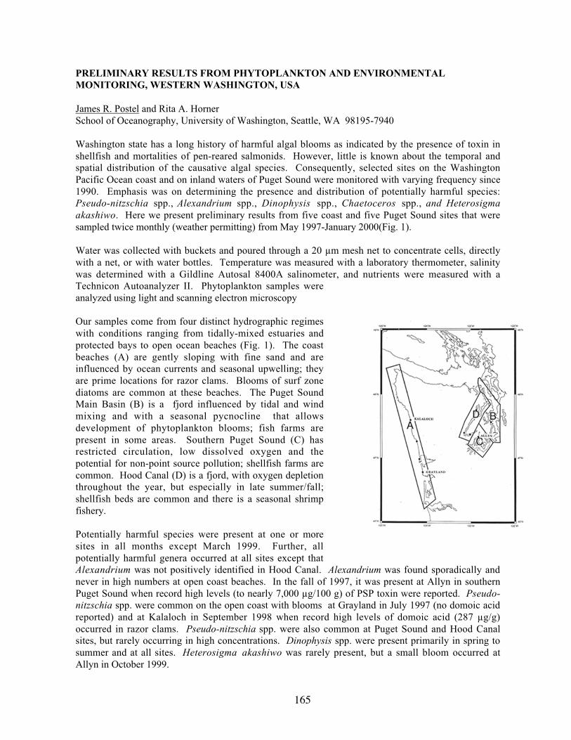
165
PRELIMINARY RESULTS FROM PHYTOPLANKTON AND ENVIRONMENTALMONITORING, WESTERN WASHINGTON, USA
James R. Postel and Rita A. HornerSchool of Oceanography, University of Washington, Seattle, WA 98195-7940
Washington state has a long history of harmful algal blooms as indicated by the presence of toxin inshellfish and mortalities of pen-reared salmonids. However, little is known about the temporal andspatial distribution of the causative algal species. Consequently, selected sites on the WashingtonPacific Ocean coast and on inland waters of Puget Sound were monitored with varying frequency since1990. Emphasis was on determining the presence and distribution of potentially harmful species:Pseudo-nitzschia spp., Alexandrium spp., Dinophysis spp., Chaetoceros spp., and Heterosigmaakashiwo. Here we present preliminary results from five coast and five Puget Sound sites that weresampled twice monthly (weather permitting) from May 1997-January 2000(Fig. 1).
Water was collected with buckets and poured through a 20 µm mesh net to concentrate cells, directlywith a net, or with water bottles. Temperature was measured with a laboratory thermometer, salinitywas determined with a Gildline Autosal 8400A salinometer, and nutrients were measured with aTechnicon Autoanalyzer II. Phytoplankton samples wereanalyzed using light and scanning electron microscopy
Our samples come from four distinct hydrographic regimeswith conditions ranging from tidally-mixed estuaries andprotected bays to open ocean beaches (Fig. 1). The coastbeaches (A) are gently sloping with fine sand and areinfluenced by ocean currents and seasonal upwelling; theyare prime locations for razor clams. Blooms of surf zonediatoms are common at these beaches. The Puget SoundMain Basin (B) is a fjord influenced by tidal and windmixing and with a seasonal pycnocline that allowsdevelopment of phytoplankton blooms; fish farms arepresent in some areas. Southern Puget Sound (C) hasrestricted circulation, low dissolved oxygen and thepotential for non-point source pollution; shellfish farms arecommon. Hood Canal (D) is a fjord, with oxygen depletionthroughout the year, but especially in late summer/fall;shellfish beds are common and there is a seasonal shrimpfishery.
Potentially harmful species were present at one or moresites in all months except March 1999. Further, allpotentially harmful genera occurred at all sites except thatAlexandrium was not positively identified in Hood Canal. Alexandrium was found sporadically andnever in high numbers at open coast beaches. In the fall of 1997, it was present at Allyn in southernPuget Sound when record high levels (to nearly 7,000 µg/100 g) of PSP toxin were reported. Pseudo-nitzschia spp. were common on the open coast with blooms at Grayland in July 1997 (no domoic acidreported) and at Kalaloch in September 1998 when record high levels of domoic acid (287 µg/g)occurred in razor clams. Pseudo-nitzschia spp. were also common at Puget Sound and Hood Canalsites, but rarely occurring in high concentrations. Dinophysis spp. were present primarily in spring tosummer and at all sites. Heterosigma akashiwo was rarely present, but a small bloom occurred atAllyn in October 1999.

166
Environmental conditions included extended periods of < 0.5 µM nitrate on the ocean beaches withperiodic replenishment by upwelling events or seasonal changes in the coastal currents. The PugetSound Main Basin seldom had < 5 µM nitrate due to strong tidal mixing and weak stratification; HoodCanal and southern Puget Sound generally had < 0.5 µM nitrate concentrations throughout the summerperiods.

167
DYNAMICS OF ALEXANDRIUM CATENELLA BLOOMS IN QUARTERMASTERHARBOR, WA
James R. Postel, Paul N. Rudell, and Rita A. HornerSchool of Oceanography, Box 357940,University of Washington, Seattle, WA 98195-7940
Alexandrium catenella, that causes paralytic shellfish poison(PSP) in Puget Sound, WA, was studied in QuartermasterHarbor (Fig. 1), a small, semi-enclosed, seasonally stratified bayin central Puget Sound to determine the effects of biological,chemical, and physical factors on the formation, maintenance,and decline of blooms. A shore station was sampled near mid-bay 1-3 times per week from May to September 1998 todetermine the environmental conditions of the surface layer inthe bay during the optimal growing season for this species.Additionally, three cruises occupied a station at 20 m depth in1997 and 1998 to study the diel migration pattern of theAlexandrium population and to estimate growth and mortality(from grazing) rates of the total dinoflagellate population fromdilution experiments. Estimates of tidal flushing were made bycalculating the volumes of the bay at various tide heights and thevolumes of typical tidal prisms for mean, neap, and spring tideranges.
Figure 1. Quartermaster Harbor, Vashon Island, WAPSP concentrations in caged mussels at 1 m depth exceeded 80 µg 100 g-1 mussel meat twice during1997 and 1998, both times following the occurrence of A. catenella concentrations > 10,000 cells L-1.The species was present continuously from late May through September 1998, blooming in July insurface water. Over this time interval surface temperatures varied from 14-20 °C; nitrate levelsremained near-zero; phosphate and silicate were low, but measurable; and salinity ranged from 25-30psu. Thermal stratification beginning in late spring led to the development of a shallow, low-nutrientsurface layer (upper 1-3 m), a distinct pycnocline (3-5 m depth), and a deeper, nutrient-replete layer(5-20 m). During the July 1998 cruise strong spring tidal cycles eroded the pycnocline and mixed thewater column within 24 h. This disrupted the distribution of A. catenella and contributed to bloomdecline.
The diel vertical migration pattern of A. catenella, studied in 1997 and 1998, was limited to the upper10 m of the water column with migration rates varying from 0.4 - 2.0 m h-1. Maximal A. catenella cellnumbers varied from < 10,000 - > 60,000 cells L-1, but the phytoplankton assemblage was dominatedby large diatoms which were present at > 500,000 cells L-1. Dilution experiments in 1998 indicatedthat the total dinoflagellate population grew at about the same rate as the diatom population but wasgrazed more heavily by microzooplankton. If estimated loss due to tidal flushing of the population isadded to grazing rate, the dinoflagellate population growth rate was less than the combined loss rate,but growth still exceeded loss for the diatoms during spring tidal exchange periods. However, duringmean or neap tidal periods, population growth rate was higher than loss rate for both dinoflagellatesand diatoms. Assuming that the results from the overall dinoflagellate population are applicable to A.catenella, it appears that when water temperature exceeds 13 °C, permitting active A. catenellagrowth, blooms are rarely generated and then only for a limited period each summer. A combinationof biological, chemical, and physical factors can lead to decline of the bloom within the bay andprevent an accumulation of PSP in the shellfish.

168
DEVELOPMENT OF A PROTOCOL FOR DETERMINATION OF DOMOIC ACID INMOLE CRABS (EMERITA ANALOGA): A POSSIBLE NEW INDICATOR SPECIES
Christine L. Powell1, M.E. Ferdin2, Carolyn Bretz2, Mark Busman1, Rikk G. Kvitek2, and Gregory J.Doucette1
1Marine Biotoxins Program, NOAA/NOS/CCEHBR, Charleston, SC 294122California State University, Monterey Bay (CSUMB), CA 93955
The transfer of algal toxins through marine food webs has important implications for not only publichealth, but also the health of ecosystems and their trophic structure. Impacts of algal toxin trophictransfer are manifested as contaminated commercial and recreational fishery resources, as well asmortality events involving a variety of birds and mammals. Protection of public health requiresidentification of an indicator or sentinel species that rapidly accumulates toxin upon its appearance inlocal waters. Monitoring the movement of an algal toxin through food webs requires identification ofpotential points of entry and thus vectors for toxin transfer. In the case of domoic acid (DA), molecrabs (Emerita analoga) have the potential to serve both as a sentinel species and as a vector fortrophic transfer of this potent neurotoxin produced by members of the diatom genus, Pseudo-nitzschia.
Monterey Bay, California is a site of recurrent, toxic and non-toxic Pseudo-nitzschia blooms, as wellas extensive mole crab populations. This location was therefore selected to begin evaluating the utilityof E. analoga as an indicator for DA presence in local waters. Moreover, one of the current sentinelorganisms, the sea mussel (Mytilus californianus), shows minimal or undetectable toxicity duringsome bloom events in Monterey Bay. It is also anticipated that data collected during this project willprovide information on the efficacy of mole crabs as a vector for DA transfer to higher trophic levels.
The efficiency of extracting algal toxins from diverse sample types is highly variable and depends onthe individual sample matrix. Using a conventional aqueous methanol extraction method for shellfishas a starting point, we have developed and validated a highly efficient protocol for extracting DA fromwhole mole crabs that yields toxin recoveries of 96.5 + 2.9 percent. We also confirmed by LC-MS/MSthat mole crabs accumulated measurable amounts of DA during toxic Pseudo-nitzschia bloom events(0.5-10 micrograms DA/g tissue), while the blue mussel showed no detectable toxin. In addition, anextensive comparison (n = 60) of inter-animal variability in DA content revealed values ranging overan order of magnitude (ca. 0.5 to 5 micrograms DA/g tissue) and no consistent trend with size class,based on either animal weight or length. These data on the toxicity of individual animals will beuseful in designing an appropriate sampling strategy and, importantly, indicate that mole crabs do notappear to progressively bioaccumulate DA with age. In fact, initial observations by Ferdin et al. (thisconference) revealed a rise and fall of DA toxicity in mole crabs, coinciding with similar changes inPseudo-nitzschia cell concentrations. The extraction protocol developed herein will be implementedduring upcoming field trials to further compare the DA toxicity of mole crabs vs. blue mussels and itscorrelation to the concentrations of toxic Pseudo-nitzschia cells in local waters. Mole crab toxicityvalues will also be available for reference, in the event of suspected DA-associated mortality eventsinvolving consumers of E. analoga.

169
FISH BIOASSAYS AND PILOT-SCALE APPLICATION OF CLAY TO CONTROL BLOOMSOF HETEROSIGMA AKASHIWO AT PACIFIC NORTHWEST SALMON NET PENS
J.E. Jack Rensel1, Donald M. Anderson2, Laurie B. Connell3
1Rensel Associates Aquatic Science Consultants, Arlington WA 982232Woods Hole Oceanographic Institution, Woods Hole, MA 025433National Marine Fisheries Service, Seattle, WA 98112
Fish killing microalgae are a major impediment to the increasing fish mariculture activities in manylocations around the world. Fish farmers use a variety of physical methods to mitigate effects ofharmful algal blooms (HABs) including towing of pens, perimeter skirts, upwelling systems and othermeans. But these are not without risks, such as ripping of pens when towing and inefficientdisplacement of blooms by upwelling due to unpredictable vertical distribution of algae. Certain typesof clay flocculation appears to be a promising means of treating some fish killing HABs in or aroundfish farms, based on laboratory tests and reports from large-scale applications in Korea and Japan.Recurring wide-scale blooms of Heterosigma akashiwo have caused major fish losses in the PacificNorthwest Coast in Washington State, but more so in British Columbia, where the industry is muchlarger and broadly distributed. Since Heterosigma does not produce a persistent toxin, but apparentlykills fish through the action of reactive oxygen species that quickly dissipate in seawater, the alga is agood candidate for the use of clay mitigation. (Note there is a possibility that Heterosigma mayproduce a weak brevetoxin-like compound, based on one documented case). This work reports initiallaboratory results of clay bioassays with Atlantic salmon (Salmo salar) and plans to test the mitigationmethod at a commercial fish farm using perimeter skirts to retain the clay in a small area and reducereintroduction of the alga.
Replicated laboratory bioassays were conducted with clay concentrations ranging from 30 to 100 mg/Lclay, held in suspension by air lift pumps for five hours, much longer than would occur in the field.Turbidity and total suspended solids were used as indices of clay loading, and were positivelycorrelated to a high degree. Turbidity is the preferred measurement for practical and water qualitystandard reasons. Fish began “coughing” immediately upon introduction of the clay in the aquaria, anatural reaction to many types of irritants, and cough frequency occurred in a dose-related fashion upto 10 times per minute. The nature of the coughing was different from that previously seen with fishexposed to harmful Chaetoceros concavicornis; it was shallower and less pronounced. Coughingceased immediately upon transfer of the fish to clean seawater at the conclusion of the bioassay. Gilltissue samples of other treated fish were collected for histological analysis. There was some mucuspresent on the affected gills, but not an usually large amount. These results suggest that there will notbe any significant or permanent effects of clay on treated fish, as high levels of turbidity occurnaturally during the freshwater phase of salmonid life. Prior studies have shown that juveniles ofseveral salmon species tolerate much higher concentrations of silts or clays than were used in ourexperiments, and for much longer periods.
Our null hypothesis for the sediment chemistry and infauna fieldwork is that there will be nomeasurable impact on the seabottom or benthos of clay application. Pacific Northwest fish farms arerequired to locate where there are no other significant aquatic resources, including shellfish beds.Moreover, there is little permanent deposition of the wastes from these farms, as they are withoutexception located in current swept areas with erosional bottoms. Although Heterosigma occursfrequently each spring and fall at many farm sites, concentrations are usually less than that which willcause fish mortality in about 9 out 10 years at a given farm site. A typical farm producing severalmillion pounds of fish annually will therefore only require an annual clay application (pro rata over 10years) of about 40 kg per farm [(0.4 ha per farm x 0.1 kg m2)/10]. There are only 9 large, separatecommercial pens in the 0.8 million hectares of Puget Sound, so cumulatively the required clay amount

170
is trivial. The volume of treatment clay needed pales in comparison to the estimated 6 million metrictons of sediment including clay entering Puget Sound each year from riverine and shoreline erosion(Dexter et al. 1981 NOAA Tech. Memo. OMPA-13).

171
ESTABLISHING AND MAINTAINING CLONAL CULTURES OF GYMNODINIUM BREVE
Bill RichardsonFlorida Fish & Wildlife Conservation Commission, Florida Marine Research Institute100 Eighth Ave SE, St. Petersburg, FL 33701
The toxic dinoflagellate Gymnodinium breve has been grown and studied in the laboratory for almostfive decades. Most of the early physiological studies were performed using the Wilson clone (isolatedby W.B. Wilson from John Pass, Florida in 1953, CCMP718). Renewed interest in the physiology andgenetics of G. breve has emphasized the need for the establishment and long term maintenance ofclonal cultures from a broad geographic range. Until recently, the former successes in establishingcultures for study had not been widely reproduced. One of the primary reasons is that G. breve cellsare fragile and are easily stressed. The method used in our laboratory is presented as a simplecombination of established isolation and culture methodologies. The techniques have been used toestablish and maintain 20 different clonal isolates of G. breve from the Gulf of Mexico and Atlanticwaters for as long as 4 years.
Growth medium is prepared using oligotrophic seawater collected by lowering a submersible pump(all plastic components) over the side of a research vessel. The water is stored, prior to use, in the darkat 5°C in 20 liter polycarbonate carboys. The seawater is 0.45 µm filtered and then sterilized byautoclaving in 1.5 liter teflon bottles. Established cultures are maintained in growth medium preparedby adding sterile filtered nutrients as in “L1” (Guillard and Hargraves, 1993) except for the vitaminadditions, which are added as in NH-15 (Gates and Wilson, 1960 ).
Samples for isolation are usually received as whole water samples shipped to the laboratory in plasticbottles or bags. Individual cells are drawn up into sterile micro-capillary borosilicate glass pipets(~300 µm diameter). Individual cells are then repeatedly washed by transferring them by micro-pipet
to drops of sterile filtered oligotrophic seawater on a glass slide spot plate. All mouth pipeting is doneon an inverted microscope (40X) under a laminar flow hood. After several washes, individual cells areplaced in polystyrene tissue culture well plates containing 1 mL of growth media. The growth mediaused to establish the single cells in culture uses the medium described above at 1/10th concentration insterile filtered oligotrophic water. This media may be diluted 50% with GF/F filtered water from anestablished culture in an effort to decrease the shock that newly isolated single cells might experiencewhen placed in water not “conditioned” by G. breve cells and their associated bacteria. The isolatecultures are allowed to reach densities of at least 25 cells/mL before additional media is added to thewell. Additional media is added 2-3 times per week in small amounts, not exceeding 10% of theexisting culture volume. This minimizes the stress to the growing cells while still meeting their needs.The isolates are transferred to successively larger flasks until they are finally moved to 13 liter Pyrexglass carboys. The isolates are maintained in these carboys in volumes of 3-8 liters at S=34, 23°C, and
under a 12:12 L:D cycle at 60 µEm-2sec-1. Transfers of established cultures in carboys are made every
3-4 weeks using cultures with densities of approximately 107 cells/L to establish new cultures withdensities of approximately 105 cells/L. Isolate cultures are maintained at growth rates between 0.2 and0.3 divisions da-1. Both “grandparent” and “parent” cultures are kept as a safeguard against unexpectedculture mortality. The combination of basic culture methods described above, could be easilyreplicated and improved upon, resulting in the establishment and maintenance of additional G. breveor other Gymnodinium clonal isolates for use in taxonomic, physiological, and genetic investigations.

172
References:
Gates, J.A. and W.B. Wilson. 1960. The toxicity of Gonyaulax monilata Howell to Mugil cephalis.Limnol. Oceanogr. 5, 171-174.
Guillard, R.R.L. and P.E. Hargraves. 1993. Stichochrysis immobilis is a diatom, not a chrysophyte.Phycologia, 32, 234-236.
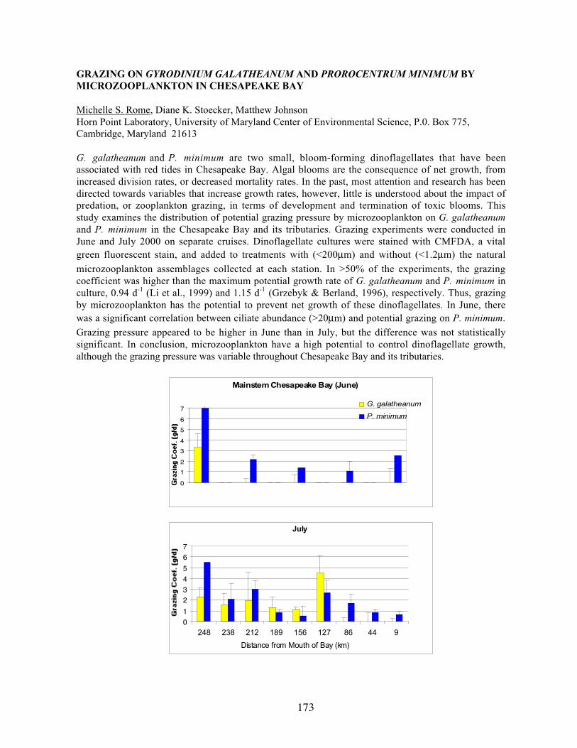
173
GRAZING ON GYRODINIUM GALATHEANUM AND PROROCENTRUM MINIMUM BYMICROZOOPLANKTON IN CHESAPEAKE BAY
Michelle S. Rome, Diane K. Stoecker, Matthew JohnsonHorn Point Laboratory, University of Maryland Center of Environmental Science, P.0. Box 775,Cambridge, Maryland 21613
G. galatheanum and P. minimum are two small, bloom-forming dinoflagellates that have beenassociated with red tides in Chesapeake Bay. Algal blooms are the consequence of net growth, fromincreased division rates, or decreased mortality rates. In the past, most attention and research has beendirected towards variables that increase growth rates, however, little is understood about the impact ofpredation, or zooplankton grazing, in terms of development and termination of toxic blooms. Thisstudy examines the distribution of potential grazing pressure by microzooplankton on G. galatheanumand P. minimum in the Chesapeake Bay and its tributaries. Grazing experiments were conducted inJune and July 2000 on separate cruises. Dinoflagellate cultures were stained with CMFDA, a vitalgreen fluorescent stain, and added to treatments with (<200µm) and without (<1.2µm) the naturalmicrozooplankton assemblages collected at each station. In >50% of the experiments, the grazingcoefficient was higher than the maximum potential growth rate of G. galatheanum and P. minimum inculture, 0.94 d-1 (Li et al., 1999) and 1.15 d-1 (Grzebyk & Berland, 1996), respectively. Thus, grazingby microzooplankton has the potential to prevent net growth of these dinoflagellates. In June, therewas a significant correlation between ciliate abundance (>20µm) and potential grazing on P. minimum.Grazing pressure appeared to be higher in June than in July, but the difference was not statisticallysignificant. In conclusion, microzooplankton have a high potential to control dinoflagellate growth,although the grazing pressure was variable throughout Chesapeake Bay and its tributaries.
Mainstem Chesapeake Bay (June)
0
1
2
3
4
5
6
7G. galatheanum
P. minimum
July
0
1
23
4
5
6
7
248 238 212 189 156 127 86 44 9
Distance from Mouth of Bay (km)

174
INHIBITION OF ALEXANDRIUM FUNDYENSE STR. CB301-A BY MARINE BACTERIA
Juliette N. Rooney-Varga, Mary Savin, Michael Ferrier, Andrew B. GolayBiological Sciences Department, University of Massachusetts Lowell, 1 University Avenue, Lowell,MA 01854
Bacterial influences on toxic algal blooms have been studied extensively in several regions of theworld but remain poorly understood in the Gulf of Maine, where Alexandrium spp. blooms havebecome a chronic problem. In the current study, we describe a bacterial assemblage and two bacterialisolates that either inhibited or killed Alexandrium fundyense. A bacterial assemblage from a non-axenic, late-exponential phase A. fundyense culture was inoculated into axenic A. fundyense str.CB301-A, which has been maintained as an axenic culture for about one year. This bacterialassemblage was found to cause the decline of A. fundyense CB301-A cells within 6 days ofinoculation. Three members of this assemblage were isolated on agar plates containing f/2 mediumamended with 0.5% peptone, 0.5% glucose, and 0.5% yeast extract. Of the three bacteria isolated, twostrains (B1 and B2) inhibited CB301-A growth, while the third (B3) had no effect. 16S rRNAphylogenetic analysis revealed that isolate B1 was most closely related to strain S34, anuncharacterized member of the alpha-Proteobacteria that was isolated from the Sargasso Sea. StrainB2 was extremely closely related to Marinobacter sp. PCOB-2, a member of the familyAlteromonodaceae isolated from a marine algal culture. We are currently continuing ourcharacterization of these bacterial isolates and plan to determine their effects on other Alexandriumstrains as well as non-toxic marine algae found in the Gulf of Maine.
175
DEVELOPMENT AND VALIDATION OF A PCR-BASED DETECTION ASSAY FORPFIESTERIA PISCICIDA IN WATER AND SEDIMENT ENVIRONMENTAL SAMPLES
Keiko Saito, José- Antonio F. Robledo, and Gerardo R. VastaCenter of Marine Biotechnology, Maryland Biotechnology Institute, 701 E. Pratt St. Baltimore, MD21202
Pfiesteria piscicida is a heterotrophic dinoflagellate that has been associated with massive fishmortalities, and, in some cases, associated with health problems in watermen working along the lowerEastern Shore of the Chesapeake Bay. Similar episodes, although of lesser scale, have occurred inChesapeake Bay tributaries. Plate tabulation has been the routine method for the identification ofdinoflagellate species. Groups of species are characterized by a particular formula, and differences inshape or size of some of the plates can define distinct species within the group. This technique is time-consuming and requires special training. We have developed a rapid, sensitive, specific, and easy toimplement in any laboratory PCR-based assay for the diagnostics of P. piscicida. Based on geneticinformation, the PCR assay uses primers from P. piscicida NTS and SSU regions yielding an ampliconof 429 bp. We have been routinely applying this assay in the core facility to identify P. piscicida inenvironmental water and sediment samples. Although the PCR assay is a very sensitive technique forthe detection of specific sequences in complex DNA mixtures, the amplification reaction can be easilyinhibited by compounds present in the sample. Indeed it is fundamental to develop controls thatguarantee the detection of P. piscicida in a sample as a consequence of the absence and not the resultof PCR inhibition. Here we present data on the effect of inhibitors from water and sediment samplesin the PCR assay and we developed an internal standard that uses the same priming sites that the PCR-based assay for P. piscicida. DNA from environmental samples is spiked with the internal control andan aliquot of the mixture used PCR. If the internal control is detected, samples are subsequently testedfor P. piscicida. When the internal control is not detected by PCR, serial dilution of the sample areprepared and used to establish the dilution of the sample at which there is no inhibition of the PCR.This sample dilution is then used for detection of P. piscicida. The use of internal standards wouldguarantee the quality of the sample and avoid false negatives in environmental samples and, based ondata, we suggest its use for routine diagnostics of P. piscicida.

176
PFIESTERIA PISCICIDA IN SEDIMENT SAMPLES FROM FISH-FARMING PONDS: INVITRO EFFECT(S) OF LIVE FISH ON THE EARLY PCR-BASED DETECTION OFZOOSPORES IN THE SUPERNATANT WATER
Keiko Saito, José-Antonio F. Robledo, and Gerardo R. VastaCenter of Marine Biotechnology, University of Maryland Biotechnology Institute, 701 E. Pratt St.,Baltimore, MD 21202
Pfiesteria piscicida is a heterotrophic dinoflagellate that has been associated with massive fish killsduring recent years in the mid-Atlantic estuaries of North America. Thus, the effects of live fish, andtheir secretions, excretions or metabolic products on the life cycle, proliferation, and toxicity of P.piscicida has become a research area of great interest. In this preliminary study, we examined thepossible effect(s) of live fish on the presence and proliferation of P. piscicida in water or culturemedium that has been incubated with sediments potentially containing putative life stages of thisdinoflagellate species. Sediments (10 ml) from 10 active or drained ponds from a fish farming facilitywere sieved through a 60 _m mesh, and cultured in 750 ml flasks with 500 ml of artificial seawater (7ppt; pH 8) containing one Sheepshead Minnow (Cyprinodon variegatus). (Controls were flasks withwater containing no fish, or with culture medium (F/2) containing no fish.) Flasks were maintained at23 ºC and 14 h light/10 h dark cycle. Enumeration of dinoflagellate zoospores in the supernatant andPCR-based specific detection of P. piscicida were carried out daily for 21 days, and the health of thefish was monitored throughout the experiment. Numbers of P. piscicida-like zoospores increasedprogressively, and positive PCR results for P. piscicida were obtained in flasks corresponding to allten ponds. In flasks containing fish, however, P. piscicida could be detected as early as the secondday, and flasks remained PCR-positive throughout the duration of the experiment. No fish deaths wererecorded.

177
EFFECTS OF ALEXANDRIUM FUNDYENSE, ON NEWLY SETTLED JUVENILE WINTERFLOUNDER, PLEURONECTES AMERICANUS
Jennifer C. Samson1, Judith S. Weis1, and Sandra E. Shumway2
1Rutgers University, Newark, NJ 071022Southampton Campus, Long Island University, Southampton, Long Island, NY 11968
Alexandrium fundyense, a toxic dinoflagellate that produces paralytic shellfish toxins (PST; saxitoxinand its derivatives) has been implicated in fish kills during bloom conditions. Alexandrium is at thebase of the aquatic food web, and is readily consumed by a variety of organisms, which canconsequently accumulate the toxin. Alexandrium occurs temporally and spatially in estuaries withmany species of larval and juvenile fish during the summer and fall. Larval winter flounder,Pleuronectes americanus, settle in their natal estuaries and shallow embayments during spring, wherethe newly settled juveniles remain for up to two years. Fidelity to specific spawning areas makes thiscommercially important species particularly sensitive to annual blooms of Alexandrium. Weinvestigated the lethal and sublethal (behavioral) effects of trophic transfer of PST on newly settledjuvenile winter flounder, using Alexandrium→copepod, Coullana canadensis→flounder, food chain.
The harpacticoid copepod, C. canadensis, was exposed to a toxic strain of A. fundyense for 4consecutive days, as a mixture of 50:50 by volume, green algae and A. fundyense (500 cells/ml).Control copepods were fed a non-toxic strain of A. tamerense. Copepods actively consumed thedinoflagellates. Following exposure, toxic copepods were fed to newly settled fish (8-10mm TL).
In mortality experiments fish were fed 1 toxic copepod every 15 minutes. Feeding was halted whenfish began swimming erratically in somersaults, eventually ending upside down on bottom, where theyremained until the heart stopped beating. Erratic swimming occurred immediately after consuming thefinal copepod (6 ± 2.5 copepods per fish). Fish fed 6 control (non-toxic) copepods never exhibited this
behavior.
Observation during preliminary experiments indicated that fish fed only 5 toxic copepods (sublethaldose) could be fed an additional 5 toxic copepods 24 hours later with no mortality. To elucidatefurther this apparent recovery, a time to death experiment was run. Fish were individually fed 5 toxiccopepods, and then fed a second set of 5 toxic copepods, either 1, 2, 4 or 8 hours after completion ofthe first feeding. Survival analysis indicated a significant difference in the mean survival rate of eachgroup (chi-square=10.964, df=3, n=33, p≤0.01) with high mortality in fish given only 1 and 2 hours
between feedings and little or no mortality in the 4 and 8 hour groups. These data indicate a recoverywith time. Observations during data collection suggest a loss of toxin in the feces, as some fish reviveafter defecation.
Experiments were also conducted to evaluate sublethal effects on behavior. Fish were fed either 5toxic or control, copepods. Two hours after feeding, individuals were placed in watch glasses andgiven 5 minutes to eat 5 unexposed copepods. Number of attempts and captures, time spent in motion,and distance traveled, were recorded. There was no difference in attempts or captures betweenexposed and unexposed juvenile flounder (n=20/treatment); however exposed fish were not as activeas control fish, resulting in a significant difference between the two treatments for time spent in motion(t=2.539, p≤0.01) and distance traveled (t=2.067, p≤0.05). Results indicate some effects of saxitoxin
on behavior but not enough to impair feeding ability. Thus, newly settled winter flounder exposed toPST via trophic transfer exhibit both lethal and sublethal (behavioral) reactions to exposure, dependentupon number of toxic copepods eaten and rate of feeding.

178
LABORATORY ADVANCES AND THE EPIDEMIOLOGY OF MARINE TOXIN DISEASES
Helen Schurz-Rogers1, Lorraine C. Backer1, and Lora E. Fleming2
1National Center for Environmental Health, Centers for Disease Control and Prevention, Atlanta, GA30329 USA 2University of Miami School of Medicine, Miami, FL 33136 USA
Advances in marine toxin laboratory science form the scientific basis for understanding the biology andepidemiology of the human illnesses caused by these toxins. We will review how both the advances inlaboratory science and the lack of such advances have affected our understanding of the epidemiologyof human illnesses caused by exposure to two dinoflagellate organisms, Gymnodinium breve andPfiesteria piscicida.
Gymnodinium breve is a marine dinoflagellate that produces polyether toxins, called brevetoxins. Fishkills associated with blooms of G. Breve, called red tides, have been reported since 1844. Foodpoisonings (neurotoxic shellfish poisoning [NSP]) from consuming shellfish from Florida have beenreported since the early 1900s, but the causative organism was not identified until the 1960s (after thediscovery of G. breve in 1947). Work conducted in the 1980s identified and characterized thebrevetoxins, allowing researchers to determine their biochemical activity (induction of channel-medicated Na+ ion influx), pharmacology, biological activity (depolarization of muscle fibermembranes), and physiological effect (neurotoxicity). Brevetoxins can be detected and quantified inshellfish as well as in water and air samples and human exposure to brevetoxins occurs either fromconsuming contaminated shellfish or from inhaling sea spray containing fragments of the cells or thetoxin. Therefore, because we have considerable information about routes of exposure, biochemicalactivity, and human symptoms, it is possible to conduct the appropriate epidemiologic studies toevaluate the public health importance of G. breve-associated diseases in human populations.
By contrast, despite the availability of molecular biology and analytical chemistry tools, the toxinsassociated with Pfiesteria piscicida, as well as their specific biochemical, pharmacological, biological,and physiologic activities, remain elusive. Pfiesteria piscicida’s fish killing ability was only identified10 years ago, and the first human health effects, associated with concentrated aerosol exposures in thelaboratory, were reported seven years ago. In addition, these organisms are difficult to identify, appearto form fleeting blooms, and can be resistant to culturing. Thus, at this time, we cannot accuratelyquantify environmental exposure to the organism or to any toxin(s) it may produce. New molecularbiology techniques may allow at least a rapid identification and enumeration of cells during a bloom,which could provide a relative estimate of exposure. However, without knowing the biochemicalactivity of the toxin(s) or an accurate assessment of exposure, it is difficult to develop a case definitionfor human illness. Because we have very little information about exposure and human symptoms,epidemiologic studies of human health effects from exposure to P. piscicida will produce limitedinformation in evaluating the public health importance of this organism.
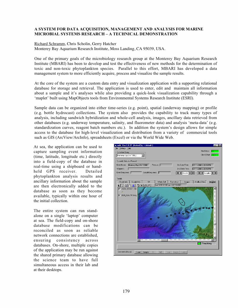
179
A SYSTEM FOR DATA ACQUISITION, MANAGEMENT AND ANALYSIS FOR MARINEMICROBIAL SYSTEMS RESEARCH – A TECHNICAL DEMONSTRATION
Richard Schramm, Chris Scholin, Gerry HatcherMonterey Bay Aquarium Research Institute, Moss Landing, CA 95039, USA.
One of the primary goals of the microbiology research group at the Monterey Bay Aquarium ResearchInstitute (MBARI) has been to develop and test the effectiveness of new methods for the determination oftoxic and non-toxic phytoplankton species. Parallel to this effort, MBARI has developed a datamanagement system to more efficiently acquire, process and visualize the sample results.
At the core of the system are a custom data entry and visualization application with a supporting relationaldatabase for storage and retrieval. The application is used to enter, edit and maintain all informationabout a sample and it’s analyses while also providing a quick-look visualization capability through a‘maplet’ built using MapObjects tools from Environmental Systems Research Institute (ESRI).
Sample data can be organized into either time-series (e.g. point), spatial (underway mapping) or profile(e.g. bottle hydrocast) collections. The system also provides the capability to track many types ofanalysis, including sandwich hybridization and whole-cell analysis, images, ancillary data retrieved fromother databases (e.g. underway temperature, salinity, and fluorometer data) and analysis ‘meta-data’ (e.g.standardization curves, reagent batch numbers etc.). In addition the system’s design allows for simpleaccess to the database for high-level visualization and distribution from a variety of commercial toolssuch as GIS (ArcView/ArcInfo), spreadsheets (Excel) or via the World Wide Web.
At sea, the application can be used tocapture sampling event information(time, latitude, longitude etc.) directlyinto a field-copy of the database inreal-time using a shipboard or hand-held GPS receiver. Detailedphytoplankton analysis results andancillary information about the sampleare then electronically added to thedatabase as soon as they becomeavailable, typically within one hour ofthe initial collection.
The entire system can run stand-alone on a single ‘laptop’ computerat sea. The field-copy and on-shoredatabase modifications can bereconciled as soon as reliablenetwork connections are established,ensuring consistency acrossdatabases. On-shore, multiple copiesof the application may be run againstthe shared primary database allowingthe science team to have fullsimultaneous access in their lab andat their desktops.
180
ECOHAB, THE U.S. INTERAGENCY PROGRAM TO SUPPORT RESEARCH ON THEECOLOGY AND OCEANOGRAPHY OF HARMFUL ALGAL BLOOMS
Kevin G. SellnerECOHAB Coordinator, NOAA/NOS/CSCOR, N/SCI2, 1315 E-W Highway, Silver Spring, MD20910
The study of harmful algal blooms (HABs) in the U.S. has recently received major commitments fromfive U.S. Federal agencies (National Oceanic and Atmospheric Administration, EnvironmentalProtection Agency, National Science Foundation, Office of Naval Research, National AeronauticsSpace Administration) for long-term funding through the interagency ECOHAB Program (Ecologyand Oceanography of Harmful Algal Blooms). Arising from the community-driven 1995 report of thesame name, ECOHAB now supports >30 research projects at over $40M U.S. The purpose of theprogram is to support research to increase general knowledge and understanding of HAB ecology,physiology, behavior, and toxin synthesis and links to environmental conditions including trophiccomplexity, water quality, local and regional circulation, bathymetry, and meteorology, as well aseconomic assessments and outreach. Projects range from targeted studies of one to severalinvestigators for 1-3 years up to large multi-investigator, multi-institution, and multi-disciplinaryregional studies for the development of biophysical models of bloom expression and distribution.Tools developed in these research projects, such as molecular probes, toxin assays, techniques forpossible use in the prevention, control, and mitigation of HABs and their impacts, and biophysicalmodels for ‘forecasting’ bloom distributions, are then considered for inclusion in standard monitoringprograms fostered through several agencies and non-Federal public departments. In the future, it ishoped that research results from ECOHAB-supported research can become part of the internationalHAB research, monitoring, and event response programs now being initiated in the new EUROHABand GEOHAB programs.
181
DETERMINING THE CAUSES OF MORTALITY IN BIOTOXICITY ASSAYS WITHPFIESTERIA: THE DEVELOPMENT OF FRACTIONATION STUDIES
Jeffrey D. Shields, Wolfgang K. Vogelbein, Yasunari Kiryu, Alynda Miller, Christopher M. Squyars,Larry W. Haas, Kimberly S. Reece, Nancy A. Stokes, Vincent J. Lovko, and Howard KatorVirginia Institute of Marine Science, Gloucester Point, Virginia 23062, USA.
Aquarium-scale biotoxicity assays with Pfiesteria pose inherently difficult problems. Fish health mustbe carefully balanced with issues such as frequency of water changes, degradation of water quality, thepresence of pathogens, and the density of the background community of micro-organisms andprotozoa. Add to the equation, long-term exposures of fish to a variably toxic species such asPfiesteria piscicida, or the potential for contamination with related species (Pfiesteria sp. B), and theproblems quickly appear insurmountable. We undertook long-term (80+ d) bioassays of laboratory andfield isolates (Neuse and Pamlico rivers) of several Pfiesteria-like dinoflagellates (P. piscicida,Pfiesteria sp. B., "Lucy," and "Shepherds Crook"). For field isolates, fish were immediately placed inwater collected for biotoxicity assays. Subsamples were processed for counts of heterotrophicdinoflagellates, DNA analysis, and SEM verification. Exposures consisted of 25-50 fish in eachaquarium (38 L, 12‰ artificial seawater in de-ionized water, preconditioned Whisper filters, 20° C)inoculated with isolates from cultures, sediments or freshly collected water samples. Fish were fedthree times per week and monitored for signs of morbidity, lesions, and mortality. Controls consistedof fish in replicate treatments without dinoflagellates (unexposed). Thirty long-term toxicity bioassays(17 field isolates, 3 subcultures, and 10 clonal cultures, with controls) were conducted. Two aquariashowed moderate mortalities, and four aquaria showed high mortalities in relation to the density ofPfiesteria sp. B. Mortality events typically lasted 2-3 weeks with 1-2 sequelae shortly thereafter. Infour cultures, heavy bacterial loads (Vibrio spp. and Shewanella putrefasciens) were present, and waterquality was degraded due to low dissolved oxygen (high BOD), high ammonia and nitrite, and low pHin relation to organic wastes. High bacterial loads and low pH (5.8-6.0) occurred in the other twoaquaria, but other conditions were ideal to support fish health. All aquaria possessed numerous ciliates(holotrichs, and hymenotrichs), rotifers, amoebae, microflagellates (bicoecids), and other taxa. Thus,even though densities of Pfiesteria were high (2.0-20.0 x 10³ cells ml-1), it was difficult to rule outother causal or contributory factors in fish deaths.
Water quality studies were undertaken with an appraisal of ammonia toxicity in tilapia, Oreochromisniloticus, at varying pH. Tilapia were resistant to high levels of reactive ammonia (LC50 = 9.72 ppm),and low pH (5.0, 6.5). Thus, ammonia was not a significant factor in the observed mortalities. Wethen developed a "fractionation" assay where small volumes (1-2 l) of "toxic" aquarium culture werefiltered and centrifuged into different components: dinoflagellates (and other protozoa), bacteria (andmicroflagellates), and the "soluble toxin" fractions. Controls consisted of raw culture water, and sterileartificial seawater. Fractionation assays with tilapia in 500 ml volumes were inconclusive. However,fractionation assays with mummichogs in 15 ml volumes indicated conclusively that the dinoflagellatefraction caused significant mortality compared to different controls. Bacterial and "soluble" fractionsdid not cause appreciable mortality. Fractionation studies with a sensitive species or sensitive life-history stage, such as larval mummichogs, provide a reliable tool for assessing fish mortality in toxicexposures with Pfiesteria.
In conclusion, during our biotoxicity assays, several inherent issues arose regarding culture conditionsto support optimal fish health, yet provide suitable conditions for the growth and assessment ofPfiesteria. We developed a protocol that accounts for water quality, the possibility of contamination,and an assessment of toxicity, but from our standpoint questions remain regarding the toxigenic natureof Pfiesteria sp. B.
182
FREE AMINO ACID (FAA) POOLS IN SPECIES AND STRAINS OF Pseudo-nitzschia FROMMONTEREY BAY, CA: DYNAMICS OF FAA PROFILES DURING A PSEUDO-NITZSCHIABLOOM
G. Jason Smith and Nicolas LadizinskyMoss Landing Marine Laboratories, 8272 Moss Landing Rd., Moss Landing, CA 95039
The phenomenology of domoic acid (DA) production by strains of the Pseudo-nitzschia speciescomplex has received considerable attention, leading to general observations that DA accumulation isstimulated by growth limiting or stress conditions. Our efforts have been guided by the hypothesis thatDA, in addition to its obvious structural similarity, also serves as a functional analogue of the aminoacid proline and is derived from proline catabolism. Ongoing studies have used HPLC-UV profilingof phenylthiocarbamyl amino acid (PTC-AA) derivatives to characterize the FAA composition andFAA pool dynamics in clonal isolates of several Pseudo-nitzschia species from Monterey Bay, CA.DA accumulation varied by 2-orders of magnitude among independent isolates of P. multiseries and P.australis, with the isolates of the latter species exhibiting consistently higher cellular yields of DA.Proline content was lower in cells accumulating high levels of DA (>1 fmole/cell) consistent with thehypothesis that DA is derived in part from proline catabolism. All Pseudo-nitzschia speciesaccumulated large pools of taurine (≥ 50% of total FAAs) when grown in Monterey Bay seawater (34ppt). This amino acid was not detected in other diatom species when grown under equivalentconditions. As taurine content covaried with DA accumulation in Pseudo-nitzschia, it may provide auseful biomarker for potentially toxic bloom events. Monterey Bay is presently experiencing a largeand persistent summer bloom initially dominated by P. australis. This event provides a novelopportunity to compare FAA compositions in natural populations and laboratory cultures to determinewhether the unique FAA profiles characterizing Pseudo-nitzschia are evident in heterogenous samples.At a nearshore sampling station (Monterey Warf 2, 36o 36’ 18”N, 121o 53’ 23”W), Pseudo-nitzschiacell abundance has remained near 105 cells L-1 in the surface waters (0-2 m) through mid-September.DA content of the particulate fraction (≥ 20 µ net fraction) varied between 0.7 and 3.5 nM with
estimates of cell specific content ranging between 6 and 30 fmole cell-1. FAA pool compositions willbe determined for samples encompassing transitions from high to low and low to high DA content andcompared to datasets from previous laboratory cultures as well as Pseudo-nitzschia isolates from thecurrent bloom. It is anticipated that taurine content in the field samples will not only trackPseudonitzschia spp. abundance but will also covary with cellular DA content. Possible interactionsbetween DA, proline and taurine metabolism will be discussed.

183
INTRASPECIFIC VARIATION AMONG CULTURES AND BLOOM SAMPLES OFAUREOCOCCUS ANOPHAGEFFERENS
Joseph Stabile1,2, Grace Montemarano2, Frank Fazio1 and Isaac Wirgin2
1Iona College, Department of Biology2New York University School of Medicine, Department of Environmental Medicine
During the past decade blooms of the brown tide microalga, Aureococcus anophagefferens, haveoccurred sporadically in Peconic Bay and Great South Bay of Long Island (LI), NY. Blooms varyannually in the timing of their onset, duration and intensity. We hypothesize that temporal and spatialvariability in bloom characteristics is due to underlying genetic variation among populations. Thishypothesis was tested by sequence analysis of the internal transcribed spacer regions (ITS1 and ITS2)of rDNA. Brown tide homologous PCR primers were developed and used to amplify ITS sequencesdirectly from water samples and cultured isolates. PCR products were cloned into pCR2.1 vector and15 to 25 recombinant clones per sample were sequenced. Sequence data were obtained from 1995summer bloom samples from two sites in Peconic Bay and one locale in Great South Bay and a 1999winter bloom sample from Great South Bay. Sequence data were also obtained for cultured isolatesCCMP 1784, 1785 and 1790 from LI and CCMP 1794 from Barnegat Bay, NJ. A. anophagefferens isunique among eukaryotes in that it has extremely high numbers of polymorphic ITS sequences withinindividuals. Cloned PCR fragments were assigned composite "types" on the basis of having uniquecombinations of polymorphic nucleotides. A total of 46 and 43 composite types were observed amongsamples for ITS1 and ITS2, respectively. Monte Carlo based chi-square analysis was performed todetermine if there were significant differences in the frequencies of ITS types within and among bloomsamples and cultured isolates. Statistically significant differences in the frequency of ITS types wereobserved between cultured isolates CCMP 1785 (LI) and CCMP 1794 (NJ) in comparison to all otherisolates and LI bloom samples. This indicates that not all cultured isolates are representative of A.anophagefferens blooms and that there is geographic differentiation between some coastal sites. Inaddition, restriction fragment length polymorphism analysis of chloroplast DNA confirmed that thereare genetic differences among cultured isolates. Interestingly, there were no significant differences inthe frequency of ITS types between the 1995 summer and 1999 winter bloom samples from LI. Thissuggests variability at the peak of a bloom is low or that the resolution of the PCR-cloning technique isinsufficient to distinguish closely related populations. Currently, Single Stranded ConformationPolymorphism analysis is being used to re-evaluate the data obtained from the rRNA analyses.

184
OBSERVATIONS OF SEA SURFACE TEMPERATURE AND WINDS ASSOCIATED WITHFLORIDA, USA, RED TIDES (GYMNODINIUM BREVE BLOOMS)
Richard P. Stumpf1, Varis Ransibrahmanakul1, Karen A. Steidinger2 and Patricia A. Tester3
1 National Ocean Service, NOAA 1315 East-West Highway, Silver Spring MD 209102 Florida Marine Research Institute, FL DEP, 100 8th Ave. SE, St. Petersburg, FL 337013 National Ocean Service, NOAA, 101 Pivers Island Rd. Beaufort, NC 28516
Blooms of Gymnodinium breve on the west coast of Florida USA are commonly initiated during thesummer but rarely in the winter, with greatest frequency of occurrence at the coast in the fall. Theseasonal changes in bloom occurrence correlates to seasonal variations in the wind and sea surfacetemperature. Blooms typically initiate offshore in the summer, when the winds are weakest.However, they are maintained at the coast during the fall, a period of strong winds blowing offshore.During January and February, satellite imagery shows cool water spreading from the shore towardsoffshore, raising the possibility that conditions may be less favorable for bloom initiation andmaintenance. Also, the winds weaken during the same time. While providing these climatologicalrelationships, our data set cannot resolve the circumstances around particular bloom events, includingmajor winter blooms, probably because of lack of data on the offshore initiation and presence of thebloom.
PSP TOXIN RETENTION BY ZOOPLANKTON FEEDING ON ALEXANDRIUMFUNDYENSE – VECTOR OR SINK?
Gregory J. Teegarden1, Allan D. Cembella2, and Edward G. Durbin3
1Bowdoin College, 6500 College Station, Brunswick, Maine 040112Institute for Marine Biosciences, NRC, 1411 Oxford St., Halifax, Nova Scotia, Canada3Graduate School of Oceanography, University of Rhode Island, Narragansett, Rhode Island
Zooplankton can consume toxic Alexandrium spp. dinoflagellates in the Gulf of Maine and retain PSPtoxins, potentially acting as vectors of toxin. Experiments were designed to determine toxin budgetsfor common species of coastal copepods feeding on toxic Alexandrium fundyense, offered asmonocultures or in mixtures of algal prey, by comparing calculated toxin ingestion rates and toxincontent of copepod body tissue and fecal pellets. Both copepod tissue and fecal pellet fractionsaccounted for <5% each of the calculated ingested toxin, and thus >90% was lost as dissolved fractiontoxin. The presence of alternate food did not significantly alter the efficiency of toxin retention.Experiments using varying concentrations of A. fundyense and alternate non-toxic species show noeffect of cell concentration on toxin retention efficiency. Sloppy feeding is the probable mechanismfor release of dissolved toxin.
Selective feeding was evident at high, but not low, concentrations of A. fundyense, suggesting that PSPtoxins deter grazers more effectively when higher Alexandrium spp. cell concentrations are achieved.Total toxin retained and efficiency of retention varied among copepod species. Toxin profiles (%molar composition) of dinoflagellates and copepod tissues differed, indicating some metabolictransformation, while fecal pellets had toxin profiles that were intermediate in composition betweencopepods and dinoflagellates. Because of their low toxin retention efficiency, copepod grazers areprobably a net sink for PSP toxins produced by Alexandrium spp., especially when cell concentrationsare low (<104 cells L-1) and relatively high zooplankton clearance rates are attained. Nevertheless,zooplankton can attain toxin body burdens sufficient to contribute to propagation of PSP toxins toother trophic levels.

185
THE EFFECT OF BARLEY STRAW EXTRACT ON THE GROWTH OF ESTUARINEDINOFLAGELLATES
Daniel E. Terlizzi1, M. Drew Ferrier2, Erin Armbrester2, and Kara Anlauf2
1Maryland Sea Grant Extension Program, Baltimore, MD 212022Hood College, Frederick, MD 21701
Harmful algal blooms occur frequently in nutrient-enriched estuarine systems. They can be particularlyproblematic in impoundments used for the culture of fish or shellfish. There is a growing body ofevidence that decomposing barley straw can exert an algistatic effect on certain nuisance freshwateralgae; however, this method of control has not been explored for estuarine dinoflagellates. We assayedextract from decomposing barley straw for its ability to affect the growth rates of unialgal batch culturesin a number of dinoflagellate genera (including Gymnodinium, Gyrodinium, Prorocentrum,Heterocapsa, and Peridinium). After three weeks, growth of Heterocapsa pygmaea and H. triquetrawas significantly inhibited (p < 0.01) in the presence of the extract (Fig. 1). Growth of Prorocentrumminimum was significantly enhanced (Fig. 2). The growth of other species was not significantlyaffected. Thus, pending further assays at larger scales, we conclude that barley straw shows promise asa component of integrated algal control for some dinoflagellate blooms.
Figure 1. Mean growth response ofHeterocapsa sp. to straw extract. Squaresdepict treatment with extract; diamonds arecontrols. Error bars represent +/- 1 standarddeviation.
Figure 2. Growth response of Prorocentrumminimum to straw extract. Squares depicttreatment with extract; diamonds are controls.Error bars represent +/- 1 standard deviation.

186
ANNUAL EXCYSTMENT OF ALEXANDRIUM HYPNOZYGOTES FROM EASTERN GULFOF MAINE POPULATIONS
Brian Thompson, Patricia Matrai, and Maureen Keller*Bigelow Laboratory for Ocean Sciences, W. Boothbay Harbor, ME 04575, USA
A germination experiment was begun last year in order to explore the possibility that an annualendogenous clock may help determine excystment of the Alexandrium hypnozygote from eastern Gulfof Maine populations, as has been shown for populations from Casco Bay, in the western side of theGulf. Sediment samples were collected from three stations in the eastern Gulf of Maine (offMonhegan and Matinicus Islands and in the Bay of Fundy); subsamples have been kept refrigerated,under nitrogen, in the dark. Subsamples are incubated in f/2 growth medium biweekly, under constanttemperature (15°C) and light conditions, and excystment determined over a 6-week period.Preliminary results show that germination started in February, continued through May-June, anddecreased to a minimum by the end of July in all 3 stations, except for off Matinicus Island where itended in mid-September. This information is significant in bloom prediction models.
*This work was started under the leadership of the late Dr. Maureen D. Keller.

187
BACTERIA AND THE SEXUAL REPRODUCTION OF THE DIATOM PSEUDO-NITZSCHIAMULTISERIES (HASLE) HASLE
Susan Thompson1, Stephen S. Bates2, Irena Kaczmarska1 and Claude Léger2
1Department of Biology, Mount Allison University, Sackville, NB E4L 1G7, Canada2Fisheries & Oceans Canada, Gulf Fisheries Centre, P.O. Box 5030, Moncton, NB E1C 9B6, Canada
It is becoming increasingly clear that bacterial-phytoplankton interactions are an intrinsic componentof harmful algal bloom (HAB) ecology and physiology (Doucette et al. 1998). Bacteria have beenshown to directly or indirectly take part in biotoxin production, promote or inhibit the growth of HABspecies, and stimulate or inhibit phytoplankton sexual reproduction. Here, we investigated thepossible role of bacteria in mediating the sexual reproduction of the domoic-acid-producing diatomPseudo-nitzschia multiseries (Bates et al. 1998), by mating pairs of axenic or non-axenic clones:CLN-8 x CLN-14 and CLN-7 x CLN-11.
Clones were rendered axenic by treatment with antibiotics and were tested for the absence of bacteriausing BactoPeptone broth. The control non-axenic clones retained their natural composition ofbacteria. Sexually compatible clones of either axenic or non-axenic cultures were mated in six-wellpetri dish clusters containing sterile f/2 medium, according to Davidovich and Bates (1998). Amixture of bacteria, isolated from each pair of non-axenic clones, was reintroduced into thecorresponding mixture of axenic clones. Evidence of sexual reproduction was quantified under a lightmicroscope by determining the percentage of gametes and auxospores, relative to the number ofvegetative cells.
There was no sexual reproduction (i.e., no gametes or auxospores) in mixtures of axenic clones CLN-8and CLN-14. Moreover, the reintroduction of bacteria to axenic mixtures restored sexual activity. Inthe mixtures to which bacteria were added, both gametes and auxospores were observed at a maximumpercentage of 24.1% and 3.5%, respectively. In contrast, sexual reproduction did occur in axenicmixtures of clones CLN-7 and CLN-11. Both gametes and auxospores were observed in the axenicmixtures of this pair and in the mixtures to which bacteria were added. In the axenic mixtures, themaximum percentage of gametes and auxospores was 32.0% and 5.0%, respectively. In the mixturesto which bacteria were added, the maximum percentage of gametes and auxospores was 39.8% and18.5%, respectively. It is possible that viable extracellular or intracellular bacteria remained in theantibiotic-treated clones CLN-7 and CLN-11; this could account for the observed sexual activity inthese "axenic" clones. Experiments are continuing to resolve the discrepancy between the results ofthe axenic mixtures of these two pairs of clones, in order to unambiguously determine whether or notbacteria are required for the sexual reproduction of P. multiseries.
References:Bates, S.S., D.L. Garrison, and R.A. Horner. 1998. Bloom dynamics and physiology of domoic-acid-producing Pseudo-nitzschia species, p. 267-292. In: Anderson, D.M., A.D. Cembella, and G.M.Hallegraeff [eds.] Physiological ecology of harmful algal blooms. Springer-Verlag, Heidelberg.
Davidovich, N.A. and S.S. Bates. 1998. Sexual reproduction in the pennate diatoms Pseudo-nitzschiamultiseries and P. pseudodelicatissima (Bacillariophyceae). J. Phycol. 34: 126-137.
Doucette, G.J., M. Kodama, S. Franca, S., and S. Gallacher. 1998. Bacterial interactions with harmfulalgal bloom species: bloom ecology, toxigenesis, and cytology, p. 619-647. In: Anderson, D.M., A.D.Cembella, and G.M. Hallegraeff [eds.] Physiological ecology of harmful algal blooms. Springer-Verlag, Heidelberg.

188
EXTRACELLULAR COMPOUNDS THAT UNBALANCE CALCIUM-MEDIATEDMETABOLISM AS POSSIBLE TOXINS FROM HETEROSIGMA AKASHIWO
Michael J. Twiner1, S. Jeff Dixon2, Peter Chidiac3, and Charlie G. Trick1
1Department of Plant Sciences, The University of Western Ontario, London, ON, N6A 5B7, Canada2Department of Physiology, and Division of Oral Biology, School of Dentistry, The University ofWestern Ontario, London, ON, N6A 5C1, Canada3Department of Pharmacology and Toxicology, The University of Western Ontario, London, ON, N6A5C1, Canada
Toxin(s) from the ichthyotoxic alga Heterosigma akashiwo have been responsible for millions ofdollars of lost aquaculture stocks around the globe. Suggested mechanisms of toxicity include theproduction of reactive oxygen species (ROS), the release of large quantities of mucus from mucocysts,and/or the production of an organic toxin that may be brevetoxin-like in structure (Khan et al. 1997).However, to date, none of these postulates has been confirmed and recent evidence suggests that analternate proposal is needed.
We have taken unique steps in order to collect and test the bioactivity of a mixture of extracellularorganics from H. akashiwo cultures on a variety of cultured carcinoma cell lines. Chronic exposure(12 to 24 hrs) of these extracellular organics to mammalian cell lines (UMR and HEK) have beenshown to cause extraordinary effects on these cell lines, specifically by elevating cellular metabolismup to 10 fold basal. This was determined by monitoring mitochondrial succinate dehydrogenaseactivity via a tetrazolium-dye assay. With continued exposure (>36 hrs), cell death became evidentwhich was preceded by detachment of cells from an original, confluent monolayer.
Acute exposure of H. akashiwo extracellular organics to the model insect cell line SF9, elevatesintracellular calcium quickly and transiently by over 100 nM, followed by maintenance of elevatedintracellular calcium concentrations for prolonged periods of time. However, this response can onlybe significantly observed by SF9 cultures that have been transfected with membrane-bound receptorsthat couple to G-protein specific pathways. Control cells and cells transfected with the vector alone(baculovirus) only elicit a minimal response suggesting pathway-specific effects of the bioactivecompounds collected from H. akashiwo cultures.
Cell-assay-mediated determination of bioactive compounds from cultures of H. akashiwo have thus farshown that extracellular compounds from this alga can elicit biological effects towards multiple,cultured cell lines and may serve to isolate and identify a potential ichthyotoxic compound from thisalga.

189
A FIELD TRIAL MONITORING HETEROSIGMA AKASHIWO IN PUGET SOUND,WASHINGTON USING THE SANDWICH HYBRIDIZATION ASSAY
John V. Tyrrell1, Laurie Connell2 and Chris A. Scholin1
1Monterey Bay Aquarium Research Institute, Moss Landing, CA 950392NOAA, Environmental Conservation Division, Seattle, WA 98112
Sandwich hybridization technology has been applied to detecting Heterosigma akashiwo, Fibrocapsajaponica and Chattonella spp. (Tyrrell et al. in review, Scholin et al. 1999). Preliminary field testingof the H. akashiwo and F. japonica sandwich hybridization assays (SHA) in New Zealand has shownthey detect target species at levels below that of concern (Rhodes et al. in review). Field testing of theSHA for H. akashiwo is also currently underway in Puget Sound, Washington. The objectives of thatstudy are: 1) to determine the specificity and sensitivity of the SHA in the context of field samples; 2)to establish methodology for archiving field samples for future examination, and; 3) to determine thepotential of the assay as a monitoring tool. In addition, the utility of the SHA is being examined forthe detection H. akashiwo in clay mitigation experiments/trials.
All field samples from Puget Sound shown to contain H. akashiwo by light microscopy were alsodetected by the SHA. To date, no false positives have been returned using the SHA against a widepanel of organisms, including detritus and flocculent material. Preliminary data suggests that fieldsamples can be preserved with acid Lugol’s, or stored as frozen filters or frozen lysates for up to 30days with no loss of signal. Also it appears that samples preserved with acid Lugol’s may enhance thesignal of the assay. This enhancement is currently under study. The results from preliminary trials inthe laboratory show no interference to the SHA from clay at the maximum proposed application ratesfor clay mitigation. Results to date are extremely encouraging and together with the results fromearlier trials in New Zealand indicate that the assay has potential for monitoring purposes. H.akashiwo is detected at levels well below the level of concern, thus allowing ample time to map thegeographic boundaries of a bloom and whether it is growing or in decline. The SHA may be improvedfor monitoring purposes by integrating the plate reader with the robotic processor, making it moresimplistic and easier to use. The SHA microtiter format can be transferred onto a solid support (filtermembrane) and used on a remote in situ processor, which ultimately may be mounted onto a buoy,drifting arrays, ROVs, on-board ship, etc (Scholin et al. 1998). The SHA could also be applied to adipstick format, which in comparison to the microtiter format would be cheaper, easier to use, but lessquantitative.
Rhodes, L., Scholin, C., Tyrrell, J., Adamson, J. and Todd, K. (2000). The Integration of DNA Probesinto New Zealand’s Routine Phytoplankton Monitoring Program. In Review.
Scholin, C., Massion, G., Mellinger, E., Brown, M., Wright, D. and Cline, C. (1998). TheDevelopment and Application of Molecular Probes and Novel Instrumentation for Detection ofHarmful Algae. Ocean Community Conference Proceedings. Marine Technology Society. 1:367-370.
Scholin, C.A., Marin III, R., Miller, P.E., Doucette, G.J., Powell, C.L., Haydock, P., Howard, J. andRay, J. (1999). DNA Probes and a Receptor-binding Assay for Detection of Pseudo-nitzschia(Bacillariophyceae) Species and Domoic Acid Activity in Cultured and Natural Samples. J. Phycol.35:1356-1367.
Tyrrell, J.V., Scholin, C.A., Bergquist, P.R. and Bergquist, P.L. (2000). Detection and Enumeration ofHeterosigma akashiwo and Fibrocapsa japonica (Raphidophyceae) using rRNA-targetedOligonucleotide Probes. In Review.

190
TROPHIC EFFECTS OF ESTUARINE BLOOM SPECIES UPON BENTHIC ANDPLANKTONIC GRAZERS
G.H. Wikfors1, H.G. Dam2, G.B. McManus2, S.E. Shumway3, and R.M. Smolowitz4
1NOAA Fisheries, NEFSC, Milford, CT 064602Department of Marine Sciences, University of Connecticut, Groton, CT 063403Southampton College of Long Island University, Southampton, NY 11968 and Bigelow Laboratoryfor Ocean Sciences, West Boothbay Harbor, ME 045754Marine Biological Laboratory, Woods Hole, MA 02543
Harmful algal blooms (HAB’s) may result from higher growth rates, lower mortality, physicalaggregation, or some combination of these factors. We have been focusing on grazing mortality as amechanism that may be involved in both bloom initiation and demise. In laboratory experiments, weare examining the effects of several estuarine HAB species on common benthic and planktonicgrazers, including larval and juvenile bay scallops (Argopecten irradians), the copepod Acartia tonsa,and a variety of ciliate microzooplankton. We are measuring feeding and growth rates and examiningsublethal effects using histological methods. To date, most of our efforts have been concentrated onthe dinoflagellate Prorocentrum minimum (strain EXUV), the raphidophyte Heterosigma carterae(strain OL), and the prymnesiophyte Prymnesium parvum (several isolates). In temperate estuaries ofthe US Atlantic coast, these species frequently bloom in summer, and blooms appear to originate inbays and subestuaries, subsequently proliferating into open estuarine waters. Our preliminary resultsare consistent with the idea that inability of benthic grazers to control these species in shallownearshore waters results in high populations that are exported to deeper waters of the estuary, whereplanktonic grazers gradually reduce their numbers. P. minimum, for example, is readily eaten by avariety of ciliates, as well as the copepod, but rejected in pseudofeces by the scallop. In addition, thisHAB species causes pathological changes in the digestive systems of juvenile scallops when it isingested. Some isolates of Prymnesium parvum are highly toxic to some stages of the bay scallop,affecting multiple organ systems, as well as to several ciliates tested (see McManus abstract). Theproject, which received funding from both ECOHAB and Connecticut Sea Grant, will result in acompendium of effects of several HAB species upon potential grazers and working hypothesesconcerning the role of grazing suppression in population dynamics of the HAB species.

191
AUTHOR INDEX

192

193
Abbott, J. 87Adams, N. 88Adolf, J. 50, 162Alavi, M. 89Allen, C. 62Andersen, R. 90Anderson, D. 15, 16, 17, 58, 74,
91, 117, 125, 130,140, 141, 142, 169
Anderson, O. 29Anlauf, K. 185Archambault, M. 91Armbrester, E. 185Armstrong, T. 92Ault, D. 76Backer, L. 178Ba de n, D. 15 6Barbier, M. 75, 93Bargu, S. 94Bates, S. 187Baya, A. 28Bean, L. 150Belas, R. 89, 154Bendis, B. 45Bennett, S. 95Berg, G. 18Berman, F. 57Bissett, W. 82Blazer, V. 42, 81Blum, P. 122, 161Boicourt, B. 19Borkman, D. 20, 66Bo ur del ais , A. 15 6Bowers, H. 51, 62, 70, 96, 97Boyer, G. 21, 32, 98Brainard, M. 80Bretz, C. 115, 134, 168Bricelj, V. M. 22, 91, 103Brown, B. 31, 157Bruland, K. 63Burkholder, J. 23, 24, 32, 34, 47
59, 61, 64, 96, 127Burns, C. 25, 159Burreson, E. 60Busman, M. 100, 168Campbell, L. 148Campbell, R. 69, 99Cancellieri, P. 23Caron, D. 26Carpenter, E. 143
Cary, C. 27, 47Cembella, A. 69, 99, 184Cerrato, R. 26Ch’ng, M. 100Chancy, C. 36Chapman, D. 113Chapman, P. 136Chechowitz, M. 100Chidiac, P. 188Choich, J. 127Churchill, J. 16Clauss, R. 146Coale, S. 65, 101, 139Codispoti, L. 19Colin, S. 102Connell, L. 103, 169, 189Cook, S. 45Cornwell, J. 49Cosper, E. 29Coss, C. 109Costa, K. 115Coyne, K. 27Cullen, J. 58Dam, H. 102, 190Deamer, N. 127Deamer-Melia, N. 23, 24, 33, 59, 61Deeds, J. 105, 162Deitz, A. 106Demir, E. 107Devereux, R. 138Dickey, R. 27Dieterle, D. 82Dixon, S. 188Dobbs, F. 107Doblin, M. 107Dortch, Q. 28Doucette, G. 28, 29, 53, 65, 74,
94, 115, 139, 168Dovel, S. 65, 101, 139Downes Gastrich, M. 29Drake, L. 107Drgon, T. 108, 109Drgonova, J. 108, 109Driscoll, C. 128Dubois, A. 50Dunstan, W. 52Durbin, E. 69, 99, 184Dyble, J. 110Dyer, B. 52Etheridge, S. 111
194
Evens, T. 41, 113Ewert, L. 114Fahnenstiel, G. 41Fairey, E. 55, 59Fanning, K. 76Faraldos, J. 32Fazio, F. 183Feinstein, T. 115Ferdin, M. 115, 168Ferrier, M. 174Ferrier, M. D. 185Fleming, L. 178Flewelling, L. 44Forstchen, A. 45Foster, V. 60, 116, 120Fournie, J. 44Frank, D. 114Fryxell, G. 28Garman, G. 31, 157Genthner, F. 36Gerwick, W. 57Geyer, W. 16Gifford, D. 116Gillevet, P. 31, 157, 158Giner, J. 32Gipson, G.Glasgow Jr., H. 23, 24, 33, 34, 47
59, 61, 64, 96Glibert, P. 19, 23, 34, 117Gobler, C. 35, 56Goddard, G. 98Golay, A. 174Goldman, J. 133Gordon, A. 52Goshorn, D. 51, 97Granéli, E. 118Grant, J. 91Greene, R. 36, 126, 140Gribble, K. 130Griffin, D. 158Gronefeld, J. 43, 134Gulland, F. 100, 139Guo, M. 158Gustafson Jr, D. 68Haab, T. 120Haas, L. 60, 81, 116, 120, 129, 181Hall, S. 163Hare, C. 27Hargraves, P. 150Harris, J. 62Harris, M. 92
Hartsig, A. 43Hata, D. 129Hatcher, G. 179Hayes, K. 47, 121He, R. 83Heil, C. 76Hemme, W. 83Henry, M. 44, 122, 131, 161Hickey, B. 37Hoagland, P. 38Holland, A. 47Holliday, V. 19Horner, R. 124, 165, 167Houchin, L. 158Humphries, E. 134Hurst, J. 150Hutchins, D. 27, 164Hyatt, J. 125Imirie, A. 43Janowitz, G. 40Jellett, J. 39Jin, D. 38Johnson, C. 126Johnson, M. 173Kaczmarska, I. 187Kamykowski, D. 40Kana, T. 49Kane, A. 127, 128Kane, T. 131Karrh, R. 97Kator, H. 42, 129, 149, 181Keafer, B. 16, 17, 74, 125, 130Keller, M. 17, 92, 186Kimm-Brinson, K. 59Kirkley, J. 119Kirkpatrick, B. 131Kirkpatrick, G. 41, 67, 113Kirn, S. 132Kiryu, Y. 42, 149, 181Kite-Powell, H. 38Krupatkina, D. 108, 109Kudela, R. 101, 133Kulis, D. 74, 130Kurtz, J. 36Kvitek, R. 65, 115, 134, 168Lacouture, R. 43, 134Ladizinsky, N. 135, 182La nc ast er, J . 15 6Landsberg, J. 44, 45LaRoche, J. 18Law, J. 47
195
Leblond, J. 136, 138Lee, C. 34, 56Lefebvre, K. 65, 139Léger, C. 187LeGresley, M. 151Leighfield, T. 44, 75, 93, 100Lesoing, M. 88Lester, K. 76Lewis, M. 140Lewitus, A. 23, 34, 47, 121Li, A. 140, 141Li, W. 57Li , X. 32Li, Y. 48Li, Z. 82, 83Lilly, E. 142Lin, S. 115, 143Lipiatou, E. 144Lipton, D. 119Litaker, W. 60, 145Liu, G. 40Loder III, T. 146Lohrenz, S. 41Lomas, M. 49, 147Lonsdale, D. 26Loret, P. 148Lovko, V. 149, 181Luerssen, R. 150Lund, E. 162Lynch, D. 54MacIntyre, H. 19, 49, 50MacIntyre, J. 58MacQuarrie, S. 22, 103Magaletti, E. 143Magaña, H. 80Magnien, R. 19, 51, 97Maier, A. 28Maldonado, M. 63Mallin, M. 61Maranda, L. 150Marin, R. 130Marshall, H. 52, 134Martin, J. 151Maske, H. 152Mason, P. 60, 81Matassa, K. 100Matrai, P. 17, 92, 186Matweyou, J. 163Mayali, X. 53McClinton, E. 155McEachron, L. 80
McGillicuddy Jr., D. 54McGowan, J. 150McGuire, P. 75McManus, G. 153, 190Melo, A. 59Merkt, R. 76Michael, B. 19Mikulski, T. 55Miller, A. 181Miller, P. 133Miller, T. 154Millie, D. 41, 113Milligan, E. 40Mitchell, B. 55, 59Moeller, P. 55, 59Moisander, P. 110, 155Montemarano, G. 183Morin, T. 38Morton, S. 55, 59, 78Mueller, J. 152Mulholland, M. 34, 56Murray, T. 57Murrell, M. 36Na ar , J . 15 6Neely, B. 76Nerad, T. 31, 157, 158O'Kelly, C. 31, 158Okino, T. 57Oldach, D. 51, 62, 70, 96, 97Ott, L. 116, 120Paerl, H. 110, 155Parrow, M. 23, 24Parsons, G. 119Parsons, M. 28Paul, J. 158Peglar, M. 31, 157, 158Pennock, J. 25, 159Penta, B. 82Penta, H. 160Pettigrew, N. 72Pierce, R. 44, 122, 161Place, A. 105, 162Plakas, S. 27Plumley, F. 163Poli, M. 127Popels, L. 164Postel, J. 165, 167Poulton, N. 58Powell, C. 28, 74, 94, 115, 168Ramsdell, J. 55, 59Ransibrahmanakul, V. 184
196
Redalje, D. 41Reece, K. 60, 81, 145, 181Reed, R. 40, 61Reimschuessel, R. 127, 128Rensel, J. 169Repeta, D. 18Richardson, B. 55, 75, 171Roberts, A. 133Robledo, F. 175, 176Rocha, A. 120Roesler, C. 111Rogers, J. 36, 138Roman, M. 19Rome, M. 173Rooney-Varga, J. 174Rose, J. 26Rosetta, C. 153Roubal, W. 103Rowles, T. 100Rublee, P. 45, 47, 62, 96Rudell, P. 167Rue, E. 63Saito, K. 175, 176Samson, J. 177Savin, M. 174Sawyer, T. 158Schaefer, E. 62Schaffner, R. 26Schofield, O. 41Scholin, C. 115, 130, 133, 163, 179, 189Schott, E. 109Schramm, R. 179Schurz-Rogers, H. 178Scott, P. 45, 109Seaborn, D. 52Seaborn, M. 52Sellner, K. 180Sellner, S. 43Sengco, M. 141Shea, D. 44, 161Shedd, T. 127Shields, J. 42, 60, 81, 129, 149, 181Shumway, S. 64, 114, 177, 190Signell, R. 16, 54, 125Silbergeld, E. 127Silver, M. 65, 94, 101, 115, 139Singh, E. 45Slifko, T. 158Smayda, T. 20, 48, 66Smith, G. 135, 182Smolowitz, R. 33, 190
Sosa, E. 44, 45Springer, J. 64Squyars, C. 181Stabile, J. 183Stanley, R. 36Steely, T. 160Steidinger, K. 45, 55, 67, 75, 76,
87, 145, 160, 184Stock, C. 54Stockwell, D. 163Stoecker, D. 68, 70, 105, 173Stokes, N. 60, 145, 181Stumpf, R. 184Sullivan, B. 90Tango, P. 97Taroncher-Oldenburg, G. 142Teegarden, G. 69, 99, 184Tengs, T. 51, 62, 70, 96, 97Terlizzi, D. 105, 185Tester, P. 44, 145, 161, 184Thessen, A. 28Thomas, A. 72, 150Thomas, K. 134Thompson, A. 54Thompson, B. 92, 186Thompson, S. 187Tjeerdema, R. 65, 101, 139Tomas, C. 71, 76, 156Townsend, D. 72, 95, 106, 132Trainer, V. 22, 73, 88, 103, 163Trees, C. 152Trick, C. 188Truby, E. 87Turner, J. 74Twarog, B. 22, 103Twiner, M. 188Tyrrell, J. 189Van der Schalie, W. 127Van Dolah, F. 44, 75, 93, 100Vargo, G. 76Varnam, S. 44Vasta, G. 109, 109, 175, 176Villareal, T. 78, 80Vogelbein, W. 42, 60, 81, 129, 149, 181Walker, C. 36Walsh , J. 67, 76, 82Ward, J. 114Way, R. 105, 162Weatherbee, R. 150Webb, S. 31, 157Weis, J. 177

197
Weisberg, R. 82, 83Wells, M. 63Whitehead, J. 119Whitledge, T. 160Widder, M. 127Wikfors, G. 190Wirgin, I. 183Wolny, J. 45
Wood, R. 45Yang, H. 82, 83Zhang, H. 143Zhao, H. 32Zheng, C. 23, 24Ziman, A. 70, 97Zwerner, D. 42, 81, 129, 149

198

199
LIST OF PARTICIPANTS

200

201
Nicolaus AdamsNOAA-NMFS2725 Montlake Blvd. E.Seattle, WA 98112Tel: (206) 860-6787Fax: (206) [email protected]
Mohammad AlaviCenter of Marine BiotechnologyUniversity of Maryland Biotechnology
InstituteUniversity of Maryland701 East Pratt StreetBaltimore, MD 21202Tel: (410) [email protected]
Robert AndersenBigelow Laboratory for Ocean Sciences180 McKown Point RoadW. Boothbay Harbor, ME 04575Tel: (207) 633-9632Fax: (207) [email protected]
Donald M. AndersonBiology Department, MS #32Woods Hole Oceanographic InstitutionWoods Hole, MA 02543Tel: (508) 289-2351Fax: (508) [email protected]
Marie-Claude ArchambaultOceanography Department1355 Oxford StreetLife Science CenterDalhousie UniversityHalifax, Nova Scotia B3H 4J1Tel: (902) 494-3675Fax: (902) [email protected]
Tamieka ArmstrongBigelow Labs, c/o P. Matrai180 McKown Pt.W. Boothbay Harbor, ME 04575Tel: (207) 633-9600Fax: (207) [email protected]
Carol AuerNOAA Coastal Ocean Office1315 East-West HighwaySSMC3, Room 9700Silver Spring, MD 20910Tel: (301) 713-3338 x123Fax: (301) [email protected]
Daniel BadenUniversity of North Carolina at
WilmingtonOne Marvin K. Moss LaneWilmington, NC 28409Tel: (910) 962-2300Fax: (910) [email protected]
Susan BanahanNOAA Coastal Ocean Program, N/SCI21315 East-West Highway, Room 9700Silver Spring, MD 20910-3282Tel: (301) 713-3338 x115Fax: (301) [email protected]
Christina Band SchmidtBiology Department, MS #32Woods Hole Oceanographic InstitutionWoods Hole, MA 02543Tel: (508) 289-3608Fax: (508) [email protected]
Michele BarbierNOAA – National Ocean Service219 Fort Johnson RoadCharleston, SC 29412Tel: (843) 762-8649Fax: (843) [email protected]

202
Sibel BarguInstitute of Marine SciencesUniversity of California, Santa CruzSanta Cruz, CA 95064Tel: (831) 459-2948Fax: (831) [email protected]
Stephen S. BatesFisheries and Oceans CanadaGulf Fisheries CentreP.O. Box 5030Moncton, NB CANADA E1C 9B6Tel: (506) 851-3982Fax: (506) [email protected]
Laurie BeanDepartment of Marine ResourcesP.O. Box 8West Boothbay Harbor, ME 04575Tel: (207) 633-9555Fax: (207) [email protected]
Robert BelasCenter of Marine BiotechnologyUniversity of Maryland701 East Pratt StreetBaltimore, MD 21202Tel: (410) 234-8876Fax: (410) [email protected]
Stephanie Bennett5741 Libby HallSchool of Marine SciencesUniversity of MaineOrono, ME 04469Tel: (207) 581-4348Fax: (207) [email protected]
Gry Mine BergInstitut für MeereskundeKiel UniversityDüsternbrookerweg 2024105 Kiel, GermanyTel: 49 431 597 3816Fax: 49 431 [email protected]
Paul BissettFlorida Environmental Research Institute4807 Bayshore Blvd., Suite 101Tampa, FL 33611Tel: (813) 837-3374 x102Fax: (813) [email protected]
Bill BoicourtUniversity o Maryland Center for
Environmental ScienceHorn Point LaboratoryP.O. Box 775Cambridge, MD 21613Tel: (410) 221-8426Fax: (410) [email protected]
Katie Rose Boissoneault CellinariBerwick Academy6 Darby Field CommonLee, NH 03824Tel: (603) [email protected]
H. Suzanne BoltonOffice of Science and TechnologyNMFS1315 East-West HighwaySilver Spring, MD 20910Tel: (301) 713-2363Fax: (301) [email protected]
David BorkmanGraduate School of OceanographyUniversity of Rhode IslandKingston, RI 02881Tel: (401) 874-6686Fax: (401) [email protected]
Holly BowersInstitute of Human VirologyRoom N557725 W. Lombard StreetBaltimore, MD 21201Tel: (410) 706-4654Fax: (410) [email protected]

203
Greg Boyer320 Jahn LabState University of New YorkCESFSyracuse, NY 13210Tel: (315) 470-6825Fax: (315) [email protected]
V. Monica BriceljInstitute for Marine BiosciencesNational Research Council1411 Oxford StreetHalifax, N.S. B3H 3Z1 CANADATel: (902) 426-8005Fax: (902) [email protected]
JoAnn M. BurkholderCenter for Applied Aquatic EcologyNorth Carolina State University620 Hutton St. – Suite 104Raleigh, NC 27606Tel: (919) 515-2726Fax: (919) [email protected]
Lisa CampbellDepartment of Oceanography3146 TAMUTexas A&M UniversityCollege Station, TX 77843Tel: (979) 845-5706Fax: (979) [email protected]
Robert CampbellGraduate School of OceanographyUniversity of Rhode IslandSouth Ferry RoadNarragansett, RI 02882-1197Tel: (401) 874-6692Fax: (401) [email protected]
David A. CaronDept. of Biological Sciences3616 Trousdale Parkway AHF 301University of Southern CaliforniaLos Angeles, CA 90089-0371Tel: (213) 740-0203Fax: (213) [email protected]
Craig CaryCollege of Marine StudiesUniversity of Delaware700 Pilottown RoadLewes, DE 19958Tel: (302)645-4078Fax: (302) [email protected]
Peter J. ChapmanU.S. EPAGulf Ecology Division1 Sabine Island DriveGulf Breeze, FL 32561Tel: (850) 934-9261/9200Fax: (850) [email protected]
James ChurchillPhysical Oceanography Dept., MS #21Woods Hole Oceanographic InstitutionWoods Hole, MA 02543Tel: (508) 289-2807Fax: (508) [email protected]
Susan CoaleInstitute of Marine SciencesUniversity of California, Santa CruzSanta Cruz, CA 95064Tel: (831) 459-2948Fax: (831) [email protected]
Christel CothranIrvine Nature Center8400 Greenspring AvenueStevenson, MD 21120Tel: (410) 484-2413Fax: (410) [email protected]

204
Sean ColinDept. of Marine SciencesUniversity of CT1084 Shennecossett Rd.Groton, CT 06340Tel: (860) 405-9097Fax: (860) [email protected]
Laurie ConnellUniversity of WashingtonSchool of FisheriesNWFSC2725 Montlake Blvd. EastSeattle, WA 98112Tel: (206) 860-3464Fax: (206) [email protected]
Kathryn CoyneCollege of Marine StudiesUniversity of Delaware700 Pilottown RoadLewes, DE 19958Tel: (302) 645-4288Fax: (302) [email protected]
John CullenDepartment of OceanographyDalhousie UniversityHalifax, NS CANADA B3H 4J1Tel: (902) 494-6667Fax: (902) [email protected]
Mary CulverNOAA Coastal Services Center2234 South Hobson AvenueCharleston, SC 29405-2413Tel: (843) 740-1250Fax: (843) [email protected]
Jonathan DeedsCenter of Marine Biotechnology701 East Pratt St., Suite 236Baltimore, MD 21202Tel: (410) 234-8830Fax: (410) [email protected]
Abigail Deitz5741 Libby HallUniversity of MaineOrono, ME 04469Tel: (207) 581-4314Fax: (207) [email protected]
Elif DemirOEAS, Old Dominion University4600 Elkhorn AvenueNorfolk, VA 23529-0276Tel: (757) 683-5976Fax: (757) [email protected]
Robert DickeyFDA Gulf Coast Seafood Laboratory1 Iberville StreetP.O. Box 158Dauphin Island, AL 36528Tel: (334) 694-4480 x249Fax: (334) [email protected]
Paul DiGiacomoJet Propulsion LaboratoryMS 300-3234800 Oak Grove DrivePasadena, CA 91109-8099Tel: (818) 354-0697Fax: (818) [email protected]
Megan DiPirro5741 Libby Hall, Room 204University of MaineOrono, ME 04469Tel: (207) 581-4348Fax: (207) [email protected]
Martina DoblinDept. of Ocean, Earth & AtmosphericSciencesOld Dominion University4600 Elkhorn AvenueNorfolk, VA 23503Tel: (757) 683-5980Fax: (757) [email protected]

205
Patrick Dooley121 Discovery HallSUNY at Stony BrookStony Brook, NY 11794-5001Tel: (631) 632-6906Fax: (631) [email protected]
Quay DortchLouisiana Universities Marine
Consortium8124 Highway 56Chauvin, LA 70344Tel: (504) 851-2821Fax: (504) [email protected]
Gregory DoucetteMarine Biotoxins ProgramNational Ocean Service219 Fort Johnson RoadCharleston, SC 29412Tel: (843) 762-8528Fax: (843) [email protected]
Shonna DovelInstitute of Marine SciencesUniversity of CaliforniaSanta Cruz, CA 95064Tel: (831) 459-2948Fax: (831) [email protected]
Mary Downes-GastrichNJ Dept. of Environmental ProtectionDivision of Science, Research andTechnologyP.O. 409401 E. State St.Trenton, NJ 08625Tel: (609) 292-1895Fax: (609) [email protected]
Jana DrgonovaCenter of Marine BiotechnologyUniversity of Maryland701 E. Pratt StreetBaltimore, MD 21202Tel: (410) 234-8827Fax: (410) [email protected]
Julianne DybleUniversity of North Carolina at
Chapel HillInstitute of Marine Sciences3431 Arendell StreetMorehead City, NC 28557Tel: (252) 726-6841Fax: (252) [email protected]
Sonya DyhrmanBiology Department, MS #32Woods Hole Oceanographic InstitutionWoods Hole, MA 02543Tel: (508) 289-3608Fax: (508) [email protected]
Stacey EtheridgeBigelow Laboratory180 McKown PointW. Boothbay Harbor, ME 04575Tel: (207) 633-9669Fax: (207) [email protected]
Terence EvensUSDA – Agricultural Research ServiceSRRC 1100 Robert E. Lee Blvd.New Orleans, LA 70124Tel: (504) 286-4492Fax: (504) [email protected]
Betty June Farkas18032 Chalet Drive #301Germantown, MD 20874Tel: (202) 514-5226Fax: (202) [email protected]
206
Timothy FeinsteinUCONNDepartment of Marine Sciences1084 Shennecossett DriveGroton, CT 06340Tel: (860) 405-9233Fax: (860) [email protected]
Maria FerdinCalifornia State University, Monterey
BayP.O. Box 658Marina, CA 93933Tel: (831) [email protected]
Drew FerrierHood College401 Rosemont AvenueFrederick, MD 21701Tel: (301) 696-3660Fax: (301) [email protected]
Michael FerrierDept. of Biological SciencesUniversity of Mass at Lowell1 University AvenueLowell, MA 01854Tel: (978) 934-2872Fax: (978) [email protected]
Jim FitzpatrickHydroQual, Inc.1 Lethbridge PlazaMahwah, NJ 07430Tel: (201) 529-5151Fax: (201) [email protected]
Vicki FosterVirginia Institute of Marine ScienceSchool of Marine ScienceCollege of William and MaryP.O. Box 1346Gloucester Point, VA 23062-1346Tel: (804) 684-7732Fax: (804) [email protected]
Sylvia GallowayNOAA/NOS/CCEHBR219 Ft. Johnson Rd.Charleston, SC 29412Tel: (843) 762-8525Fax: (843) [email protected]
David L. GarrisonNational Science FoundationRoom 7254201 Wilson BoulevardArlington, VA 22230Tel: (703) 292-8582Fax: (703) [email protected]
Dian GiffordGraduate School of OceanographyUniversity of Rhode IslandNarragansett, RI 02882-1197Tel: (401) 874-6690Fax: (401) [email protected]
Patrick GillevetGeorge Mason University10900 University Blvd., MSN 4E3Manassas, VA 20110Tel: (703) 993-1057Fax: (703) [email protected]
José-L. GinerDepartment of ChemistrySUNY-ESFSyracuse, NY 13210Tel: (315) 470-6895Fax: (315) [email protected]

207
Howard GlasgowCenter for Applied Aquatic EcologyNorth Carolina State University620 Hutton St. – Suite 104Raleigh, NC 27606Tel: (919) 515-3421Fax: (919) [email protected]
Pat GlibertHorn Point LaboratoryP.O. Box 775Cambridge, MD 21613Tel: (410) 221-8422Fax: (410) [email protected]
Christopher GoblerNatural Science DivisionSouthampton CollegeLong Island UniversitySouthampton, NY 11968Tel: (631) 287-8397Fax: (631) [email protected]
David GoshornMaryland Department of Natural
Resources580 Taylor Avenue, D-2Annapolis, MD 21401Tel: (410) 260-8639Fax: (410) [email protected]
Edna GraneliMarine Sciences DepartmentKalmar University391 82 Kalmar, SWEDENTel: +46-480-447307Fax: [email protected]
Kristin GribbleBiology Department, MS #32Woods Hole Oceanographic InstitutionWoods Hole, MA 02543Tel: (508) 289-2569Fax: (508) [email protected]
Larry HaasVirginia Institute of Marine ScienceSchool of Marine ScienceCollege of William and MaryP.O. Box 1346Gloucester Point, VA 23062-1346Tel: (804) 684-7248Fax: (804) [email protected]
Sheean HaleyUniversity of Connecticut1084 Shennecossett RoadGroton, CT 06340Tel: (860) [email protected]
Sherwood HallFDA HFS-426260 C Street, SWWashington, DC 20204Tel: (202) 205-4818Fax: (202) [email protected]
Clinton HareCollege of Marine StudiesUniversity of Delaware700 Pilottown RoadLewes, DE 19958Tel: (302) 645-4008Fax: (302) [email protected]
Paul HargravesGraduate School of OceanographyUniversity of Rhode IslandNarragansett, RI 02882-1197Tel: (401) 874-6241Fax: (401) [email protected]
Matthew HarrisBigelow Labs, c/o P. Matrai180 McKown Pt.W. Boothbay Harbor, ME 04575Tel: (207) 633-9600Fax: (207) [email protected]

208
Kenneth HayesBaruch Marine LaboratoryP.O. Box 1630Georgetown, SC 29442Tel: (843) 546-3623Fax: (843) [email protected]
Cynthia HeilUniversity of South FloridaDepartment of Marine Science140 7th Avenue, S.St. Petersburg, FL 33701Tel: (727) 553-1667Fax: (727) [email protected]
John HeislerEPA1200 Pennsylvania Ave., NW (4504F)Washington, DC 20460Tel: (202) 260-8632Fax: (202) [email protected]
Robert Hetland3146 Texas A&M UniversityDepartment of OceanographyCollege Station, TX 77843-3146Tel: (979) 458-0096Fax: (979) [email protected]
Barbara HickeyUniversity of WashingtonSchool of OceanographyBox 357940Seattle, WATel: (360) [email protected]
Porter HoaglandMarine Policy Center, MS #41Woods Hole Oceanographic InstitutionWoods Hole, MA 02543Tel: (508) 289-2867Fax: (508) [email protected]
Rita HornerSchool of OceanographyBox 357940University of WashingtonSeattle, WA 98195-7940Tel: (206) 543-8599Fax: (206) [email protected]
Edythe HumphriesDNRED-DWR Environmental Lab.89 Kings HighwayDover, DE 19901Tel: (302) 739-4771Fax: (302) [email protected]
John HurstDepartment of Marine ResourcesP.O. Box 8West Boothbay Harbor, ME 04575Tel: (207) 633-9555Fax: (207) [email protected]
David HutchinsAssistant Professor of OceanographyCollege of Marine StudiesUniversity of Delaware700 Pilottown RoadLewes, DE 19958Tel: (302) 645-4079Fax: (302) [email protected]@epa.gov
Jason HyattPhysical Oceanography Dept.Clark 317, MS #21Woods Hole Oceanographic InstitutionWoods Hole, MA 02543Tel: (508) [email protected]

209
Joanne JellettJellett Biotek LimitedP.O. Box 790Dartmouth NSB2Y 3Z7 CANADATel: (902) 424-8670 x147Fax: (902) [email protected]
David JohnsonNOS, CSCOR1315 East-West HighwaySilver Spring, MD 20910(301) 713-3338 x134(301) [email protected]
Christopher JohnsonFlorida State University3500 Creighton Road, Apt. #S-1Pensacola, FL 32504Tel: (850) 934-2453Fax: (850) [email protected]
Daniel Kamykowski1125 Jordan HallDept. of Marine, Earth & Atmos. Sci.North Carolina State UniversityRaleigh, NC 27695-8208Tel: (919) 515-7894Fax: (919) [email protected]
Howard KatorVirginia Institute of Marine ScienceSMSGloucester Point, VA 23062Tel: (804) 684-7341Fax: (804) [email protected]
Bruce A. KeaferBiology Department, MS #32Woods Hole Oceanographic InstitutionWoods Hole, MA 02543Tel: (508) 289-2509Fax: (508) [email protected]
Ruth KeltyNational Center for Coastal Ocean ScienceNOS/NOAA1305 East-West Hwy, SSMC4, Room 9224Silver Spring, MD 29010-3281(301) 713-3020 x124(301) [email protected]
Steve KiblerNational Ocean Service, NOAA101 Pivers Island RoadBeaufort, NC 28516Tel: (252) 728-8735Fax: (252) [email protected]
John Kieser4980 N. Main StreetBldg. 6, Apt. 5Fall River, MA 02720Tel: (508) 999-8484Fax: (508) [email protected]
Barbara KirkpatrickMote Marine Laboratory1600 Ken Thompson ParkwaySarasota, FL 34236Tel: (941) 388-4441 x226Fax: (941) [email protected]
Gary KirkpatrickMote Marine Laboratory1600 Ken Thompson ParkwaySarasota, FL 34236Tel: (941) 388-4441 x389Fax: (941) [email protected]
Sarah Kirn5741 Libby HallSchool of Marine SciencesUniversity of MaineOrono, ME 04469Tel: (207) 581-4348Fax: (207) [email protected]

210
Yasunari KiryuDepartment of Environmental SciencesVirginia Institute of Marine ScienceGloucester Point, VA 23062Tel: (804) 684-7771Fax: (804) [email protected]
Judy KleindinstBiology Department, MS #32Woods Hole Oceanographic InstitutionWoods Hole, MA 02543Tel: (508) 289-2745Fax: (508) [email protected]
Danara KrupatkinaCenter of Marine Biotechnology701 East Pratt Street, Suite 236Baltimore, MD 21203Tel: (410) 234-8884Fax: (410) [email protected]
Julia KubanekCenter for Marine ScienceUniv. of N. Carolina at Wilmington1 Marvin Moss LaneWilmington, NC 28409Tel: (910) 962-2365Fax: (910) [email protected]
Raphael KudelaOcean SciencesUCSC1156 High StreetSanta Cruz, CA 95064Tel: (831) 459-3290Fax: (831) [email protected]
David KulisBiology Department, MS #32Woods Hole Oceanographic InstitutionWoods Hole, MA 02543Tel: (508) 289-2859Fax: (508) [email protected]
Jan KurtzU.S. EPA, Gulf Ecology Division1 Sabine Island DriveGulf Breeze, FL 32561Tel: (850) 934-9212Fax: (850) [email protected]
Rikk KvitekCalifornia State UniversityMonterey Bay100 Campus CenterSeaside, CA 93955Tel: (831) 582-3529Fax: (831) [email protected]
Richard LacoutureAcademy of Natural Sciences10545 Mackall RoadSt. Leonard, MD 20685Tel: (410) 586-9721Fax: (410) [email protected]
Nicolas LadizinskyMoss Landing Marine Labs.8272 Moss Landing RoadMoss Landing, CA 95039-9647Tel: (831) 633-7270 x*12Fax: (831) [email protected]
Jan LandsbergFlorida Marine Research InstituteFlorida Fish and Wildlife Commission100 Eighth Avenue, S.E.St. Petersburg, FL 33701Tel: (727) 896-8626Fax: (727) [email protected]
Julie LaRocheInstitut für MeereskundeDüstenbrooker Weg 20Kiel 24105 GERMANYTel: 0049-431-597-4030Fax: [email protected]

211
Jeffrey LeblondDepartment of BiologyP.O. Box 60Middle Tennessee State UniversityMurfreesboro, TN 37132Tel: (615) [email protected]
Kathi LefebvreInstitute of Marine SciencesUniversity of California, Santa CruzSanta Cruz, CA 95064Tel: (831) [email protected]
Tod LeighfieldNOAA – National Ocean Service219 Fort Johnson RoadCharleston, SC 29412Tel: (843) 762-8631Fax: (843) [email protected]
Mitch LesoingQuileute TribeP.O. Box 187La Push, WA 98350Tel: (360) 374-5695Fax: (360) [email protected]
Alan LewitusMarine Resources Research InstituteP.O. Box 12559Charleston, SC 29422-2559Tel: (843) 762-5415Fax: (843) [email protected]
Aishao LiBiology Department, MS #32Woods Hole Oceanographic InstitutionWoods Hole, MA 02543Tel: (508) 289-2688Fax: (508) [email protected]
Yaqin LiGraduate School of OceanographyUniversity of Rhode IslandNarragansett, RI 02882Tel: (401) 874-6686Fax: (401) [email protected]
Emily LillyBiology Department, MS #32Woods Hole Oceanographic InstitutionWoods Hole, MA 02543Tel: (508) 289-3633Fax: (508) [email protected]
Senjie LinDepartment of Marine SciencesUniversity of ConnecticutGroton, CT 06340Tel: (860) 405-9168Fax: (860) [email protected]
Elisabeth LipiatouEuropean Commission200 Rue de la LoiB-1049 Brussels, BELGIUMTel: 322-2966286Fax: [email protected]
R. Wayne LitakerMedical SchoolUniversity of North Carolina
at Chapel HillCB #7100, 442 Taylor HallChapel Hill, NC 27599-7100Tel: (919) 966-1730Fax: (919) [email protected]
Theodore LoderEOS, 348 Morse HallThe University of New HampshireDurham, NH 03824Tel: (603) 862-3151Fax: (603) [email protected]

212
Michael LomasHorn Point LaboratoryP.O. Box 775Cambridge, MD 21613Tel: (410) 221-8230Fax: (410) [email protected]
Darcy LonsdaleMarine Sciences Research CenterState University of New YorkStony Brook, NY 11794-5000Tel: (631) 632-8712Fax: (631) [email protected]
Pascale LoretTexas A&M UniversityDepartment of OceanographyEller O & M Bldg.College Station, TX 77843-3146Tel: (979) [email protected]
Remy Luerssen21 Centre Drive, Apt. 7DOrono, ME 04473Tel: (207) [email protected]
Danielle LuttenbergNOAANational Centers for Coastal Ocean Science1305 East-West Highway (N/SC1)Washington, DC 20910(301) 713-3020 x120(301) [email protected]
Daniel LynchDartmouth CollegeHB 8000Hanover, NH 03755Tel: (603) 646-2308Fax: (603) [email protected]
Hugh MacIntyreHorn Point LaboratoryP.O. Box 775Cambridge, MD 21613Tel: (410) 221-8430Fax: (410) [email protected]
Scott MacQuarrieNational Research Council of Canada1411 Oxford St.Halifax, Nova ScotiaCanada B3H 3Z1Tel: (902) 426-2239Fax: (902) [email protected]
Hugo MaganaUniversity of Texas Marine Science750 Channel View DrivePort Aransas, TX 78373Tel: (361) [email protected]
Robert MagnienMaryland Department of Natural
Resources580 Taylor Avenue, D-2Annapolis, MD 21401Tel: (410) 260-8630Fax: (410) [email protected]
Lucie MarandaGraduate School of OceanographyUniversity of Rhode IslandNarragansett, RI 02882Tel: (401) 874-6216Fax: (401) [email protected]
Roman Marin IIIMonterey Bay Aquarium Research Inst.7700 Sandholdt RoadMoss Landing, CA 95039Tel: (831) 775-1859Fax: (831) [email protected]

213
Harold MarshallDepartment of Biological SciencesOld Dominion UniversityNorfolk, VA 23529-0266Tel: (757) 683-4204Fax: (757) [email protected]
Jennifer MartinFisheries & Oceans Canada531 Brandy Cove Rd.St. Andrews, NB ESB 2L9Tel: (506) 529-5921Fax: (506) [email protected]
Helmut MaskeCHORS/SDSU6505 Alvarado Road, Suite 206San Diego, CA 92120-5005Tel: (617) 594-2272Fax: (617) [email protected]
Gene MassionMBARI7700 Sandholdt RoadMoss Landing, CA 95039Tel: (831) 775-1922Fax: (831) [email protected]
Patricia MatraiBigelow Laboratory for Ocean Sciences180 McKown Pt.W. Boothbay Harbor, ME 04575Tel: (207) 633-9614Fax: (207) [email protected]
Xavier MayaliNOS/CCEHBR219 Fort Johnson RoadCharleston, SC 29412Tel: (843) 762-8511Fax: (843) [email protected]
Dennis McGillicuddyAOPE Department, MS #11Woods Hole Oceanographic InstitutionWoods Hole, MA 02543Tel: (508) 289-2683Fax: (508) [email protected]
Jay McGowanDepartment of Marine ResourcesP.O. Box 8West Boothbay Harbor, ME [email protected]
Gene MassionMBARI7700 Sandholdt RoadMoss landing, CA 95039Tel: (831) 775-1922Fax: (831) 775-1646Mag [email protected]
Todd MillerUniv. of MD Biotechnology InstituteCenter of Marine Biotechnology701 E. Pratt St., Suite 236Baltimore, MD 21202Tel: (410) 234-8877Fax: (410) [email protected]
Michael MitchellUnity CollegeHC-78, Box 593Unity, ME 04988Tel: (207) [email protected]
Pia MoisanderInstitute of Marine SciencesUNC-Chapel Hill3431 Arendell StreetMorehead City, NC 28557Tel: (252) 726-6841 x135Fax: (252) [email protected]
214
Steve MortonNOAA/NOS219 Ft. Johnson Rd.Charleston, SC 29412Tel: (843) 762-8501Fax: (843) [email protected]
Margaret MulhollandMarine Sciences Research CenterSUNY Stony BrookStony Brook, NY 11794-5000Tel: (631) 632-3163Fax: (631) [email protected]
Thomas MurrayDept. of Physiology & PharmacologyCollege of Veterinary MedicineThe University of GeorgiaAthens, GA 30602Tel: (706) 542-3014Fax: (706) [email protected]
Jerome NaarCenter for Marine ScienceUNC at Wilmington1 Marvin K. Moss LaneWilmington, NC 28409Tel: (910) [email protected]
Kerry NortonBiology Department, MS #32Woods Hole Oceanographic InstitutionWoods Hole, MA 02543Tel: (508) 289-29215Fax: (508) [email protected]
Thomas NeradAmerican Type Culture Collection10801 University BoulevardManassas, VA 20110--2209(703) 365-2722(703) [email protected]
Shane O'BoyleMartin Ryan InstituteNUIGalway, IRELANDTel: 353 91 524411, x 3188Fax: 353 91 [email protected]
Mr. Micheal O’CinneideMarine Environment & Health Services Div.Abbotstown Laboratory ComplexSnugbord Road, CastleknockDublin 15, IrelandTel: 353 1 8210111Fax: 353 1 [email protected].
Chris O’Halloran224 Baldwin StreetSanta Cruz, CA 95060(831) 421-0887Tel: (831) 421-0887Fax: (831) 466-7859 (pager)[email protected]
John PaulUniversity of South Florida140 Seventh Avenue, S.St. Petersburg, FL 33701Tel: (727) 553-1168Fax: (727) [email protected]
Michael T. PeglarAmerican Type Culture CollectionProtistology Department10801 University BoulevardManassass, VA 20110-2209Tel: (703) 365-2700 [email protected]
Jonathan PennockDauphin Island Sea Lab101 Bienville Blvd.Dauphin Island, AL 36528Tel: (334) 861-7531Fax: (334) [email protected]

215
Heather PentaFlorida Marine Research Institute100 8th Ave., SESt. Petersburg, FL 33701Tel: (727) 896-8626Fax: (727) [email protected]
Neal PettigrewSchool of Marine SciencesLibby HallUniversity of MaineOrono, ME 04469Tel: (207) 581-4384Fax: (207) [email protected]
Richard PierceMote Marine Laboratory1600 Ken Thompson ParkwaySarasota, FL 34236Tel: (941) 388-4441Fax: (941) [email protected]
Allen PlaceCenter of Marine BiotechnologyColumbus Center, Suite 236701 East Pratt StreetBaltimore, MD 21202Tel: (410) 234-8828Fax: (410) [email protected]
Steven PlakasU.S. FDA1 Iberville DriveDauphin Island, AL 36528Tel: (334) 694-4480, x.251Fax: (334) [email protected]
F. Gerald (Gerry) PlumleyInstitute of Marine ScienceUniversity of AlaskaFairbanks, AK 99755Tel: (907) 474-6786Fax: (907) [email protected]
Linda PopelsCollege of Marine StudiesUniversity of Delaware700 Pilottown RoadLewes, DE 19958Tel: (302) 645-4257Fax: (302) [email protected]
James PostelUniversity of WashingtonSchool of OceanographyBox 357940Seattle, WA 98195-7940Tel: (206) 543-4485Fax: (206) [email protected]
Nicole PoultonBiology Department, MS #32Woods Hole Oceanographic InstitutionWoods Hole, MA 02543Tel: (508) 289-2584Fax: (508) [email protected]
Christine PowellMarine Biotoxins ProgramNOAA/NOS219 Fort Johnson RoadCharleston, SC 29412Tel: (843) 762-8627Fax: (843) [email protected]
Frances PustizziUniversity of DelawareCollege of Marine Studies700 Pilottown RoadLewes, DE 19958Tel: (302) 645-4257Fax: (302) [email protected]
Robin RaineMartin Ryan InstituteNational University of IrelandGalway, IrelandTel: 353 91 52 4411Fax: 353 91 52 [email protected]

216
John RamsdellCoastal Research BranchNOAA – National Ocean Service219 Fort Johnson RoadCharleston, SC 29412Tel: (843) 762-8510Fax: (843) [email protected]
Kimberly ReeceVirginia Institute of Marine ScienceThe College of William and MaryP.O. Box 1346Gloucester Point, VA 23062Tel: (804) 684-7407Fax: (804) [email protected]
Robert ReedCenter for Applied Aquatic EcologyNorth Carolina State University620 Hutton St. – Suite 104Raleigh, NC 27606Tel: (919) 515-3421Fax: (919) [email protected]
Karin RengeforsBiology Department, MS #32Woods Hole Oceanographic InstitutionWoods Hole, MA 02543Tel: (508) 289-2948Fax: (508) [email protected]
J.E. Jack RenselRensel Associates Aquatic Science Consultants4209 234th Street, N.E.Arlington, WA 98223Tel: (360) 435-3285Fax: (360) [email protected]
Daniel RepetaChemistry Dept., MS #4Woods Hole Oceanographic InstitutionWoods Hole, MA 02543Tel: (508) 289-2635Fax: (508) [email protected]
Lesley RhodesSenior Research ScientistCawthron InstitutePrivate Bag 2Nelson, New ZealandTel: +64 3 5482319Fax: +64 3 [email protected]
Bill RichardsonFlorida Fish & Wildlife Cons. Comm.Florida Marine Research Institute100 Eighth Ave., S.E.St. Petersburg, FL 33701Tel: (727) 896-8626Fax: (727) [email protected]
Raymond RobertsJellett Biotek Ltd.101 Research DriveDartmouth, Nova ScotiaB2Y 4T6, CANADATel: (902) 424-8670, x118Fax: (902) [email protected]
Helen RogersCDC-NCEH1600 Clifton Road, MS E-23Atlanta, GA 330333(404) 639-2561(404) [email protected]
John RogersU.S. EPAGulf Ecology Division1 Sabine Island DriveGulf Breeze, FL 32561Tel: (850) 934-9326Fax: (850) [email protected]
Michelle RomeBrown UniversityBox 2740Providence, RI 02912Tel: (401) 867-4243Fax: (401) [email protected]

217
Juliette Rooney-VargaBiological Sciences Department1 University AvenueUniversity of Massachusetts, LowellLowell, MA 01854Tel: (978) 934-4715Fax: (978) [email protected]
Carol RosettaThe University of ConnecticutDepartment of Marine Sciences1084 Shennecossett RoadAvery Point, CT 06340-6097Tel: (860) 405-9090Fax: (860) [email protected]
Parke RubleeUniversity of North Carolina
at GreensboroP.O. Box 26174Greensboro, NC 27402-6174Tel: (336) 256-0067Fax: (336) [email protected]
Eden Rue1156 High StreetEarth & Marine Sciences BuildingUniversity of California, Santa CruzSanta Cruz, CA 95064Tel: (831) 459-5152Fax: (831) [email protected]
Keiko SaitoCenter of Marine BiotechnologyUniversity of Maryland Biotech. Inst.701 E. Pratt Street, #3061Baltimore, MD 21202Tel: (410) 234-8827Fax: (410) [email protected]
Amalia SalditosBiology Department, MS #32Woods Hole Oceanographic InstitutionWoods Hole, MA 02543Tel: (508) 289-2921Fax: (508) [email protected]
Jennifer SamsonRutgers University, NewarkDepartment of Biological Sciences101 Warren Street – Smith HallNewark, NJ 07102Tel: (973) 353-5387Fax: (973) [email protected]
Mary SavinUMASS, Lowell23 Wm. Fairfield DriveWenham, MA 01984Tel: (978) 934-2872Fax: (978) [email protected]
Rebecca SchaffnerDept. of Biological Sciences3616 Trousdale Pkwy AHF 301University of Southern CaliforniaLos Angeles, CA 90089-0371Tel: (213) 821-1800Fax: (213) [email protected]
Cornelia Schlenk121 Discovery HallSUNY at Stony BrookStony Brook, NY 11794-5001Tel: (631) 632-6906Fax: (631) [email protected]
Chris ScholinMonterey Bay Aquarium Research Inst.7700 Sandholdt RoadMoss Landing, CA 95039Tel: (831) 775-1779Fax: (831) [email protected]
218
Richard SchrammMonterey Bay Aquarium Research Inst.7700 Sandholdt RoadMoss Landing, CA 95039Tel: (831) 775-1712Fax: (831) [email protected]
Kevin SellnerNOAA N/SC12Room 9752, Bldg. 31315 East-West HighwaySilver Spring, MD 20910Tel: (301) 713-3338Fax: (301) [email protected]
Stella SellnerAcademy of Natural SciencesEstuarine Research CenterSt. Leonard, MD [email protected]
Mario SengcoBiology Department, MS #32Woods Hole Oceanographic InstitutionWoods Hole, MA 02543Tel: (508) 289-2749Fax: (508) [email protected]
Jeffrey ShieldsVirginia Institute of Marine ScienceP.O. Box 1345Gloucester Point, VATel: (804) 684-7128Fax: (804) [email protected]
Sandra ShumwayNatural Science DivisionSouthampton CollegeLong Island UniversitySouthampton, NY 11968Tel: (631) 287-8407Fax: (631) [email protected]
Richard P. SignellU.S. Geological Survey384 Woods Hole RoadWoods Hole, MA 02543Tel: (508) 457-2229Fax: (508) [email protected]
Mary SilverInstitute of Marine SciencesUniversity of California, Santa CruzSanta Cruz, CA 95064Tel: (831) 459-2908Fax: (831) [email protected]
Ted SmaydaProf. OceanographyGraduate School of OceanographyUniversity of Rhode IslandKingston, RI 02881Tel: (401) 874-6171Fax: (401) [email protected]
G. Jason SmithMoss Landing Marine Laboratories8272 Moss Landing RoadMoss Landing, CA 95039Tel: (831) 633-7270Fax: (831) [email protected]
Joseph StabileIona CollegeNYU Medical Center715 North AvenueNew Rochelle, NY 10801Tel: (914) 633-2253Fax: (914) [email protected]
Karen SteidingerFlorida Marine Research Institute100 Eighth Avenue, S.E.St. Petersburg, FL 33701Tel: (727) 896-8626Fax: (727) [email protected]
219
Charles StockAOP&E Department, MS #9Woods Hole Oceanographic InstitutionWoods Hole, MA 02543Tel: (508) 289-2611Fax: (508) [email protected]
Andrew StoddardAndrew Stoddard & Associates112 Orchard circleHamilton, VA 20158-9734Tel: (540) 338-3649Fax: (540) [email protected]
Diane StoeckerHorn Point LaboratoryUMCESP.O. Box 775Cambridge, MD 21664Tel: (410) 221-8407Fax: (410) [email protected]
Nancy StokesVirginia Institute of Marine ScienceP.O. Box 1346Gloucester Point, VA 23062Tel: (804) 684-7410Fax: (804) [email protected]
Gregory TeegardenBowdoin College6500 College StationBrunswick, ME 04011Tel: (207) 725-3213Fax: (207) [email protected]
Torstein TengsInstitute of Human VirologyUniversity of Maryland725 West Lombard St., Room N557Baltimore, MD 21201Tel: (410) 706-4654Fax: (410) [email protected]
Dan TerlizziCenter of Marine Biotechnology701 E. Pratt StreetBaltimore, MD 21201Tel: (410) 234-8837Fax: (410) [email protected]
Patricia A. TesterNational Ocean Service, NOAA101 Pivers Island RoadBeaufort, NC 28516Tel: (252) 728-8792Fax: (252) [email protected]
Andrew ThomasSchool of Marine SciencesUniversity of MaineLibby HallOrono, ME 04469Tel: (207) 581-4335Fax: (207) [email protected]
Brian ThompsonBigelow Laboratory for Ocean SciencesP.O. Box 475McKown PointWest Boothbay Harbor, ME 04575-0475Tel: (207) 633-9600Fax: (207) [email protected]
Carmelo TomasUniversity of North Carolina at
WilmingtonCenter for Marine Sciences1 Marvin K. Moss LaneWilmington, NC 28409Tel: (910) 962-2385Fax: (910) [email protected]

220
Shelly TomlinsonNODC/NOAA/NESDISF10C11315 East-West Highway #4660Silver Spring, MD 20910Tel: (301) 713-3272 x122Fax: (301) [email protected]
David W. TownsendUniversity of Maine5741 Libby HallOrono, ME 04469Tel: (207) 581-4367Fax: (207) [email protected]
Vera L. TrainerMarine BiotoxinsNWFSC-NMFS2725 Montlake Blvd. E.Seattle, WA 98105Tel: (206) 860-6788Fax: (206) [email protected]
Megan TremlNOAA Coastal Services Center2234 South Hobson Ave.Charleston, SC 29405Tel: (843) 740-1212Fax: (843) [email protected]
Charlie TrickUniversity of Western OntarioDepartment of Plant SciencesBiology and Geology BuildingLondon, Ontario, Canada N6A 5B7Tel: (519) 661-3899Fax: (519) [email protected]
Jefferson T. TurnerUniv. of Massachusetts, Dartmouth285 Old Westport RoadNo. Dartmouth, MA 02747-2300Tel: (508) 910-6332Fax: (508) 999-8901Email: [email protected]
Betty TwarogDarling Marine CenterUniversity of Maine193 Clark’s Cove roadWalpole, ME 04573Tel: (207) 563-3146 x290Fax: (207) [email protected]
Mike TwinerUniversity of Western OntarioDepartment of Plant SciencesBiology and Geology BuildingLondon, Ontario, Canada N6A 5B7Tel: (519) 661-2111 x86470Fax: (519) [email protected]
John TyrellMonterey Bay Aquarium Research Inst.7700 Sandholdt RoadMoss Landing, CA 95039Tel: (831) 775-1903Fax: (831) [email protected]
Mark VanderseaNational Ocean Service, NOAA101 Pivers Island RoadBeaufort, NC 28516Tel: (252) 728-877?Fax: (252) [email protected]
Frances Van DolahNOAA – National Ocean Service219 Fort Johnson RoadCharleston, SC 29412Tel: (843) 762-8529Fax: (843) [email protected]
Gabriel VargoCollege of Marine ScienceUniversity of South Florida140 7th Avenue, SouthSt. Petersburg, FL 33701Tel: (727) 553-1167Fax: (727) [email protected]

221
Sabrina VarnamNational Ocean Service, NOAANorth Carolina State University101 Pivers Island RoadBeaufort, NC 28516Tel: (252) 728-8735Fax: (252) [email protected]
Tracy VillarealMarine Science InstituteUniversity of Texas – Austin750 Channel View DrivePort Aransas, TX 78373Tel: (361) 749-6732Fax: (361) [email protected]
Wolfgang VogelbeinDepartment of Environmental SciencesVirginia Institute of Marine ScienceRoute 1208Gloucester Point, VA 23061Tel: (804) 684-7261Fax: (804) [email protected]
Calvin C. WalkerU.S. EPAGulf Ecology Division1 Sabine Island DrivePensacola, FL 32503Tel: (850) 934-9245Fax: (850) [email protected]
John J. WalshCollege of Marine ScienceUniversity of South Florida140 7th Avenue, SouthSt. Petersburg, FL 33701Tel: (727) 553-1164Fax: (727) [email protected]
Qian WangCenter of Marine BiotechnologyUniversity of Maryland Biotech. Inst.701 East Pratt Street, Suite 236Baltimore, MD 21202Tel: (410) 234-8877Fax: (410) [email protected]
Ryan WeatherbeeSchool of Marine SciencesUniversity of MaineRoom 214, Libby HallOrono, ME 04468Tel: (207) 581-4334Fax: (207) [email protected]
Robert WeisbergCollege of Marine ScienceUniversity of South Florida140 7th Avenue S.St. Petersburg, FL 33701Tel: (727) 553-1568Fax: (727) [email protected]
Gary H. WikforsNOAA/NEFSC212 Rogers AvenueMilford, CT 06460Tel: (203) 579-7025Fax: (203) [email protected]
Jennifer WolnySouth Carolina Department of Natural ResourcesMarine Resources DivisionBelle Baruch Marine Field LaboratoryHobcaw Barony Highway 17 NorthGeorgetown, SC 29440(843) 546-3623(843) [email protected]
222
Bryan YonishThe Program in Molecular Biology and
BiotechnologyUniversity of North Carolina Medical
SchoolCB#7100, 101 MBBRLChapel Hill, NC 27599Tel: (919) 966-1730Fax: (919) [email protected]
Huan ZhangDepartment of Marine SciencesUniversity of ConnecticutGroton, CT 06340Tel: (860) 405-9233Fax: (860) [email protected]