
Sydenham Chorea: MR Manifestations in Two Cases
4
73 Sydenham Chorea: MR Manifestations in Two Cases Gregery D. Kienzle, 1 Robert K. Breger, 1 Raymond W. M. Chun , 2 Mary L. Zupanc, 2 and Joseph F. Sackett 1 The acute onset of a unilateral choreiform movement dis- order in a child with a recently documented febrile illness or streptococcal pharyngitis is suggestive of Sydenham chorea. The MR manifestations of two clinically corroborated cases of Sydenham chorea are summarized. Increased signal inten- sity in the corpus striatum contralateral to the patients' symp- tomatic side was observed on T2-weighted images. Focal enhancement in the involved head of the caudate was iden- tified after IV administration of gadopentetate dimeglumine. The MR findings correlated with anatomic regions , which have been described to be susceptible to cross reaction with lgG antibodies that are formed in association with streptococcal infection. Case Reports Case 1 An 8-year-old girl developed bitemporal headaches associated with a mild fever (38.5°C). The next morning she developed left arm and Fig . 1.-Case 1: Initial MR study in 8-year-old girl obtained 3 days after onset of chorea. A, Coronal T1-weighted (600/20/2) image at level of caudate nuclei. The head of the right caudate nucleus is enlarged, causing compres- sion of adjacent frontal horn of lateral ventricle (arrow). B, Axial T2-weighted (2500/80/2) image shows increased signal intensity involving the head of the right caudate nucleus and contig- uous anterior putamen (arrows) . Rece ived July 3, 1990; accepted August 15, 1990. A leg choreiform movements. Facial weakness and drooling were pres- ent, and her speech was slurred . No other symptoms were noted. There was no known exposure to toxin s or heavy metals. Phy sical examination documented choreoathetoid movements of her left arm and leg , which made examination for strength difficult. She had a positive pronator sign and milkmaid grip on the left. No fasciculations or myoclonus were present, and no skin ra sh or joint abnormalities were detected. The remainder of her neurologic and physical exami- nation was normal. An initial throat culture was negative. Repeat throat cu lture was positive for /3-hemolytic streptococci. CSF evaluation, including pro- tein , glucose, and cytology, was normal, as was an echocardiogram. Routine chemistry and hematology profiles were within normal limits. MR imaging was performed on three separate occasions within a 44-day period after the onset of the patient's symptoms. The initial study was performed on a 1.5-T Gyroscan (Phi lips, Eindhoven , the Netherlands) 3 days after the onset of symptoms. The examination demonstrated increased signal intensity on T2-weighted images, 2500/80/2 (TR/TEjexcitations), involving the head of the right cau- date nucleus and contiguous portion of the putamen and bridging st riations (Fig . 1 A) . Mass effect on the adjacent frontal horn of the right lateral ventricle (Fig. 1 B) was visualized on T1-weighted (600/ B ' Department of Radiology, University of Wiscon sin Health Center, 600 Highland Ave., Madison, WI 53792. Address reprint requests to J. F. Sackett. 2 Department of Pediatric Neurology, Uni ve rsity of Wisconsin Health Center, Madison, WI 53792. AJNR 12:73-76, January/February 1991 0195-6108/ 91 / 1201 - 0073 © American Society of Neuroradiology
Transcript of Sydenham Chorea: MR Manifestations in Two Cases
73
Sydenham Chorea: MR Manifestations in Two Cases Gregery D. Kienzle, 1 Robert K. Breger,1 Raymond W. M. Chun ,2 Mary L. Zupanc,2 and Joseph F. Sackett1
The acute onset of a unilateral choreiform movement disorder in a child with a recently documented febrile illness or streptococcal pharyngitis is suggestive of Sydenham chorea. The MR manifestations of two clinically corroborated cases of Sydenham chorea are summarized . Increased signal intensity in the corpus striatum contralateral to the patients ' symptomatic side was observed on T2-weighted images. Focal enhancement in the involved head of the caudate was identified after IV administration of gadopentetate dimeglumine. The MR findings correlated with anatomic regions , which have been described to be susceptible to cross reaction with lgG antibodies that are formed in association with streptococcal infection.
Case Reports
Case 1
An 8-year-old girl developed bitemporal headaches associated with a mild fever (38.5°C). The next morning she developed left arm and
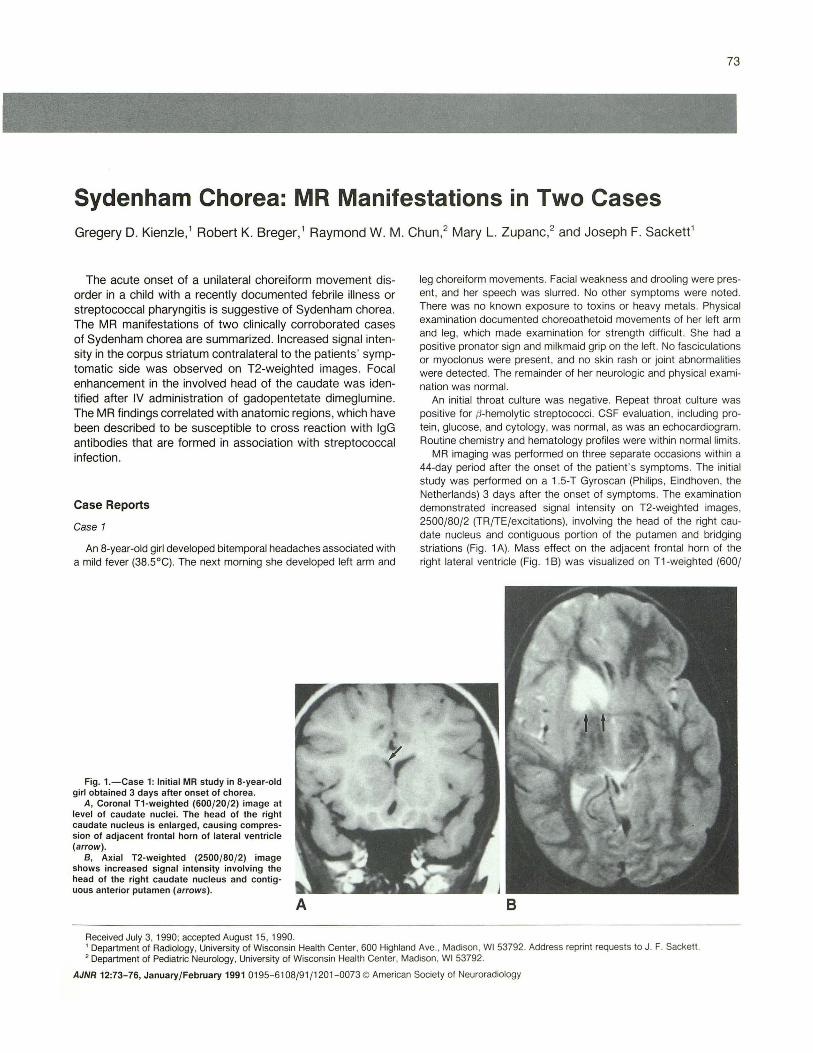
Fig. 1.-Case 1: Initial MR study in 8-year-old girl obtained 3 days after onset of chorea.
A, Coronal T1-weighted (600/20/2) image at level of caudate nuclei. The head of the right caudate nucleus is enlarged, causing compression of adjacent frontal horn of lateral ventricle (arrow).
B, Axial T2-weighted (2500/80/2) image shows increased signal intensity involving the head of the right caudate nucleus and contiguous anterior putamen (arrows) .
Received July 3, 1990; accepted August 15, 1990.
A
leg choreiform movements. Facial weakness and drooling were present, and her speech was slurred . No other symptoms were noted. There was no known exposure to toxins or heavy metals. Physical examination documented choreoathetoid movements of her left arm and leg, which made examination for strength difficult. She had a positive pronator sign and milkmaid grip on the left . No fasciculations or myoclonus were present , and no skin rash or joint abnormalities were detected. The remainder of her neurologic and physical examination was normal.
An initial throat culture was negative. Repeat throat culture was positive for /3-hemolytic streptococci. CSF evaluation, including protein , glucose, and cytology, was normal , as was an echocardiogram. Routine chemistry and hematology profiles were within normal limits.
MR imaging was performed on three separate occasions within a 44-day period after the onset of the patient 's symptoms. The initial study was performed on a 1.5-T Gyroscan (Phi lips, Eindhoven, the Netherlands) 3 days after the onset of symptoms. The examination demonstrated increased signal intensity on T2-weighted images, 2500/80/2 (TR/TEjexcitations) , involving the head of the right caudate nucleus and contiguous portion of the putamen and bridging striations (Fig . 1 A). Mass effect on the adjacent frontal horn of the right lateral ventricle (Fig. 1 B) was visualized on T1-weighted (600/
B
' Department of Radiology, University of Wisconsin Health Center, 600 Highland Ave., Madison, WI 53792. Address reprint requests to J. F. Sackett. 2 Department of Pediatric Neurology, University of Wisconsin Health Center, Madison, WI 53792.
AJNR 12:73-76, January/February 1991 0195-6108/91 / 1201 - 0073 © American Society of Neuroradiology

74 KIENZLE ET AL. AJNR:12, January/February 1991
20/2) coronal images. A follow-up examination performed on a 0.38-T scanner (Resonex, Sunnyvale, CA) was done 15 days after onset of chorea. Axial T2-weighted (2000/75/2) images demonstrated an interval decrease in the size of the right caudate and putamina! abnormalities (Fig . 2A) . After administration of gadopentetate dimeglumine (0. 1 mmol/kg, Berlex , Cedar Knolls, NJ), enhancement of the right caudate head and adjacent putamen was present on axial T1 -weighted (700/20/2) images (Fig. 28 ).
A clinical diagnosis of Sydenham chorea was made, and antibiotic therapy was begun. Reevaluation at day 44 revealed considerable improvement in choreiform movements. The patient 's speech, strength , and coordination had returned to normal. The only residual neurologic sign was occasional choreic movement of her left arm . MR examination was performed at this time on a 0.38-T Resonex imager. T2-weighted (2000/75/2) axial scans demonstrated partial resolution of the abnormal signal intensity of the right caudate and putamen. Axial T1 -weighted (700/20/2) images enhanced with 0.1
A 8
c D
mmolfkg of gadopentetate dimeglumine showed no interval change in the discrete enhancement of the head of the caudate.
Case 2
An 8-year-old boy presented for evaluation of a left-sided movement disorder of gradual onset over several days. Four months earlier he had a documented streptococcal pharyngitis. He had a history of a petit mal seizure disorder that was well controlled with oral anticonvulsant therapy. He was otherwise in good health. There was no history of rash, joint symptoms, or emotional lability .
On physical examination he had sudden jerking movements of the left arm and leg . His gait was profoundly abnormal , and he had marked incoordination of the upper and lower extremities. Reflexes were symmetrical. The pronator and milkmaid signs were positive on the left . Neurologic and general physical examinations were otherwise not contributory.
Fig. 2.-Case 1: Follow-up MR examinations acquired 15 (A and B) and 44 (C and D) days alter onset of chorea.
A, T2-weighted (2000/75/2) axial image illustrates partial resolution of the increased signal intensity in the region of the head of the right caudate nucleus and putamen (arrows) since the initial study (Fig. 1 ).
B, T1-weighted (700/ 20/ 2) axial image alter administration of gadopentetate dimeglumine shows unilateral enhancement of the head of the right caudate nucleus (arrow) .
C, T2-weighted (2000/ 75 /2) axial image obtained 44 days after onset of chorea shows further interval decrease in area of abnormal signal in caudate nucleus and putamen (arrow).
D, The degree of enhancement of the head of the right caudate nucleus (arrow) appears unchanged on T1 -weighted (750/ 20/ 2) image after administration of gadopentetate dimeglumine.

AJNR:12, January/February 1991 MR OF SYDENHAM CHOREA 75
Laboratory findings included a positive throat culture for ,6-hemolytic streptococci. There was an elevated antistreptolysin titer of 680 Tu (n = 0-120 Tu), and elevated anti-DNase B of 960 U (n = 0-480 U).
MR imaging was done 11 days after recognition of the patient 's symptoms on a 0.38-T Resonex scanner. Increased signal intensity was identified in the head of the right caudate nucleus and putamen on axial T2-weighted (2000/75/2) images in a remarkably similar distribution to that observed in case 1 (cf Figs. 1 B and 3A). A 2.5-cm-diameter lesion of decreased signal intensity on T1-weighted (700/20/2) axial scans was seen to involve the entire head of the right caudate nucleus. However, increased signal intensity, thought to represent petechial hemorrhage, was present along the posterolateral margin of the abnormal area (Fig. 3B). The enlarged head of the caudate compressed the anterior horn of the right lateral ventricle. After administration of 0.1 mmol/kg of gadopentetate dimeglumine, a portion of the head of the right caudate nucleus enhanced intensely (Fig. 3C) on T1-weighted (700/20/2) coronal images. The clinical and radiologic diagnosis was Sydenham chorea.
Discussion
The onset of chorea is a dramatic clinical entity, but is often a benign, self-limiting disease. The clinical syndrome referred to as Sydenham chorea has multiple causes, but is most commonly associated with post-streptococcal infection (rheumatic-related chorea). Other possible causes include pregnancy, lupus erythematosus, thyrotoxicosis , myeloproliferative disorders, rubella and pertussis infection, and drug reactions (phenothiazines, levodopa, phenytoin , oral contraceptives). The latency period for onset of rheumatic-related chorea following streptococcal infection may vary from 1 to 6 months [1]. Symptoms may be unilateral (19%) or bilateral (81 %). They are characterized by choreoathetoid movements that vary from purposeless, sudden jerking motions to more
A 8
prolonged, grotesque posturing superimposed on volitional movements. Associated neurologic symptoms may include dysarthria, encephalopathy with personality changes , emotional lability, disorientation, confusion, weakness , and reflex changes. The presentation of rheumatic-related chorea in absence of symptoms or signs of associated joint or heart disease is not uncommon. The rate of occurrence of rheumatic heart disease is low at the initial presentation of chorea, but increases with time. Most patients developing Sydenham chorea are young, with 75% presenting between the ages of 7-12 years old. Recurrent attacks occurred in 20% of patients in one study, but ultimate complete recovery or minimal neurologic sequelae is the rule [1-4] .
Few studies have been performed, and pathologic correlation is limited. In some patients a vasculitis has been demonstrated in the affected regions of the brain [5 , 6]. Of specific interest is an investigational study that reported a high rate of positive sera (46.6%) for lgG antibodies in patients with rheumatic fever. These antibodies demonstrated cross-reactivity with high affinity for neuronal cytoplasmic antigens from the caudate and subthalamic nuclei [7] . This has striking correlation with our observation of selective abnormal signal intensity involving the head of the caudate nucleus and contiguous putamen in each of our patients and the presence of focal blood-brain barrier disruption confined to the region of the head of the caudate nucleus in each case. Presumably, cross-reacting antibody formed in response to the streptococcal infection results in a focal antigen-antibody mediated reaction confined to a specific antigen localized to , or at least concentrated in , the region of the head of the caudate nucleus and adjacent putamen. The resulting vasculitis is thought to result in edema and focal blood-brain barrier disruption , as demonstrated by MR imaging. While this mechanism is an attractive explanation for at least some patients with rheu-
c Fig. 3.-Case 2: MR study in 8-year-old boy obtained 11 days after symptom recognition. A, T2-weighted axial scan (2000/20/2) depicts abnormal signal in head of right caudate nucleus and contiguous putamen (arrows). Note the striking
similarity of distribution to case 1 (Fig. 1 ). B, Parasagittal T1-weighted image (700/20/2) through head of right caudate nucleus. An area of decreased signal intensity is seen involving the
majority of caudate head. Focal area of increased signal intensity in posterior aspect of caudate head is thought to represent petechial hemorrhage (arrow).
C, T1-weighted (700/20/2) postcontrast image shows mass effect and local enhancement confined to head of right caudate nucleus (arrow).

76 KIENZLE ET AL. AJNR:12, January/February 1991
matic-related chorea, other unknown mechanisms may also be involved, and of course an immunologic explanation is not suggested in the many other causes of chorea.
It is important to recognize this disease clinically and radiographically because of its benign nature and excellent prognosis. In the proper clinical context , the abnormal MR findings described should not be interpreted as representing neoplasm or infarction even in the presence of mass effect and abnormal enhancement. Conservative management and prophylaxis against streptococcal infection are warranted in these patients , and interval change may be seen clinically and by MR imaging over the course of days to weeks .
ACKNOWLEDGMENT
We thank Robert T. Schmidt for referral of case 1.
REFERENCES
1. Nausieda PA, Burton BJ , Koller WC, et al. Sydenham chorea: an update. Neurology 1980;30 :331-334
2. McDowell FH , Cedarbaum JM. The extrapyramidal system and disorders of movement. In: Joynt RJ, ed. Clinical neurology, vol. 3. Philadelphia: Lippincott, 1989 :57-59
3. Adams RD , Victor M. Principles of neurology. New York: McGraw-Hill , 1989
4. Mitkov V. Cerebral manifestations of rheumatic fever. World Neural 1961 ;2:920-924
5. Colony S. Sydenham's chorea: a clinicopathologic study. Neurology 1956;6: 672- 676
6. Greenfield JG, Wolfsohn JM. The pathology of Sydenham's chorea. Lancet 1922;2 : 603-606
7. Husby G, van de Riju I, Zabrinskie JB, et al. Antibodies reacting with the cytoplasm of subthalamic and caudate nuclei neurons in chorea and acute rheumatic fever. J Exper Med 1976;144: 1094-1110
The reader 's attention is directed to the commentary on this article, which appears on the following page.