Syddansk Universitet Essence and Energy: An Exploration in ...
University of Southern Denmark
Genetic Variants Involved in Mitochondrial Oxidative Metabolism are associated with Type 2Diabetes Mellitus in studies of 9,132 Danes
Snogdal, Lena Sønder
Publication date:2010
Document version:Submitted manuscript
Citation for pulished version (APA):Snogdal, L. S. (2010). Genetic Variants Involved in Mitochondrial Oxidative Metabolism are associated withType 2 Diabetes Mellitus in studies of 9,132 Danes. Poster session presented at XIth International Symposiumon Insulin Receptors and Insulin Action, Naples, Italy.
Go to publication entry in University of Southern Denmark's Research Portal
Terms of useThis work is brought to you by the University of Southern Denmark.Unless otherwise specified it has been shared according to the terms for self-archiving.If no other license is stated, these terms apply:
• You may download this work for personal use only. • You may not further distribute the material or use it for any profit-making activity or commercial gain • You may freely distribute the URL identifying this open access versionIf you believe that this document breaches copyright please contact us providing details and we will investigate your claim.Please direct all enquiries to [email protected]
Download date: 28. Dec. 2021
XIInternatIonal
SympoSIum on InsulIn ReceptoRs
and InsulIn actIon
naples, ItalyOctober 28 - 30, 2010
“Federico II” University of Naples

2
XIInternatIonalSympoSIum on InsulIn ReceptoRsand InsulIn actIon
naples, ItalyOctober 28 - 30, 2010
3
CONTENTS
letter from the chair of the local organizing committee ............................................ 4
sponsors ....................................................................................................................... 5
supporting Institutions, scientific, Honorary and organizing committees .................... 6
General Information ...................................................................................................... 7
social program ............................................................................................................. 9
transportation .............................................................................................................. 10
Information for speakers and abstract presenters ...................................................... 11
scientific program ........................................................................................................ 13
speakers abstracts ....................................................................................................... 19
oral presentations and poster abstracts ...................................................................... 33
presenters list ............................................................................................................. 00
4
Dear Colleague,
On behalf of my fellow members of the International and Local Organizing Committees, it is a pleasure to welcome you to the XI International Symposium on Insulin Receptors and Insulin Action.
The congress provides continuity to a series of outstanding symposia started in Italy in 1980. Since then, the symposium has been organized every three years in Europe and has brought forward recent advances in the description of insulin signal transduction and metabolism related to metabolic disorders such as Diabetes mellitus and Obesity. After 20 years since the Verona edition, the symposium is now returning to Italy.
The understanding of the pathogenesis of Diabetes mellitus and related disorders is being transformed by a number of discoveries. As a result of this activity, our perception of Diabetes and related disorders is changing both at the mechanistic and the clinical levels. The “XI International Symposium on Insulin Receptors and Insulin Action” will address various key topics related to the “New face of Diabetes in the XXI century”.
The meeting is composed of keynote lectures, plenary sessions, short oral presentations and poster sessions. We hope that you will find plenty of time for informal and formal discussions. We also wish you to take time to explore Naples and appreciate the strength of its history and long lasting traditions. Our intention is that this meeting will foster constructive scientific interactions and collaborations in the very unique and inspiring scenario of the bay of Naples.
I hope your visit will be memorable and wish to welcome you to Naples!
Francesco Béguinot, MD, PhDProfessor of Clinical Pathology,Director, Dept. of Clinical Pathologyand Metabolic Disease Unit“Federico II” University of Naples,Chair, IR10ph. +39 081 746 3248fax +39 081 746 [email protected]

naples, ItalyOctober 28 - 30, 2010
5
XIInternatIonalSympoSIum on InsulIn ReceptoRsand InsulIn actIon
IR10 SPONSOR COMPANIESWe would like to thank all Companies offering their generous sponsorship to our Symposium:
GOLD
SILVER
PLATINUM
BRONZE
AND
6
The Symposium is under the Aegis of:European Association for the Study of Diabetes (EASD)Ministero del Lavoro, della Salute e delle Politiche SocialiConsiglio Nazionale delle RicercheConsiglio Universitario NazionaleConferenza dei Rettori delle Università ItalianeIstituto Superiore di SanitàAssessorato alla Sanità della Regione Campania
and is supported by:Polo delle Scienze e Tecnologie per la vita, Università degli Studi di Napoli Federico II
International Scientific CommitteeHans Ulrich Häring (Germany), President IR10Domenico Accili (USA) Henning Beck-Nielsen (Denmark)Francesco Béguinot (Italy), Chair IR10 Jean-Louis Carpentier (Belgium)Michael P. Czech (USA)Pierre De Meyts (Denmark)Suad Efendic (Sweden)Barbara B. Kahn (USA)C. Ronald Kahn (USA)Eddy Karnieli (Israel)Masato Kasuga (Japan)Yannick Le-Marchand-Brustel (France)Jesse Roth (USA)Alan R. Saltiel (USA)Simeon Taylor (USA)Emmanuel Van Obberghen (France)Morris F. White (USA)Yehiel Zick (Israel)Juleen Zierath (Sweden), Scientific Secretary IR10
Local Organizing CommitteeFrancesco BéguinotPietro FormisanoFrancesco GiorginoClaudia MieleFrancesco OrienteLuca UlianichPaola UngaroRossella Valentino
Honorary CommitteeRenato Lauro (IR10 Honorary President)Eduardo Consiglio and Vincenzo Macchia (Honorary Committee Members)
Previous Editions1980 Rome, Italy 1983 Rome, Italy 1987 Madrid, Spain 1990 Verona, Italy 1993 Munich, Germany 1996 Copenhagen, Denmark 1998 Jerusalem, Israel 2001 Geneva, Switzerland 2004 Nice, France 2007 Stockholm, Sweden
XIInternatIonalSympoSIum on InsulIn ReceptoRsand InsulIn actIon
naples, ItalyOctober 28 - 30, 2010
7
GENERAL INFORMATION
CONGRESS SECRETARIATI&C SrlVia Andrea Costa 202/6 40134 Bologna (Italy) Ph. +39 051 6144004Fax +39 051 [email protected] [email protected]
CONFERENCE VENUERoyal Continental HotelVia Partenope 38/4480121 NaplesPh. +39 081 2452068 The Royal Continental Hotel is located on the Naples waterfront and offers an ideal location with superb views of the gulf and of Castel dell’Ovo.
BADGESParticipants are requested to wear their conference badge during all sessions, coffee breaks, lunches and social events.
ITALIAN CME CREDITSN. 11 CME credits have been assigned to the Congress from the Board for Accreditation of the Italian Ministry of Health for the professional figure of medical doctors and surgeons.
CONTACT INFORMATION DURING CONFERENCEDuring the conference (October 28-29-30), messages to participants may be sent to the registration desk at the venue, Royal Continental Hotel: Federica Schiassi (+39 347 7865104), Stefania Parolari (+39 346 2217620); Fax: +39 081 7644616; e-mail: [email protected]; [email protected]
INTERNET CONNECTIONInternet access will be provided free of charge at the Congress Venue.
DRESS CODEDress will be informal throughout the conference, however jacket is recommended for the Gala Dinner.S. Lorenzo Complex is an ancient building and no heating system is provided. Although Naples has usually a temperate climate, we suggest warm clothes for the dinner.
REGISTRATION DESkThe desk, at Royal Continental Hotel, will be open during the conference as follows:u Wednesday October 27 from 15:00 to 20:00u Thursday October 28 from 08:00 to 18:00u Friday October 29 from 08:00 to 18:00u Saturday October 30 from 08:00 to 19:00

8
XIInternatIonalSympoSIum on InsulIn ReceptoRsand InsulIn actIon
naples, ItalyOctober 28 - 30, 2010
9
SOCIAL PROGRAM
Participation requires registration. Please contact the registration desk for the payment of the fee for accompanying persons. Transportation has to be arranged personally. Accompanying persons have to wear their conference badge during all sessions, coffee breaks, lunches and social events.
u THURSDAy, OCTOBER 28 – H. 20:00Dinner at the Royal Continental Hotel Restaurant accompanied by live music entertainment
u FRIDAy, OCTOBER 29 – H. 08:30RESERVED TO THE ACCOMPANYING PERSONSSightseeing Excavations of ErcolanoH. 08:15 Meeting point in the Royal Continental Hotel lobbyH. 08:30 Transfer by bus to the Excavations of ErcolanoH. 12:00 Transfer by bus to Pizzeria Regina Margherita, on the seafront of NaplesH. 13:00-14:30 Lunch and transfer to Royal Continental HotelIn case of bad weather, the visit will be organized to the National Archeological MuseumPlease confirm your participation to the Congress Secretariat within October 28 morning.
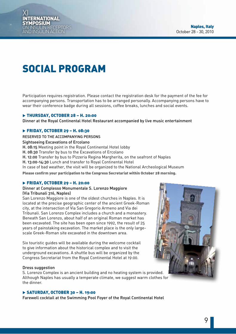
u FRIDAy, OCTOBER 29 – H. 20:00Dinner at Complesso Monumentale S. Lorenzo Maggiore (Via Tribunali 316, Naples)San Lorenzo Maggiore is one of the oldest churches in Naples. It is located at the precise geographic center of the ancient Greek-Roman city, at the intersection of Via San Gregorio Armeno and Via dei Tribunali. San Lorenzo Complex includes a church and a monastery. Beneath San Lorenzo, about half of an original Roman market has been excavated. The site has been open since 1992, the result of 25 years of painstaking excavation. The market place is the only large-scale Greek-Roman site excavated in the downtown area.
Six touristic guides will be available during the welcome cocktail to give information about the historical complex and to visit the underground excavations. A shuttle bus will be organized by the Congress Secretariat from the Royal Continental Hotel at 19:00.
Dress suggestionS. Lorenzo Complex is an ancient building and no heating system is provided. Although Naples has usually a temperate climate, we suggest warm clothes for the dinner.
u SATURDAy, OCTOBER 30 – H. 19:00Farewell cocktail at the Swimming Pool Foyer of the Royal Continental Hotel
10
TRANSPORTATION
TAXI: Radiotaxi Naples Telephone + 39 081 5564444.
PUBLIC TRANSPORTATION (TAXI EXCLUDED):UNICO NAPOLI: is the ticket that allows you to travel on all means of transportation in the Naples urban area.The main fares are:HOURLY TICKET: e 1,10 (valid for 90 minutes from validation)DAILY TICKET: e 3,10 (valid from 00.00 to midnight of the day of validation). You must fill in the appropriate blanks with your name and date of birth and keeping with you an identity document to be shown if requested.Tickets can be purchased in most of tobacco shops and newsstands. Once on the travel mean, you must validate your ticket in the ticket-machine. If it is out of order you can valid writing by pen date and time.
HOw TO REACH:THE CONGRESS VENUE ROYAL CONTINENTAL HOTEL (VIA PARTENOPE 38/44):
BusANM – 152STOP AT CASTEL DELL’OVO (bus stop in front of the hotel)
BusANM – 601STOP AT PIAZZA VITTORIA (bus stop behind the hotel)
THE MONUMENTAL COMPLEx S. LORENZO MAGGIORE (VIA TRIBUNALI 316):
Shuttle BusShuttle busses will be organized by the Congress Secretariat leaving the Royal Continental Hotel on Friday October 29 at h 19:00. Their use to reach S. Lorenzo Complex is recommended.
Other travel means:
Trolleybus ANM - 201 STOP AT PIAZZA CAVOUR
Busses SITA - Linea: Napoli - Pertosa STOP AT NAPOLI (VARCO IMMACOLATELLA)
ANM - C47 STOP AT PIAZZA CAVOUR
ANM - E1 STOP AT PIAZZA DEL GESÙ
ANM - R2 STOP AT PIAZZA NICOLA AMORE
ANM - E1 STOP AT VIA CIRILLO
ANM - E1 STOP AT VIA DUOMO
ANM - CS STOP AT VIA DUOMO
ANM - E1 STOP AT VIA FORIA
naples, ItalyOctober 28 - 30, 2010
11
XIInternatIonalSympoSIum on InsulIn ReceptoRsand InsulIn actIon
INFORMATION FOR SPEAkERS AND ABSTRACT PRESENTERS
SPEAkERS AND ORAL PRESENTERS
Slide center opening times:October 27: 16:00-20:00October 28-30: 07:30-19:00You are kindly requested to report to the technical support team in the slide center no later than 2 hours before the respective scheduled session in order to copy the presentation onto the meeting computer.Presentations must be in Power Point and no personal PC will be allowed.The operating system is Microsoft Windows Office 2007 and converter for Macinthosh is available.
POSTER PRESENTERS
Mounting Time:October 27 – from 18:00 to 19:00; October 28 – from 08:00 to 09:00
Poster Area: you will find a dedicated Area for posters near the Lunch Area; don’t forget the number assigned to your Abstract; you will have to fix your Poster in the Panel showing the same number. In the Poster Area, you will find the necessary material to hang your poster.
Dismounting Time: October 30 – 19:00 to 20:00; our Organization will remove and eliminate all posters not dismounted by October 30, 20:00.
Discussion Time: your presence near your Poster is advised at all times during the Poster Session. However we remind you that you will be asked to be present on the day and at the time communicated in the acceptance letter.

12

XIInternatIonalSympoSIum on InsulIn ReceptoRsand InsulIn actIon
naples, ItalyOctober 28 - 30, 2010
13
WEDNESDAY, OCTOBER 2715:00-20:00REGISTRATION DESK OPENING
THURSDAY, OCTOBER 2809:00-09:30OPENING CEREMONY AND WELCOME ADDRESSChairs: Hans Ulrich Häring (Tübingen, DE) and Francesco Béguinot (Naples, IT)Welcome address of the President and the Organizer
Ulf Smith (Gothenburg, SE)Welcome address of the President of the European Association for the Study of Diabetes
Gabriele Riccardi (Naples, IT)Welcome address of the President of the Italian Diabetes Society
Gaetano Lombardi (Naples, IT)Welcome address of the President of the Italian Endocrinology Society
09:30-10:30MEETING INTRODUCTIONINSULIN ACTION IN 2010: PERSPECTIVES IN THE COMING DECADEChairs: Edwin Gale (Bristol, UK) and Jesse Roth (Whitestone, NY USA)
09:30-09:50Introduction - Jesse Roth (Whitestone, NY USA) Paired revolutions in intercellular communication: the tenth day of creation
09:50-10:10Lecture 01 - C. Ronald Kahn (Boston, MA USA)Continuing evolution in our concepts of insulin action and insulin resistance
10:10-10:30Lecture 02 - Ulf Smith (Gothenburg, SE)Understanding insulin signaling and action-also a future challenge
10:30-11:00COFFEE BREAK
11:00-12:30ORAL SESSION 01NEw CONCEPTS IN INSULIN SIGNAL TRANSDUCTIONChairs: Masato Kasuga (Tokyo, JP) and Emmanuel Van Obberghen (Nice, FR)
11:00-11:30Lecture 03 - Domenico Accili (New York, NY USA)Unusual sites of insulin resistance
11:30-12:00Lecture 04 - Takashi Kadowaki (Tokyo, JP)Adipocyte control of insulin action
12:00-12:15Oral Presentation 1 (C1-1) - Qiong L. ZhouA novel protein enhances insulin-stimulated akt phosphorylation and glut4 translocation in adipocytes
12:15-12:30 Oral Presentation 2 (C1-2) - Barbara SalaniIGF-IR internalizes with caveolin-1 and ptrf/cavin in hacat cells
12:30-14:30POSTER SESSION 01 and BUFFET LUNCH
SCIENTIFIC PROGRAM
14
THURSDAY, OCTOBER 2814:30-15:30ORAL SESSION 02THE ROLE OF ADIPOSE TISSUE IN INSULIN ACTIONChairs: Eddy Karnieli (Haifa, IL) and Francesco Purrello (Catania, IT)
14:30-15:00Lecture 05 - Francesco Giorgino (Bari, IT)The biological specificity of visceral fatwith the Unrestricted Educational Grant of Eli Lilly Italia
15:00-15:15Oral Presentation 3 (C2-3) - Karin StenkulaInsulin regulates the spatial distribution of glucose transporter-4 on the cell surface
15:15-15:30Oral Presentation 4 (C2-4) - Gregory RacitiANKRD26 gene ablation causes diabetes in mice
15:30-16:00COFFEE BREAK
16:00-17:00ORAL SESSION 03INSULIN SIGNALLING IN PERIPHERAL TISSUES Chairs: Amira Klip (Toronto, CA) and Robert J. Smith (Providence, RI USA)
16:00-16:30Lecture 06 - Timothy E. McGraw (New York, NY USA)AKT2 regulation of GLUT4 trafficking
16:30-16:45Oral Presentation 5 (C3-5) - Isabelle Castan-LaurellChronic apelin treatment in HFD-fed mice improves b-oxydation and insulin resistance
16:45 - 17:00Oral Presentation 6 (C3-6) - Kurt HøjlundIs defect insulin signaling to mitochondria linked to insulin resistance in human muscle?
17:00-17:10Francesco Béguinot (Naples, IT)The past, the present and the future of the IR meeting communityTalk not included in the CME credit program
17:10-17:55ORAL SESSION 04INSULIN ACTION IN OBESITyRoberto De Pirro Memorial CeremonyChairs: Samuel Cushman (Bethesda, MD USA) and Ulf Smith (Gothenburg, SE)
17:10-17:40Lecture 07 - Hadi Al-Hasani (Nuthetal, DE)Roberto De Pirro Memorial Lecture - Obesity genes and their impact on insulin action: lesson from mouse geneticswith the Unrestricted Educational Grant of Sanofi Aventis
17:40-17:55Oral Presentation 6bis (C4-6bis) - Paul PilchAdiporedoxin, an adipocyte-enriched novel regulator of adiponectin assembly/secretionTalk not included in the CME credit program
19:30DINNER AT ROYAL CONTINENTAL HOTEL RESTAURANT

naples, ItalyOctober 28 - 30, 2010
15
XIInternatIonalSympoSIum on InsulIn ReceptoRsand InsulIn actIon
FRIDAY, OCTOBER 2908:30-10:00ORAL SESSION 05GENETIC AND EPIGENETIC CONTROL OF INSULIN ACTION Chairs: Henning Beck-Nielsen (Odense, DK) and Michael P. Czech (Worcester, MA USA)
08:30-09:00Lecture 08 - Markku Laakso (Kuopio, FI)Human gene variation affecting insulin action and secretion
09:00-09:30Lecture 09 - I. Karen Temple (Southampton, UK)Epigenetics and diabetes: lesson from neonatal diabetes and disorders of imprinting
09:30-09:45Oral Presentation 7 (C4-7) - Raffaele TeperinoEnvironmental and epigenetic regulation of the diabetes-related PED/PEA-15 gene
09:45-10:00Oral Presentation 8 (C4-8) - Stephan ScherneckZFP69 as a potential modulator of obesity-associated diabetes
10:00-10:30COFFEE BREAK
10:30-12:00ORAL SESSION 06ENVIRONMENTAL CONTROL OF INSULIN ACTION AND NUTRIENT METABOLISMChairs: Angelika Bierhaus (Heidelberg, DE) and Philipp Gorden (Bethesda, MD USA)
10:30-11:00Lecture 10 - Juleen Zierath (Stockholm, SE)Metabolic regulation in diabetes through DNA methylation
11:00-11:30Lecture 11 - Christopher B. Newgard (Durham, NC USA)Metabolic drivers of insulin resistance and related disorders
11:30-11:45Oral Presentation 9 (C5-9) - Katy RaddatzPkc epsilon regulates hepatic lipid metabolism during short term lipid oversupply
11:45-12:00Oral Presentation 10 (C5-10) - Assaf RudichActivation of ASK1 in omental fat links adipocyte stress to whole-body insulin resistance
12:00-14:00POSTER SESSION 02 and BUFFET LUNCH
16
FRIDAY, OCTOBER 2914:00-15:30ORAL SESSION 07b- AND α-CELLS IN INSULIN-RESISTANCE STATES Chairs: Jean-Louis Carpentier (Namur, BE) and Piero Marchetti (Pisa, IT)
14:00-14:30Lecture 12 - Bernard Thorens (Lausanne, CH)IGF-2 and IGF-1 receptor in the control of b-cell mass and function
14:30-15:00Lecture 13 - Rohit N. Kulkarni (Boston, MA USA)Intra-islet insulin signalling regulates α-cell function in vivo
15:00-15:15 Oral Presentation 11 (C6-11) - Eugenia Tiziana ManiscalchiMAPK activation in α TC1 cells after chronic palmitate exposure
15:15-15:30Oral Presentation 12 (C6-12) - Avital BeckSirna screening for genes involved in cytokine-induced pancreatic b cell death
15:30-16:00COFFEE BREAK
16:00-17:30ORAL SESSION 08INSULIN ACTION IN THE CNS Chairs: Ira D. Goldfine (San Francisco, CA USA) and Giuseppe Paolisso (Naples, IT)
16:00-16:30Lecture 14 - Jens C. Brüning (Cologne, DE)Central regulation of energy metabolism by insulin
16:30-17:00Lecture 15 - Suzanne Craft (Seattle, WA USA)Insulin-resistance and neurodegeneration: novel mechanisms and therapeutic implications
17:00-17:15Oral Presentation 13 (C7-13) - Toshiyasu SasaokaImpact of the elevation of lipid phosphatase ship2 on brain insulin action for the cognitive function in mice
17:15-17:30Oral Presentation 14 (C7-14) - Tina SartoriusToll-like receptor2/4 deficiency protects from diet-induced brain insulin resistance
19:00MEETING POINT IN THE ROYAL CONTINENTAL LOBBY FOR THE TRANSFER TO THE MONUMENTAL COMPLEx S. LORENZO MAGGIORE FOR THE GALA DINNER
20:00GALA DINNER AT THE MONUMENTAL COMPLEx S. LORENZO MAGGIORE
naples, ItalyOctober 28 - 30, 2010
17
XIInternatIonalSympoSIum on InsulIn ReceptoRsand InsulIn actIon
SATURDAY, OCTOBER 3009:30-10:45ORAL SESSION 09METABOLISM AND CANCER Chairs: Pietro Formisano (Naples, IT) and Derek Le Roith (New York, NY USA)
09:30-10:00Lecture 16 - Reuben J. Shaw (La Jolla, CA USA)AMPK signaling in glucose metabolism and growth control
10:00-10:30Lecture 17 - Michael Pollak (Montreal, CA)Insulin signalling and metformin in relation to cancer: new developmentswith the Unrestricted Educational Grant of Sanofi Aventis
10:30-10:45Oral Presentation 15 (C8-15) - Jutta Nagel Glargine and NPH insulin similarly increase colonic cell proliferation and ACF formation
10:45-11:15COFFEE BREAK
11:15-12:30ORAL SESSION 10 INSULIN-ACTION AND CANCERSession not included in the CME credit program
Chairs: Ira Pastan (Bethesda, MD USA) and Riccardo Vigneri (Catania, IT)
11:15-11:45Lecture 18 - Morris F. White (Boston, MA USA)The dark side of IRS-2: cancer and neuro-degeneration
11:45-12:15Lecture 19 - Norbert Tennagels (Frankfurt am Main, DE)Metabolic and mitogenic signalling of exogenous insulin(s)
12:15-12:30Oral Presentation 16 (C9-16) - Michael Greene Breast cancer growth and activation of insulin/IGF-1 signaling by circadian disruption
12:30-14:30POSTER SESSION 03 and BUFFET LUNCH
18
SATURDAY, OCTOBER 3014:30-16:30ORAL SESSION 11FUTURE STRATEGIES FOR TREATING DIABETES Chairs: Stefano Del Prato (Pisa, IT) and Giorgio Sesti (Catanzaro, IT)
14:30-15:00Lecture 20 - Fatima Bosch (Barcelona, ES)Gene therapy in diabetes
15:00-15:30Lecture 21 - Markus Stoffel (Zürich, CH)MicroRNAs: regulators of metabolism and therapeutic targets
15:30-15:45Oral Presentation 17 (C10-17) - Lucie Popineau GRB14 inhibition in mouse liver improves glucose tolerance and hepatic steatosis
15:45-16:00Oral Presentation 18 (C10-18) - Olivier Bezy PKCD is a regulator of hepatic insulin sensitivity and modifies hepatosteatosis risk
16:00-16:15Oral Presentation 19 (C10-19) - Shiri Karni Ahkenazi NFKB paradox: suppression of glut4 expression with concomitant translocation enhancement
16:15-16:30Oral Presentation 20 (C10-20) - Mashito Sakai Cited2 regulates hepatic gluconeogenesis by controlling PGC-1α activity
16:30-17:00COFFEE BREAK
17:00-18:30ORAL SESSION 12DIABETES COMPLICATIONSChairs: Michele Muggeo (Verona, IT) and Yehiel Zick (Rehovot, IL)
17:00-17:15Oral Presentation 21 (C11-21) - E. Dale Abel Insulin signaling is a physiological regulator of myocardial autophagy in vivo
17:15-17:30Oral Presentation 22 (C11-22) - Rossella Menghini Increased expression of TIMP3 protects against diabetes and atherosclerosis
17:30-17:45Oral Presentation 23 (C11-23) - Janice Zabolotny Protein tyrosine phosphatase 1b (PTP1B) regulates wound healing
17:45-18:00Oral Presentation 24 (C11-24) - Eugenia Carvalho Immunosuppressive agents alter glucose metabolism, blood pressure and heart rate in rats
18:00-18:30Lecture 22 - Hans Ulrich Häring (Tübingen, DE)President lecture - Insulin action in human brain
18:30-19:00CONCLUSION CEREMONY
18:30-18:45Francesco Béguinot (Naples, IT)Conclusion remarks
18:45-19:00XII meeting announcement
19:00FAREWELL COCKTAIL AT THE SWIMMING POOL FOYER OF THE ROYAL CONTINENTAL HOTEL
naples, ItalyOctober 28 - 30, 2010
19
XIInternatIonalSympoSIum on InsulIn ReceptoRsand InsulIn actIon
SPEAkERS ABSTRACTS
9:30-9:50 Thursday, October 28, 2010• Jesse Roth
Receptors and intercellular communication: where we are and how we got here Jesse RothFeinstein Institute for Medical Research, North Shore-Long Island, New York (NY USA)
The Earth is three or four billion years old. Homo sapiens has been around for roughly 100.000 years. Written language, which marks the beginning of “history”, is 5.000 years old. Adults who are over 50 have witnessed 1% of all history. The modern era of biology began about 150 years ago (L. Pasteur, C. Bernard, et al.). Those same adults have witnessed one third of all of the history of modern biology.When I started medical school about fifty years ago, intercellular communication was the exclusive province of the nervous system and the endocrine system. The nervous system was largely hard wired and electrical transmission a prominent concept. The total number of neurotransmitters could be counted on one’s fingers (with no need for polydactyly), and this exclusive club was resisting the admission of new members. The endocrine system included a finite number of glands, each producing one or a few hormones that travelled through the blood to a limited roster of target cells.Measurements of hormone levels in vivo (outside of the glands) were limited to steroid hormones and hCG in urine and thyroid hormones in blood (Tri-iodothyronine was just discovered the year before). Since these endocrine glands arose evolutionarily with the vertebrates, the hormones were thought to be restricted to vertebrates (Neurons were about twice as old, emerging with multicellularity).In 2010, we know that every cell at every moment is releasing a set of (hormone-like) intercellular
communication molecules to affect itself, its neighbors and cells at a distance. It is receiving many-fold more communication molecules from other cells in the body. Even bacteria are known to be communicating among one another within and across species (“quorum sensing”). There is a growing body of data that vertebrate and bacterial cells (e.g. in the intestine) are communicating.Fifty years ago, we did not know how a hormone was recognized by its target cell. Second messengers, intracellular receptors, cell surface receptors were not even conceived of. They were absent from the textbooks.A search of JBC, PNAS, and JCI titles and abstracts for the word “receptor” shows essentially no entries at all from 1940 to 1970. Then receptors suddenly took off. Since then, receptor papers represented a substantial minority of all papers in all three publications, right through December 2009. What were people thinking before “receptors”? How did receptors emerge? This assembly in its history captures that transformation.
9:50-10:10 Thursday, October 28, 2010• C. Ronald kahn
Continuing evolution in our concepts of insulin action and insulin resistanceC. Ronald KahnJoslin Diabetes Center and Harvard Medical School, Boston (MA USA)
Since the discovery of the insulin receptor tyrosine kinase – now 30 years ago – the entire field of insulin action and insulin resistance has exploded with new information and new insights. Following the activation of the insulin receptor, multiple intracellular substrates and signaling proteins form a complex, diverging signaling network. Through various combinations of these molecules, over 2.000 different potential pathways of insulin signaling can be activated. By knocking-out and knocking-down specific components of the signaling system in isolated cells and specific tissues of intact animals, it has been possible to define many of the specific pathways that lead to various insulin actions and determine how they are modified in disease
20
states. Using this approach, for example, we have been able to distinguish different pathways involved in control of glucose and lipid metabolism, as well as those pathways and genes that are primarily regulated by insulin itself versus those that are secondarily regulated by either diet or the metabolic alterations in diabetes. These approaches have allowed us to define the specific pathways responsible for hyperglycemia versus those responsible for the hyperlipidemia and accelerated atherosclerosis of type 2 diabetes. Likewise, pathways relating insulin action and insulin resistance have been mapped for various complications of the metabolic syndrome including formation of gallstones, increased risk for Alzheimer’s disease, and alterations in bone density and osteoporosis. Links between insulin action and mitochondrial dysfunction have also been established and shown to play a role in the altered metabolism of diabetes. Conversely, diabetes itself has been shown to alter insulin signaling and metabolism in both classical tissues of insulin action, such as liver, muscle and fat, and non-classical tissues, such as brain and b-cell. New components of the insulin action/resistance network are being identified, including a number of transcription factors, co-activators and co-repressors, and members of the sirtuin family of protein deacetylases. However, important challenges remain in both defining basic aspects of insulin action – for example, how do insulin and IGF-1 act through similar signaling networks but produce such different physiological effects – and basic aspects of insulin resistance – such as, where is the primary site of insulin resistance in people with genetic risk of type 2 diabetes. Solving these problems is not only of academic interest, it will provide insights into development of new treatments for diabetes, obesity and metabolic syndrome–epidemic diseases of the 21st century.
10:10-10:30 Thursday, October 28, 2010• Ulf Smith
Do we need to know more about insulin signalling and action?Ulf SmithDivision of EndocrinologyThe Hallett Center for Diabetes and EndocrinologyRhode Island HospitalAlpert Medical School of Brown University, Providence (RI USA)
Our understanding of insulin signalling and action has dramatically increased over the last decades. We also know a number of perturbations in the intracellular signalling cascade and action in insulin resistance but we still have had very limited information of potential genetic factors. In fact, the only common risk genotypes are related to polymorphisms in the PPARγ and IRS-1 genes. This is in contrast to the expanding number of identified risk genes for Type 2 diabetes which primarily appear to influence insulin secretion. So is there then a future for young scientists who want to study insulin signalling and action? In my view, there is a great need to further understand how and where insulin resistance develops. Obesity is by far the most common cause of insulin resistance emphasizing that the adipose tissue may play an important role. However, it is also possible that insulin resistance is primarily an environmental factor while the genetic predisposition for Type 2 diabetes involves the b-cells and insulin secretion.Irrespective of this, we badly need new therapeutic agents which improve insulin sensitivity and cardiovascular risk. The diabetes drugmarket has a global sale of around $ 29 billion and in Europe alone it is $ 7 billion. This is predicted to further grow as the Type 2 diabetes epidemic continues.Recent studies have shown additional health problems for patients with Type 2 diabetes since morbidity and mortality in certain forms of cancer are increased. Insulin resistance has also been implicated in this and we need to understand the signalling pathways for the growth-promoting effect of insulin and the cross-talk with the IGF-receptors.
naples, ItalyOctober 28 - 30, 2010
21
XIInternatIonalSympoSIum on InsulIn ReceptoRsand InsulIn actIon
These new challenges combined with the need for novel therapy in Type 2 diabetes should give a further boost to the research area and ensure future symposia on insulin receptors and action.
11:00-11:30 Thursday, October 28, 2010• Domenico Accili
11:30-12:00 Thursday, October 28, 2010• Takashi kadowaki
Adipocyte control of insulin actionTakashi KadowakiDepartment of Diabetes and Metabolic Diseases Graduate School of Medicine, University of Tokyo (JP)
Adiponectin is an anti-diabetic adipokine, which facilitates glucose and lipid metabolism to increase insulin sensitivity. Decreased plasma adiponectin in obesity is causally involved in insulin resistance and metabolic syndrome. Adiponectin receptors, AdipoR1, AdipoR2, possess seven-transmembrane topology with the amino terminus located intracellularly, which is opposite to G-protein coupled receptors. In liver, activation of AdipoR1 by adiponectin stimulates AMP kinase (AMPK) pathway and that of AdipoR2 stimulates PPARα pathway, thereby decreasing gluconeogenesis and lipogenesis, increasing fatty acid oxidation, simultaneously causing suppression of inflammation and oxidative stress. In muscle, adiponectin- AdipoR1 regulate PGC-1α expression via Ca2+/CaMKKb pathway and PGC-1α deacetylation and activation via and AMPK/SIRT1, respectively, leading to increased mitochondrial biogenesis impaired, exercise endurance and insulin resistance. In fact, skeletal muscle-specific AdipoR1 showed decreased PGC-1α expression and activity, decreased mitochondrial biogenesis, decreased type1 fibre, insulin resistance, endurance. Decreased adiponectin and adiponectin receptors in obesity are causally involved in obesity-linked diseases such as insulin resistance, metabolic syndrome, diabetes, decreased exercise endurance and vascular inflammation, and thus adiponectin and
adiponectin receptors would serve as molecular targets of treatment strategy of these diseases.
REFERENCES1) Nature Medicine 7:941-946, 2001, 2) Nature Medicine 8: 1288-1295, 2002, 3) Nature 423: 762-769, 2003, 4) J. Biol. Chem. 279: 30817-30822, 2004, 5) Endocrine Reviews 26: 439-451, 2005, 6 ) J. Biol. Chem. 281: 8748-8755, 2006, 7) J. Clin. Invest. 116: 1784-1792, 2006, 8) Nature Medicine 13: 332-339, 2007, 9) Cell Metabolism 6:55-68, 2007, 10) FEBS Letters, 582: 74-80, 2008, 10) Nature, published online March 31 2010
22
14:30-15:00 Thursday, October 28, 2010• Francesco Giorgino
The biological specificity of visceral fatFrancesco GiorginoDepartment of Emergency and Organ TransplantationSection of Internal Medicine,Endocrinology and Metabolic Diseases University of Bari School of Medicine (IT)
With excess energy storage, obesity develops, leading to increased risk for type 2 diabetes and cardiovascular diseases. The distribution of body fat appears to be even more important than the total amount of fat. Abdominal and, in particular, visceral adiposity is strongly linked to insulin resistance, type 2 diabetes, hypertension, dyslipidaemia, sleep apnea, and other complications of obesity. Visceral adiposity, manifested as a high waist circumference, is now accepted as a major component of the metabolic syndrome. Although the epidemiological association between visceral fat accumulation and excess risk of diabetes and of cardiovascular disease is striking, the biological mechanisms underlying the adverse impact of adipose tissue remain incompletely defined. Adipose tissue is a critical regulator of energy balance and substrate metabolism, and synthesizes several different substances with endocrine or paracrine functions, which regulate the overall energetic homeostasis. Evidently, adipose tissue is not only responsible for storage of energy, but operates as a highly active and dynamic tissue. Adipocytes produce hormones and cytokines, collectively called «adipokines», with pleiotropic effects on multiple tissues, leading to fine tuning of fuel utilization, energy homeostasis, and cardiovascular function. It should be also recognized that the functional attitude of visceral and subcutaneous adipocytes is programmed quite early during development and differentiation, due to inherent characteristics of the adipocyte precursors, multipotent cells that are resident in each fat depot and possess defined depot-specific genetic, biochemical and metabolic features. Thus, the biological specificity of the distinct adipose tissue depots may reside in the different biological profile of the resident cells, including the adipocyte precursors. In particular, adult
adipose tissue contains a pool of abundant and accessible multipotent stem cells, generally termed adipose-derived stem cells (ASCs), that are able to replicate as undifferentiated cells, to develop as mature adipocytes, and to differentiate into multiple other cell types along the mesenchymal lineage, including chondrocytes, myocytes and osteoblasts, and also into cells of neuroectodermal origin, including neurons and astrocytes. Different populations of multipotent precursor cells can be isolated from human fat fragments. Thus, adipose precursors cells are a heterogeneous cells population. An impairment in the differentiation potential and biological function of ASCs may contribute to the development of obesity and related diseases.
16:00-16:30 Thursday, October 28, 2010• Timothy E. McGraw
AkT2 regulation of GLUT4 traffickingTimothy E. McGrawDepartment of BiochemistryWeill Medical College of Cornell University of New York (NY USA)
Insulin stimulates glucose transport by recruiting the GLUT4 glucose transporter to the plasma membrane. Here we use total internal reflection fluorescence microscopy to show that two trafficking motifs of GLUT4, a FQQI motif and a TELE-based motif, target GLUT4 to specialized vesicles that accumulate adjacent to the plasma membrane of unstimulated adipocytes. Mutations of these motifs redistributed GLUT4 to transferrin-containing recycling vesicles adjacent to the plasma membrane, and the degree of redistribution correlated with the increases of the GLUT4 mutants in the plasma membrane of basal adipocytes. These results establish that GLUT4 defaults to recycling endosomes when trafficking to specialized vesicles is disrupted, supporting the hypothesis that the specialized vesicles are derived from an endosomal compartment. Insulin stimulates both the accumulation of GLUT4 in the evanescent field and the fraction of this GLUT4 that is inserted into the plasma
naples, ItalyOctober 28 - 30, 2010
23
XIInternatIonalSympoSIum on InsulIn ReceptoRsand InsulIn actIon
membrane. Unexpectedly, these two steps are differentially affected by the development of insulin resistance. We ascribe this selective insulin resistance to inherent differences in the sensitivities of GLUT4 vesicle accumulation and insertion into the plasma membrane to insulin. Differences in insulin sensitivities of various processes may be a general mechanism for the development of the physiologically important phenomenon of selective insulin resistance. IRAP, a transmembrane aminopeptidase, is also cargo of the insulin-regulated membrane trafficking pathway. Here we show that it is essential for GLUT4 sorting to GSV. In unstimulated IRAP knockdown adipocytes (IRAP-KD), plasma membrane GLUT4 levels are elevated due to increased exocytosis. These changes correlate with a re-localization of GLUT4 from specialized compartments to endosomes. Thus IRAP controls sorting GLUT4 to GSV, and therefore has a fundamental role in basal GLUT4 retention. The IRAP cytoplasmic domain rescues the IRAP-KD phenotype, demonstrating that the aminopeptidase activity is not required. Current evidence supports the model that AS160 RabGAP, which is required for basal intracellular retention of GLUT4, is recruited to GSV via an interaction with IRAP. However, AS160 recruitment to GLUT4 compartments and AS160 regulation of GLUT4 trafficking were unaffected by IRAP knockdown. These results demonstrate that AS160 is recruited to membranes by an IRAP-independent mechanism, and that IRAP and AS160 have independent roles in basal GLUT4 retention.
17:10-17:40 Thursday, October 28, 2010• Roberto De Pirro Memorial Lecture• Hadi Al-Hasani
Obesity genes and their impact on insulin action: lessons from mouse geneticsHadi Al-Hasani German Institute for Human Nutrition, Potsdam (DE)
Over the past years, human genome-wide association studies (GWAs) have revealed numerous genetic loci, some of which have been
implicated to play roles in the pathogenesis of obesity and diabetes. However, very little is known about the molecular mechanisms by which these SNPs exert their function on the molecular and physiological level. Moreover, identification of relevant susceptibility genes for obesity and diabetes has been difficult, mainly because the risk has been attributed to the interaction of multiple variant genes with the environment, where each of these genes only makes a small contribution to overall heritability.In addition to studies in humans, numerous genome-wide searches for obesity and diabetes genes, respectively, have been performed in mouse models. In this approach, two parental lines differing in a diabetes - and/or obesity - related phenotype are crossed to produce outcross populations that are then analyzed for molecular markers across the genome to search for obesity susceptibility loci (quantitative trait loci, QTL). These studies have shown a very complex picture of numerous QTL, contributing to the phenotype and gene/diet interactions. Nevertheless, identification of disease genes from these QTL has been mostly not successful.Recently, we and others have succeeded in positional cloning of susceptibility genes for obesity and diabetes from polygenic mouse models. In this approach, subcongenic mouse strains were generated to establish minimal critical segments of the respective QTL, followed by gene expression profiling and sequencing of positional candidate genes. Among other allelic variants, we identified a loss-of-function mutation in the gene Tbc1d1 which is predominantly expressed in skeletal muscle. In mice, inactivation of Tbc1d1 results in increased fatty acid oxidation and decreased glucose utilization in skeletal muscle. This shift in energy substrate preference from glucose to lipids as fuel in muscle confers leanness, improves glycemia, and leads to protection from diet-induced obesity and diabetes. Our data indicate that dysregulation of substrate partitioning and adaptive organ crosstalk are key features in the development of obesity and diabetes.
24
8:30-9:00 Friday, October 29, 2010• Markku Laakso
Human gene variation affecting insulin action and secretionMarkku LaaksoDepartment of Medicine, University of Eastern Finland, Kuopio (FI)
Type 2 diabetes is a strongly inherited disease, but the genetic basis of this disease has remained largely unknown until recent years. Both impaired insulin secretion and insulin resistance, two main pathophysiological mechanisms leading to type 2 diabetes, have a significant genetic component. Genome-wide association studies (GWAS) published since 2007 have substantially increased our knowledge on the genes increasing the risk of type 2 diabetes. There are currently about 20 common loci that increase the risk of type 2 diabetes (> 10 new SNPs will be shortly published). Only a few of these SNPs are actually known to affect the structure of a gene product or gene expression with a credible link to glucose metabolism. Therefore, for the majority of the SNPs the functionality has not been proven yet, and it is possible that the causal variants are not the SNPs published, but are located at varying distances from the ‘real’ functional gene. However, the unexpected conclusion from these studies is that a large number of diabetes SNPs are close to genes expressed highly in the pancreas and several of these SNPs have been shown to be associated with reduced b-cell dysfunction in non-diabetic subjects. Only a few SNPs have been associated with insulin resistance (PPARG2, IRS1). Interestingly, type 2 diabetes risk SNPs do not much overlap with the SNPs regulating fasting or 2-hour glucose levels. The conclusion from these studies seems to be that in a majority of cases type 2 diabetes develops as a consequence of a combination of inherited b-cell defect and acquired insulin resistance (obesity, lack of exercise, etc.). Copy number variants are not likely to be important genetic determinants of type 2 diabetes. Genome-wide sequencing and large scale studies aiming to investigate the interaction between gene variants and lifestyle/environmental factors will substantially increase
our knowledge on gene-gene and gene-lifestyle interactions that are determining the “epidemic” of type 2 diabetes worldwide.
9:00-9:30 Friday, October 29, 2010• I. karen Temple
Epigenetics and diabetes; lessons from transient neonatal diabetes and disorders of imprintingI. Karen TempleSchool of Medicine, University of Southampton (UK)
Different cells and tissue types in the body are characterised by different levels of gene expression despite each cell sharing the same genetic code. This variation in gene activity from cell to cell is achieved by different configurations of chromatin (DNA and associated proteins) that generally exists in either a transcriptionally permissive or silent state. Chromatin is modified partly through the addition or loss of small molecules such as acetyl groups to histone proteins or methyl groups to cytosine residues within CpG dinucleotides in the DNA sequence. These epigenetic changes can therefore alter gene expression without altering the DNA sequence. The reversibility of the epigenetic ‘marks’ sets them apart from DNA mutation and they are heritable over rounds of cell division. Epigenetic modification can be influenced by environment, gene interaction or by stochastic error and there is a two fold higher rate of epimutation than DNA mutation.
Epigenetic modification is well recognised as a cause of human disorders and is likely to play a pivotal role in the cause of complex disorders. The challenge is to identify consistent epigenetic alterations of aetiological significance, given that epigenetic modification of DNA differs between tissues, at different times of development within the same tissue and is sensitive to continual environmental factors.
Genomic imprinting is one of the best understood examples of epigenetic regulation of gene expression. The expression patterns of imprinted genes are characterised by expression
naples, ItalyOctober 28 - 30, 2010
25
XIInternatIonalSympoSIum on InsulIn ReceptoRsand InsulIn actIon
from only one allele (of the pair) in a consistent parent of origin manner. The pattern is set by targeted methylation within the male or female germ line that resists the post fertilisation waves of demethylation of the zygote. Imprinted genes are thought to play an important role in fetal growth and their carefully regulated expression is important for normal cellular metabolism. Imprinted genes are therefore candidate predisposition genes for disorders such as diabetes.
Several well known disorders of imprinting predispose to type 2 diabetes including Prader Willi syndrome. However the main imprinted locus clearly linked to diabetes, is PLAGL1 at 6q24. Genetic and epigenetic aberrations that result in overexpression of PLAGL1, cause transient neonatal diabetes (TND) with a high risk of type 2 diabetes in later life. 20% of TND (type 1) cases have an epigenetic loss of methylation to account for the phenotype. Studies to determine the cause of epigenetic aberrations identified in imprinting disorders may provide helpful insights into the causes of epigenetic aberrations in general. For example the work has led to the identification of ZFP57 as a methylation control gene. Imprinted regions are also a possible explanation for the recent observation of sex dependent risk of transmission for more common types of Diabetes (DM1 and DM2).
10:30-11:00 Friday, October 29, 2010• Juleen Zierath
Gene/environment influence on skeletal muscle insulin sensitivity in type 2 diabetic patientsJuleen R. ZierathSection of Integrative Physiology, Dept. of Molecular Medicine and Surgery, Karolinska Institutet, Stockholm (SE)
Skeletal muscle is an important site of insulin-mediated glucose uptake and defects in this insulin target tissue precede the manifestation of Type 2 diabetes. Our central hypothesis is that activation of insulin-independent pathways to glucose transport in skeletal muscle may
overcome the profound impairment in whole body glucose homeostasis associated with Type 2 diabetes. Intensive research efforts have been directed towards understanding the regulation of insulin-dependent and insulin-independent pathways governing glucose metabolism and the factors causing insulin resistance in Type 2 diabetes. Physical exercise/muscle contraction elicits an insulin-independent increase in glucose transport and perturbation of this pathway may bypass defective insulin signaling. To date, the exercise-responsive signaling molecules governing glucose metabolism in skeletal muscle are largely unknown. Epigenetic modification through DNA methylation is implicated in metabolic disease and may play a role in the mechanism by which environmental factors influence metabolic responses in diabetes. Using whole genome promoter methylation analysis of skeletal muscle from normal glucose tolerant and Type 2 diabetic subjects, we identified cytosine hypermethylation of Peroxisome Proliferator-Activated Receptor γ Coactivator-1 α (PGC-1α) in diabetic subjects. Methylation levels were negatively correlated with PGC-1α mRNA and mitochondrial DNA (mtDNA). Bisulfite sequencing revealed that the highest proportion of cytosine methylation within PGC-1α was found within non-CpG nucleotides. Non-CpG methylation was acutely increased in human myotubes by exposure to tumor necrosis factor-α (TNF-α) or free fatty acids, but not insulin or glucose. Selective silencing of the DNA methyltransferase 3B (DNMT3B), but not DNMT1 or DNMT3A, prevented palmitate-induced non-CpG methylation of PGC-1α and decreased mtDNA and PGC-1α mRNA. We provide evidence for PGC-1α hypermethylation, concomitant with reduced mitochondrial content in Type 2 diabetic patients, and link DNMT3B to the acute fatty-acid induced non-CpG methylation of PGC-1α promoter. By identifying the molecular mechanisms controlling insulin sensitivity, future development of pharmacological and physiological (exercise and diet) intervention strategies aimed to improve glucose homeostasis may be possible.
26
11:00-11:30 Friday, October 29, 2010• Christopher B. Newgard
Metabolic drivers of insulin resistance and related disordersChristopher B. NewgardSarah W. Stedman Nutrition and Metabolism Center &Department of Pharmacology & Cancer BiologyDuke University Medical Center, Durham (NC USA)
We seek to apply comprehensive metabolic analysis tools (sometimes called “metabolomics”) for understanding of mechanisms underlying chronic human diseases such as diabetes, obesity, and cardiovascular disease. Current approaches include analysis of metabolic flux by 13C NMR-based mass isotopomer analysis (in collaboration with Drs. Shawn Burgess and A. Dean Sherry and associates, Dallas, TX) and metabolic profiling of important groups of metabolic intermediates by both “targeted” and “unbiased” mass spectrometry (in collaboration with Drs. James Bain, Robert Stevens, Olga Ilkayeva, Brett Wenner, Michael Muehlbauer, Mark Butler, and David Millington at Duke). These tools have been used to investigate the metabolic mechanisms underlying development of peripheral insulin resistance in animals and humans. Studies led by Drs. Debbie Muoio and Tim Koves at the Stedman Center have demonstrated a “disconnect” between mitochondrial b-oxidation and TCA cycle activity that leads to accumulation of incompletely oxidized lipids in the mitochondria and impairment of insulin actions. We have also recently identified perturbations of branched chain amino acid (BCAA) catabolism in multiple cohorts of insulin resistant humans compared to normally insulin sensitive controls and have translated these findings to rodent models to demonstrate a contribution of BCAA to development of insulin resistance that is independent of body weight. We have also found that BCAA supplementation induces behavioral changes in rats that can be reversed by provision of tryptophan in the drinking water. Finally, in collaboration with Dr. Alan Attie at the University of Wisconsin, we have integrated transcriptomic and metabolomic analysis to identify new pathways that control hepatic gluconeogenesis and PEPCK expression. These
examples will serve to illustrate the potential of comprehensive metabolic profiling methods for providing insights into diabetes and obesity mechanisms.
14:00-14:30 Friday, October 29, 2010• Bernard Thorens
IGF-2 and IGF-1 receptor in the control of b-cell mass and function Bernard ThorensDepartment of Physiology and Center for Integrative GenomicsUniversity of Lausanne (CH)
To preserve glucose homeostasis in face of increased metabolic demand, the pancreatic beta-cells increase their total number and/or insulin secretion capacity and type 2 diabetes is caused by defects in this adaptive mechanisms leading to reduced b-cell mass and reduced glucose competence. The molecular mechanisms that control b-cell replication or neogenesis from precursors, their glucose competence and protection against apoptosis are still uncompletely defined. The gluco-incretin hormones GLP-1 and GIP can induce b-cell proliferation, increased glucose competence and protection against apoptosis. To identify novel regulatory pathway controlling these trophic effects, we performed comparative transcriptomic analysis of islets from control and GipR-/-;Glp-1-R-/- mice, which show reduced glucose competence and increased sensitivity to cytokine-induced apoptosis. We found that IGF-1 receptor expression was markedly reduced in the mutant islets. As the IGF-1 receptor signaling pathway is known for its anti-apoptotic effect, we explored the relationship between gluco-incretin action, IGF-1 receptor expression and signaling, and glucose competence, proliferation and apoptosis. These studies led us to identify the presence in b-cells of an autocrine loop that depends on IGF-1R expression and secretion of IGF-2. We demonstrated that GLP-1 induced IGF-1R expression by a translational mechanism and a parallel phosphorylation of Akt. Akt phosphorylation was however dependent on IGF-2 secretion by the b-cells. Furthermore, we
naples, ItalyOctober 28 - 30, 2010
27
XIInternatIonalSympoSIum on InsulIn ReceptoRsand InsulIn actIon
showed that GLP-1-induced b-cell proliferation, protection against cytokine-induced apoptosis and increased glucose competence were dependent on IGF-1R up-regulation and IGF-2 secretion. Thus, an IGF-2/IGF-1 receptor autocrine loop operates in b-cells and its activation is required for all GLP-1 trophic effects on b-cells. These data provides a novel integrated view of the GLP-1 and IGF-R signaling pathway in b-cell physiology.
14:30-15:00 Friday, October 29, 2010• Rohit N. kulkarni
Intra-islet insulin signalling regulates α-cell function in vivoRohit N. Kulkarni Islet Biology and Regenerative Medicine, Joslin Diabetes Center and Department of Medicine, Harvard Medical School, Boston (MA USA)
Glucagon is an important counter-regulatory hormone to insulin, and counters hypoglycemia by enhancing hepatic glucose output and regulating gluconeogenesis. The secretion of glucagon from α cells is promoted by hypoglycemia, and inhibited by hyperglycemia as well as by insulin. In type 2 diabetes, enhanced glucagon secretion is often seen under hyperglycemia. On the other hand, glucagon non-responsiveness to hypoglycemia is observed to worsen in patients with type 1 diabetes on long-standing insulin therapy. Data will be presented from studies to investigate the importance of insulin signaling in α cells using α cell specific insulin receptor (IR) knockout mice and will involve discussion regarding the role of intra-islet signaling in the control of α cell function and proliferation.
16:00-16:30 Friday, October 29, 2010• Jens C. Brüning
Central regulation of energy metabolism by insulinJens C. BrüningInstitute for Genetics, University of Cologne (DE)
Insulin action in the CNS plays a critical role in the regulation of energy homeostasis and peripheral glucose metabolism. In turn, insulin resistance in the CNS significantly contributes to altered peripheral glucose metabolism. We have revealed a critical role for insulin action in agouti peptide related peptide (AgRP)-expressing neurons in mediating insulin’s ability to suppress hepatic glucose production. The presentation will focus on the role of insulin action in other defined neuronal populations and on the molecular mechanisms, how inflammatory signals inhibit neuronal insulin action.
16:30-17:00 Friday, October 29, 2010• Suzanne Craft
Insulin-resistance and neurodegeneration: novel mechanisms and therapeutic implicationsSuzanne CraftProfessor of Psychiatry and Behavioral SciencesUniversity of Washington School of MedicineSeattle (WA USA)
Our knowledge of the multifaceted role of insulin in the central nervous system has expanded rapidly in recent years. It is now apparent that perturbation of this role by insulin resistance and hyperinsulinemia can increase the risk for aging-related neurodegenerative disorders such as Alzheimer’s disease a number of mechanistic pathways, delineated in elegant in vitro and animal studies. We have investigated these pathways in humans using models of insulin resistance and hyperinsulinemia. We will present results suggesting that insulin-associated effects on cerebral glucose metabolism, Ab regulation and inflammation contribute to AD pathophysiology. This premise raises the possibility that treatments aimed at
28
improving or overcoming insulin resistance will benefit patients with AD. We will present data testing this possibility in pilot therapeutic trials using insulin intranasal insulin.
9:30-10:00 Saturday, October 30, 2010• Reuben J. Shaw
AMPk signaling in glucose metabolism and growth controlReuben J. Shaw Howard Hughes Medical Research InstituteMolecular and Cell Biology LaboratoryDulbecco Center for Cancer ResearchSalk Institute for Biological Studies La Jolla (CA USA)
AMPK is a highly conserved sensor of cellular energy status found in all eukaryotic cells which reprograms metabolism and suppresses cell growth upon its activation. The serine/threonine kinase LKB1 is the upstream kinase which activates AMPK under conditions of low intracellular ATP. In addition, LKB1 is a human tumor suppressor gene mutated in the familial cancer condition Peutz-Jeghers syndrome (PJS), as well as in 30% of sporadic non small cell lung cancer (NSCLC). Consistent with playing key roles in tumor suppression and metabolic control, loss of function of LKB1 in genetically engineered mice leads to tumor formation or metabolic disease depending on the tissue deletion is induced in. In addition, utilizing liver-specific LKB1 knockout mice, we demonstrated that the widely used type 2 diabetes therapeutic metformin acts to lower blood glucose through the LKB1 pathway in liver. To try to further undestand the molecular bases behind these profound anti-cancer and anti-diabetes effects, we have sought to identify new components of the AMPK pathway. As one arm of that effort, using a proteomic and bioinformatics approach, we have performed a screen to identify novel AMPK substrates which mediate its effects on cell growth and metabolism. One of the first new substrates we identified was the mTOR-binding subunit raptor, which we demonstrated as a critical substrate of AMPK mediating its effects on growth control. Phosphorylation of raptor by
AMPK is required for the inhibition of mTORC1 and for cell cycle arrest following energy stress. These findings uncover a novel conserved effector of AMPK that mediates its role as a metabolic checkpoint coordinating cell growth with energy status. More recently, we have also characterized novel substrates of AMPK involved in its transcriptional control of glucose metabolism in liver that relate to the ability of this pathway to control type 2 diabetes, which will be discussed.
10:00-10:30 Saturday, October 30, 2010• Michael Pollak
Insulin signaling and metformin in relation to cancer: new developmentsMichael PollakMcGill University, Montreal (CA)
In contrast to insulin receptors (IRs) present on classic insulin target tissues, IRs of normal and transformed epithelial cells have no established role in the regulation of energy metabolism at the whole organism level. Rather, they may have physiologic roles resembling those of insulin-like signaling in primitive organisms, where receptor activation regulates apoptosis and proliferation. Much experimental and epidemiologic data support the hypothesis that a subset of cancers are responsive to physiologic insulin concentrations (reviewed in 1,2): as examples, (a) higher insulin levels are associated with poor prognosis of several common cancers such as prostate, colon, and breast, (b) insulin can stimulate cancer proliferation in vitro, and (c) there is circumstantial evidence that insulin is one of the mediators of the adverse effect of obesity on cancer prognosis.In view of the above, research regarding targeting of insulin signaling (often in conjunction with targeting of IGF-I signaling) as a cancer treatment strategy has been proposed. Early efforts included the use of somatostatin analogues to lower insulin and IGF-I levels, but this was unsuccessful as the intervention achieved only minor reduction in ligand levels (3). Metformin is currently under study in this context, as it is known to reduce insulin
naples, ItalyOctober 28 - 30, 2010
29
XIInternatIonalSympoSIum on InsulIn ReceptoRsand InsulIn actIon
levels in settings of host insulin resistance and hyperinsulinemia, but metformin has little effect on IGF-I levels, or on insulin levels in the absence of baseline hyperinsulinemia. Small molecule insulin/IGF-I receptor kinase inhibitors are also being evaluated, and initial results justify further investigation as the expected metabolic toxicity is less than anticipated, and there is evidence of activity for particular subsets of cancers for which insulin and/or IGFs stimulate proliferation. Recent data in this area, including the use of treatment-induced hyperinsulinemia as a pharmacodynamic marker, will be reviewed. A related area of investigation concerns the hypothesis that exogenous insulin administration (or the administration of specific insulin analogues) may increase neoplastic proliferation. This is biologically plausible, but clinical and epidemiologic data remain controversial. While much attention has been given to the important issue of affinity of specific ligands to the various receptors in the insulin/IGF receptor family, pharmacokinetic aspects are also important, as many forms of insulin therapy result in continuous exposure, in contrast to the more transient exposure seen in normal physiology. While it has been proposed that insulin therapy may slow cancer growth by reducing glucose levels (4), the fact that experimental tumors are growth inhibited in the hyperglycemic hypoinsulinemic environment of type I diabetes suggests that for some cancers, normoglycemic environments do not limit proliferation, but insulin facilitates aggressive behavior (5). Metformin and other biguinides are under study not only in relation to their insulin-lowering properties mentioned above, but also in relationship to their action in activating AMP kinase and altering cellular energetics by inhibiting oxidative phosphorylation. While anti-neoplastic activity has been shown in many preclinical models, optimum design of clinical trials will require more information concerning pharmacokinetics and drug distribution, as well as identification of predictive biomarkers. This research direction is justified in part by epidemiologic research that associates metformin use in diabetes treatment with reduced cancer risk and cancer mortality – but
relevance of these findings to non-diabetic populations remains to be established (6).
1. Pollak M. Insulin and insulin-like growth factor signaling in neoplasia. Nat Rev Cancer 2008;8:915-282. Pollak M. Do cancer cells care if their host is hungry? Cell Metab. 2009 May;9(5):401-4033. Pritchard K, Chapman M, Shepherd L, Pollak M. Octreotide plus tamoxifen in adjuvant treatment of breast cancer: 10 year follow up of the NCIC MA.14 study. Submitted, 20104. Gerstein HC. Does insulin therapy promote, reduce, or have a neutral effect on cancers? JAMA. 2010;303(5):446-7.5. Johnson JA, Pollak M, Insulin, glucose and the increased risk of cancer in patients with type 2 diabetes, in press Diabetoligica, 20106. Giovannucci E, Harlan DM, Archer MC, Bergenstal RM, Gapstur SM, Habel LA, Pollak M, Regensteiner JG, Yee D. Diabetes and cancer: a consensus report. Diabetes Care. 2010 Jul;33(7):1674-85
11:15-11:45 Saturday, October 30, 2010• Morris F. white
The dark side of IRS-2: cancer and neurodegenerationMorris F. WhiteChildren’s Hospital Boston and Harvard Medical School, Boston (MA USA)
Dysregulation of the PI 3-kinase→AKT cascade contributes to metabolic disease and to oncogenesis. Insulin receptor (IR) and the type 1 insulin-like growth factor receptor (IGF1R) are two of the major regulators of PI3K signaling. Upon ligand-binding, the IR and IGF1R are autophosphorylated and recruit the adaptor proteins IRS1 and IRS2, which are phosphorylated by the receptor tyrosine kinase. The phospho-IRS proteins recruit the 85 kDa regulatory subunit of PI3K, which activates the 110 kDa catalytic subunits, generates PtdIns-3-4-5-P3, and activates PDK1, AKT1, AKT2, mTOR, and S6K cascades. Pten+/--mice develop tumors in multiple organs because of the reduced hydrolysis of PtdIns-3-4-5-P3. Although activation of PI3K→AKT cascade usually inhibits IRS2 expression, an elevation of IRS2 is often detected in tumor samples in which PTEN levels were compromised. To determine the potential
30
contribution of IRS2 to tumor progression, Pten+/--mice were crossed with IRS2+/--mice. Deletion of IRS2 did not affect the initiation of neoplasia found in Pten+/--mice, but suppressed cancer cell growth, proliferation, and invasion through the basement membrane. Deletion of IRS2 also attenuated the expression of Myc in prostatic intraepithelial neoplasia in Pten+/--mice. In addition, the expression levels of IRS2 and MYC were highly correlated in human prostate cancer, and IRS2 stimulated MYC expression in cultured cells. Thus, IRS2 promotes tumor progression in Pten+/--mice by stimulating both Myc and DNA synthesis (Szabolcs M et al. Am J Pathol 2009, 174:276–286). IRS2 also plays a critical role in nutrient homeostasis because it mediates insulin signaling in liver, skeletal and cardiac muscle, b-cells, endothelial cells and brain. Peripheral insulin resistance contributes to metabolic disease and type 2 diabetes, whereas reduced neuronal insulin-like signaling can delay ageing, and less IRS2 signaling throughout the body or just in the brain extends mouse life span (Taguchi A et al. Science 2007, 317:369-372). Aging is a major risk factor for the progression of neurodegenerative disease. To determine whether IRS2 signaling modulates neurodegeneration we investigated R6/2-mice that develop Huntington’s disease (HD) and die around 15 weeks of age. Increasing IRS2 expression in the R6/2-brain (R6/2•IRS2ntg) reduced the life span to 11 weeks, whereas decreasing IRS2 throughout the body—except in the b-cells where it was needed to prevent diabetes (R6/2•IRS2+/- •IRS2btg)—extended life-span to 25 weeks. The slower progression of HD was associated with more nuclear FoxO1, and increased concentration of Sod2 and Pparg1α. Aggregate formation typical of HD progression, along with astrocytosis and oxidative stress decreased in R6/2•IRS2+/-•IRS2btg-mice, while the number of autophagosomes increased and motor function improved. Our results show how the progression of HD—and life span in general—can be linked to the insulin-like signaling cascade through the IRS2-branch of the pathway (Sadagurski M. et al. submitted). In summary, IRS2 signaling is critical for nutrient homeostasis, but it also promotes tumor progression and neurodegeneration that
diminishes longevity.11:45-12:15 Saturday, October 30, 2010• Norbert Tennagels
Metabolic and mitogenic signaling of exogenous insulin(s)Norbert Tennagels R&D BD Diabetes, Frankfurt (DE)
Introduced more than 10 years ago, the treatment of diabetic patients with insulin analogs has been shown to provide a more efficient, reproducible, and convenient therapy than regular insulin. Depending on the pharmacokinetic and –dynamic profile and the underlying mode of action, which is achieved either by sequence or secondary modifications, insulin analogs may vary from insulin with respect to metabolic potency, stability, tissue specificity, onset and duration of action. Since these changes may lead to an altered interaction or activation profile of insulin and/or IGF receptor signaling pathways and in consequence may change metabolic or mitogenic responses, a careful investigation of acute and long-term effects has to be a major focus in the development of these molecules. One prominent example for an unwanted effect is [AspB10]insulin, which has a higher affinity towards both insulin receptor (IR) and IGF-1 receptor (IGF1R) than insulin, a prolonged occupancy time for the IR and has induced a higher incidence of breast cancer in rats and a higher proliferation rate of mammalian cell lines. This has led to the contention that insulins having a similar profile - especially an increased affinity towards IGF1R in vitro - might have a potential cancer promoting activity as well. However, the relevance of binding affinities to IGF1R and insulin therapy is still a matter of debate, since i) variable results for different insulin analogs from in vitro proliferation assays do not support this view with confidence and ii) additional aspects reflecting the in vivo situation e.g. the competition with endogenous free IGF-1 or the distribution and the metabolism of an insulin analogs, critically affect the initiation of metabolic and mitogenic signaling as well. Insulin glargine, a long-acting basal insulin analog, displays the slightly increased IGF1R activity comparable to that of [AspB10]insulin
naples, ItalyOctober 28 - 30, 2010
31
XIInternatIonalSympoSIum on InsulIn ReceptoRsand InsulIn actIon
in vitro but did not show any difference in the incidence of mammary tumors in mice and rats compared to controls or NPH insulin in 2-year carcinogenicity studies. The analog undergoes rapid and significant metabolism in humans and animals leading to the formation of two main metabolites, M1 and M2, which have a metabolic profile comparable to the parent molecule but at the same time a mitogenic profile identical to insulin in vitro. Therefore it’s suggested that the degradation and the action of the metabolites significantly contribute to the established efficacy and safety of insulin glargine as observed in clinical practice and particularly in long-term clinical trials and emphasize the importance of examining all steps in the action of an insulin analog in vitro and in vivo.
14:30-15:00 Saturday, October 30, 2010• Fatima Bosch
Gene therapy in diabetesFatima BoschCenter of Animal Biotechnology and Gene Therapy and Department of Biochemistry and Molecular Biology, Universitat Autonoma de Barcelona, Bellaterra, and CIBER de Diabetes y Enfermedades Metabólicas Asociadas (CIBERDEM), Barcelona (ES)
Diabetes mellitus is a chronic disease with severe secondary complications driven by poor glycemic control for which there is no cure. Although insulin therapy allows type 1 diabetic patients to lead normal, active lives, it has associated risks, mainly acute hypoglycaemia and development of retinopathy, nephropathy and neuropathy. Gene therapy is a new approach to treat diabetes where the main aim is to develop a way to engineer tissues to maintain normoglycaemia. We previously demonstrated the feasibility of a potential gene therapy strategy based upon engineering skeletal muscle to take up glucose by co-expressing insulin and glucokinase (GK). Both genes act synergistically such that local production of human insulin (hIns) improves glucose uptake, whereas GK acts as a glucose sensor in a concentration-dependent manner to facilitate glucose disposal. We showed reversal of diabetic hyperglycemia in AAV1-treated diabetic mice. Our objective was to undertake a pre-clinical
assessment of this combined therapy in diabetic beagle dogs as a step towards treatment of human diabetes. We wished to determine (1) the feasibility and relative efficiency of AAV1-CMV-hIns treatment in large animals; (2) the minimal effective dose of AAV1-CMV-hIns; (3) the feasibility of co-injecting AAV1-CMV-hIns and AAV1-CMV-GK; and finally (4) the duration of effects, as well as any biochemical and safety issues. Our results indicate that a single viral injection can mediate long term survival for greater than two years with sustained benefit to glycemic control, body weight and with no detriment to physical performance. In this presentation, this new gene therapy approach for type 1 diabetes from our group will be discussed.
15:00-15:30 Saturday, October 30, 2010• Markus Stoffel
18:00-18:30 Saturday, October 30, 2010• Ulrich Häring

32
XIInternatIonalSympoSIum on InsulIn ReceptoRsand InsulIn actIon
naples, ItalyOctober 28 - 30, 2010
33
ORAL PRESENTATIONS AND POSTER ABSTRACTS
ORAL PRESENTATIONS
C01.1 - New concepts in insulin signal transductionA NOVEL PROTEIN ENHANCES INSULIN-STIMULATED AKT PHOSPHORYLATION AND GLUT4 TRANSLOCATION IN ADIPOCYTESZhou Qiong L1, Jiang Zhen Y2, Holik John1, Mabardy Allan1, Straubhaar Juerg1, Nicoloro Sarah1, Chawla Anil1, Del Campo Claudia M1, Lambright David G1, Czech Michael P1
1University Of Massachusetts Medical School, Worcester, Usa; 2Burnham Institute For Medical Research, Orlando, UsaProtein kinase B/Akt protein kinases control an array of diverse functions including cell growth, survival, proliferation and metabolism. We report here the identification of Pleckstrin Homology-like domain family B member 1 (PHLDB1) as an insulin responsive protein that enhances Akt activation. PHLDB1 contains a PH domain, which we demonstrated binds phosphatidylinositol (PI)(3,4)P2, PI(3,5)P2 and PI(3,4,5)P3, as well as an FHA domain and coiled coil regions. PHLDB1 expression is increased during adipocyte differentiation, and it is abundant in many mouse tissues. Both endogenous and HA or GFP tagged PHLDB1 displayed a cytoplasmic disposition in unstimulated cultured adipocytes, but translocated to the plasma membrane in response to insulin. Depletion of PHLDB1 by siRNA inhibited insulin stimulation of Akt phosphorylation but not IRS-1 phosphorylation. RNAi-based silencing of PHLDB1 in cultured adipocytes also attenuated insulin-stimulated deoxyglucose transport and myc-GLUT4-EGFP translocation to the plasma membrane, while knockdown of the PHLDB1 isoform PHLDB2 failed to attenuate insulin-stimulated deoxyglucose transport. Furthermore, adenovirus-mediated expression of PHLDB1 in adipocytes enhanced insulin-stimulated Akt and p70S6 kinase phosphorylation, as well as GLUT4 translocation. These results indicate that PHLDB1 is a novel modulator of Akt protein kinase activation by insulin.
C01.2 - New concepts in insulin signal transductionIGF-IR INTERNALIZES WITH CAVEOLIN-1 AND PTRF/CAVIN IN HACAT CELLSSalani Barbara1, Passalacqua Mario2, Maffioli Sara1, Briatore Lucia1, Contini Paola3, Cordera Renzo1, Maggi Davide1
1Department Of Endocrinology And Medicine (DISEM), University Of Genova, Genova, Italy ; 2Department Of Experimental Medicine (dimes), University Of Genova, Genova, Italy; 3Department Of Internal Medicine And Medical Specialties (DIMI), University Of Genova, Genova, ItalyInsulin like growth factor-I receptor (IGF-IR) is a tyrosine kinase receptor (RTK) that associates with caveolae, invaginations of the plasma membrane that regulate vesicular transport, endocytosis and intracellular signaling. IGF-IR internalization represents a key mechanism of down-modulation of receptor number on plasma membrane. IGF-IR interacts directly with Caveolin-1 (Cav-1), the most abundant protein of caveolae. Recently it has been demonstrated that the Polymerase I and Transcript Release Factor I (PTRF/Cavin) is required for caveolae formation and it affects their morphology. The role of Cav-1 and PTRF/Cavin in IGF-IR receptor internalization is still to be clarified. We have investigated the interaction of IGF-IR with Caveolin and PTRF/Cavin in the presence of IGF-I in human Hacat cells. We show that IGF-IR internalization triggers Cav-1 and PTRF/Cavin translocation from plasma membrane to cytosol and increases its interaction with these proteins. In fact Caveolin-1 and PTRF/Cavin co-immunoprecipitate with IGF-IR during receptor internalization. We found a different time course of coimmunoprecipitation between IGF-IR and Cav-1 compared to IGF-IR and PTRF/Cavin. Cav-1 and PTRF/Cavin silencing by siRNA differently affect IGF-IR plasma membrane expression following IGF-I treatment: Cav-1 silencing affects significantly IGF-IR rate of internalization whereas PTRF/Cavin silencing decreases IGF-IR plasma membrane recovery. These data indicate that Caveolae play a role in IGF-IR internalization. Based on these findings, Cav-1 and PTRF/Cavin, new player in IGF-IR trafficking, could represent two relevant and distinct targets to modulate IGF-IR function.
34
C02.3 - The role of adipose tissue in insulin actionINSULIN REGULATES THE SPATIAL DISTRIBUTION OF GLUCOSE TRANSPORTER-4 ON THE CELL SURFACEStenkula Karin G1, Lizunov Vladimir A2, Cushman Samuel W1, Zimmerberg Joshua2
1National Institute Of Diabetes And Digestive And Kidney Diseases, Nih, Bethesda, Usa; 2Eunice Kennedy Schriver National Institute Of Child Health And Human Development, Nih, Bethesda, UsaGlucose transporter-4 (GLUT4) are key players in insulin-regulated glucose metabolism, insulin resistance, and type-2 diabetes. Nevertheless, the dynamic spatial organization of GLUT4 in the plasma membrane (PM) is unclear. Here, we applied multi-color TIRF microscopy to visualize HA-GLUT4-GFP/mCherry dynamic recycling in live adipose cells. We demonstrate that insulin regulates not only the exocytosis of GLUT4 storage vesicles, but also the PM distribution of GLUT4 themselves. In basal cells, we find the majority of GLUT4 distributed in bright spots close to PM that may represent GLUT4 vesicles, and intermediates of exo- and endocytosis of GLUT4, as well as domains/clusters of GLUT4 molecules in PM. We detected fusion of single GLUT4 vesicles by doubly labelling them with GLUT4-mCherry and insulin-regulated aminopeptidase (IRAP)-pHluorin. Indeed, domains (clusters) of GLUT4 molecules in PM are created by a mode of exocytosis that retains GLUT4 at the fusion site. Surprisingly, when insulin induces a burst of GLUT4 exocytosis, it does not merely accelerate this basal exocytosis, but rather stimulates ~60-fold another mode of exocytosis that disperses GLUT4 into PM, bypassing the domains. We further tested GLUT4 association with clathrin-coated pits which has been implicated in the recycling of GLUT4. However, in contrast to previous findings, clathrin does not initiate clustering of GLUT4. Rather, internalization of most GLUT4, regardless of insulin, occurs from pre-existing clusters via the subsequent recruitment of clathrin. Taken together, these data fit a new kinetic model that features multifunctional clusters as intermediates of exocytosis and endocytosis.
C02.4 - The role of adipose tissue in insulin actionANKRD26 GENE ABLATION CAUSES DIABETES IN MICERaciti Gregory A1, Bera Tapan1, Gavrilova Oksana2, Pastan Ira1
1Laboratory Of Molecular Biology, Nci, Nih, Bethesda, Usa; 2Mouse Metabolism Core, Niddk, Nih, Bethesda, UsaBackground and aims: the ANKRD26 gene is expressed in most organs of humans and mice with expression in brain being particularly high. Mice with partial inactivation of Ankrd26 have hyperphagia, extreme obesity and an increase in body size. Mice with complete inactivation of the gene are homozygous lethal. Since obesity is one of the major risk factors for diabetes, we performed metabolic characterization of the Ankrd26 MT mice, to examine the possible role of the Ankrd26 gene in the regulation of glucose homeostasis. Results: six-month old Ankrd26 MT mice with extreme obesity, weighing ~ 55 grams, developed a severe impairment of glucose tolerance during GTT. This defect was accompanied by an impairment of glucose-induced insulin secretion. The MT mice also showed a decreased response to insulin during an ITT and had high fasting insulin levels, suggesting the presence of insulin resistance. To assess insulin resistance in individual tissues, we performed hyperinsulinemic-euglycaemic clamps in the Ankrd26 MT mice. The mice exhibited whole body insulin resistance as measured by a reduced glucose infusion rate and glucose disposal. The effect was due to hepatic suppression of endogenous glucose production and reduction of 2-deoxy-glucose (DG) uptake into skeletal muscle and BAT. But 2DG uptake into WAT was not affected. Conclusion: the partial inactivation of the Ankrd26 gene in mice is sufficient to impair both insulin sensitivity and insulin secretion leading to the development of diabetes in MT mice. Further studies will be directed at determining the molecular mechanisms by which partial Ankrd26 inactivation leads to these abnormalities.
C03.5 - Insulin signalling in peripheral tissuesCHRONIC APELIN TREATMENT IN HFD-FED MICE IMPROVES b-OxYDATION AND INSULIN RESISTANCECamille Attané1, Chloé Rancoule1, Sophie Le Gonidec1, Camille Foussal1, Alexandre Bénani2, Cédric Dray1, Rocio Guzman-Ruiz3, Danièle Daviaud1, Mariano Ruiz-Gayo3, Luc Pénicaud2, Philippe Valet1, Isabelle Castan-Laurell1
1Inserm U858, Toulouse, France; 2Centre Des Sciences Du Gout Et De L’Alimentation, Dijon, France; 3University Of Ceu-San Pablo, Madrid, SpainWe have recently shown that acute apelin treatment decreases glycemia in mice and stimulates glucose uptake in skeletal muscle through an AMPK-dependent pathway. In insulin-resistant mice, apelin also increases total glucose utilization. Since apelin appears as a beneficial peptide acting on glucose metabolism, the aim of the present study was to know the impact of chronic apelin treatment in insulin-resistant mice on lipid metabolism by studying fatty acid b-oxidation and on insulin sensitivity. For this purpose, HFD-fed mice were treated by an ip injection of either apelin (0.1 µmole/kg/d) or PBS during 28 days. Apelin treatment decreased the respiratory quotient (0.74±0.01 vs 0.70±0.01, n= 12 p< 0.05) in HFD-mice suggesting a better utilization of lipids. Indeed, apelin treatment increased complete oxidation of [14C]palmitate in soleus muscle (161±15 vs 332±61 nmol CO2 released/g protein, n=10, p< 0.01). This effect was abolished in muscle of DN-AMPK mice where AMPK is inactive. A better mitochondrial oxidative capacity was observed in soleus of apelin-treated mice since the impaired respiratory index (1.27±0.03 vs 1.19±0.04, n=7) was corrected and the citrate synthase activity increased. Moreover, apelin-treated mice exhibited a better insulin sensitivity due to a) a decrease of C18-1 acylcarnitines levels in muscle homogenates (24.8±6.6 vs 12.7±3.7 nmol/g prot, n= 7), b) a better glucose tolerance and c) an increase of insulin-stimulated glucose uptake in isolated skeletal muscle (2.4 fold increase vs 1.4 in PBS-treated mice). Thus, chronic apelin treatment improves insulin resistance by acting on both glucose and lipid metabolism.
naples, ItalyOctober 28 - 30, 2010
35
XIInternatIonalSympoSIum on InsulIn ReceptoRsand InsulIn actIon
C03.6 - Insulin signalling in peripheral tissuesIS DEFECT INSULIN SIGNALING TO MITOCHONDRIA LINKED TO INSULIN RESISTANCE IN HUMAN MUSCLE?Højlund Kurt1, Zhao Xiaolu2, Kristensen Jonas M3, Skov Vibe H.1, Jensen Ole N2, Yi Zhengping4, Mandarino Lawrencce J4, Wojtaszewski Jørgen FP3, Beck-Nielsen Henning1
1Odense University Hospital, Odense, Denmark; 2Protein Research Group, University Of Southern Denmark, Odense, Denmark; 3Copenhagen Muscle Research Centre, University Of Copenhagen, Copenhagen, Denmark; 4Center For Metabolic Biology, Arizona State University, Tempe, Arizona, United StatesRecent data suggest a link between insulin resistance and impaired mitochondrial oxidative phosphorylation (OxPhos) in human skeletal muscle. It remains, however, to be clarified whether mitochondrial dysfunction is a cause or consequence of insulin resistance. In addition to decreased abundance of OxPhos genes and proteins, we have shown compromised intrinsic function of mitochondria in muscle in type 2 diabetes (T2D) indicating a role for post-translational regulatory mechanisms. Supporting the idea that an impaired biological response to insulin contributes to mitochondrial dysfunction, we have now data showing that genetically determined insulin resistance due to a mutation in the insulin receptor gene (Arg1174Gln) causes down-regulation of OxPhos genes and proteins in muscle in vivo. There is growing evidence that several subunits in OxPhos are phosphorylated, and that several cytosolic kinases translocate into mitochondria in response to different stimuli. Recently, we reported that the catalytic beta-subunit in the ATP synthase complex are phosphorylated at several sites, and that some of these sites showed altered phosphorylation in muscle in obesity and T2D. In mitochondria isolated from human muscle, we have now identified 126 phosphorylation sites in 63 mitochondrial proteins using phosphoproteomics. We are currently investigating how these phosphorylation sites respond to insulin. Based on these observations, we hypothesize that altered phosphorylation of OxPhos proteins in response to different stimuli including insulin could play a role for the association of mitochondrial dysfunction with insulin resistance, and hence contribute to the pathogenesis of T2D.
C04.6 bis - Insulin action in obesityADIPOREDOxIN, AN ADIPOCYTE-ENRICHED NOVEL REGULATOR OF ADIPONECTIN ASSEMBLY/SECRETIONPilch Paul1, Jedrychowski Mark1, Meshulam Tova1, Ding Shi-ying1, Fried Susan1
1Boston University School Of Medicine, Boston, UsaWe have characterized by mass spectrometry the protein content of insulin-responsive Glut4 storage vesicles (GSVs) and GSV precursor compartments (JBC 2010, 285:104-14). In the latter compartments, we identified a member of the thioredoxin superfamily, whose members function to regulate cellular redox status and disulfide bond formation. We named this protein adiporedoxin (Arx) because it is expressed at appreciable levels only in brown and white fat and in testes, and it is markedly induced upon the differentiation of cultured adipocytes. Arx is a resident of the endoplasmic reticulum where secretory protein assembly and disulfide bond formation takes place, including for adiponectin, the insulin-sensitizing adipokine. To test the hypothesis that Arx might be involved in the assembly and/or secretion of adiponectin, we engineered fat cells over and under expressing Arx and determined that over expression enhanced adiponectin expression and secretion whereas under expression reduced adiponectin secretion. Chronic high fat feeding (HFD) of mice results in reduced adiponectin levels and insulin resistance, and therefore, we subjected mice to an HFD for 12 weeks, which resulted in a concomitant 90% loss of both adiponectin and Arx content in fat cells. Finally, we obtained mRNA array data from a cohort of obese humans and found that the expression of these two proteins was highly positively correlated (R = 0.77, P<0.05). Taken together, these data support the hypothesis that Arx is likely to be the principal regulator of adiponectin assembly and secretion, and thus its regulation and expression are likely to have important consequences for insulin sensitivity.
C04.7 - Genetic and epigenetic control of insulin actionENVIRONMENTAL AND EPIGENETIC REGULATION OF THE DIABETES-RELATED PED/PEA-15 GENETeperino Raffaele1, Ciccarelli Marco2, Longo Michele2, Mirra Paola2, Manzo Italia2, Béguinot Francesco2, Ungaro Paola2
1Ecole Polytechnique Fèdèrale De Lausanne, Lausanne, Switzerland ; 2DBPCM, University Of Naples & IEOS-CNR, Naples, ItalyThe molecular details of gene–environment interplay in type 2 diabetes (T2D) onset have recently received great attention. PED/PEA-15 is a gene commonly overexpressed in tissues from T2D individuals and healthy subjects at high risk of developing diabetes (such as first degree relatives). Indeed, overexpression of the ped/pea-15 gene in mice, impairs glucose tolerance and leads to diabetes in conjunction with high-fat diet treatment. We have evaluated the influence of reactive oxygen species (ROS) on PED/PEA-15 regulation, representing oxidative stress a mechanism by which aberrant epigenetic programming leads to T2D. Incubation of THP-1 cells with hydrogen peroxide determines an increase in the binding of the transcription factor nuclear factor kB (NF-kB) to its response element on PED/PEA-15 promoter causing an up-regulation of PED/PEA-15 gene. This effect was accompanied by an increase both in dimethylation of lysine 4 (H3K4me2) and acetylation of Lysine 9 and Lysine 14 (AcH3) at PED/PEA-15 promoter and coding region, indicating that ROS determine chromatin modifications on PED/PEA-15 gene. Further co-immunoprecipitations experiments performed using NF-kB-p65 followed by H3K4me2 and AcH3 antibodies revealed both H3K4me2/NF-kB and AcH3/NF-kB complexes at the PED/PEA-15 promoter indicating that NF-kB recruitment is accompanied by histone di-methylation and acetylation at PED/PEA-15 promoter. These results suggest that an inflammatory stimulus like hydrogen peroxide alter the epigenetic regulation of the PED/PEA-15 gene determining an increase of its expression. However, the former input starting the series of events leading to this alteration remains to be clarified.
36
C04.8 - Genetic and epigenetic control of insulin actionZFP69 AS A POTENTIAL MODULATOR OF OBESITY-ASSOCIATED DIABETESScherneck Stephan2, Vogel Heike2, Nestler Matthias2, Blüher Matthias1, Urbanski Sina2, Schulz Nadja2, Kluge Reinhart2, Schürmann Annette2, Joost Hans-Georg2
1University Of Leipzig, Leipzig, Germany; 2German Institute Of Human Nutrition Potsdam-rehbruecke, Nuthetal, GermanyType 2 diabetes is a polygenic disease resulting from a combination of adipogenic and diabetogenic alleles. Here we report the identification of a candidate gene for the hyperglycemic effect of the mouse quantitative-trait locus (QTL) Nidd/SJL. The diabetogenic allele was contributed by the lean Swiss Jim Lambert (SJL) inbred mouse strain in an outcross population with New Zealand Obese (NZO) mice. In order to define a critical region of Nidd/SJL, we generated recombinant-congenic mouse lines carrying different intervals of the locus on the C57BL/6J background. These lines were crossed with NZO mice (reporter cross). Characterisation of the obese progeny led to the identification of a critical region of chromosome 4 comprising ten genes. Sequencing and expression profiling identified a striking variant of zinc finger domain transcription factor 69 (Zfp69). In NZO and C57BL/6J mice, an IAPLTR1a retrotransposon in intron 3 disrupted the gene by formation of a prematurely polyadenylated mRNA that lacked the coding sequence for the Krüppel-associated box (KRAB) and Znf-C2H2 domains of Zfp69. In contrast, the diabetogenic SJL allele generated a functional mRNA. When transferred to the B6.V-Lepob background, the diabetogenic Zfp69SJL allele produced hyperglycemia, reduced gonadal fat pads, and increased plasma and liver triglycerides. In addition, mRNA levels of the human ortholog of Zfp69, ZNF642, were significantly increased in white adipose tissue of patients with type 2 diabetes. Thus, the expression of the transcription factor Zfp69/ZNF642 in adipose tissue may play a role in the pathogenesis of type 2 diabetes.
C05.9 - Environmental control of insulin action and nutrient metabolismPKC EPSILON REGULATES HEPATIC LIPID METABOLISM DURING SHORT TERM LIPID OVERSUPPLYRaddatz Katy2, Turner Nigel2, Raftery Mark1, Frangioudakis Georgia2, Wilks Donna2, Preston Elaine2, Hegarty Bronwyn2, Leitges Michael3, Schmitz-Peiffer Carsten2
1University Of New South Wales, Sydney, Australia; 2Garvan Institute, Sydney, Australia; 3University Of Oslo, Oslo, NorwayLipid oversupply is a major contributor to the development of insulin resistance. We have previously shown that deletion of the lipid-activated protein kinase C, PKCε, augmented circulating insulin levels in long term fat-fed mice, thus maintaining glucose tolerance. However, an improvement of insulin action was not observed. Here, we used a one week high-fat diet to investigate the role of PKCε during the early phase of insulin resistance. While WT mice became glucose intolerant, PKCε-/- mice were protected. In contrast to our long term study this was accompanied by lower insulin levels indicating that insulin resistance was prevented. Proximal insulin signalling and glucose uptake into skeletal muscle was not improved in the PKCε-/- mice, suggesting the involvement of alternative mechanisms. Using iTRAQ as a quantitative proteomic approach, we identified several proteins involved in lipid metabolism and oxidative phosphorylation, which were differentially expressed in the liver of fat-fed PKCε-/- and WT mice, indicating a regulatory role of PKCε in these pathways. A more detailed analysis revealed increased levels of triglycerides, diacylglycerides, and cholesterol in the liver of fat-fed PKCε-/- mice due to a shift in lipid partitioning towards esterification instead of b-oxidation of fatty acids. The contribution of these alterations to improved insulin action is currently under investigation. Taken together, our data show that the role of PKCε changes during the development of glucose intolerance, contributing to insulin resistance only at an early stage and that the underlying mechanisms may include extensive changes in lipid metabolism.
C05.10 - Environmental control of insulin action and nutrient metabolismACTIVATION OF ASK1 IN OMENTAL FAT LINKS ADIPOCYTE STRESS TO WHOLE-BODY INSULIN RESISTANCEBluher Matthias1, Tarnovscki Tanya2, Beck-Haim Yulia2, Palgi Niv2, Kloting Nora1, Harman-Boehm Ilana2, Bashan Nava2, Rudich Assaf2
1University Of Leipzig, Leipzig, Germany; 2Ben-Gurion University, Beer-Sheva, IsraelIn obesity adipocytes are exposed to various potential stressors, but what type of stress predominates in human obesity, and which stress-sensing signaling cascades are activated to induce adipose dysfunction remains poorly characterized. We have previously reported in humans that omental (OM), but not subcutaneous (SC) adipose tissue exhibits increased activation of JNK and p38MAPK. In analyzing upstream kinases, a signaling cascade involving the MKK3/6 and 4 (but not MKK7), and the MAP3K Ask1 (but not Tak1 or MLK3) could be verified by phosphospecific antibodies and inter-correlation analyses. Ask1 was more highly phosphorylated on the Thr845 activatory site in OM compared to SC, and compared to OM of lean persons, while the inhibitory Ser83 and Thr966 sites were phosphorylated to varying degrees. In TNFα-treated depot-specific adipocyte cell line, Thr845 phosphorylation occurred earlier than the increase in pSer83 and Thr966, and was more robust in intra-abdominal adipocytes than in SC. Protein and mRNA expression of Ask1 in human OM fat was higher than in SC, correlated with the degree of macrophage infiltration, but could be attributed at least partly to the adipocyte cell fraction. In-vitro, Ask1 mRNA was increased in intra-abdominal adipocytes in response to TNFα, FasL and oxidative stress, but not in response to the ER-stress inducer tunicamycin. Remarkably, a multivariate model revealed OM-Ask1-mRNA as an independent predictor of whole-body insulin resistance assessed by clamp (b=-0.308, p=0.001, n=196). Collectively, OM Ask1 is regulated by combinatorial phosphorylation input and at the expression level, linking adipose stress with whole-body insulin resistance.
naples, ItalyOctober 28 - 30, 2010
37
XIInternatIonalSympoSIum on InsulIn ReceptoRsand InsulIn actIon
C06.11 - b- and α-cells in insulin-resistance statesMAPK ACTIVATION IN ALPHA TC1 CELLS AFTER CHRONIC PALMITATE ExPOSUREManiscalchi Eugenia Tiziana1, Piro Salvatore1, Monello Adelina1, Mascali Loriana Grazia1, Urbano Francesca1, Iacoviello Linda1, Rabuazzo Agata Maria1, Purrello Francesco1
1Dipartimento Di Medicina Interna E Specialistica, Lab. di Medicina Molecolare, Università Degli Studi Di Catania, Ospedale Garibaldi, Catania, ItalyT2DM is characterized by insulin resistance at the cellular level, and by a concomitant enhanced glucagon release from alpha cells. Since MAPK can regulate Pax 6, a transcription factor that controls glucagon expression, an alteration in MAPK way could explain some aspects in the physiopathology. In this study we investigated the effects of chronic exposure to palmitate on IR, IGF-1R and MAPK way in pancreatic alpha cells. We cultured α-TC1-6 with palmitate (0.5 mM) for 48 h. After chronic palmitate exposure, an acute insulin stimulation was performed and IR, IGF-1R, MAPK, Pax 6 and glucagon expression were investigated. In our model, after chronic palmitate exposure, IR-P was impaired and acute insulin stimulation did not increase the phosphorylation. In contrast, after palmitate culture, basal IGF-1R-P was significantly higher than in control cells (p<0.05). Moreover, insulin already at 10-10 M, significantly increased this activity (p<0.01). In cells cultured with palmitate, basal P-Erk 44/42 and P-p38 was significantly (p<0.05) higher than in control cells, and insulin, already at low concentrations, further increased this activity. Pax 6 protein expression was also increased compared to control cells (p<0.01). Glucagon protein expression was 2.2 ± 1 AU in control α−TC1 cells, and 23.4 ± 2 AU (p<0.01) in cells cultured 48 h with palmitate. These results indicate that alpha cells exposed to palmitate show an activation in IGF-1R/MAPKs pathway. Through these activations both Pax6 and glucagon expression were increased. Our data support the hypothesis that the chronic elevation of fatty acids contribute to alpha cell alterations frequently observed in Type 2 Diabetes.
C06.12 - b- and α-cells in insulin-resistance statesSIRNA SCREENING FOR GENES INVOLVED IN CYTOKINE-INDUCED PANCREATIC BETA CELL DEATHBeck Avital1, Lavelin Irena1, Hart Yuval1, Volberg Tova1, Geiger Benjamin1, Zick Yehiel1
1Weizmann Institute Of Science, Rehovot, IsraelPro-inflammatory cytokines are involved in inducing pancreatic b cell death, leading to the development of Type 1 diabetes. To elucidate the mechanisms underlying this process, a high throughput screen of 3850 mouse siRNAs was performed in cytokine-treated Min6 b cell line. Cells were transfected with the various siRNAs and then treated with a combination of TNFα, IL-1b, and IFNγ. Apoptotic and necrotic activities were monitored by automatically assessing caspase-3/7 activity, cellular ATP content, and cell morphology. The resulting data were compared to control samples, and the corresponding Z-scores calculated. Several gene families were identified as promoting cytokine-induced b cell apoptosis, the most prominent being the cytokine receptor serine/threonine kinases, and the ubiquitin ligases. Conversely, deubiquitinating enzymes appeared to reduce cytokine-induced b cell apoptosis. Protein phosphatases were mainly associated with necrotic, rather than apoptotic, b cell death. The screen confirmed the involvement of genes such as IRF7, CEBPB, JAK2 and Atg4, previously implicated in cytokine-induced b cell death, and reveled the involvement of several novel validated hits in this process. One of these hits, which belongs to the deubiquitinating enzyme family, was shown to act as novel mediator of cytokine-induced b cell death and insulin secretion in primary cultures of murine and human islets. Our results indicate that b cell death occurs via a combination of apoptotic and necrotic processes, mediated by novel as well as known genes. These findings open up a new prospect of genes involved in beta cell death and in the pathogenesis of diabetes.
C07.13 - Insulin action in the CNSIMPACT OF THE ELEVATION OF LIPID PHOSPHATASE SHIP2 ON BRAIN INSULIN ACTION FOR THE COGNITIVE FUNCTION IN MICESasaoka Toshiyasu1, Soeda Yoshiyuki1, Muranaka Hayato1, Mori Norihiko1, Wada Tsutomu1, Tsuneki Hiroshi11University Of Toyama, Toyama, JapanImpaired insulin signaling in brain is implicated in the development of neurological disorders. However, the deteriorating mechanism of insulin action in brain is largely unknown. In the present study, we found that SHIP2, a negative regulator of insulin signaling, is expressed in brain of adult mice. When we expressed a dominant-negative mutant of SHIP2 in primary culture neurons, insulin signaling was augmented, implying physiological significance of endogenous SHIP2 in neurons. Interestingly, SHIP2 mRNA and protein levels were significantly increased in the brain of diabetic db/db mice. To reveal the impact of elevated expression of SHIP2, we investigated alterations of neuronal functions in transgenic mice overexpressing SHIP2. In the hippocampus and cerebral cortex of SHIP2-Tg mice, icv injection of insulin-induced phosphorylation of Akt was diminished. Neuroprotective effects of insulin, assessed by MTT assay, were significantly attenuated in cultured cerebellar granule neurons from SHIP2-Tg mice. Consistently, TUNEL assay demonstrated that the number of apoptosis-positive cells was increased in cerebral cortex of aged SHIP2-Tg mice. Furthermore, SHIP2-Tg mice exhibited impaired performance in two memory tests, the Morris water maze and a novel object recognition test. Importantly, impairment of synaptic long-term potentiation in hippocampus of db/db mice was effectively ameliorated by treatment with a SHIP2 inhibitor (AS1949490). These results suggest that maintenance of adequate SHIP2 levels is critical for the insulin action in brain, and inhibition SHIP2 is effective therapeutic approach to protect from the development of memory impairment in diabetes.
38
C07.14 - Insulin action in the CNSTOLL-LIKE RECEPTOR2/4 DEFICIENCY PROTECTS FROM DIET-INDUCED BRAIN INSULIN RESISTANCETina Sartorius1, Anita M. Hennige1, Stefan Z. Lutz1, Cora Weigert1, Hans-Ulrich Häring1
1University Of Tübingen, Dept. Of Internal Medicine, Tübingen, GermanyIn diet-induced obesity, fatty acids are known to activate inflammatory pathways that finally lead to insulin resistance. One potential mediator that links fatty acid signaling to insulin resistance is the toll-like receptor (TLR) family. To address the impact of TLR on insulin action in brain, primary astrocytes from TLR2/4-deficient as well as control mice were isolated. In astrocytes of mutants, insulin-mediated phosphorylation of AKT (S473) was enhanced and resistant to an inhibition by palmitate as observed in controls. While glucose uptake in presence of palmitate was diminished in control astrocytes, mutant cells were partly protected, accompanied by an elevated glycogen synthesis rate. In vivo, high-fat diet (HFD) fed mice deficient of TLR2/4 were protected from glucose intolerance and displayed lower IL-6 levels. Moreover, insulin-mediated tyrosine phosphorylation of IR and Irs2 was still present in brain of HFD fed mutants where it was absent in controls. In parallel, TLR2/4-deficient mice displayed an improved spontaneous locomotor activity accompanied by an increase in cortical activity assessed by radiotelemetry. To elucidate a mechanism in brain that links fatty acid signaling and inflammation to insulin resistance, we injected an IL-6-neutralizing antibody in HFD fed controls. Thereby, cortical activity as well as locomotion of controls was enhanced to levels of the mutants and tyrosine phosphorylation of IR and Irs2 in brain of HFD fed controls were restored. Taken together, our study suggests that palmitate induces cytokine expression that leads to impaired insulin action in brain via an IL-6 mediated mechanism that is absent in mice deficient of TLR2/4.
C08.15 - Metabolism and cancerGLARGINE AND NPH INSULIN SIMILARLY INCREASE COLONIC CELL PROLIFERATION AND ACF FORMATIONNagel Jutta M.6-7, Staffa Juergen6, Renner-Mueller Ingrid1, Horst David2, Vogeser Michael3, Langkamp Markus4, Hoeflich Andreas5, Goeke Burkhard6, Kolligs Frank T.6, Mantzoros Christos M.71Institute Of Molecular Animal Breeding And Biotechnology, Gene Center, Lmu Munich, Munich, Germany; 2Institute Of Pathology, Lmu Munich, Munich, Germany; 3Department Of Clinical Chemistry, Klinikum Grosshadern, Lmu Munich, Munich, Germany; 4Mediagnost Gmbh, Reutlingen, Germany; 5Laboratory Of Mouse Genetics, Research Institute For The Biology Of Farm Animals (fbn), Dummerstorf, Germany; 6Department Of Medicine II, Klinikum Grosshadern, Lmu Munich, Munich, Germany; 7Beth Israel Deaconess Medical Center, Boston, UsaAbstract Aim: the insulin analogue glargine has a higher binding affinity than regular insulin for the IGF-1-receptor in vitro, raising questions about increased mitogenicity in vivo. Observational studies in humans have recently reported a potential association between insulin glargine and malignancies, but available evidence remains inconclusive. We directly compared glargine vs. NPH insulin’s effects on cell proliferation in colonic mucosa and on formation of aberrant crypt foci in diabetic mice, i.e. early stages of colorectal cancer development. Materials and Methods: mice (BKS.Cg-+Leprdb/+Leprdb/OlaHsd) were treated with insulin glargine (G), NPH insulin (NPH), or saline (NaCl). We assessed epithelial proliferation after long-term insulin treatment (18 weeks) by BrdU and Ki67 staining and analyzed the formation of aberrant crypt foci (ACF) in mice treated with insulin glargine or NPH insulin or 10 weeks after initiation with 1,2-dimethylhydrazine. Results: insulin glargine treatment did not result in significantly different epithelial colonic proliferation compared to NPH insulin (G: 137±22; NPH: 136±15; NaCl: 100±20 (relative proliferation index)), but both insulin treated groups of mice had a higher proliferation index compared to the NaCl control group (p<0.001). Similarly, we observed no difference in ACF formation between glargine and NPH insulin treated mice (G: 132±12; NPH: 138±9; NaCl: 100±7 (relative number of ACF)), but ACF formation was significantly higher in insulin treated mice than in NaCl-treated control mice (p=0.001).
C09.16 - Insulin-action and cancerBREAST CANCER GROWTH AND ACTIVATION OF INSULIN/IGF-1 SIGNALING BY CIRCADIAN DISRUPTIONGreene Michael1, Wu Jinghai1, Dauchy Robert2, Blask David2, Dauchy Erin2, Jitawatanarat Potjana1, Tirrell Paul1
1Bassett Research Institute, Cooperstown, Usa; 2Tulane University School Of Medicine, New Orleans, UsaDisruption of either the central circadian system or the cell cycle increases the risk of cancer in humans. To investigate the role of circadian disruption in breast cancer growth in vivo, female nude rats bearing human breast cancer xenografts were exposed to a normal 12L:12D circadian condition or to low intensity of light at night (LAN) (0.08 µW/cm2). Exposure of tumor-bearing rats to LAN resulted in significantly accelerated growth of these xenografts. PCNA protein, a marker of cell proliferation, was continuously expressed at a high level throughout the 24 h period in breast cancer xenografts growing in nude rats exposed to the LAN. Daily rhythms of Akt and MAPK activation in the human breast cancer tumors were disrupted by LAN but did not track the changes in PCNA expression; however, PDK1 activation directly correlated with PCNA expression. Given PDK1’s pivotal role in insulin and IGF-1 signaling, we questioned whether LAN disrupted circulating insulin or IGF-1 hormone levels in tumor-bearing rats. Serum insulin levels were dramatically elevated throughout the 24 h period in LAN-exposed tumor-bearing nude rats. Further, IGF-1 concentrations showed circadian disruption in LAN-exposed tumor-bearing nude rats. The essential role of PDK1 in breast cancer growth was further explored using cultured human breast cancer cells. In PDK1 knockdown human breast cancer cell lines, a reduction in PCNA protein level was observed, and serum stimulated proliferation was reduced by approximately 50%. Thus, interruption of the insulin/IGF-1 signaling pathway may constitute a novel molecular mechanism of circadian regulated tumor growth.
naples, ItalyOctober 28 - 30, 2010
39
XIInternatIonalSympoSIum on InsulIn ReceptoRsand InsulIn actIon
C10.17 - Future strategies for treating diabetesGRB14 INHIBITION IN MOUSE LIVER IMPROVES GLUCOSE TOLERANCE AND HEPATIC STEATOSISLucie Popineau1, Nadège Carré1, Michèle Caüzac1, Véronique Fauveau1, Catherine Postic1, Jean Girard1, Anne-Francoise Burnol1
1Institut Cochin, Inserm U1016, Cnrs Umr 8104, Université Paris Descartes, Paris, FranceThe molecular adapter Grb14 is specifically expressed in insulin sensitive tissues and modulates insulin signaling and action through its direct inhibitory effect on the receptor catalytic activity. In addition, insulin sensitivity is negatively correlated with Grb14 expression level in adipose tissue in different models of obesity or type 2 diabetes. Here we investigated the consequences of Grb14 depletion in the liver on the regulation of the glucido-lipidic metabolism. C57Bl6 and ob/ob mice were injected intraveinously with either a Grb14 adeno-shRNA or a control adenovirus and studied a week later. Decreased Grb14 expression induced an increase in insulin signaling (pAkt, pERK) and an improvement in glucose tolerance. Glycemia was decreased in shGrb14 treated mice, and was restored to control levels in ob/ob mice. In agreement with the improvement in liver insulin action, the expression of gluconeogenic genes (PEPCK, G6Pase) was decreased. However, unexpectedly, Grb14 depletion induced a striking decrease in glycolytic and lipogenic gene expression (SREBP-1c, ChREBP, ACC, FAS, SCD1), leading to a reduction in glycogen and lipid content in fed mice. In ob/ob mice Grb14 depletion induced a 70% decrease in liver TG content. The paradoxical effect of Grb14 depletion on gluconeogenesis on one hand and on lipogenesis on the other hand suggests that Grb14 can control insulin signaling by acting at a step downstream from the insulin receptor. Furthermore the combination of these two effects should be beneficial to prevent obesity and type 2 diabetes which are both characterized by glucose intolerance and fatty liver, pointing out Grb14 as a potential new therapeutical target.
C10.18 - Future strategies for treating diabetesPKCDELTA IS A REGULATOR OF HEPATIC INSULIN SENSITIVITY AND MODIFIES HEPATOSTEATOSIS RISKOlivier Bezy1, Thien T. Tran1, Ryo Suzuki1, Jonathan Winnay1, Brice Emanuelli1, Marcelo Mori1, George L. King1, C. Ronald Kahn1
1Joslin Diabetes Center, Boston, UsaC57BL/6 (B6) and 129S6/Sv (129) mice strains differ dramatically in their susceptibility to develop diabetes in response to genetic or diet-induced insulin resistance. This difference has been mapped to a region on chromosome 14 which contain the PKCdelta gene. PKCdelta expression in liver is higher in several mice models of diabetes, 2-fold higher in B6 mice compared to 129 and in B6 mice, but not 129, increases in response to high-fat diet. Mice with global inactivation of the PKCdelta gene display increased insulin sensitivity with higher suppression of hepatic gluconeogenesis, improved hepatic insulin signaling, decreased gluconeogenic and lipogenic genes expression. Liver specific deletion of the PKCdelta gene also results in improved glucose tolerance, restores hepatic insulin signaling and protects mice on high fat diet from hepatosteatosis. Conversely, mice with liver specific PKCdelta overexpression develop hepatic insulin resistance and hepatosteatosis, with decreased insulin signaling and enhanced lipogenic genes expression. Thus, PKCdelta plays an important role in hepatic insulin resistance, hepatosteatosis and genetic differences controlling hepatic insulin resistance. PKCdelta may be a new target for treatment of metabolic syndrome.
C10.19 - Future strategies for treating diabetesNFKB PARADOx: SUPPRESSION OF GLUT4 ExPRESSION WITH CONCOMITANT TRANLOCATION ENHANCEMENTKarni Ashkenazi Shiri1, Armoni Michal2, Rozentzweig Ben Yosef Dafna2, Harel Chava2, Volpin Gershon3, Karnieli Eddy4
1B. Rappaport Faculty Of Medicine, Technion-Israel Institute Of Technology And Institute Of Endocrinology, Diabetes And Metabolism, Rambam Medical Center, Haifa, Israel; 2Institute Of Endocrinology, Diabetes And Metabolism, Rambam Medical Center, Haifa, Israel; 3Orthopedic Dept. West Galilee Medical Center, Nahariya, Israel; 4B. Rappaport Faculty Of Medicine, Technion-Israel Institute Of Technology And Institute Of Endocrinology, Diabetes And Metabolism, Rambam Medical Center, Haifa, IsraelThe link between inflammation, insulin resistance and type 2 diabetes mellitus (DM2) is now well-established. While NFkB activity is enhanced in DM2, its role in regulating the insulin-responsive glucose transporter 4 (GLUT4) gene expression and function is yet unclear. GLUT4 regulation at transcriptional level was studied in vitro in skeletal muscle-derived L6 myotubes using transient co-expression assays. Co-expression of NFkB subunits p65, p50 or both with a GLUT4 promoter reporter, dose-dependently suppressed basal promoter activity by 90%, 60% and 80%, respectively. GLUT4 translocation was assessed by measuring cell surface levels of GLUT4 (CS-GLUT4) in GLUT4-myc L6 myoblasts. Co-expression of p65 increased basal CS-GLUT4 by 200%, similar to the effect of insulin alone, while p65 and insulin combined together induced a 250% enhancement. While p50 was without effect, when co-expressed with p65 in equimolar amounts, a 300% of CS-GLUT4 was evident, and insulin had no additional effect. Interestingly, co-expression of p65 and p50 increased cellular levels of the 14-3-3 protein, while their silencing by siRNA reduced 14-3-3 expression. Most importantly, using co-IP, we found that p65 enhanced a critical step in Glut4 translocation, namely, the interaction of 14-3-3 with AS160. Thus, while NFkB represses GLUT4 gene expression on one hand, it enhances its translocation to the cell surface via enhancing 14-3-3-AS160 interaction, on the other. These novel modes of action of NFkB present potential targets for type 2 diabetes therapy.
40
C10.20 - Future strategies for treating diabetesCITED2 REGULATES HEPATIC GLUCONEOGENESIS BY CONTROLLING PGC-1α ACTIVITYMashito Sakai1, Michihiro Matsumoto1, Masato Kasuga1
1National Center For Global Health And Medicine, Tokyo, JapanHepatic gluconeogenesis is critical in the adaptation to fasting conditions and contributes to hyperglycemia under hepatic insulin resistance in diabetic patients. Many transcriptional regulators cooperatively control the gluconeogenic program during fasting. It has been reported that the CBP/p300-binding transcriptional coactivator Cited2 binds HNF4α and plays a crucial role during liver development. Despite the physiological importance of HNF4α and CBP in controlling gluconeogenesis, the role of Cited2 remains unknown. We investigated the role of Cited2 in hepatic gluconeogenesis by using gain- and loss-of-function approaches in vitro and in vivo. In hepatocytes, the overexpression of Cited2 resulted in a 2-fold increase in cAMP-induced expression of gluconeogenic genes such as G6Pase and Pepck, leading to a 2-fold increase in glucose levels in the media. Cited2 deletion in hepatocytes attenuated cAMP-dependent induction of gluconeogenic genes. Hepatic expression of Cited2 in vivo increased gluconeogenic gene expression and blood glucose levels under both fasting and fed conditions. cDNA microarray analysis revealed that Cited2 overexpression in vitro enhanced cAMP-dependent induction of genes regulated by PGC-1α, such as Ppara, Cpt1a, as well as G6Pase and Pepck. Therefore, we examined the effect of Cited2 on gluconeogenic gene expression induced by PGC-1α in vitro. Overexpression of Cited2 enhanced PGC-1α-induced gluconeogenic gene expression, but Cited2 deletion attenuated the expression. These data strongly suggest that Cited2 upregulates PGC-1α function, and the molecular mechanism underlying the upregulation is currently being investigated.
C11.21 - Diabetes ComplicationsINSULIN SIGNALING IS A PHYSIOLOGICAL REGULATOR OF MYOCARDIAL AUTOPHAGY IN VIVOZhang Quan Jiang1, Sena Sandra1, Christian Riehle1, Parra Valentina1, Abel E. Dale1
1University Of Utah, Salt Lake City, USAFasting increases autophagy in hearts, which is reversed upon re-feeding. To investigate the role of insulin signaling in this process, we generated mice with cardiomyocyte-restricted deletion (CKO) of IRS1 and IRS2. These mice exhibited increased rates of myocardial autophagy that could not be reduced upon re-feeding. The unrestrained autophagy led to progressive cardiomyocyte loss, heart failure and premature mortality. Autophagy was also increased in hearts of mice with CKO of insulin (CIRKO) but not IGF-1 (CIGFRKO) receptors respectively, indicating a specific role for insulin signaling. To determine if the suppression of autophagy by insulin signaling was mediated via an increase in nutrient uptake versus direct activation of mTOR via activation of Akt. We studied H9C2 cardiomyocytes in which autophagy was induced in serum-free media lacking glucose (G) and amino acids (AA) but supplemented with 0.5 mM pyruvate (starvation media, SM). Cells were then incubated with 100 nM insulin (INS) in the presence or absence of G (5 mM) or AA (0.1 mM). INS increased Akt phosphorylation in SM, but this was reversed by the addition of AA indicating AA-induced insulin resistance. Starvation-induced autophagy was reversed either by INS or AA +G to equivalent degrees. Addition of INS in the presence of G + AA, did not reduce autophagy further. Thus, starvation-induced autophagy in cardiomyoyctes can be reversed independently by insulin or nutrients and AA attenuates cardiomyocyte autophagy under conditions of insulin resistance. In conclusion, insulin signaling regulates cardiomyocyte autophagy in vivo independently of nutrient mediated signaling to mTOR.
C11.22 - Diabetes ComplicationsINCREASED ExPRESSION OF TIMP3 PROTECTS AGAINST DIABETES AND ATHEROSCLEROSISMenghini Rossella1, Casagrande Viviana1, Marino Arianna1, Cavalera Michele1, Lauro Davide1, Menini Stefano2, Pugliese Giuseppe2, Sbraccia Paolo1, Lauro Renato1, Federici Massimo1
1University Of Rome Tor Vergata, Rome, Italy; 2Sapienza University Of Rome, Rome, ItalyTIMP3 has been identified as a gene-modifier for diabetes phenotype in and is decreased in atherosclerotic plaque from patients with Type 2 diabetes and in tissues of humans and mice affected by obesity. To analyze the potential anti-diabetic and anti-atherosclerotic effects of TIMP3 we used a transgenic approach to reconstitute TIMP3 levels in macrophages. Transgenic (Tg) mice were identified by RT-PCR and confirmed by Southern blot. In the Diet Induced Obesity (DIO) model, we observed that Tg mice compared with WT showed a significant improved glucose tolerance and insulin sensitivity and increased insulin induced glucose uptake in forelimbs (FDG-PET scan); Histology of white adipose tissue (WAT), liver, kidney and aorta showed accumulation of lipids, intense fibrosis and increased inflammatory markers in WT compared with Tg. Tg revealed also significant reduction in visceral adipose tissue, adipose tissue vascularization and reduced mean adipocyte area. Expression profiling indicated in Tg mice a decrease of inflammatory genes and an improvement in metabolism related genes, in both WAT and liver. Next we analyzed the effect of Timp3 overexpression in an atheoslcerotic model (LDLR) fed Western Diet for 12 weeks. Histology of aortic roots from LDLR-Tg mice revealed reduction in plaque surface, F4/80, CD3 and nitrotyrosine staining, increased collagen content and no necrotic cores, when compared to LDLR. Gene expression profiles in liver and aorta suggest reduced oxidative stress and less activation of inflammatory pathways in LDLR-Tg. Our data indicate that macrophage specific overexpression of TIMP3 protects from metabolic inflammation and related metabolic disorders.
naples, ItalyOctober 28 - 30, 2010
41
XIInternatIonalSympoSIum on InsulIn ReceptoRsand InsulIn actIon
C11.23 - Diabetes ComplicationsPROTEIN TYROSINE PHOSPHATASE 1B (PTP1B) REGULATES WOUND HEALINGZabolotny Janice M.1, Leal Ermelindo1, Kontoes Iraklis1, Kuchibhotla Sarada1, Tecilazich Francisco1, Pradhan Leena1, Veves Aristidis1
1Beth Israel Deaconess Medical Center, Boston, UsaThe major complication of diabetes leading to hospitalization is diabetic foot ulceration (DFU). DFU significantly lowers the quality of life of diabetic patients and can lead to amputation. PTP1B negatively regulates a network of growth factor signaling pathways that promote wound healing, including insulin, leptin, epidermal growth factor (EGF), platelet derived growth factor (PDGF), and vascular endothelial growth factor (VEGF), via dephosphorylation of their respective receptors or associated kinases. In mice, PTP1B expression is induced in a variety of tissues by obesity- or diabetes-associated inflammation as well as treatment with tumor necrosis factorαα. Our data indicate that diabetes was associated with increased inflammatory cytokines, aberrant growth factor levels, and elevation in the extracellular matrix metalloproteinases involved in inactivating growth factors and that these abnormalities were associated with failure to heal DFU. Immunohistochemical analysis of forearm skin biopsies and discarded foot skin specimens revealed PTP1B was elevated in skin of diabetics at risk for DFU compared to non-diabetics. Moreover, in PTP1B-deficient mice, healing of full thickness excisional skin wounds was enhanced by 37% compared to wildtype controls at day 10 post wounding. Preliminary studies also indicate wound healing was enhanced by 48% in streptozotocin (STZ)-induced diabetic PTP1B-deficient mice compared to diabetic wildtype controls. These data suggest that elevated PTP1B expression may be an additional intracellular mechanism contributing to failure to heal in diabetics with DFU and that PTP1B inhibitors may have utility to enhance healing of DFU.
C11.24 - Diabetes ComplicationsIMMUNOSUPPRESSIVE AGENTS ALTER GLUCOSE METABOLISM, BLOOD PRESSURE AND HEART RATE IN RATSPedro Joana1, Sereno Jose2, Lopes Patricia1, Reis Flavio2, Carvalho Eugenia1
1Center For Neuroscience And Cell Biology, University Of Coimbra, Coimbra, Portugal; 2Institute Of Pharmacology & Experimental Therapy At The Medicine Faculty Of The University Of Coimbra, Coimbra, PortugalImmunosuppressive agents (IA), such as, Cyclosporin A (CsA) cause post-transplant diabetes (PTD), a serious metabolic complication of solid organ transplant. Sirolimus (Sir) is a new option because of identical efficacy with apparent less side-effects, compared to CsA. Our aim is to elucidate the mechanisms by which in vivo IA treatment causes metabolic and cardiovascular abnormalities. Rats were treated with CsA or Sir for 3, 6 and 9 weeks. Glucose uptake in isolated adipocytes. glucose tolerance tests (GTT), heart rate and blood pressure were measured. Already after 3 weeks of treatment with CsA animals show a significant increase in blood glucose during a GTT as compared to vehicle treated animals at 15 (333.0±39.3 vs 196.0±44 p<0.01) and 30 min (304±32 vs 180±34 p<0.05) time points. This was not observed in the Sir treated animals for 3 weeks. These effects worsened with 6 and 9 weeks of treatment. CsA-treated rats display significantly increased SBP (p<0.001) at 6 and 9 weeks and DBP at all times (p<0.01), when compared with vehicle treated rats. The same profile was obtained with Sir for SBP and DBP vs the control group (p<0.05). The CsA induced increased HR after 6 weeks of treatments (p<0.005) was not observed in the Sir-treated rats. In addition, insulin-stimulated glucose uptake is totally impaired already after 3 weeks exposure to either drug compared to vehicle treated animals. These results demonstrate that the in vivo studied IAs not only impair glucose uptake in adipocytes but they also disrupt blood pressure and heart rate as compared to vehicle treated animals. These alterations can contribute to development of PTD.
POSTER ABSTRACTS
P01.1 - New concepts in insulin signal transductionA NOVEL DOMAIN MEDIATES INSULIN INDUCED PROTEASOMAL DEGRADATION OF IRS-1Boura-Halfon Sigalit1, Shuster-Meiseles Timor1, Beck Avital1, Petrovich Katia1, Gurevitch Diana1, Ronen Denise1, Zick Yehiel1
1Weizmann Institute Of Science, Rehovot, IsraelInsulin receptor substrate-1 (IRS-1) plays a pivotal role in insulin signaling therefore its degradation is exquisitely regulated. Here we show that insulin-stimulated degradation of IRS-1 requires the presence of a highly conserved Ser/Thr-rich domain, we named DIDI (Domain Involved in Degradation of IRS-1). DIDI (aa 386-430 of IRS-1) was identified by comparing the intracellular degradation rate of several truncated forms of IRS-1 transfected into CHO cells. The isolated DIDI domain, underwent insulin stimulated Ser/Thr phosphorylation, suggesting that it serves as a target for IRS-1 kinases. The effects of deletion of DIDI were studied in Fao rat hepatoma and in CHO cells infected with adenoviral constructs expressing Myc-IRS-1WT or Myc-IRS-1D386-430. Deletion of DIDI maintained the ability of IRS-1D386-434 to undergo ubiquitination while rendering it insensitive to insulin-induced proteasomal degradation which affected IRS-1WT (80% at 8h). Consequently, IRS-1D386-434 mediated insulin signaling (activation of Akt, and glycogen synthesis) better than IRS-1WT. IRS-1D386-434 exhibited a significant greater preference for nuclear localization, compared to IRS-1WT. Higher nuclear localization was also observed when cells expressing IRS-1WT were incubated with the proteasome inhibitor MG-132. The sequence of DIDI is conserved >93% across species, from fish to mammals, as opposed to ~40% homology of the entire IRS-1. These findings implicate DIDI as a novel, highly conserved domain of IRS-1 which mediates its cellular localization, rate of degradation and biological activity, with a direct impact on insulin signal transduction.

42
P01.2 - New concepts in insulin signal transductionPREP1 REGULATES HEPATIC LIPOGENESIS THROUGH SHIP2-MEDIATED AMPK INHIBITIONCabaro Serena1, Oriente Francesco1, Iovino Salvatore1, Cassese Angela1, Blasi Francesco2, Formisano Pietro1, Béguinot Francesco1
1DBPCM & IEOS-CNR, Università Degli Studi Di Napoli “Federico II”, Naples, Italy; 2IFOM (firc Institute Of Molecular Oncology), Milan, ItalyPrep1 is an homeodomain transcription factor belonging to TALE (Three Aminoacid Loop Extension) proteins, which regulate the overall size of the organism and individual organs. Our preliminary experiments have shown that the hepatic Prep1 levels are increased in high fat diet treated mice. To further study the function of Prep1 on hepatic lipid metabolism, we have used Prep1 hypomorphic mice (Prep1i/+), which express only 55-57% of protein. Triglyceride levels measured both in the blood and in the liver of Prep1i/+ mice are reduced by 35 and 75%, respectively. In parallel, the expression of the lipogenetic enzyme FAS (Fatty Acid Synthase) is decreased. Western Blot analysis has shown in Prep1i/+ mice liver increased phosphorylation of PKCzeta, LKB1, AMPK and ACC, which may control FAS expression and triglycerides production. mRNA and protein levels of the lipid phosphatase SHIP2, an inhibitor of PI3Kinase/PKCzeta signaling, are reduced by 40% in Prep1i/+ mice liver. In order to confirm these data, Prep1 has been overexpressed in HepG2 (Human Hepatoma cell line) cells. High levels of Prep1 induce an increase of triglycerides production and FAS expression, while PKCzeta, LKB1, AMPK and ACC phosphorylation is strongly reduced. Moreover, SHIP2 levels are increased by 50% and chromatin IP experiments indicate that Prep1 can bind SHIP2 promoter region. These data suggest that Prep1 increases hepatic triglycerides production by increasing SHIP2 levels thereby inhibiting the PKCzeta/AMPK signaling.
P01.3 - New concepts in insulin signal transductionERK1/2- AND MTOR-DEPENDENT PHOSPHORYLATION OF IRS-2: REGULATION AND FUNCTIONFritsche Louise1, Neukamm Sabine1, Kremmer Elisabeth2, Lehmann Rainer1, Schleicher Erwin D.1, Häring Hans U.1, Weigert Cora1
1University Hospital Tübingen, Department Of Internal Medicine IV, Tübingen, Germany; 2Institute Of Molecular Immunology, Helmholtz Center, Munich, GermanyThe IRS proteins are essential transducers of the insulin signal and their function is regulated via multiple phosphorylations. The dysregulation of IRS-1 phosphorylation is implicated in the development of insulin resistance and type II diabetes. The aim of our study was to identify and characterise novel Ser phospho-sites of IRS-2 and investigate their role in (patho)physiological insulin signaling. Monoclonal antibodies were generated against potential phospho-sites which were identified with in vitro kinase assays and with bioinformatical programs. Phospho-site specific functions were studied with Ala-mutants. In vivo Ser phosphorylation of IRS-2 was studied in hepatic tissue of mice. Insulin induced a strong phosphorylation on Ser 641, Ser 675 (both located in the IRS-2-unique KRLB domain), and Ser 907 (adjacent to Grb2-binding Tyr 911) in Fao hepatoma cells (endogenous IRS-2) and in HEK293 cells (ectopically expressed IRS-2). Phosphorylation on these sites was a late event (60 to 90 min) of insulin signaling and could also be demonstrated in mice. Experiments with pharmacological inhibitors and siRNA indicated that Ser 907 is ERK1/2-dependently phosphorylated and mTOR, but not p70S6K1, phosphorylated Ser 675. Inhibition of mTOR with rapamycin prevented the insulin-induced IRS-2 degradation in Fao cells and the introduction of a 675 Ala-mutation led to increased half-life of ectopically expressed IRS-2 in HEK293 cells. IRS-2 has multiple potential Ser/Thr phospho-sites, three of which were identified and characterised in this study. Ser 675 might be involved in the regulation of IRS-2 protein stability; the function of other sites is currently under investigation.
P01.4 - New concepts in insulin signal transductionROLE OF STRA13 IN INSULIN REGULATION OF HEPATIC LIPID METABOLISMNomura Kazuhiro1, Ogawa Wataru1, Emi Aki1, Hosooka Tetsuya1, Kasuga Masato2
1Kobe University Graduate School Of Medicine, Kobe, Japan; 2International Medical Center Of Japan, Tokyo, JapanIn the liver, insulin stimulates the synthesis of fatty acids thorough the up-regulation of the transcription factor SREBP1c. The mechanism how insulin regulates the expression of SREBP1c is still ambiguous, however. We have now found that hepatic expression of Stra13, a member of bHLH family transcription factors, was stimulated by insulin both in vitro and in vivo. In primary cultured hepatocytes, overexpression of Stra13 resulted in the up-regulation of SREBP1c, and the reduction of Stra13 with the use of short hairpin RNA (shRNA) inhibited insulin-induced expression of SREBP1c, indicating that Stra13 contributes to insulin-induced expression of SREBP1c. Stra13 was found to suppress and enhance the promoter activity of the SREBP1c gene by binding to an E-Box in the promoter region of and to the intron1 of the SREBP1c gene, respectively. Insulin prevents the binding of Stra13 to the suppressor region and promotes it to the enhancer region. In obese KKAy mice, the hepatic abundance of Stra13 as well as of SREBP1c was greater than those of control mice. The reduction of Stra13 in the liver with the use of an adenovirus vector encoding shRNA of Stra13 decreased the expression of SREBP1c along with that of the downstream genes of SREBP1c including stearoyl-CoA desaturase1 or fatty acid synthase. The reduction of hepatic Stra13 in KKAy mice markedly ameliorated hypertriglyceridemia. These results suggest that Stra13 controls hepatic lipid metabolism thorough the regulation of SREBP1c and that the Stra13 pathway serves as a potential therapeutic target of dyslipidemia of obese individuals.
naples, ItalyOctober 28 - 30, 2010
43
XIInternatIonalSympoSIum on InsulIn ReceptoRsand InsulIn actIon
P01.5 - New concepts in insulin signal transductionTHE ROLE OF THE AKT SUBSTRATE APE (GIRDIN, GIV) IN INSULIN SIGNALLINGHartung Angelika1, Häring Hans-Ulrich1, Lammers Reiner1
1Department Of Internal Medicine IV, University Of Tübingen, Tübingen, GermanyAkt kinases are important mediators of the insulin signal. Several Akt substrates are directly involved in glucose homeostasis, e.g. GSK3 and AS160. Recently, Akt phosphorylation enhancer (APE) has been described as a new Akt substrate. It is expressed ubiquitously in mammals and highly in a variety of cancer cell lines. Based on its actin and microtubule binding domains it is involved in remodelling of actin cytoskeleton and cell motility. In addition, APE is also a Gαi–interacting protein and controls the Gαi triggered cell migration. However, not much is known about its role in insulin signalling. To investigate the function of APE we established C2C12 cells which permanently overexpress APE. With this approach we found that Akt is constitutively active, as determined by its phosphorylation at Thr308. Consequently, GSK3 phosphorylation was also increased after insulin stimulation. One possible reason for this are changes in the expression of several proteins. The insulin receptor was upregulated and tyrosine phosphorylation of IRS-1 enhanced. For medium-term insulin responses, the cells had higher insulin sensitivity with glycogen synthesis peaking at 1 nM of insulin stimulation. Of note, these effects were independent of APE phosphorylation by Akt, as an Akt phosphorylation site mutant behaved similar to wild type. We conclude that APE could be an important mediator of the insulin dependent Akt signalling in muscle cells.
P01.6 - New concepts in insulin signal transductionTHE INSULIN RECEPTOR ISOFORM A PLAYS A ROLE IN THYROSPHERES GROWTH AND DIFFERENTIATIONMalaguarnera Roberta1, Pandini Giuseppe2, Vigneri Riccardo2, Belfiore Antonino1
1Endocrinology Unit, Department Of Clinical And Experimental Medicine, University Of Catanzaro, Catanzaro, Italy; 2Endocrinology Unit, Department Of Internal Medicine And Medical Specialties, University Of Catania, Catania, ItalyThe isoform A of the insulin receptor (IR-A) is a high affinity receptor not only for insulin but also for IGF-II, at variance with the second IR isoform (IR-B), which only binds insulin. IR-A is preferentially expressed in prenatal life and it contributes to development through IGF-II mediated activation. Previously, we demonstrated that increased IR-A is associated to loss of differentiation. We now hypothesized that IR-A may play a role in stem cells biology. We aimed to evaluate IR-A expression and function in thyroid stem cells. We established cultures of stem cells from normal human thyroid tissues as non-adherent thyrospheres. Thyrospheres were then allowed to differentiate under adherent conditions in presence of serum and TSH. mRNA and protein expression of IR isoforms, as well as of IGF-II, were assessed before and after differentiation, by quantitative real time PCR and western blot analysis. The effects of insulin and IGF-II on stem cells growth and self-renewal ability were also evaluated. We found that IR-A and IGF-II were expressed at high levels in normal thyrospheres and decreased in differentiating cells. The IR-A was the predominant isoform in thyrospheres while IR-B was predominant in differentiating cells. Insulin and IGF-II were both able to stimulate thyroid cancer stem cell growth but not self-renewal. Our data suggest that IR-A and autocrine IGF-II play a critical role in the biology of thyroid stem cells. While high IR-A levels are associated with cell stemness, IR downregulation and isoform switch to a predominant IR-B are associated with cell differentiation. IR-A appears, therefore, a new player in human stem cell growth and differentiation.
P01.7 - New concepts in insulin signal transductionNEW SUBSTRATES INVOLVED IN INSULIN RECEPTOR ISOFORM A SIGNALINGMorcavallo Alaide1, Gaspari Marco1, Pandini Giuseppe2, Cuda Giovanni1, Larsen Martin Røssel 3, Vigneri Riccardo2, Belfiore Antonino1
1University Magna Graecia, Catanzaro, Italy; 2University Of Catania, Catania, Italy; 3University Of Southern Denmark, Odense, DenmarkRecent evidences suggest that the two IR isoforms (IR-A and IR-B) may have partially different functions. IR-B is the predominant isoform in adult tissues and binds only insulin, while IR-A is predominantly expressed in fetal and transformed cells and binds both insulin and IGF-II. In order, to clarify the biological significance of IR-A we aimed at obtaining an unbiased map of IR-A signalsome after stimulation with either insulin or IGF-II. We used R-/IR-A cells, fibroblasts overexpressing the human IR-A and knocked-out for the IGF-I receptor, and applied a proteomics strategy based on stable isotope labeling aminoacid in cell culture (SILAC). Cells were stimulated with either insulin or IGF-II (10 nM), subjected to phosphotyrosine proteins enrichment and to LC-MS/MS analysis. We identified 57 IR-A substrates: 19 were known IR mediators while 38 were not previously related to IR signaling. Only 20 substrates were activated by both ligands while the remaining 37 were activated by only one ligand. These novel IR-A mediators fell into the following functional categories: signal transduction, proliferation, cell motility, cytoskeleton rearrangement, apoptosis, cell to matrix interaction, cell transformation, DNA-repair, endocytosis, metabolism, angiogenesis, tyrosine kinase receptor. The involvement of a subset of these mediators was validated by western blot. In conclusion, we identified novel IR-A signaling mediators, which will help defining the biological functions of this IR isoform. The majority of these mediators are differentially activated by insulin and IGF-II, implying that IR-A may elicit different functions in response to different ligands.

44
P01.9 - New concepts in insulin signal transductionINSULIN RECEPTOR ISOFORM SELECTIVE ANALOGUES GIVE TISSUE-PREFERENTIAL BIOLOGICAL EFFECTSVienberg Sara1, Bouman Stephan1, Sørensen Heidi1, Stidsen Carsten1, Kjeldsen Thomas1, Glendorf Tine1, Sørensen Anders1, Olsen Grith1, Andersen Birgitte1, Nishimura Erica1
1Novo Nordisk A/s, Maaloev, DenmarkThe relative expression patterns of the two insulin receptor isoforms, +/- exon11 (IR-B/A respectively), are tissue dependent. Therefore we have developed insulin analogues with different binding affinities for the two insulin receptor isoforms, to test whether tissue-preferential biological effects can be attained. In rats and mice, it was found that IR-B was the most prominent isoform in liver (>95%) and fat (>90%), whereas in muscles IR-A was the dominant isoform (>95%). As a consequence, insulin analogue INS-A, which has a higher relative affinity for the human IR-A, was found to have a higher relative potency (compared to human insulin, HI) for glycogen synthesis in rat muscle strips (26%) than for glycogen accumulation in rat hepatocytes (5%) and for lipogenesis in rat adipocytes (4%). In contrast, the INS-B analogue, which has an increased affinity for human IR-B, had higher relative potencies (compared to HI) for inducing glycogen accumulation (75%) and for lipogenesis (130%) than for affecting muscle (45%). For the same blood glucose lowering effect upon acute i.v. dosing to male NMRI mice, INS-B gave a significantly (p<0.01) higher degree of IR phosphorylation in liver than HI. These in vitro and in vivo results indicate that insulin analogues with IR isoform-preferential binding affinity are able to elicit tissue-selective biological responses, depending on the IR-A/B expression pattern.
P01.10 - New concepts in insulin signal transductionA NOVEL ROLE FOR UNOCCUPIED INSULIN AND IGF-1 RECEPTORS IN THE CONTROL OF APOPTOSISBoucher Jeremie1, Macotela Yazmin1, Bezy Olivier1, Mori Marcelo1, Kriauciunas Kristina1, Kahn C. Ronald1
1Joslin Diabetes Center, Boston, UsaInsulin and IGF-1 are potent and complementary anti-apoptotic hormones. In wild-type (WT) brown preadipocytes with both receptors intact, or with knockout of either the insulin receptor (IR) or the IGF-1 receptor (IGF1R), serum withdrawal induces rapid apoptosis that can be rescued by addition of insulin or IGF-1. Surprisingly, double knockout (DKO) cells lacking both insulin and IGF-1 receptors, show remarkable resistance to apoptosis. Serum deprivation induced strong activation of caspase 3, DNA fragmentation, and apoptosis as indicated by FACS analysis of annexin binding in WT, IRKO and IGFRKO cells, but not in DKO cells. Resistance to apoptosis was observed in both the extrinsic pathway induced by TNFα or FAS ligand and the intrinsic pathway induced by growth factor deprivation, DNA damage or oxidative stress. The apoptosis resistance in DKO cells was due to decreased levels of the pro-apoptotic protein Bax and an increase in the anti-apoptotic proteins Bcl2, Bcl-xL, XIAP and Flip, which occurred through post-transcriptional control of expression. Furthermore, expression of IR and/or IGF1R in DKO cells restored their sensitivity to apoptosis. More surprisingly, expression of a kinase dead mutant of the IR restored apoptosis sensitivity, indicating that the kinase activity of the IR is not required for the pro-apoptotic effect of the unoccupied receptor. Thus, the insulin and IGF-1 receptors have unique, bidirectional roles in the control of cell survival. Insulin and IGF-1 binding to and activating their receptors block apoptosis, whereas unliganded insulin and IGF-1 receptors acting independent of kinase activity provide a permissive effect in cell death.
P01.11 - New concepts in insulin signal transductionTHE MOTOR PROTEIN KIF1B-ALPHA IS A NOVEL AKT SUBSTRATEKovacs Anna-Maria1, Häring Hans-Ulrich1, Lammers Reiner1
1Department Of Internal Medicine IV, Tübingen, GermanyAkt is an important player in insulin signalling, and therefore it is important to identify and investigate new Akt substrates. Here we investigated the role of the new Akt substrate KIF1B-alpha, a motor protein which is involved in mitochondrial transport. Heterozygous knockout mice suffer from progressive muscle weakness. KIF1B-alpha may play a role in mitochondrial dysfunction that was shown to be associated with type 2 diabetes. Computer analysis indicated that KIF1B-alpha is an Akt substrate. The phosphorylation of KIF1B-alpha by Akt was shown in an in vitro kinase assay. IGF1-induced phosphorylation of KIF1B-alpha was prevented by inhibition of Akt with the inhibitor Akti-1/2. This phosphorylation site is additionally a target for several other kinases and one of two 14-3-3 binding sites. Overexpression of 14-3-3 binding deficient KIF1B-alpha mutants resulted in delayed cell spreading accompanied by impaired reorganization of the microtubules and disturbed mitochondrial relocalisation. Our results show that KIF1B-alpha is a convergence point for different pathways, including the Akt pathway. The phosphorylation and 14-3-3 binding is important for the correct function of KIF1B-alpha. Furthermore, KIF1B-alpha is not only transporting mitochondria along microtubules but is also involved in microtubule organisation and cell spreading. Therefore, KIF1B-alpha may be important for the function of mitochondria, but may also be involved in the development or repair of tissues.
naples, ItalyOctober 28 - 30, 2010
45
XIInternatIonalSympoSIum on InsulIn ReceptoRsand InsulIn actIon
P01.12 - New concepts in insulin signal transductionCGMP-DEPENDENT PROTEIN KINASE DEFICIENCY CAUSES LIVER INFLAMMATION AND HYPERGLYCEMIAStefan Z. Lutz1, Anita M. Hennige1, Andreas Peter1, Cora Weigert1, Susanne Feil1, Juergen Machann1, Robert Feil1, Hans-Ulrich Häring1
1University Of Tübingen, Tübingen, GermanyThe NO⁄cGMP⁄cGMP-dependent protein kinase I (cGKI) signaling pathway regulates cell functions that play a pivotal role in the pathogenesis of type 2 diabetes mellitus. However, the impact of a dysfunction of this pathway for glucose metabolism in vivo is unknown. Therefore, the consequences of a genetic deletion of cGKI were analyzed in mice that express cGKI selectively in smooth muscle (cGKI-SM) to obtain a viable phenotype. In controls, cGKI protein was detected in hepatic stellate cells, but not in hepatocytes, skeletal muscle, fat cells or pancreatic islets. As compared to control animals, cGKI-SM mice had higher energy expenditure associated with lower body weight and fat mass and increased insulin sensitivity. Mutant mice showed higher fasting glucose levels, while insulin levels and glucose tolerance tests were similar to controls. IL-6 expression was strongly upregulated in the liver of cGKI-SM mice and accompanied by increased STAT3 (Tyr 705) phosphorylation and serum IL-6 levels. Moreover, the number of macrophages in the liver was higher in cGKI-SM mice. Insulin-stimulated tyrosine phosphorylation of the insulin receptor (IR) in the liver was impaired in cGKI-SM mice. In contrast to cGKI-SM mice, conditional knockout mice lacking cGKI only in the nervous system had normal levels of fasting glucose as well as hepatic IL-6 expression and IR phosphorylation. Together, a loss of cGKI in hepatic stellate cells affects liver metabolism via a paracrine mechanism by enhanced IL-6 signaling and macrophage infiltration that finally results in a complex metabolic phenotype including hepatic insulin resistance and fasting hyperglycemia.
P01.13 - New concepts in insulin signal transductionEFFECTS OF LIPID-INDUCED ENDOPLASMIC RETICULUM STRESS ON INSULIN ACTION IN LIVER CELLSAchard Caroline 1, Laybutt Ross1
1Garvan Institute Of Medical Research, Sydney, AustraliaObesity and associated lipid oversupply is a likely cause of insulin resistance (IR) in peripheral tissues incuding liver, but the mechanisms responsible remain unknown. We investigated the role of endoplasmic reticulum (ER) stress in hepatic IR induced by palmitate, a saturated fatty-acid. In human hepatoma HepG2 cells and in mouse isolated hepatocytes, exposure to palmitate dose- (250-750 µmol/l) dependently led to activation of two arms of the ER stress response (increased PERK and EIF2α phosphorylation, XBP1 splicing and CHOP expression, JNK activation), associated with defective insulin signalling (decreased insulin-stimulated Akt phosphorylation) and reduced insulin action (insulin-stimulated glycogen synthesis). In presence of chemical chaperones (trimethylamine N-oxide or tauroursodeoxycholic acid), palmitate-induced insulin signalling defect was partially reversed, demonstrating that ER stress makes a necessary contribution to lipid-induced IR in liver cells. As ER stress response has been described as a protective signalling pathway, we also investigated the consequences of chronic mild ER stress on insulin signalling. Strikingly, chronic exposure of HepG2 cells to low level thapsigargin, tunicamycin or palmitate led to enhanced insulin signalling. This was associated with an attenuated ER stress activation in response to subsequent high-level palmitate or thapsigargin exposure. Our results suggest chronic exposure of liver cells to low-level palmitate induces an adaptive ER stress response that enhances insulin signalling. However, more severe ER stress induced by chronically elevated palmitate overwhelms the adaptive response and contributes to hepatic IR.
P01.14 - New concepts in insulin signal transductionIDENTIFICATION OF NOVEL PHOSPHORYLATION SITES IN IRS2 AND INTERACTION WITH 14-3-3 PROTEINSNeukamm Sabine1, Lehmann Rainer1, Häring Hans-Ulrich1, Schleicher Erwin D.1, Weigert Cora1, Mackintosh Carol2, Morrice Nick2, Kalbacher Hubert3
1Internal Medicine IV, University Hospital Tübingen, Tübingen, Germany; 2Mrc Protein Phosphorylation Unit, College Of Life Sciences, University Of Dundee, Dundee, Scotland, Uk; 3Interfacultary Institute Of Biochemistry, University Of Tübingen, Tübingen, GermanyApproach 14-3-3 proteins are dimers that dock onto two phosphorylated serine-/threonine residues on hundreds of intracellular proteins, including sites that are activated upon insulin stimulation. The 14-3-3s can integrate signals from different signalling pathways, block the interactions of targets with other proteins or alter the conformation of the target protein. In the diabetic state the regulation of insulin signalling is disturbed, therefore the investigation of the interaction of 14-3-3s with IRS-2 is one more step in the understanding of the insulin signalling network. Methods HEK293 cells were transiently transfected with GFP-IRS2 and after stimulation IRS2 was purified using the GFP-nanotrap immobilized antibody, which was followed by Mass Spectometric analysis to identify phosphorylated peptides. To show the interaction of 14-3-3 with IRS2 the Far-Western Blotting technique using recombinant 14-3-3 or a mix of BMH1 and BMH2 (yeast forms of 14-3-3) was used. Results/Outlook 14-3-3 proteins interact with IRS2 in an insulin-dependent manner. Twenty-nine phosphorylated amino acid residues were identified on IRS2, including sites that are being tested for their potential binding to 14-3-3s. Insulin-dependent phosphorylation of specific serines was verified in cell culture studies. Among the other sites, one is located in the pleckstrin homology domain. Whether this phosphorylation affects the function of IRS2 by altering recruitment to the plasma membrane or interaction with components of the signal transduction pathway is currently under investigation.
46
P01.15 - New concepts in insulin signal transductionPROTEIN KINASE C EPSILON ABLATION IMPAIRS INSULIN RECEPTOR TRAFFICKING BUT NOT SIGNALLINGPedersen David1, Schmitz-Peiffer Carsten1
1Garvan Institute Of Medical Research, Sydney, AustraliaWe have previously shown that protein kinase C epsilon knockout (PKCε KO) mice are protected from glucose intolerance induced by a high fat diet, in part through reduced hepatic insulin clearance. Understanding the PKCε-dependent regulation of insulin receptor (IR) trafficking may enable modulation of insulin clearance to treat glucose intolerance through enhanced insulin availability. We investigated the role of PKCε in this process using wild type (WT) and PKCε KO mouse embryonic fibroblasts (MEFs). PKCε KO MEFs had greatly reduced fluorescently-labelled insulin uptake and this was associated with a reduction in the insulin-stimulated IR redistribution within the cell, analysed by immunoblotting of fractions derived from Opti-Prep density gradient centrifugation. Insulin-stimulated IR Tyr 1162/1163 phosphorylation was also reduced, with a maximum difference at 2 min. This defect in IR phosphorylation did not, however, translate to defective downstream signalling with PKCε KO MEFs tending to have enhanced Akt/PKB, Erk1/2 and IRS-1 tyrosine phosphorylation over a 30min insulin-stimulated time course. A potential candidate for mediating these effects is growth factor receptor-bound protein 14 (Grb14) which can act to prevent tyrosine dephosphorylation specifically of the IR. We found Grb14 protein expression was reduced in PKCε KO MEFs, which may explain the alterations in both IR phosphorylation and trafficking. Thus reduced insulin uptake by PKCε KO MEFs is associated with altered IR trafficking and reduced IR phosphorylation, however, downstream insulin action is not inhibited, allowing for alterations in insulin clearance without defects in liver insulin action.
P01.16 - New concepts in insulin signal transductionREGULATION OF PED/PEA-15 ExPRESSION LEVELS BY E3 LIGASE ITCH/AIP4Perfetti Anna1, Porcaro Grazia1, Béguinot Francesco1, Formisano Pietro1
1DBPCM & IEOS/CNR Università Degli Studi Di Napoli Federico II, Napoli, ItaliaPhosphoprotein enriched in diabetes/phosphoprotein enriched in astrocytes (PED/PEA)-15 is an anti-apoptotic protein originally identified in astrocytes as a protein kinase C (PKC) substrate and found upregulated in insulin target tissues of patients with type 2 diabetes. PED/PEA-15 has been described to be upregulated also in several human tumor cell lines and following experimental skin carcinogenesis. Thus, the increase of PED/PEA-15 appears to be involved in different pathogenetic states. We have previously shown that PED/PEA-15 is an ubiquitinated protein and it is degraded by the proteosome. However, the protein/s responsible for PED/PEA-15 ubiquitination has/have not been identified so far. Here we show that the E3 ligase Itch/AIP4 interacts with PED/PEA-15 in the keratinocyte cell line HaCat, that expresses endogenous levels of both proteins. Itch/AIP4 has been reported to be phosphorylated and, thus, activated by JNK. Accordingly, treatment of HaCat cells with anisomycin, a well known JNK activator, induced PED/PEA-15 ubiquitination and degradation. Moreover, when HaCat cells were treated with anisomycin in the presence of the JNK inhibitor SP600125, PED/PEA-15 ubiquitination and degradation were inhibited, in spite of a preserved interaction between the two proteins. In perspective, these data might contribute to understand the ubiquitination process of PED/PEA-15 and the role that its eventual deregulation might play in different pathogenetic conditions, such as diabetes and cancer.
P01.17 - New concepts in insulin signal transductionGENETIC MUTATION OF P66SHC PROTEIN AT SER36 PREVENTS OxIDATIVE STRESS IN HUMAN LIVER CELLSPerrini Sebastio1, De Stefano Francesca1, Natalicchio Annalisa1, Tortosa Federica1, Peschechera Alessandro1, Cignarelli Angelo1, Ficarella Romina1, Melchiorre Mariangela1, Losurdo Fabrizio1, Laviola Luigi1, Giorgino Francesco1
1Endocrinology & Metabolic Diseases, University Of Bari, Bari, ItalyThe p66 Shc protein isoform (p66Shc), through its phosphorylation (Ph) on Ser36, modulates the intracellular oxidative balance. In this study, the role of p66Shc in cellular responses to oxidative stress was studied in human HepG2 liver cells. Wild-type p66Shc and mutant p66Shc, in which Ser36 had been replaced by Ala (p66Shc-Ala36), were selectively overexpressed in HepG2 cells following infection with recombinant adenoviruses. Intracellular ROS generation, in the presence of the DHE probe, was evaluated by fluorimetric analysis. Subcellular FoxO3a localization was investigated by confocal microscopy. Overexpression of wild-type p66Shc was associated with increased p66Shc Ph on Ser36, both under basal conditions and following exposure to H2O2 (by 8.0- and 10-fold, respectively; p<0.001 vs mock-infected or non-infected control cells). HepG2/p66Shc cells showed increased ROS synthesis, both basally and after H2O2 exposure (p<0.005). In addition, Erk-1/2 and Akt Ph levels were increased, and FoxO3a also showed enhanced Ph and consequently reduced nuclear localization (p<0.01 vs controls). Pretreatment of HepG2/p66Shc cells with the MEK inhibitor UO126 significantly reduced Erk-1/2 Ph and p66Shc Ph on Ser36 (p<0.001 vs controls), and this was associated with reduced basal and H2O2-stimulated ROS synthesis (p<0.01 vs controls). In contrast, HepG2/p66Shc-Ala36 cells showed blunted responses to oxidative stress, since following H2O2 exposure Ph of Erk-1/2, Akt and FoxO3a was markedly reduced, and ROS synthesis was completely abolished (p<0.001 vs. HepG2/p66Shc cells). The p66Shc/Erk/Foxo3a pathway represents a novel modulator of oxidative stress-induced injury in liver cells.
naples, ItalyOctober 28 - 30, 2010
47
XIInternatIonalSympoSIum on InsulIn ReceptoRsand InsulIn actIon
P01.18 - New concepts in insulin signal transductionWNT CO-RECEPTORS LRP5/6 MODULATE PHOSPHORYLATION LEVELS OF KEY INSULIN SIGNALING MEDIATORSPalsgaard Jane1, Emanuelli Brice1, Kahn C. Ronald1
1Joslin Diabetes Center, Harvard Medical School, Boston, UsaLow-density lipoprotein receptor-related proteins 5 and 6 (LRP5 and LRP6) are important co-receptors in the wnt signaling pathway. The wnt signaling system is a major regulator of embryonic development and activation of the pathway inhibits adipogenesis. Crosstalk between insulin and wnt signaling pathways have been indicated in several ways. One example is the tyrosine phosphorylation of LRP6 in response to insulin which was recently reported in a phosphoproteomic study. We created LRP5, LRP6, and LRP5/6 double knockdown cell lines using a lenti virus shRNA approach in 3T3L1 preadipocytes. These cells had a blunted response to wnt3a treatments assessed by beta-catenin accumulation and LRP6 serine phosphorylation. However, decreased levels of LRP5/6 did not abolish the inhibitory effect of wnt3a on adipogenesis. Interestingly, LRP5 knockdown cells had more or less no response to insulin treatments in terms of GSK3beta, ERK1/2, MEK1/2 and SHC phosphorylation. Phosphorylation of mTOR and p70 S6 Kinase was greatly impaired in both LRP5 and LRP6 knockdown cells. This impairment of the insulin signaling pathway was not found when treating with IGF1. Whether or not this finding represents a true difference in insulin and IGF1 signaling mechanisms needs to be verified. Additionally, we observed an aggregation of LRP5 and the insulin receptor in response to insulin which further supports that the two pathways interact. This data suggest that especially LRP5 plays an important role in insulin signaling and identifies a new level of insulin and wnt signaling crosstalk. Whether these findings play a role in the insulin response in vivo will need to be determined in future studies
P01.19 - New concepts in insulin signal transductionIMMUNE-METABOLIC PICTURE OF T1D: A FOLLOW-UP STUDY DURING INSULIN TREATMENTGalgani Mario2, Nugnes Rosa1, Franzese Adriana1, Matarese Giuseppe2
1Dipartimento Di Pediatria Università Degli Studi Di Napoli, Naples, Italy; 2Istituto Di Endocrinologia Ed Oncologia Sperimentale IEOS - CNR, Naples, ItalyType 1 diabetes (T1D) is an immune-mediated disease originating from the selective destruction of beta cells following immune infiltration of the islets of Langerhans. Immune response and metabolic state are reciprocally influenced during autoimmune attack associated with T1D and both are involved in its pathogenesis. Our study intends to analyze in details the immune/metabolic profile of pediatric patients with T1D at the onset and after 12-18 months of disease. Circulating levels of several molecules associated with inflammation and metabolic responses (Leptin, LepR, MPO, sICAM, sCD40L, and Resistin) were analyzed in 48 patients (23 M, 15 F) with new onset T1D (age 3.4-15; BMI 17.77±3.54) and 23 age-BMI-matched controls. Citofluorimetric analysis of immune cells population (CD4+, CD8+, DCs, NK cells) was concomitantly performed in both groups. Circulating levels of metabolic parameters were significantly different in two study groups, in particular patients showed higher levels of LepR, sICAM and lower levels of MPO, Leptin and Resistin when compared to age-matched control subjects. Immune cell analysis showed an increasing number of NK cells in T1D subjects associated with reduced number of T cell memory population. Immune-metabolic analysis performed after 12-18 months of disease displayed a significant reduction of LepR and further increase of NK cell. Our data show the existence of a characteristic immuno-metabolic profile in patients and represent the starting point for future more extensive and correlative studies aiming at better define specific diagnostic immune-metabolic profile that reflects disease staging and severity in relationship with insulin signalling
P01.20 - New concepts in insulin signal transductionCHARACTERIZATION OF ADP1- POLYPHOSPHOINOSITIDE PHOSPHATASE INVOLVED IN INSULIN SIGNALINGKryvych Sergiy1, Behrens Verena1, Himmelbauer Heinz 2, Hase Marie1, Schulz Nadja1, Scherneck Stephan1, Schürmann Annette1
1German Institute Of Human Nutrition Potsdam-Rehbrücke, Potsdam, Germany; 2Centre For Genomic Regulation, Barcelona, SpainType 2 diabetes and obesity represent one of the major health problems of modern civilization. Therefore molecular mechanisms of insulin action became an objective of numerous investigations. One of the major players of insulin signaling cascade is phosphatidylinositol 3,4,5- triphosphate (PIP3), which is required for the recruitment and activation of protein kinase B (PKB)/Akt, which in turn mediates downstream effects of insulin. Inositol polyphosphate phosphatases transform PIP3 into its inactive form via dephosporylation thus eventually decreasing amount of active PKB/Akt. ADP1 is a polyphosphoinositide phospatase specific for PIP3. We investigated the effects of its loss in mice. Adp1 knockout mice were produced by means of gene trapping. They displayed gender-dimorphic changes in insulin signalling. Male knockout mice showed lower insulin secretion rates and a slight tendency towards enhanced insulin sensitivity when compared to wild-type littermates. Such effect is likely to occur due to negative feedback mechanism activated by insulin in pancreatic beta-cells. In contrast, female mice did not demonstrate any alterations in insulin secretion levels and insulin sensitivity. However slight but significant increase in their body mass was observed. This increase was due to higher lean mass and to lesser extent due to elevated body fat content of these mice. Such changes might be a consequence of central alteration of insulin action. In summary, loss of function of ADP1 causes gender-dimorphic alterations in insulin secretion. Furthermore central insulin action is likely to be affected in female mice
48
P01.21 - New concepts in insulin signal transductionSOLUBLE INSULIN RECEPTOR ECTODOMAIN IS ELEVATED IN THE PLASMA OF PATIENTS WITH DIABETESYuasa Tomoyuki3, Nagaya Hisao3, Hashida Seiichi1, Umehara Asako1, Yokota Ichiro2, Obata Toshiyuki3, Ebina Yousuke3
1Department Of Human Life Science Division Of Life Style Diseases Institutes For Health Science Tokushima Bunri University, Tokushima, Japan; 2Department Of Pediatrics Graduate School Of Medicine The University Of Tokushima, Tokushima, Japan; 3Division Of Molecular Genetics Institute For Enzyme Research The University Of Tokushima, Tokushima, JapanSoluble insulin receptor ectodomain, which consists of a whole α subunit and a part of b subunit (soluble IRα), was found to exist in human plasma. We developed the ELISA system for human specific insulin receptor α subunit using two kinds of monoclonal antibodies against distinct epitopes on human insulin receptor α subunits. The plasma soluble IRα levels of both type 2 and type 1 diabetic patients undergoing various treatments were significantly elevated compared with the control group which had been confirmed to be normoglycemic by oral glucose tolerance test. According to the analysis of the correlations of soluble IRα levels with the clinical parameters of type 2 diabetic patients, there were significant positive correletions between soluble IRα and blood glucose levels, including fasting blood glucose and A1c levels. We further confirmed that soluble IRα levels of new-onset type 1 diabetic patients decreased parallel to blood glucose levels by intensive insulin therapy. In addition, soluble IRα levels had reached plateau earlier than glycoalbumin and A1c levels. Furthermore, we found that urinary soluble IRα levels had a correlation with soluble IRα levels in the serum, and the urinary soluble IRα levels in patients with diabetes was significantly higher than those in healthy volunteers. We have continued to determine a biological meaning of soluble IRα through various clinical investigations. Regarding the mechanisms of soluble IRα generation, we have proposed the hypothesis that insulin receptors on the plasma membrane receive the “shedding”, meaning that a proteinase is activated by a stimuli (e.g. hyperglycemia) and then cleaves insulin receptors.
P01.22 - New concepts in insulin signal transductionTOWARDS THE STRUCTURAL BASIS OF “INSULIN RECEPTOR SUBSTRATE-1 (IRS-1)” FUNCTIONKhan Zaigham M1, Mueller Steffen1, Persson David1, Kambach Christian 2, Kalbacher Hubert1, Lehmann Rainer3, Schleicher Erwin D3, Häring Hans-Ulrich3, Voelter Wolfgang1, Stehle Thilo4
1Interfaculty Institute For Biochemistry, University Of Tübingen, Tübingen, Germany; 2Paul Scherrer Institut, Life Sciences, Ch-5232 Villigen, Switzerland; 3Division Of Clinical Chemistry And Pathobiochemistry, University Of Tübingen, Tübingen, Germany; 4Department Of Pediatrics, Vanderbilt University School Of Medicine, Nashville, Tn 37232, UsaInsulin Receptor Substrate-1 (IRS-1) is a key component of multiple intracellular signaling pathways. Human IRS-1 harbors numerous physiologically important phosphorylation motifs. It is well established that phosphorylation of serine residues results in inhibition of the association of IRS-1 with its cognate binding partners. However, the structural basis underlying this process is not understood. Moreover, the studies conducted previously mostly focused on “one site-one effect” phenomenon due to the unavailability of phosphorylated-residue-specific antibodies. Nevertheless, the disruption of association of IRS-1 with its binding partners has been reported for multiple different residues. Therefore, it might be the phosphorylation of the Ser-rich domain rather than a single Ser-residue that subsequently abolishes the binding. We focused on the association of IRS-1 with the insulin receptor to address this issue. We designed different boundaries of the hypothetical “PTBD-associated-Ser-rich domain” and generated glutamate mutants of pertinent serine residues and the combination thereof for Surface Plasmon Resonance analysis. The bacterially expressed protein fragments were characterized to ensure their proper fold. We are currently screening the mutants individually to estimate the strength of inhibition. This will be followed by testing of the cohort of mutants to identify the minimum number of the Ser-residues required to abrogate the binding per se. We anticipate that our finding will illuminate the determinants of interactions of IRS-1 with its binding partner(s). The elucidation of the structural basis of the IRS-1 function is the ultimate goal of our project.
P01.23 - New concepts in insulin signal transductionOLEATE REGULATES HEPATOMA PROLIFERATION AND SENSITIVITY TO RAPAMYCIN THROUGH MTORArous Caroline1, Naïmi Mourad1, Van Obberghen Emmanuel1
1Inserm U907, Medecine Faculty, Nice-Sophia Antipolis Faculty, Nice, FranceEpidemiological studies indicate that high-fat diet and obesity are associated with increased risk of liver cancer. While the impact of fatty acids on cancer development is highly controversial, several studies suggest the involvement of fatty acids in generation of liver tumors. Here we explored the effects of the monounsaturated fatty acid, oleate, on signaling pathways and proliferation in the human hepatoma cell line, HepG2. We found that a low dose of oleate increased HepG2 cell proliferation, whereas a high dose inhibited it. In addition, oleate affected major growth-promoting signaling pathways in HepG2 cells. Remarkably, we found that the mTor pathway appears to be the only one activated by a low dose of oleate, but inhibited by a high dose. In contrast, the PI3K and MAPK pathways were not differentially modulated by oleate. These preliminary results suggest the involvement of mTor in the oleate-mediated proliferative effects. Further, oleate at low and high dose increased and decreased, respectively, the translation of cell cycle regulators. Importantly, the mTor inhibitor rapamycin reduced the translation of those cell cycle regulators and the proliferation of HepG2 cells in absence of oleate, but not when present at low concentration. This suggests the occurrence of an oleate-mediated regulation of mTor, which may render mTor resistant to rapamycin. While caution is required in extrapolating to the human situation, our results support the notion that western diet promotes hepatoma proliferation through oleate-mediated mTor activation, and may limit the therapeutic use of rapamycin-derived mTor inhibitors to reduce deregulated hepatic proliferation.
naples, ItalyOctober 28 - 30, 2010
49
XIInternatIonalSympoSIum on InsulIn ReceptoRsand InsulIn actIon
P01.24 - New concepts in insulin signal transductionCOMPARATIVE PATHWAYS OF THE INSULIN RECEPTOR AND INSULIN UPON INTERNALIZATION IN RAT LIVERBernard Desbuquois1, Khadija Tahiri21Institut Cochin, Université Paris Descartes, Cnrs Umr 8104, Inserm U 567, Paris, France; 2Inserm Umr-s 747, Université Paris Descartes, Centre Universitaire Des Saints-Pères, Paris, FranceEndosomes (ENs) have been identified as the main locus at which both insulin and the activated insulin receptor (IR) accumulate upon internalization in rat liver. In this study analytical cell fractionation methods have been used to compare the pathways of the IR and insulin in liver and to assess their association with lysosomes (LYs). Following injection of insulin to rats (25 µg/100 g body wt), the bulk of liver IRs as detected by ligand binding and Western blot underwent a rapid transfer from high density elements sedimenting in nuclear (N) and microsomal (P) fractions, enriched in plasma membrane (PM) markers, to low density elements sedimenting in the P fraction, enriched in Golgi and EN markers. This change was detectable at 2 min, maximal at 15 min and fully reversed in 2 h. At 2-5 min, the acid-extractable insulin associated with P and EN fractions distributed similarly to the IR, but after 30 min it became in part separable from the IR, being shifted towards higher densities. Although insulin injection led to a slight increase in IR and insulin content in large granule (ML) fractions, most of this material migrated at lower densities than LY markers on density gradients. However, an EN to LY transfer of both insulin and IR, accompanied by a degradation of IR-b subunit, did occur in a liver cell-free system, and under these in vitro conditions ENs containing biotinylated insulin were shown to fuse with avidin-loaded LYs. It is concluded that, following co-translocation of insulin and the IR from the PM to ENs, the IR progressively segregates from insulin, and although capable of undergoing lysosomal transfer in vitro, is in major part recycled.
P01.24 bis - New concepts in insulin signal transductionFUNCTIONAL CHARACTERIZATION OF THE 5’FLANKING REGION OF THE RAT INSULIN RECEPTOR GENEMartine Collinet1, Sylvie Bortoli1, Bernard Desbuquois2
1Inserm Umr S-747, Université Rene Descartes, Centre Universitaire Des Saints-Pères, Paris, France; 2Inserm U567, Cnrs Umr 8104, Institut Cochin, Université Paris Descartes, Paris, FranceWe have previously cloned a 0.9 kb portion of the 5’flanking region of the rat insulin receptor (IR) gene (-1143 to -252 bp upstream from the ATG) and shown that the region extending from -536 to -386 displays a marked sequence identity with the corresponding region of the mouse (92%) and human (81%) genes (Genebank data base, accession number AJ06071). In the present study, some functional properties of the -1143/-252 fragment of the rat IR gene have been characterized. Several transcriptional initiation sites (two major sites at -455 and -394, and four minor sites at -430, -381, -367 and -361) have been identified using a RNase protection assay. When fused to the luciferase reporter gene in the PGL3 expression vector and transiently transfected into Fao rat hepatoma cells, the -1143/-252 fragment displayed promoter activity. Deletion of the -1143/-760 region little affected activity, but additional deletion of the -759/-464 region led to a 90% decrease in activity. Treatment of cells with vanadate, previously shown to inhibit the transcriptional activity of the endogenous IR gene, led to a significant, dose-dependent decrease in the activity of the -1143/-252 IR-luciferase construct (40% at 0.125 mM and 70% at 0.250 mM). Vanadate treatment also led to a significant, comparable decrease in the activity of the -759/-252 and -463/-252 IR-luciferase constructs. These data indicate that the -759/-464 region of the rat IR gene is important for promoter activity and suggest that DNA elements localized downstream this region, which remain to be identified, are involved in the ability of vanadate to inhibit activity.
P02.25 - The role of adipose tissue in insulin actionA ROLE FOR G PROTEIN-COUPLED RECEPTOR KINASE 2 (GRK2) IN INSULIN RESISTANCE AND OBESITYGarcia-Guerra Lucia3, Nieto-Vazquez Iria3, Vila-Bedmar Rocio3, Jurado-Pueyo Maria2, Zalba Guillermo1, Diez Javier1, Murga Cristina2, Fernandez-Veledo Sonia3, Mayor Jr. Federico2, Lorenzo Margarita3
1Division Of Cardiovascular Sciences, Center For Applied Medical Research, University Of Navarra, Pamplona, Spain; 2II Dpto. BiologÍa Molecular And Centro De BiologÍa Molecular Severo Ochoa (csic-uam), Madrid, Spain; 3Dpto. BioquÍmica Y BiologÍa Molecular II. Facultad De Farmacia. Universidad Complutense De Madrid And Ciber De Diabetes Y Enfermedades Metabólicas Asociadas (ciberdem), Madrid, SpainInsulin resistance is associated with the pathogenesis of metabolic disorders as type-2-diabetes and obesity. Given the sociopathological importance of these syndromes, we set out to explore the possible role that G protein-coupled receptor kinase 2 (GRK2), first identified as a G protein-coupled receptor regulator, could have as a modulator of insulin responses. We analyzed the influence of GRK2 levels in the development of insulin resistance in myoblasts and adipocytes with experimentally increased or silenced levels of GRK2, and in GRK2+/- animals expressing 50% lower levels of this kinase by using three different models of insulin resistance: TNFα infusion, aging and high fat (HFD) feeding. Glucose transport, whole body glucose and insulin tolerance, the activation status of insulin pathway components, and the circulating levels of important mediators were measured, and the development of obesity and adipocyte size with age and HFD was analyzed. Altering GRK2 levels markedly modifies insulin-mediated signaling in cultured adipocytes and myocytes. GRK2 levels are increased by approximately 2-fold in muscle and adipose tissue in the animal models tested, and in lymphocytes from metabolic syndrome patients. In contrast, hemizygous GRK2 mice show enhanced insulin sensitivity and do not develop insulin resistance by TNFα aging or HFD. Furthermore, reduced GRK2 levels induce a lean phenotype and decrease age-related adiposity. Overall, our data identify GRK2 as an important negative regulator of insulin effects, key to the etiopathogenesis of insulin resistance and obesity, what uncovers this protein as a potential therapeutic target in the treatment of these disorders.
50
P02.26 - The role of adipose tissue in insulin actionADIPOSE MAP4K4 DEFICIENCY ENHANCES INSULIN SENSITIVITY AND LOWERS HEPATIC GLUCOSE OUTPUTGuntur Kalyani1, Aouadi Myriam 1, Tesz Gregory1, Nicoloro Sarah1, Hagan Nana1, Wang Mengxi1, Jung Dae Young 1, Friedline Randall 1, Kim Jason1, Czech Michael 11University Of Massachusetts Medical School, Worcester, UsaIn an RNAi based screen, the Ste20 protein kinase ortholog Map4k4 was identified in our laboratory as an inhibitor of PPARγ expression and triglyceride deposition in 3T3-L1 adipocytes. The effect of Map4k4 on PPARγ appears to be mediated through regulation of protein synthesis rather than transcription or protein degradation. To investigate the role of Map4k4 suppression on adipose tissue function and whole-body metabolism, we produced transgenic mice that express an shRNA that attenuates Map4k4 expression selectively in adipose tissue. Map4k4 AdKD mice became obese on CHO or HFD and displayed significantly more adipose tissue, enhanced PPARγ protein levels, but lower lean mass than control mice. Remarkably, adipocytes from Map4k4 AdKD mice exhibited striking hypertrophy with increased triglyceride storage, but this was accompanied by markedly reduced macrophage infiltration. This increased adiposity in Map4k4 AdKD mice was not due to changes in energy expenditure, physical activity or caloric intake compared to control mice. Importantly, although Map4k4 AdKD mice were more obese than controls, fasting glycemia was lowered, GTT was normal and they exhibited enhanced insulin sensitivity compared to control littermates matched for weight. This could be attributed to reduced hepatic glucose production observed in Map4k4 AdKD mice under basal and hyperinsulinemic clamp conditions. Thus, Map4k4 deficiency in adipose cells improves adipose function to store triglyceride, with beneficial effects on liver glucose production and whole body glucose homeostasis. This increased insulin sensitivity occurs in spite of the obesity displayed by the Map4k4 AdKD mice.
P02.28 - The role of adipose tissue in insulin actionDGK DELTA ExPRESSION IS REQUIRED FOR ADIPOGENESISLowe Chris2, Zierath Juleen1, O’Rahilly Stephen2, Rochford Justin2
1Karolinska Institute, Stockholm, Sweden ; 2Institute Of Metabolic Science, Cambridge, UkDiacylglycerol (DAG) is both a second messenger of intracellular signalling and a key intermediate in the synthesis of lipids. Consequently, intracellular DAG levels are tightly regulated by DAG kinases such as DGK delta. Mice lacking DGK delta show skeletal muscle insulin resistance and develop metabolic disease whilst decreased DGK delta is observed in the skeletal muscle of humans with insulin resistance. Although DGK delta is also highly expressed in adipose tissue its involvement in adipocyte development and function has not been investigated. Here we show that DGK delta is highly induced during adipogenesis and abundantly and selectively expressed in the mature adipocyte fraction of adipose tissue. Further, we show that inhibition of DGK delta expression, using shRNA, leads to an inhibition of adipogenesis. We find a marked reduction in lipid accumulation and reduced expression of key adipogenic markers. Notably, the early induction of the essential transcription factor PPAR gamma appears normal. However, this expression is not maintained as adipogenesis proceeds, implying an impairment of PPAR gamma activity. Together, these data demonstrate that DGK delta is required for adipogenesis.
P02.29 - The role of adipose tissue in insulin actionAHNAK- A NOVEL REGULATOR OF GLUT4: ROLE IN OBESITY AND INSULIN RESISTANCERozentzweig Ben-Yosef Dafna1, Harel Chava1, Armoni Michal1, Ciaraldi Theodore P2, Henry Robert R2, Karnieli Eddy1
1Institute Of Endocrinology, Diabetes And Metabolism, Rambam Medical Center And B. Rappaport Faculty Of Medicine, Technion -Israel Institute Of Technology, Haifa, Israel; 2San Diego Healthcare System And Department Of Medicine, University Of California, San Diego, United StatesHigh levels of free fatty acids such as arachidonic acid (AA) play an important role in insulin resistance, obesity and type 2 diabetes (DM2). This lipotoxicity is associated with reduced GLUT4 expression and function. Elevated levels AA repress GLUT4-promoter (GLUT4-P) via specific region but the mediator remains elusive. Using this region as a bait in AA-treated cardiomyocytes, followed by mass spectrometry analysis, we identified AHNAK, a giant phosphoprotein, known to be involved in calcium and lipid metabolism, attached to GLUT4-P. This was subsequently confirmed by ChIP assay. In subcutaneous adipose tissue obtained from obese patients undergoing bariatric surgery, AHNAK mRNA correlated with weight loss degree of (R2=0.943 ; p<0.005). Similarly, compared to lean controls, AHNAK mRNA were ~2 fold increased in adipocytes of aged/obese rats. Transient expression of AHNAK in primary rat adipocytes repressed transcription from GLUT4-P which was partially curtailed by insulin, via nuclear exclusion of AHNAK, as observed by immunofluorescent staining. Further, AHNAK silencing enhanced Glut4 protein by 2-fold, and protected it from AA-induced repression. The potential role of AHNAK on GLUT4 translocation was studied in muscle–derived L6 GLUT4myc cells. Compared to mock transfection, AHNAK silencing increased basal and insulin-stimulated cell surface levels of GLUT4 to ~150% and ~230%, respectively. In conclusion, AHNAK is a novel regulator of GLUT4, reduces its gene expression, impairs its translocation machinary, thus contributing to the pathogenesis of insulin resistance. Hence, AHNAK may serve as potential molecular therapeutic target in obesity and DM2
naples, ItalyOctober 28 - 30, 2010
51
XIInternatIonalSympoSIum on InsulIn ReceptoRsand InsulIn actIon
P02.30 - The role of adipose tissue in insulin actionBMI-INDEPENDENT INFLAMMATION IN OMENTAL ADIPOSE TISSUE ASSOCIATED WITH INSULIN RESISTANCEHardy Olga2, Perugini Richard2, Nicoloro Sarah2, Gallagher-Dorval Karen2, Puri Vishwajeet1, Straubhaar Juerg 2, Czech Michael 21Boston University, Boston, Usa; 2University Of Massachusetts Medical Center, Worcester, UsaObesity is an important risk factor for resistance to insulin-mediated glucose disposal, a precursor of type 2 diabetes and other disorders. To identify molecular pathways in adipose tissue that may result in obesity-associated insulin resistance in human subjects, we exploited the fact that not all obese individuals are prone to insulin resistance. Thus, the degree of obesity as a variable was removed by studying morbidly obese subjects of similar body mass index (BMI) values who are insulin-sensitive (IS) versus insulin-resistant (IR). Combining gene expression profiling with computational approaches, we determined the global gene expression signatures of omental and subcutaneous adipose tissue samples obtained from 10 obese-IR and 10 obese-IS patients undergoing gastric bypass surgery. Gene sets related to chemokine activity and chemokine receptor-binding were identified as most highly expressed in the omental tissue from obese-IR compared to obese-IS subjects, independent of BMI. These upregulated genes included chemokines CCL2, CCL3, CCL4, CCL8, CXCL2, CXCL10 and IL8/CXCL8, and were not differentially expressed in subcutaneous adipose tissues between the two groups of subjects. Strikingly, insulin resistance, but not BMI, was associated with increased macrophage infiltration in the omental adipose tissue, as was adipocyte size, in these morbidly obese subjects. Our findings strongly support the hypothesis that inflammation of omental adipose tissue mediates insulin resistance in human obesity.
P02.31 - The role of adipose tissue in insulin actionTHE HUMAN LIPODYSTROPHY PROTEIN BSCL2/SEIPIN BINDS THE KEY LIPOGENIC ENZYME LIPIN 1Aubry Evelyne1, Dennis Rowena1, Lowe Chris1, Sembongi Hiroshi1, Ito Daisuke2, O’Rahilly Stephen1, Siniossoglou Symeon1, Rochford Justin1
1University Of Cambridge, Cambridge, Uk; 2Keio University, Tokyo, JapanPatients with congenital generalised lipodystrophy (CGL) suffer severe insulin resistance demonstrating the importance of functional adipose tissue for metabolic homeostasis. Homozygous loss of function mutations in BSCL2 cause the most severe form of CGL reported. The protein encoded by this gene, BSCL2/seipin, is localised to the endoplasmic reticulum (ER) membrane and plays an essential role in adipocyte differentiation, whilst disruption of BSCL2/seipin in non-adipose cells impairs the formation of lipid droplets. However, the molecular function of BSCL2/seipin in adipocyte development remains unknown. Lipin 1 is a key enzyme of triglyceride synthesis which, unlike other enzymes in this process, lacks a transmembrane domain. Here we demonstrate that in developing adipocytes lipin 1 translocates to the ER under lipogenic conditions where it binds BSCL2/seipin. Moreover, BSCL2/seipin and lipin 1 are co-localised where the ER contacts small, newly-formed lipid droplets. The ability of BSCL2/seipin to act as an ER membrane anchor for Lipin 1 during lipogenesis suggests the first molecular mechanism whereby congenital BSCL2/seipin deficiency may cause severe lipodystrophy.
P02.32 - The role of adipose tissue in insulin actionDEFICIENCY IN ERK1 PROTECTS OB/OB MICE FROM INSULIN RESISTANCE WITHOUT AFFECTING OBESITYJennifer Jager2, Véronique Corcelle2, Thierry Gremeaux2, Kathiane Laurent2, Aurélie Waget1, Yannick Le Marchand-Brustel2, Rémy Burcelin1, Frédéric Bost2, Jean-François Tanti21Inserm U858, Toulouse, France; 2Inserm U895, Team 7, Nice, FranceERK activity is increased in adipose tissue from obese and type II diabetic animals and patients. Strong evidences suggest that activation of ERK is implicated in the down-regulation of insulin signaling and action in insulin resistance state. Here, we perform genetic inactivation of ERK1 in leptin-deficient mice (ob/ob) to determine whether the lack of ERK1 could improve insulin sensitivity and metabolic disorders in obese mouse models. ob/ob-ERK1-/- mice were obtained by crossing ERK1-/- mice with ob/ob mice. Mice were studied at 12 weeks of age. Body weight and adiposity were comparable between ob/ob-ERK1-/- and ob/ob mice. However, ob/ob-ERK1-/- mice did not develop hyperglycemia and their glucose tolerance was improved compared to ob/ob mice. Using hyperinsulinemic euglycemic clamp, we showed an improved whole-body insulin sensitivity of ob/ob-ERK1-/- mice, due to an increase of glucose uptake in skeletal muscles. Moreover, ob/ob-ERK1-/- mice were partially protected against hepatic steatosis with a strong reduction in Acetyl-CoA Carboxylase expression. The ob/ob-ERK1-/- mice also showed markedly reduced adipose tissue inflammatory markers expression, and the adipose tissue insulin sensitivity during euglycemic clamp was improved. Our results indicate that the specific targeting of ERK1 could protect obese mice against insulin resistance and liver steatosis by decreasing adipose tissue inflammation and by increasing muscle glucose uptake. Our results indicate that deregulation of ERK1 pathway could be an important component in obesity-associated metabolic disorders.

52
P02.33 - The role of adipose tissue in insulin actionTWEAK PREVENTS TNF-α-INDUCED INSULIN RESISTANCE IN HUMAN VISCERAL ADIPOCYTESVazquez-Carballo Ana1, Chacon Matilde R.2, Vila-Bedmar Rocio1, Lorenzo Margarita1, Vendrell Joan2, Fernandez-Veledo Sonia1
1Complutense University, Madrid, Spain; 2Pere Virgili Institute, University Hospital Of Tarragona Joan XXIII, Tarragona, SpainInsulin resistance is an important contributor to the pathogenesis of type 2 diabetes and obesity is a risk factor for its development. In the obese state an altered secretion pattern is observed and mediators released from adipocytes and macrophages, including TNF family members, have been suggested to impair insulin action in peripheral tissues. Tumor necrosis factor-like weak inducer of apoptosis (TWEAK) is a recently identified pro-inflammatory cytokine that regulates multiple cellular responses. Tweak could be involved in the pathogenesis of chronic inflammatory diseases but its physiological role in adipose tissue is still not known. The objective of the present study was to dissect the differential effects of Tweak vs. TNF-α on human visceral adipocytes. We analyzed the impact of these cytokines on glucose uptake and insulin action in a human visceral adipocytic cell line with high capacity to differentiate. TNF-α and Tweak activates different intracellular signaling pathways. TNF-α induces insulin resistance by JNK1/2-dependent mechanism, impairing insulin-stimulated glucose uptake and insulin signaling at the IRS-1/Akt level. By contrast, Tweak does not induce JNK1/2 activation and in consequence insulin sensitivity on glucose uptake is not affected. Moreover, pre-treatment with Tweak prevented TNF-α-dependent JNK1/2 activation, restoring insulin signaling and insulin-induced glucose uptake. In conclusion, Tweak shows a protective role against TNF-α-induced insulin resistance in human visceral adipocytes. Our results suggest the balance of TNF family members on adipose tissue as a key factor in the pathogenesis of obesity-associated metabolic disorders.
P02.34 - The role of adipose tissue in insulin actionPHLPP1 INCREASES IN ADIPOSE TISSUE OF OBESE SUBJECTS CORRELATING WITH INSULIN RESISTANCEMannino Gaia1, Andreozzi Francesco1, Greco Annalisa1, Hribal Marta Letizia1, Raciti Gregory Alexander2, Iadicicco Claudia2, Miele Claudia2, Pontiroli Antonio E.3, Folli Franco4, Sesti Giorgio1
1University Of Catanzaro, Catanzaro, Italy; 2IEOS-CNR E DBPCM, University Of Naples “Federico II”, Naples, Italy; 3University Of Milan, Milano, Italy; 4University Of Texas Health Science Center, San Antonio, UsaInsulin signalling pathway is altered in the adipose tissue of obese subjects. Akt plays a key role in insulin signal transduction. It was recently identified an Akt-specific phosphatase named PH domain leucine-rich repeat protein phosphatase (PHLPP) that dephosphorylates Akt in the Ser473 and has two isoforms (PHLPP1/2). It is not known whether PHLPP1/2 levels are increased in the target tissues of insulin action in the presence of insulin-resistance. To address this issue, we have analysed the expression of the two PHLPP isoforms in the adipose tissue of 70 obese (BMI=44±8) and 10 normoglycemic normoweight control subjects (BMI=25±3). Of the 70 obese subjects, 26 were NGT, 11 IFG, 17 IGT, 16 T2D. PHLPP1 expression was increased in obese group compared with the normoweight group (p<0.001) and was inversely related with Akt phosphorylation on Ser473 (r=-0.43;p=0.03). In the obese group, PHLPP1 expression did not change between subjects with different glycemic tolerance. After correction for age and sex, PHLPP1 expression correlated with BMI (r=0.33;p=0.005), waist (r=0.34;P=0.005), fasting insulinemia (r=0.31;p=0.01) and HOMA index (r=0.26;p=0.02), but not with plasma glucose levels, in fasting condition and 2h after oral glucose loading, total cholesterol and HDL, triglycerides, leptin, TNFα and resistin. PHLPP2 expression did not correlate with anthropometric or metabolic parameters including BMI, waist, insulinemia and HOMA index. These data suggest that PHLPP1 over-expression could be a new molecular mechanism of insulin-resistance in obese subjects and that PHLPP1 could be a new therapeutic target in the treatment of insulin-resistant conditions.
P02.35 - The role of adipose tissue in insulin actionTHE ROLE OF INCREASED S-NITROSYLATION IN IMPAIRED INSULIN-INDUCED ANTILIPOLYSIS IN OBESITYOvadia Hilla1, Bashan Nava1, Rudich Assaf1
1Ben-Gurion University, Beer-Sheva, IsraelNitrosative stress occurs systemically in obesity and diabetes, and is suggested to contribute to the development of insulin resistance in muscle, but its role in adipose tissue dysfunction is unknown. In the present study we evaluated S-nitrosylation in adipose tissue mouse models of obesity, and assessed in 3T3-L1 adipocytes the ability of NO donors (GSNO, SNAP) to impair insulin action. Increased total S-nitrosylation of adipose tissue proteins was observed in high fat fed and ob/ob mice compared to lean controls, potentially due to enhanced iNOS and decreased thioredoxin reductase activities. As previously demonstrated in skeletal muscle, NO donors increased S-nitrosylation of IR, IRS1 and PKB causing mild reduction in insulin stimulated PKB phosphorylation. Yet PKB activity as demonstrated using phospho-PKB substrate antibody (PAS) and insulin-stimulated glucose uptake were not significantly impaired. Rather, exposure of 3T3-L1 adipocytes to the NO donor specifically blocked the antilipolytic action of insulin. Diminished ability of insulin to decrease p-PKA substrates phosphorylation in response to isoproteranol, including HSL and perilipin, pointed to impairment in PDE3B activation. Flag-PDE3B was readily S-nitrosylated by NO donors, and point mutants of PDE3B revealed that Cys768 and Cys1040, both adjecent to the catalytic site, superficially exposed, and predicted to be non-bonding in the native protein, were preferred sites of S-nitrosylation of PDE3B. Thus, PDE3B may constitute a newly-identified, adipose specific target of S-nitrosylation, contributing to impaired anti-lipolytic effect of insulin- a common characteristic of adipose dysfunction in obesity.
naples, ItalyOctober 28 - 30, 2010
53
XIInternatIonalSympoSIum on InsulIn ReceptoRsand InsulIn actIon
P02.36 - The role of adipose tissue in insulin actionTHE MAMMALIAN LIFE-SPAN DETERMINANT P66SHCA MEDIATES OBESITY-INDUCED INSULIN RESISTANCEChiatamone Ranieri Sofia1, Fusco Salvatore1, Panieri Emiliano1, Labate Valentina1, Mele Marina1, Tesori Valentina1, Ferrara Anna Maria1, Maulucci Giuseppe1, De Spirito Marco1, Martorana Giuseppe Ettore1, Galeotti Tommaso1, Pani Giovambattista1
1Catholic University Medical School, Rome, ItalyObesity and metabolic syndrome result from excess calories intake and genetic predisposition and are mechanistically linked to type II diabetes and accelerated body ageing; abnormal nutrient and insulin signaling participate in this pathologic process, yet the underlying molecular mechanisms are incompletely understood. Mice lacking the p66 kD isoform of the Shc adapter molecule live longer and are leaner than wild type animals, suggesting that this molecule may have a role in metabolic derangement and premature senescence by overnutrition. We found that p66 deficiency exerts a modest but significant protective effect on fat accumulation and premature death in lepOb/Ob mice, an established genetic model of obesity and insulin resistance; strikingly, however, p66 inactivation improved glucose tolerance in these animals, whitout affecting (hyper)insulinaemia and independent of body weight. Protection from insulin resistance was cell-autonomous, since isolated p66KO preadipocytes were relatively resistant to insulin desenzitization by free fatty acids. Biochemical studies revealed that p66shc promotes the signal-inhibitory phosphorylation of the major Insulin transducer IRS-1, by bridging IRS-1 and the mTOR effector p70S6 Kinase, a molecule previously linked to obesity-induced insulin resistance. Importantly, IRS-1 was strongly up-regulated in the adipose of p66KO lepOb/Ob mice, confirming that effects of p66 on tissue responsiveness to insulin are largely mediated by this molecule. Taken together these findings identify p66shc as a major mediator of insulin resistance by excess nutrients, and as a potential molecular target against obesity and type II diabetes.
P02.37 - The role of adipose tissue in insulin actionADIPOCYTE INFLAMMATION CAUSES INSULIN RESISTANCE IN LIVER-DERIVED CELLS VIA IL-1bNov Ori1, Kohl Ayelet1, Lewis Eli C.1, Bashan Nava1, Dvir Irit2, Ben-Shlomo Shani3, Fishman Sigal3, Wueest Stephan4, Konrad Daniel4, Rudich Assaf1
1Ben Gurion University, Beer Sheva, Israel; 2Sapir Academic College, Hof Ashkelon, Israel; 3Sackler School Of Medicine, Tel Aviv, Israel; 4University Of Zurich, Zurich, SwitzerlandCentral obesity is frequently associated with adipose tissue inflammation and hepatic insulin resistance. To identify individual mediators and cell-types in this process we utilized in-vitro systems and assessed if insulin resistance in liver cells could be induced by medium from TNFα pre-exposed adipocytes. Conditioned-medium (CM) from 3T3-L1 adipocytes pre-treated without or with TNFα (CM-TNF) was used to treat Fao cells or primary hepatocytes. CM-TNF-treated Fao cells exhibited ~45% diminution in insulin-stimulated phosphorylation of insulin receptor, IRS1, IRS2, PKB and GSK3, and in glycogenesis, as compared to CM-treated cells. Expression of IL-1b mRNA was elevated 3-fold in TNFα-treated adipocytes, and CM-TNF had 10-fold higher concentrations of IL-1b. IL-1b directly induced insulin resistance in primary rat hepatocytes, Fao and HepG2 cells. Moreover, when TNF-induced secretion of IL-1b from adipocytes was inhibited by the caspase-1 inhibitor YVAD, or by co-incubation with rosiglitazone, insulin resistance was prevented. Furthermore, liver-derived cells treated with IL-1 receptor antagonist (IL-1Ra) were protected against insulin resistance induced by CM-TNF. Finally, IL-1b secretion from human omental fat explants correlated with BMI (R2=0.639, p<0.01), and this conditioned medium induced insulin resistance in HepG2 cells, inhibitable by IL-1Ra. Our results suggest that adipocyte-derived IL-1b may constitute a mediator in the perturbed crosstalk between adipocytes and liver cells in response to adipose tissue inflammation.
P02.38 - The role of adipose tissue in insulin actionADP3 - A PUTATIVE GENE FOR THE REGULATION OF INSULIN SECRETIONSchulz Nadja1, Himmelbauer Heinz2, Scherneck Stephan1, Kaiser Daniel1, Vogel Heike1, Augustin Robert1, Kluge Reinhart1, Joost Hans-Georg1, Schürmann Annette1
1German Institute Of Human Nutrition Potsdam-Rehbruecke, Nuthetal, Germany; 2Centre For Genomic Regulation, Barcelona, SpainThe metabolic syndrome represents one of the most important contemporary health problems. Obesity, its central component, is associated with type 2 diabetes and cardiovascular complications. By comparing two genome wide screenings, a quantitative trait locus (QTL) analysis performed in the polygenetic obese NZO mouse and a siRNA-mediated mutagenesis in C. elegans, we selected Adp3 a gene encoding for an enzyme of mitochondrial b-oxidation. In human studies a splice site mutation of the human ortholog was shown to be associated with hyperinsulinism of infancy. Furthermore, down-regulation of its expression resulted in insulin secretion independent of the KATP-channel under basal conditions. For the characterisation of the Adp3 gene we generated an Adp3-knockout mouse with the gene trap approach and analyzed body weight, body composition, energy expenditure, and glucose tolerance. Under high-fat-diet conditions Adp3-/- mice displayed a lower body weight in comparison to Adp3+/+ mice presumably due to an elevated body temperature. Food intake and energy expenditure were not altered in knockout mice. Furthermore, Adp3-/- mice exhibited impaired glucose tolerance with lower insulin levels. However, in response to oral fat application insulin levels of Adp3-/- mice were significantly higher than those of Adp3+/+ mice. Similar effects were obtained after fasting and subsequently refeeding for two hours with the high-fat diet indicating that metabolites of the b-oxidation regulate insulin secretion. Furthermore, our data indicate that ADP3 is involved in thermogenesis and thereby in the regulation of body weight development.

54
P02.39 - The role of adipose tissue in insulin actionINSULIN INDUCES REDD1 ExPRESSION IN ADIPOCYTESClaire Regazzetti1, Karine Dumas1, Frederic Bost1, Yannick Le Marchand-Brustel1, Jean-François Tanti1, Sophie Giorgetti-Peraldi11Inserm U895, Nice, FranceObesity is associated with the development of insulin resistance in adipose tissue. Expansion of the adipose tissue leads to the generation of hypoxic areas within this tissue, and we have shown that hypoxia is involved in the development of insulin resistance through the direct inhibition of the insulin signaling pathway in adipocytes. In order to understand the mechanisms activated by hypoxia and implicated in insulin resistance development, we investigate the role of a hypoxia-responsive protein REDD1 (Regulated in DNA Damages Responses). REDD1 is induced by stresses such as hypoxia, oxidative stress and energy depletion. We have demonstrated that REDD1 expression is increased in response to hyperinsulinemia. Indeed, in murine and human adipocytes, insulin stimulates REDD1 expression through phosphoinositide 3-kinase/mTOR-dependent pathways. Moreover, using echinomycin, a hypoxia-inducible factor 1 (HIF-1) inhibitor, and HIF-1alpha small interfering RNA, we demonstrate that insulin stimulates REDD1 expression only through the transcription factor HIF-1. In addition to transcriptional regulation, insulin increases REDD1 protein level through the inhibition of its degradation, which seems to be mediated by a GSK3 dependent pathway. To study the role of REDD1 on insulin signaling pathway, we have generated 3T3-L1 adipocytes expressing inducible REDD1. These cells are used to determine the implication of REDD1 in adipocyte differenciation, insulin signalling pathway and adipocyte functions in normoxia and in hypoxia. The identification of REDD1 as a new player in insulin signaling pathway is an important step to identify new therapeutic targets to treat insulin resistance.
P02.40 - The role of adipose tissue in insulin actionABSCISIC ACID: A NEW HUMAN ADIPOSE TISSUE-DERIVED HORMONE?Basile Giovanna1, Bruzzone Santina1, Sturla Laura1, Grozio Alessia1, Fruscione Floriana2, Magnone Mirko1, Ruvolo Vittorio3, De Flora Antonio1, Zocchi Elena1
1Dept. Of Experimental Medicine, Section Of Biochemistry, And Center Of Excellence For Biomedical Research (CEBR), University Of Genova, Genova, Italy; 2Advanced Biotechnology Center, Genova, Italy; 3Santa Corona Hospital, Savona, ItalyThe plant hormone abscisic acid (ABA) has been demonstrated to behave as an endogenous pro-inflammatory hormone in humans. ABA is released by activated inflammatory cells and stimulates functional activities in these cells. Moreover, glucose induces ABA release from pancreatic beta cells and ABA, at nanomolar concentrations, stimulates glucose-dependent and -independent insulin release. A 24 h incubation of human abdominal adipose tissue biopsies (AT) with LPS or with IL-8 induced a 8- or 1.5-fold increase in ABA content, respectively (basal level 0.3±0.1 pmol/g tissue). Since administration of oral glucose to healthy subjects determines an increase of ABA concentration in plasma, we investigated whether AT might represent a source of ABA. Indeed, AT incubated for 2 h in the presence of 25 mM glucose released ABA in the culture supernatant (3-fold over control). Next, we investigated whether addition of ABA could stimulate cytokine release from AT. Supernatants from AT cultured for 24 h with (or without) 10 µM ABA were analyzed by a human cytokine array. ABA stimulated the release of IL-1b, -4, -6 and -10 by 3-, 2-, 5-, and 3-fold, respectively, as compared to controls. ABA also enhanced the release of two chemokines, RANTES and MCP-1 (2- and 2.5-fold increase relative to controls, respectively). As ABA stimulates insulin release, ABA produced by the adipose tissue may induce disregulated, glucose-independent insulin release, possibly leading to insulin-resistance. This may occur particularly in the presence of excess adipose tissue, as in the case of the metabolic syndrome, which is characterized by obesity, dyslipidemia, hypertension, insulin resistance.
P02.41 - The role of adipose tissue in insulin actionCROSS-TALK BETWEEN INSULIN AND GP130 CYTOKINES IN ADIPOCYTESWhite Ursula1, Stephens Jacqueline1
1Louisiana State University, Baton Rouge, UsaThe Interleukin (IL)-6 or gp130 family of cytokines is a group of functionally and structurally related proteins that consists of several cytokines including leukemia inhibitory factor (LIF), oncostatin M (OSM), ciliary neurotrophic factor (CNTF), cardiotrophin-1 (CT-1), and neuropoietin (NP). Gp130 cytokines are involved in the regulation of numerous biological processes. These cytokines share glycoprotein 130 as a common signal transducer in their receptor complex and typically activate STAT3. Findings in our laboratory have shown that several gp130 cytokines have differential effects on both adipogenesis and insulin-stimulated glucose uptake in adipocytes in vitro. Our current observations have examined the responsiveness of insulin sensitive tissues to gp130 cytokines in rodents. Our studies demonstrate that white adipose tissue (WAT) is the most sensitive to administration of four different gp130 cytokines as judged by the activation of STAT3 and ERKs 1 and 2. Compared to WAT, liver and skeletal muscle were not responsive to cytokine administration in the whole animal. All of the cytokines (CT-1, LIF, OSM, and NP) we examined in vivo resulted in equivalent activation of STAT3, but only two of these cytokines were capable of inhibiting insulin action in vitro as judged by AKT phosphorylation. Although gp130 cytokines are present in circulation, the roles of these cytokines in insulin responsive tissues have not been investigated. Using adipocytes as a model system, our results show that these STAT3 activating cytokines have differential capabilities in modulating insulin action and insulin sensitive glucose uptake in adipose tissue in vitro and in vivo.
naples, ItalyOctober 28 - 30, 2010
55
XIInternatIonalSympoSIum on InsulIn ReceptoRsand InsulIn actIon
P03.42 - Insulin signalling in peripheral tissuesHYPERGLYCAEMIA INDUCES INSULIN RESISTANCE THROUGH ER STRESS ACTIVATION IN L6 SKELETAL MUSCLE CELLSIadicicco Claudia1, Raciti Gregory Alexander1, Ulianich Luca1, Vinci Viviana1, Andriulo Giusi1, Buonomo Roberta1, Béguinot Francesco1, Formisano Pietro1, Miele Claudia1
1DBPCM & IEOS-CNR, Università Degli Studi Di Napoli Federico II, Naples, ItalyThe endoplasmic reticulum (ER) is responsible for the quality control of proteins. When the productive folding process in the ER is perturbed by environmental stress conditions, eukaryotic cells activate the unfolded protein response (UPR). Recently, several studies reported that ER stress is involved both in pancreatic b-cell dysfunction and in peripheral insulin resistance, the two main features of Type2 diabetes. Hyperglycaemia causes insulin resistance and this process appears to be linked to UPR activation. Even glucosamine (GlcN), generated during hyperglycaemia, causes insulin resistance and glucose toxicity in muscle. In this work we investigated the possible role of high glucose (HG)- and glucosamine-induced ER stress on insulin-resistance in L6 skeletal muscle cells. L6 cells were incubated either with 7.5mM GlcN or with 25mM glucose, in presence or absence of insulin (100nM). Upon either GlcN or HG treatment, the ER stress markers BiP, Xbp1 and ATF6 mRNA levels were increased by 6, 3 and 2 fold respectively, indicating UPR activation. Moreover both GlcN and HG blunted insulin induced 2-deoxy-D-[14C]-glucose (2DG) uptake, Akt and GSK3b phosphorylation. Furthermore both GLUT4 mRNA and protein levels decreased by 70% upon GlcN and HG treatment. Interestingly, the use of the chemical chaperon 4-Phenyl Butyric Acid (PBA) prevented the induction of ER stress by GlcN and HG and their effects on 2DG uptake, Akt phosphorylation and GLUT4 levels. In conclusion, we show for the first time that GlcN and HG are able to activate the UPR in skeletal muscle cells, and they may play a role in insulin resistance through the inhibition of Glut4 expression and/or translocation.
P03.43 - Insulin signalling in peripheral tissuesTGFBETA IMPAIRS SKELETAL MUSCLE DIFFERENTIATION AND INDUCES AUTOPHAGY BY PED/PEA-15-MEDIATED PATHWAYIovino Salvatore1, Oriente Francesco1, Botta Ginevra1, Formisano Pietro1, Béguinot Francesco1
1Department Of Cellular And Molecular Biology And Pathology, University Of Napoli “Federico II”, Napoli, ItalyLoss of muscle mass and de-differentiation occur in a variety of disease, including cancer, chronic heart failure and diabetes. Preventing muscle wasting by promoting growth and differentiation has been proposed as a possible therapeutic approach. Tgfbeta is an important negative modulator of myogenesis. Recently, it has been shown that TGF beta activates autophagy, which plays a critical role in muscle atrophy but the exact mechanisms by which autophagy and Tgfbeta might impair skeletal muscle differentiation are still unclear. Western blot and qRT-PCR analysis revealed that in L6 skeletal muscle cells, TGFbeta 1 increased by 5-fold the expression of PED/PEA-15, a gene overexpressed in type 2 diabetic patients. Moreover, Tgfbeta1 led to LC3I/II conversion and increased expression of beclin-1 in L6 cells, peculiar features of autophagy activation. These molecular events paralleled an impairment of L6 myotubes differentiation, as revealed by morphological analysis and reduction of myoD1, myogenin and glut4 gene expression. Importantly, silencing of PED/PEA-15 by specific shRNA restored the expression of myoD1, myogenin, and glut4 and L6 differentiation upon TGFbeta 1 stimulation. Interestingly, silencing of PED/PEA-15 also partially reverted the LC3I/II conversion and beclin-1 levels. Furthermore, FACS analysis showed that the transfection of PED/PEA-15 cDNA in L6 cells, activated autophagy and impaired the myotubes formation. Importantly, the block of autophagy in L6PED, restored myotubes formation. These data indicate that Tgfbeta may impair skeletal muscle differentiation by inducing autophagic process and PED/PEA-15 might be a key regulator to mediate this effect.
P03.44 - Insulin signalling in peripheral tissuesTHE PED/PEA15 DIABETOGENE AS A POTENTIAL THIAZOLIDINEDIONE TARGETMirra Paola1, Oriente Francesco1, Teperino Raffaele1, Ciccarelli Marco1, Longo Michele1, Albano Luigi1, Ungaro Paola1, Béguinot Francesco1
1DBPCM, University Of Naples C/o IEOS-CNR, Naples, ItalyThiazolidinediones (TZDs) are hypoglycemic agents used to treat type 2 diabetes (T2D). TZD target is PPARγ, a nuclear hormone receptor able to modulate gene expression upon ligand binding. The overexpression of Phosphoprotein Enriched in Diabetes/Phosphoprotein Enriched in Astrocytes (PED/PEA15), observed in tissues from T2D subjects and from their first-degree relatives, impairs insulin activation of Protein Kinase C zeta (PKCζ) and glucose disposal. Our recent evidence supported TZD down-regulation of PED/PEA15 expression. Thus, this study aimed at investigating whether and how TZDs ameliorate insulin sensitivity by modulating PED/PEA15 expression. HeLa cells treated with TPA, the prototypical AP1 activator, showed an increase in histone 3 acetylation levels at PED/PEA15 promoter according to an increase in PED/PEA15 expression. Interestingly, TPA effect was almost completely reverted by Rosiglitazone (RGTZ). Furthermore, TPA increased the binding of AP1 to CRE-like site (a putative AP1 binding site) on PED/PEA15 promoter, which was reduced by 50% in presence of RGTZ. Overexpression of histone acetyltransferase P300, a coactivator of AP1, partially relieved the suppressive effect of PPARγ, suggesting an important role of factors causing histone modifications in TZD action. In addition, in L6 skeletal muscle cells, the down-regulation of PED/PEA15 by TZDs was associated with an increase in PKCζ activation and in glucose up-take. In conclusion, this study identified PED/PEA15 gene as a potential TZD target in T2D treatment. The targeting of PED/PEA15 may provide a novel TZD-independent approach to improve insulin sensitivity, in view of concerns about TZD adverse effects.

56
P03.45 - Insulin signalling in peripheral tissuesTHE MAP KINASE ERK1 NEGATIVELY REGULATES FATTY ACIDS OxIDATION IN SKELETAL MUSCLESVéronique Corcelle3, Jennifer Jager3, Kathiane Laurent3, Gilles Pages1, Isabelle Castan-Laurell2, Philippe Valet2, Yannick Le Marchand-Brustel3, Frédéric Bost3, Jean-François Tanti31Cnrs, Umr6543, Nice, France; 2Inserm U858, Toulouse, France; 3Inserm U895, Team 7, Nice, FranceAccumulation of lipid metabolites in skeletal muscles plays a critical role in the development of insulin resistance by interference with insulin signaling and action. The activity of the MAP kinase ERK is increased in muscles of diabetic patients and we have shown that Erk1-/- mice have a higher post-prandial metabolic rate and are protected against lipotoxicity and insulin resistance on a high fat diet. Here, we investigated whether ERK1 plays a role in the regulation of fatty acids oxidation in skeletal muscles. Ten weeks old Erk1-/- and WT mice were studied after 16h of fasting in order to increase lipid flux in muscles. Erk1-/- muscles displayed a higher expression of proteins involved in fatty acids oxidation (M-CPT1, LCAD, MCAD) and in oxidative phosphorylation (mitochondrial complexes; cytochrome C; UCP3). Mitochondrial complex activity and expression of transcription factors regulating fatty acids oxidation steps (PGC1α,PPARα, ERRα, MEF2, FOXO1) as well as their targets were also increased in muscles of Erk1-/- mice. AMPK is an important energy sensor which activation promotes fatty acid oxidation by phosphorylating Acetyl CoA Carboxylase (ACC). AMPK and ACC phosphorylation were increased in muscles of Erk1-/- mice as compared to WT mice. These modifications increased palmitate oxidation rate in isolated muscles. Importantly, short-term treatment of WT mice with a MEK inhibitor increased fatty acids oxidation program in muscles. Our results demonstrate that Erk1 negatively controls fatty acids oxidation in muscles suggesting that the increased Erk1 activity in muscles of obese patients could contribute to a reduction in the dynamic of fatty acids oxidation
P03.46 - Insulin signalling in peripheral tissuesTBC1D1 DEFICIENCY PROTECTS SKELETAL MUSCLE FROM PALMITATE-INDUCED INSULIN RESISTANCEAlexandra Chadt1, Hadi Al-Hasani11German Institute Of Human Nutrition Potsdam-Rehbruecke, Nuthetal, GermanyWe previously identified Tbc1d1 as susceptibility gene for high-fat diet-induced obesity and diabetes in mice. Skeletal muscle from TBC1D1-deficient mice show increased fatty acid oxidation (FAO) with concomitant decrease in insulin-stimulated glucose uptake, indicating that TBC1D1 regulates substrate preference in skeletal muscle. An impaired adaptation of skeletal muscle fuel preference to the availability of glucose and fatty acids as energy substrates has been associated with the development of insulin resistance and diabetes. We therefore sought to investigate the role of TBC1D1 in the mediation of lipid-induced insulin resistance in skeletal muscle. In both cultured C2C12 myotubes and isolated muscles, depletion of TBC1D1 leads to enhanced FAO accompanied by increased expression of genes involved in lipid metabolism including Acadl, Cd36, Fabp3 and Ppargc1. Exposure of C2C12 myotubes and isolated skeletal muscles to palmitic acid results in a substantial and dose-dependent reduction of insulin-stimulated AKT phosphorylation, and expression of Ppargc1 mRNA, respectively. As a result, in the presence of 1 mM palmitate, the insulin-stimulated glucose uptake in isolated skeletal muscles is reduced by >50%. In contrast, depletion of TBC1D1 almost completely prevents the detrimental effect of palmitate on insulin-stimulated AKT phosphorylation, Ppargc1 expression and insulin-stimulated glucose uptake, and thus restores insulin sensitivity in muscle cells. Our data demonstrate that TBC1D1 deficiency protects skeletal muscle from lipid-induced insulin resistance, presumably by enhancing FA oxidation.
P03.47 - Insulin signalling in peripheral tissuesHYPERGLYCEMIA NORMALIZES INSULIN ACTION ON GLUCOSE METABOLISM BUT NOT ON INSULIN SIGNALINGVind Birgitte F.1, Beck-Nielsen Henning1, Højlund Kurt1, Birk Jesper B.2, Wojtaszewski Jørgen F.P.21Odense University Hospital, Odense, Denmark; 2Copenhagen Muscle Research Center, University Of Copenhagen, Copenhagen, DenmarkIn type 2 diabetes (T2D), reduced insulin-mediated glucose uptake and glycogen synthesis, is associated with defect insulin activation of glycogen synthase (GS) in skeletal muscle. Hyperglycemia may compensate for these defects, but to what extent it involves improved insulin signaling to glucose transport and glycogen synthesis remains to be clarified. Whole-body glucose metabolism was studied in patients with T2D and matched lean and obese non-diabetic controls by indirect calorimetry and euglycemic-hyperinsulinemic clamps. The diabetic subjects underwent a second isoglycemic-hyperinsulinemic clamp maintaining fasting hyperglycemia. Muscle biopsies from m. vastus lateralis were obtained for examination of GS and relevant insulin signaling components. During euglycemia, insulin-mediated glucose disposal, glucose oxidation, and non-oxidative glucose metabolism were reduced in the diabetic group compared to the control groups, and this was associated with impaired insulin-stimulated GS activity, deficient dephosphorylation at GS sites 2+2a, and reduced Thr308 and Ser473 phosphorylation of Akt. When studied under hyperglycemia, all parameters of insulin-mediated glucose metabolism were normalized compared with the weight-matched controls. However, insulin activation of GS, dephosphorylation at GS sites 2+2a, and phosphorylation of Akt at Thr308 and Ser473 remained impaired. These data confirm that hyperglycemia compensates for impaired glucose metabolism in T2D. In contrast to previous less well-controlled studies, we provide evidence that this compensatory effect of hyperglycemia does not involve normalization of insulin action on GS and Akt in skeletal muscle.
naples, ItalyOctober 28 - 30, 2010
57
XIInternatIonalSympoSIum on InsulIn ReceptoRsand InsulIn actIon
P03.48 - Insulin signalling in peripheral tissuesREDUCTION OF MUSCLE CERAMIDE IN MICE FED SATURATED OR UNSATURATED FAT DIETS BY LISOFYLLINEFrangioudakis Georgia1, Raddatz Katy1, Garrard James2, Nadler Jerry3, Mitchell Todd2, Schmitz-Peiffer Carsten1
1Garvan Institute Of Medical Research, Sydney, Australia; 2University Of Wollongong, Wollongong, Australia; 3Eastern Virginia Medical School, Norfolk, UsaLipid-induced insulin resistance is associated with accumulation of inhibitory intermediates depending on the prevalent fatty acid (FA) species. In cultured myotubes, ceramide and phosphatidic acid (PA) mediate the effects of the saturated FA palmitate and the unsaturated FA linoleate, respectively. We hypothesised that myriocin (MYR), an inhibitor of ceramide synthesis, would protect against glucose intolerance in saturated fat-fed mice, while lisofylline (LSF), a functional inhibitor of PA synthesis, would protect unsaturated fat-fed mice. Mice were fed diets enriched in saturated fat, n-6 unsaturated fat or chow for 6 weeks. Saline, LSF or MYR were administered by mini-pumps in the final 4 weeks. Both diets induced glucose intolerance and unexpectedly, both increased skeletal muscle ceramide. MYR and LSF improved glucose tolerance in unsaturated fat-fed mice, while LSF was also effective in saturated fat-fed mice. Mass spectrometry indicated that both inhibitors reduced levels of the predominant C18 ceramide species, while LSF also reduced minor longer chain ceramides. LSF was also better able to reverse the lard diet-induced increase in expression of ceramide synthase 1, the major isoform in muscle. In conclusion, fat-feeding results in muscle ceramide accumulation and impairs glucose tolerance, independently of the prevalent FA. Although LSF is not known to affect ceramide metabolism, its anti-inflammatory properties may act indirectly to reverse muscle ceramide accumulation. The demonstration that LSF can ameliorate glucose intolerance in vivo in two dietary models indicates it may be a novel intervention for the treatment of insulin resistance.
P03.49 - Insulin signalling in peripheral tissuesFA1 PER SE DOES NOT INDUCE INSULIN RESISTANCE IN HUMAN MYOTUBESAbdallah Basem2, Teisner Børge1, Beck-Nielsen Henning2, Gaster Michael2
1University Of Southern Denmark, Odense, Denmark; 2Odense University Hospital, Odense, DenmarkDelta like 1/pre-adipocyte factor-1 (Dlk1/Pref-1) is a transmembrane protein expressed by hormonal producing cells in adult human. The extracellular domain of the Dlk1 is cleaved into the circulation as a soluble active form named fetal antigen 1 (FA1). Transgenic mice with high circulating level of Dlk1/Pref-1 displayed reduced total fat pad mass and were protected against high fat diet-induced obesity, associated with increasing whole body insulin resistance due to impaired insulin induced glucose uptake in skeletal muscle and adipose tissue. In human, increasing serum levels of FA1 was shown to be associated with obesity and negatively correlated with insulin sensitivity. Thus, we aimed here to study the effects of chronic FA1 on the intermediary metabolism in myotubes established from lean, obese and type 2 diabetic (T2D) subjects. Myotubes established from lean and obese control subjects, and obese T2D subjects were treated with FA1 for 4 days supplemented with/without palmitate (PA). Lipid- and glucose metabolism were studied with labelled precursors while quantitative expression of genes were analyzed using Real-Time PCR. Diabetic myotubes express a significantly reduced insulin stimulation glucose metabolism compared to lean myotubes and a significantly decreased basal PA oxidation. Chronic FA1 exposure did not affect the intermediary metabolism in myotubes. Insulin sensitivity of glucose and lipid metabolism was not affected by chronic FA1 exposure in myotubes established from lean, obese and T2D subjects. Chronic FA1 exposure induces proinflammatoric gene expression (IL6 and CCL2) and reduced markers of adipogen differentiation (ADD1, AP2, CD36 and PPAR2). FA1 did not alter the effect of PA on the intermediary metabolism or change the Gene expression related to PA in human myotubes. Conclusions: FA1 per se does not induce insulin resistance in human myotubes established from lean, obese and Type 2 diabetic subjects.
P03.50 - Insulin signalling in peripheral tissuesANALYSIS OF INSULIN SENSITIVITY IN P66SHCA KO MICE BY HYPERINSULINEMIC EUGLYCEMIC CLAMPSorice Gianpio2, Chiatamone Ranieri Sofia1, Muscogiuri Giovanna2, Pani Giovanni1, Giaccari Andrea2
1Institute Of General Pathology, Rome, Italy; 2Division Of Endocrinology, Rome, ItalyEmerging clinical and experimental evidences point to a major role of tissue oxidative stress in linking ageing and excess body weight with insulin resistance (IR); molecular mechanism underlying this connection are however still elusive. The signaling adaptor protein p66shcA promotes mitochondrial generation of oxidant species in several models of age-related disease, and p66KO mice live 30% longer than their WT littermates; thus, this molecule may represent a critical determinant of the intracellular redox-state imbalance associated with diabetes. We have previously shown that genetically obese, hyperinsulinemic (LepOb/Ob) mice lacking p66shcA are partially protected from glucose intolerance observed in their p66shcA proficient littermates (Chiatamone et al., submitted), demonstrating that p66shcA promotes IR in this setting. However, no major effects of p66shcA on glucose homeostasis were detected in normal, lean mice by standard ipGTT, (1g glucose/Kg BW) and insulin tolerance test (0.75 U/Kg BW). Prompted by this discrepancy we have set up and performed hyperinsulinemic euglycemic clamp (18 mUI∙kg-1∙min-1) on lean mice of the two genotypes. This more reliable assay clearly demonstrated an enhanced insulin sensitivity in p66shcA-/- vs. WT mice (glucose infusion rates during the clamp ± SEM: respectively 161 mg∙kg-1∙min-1, ±35; 66 mg∙kg-1∙min-1,±4.4; p<0.02). In keeping with this finding, biochemical analysis of the insulin signalling pathway in the adipose tissue of p66shcA null mice demonstrated reduced serine phosphorylation (serine307 + serine636-639) of IRS-1, an hallmark of insulin desensitization. These findings confirm and extend our observation that p66shcA participates in the regulation of insulin action in an hyperinsulinemic setting, as it occurs in ageing-associated metabolic disorders including obesity and metabolic syndrome. While the possible connection between p66-dependent oxidative stress and IR needs to be further investigated, our data strongly encourage pharmacological research aimed at blocking the deleterious effect of the p66shcA protein to improve glucose homeostasis and prevent type 2 diabetes.

58
P03.51 - Insulin signalling in peripheral tissuesDIRECT EFFECT OF HYPERLEPTINEMIA ON INSULIN ACTION IN WISTAR RAT SKELETAL MUSCLEDe Solis Alain J.1, Perez-Pardo Paula1, Bogonez Elena1, Carrascosa Jose M.11Centro Biologia Molecular Severo Ochoa (CBMSO), UAM-CSIC, Universidad Autonoma De Madrid (UAM), Madrid, SpainWistar rats develop hyperleptinemia and whole body insulin resistance during ageing. Here we study how hyperleptinemia influences directly insulin action on skeletal muscle (SM) during ageing and the effect of moderate caloric restriction (CR). Insulin action on oxidative (soleus) and non oxidative (EDL) SM was determined by analyzing: A) the insulin-induced 2-deoxyglyucose (2-DOG) uptake in isolated muscles; and B) muscle insulin signalling after i.v. injection of leptin plus insulin. Studies were carried out on 3-, 8- and 24-months old rats fed ad libitum, and 8- and 24-months old animals after three months of CR. Insulin-induced 2-DOG uptake, phosphorylation of the insulin pathway (insulin receptor b, IRS-1, AKT) and p38MAPK phosphorylation decrease with ageing, mainly in soleus. Leptin also stimulates 2-DOG uptake and this effect decreases with ageing. However, leptin has no effect on the insulin pathway. CR restores ageing-associated effects on insulin and leptin signalling, but only in 8-month old rats. Pre-treatment with high doses of leptin decreases the insulin stimulation of 2-DOG uptake and the phosphorylation of the insulin pathway and p38MAPK in leptin sensitive muscles. These data indicate that leptin, acting directly in SM, might impair insulin action in contrast to its insulin-sensitizing effects elicited through the CNS. The results show that leptin has a moderate effect on the insulin-signalling pathway, and suggest that the blocking effect on insulin-induced glucose uptake could be mediated by the impairment of p38MAPK phosphorylation. We conclude that ageing-associated hyperleptinemia could contribute to the development of muscle insulin resistance.
P03.52 - Insulin signalling in peripheral tissuesPALMITATE INDUCES ER STRESS AND IMPAIRS INSULIN ACTION IN ISOLATED HUMAN SKELETAL MUSCLESkrobuk Paulina1, Kärkkäinen Johannes1, Koistinen Heikki A.11Department Of Medicine, Division Of Cardiology, Helsinki University Central Hospital And Minerva Research Institute, Helsinki, FinlandObesity and high fat diet have been linked to insulin resistance, and may involve endoplasmic reticulum (ER) stress. Here, we studied the effect of acute palmitate exposure on insulin action on glucose transport (GT), glycogen synthesis, insulin signaling and ER stress. We studied 18 men (47±3 years, BMI 26.2±0.8 kg/m2, fP-gluk 5.5±0.1 mM). Open muscle biopsy was obtained from m. vastus lateralis, and small muscle strips were incubated for 4 h with or without (w/o) palmitate (1mM), and with or w/o insulin (1.2 nM). Insulin increased GT (in nmol/mg protein/20 min) 1.9-fold (from 0.6±0.05 to 1.2±0.1, p<0.001). With palmitate, basal GT tended to be increased (0.9±0.1, p=0.062), insulin-stimulated GT was unchanged (1.2±0.1), and insulin action on GT (insulin-stimulated minus basal) reduced by 49 % (p=0.068). Palmitate reduced insulin-stimulated glycogen synthesis 18 % (70±13 vs 85±15 nmol/g/h, p<0.02), but did not affect AKT-Ser473 or AS160-phosphorylation. When men were divided by BMI, insulin increased GT with palmitate in lean, but not in overweight men. Similarly, when analyzed by glucose tolerance, insulin increased GT with palmitate in men with normal glucose tolerance, but not in men with impaired fasting glucose (IFG). This palmitate-induced defect was associated with blunted insulin-stimulated AS160-phosphorylation in muscle from IFG men. Palmitate induced ER stress (1.3-fold increased phosphorylation of eIF2α) in overweight but not in lean men. Thus, acute exposure to palmitate impairs insulin action in human muscle strips. This effect is more pronounced in men with overweight or IFG, and may involve activation of ER stress.
P03.53 - Insulin signalling in peripheral tissuesQUANTITATIVE PROTEOMIC STUDY OF TYPE 2 DIABETIC AND HEALTHY MYOTUBESThingholm Tine E.1, Jensen Ole N.2, Beck-Nielsen Henning 1, Gaster Michael1
1Odense University Hospital, Odense, Denmark; 2Univ. Of Southern Denmark, Odense, DenmarkOne of the hallmarks of Type 2 Diabetes (T2D) is insulin resistance (IR) in peripheral target tissues. Skeletal muscle is a key tissue site of IR, with evidence of decreased insulin-stimulated glucose uptake and storage as glycogen. Although a number of abnormalities have been identified in skeletal muscle from T2D subjects, the exact molecular mechanism for IR has not been established. Using mass spectrometry we have characterized the basal proteome of myoubes established from T2D subjects and compared them to myoubes from healthy, lean or obese subjects. Myotubes were labeled using iTRAQ as this strategy can be directly implemented on subsequent in vivo studies. Combining Strong Cation Exchange and sensitive mass spectrometry using a combination of different fragmentation strategies on a LC-ESI-LTQ-orbitrap gave a thorough and informative characterization of the myotube proteome. Human myotubes are a valuable model system as the diabetic phenotype is conserved in myotubes established from diabetic subjects. However, the basal proteome of human myotubes had still not been investigated. Therefore, we ran four comprehensive experiments in order to compare the basal proteome of myotubes from obese, lean and obese T2D subjects. We have identified 3593 unique proteins. A total of 32 proteins were found to be upregulated in T2D myotubes, whereas 25 proteins were found to be downregulated. Bioinformatics analysis showed that many enzymes playing key roles in energy-related pathways were upregulated in T2D myotubes whereas structural proteins were downregulated when compared to the two healthy controls. We are now in the process of evaluating these comprehensive datasets.
naples, ItalyOctober 28 - 30, 2010
59
XIInternatIonalSympoSIum on InsulIn ReceptoRsand InsulIn actIon
P03.54 - Insulin signalling in peripheral tissuesPTP1B PLAYS AN ESSENTIAL ROLE IN THE DEVELOPMENT OF AGE-RELATED INSULIN RESISTANCEGonzalez-Rodriguez Agueda1, Mas-Gutierrez Jose A3, Carrascosa Jose M2, Ros Manuel3, Valverde Angela M4
1Ciber De Diabetes Y Enfermedades Metabolicas Asociadas, Madrid, Spain; 2Universidad Autonoma De Madrid, Madrid, Spain; 3Universidad Rey Juan Carlos, Madrid, Spain; 4Consejo Superior De Investigaciones Cientificas, Madrid, SpainProtein tyrosine phosphatase 1B (PTP1B) is a negative regulator of insulin signaling and a therapeutic target for type 2 diabetes mellitus (T2DM). It is well known that aging is one of the risk factors to increase the susceptibility to metabolic diseases. The purpose of this study was to evaluate the differences in insulin sensitivity between wild-type and PTP1B-/- mice during aging. Wild-type mice showed augmented fat depots, increased adipocyte size and body weight at 15 months of age as compared to young mice at 12 weeks. However, adiposity did not increase in PTP1B-/- mice with aging. Levels of pro-inflammatory cytokines together with the appearance of crown-like structures positive for F4/80 staining were elevated in white adipose tissue (WAT) from wild-type mice with aging. By contrast, levels of pro-inflammatory cytokines and WAT morphology of aged PTP1B-/- mice did not differ from the young controls. ITT tests revealed insulin resistance in 15 month-old wild-type mice compared to mice lacking PTP1B at the same age. Moreover, PTP1B deficiency resulted in a more rapid clearance of glucose as compare to wild-type controls. At the molecular level, both PTP1B mRNA and protein content were up-regulated in liver and muscle from aged wild-type mice compared to the young stage. On the other hand, the expression of p85α was down-regulated in liver and muscle from PTP1B-/- with aging. In both tissues, an age-dependent activation of stress kinases and, conversely, attenuation of insulin receptor tyrosine phosphorylation and its downstream signaling was observed in wild-type mice. However, PTP1B deficiency protected against these effects. Overall, our data provide the possibility to explore the inhibition of PTP1B as a therapeutically approach against age-dependent T2DM.
P03.55 - Insulin signalling in peripheral tissuesPROTEIN-TYROSINE PHOSPHATASE 1 B CONTRIBUTION TO GLYCOGEN METABOLISMAlonso-Chamorro María3, Nieto-Vázquez Iria2-3, García-Guerra Lucía3, Montori-Grau Marta1-2, Gómez-Foix Anna María1-2, Fernández-Veledo Sonia2-3, Lorenzo Margarita2-3
1Dpto. Biochemistry. Faculty Of Biology. University Of Barcelona, Barcelona, Spain; 2Ciber Of Diabetes And Metabolic Syndromes (ciberdem), Madrid, Spain; 3Dpto. Biochemistry And Molecular Biology II. Faculty Of Pharmacy. Complutense University, Madrid, SpainProtein-tyrosine phosphatase 1 B (PTP1B) is known to be involved in many intracellular signalling processes, mainly acting as a negative regulator on the insulin signalling pathway. As many studies have described, the expression levels of this protein are higher in some insulin-resistant conditions, as obesity and type 2 diabetes. This study focuses on the role of PTP1B on glycogen metabolism in a line of immortalized myocytes deficient in this protein, in basal and under a TNF-α-induced insulin resistant condition. Glycogen content and glycogen synthesis, as well as glycogen synthase (GS) and glycogen phosphorylase (GP) enzymatic activities, were measured. The possible signalling pathways involved were assayed by western blot procedures. Our in vitro studies revealed that insulin positively stimulates GS activity and glycogen content, and this action is enhanced in PTP1B deficient myocytes. We verified the potent glycogenolytic effect of TNF-α, and the inhibitory effect of the cytokine on glycogen synthesis, content, and activation of GS. Moreover, we described a novel role for insulin on GP regulation, so that GP degradation is achieved for low insulin doses. In our model, absence of PTP1B restores insulin action in myocytes pretreated to TNF-α, thus confirming its protective role against an insulin resistant situation, probably owed to an enhanced insulin action on glucose and glycogen metabolism. This could be of special interest in the development of new treatments against type 2 diabetes. We thank the support of CIBERDEM CB07-08-0007 from ISCIII, INSINET S-SAL-159-2006 (Madrid Community) networks, and the project BFU-2008-04043 (Ministry Science and Innovation).
P03.56 - Insulin signalling in peripheral tissuesROLE OF SEx HORMONES IN GLUCOSE AND LIPID METABOLISM IN HUMAN SKELETAL MUSCLESalehzadeh Firoozeh1, Al-Khalili Lubna1
1Karolinska Institutet, Stockholm, SwedenBackground: we have shown earlier, that human skeletal muscle cells exhibit no intrinsic sex differences on glucose and lipid metabolism. Sex hormones play an important role in substrate utilization. Aims: to determine whether testosterone or estradiol treatment 1-may reveal intrinsic sex differences in metabolism, 2- have different role on lipid and glucose metabolism. Methods: Myotubes obtained from women and men donors were treated with testosterone or estradiol (10nM (4 days) or 10µM (20 min)). Insulin-stimulated glycogen synthesis and palmitate oxidation were assessed. Lysates were collected for western blotting and mRNA expression (4 days, only). Results: 4 days testosterone treatment enhanced glycogen synthesis and insulin-stimulated Akt phosphorylation in myotubes from women donors. Testosterone enhanced AMPK phosphorylation and lipid oxidation. mRNA expression showed a differential response to sex hormones treatment with no sex differences. Acute sex hormones treatment decreased basal and insulin stimulated glycogen synthesis. Testosterone treatment only impaired insulin associated lipolysis. Discussion: 4 days testosterone treatment showed sex dependent effect on lipid and glucose metabolism, but not acute treatment. Both sex hormones independent of sex origin of cells, changed the mRNA expression of several genes involved in lipid oxidation. In this study, we are able to suggest an important role of testosterone on glucose and lipid metabolism in human skeletal muscle cells. Estradiol role on metabolism was found only acutely at high concentration and there is a sexual difference on cellular metabolism with testosterone treatment.
60
P04.57 - Genetic and epigenetic control of insulin actionA GERMLINE TRUNCATING MUTATION IN PI3K (P85A) IN A PATIENT WITH SEVERE INSULIN RESISTANCEHuang-Doran Isabel5, Rocha Nuno M5, Hakeem Vaseem F1, Dattani Mehul2, Siddle Kenneth5, Savage David B5, Foukas Lazaros3, O’Rahilly Steve5, Barroso Ines4, Semple Robert K5
1Barnet And Chase Farm Hospitals Nhs Trust, Barnet, Uk; 2University College London Institute For Child Health, Great Ormond Street Hospital For Children, London, Uk; 3Institute Of Healthy Ageing & Department Of Genetics, Evolution And Environment, University College London, London, Uk; 4Metabolic Disease Group, Wellcome Trust Sanger Institute, Wellcome Trust Genome Campus, Hinxton, Uk; 5University Of Cambridge Metabolic Research Laboratories, Institute Of Metabolic Science, Addenbrooke’s Hospital, Cambridge, UkPhosphoinositide 3-kinase (PI3K) is an essential mediator of key cellular actions of insulin and other growth factors, and its activity is upregulated by somatic mutations in many neoplasia, leading to keen interest in PI3K inhibitors as cancer therapeutics. We report a lean 15 year old female with low birth weight who presented at 12 years with features of severe insulin resistance including diabetes mellitus, widespread acanthosis nigricans and severe hyperandrogenism, but no lipodystrophy. Fasting insulin was 1140pmol/L and glucose 4.7mmol/L. Plasma lipids and sex hormone binding globulin were normal, and there was no hepatic steatosis. Adiponectin was suppressed at 0.7mg/L (gender and BMI-matched normal range 2.6-12.6mg/L). Whole exome sequencing revealed a heterozygous truncating mutation in PIK3R1, Tyr657STOP, which encodes the p85α regulatory subunit of PI3K and its shorter splice variants, p55α and p50α. Levels of PIK3R1 transcripts were normal in EBV-transformed lymphoblastoid cells from the proband, while cDNA sequencing suggested stoichiometric expression of wildtype and mutant alleles. Immunoblotting revealed both full-length and truncated p85α, p55α and p50α proteins, with truncated proteins more highly expressed than their full-length counterparts. We thus describe a patient with impaired growth and severe insulin resistance associated with a germline truncating mutation in PIK3R1. Further studies will yield insights into the role of PI3K in human metabolic regulation in vivo, and may offer important lessons regarding metabolic side effects of long term PI3K inhibition.
P04.58 - Genetic and epigenetic control of insulin actionHEPATOCYTE NUCLEAR FACTOR (HNF)-4α-DRIVEN EPIGENETIC SILENCING OF HUMAN PED/PEA-15 GENEUngaro Paola1, Teperino Raffaele1, Mirra Paola1, Longo Michele1, Ciccarelli Marco1, Raciti Gregory Alexander1, Nigro Cecilia1, Miele Claudia1, Formisano Pietro1, Béguinot Francesco1
1DBPCM, University Of Naples C/o IEOS-CNR, Naples, ItalyOverexpression of PED/PEA-15 determines insulin resistance and impaired insulin secretion and may contribute to progression toward type 2 diabetes. Recently, we found that the transcription factor hepatocyte nuclear factor (HNF)-4α binds to PED/PEA-15 promoter and represses its transcription. However, the molecular details responsible for regulation of PED/PEA-15 gene remain unclear. By using gain and loss of function approaches we have investigated the hypothesis that HNF-4α controls chromatin remodelling at the PED/PEA-15 promoter in human cell lines. We have demonstrated that HNF-4α expression and binding induce chromatin remodelling at the −250 to 50 region of PED/PEA-15, indicating that remodelling is limited to two nucleosomes located at the proximal promoter. Chromatin immunoprecipitation assays also revealed concomitant HNF-4α-induced deacetylation of histone H3 at Lys9 and Lys14, and increased dimethylation of histone 3 at Lys9. The latter was followed by reduction of histone 3 Lys 4 dimethylation. HNF-4α was also shown to target the histone deacetylase complex associated with silencing mediator of retinoic acid and thyroid hormone receptor, both at the PED/PEA-15 promoter, and at GRB14 and USP21 regulatory regions, leading to a reduction of mRNA levels. Moreover, HNF-4α silencing and PED/PEA-15 overexpression were accompanied by a significant reduction of hepatic glycogen content. These results show that HNF-4α serves as a scaffold protein for histone deacetylase activities, thereby inhibiting liver expression of genes including PED/PEA-15. Dysregulation of these mechanisms may lead to upregulation of the PED/PEA-15 gene in type 2 diabetes.
P04.59 - Genetic and epigenetic control of insulin actionMTNR1B GENE VARIANTS EFFECTS IN PATIENTS WITH NEWLY DIAGNOSED TYPE 2 DIABETESBonetti Sara1, Trombetta Maddalena1, Boselli Linda1, Turrini Fabiola1, Trabetti Elisabetta2, Malerba Giovanni2, Pignatti Pier Franco2, Bonora Enzo1, Bonadonna Riccardo1
1Department Of Medicine, University Of Verona, Verona, Italy; 2Department Of Life And Reproduction Sciences, University Of Verona, Verona, ItalyBackground: the high affinity melatonin receptor, MTNR1B, is expressed in brain, retina and endocrine pancreas. In non-diabetic subjects, the MTNR1B variant rs10830963 is associated with increased levels of fasting glucose (G), reduced beta cell function (BF) and increased risk of developing type 2 diabetes (T2D). We investigated the role of MTNR1B in determining clinical and pathophysiological traits in patients with newly diagnosed T2D. Methods. 494 GAD-negative and drug-naïve patients (age 57.7±10.3 years, BMI 29.9±5.1 kg/m2, HbA1c 7.0±1.4 %) with newly diagnosed T2D underwent standard clinical characterization. Furthermore, beta cell function (BF) and insulin sensitivity (SI) were assessed by state-of-art methods.We genotyped the following tag SNPs which reportedly capture ~94% of genetic variability of MTNR1B locus: rs7102746 e rs11523890 upstream to, rs10830963 within and rs9666752 downstream to MTNR1B. Results.The rs11523890 G allele (frequency: 0.65) and the diabetes risk allele G of rs10830963 (frequency: 0.26) were both associated with reduced DC (p=0.002 and p<0.03).In the combinatory rs11523890-rs10830963 genotype, six patients were homozygous for both non risk alleleles (CC-AA) and had the best BF (p<0.01) and the worst SI (p<0.01). Finally, the rs9666752 A allele (frequency: 0.52) was associated with increased HbA1c (+0.22±0.09%, p<0.02). Conclusions: MTNR1B may play a relevant role in the regulation of G levels and BF in patients with newly diagnosed T2D. These data, if confirmed, could lead to clinical and therapeutic applications, including, but not limited to, identification of specific subgroups of patients for diagnostic-therapeutic purposes.
naples, ItalyOctober 28 - 30, 2010
61
XIInternatIonalSympoSIum on InsulIn ReceptoRsand InsulIn actIon
P04.60 - Genetic and epigenetic control of insulin actionGENETIC VARIANTS INVOLVED IN MITOCHONDRIAL OxIDATIVE METABOLISM ARE ASSOCIATED WITH TYPE 2Snogdal Lena Soender1, Wod Mette1, Højlund Kurt1, Henriksen Jan Erik1, Beck-Nielsen Henning1, Vestmar Marie2, Sparsoe Thomas2, Grarup Niels2, Pedersen Oluf B.2, Hansen Torben2, Witte Daniel3, Joergensen Torben4, Lauritzen Torsten5, Sandbaek Anneli61Dept. Of Endocrinology, Odense University Hospital, Odense, Denmark; 2Hagedorn Research Institute, Gentofte, Denmark; 3Steno Diabetes Center, Gentofte, Denmark; 4Faculty Of Health Sciences, University Of Copenhagen, Copenhagen, Denmark; 5Research Centre For Prevention And Health, Glostrup University Hospital, Glostrup, Denmark; 6Dept. Of General Practice, Faculty Of Health Sciences, University Of Aarhus, Aarhus, DenmarkType 2 Diabetes (T2D) is characterized by insulin resistance and failure of the pancreatic beta cells to compensate for this defect. Several studies have demonstrated a link between insulin resistance and impaired mitochondrial oxidative phosphorylation (OxPhos) in skeletal muscle. Recently, mitochondrial defects have also been implicated in beta-cell dysfunction in T2D. We hypothesized that single nucleotide polymorphisms (SNPs) in OxPhos genes could contribute to the pathogenesis of T2D. Exploring results from a meta-analysis of genome wide association studies (DIAGRAM Consortium), we found that among 2542 SNPs in 119 OxPhos genes, 46 SNPs showed potential association with T2D (uncorrected p<0.01). SNPs that were in linkage disequilibrium (r2>0.8) were excluded from further analysis. The resulting 10 SNPs in or near 6 OxPhos genes were genotyped in 4,089 T2D patients and 5,043 controls with normal glucose tolerance. Rs1466100 in COX5B (OR=1.67, p=0.004) and rs9915302 in COX10 (OR=1.14, p=0.02) were significantly associated with T2D. In a meta-analysis combining our results with data from an available subset of the DIAGRAM data, we demonstrated that three SNPs (rs10775377, rs8077302, rs9915302) in COX10 and a SNP (rs2267584) in a gene (UPK1A) next to COX6B1 were significantly associated with T2D. In particular, the rs9915302 variant in COX10 showed strong association with T2D (OR=1.14, p=7.7x10⁻⁶). Our data suggest that genetic variants in or near genes encoding subunits in complex IV (COX5B, COX6B1, COX10) contribute to the pathogenesis of T2D. The observed association of variants in COX5B, COX10 and NDUFV3, with markers of insulin secretion and postprandial hyperglycae.
P04.61 - Genetic and epigenetic control of insulin actionGENETIC MARKERS FOR METABOLIC SYNDROME AND HYPERADROGENY IN WOMEN WITH POLYCYSTIC OVARIESFlorin Grigorescu1, Redha Attaoua1, Serban Radian2, Felicia Hanzu1, Simona Fica2, Monica Gheorghiu2, Mihai Coculescu2
1Iurc, Molecular Endocrinology, Montpellier, France; 2Institute Of Endocrinology, Bucharest, RomaniaPolycystic ovary syndrome (PCOS) is characterized by anovulation, hyperandrogeny (HA) and insulin resistance. To understand its genetic determinants we investigated insulin receptor (INSR) and fat mass and obesity associated (FTO) genes in controls (n = 100) and PCOS women from Central Europe (n = 207) with 53% obesity, mean HOMA of 4.6 and 46.9% metabolic syndrome (MetS). For INSR, we genotyped 5 single nucleotide polymorphisms (SNPs) around exon 9 while for FTO gene, beside previously SNP rs1421085 we refined the locus by genotyping Retinoblastoma-like (RBL-2) gene (rs8043918) and other SNPs in the linkage disequilibrium (LD) block of intron 1. Genotyping was performed by SSO-PCR or sequencing (CEQ-8000) and association of haplotypes (PHASE program) was performed with logistic regression. SNP rs1421085 of FTO was strongly associated with Mets (P < 0.001, OR 3.6 95%CI 1.6-8.1) with a gene-dosage effect, but without effect on HA. RBL-2 gene did not improved association while four other SNPs confirmed association of the LD block. INSR gene by one haplotype (h9) and rs2963 was also associated with PCOS, but association with the MetS (P < 0.001, OR 6.5 95%CI 2.1-19.8) was independent from FTO. Moreover, there was a good correlation (ANOVA) between h9 and HA while subgroup analysis indicated the association of INSR to infertility. We conclude that both INSR and FTO are influential genes in PCOS but their independent effects suggest that there may be two components contributing to the severity of PCOS, one related to HA and ovarian function and another dictating systemic insulin resistance in relation to obesity and cardiovascular complications.
P05.62 - Environmental control of insulin action and nutrient metabolismCHARACTERIZATION OF TRANSGENIC MOUSE MODEL OVERExPRESSING C REACTIVE PROTEINHribal Marta Letizia1, Procopio Teresa1, Ruffo Mariafrancesca1, Fiorentino Teresa Vanessa1, Sesti Giorgio1
1University Magna Graecia, Catanzaro, ItalyIncreased plasma C reactive protein (CRP) levels are associated with type 2 diabetes and related diseases. To investigate if CRP affects glucose metabolism in vivo, we analyzed transgenic mice with PEPCK-driven CRP overexpression (TgCRP). Young TgCRP mice had a normal glucose metabolism with only slightly increased fasting glucose. However by 6-months of age, TgCRP mice had developed hyperglycemia both in a fasting (108±14, 128.9±36 mg/ml, p‹0.05) and in a fed (135±30, 160±29 mg/ml, p‹0.05, n=18-20) state and had reduced fed insulin levels (3.3±2.3, 2.33±1.2 ng/ml p‹0.02, n=10-12). Interestingly, when we performed A-GSIS tests, we found that TgCRP mice released significantly less insulin at all time points (p‹0.01). On the other hand intraperitoneal glucose tolerance tests (IPGTT) show that they had also became glucose intolerant (p‹0.05, n=10-12). We then hypothesize that the mild metabolic phenotype of TgCRP mice would be exacerbated under High Fat Diet (HFD). Indeed, after 24 weeks of HFD, TgCRP had gained more weight than WT mice (46.6±3.4 and 51.5±3.69 g, p‹0.01, n=10-12), had significantly higher fed (204±55.32, 257±50.28 mg/dl) and fasting (133.6±31.9, 147.7±30.38 mg/dl, p‹0.05, n=10-12) glucose levels and higher fed insulin levels (8±4 and 16±5 ng/ml, p‹0.05, n=8-10). IPGTT and insulin tolerance tests (ITT) show that TgCRP had developed a significantly higher degree of glucose intolerance and insulin resistance than their WT littermates (p‹0.001, for IPGTT and p‹0.05 for ITT, n=8). Our data suggest that the effect of a chronic low-grade inflammatory state, mimicked by CRP overexpression, on glucose metabolism is exacerbated by stress factors, such as aging or HFD.

62
P05.63 - Environmental control of insulin action and nutrient metabolismPKC DELTA AND METABOLIC DISEASEGreene Michael1, Burrington Christine1, Chongkrairatanakul Tepsiri1, Johnson Andrew1, Kangwanpornsiri Atipon1
1Bassett Research Institute, Cooperstown, UsaIn metabolic diseases, lipid accumulation in cells and tissues that are unaccustomed to storing excess lipids can result in insulin resistance, cellular damage, and even cell death. To determine whether PKCd plays a role in a lipid mediated metabolic disease of the liver, we determined whether hepatic PKCd is activated in the progression from steatosis to steatohepatitis. Indeed, PKCd activation occurred through out the time course of the development of steatohepatitis induced by feeding a lipogenic methionine and choline deficient (MCD) diet. PKCd activation was coincident with the activation of the ER stress response in the liver and induction of serum alanine aminotransferase (ALT). Next, MCD medium with fatty acids was found to inhibit insulin signaling in hepatic cells. Further, ALT and CHOP induction and significantly elevated PERK and JNK phosphorylation were detected in cells treated with palmitic acid in MCD medium. In contrast, ALT and CHOP induction and PERK and JNK phosphorylation were completely blocked in PKCd knockdown cells, indicating the requirement for PKCd in ER stress activation and cellular damage. To test whether PKCd was necessary for palmitic acid induced cell death, control or PKCd knockdown cells were incubated with MCD medium without or with palmitic acid. Palmitic acid significantly reduced cell viability, DNA content, and increased the number of apoptotic nuclei in control cells but not in PKCd knockdown cells. Taken together, our data indicates that PKCd regulates ER stress and cellular damage and even cell death. In summary, PKCd may be a good therapeutic target for metabolic diseases.
P05.64 - Environmental control of insulin action and nutrient metabolismTHE IMPACT OF AN HIGH PROTEIN DIET ON STRESS AND BEHAVIORCoppola Anna2, Wenner Brett2, Stevens Robert2, Levin Edward1, Newgard Christopher2
1Dept. Psychiatry, Duke University Medical Center, Durham, NC, USA; 2Sarah W. Stedman Nutrition And Metabolism Center, Duke University Medical Center, Durham, NC, USAIn the last decades the increase in protein intake reached levels two times higher than those recommended by the American Diabetes Association. More than 20% of dietary protein is comprised by Branched Chain Aminoacids (BCAAs). A previous study conducted in our laboratory demonstrated a direct effect of BCAAs supplementation in the development of insulin resistance and diabetes. In this study we investigate the role of a diet rich in protein in the pathophysiology of mood disorders and depression. Chronic elevation in BCAAs influences the transport of aromatic amino acids (tyrosine and tryptophan) across the Large neutral Amino acid Transporter 1, located at the brain blood barrier, contributing to reduced production of monoamines in the central nervous system. Rats fed with a diet supplemented with BCAAs showed a significant decrease in the concentration of tryptophan in brain tissues, and a consequent decrease in serotonin. Behavioral assessments were performed using different emotional tests, in all of which rats fed with the high BCAA diet showed an increase in depressed-like behavior compared to the control groups. Moreover, we were able to rescue the depressed behavior in rats fed with high BCAA diet by supplementing their diet with tryptophan in drinking water. These results suggest that chronic high BCAA diet in rats may lead to the development of mood disorders, likely in relation with decrease in tryptophan concentration in the brain and consequent dysregulation of the serotonin system. Such dysregulation may induce impairment of the adaptive response of neuronal pathways to stress conditions, ultimately leading to the etiology of depression.
P05.65 - Environmental control of insulin action and nutrient metabolismHIGH FAT DIET RICH IN LARD OR FISH OIL AND HEPATIC INSULIN RESISTANCE: ROLE OF MITOCHONDRIAL FUNCTIONLionetti Lillà1, Mollica Maria Pina1, Donizzetti Immacolata1, Cavaliere Gina1, Gifuni Giorgio1, Pignalosa Angelica1, De Filippo Chiara1, Gaita Marcello1, Barletta Antonio1, Putti Rosalba1
1Department Of Biological Science, Naples, ItalyIntroduction: the present study aimed to evaluate the effect of different dietary fat sources (lard or fish oil) on hepatic insulin resistance induced by chronic overfeeding. The focus was on mitochondrial function which play important roles in the development of hepatic injury and insulin resistance. Methods: three groups of rats were fed, for 6 weeks, three different diets: standard diet (N rats), high fat diet rich in lard (40% J/J, L rats), or in fish oil (40% J/J, F rats). Histological and immunohistochemical analyses were performed in liver section. Peroxisome proliferator-activated receptor α (PPARα) and transforming growth factor-b1 (TGFb-1) contents were analyzed by immunocitochemistry and western blot. Measurement of insulin stimulated phosphorylation of protein kinase B (PKB/AKT) was performed. In isolated liver mitochondria, fatty acid oxidation, carnitine-palmitoyl transferase (CPT) activity, as well as energy efficiency and oxidative stress were assessed. Results: F and L rats livers showed moderate diffusely mixed steatosis. PPARα content and fatty acid oxidation progressively increased in L and F rats compared to N ones. L rats showed: a) the widest glycogen depleted areas in PAS (Periodic acid-Schiff reactive) methods, c) the lowest insulin-induced AKT-phosphorylation d) the highest mitochondrial oxidative stress and energy efficiency and d) the highest TGFb-1 content. Conclusions: high fat diet rich in fish oil, compared to high fat diet rich in lard, leads to a relatively lower grade of hepatic fat accumulation and insulin resistance, as showed by insulin-induced AKT phosphorylation and glycogen content, by decreasing mitochondrial energy efficiency and oxidative stress.
naples, ItalyOctober 28 - 30, 2010
63
XIInternatIonalSympoSIum on InsulIn ReceptoRsand InsulIn actIon
P05.66 - Environmental control of insulin action and nutrient metabolismINSULIN RESISTANCE AND HIGH FAT DIET RICH IN LARD OR FISH OIL: SKELETAL MUSCLE MITOCHONDRIAL FUNCTIONMollica Maria Pina1, Lionetti Lillà1, Gifuni Giorgio1, Cavaliere Gina1, Pignalosa Angelica1, Gaita Marcello1, De Filippo Chiara1, Donizzetti Immacolata1, Putti Rosalba1, Barletta Antonio1
1Department Of Biological Science, Naples, ItalyThe present study is focused on the effect of high fat diet rich in lard or in fish oil on skeletal muscle mitochondrial function and on insulin resistance development taking into account that mitochondrial dysfunction plays an important role in insulin resistance. Methods: Three groups of rats were fed for 6 weeks: standard diet (N), high fat diet rich in lard (40% J/J) (L) or high fat diet rich in fish oil (40% J/J) (F). Total body oxygen consumption, energy balance analysis, serum triglycerides, cholesterol and leptin levels, and oral glucose tolerance test were performed. At skeletal muscle level, histological and immunohistochemical analyses, mitochondrial energy efficiency, oxidative stress, and measurement of insulin stimulated phosphorylation of protein kinase B (PKB/AKT) were performed. Results: L rats showed increased body weight and lipid gains, triglyceride, cholesterol and leptin serum levels and a reduced tolerance to oral glucose load compared to F rats. L rats showed: a) the widest glycogen depleted areas in PAS (Periodic acid-Schiff reactive) methods, b) the lightest immunostaining for adipocyte differentiation-related protein (ADRP), c) the lowest insulin-induced AKT-phosphorylation and d) the highest mitochondrial oxidative stress and energy efficiency. Conclusions: In chronic overnutrition, high fat diet rich in fish oil, compared to high fat diet rich in lard, induces a lower glucose dependent insulin secretion, as well as a lower adiposity by decreasing total body and skeletal muscle mitochondrial energy efficiency and by improving insulin signaling response.
P06.68 - b- and α-cells in insulin-resistance statesGLUCOSAMINE-INDUCED BETA CELL DYSFUNCTION: THE ROLE OF ENDOPLASMIC RETICULUM STRESSLombardi Angela1, De Vitis Stefania1, Turco Stefano1, Di Jeso Bruno1, Garbi Corrado2, Raciti Gregory Alexander2, Miele Claudia2, Ulianich Luca2
1Disteba, Università Del Salento, Lecce, Italy; 2DBPCM & IEOS-CNR, Università Degli Studi Di Napoli “Federico II”, Napoli, ItalyUnder diabetic conditions, ER stress is induced and the JNK pathway is activated; how this activation influences pancreatic beta cell function remains unclear. In the present study, we investigate the effects of Glucosamine (GlcN), generated by the hexosamine pathway during hyperglycaemia, on ER stress and JNK pathway activation in cultured beta cells (INS-1E) and in isolated pancreatic mouse islets. Upon GlcN treatment the mRNA levels of the ER stress markers BiP/GRP78, Chop/GADD153 and ATF6 were increased by 3.5 2 and 3 fold respectively in mouse islets. Moreover, GlcN treatment decreased by 70% the mRNA levels of both GLUT2 and GK, and decreased by 80% the mRNA levels of the insulin gene (INS1) with a parallel downregulation of the transcription factor PDX1. Similar results were obtained with INS-1E cells. Pretreatment of INS-1E cells with the chemical chaperon PBA (2.5mM) for 24h partially prevented ER stress induced by GlcN, thereby reducing BiP/GRP78 and CHOP/GADD153 mRNA levels. The inibitory effect of GlcN on GLUT2, INS1 and PDX1 mRNA levels was almost completely abolished by PBA. GlcN reduced the capability of INS-1E to secrete insulin after glucose stimulation. Glucose-induced insulin secretion was restored, when cells were preincubated with PBA for 24h in the presence of GlcN. Finally, pretreatment for 1h of INS-1E cells with the selective JNK-inhibitor SP600125 (20µM) was effective in preventing the downregulation of INS1 and its upstream regulator PDX1, exerted by GlcN. These studies suggest that glucosamine induced ER stress and subsequent activation of the JNK pathway may be responsible, at least in part, for glucotoxicity-induced beta cell dysfunction.
P06.69 - b- and α-cells in insulin-resistance statesIR-B MEDIATED PI(3,4)P2 GENERATION BY PI3K-C2A ACTIVATES AKT1 IN INSULIN-PRODUCING CELLSLeibiger Barbara2, Moede Tilo2, Uhles Sabine2, Barker Christopher J.2, Creveaux Marion2, Domin Jan1, Berggren Per-Olof2, Leibiger Ingo B.21Renal Section, Faculty Of Medicine, Imperial College, London, Uk; 2The Rolf Luft Research Center For Diabetes And Endocrinology, Karolinska Institutet, Stockholm, SwedenPhosphatidylinositide 3-kinases (PI3Ks) play central roles in insulin signal-transduction. While the contribution of class Ia PI3K members has been extensively studied, the role of class II members remains poorly understood. The diverse actions of class II PI3K-C2α have been attributed to its lipid product PI(3)P. By applying pharmacological inhibitors, transient over-expression and siRNA-based knockdown of PI3K- and PKB/Akt isoforms, together with PI-lipid profiling and life-cell confocal and TIRF microscopy, we now demonstrate that PI3K-C2α in response to insulin generates PI(3,4)P2, which allows the selective activation of PKBα/Akt1. Knockdown of PI3K-C2α expression and subsequent reduction of PKBα/Akt1 activity in the pancreatic b-cell impaired glucose-stimulated insulin release, at least in part, due to reduced glucokinase expression and increased AS160 activity. Our results identify signal-transduction via PI3K-C2α as a novel pathway whereby insulin activates PKB/Akt and thus discloses PI3K-C2α as a potential drugable target in type 2 diabetes. The high degree of co-distribution of PI3K-C2α and PKBα/Akt1 with insulin receptor B-type, but not A-type, in the same plasma membrane microdomains lends further support to the concept that selectivity in insulin signaling is achieved by the spatial segregation of signaling events. Grant support from the Swedish Diabetes Association, the Swedish Research Council, the Novo Nordisk Foundation, Eurodia (FP6-518153), Berth von Kantzow´s Foundation, JDRF, and the Family Erling-Persson Foundation.
64
P06.70 - b- and α-cells in insulin-resistance statesTHE ROLE OF SERUM GLUCOCORTICOID INDUCIBLE KINASE 1 (SGK1) IN INSULIN SECRETING CELLSSchulz-Raffelt Gabriele1, Berchtold Susanne1, Michael Diana1, Ranta Felicia1, Häring Hans-Ulrich1, Ullrich Susanne1
1Internal Medicine Iv And Clinical Chemistry, University Hospital Tübingen, Tübingen, GermanyChronic elevation of glucocorticoids is known to impair insulin secretion. The underlying mechanism involves changes in protein expression mediated by the activation of the glucocorticoid receptor. Previously, we demonstrated the prominent induction of SGK1, an enzyme with 54% homology to PKB. The aim of the present study was to examine the influence of SGK1 on insulin/IGF-1 signalling pathways in insulin secreting cells. Endogenous expression of SGK1 was induced by treatment of INS-1E cells with dexamethasone (dexa). Western blotting revealed an induction of SGK1 and a 2-fold increase of IRS-2 protein. Whereas dexa dramatically augmented SGK1 mRNA no change in IRS-2 mRNA was observed. In cells transfected with SGK1 the degradation of IRS-2 protein was significantly inhibited. Despite the increase in IRS-2 protein, dexa reduced tyrosine phosphorylation of IRS-2. The analysis of downstream signalling revealed that PKB phosphorylation was also inhibited whereas FOXO1 phosphorylation was paradoxically increased in dexa-treated cells. When SGK1 activity was selectively inhibited by GSK650394 or with siRNA against SGK1 the phosphorylation of FOXO1 was not inhibited. Phosphorylation of FOXO1 was also unchanged in cells transfected with either constitutive active or dominant negative SGK1. These data suggest that SGK1 exerts anti-apoptotic effects in insulin secreting cells by reducing IRS-2 protein degradation. Although increased phosphorylation of FOXO1 does not depend on SGK1, dexa mediated inhibition of IRS-2 and PKB phosphorylation may contribute to increased apoptosis observed under glucocorticoid excess and to the development of steroid-induced diabetes mellitus.
P06.71 - b- and α-cells in insulin-resistance statesTHE SUPPRESSOR OF CYTOKINE SIGNALLING 2, SOCS2, IS A KEY REPRESSOR OF INSULIN SECRETIONLebrun Patricia4, Cognard Emmanuelle4, Gontard Pierre4, Bellon-Paul Rachel4, Filloux Chantal4, Berthault Marie-France1, Magnan Christophe1, Ruberte Jesus3, Luppo Mariana3, Pujol Anna 3, Pachera Nathalie2, Herchuelz André2, Bosch Fatima3, Van Obberghen Emmanuel4
1Cnrs Eac 4413, Paris, France; 2Laboratoire De Pharmacodynamie Et De Thérapeutique, Bruxelles, Belgium; 3Cbateg, Ciberdem, Barcelona, Spain; 4Inserm U907, Ifr50, Nice, FranceSuppressor of cytokine signalling (SOCS) proteins are powerful inhibitors of pathways involved in survival and function of pancreatic beta cells. Whereas SOCS1 and SOCS3 have been involved in immune and inflammatory processes in beta cells, respectively, nothing is known about SOCS2 implication in the pancreas. We generated transgenic mice constitutively producing SOCS2 in beta cells (ßSOCS2) to define whether this protein is implicated in beta cell functioning and/or survival. Constitutive expression of SOCS2 in beta cells leads to hyperglycaemia and glucose intolerance. This phenotype is not a consequence of decreased beta cell mass or inhibition of insulin synthesis. However, insulin secretion to various secretagogues is profoundly altered in intact animals and isolated islets. Interestingly, constitutive SOCS2 production dampened the rise in cytosolic free calcium concentration induced by glucose, while glucose metabolism is unchanged. Moreover, tg islets have a depletion in endoplasmic reticulum Ca2+ stores, suggesting that SOCS2 interferes with calcium fluxes. Finally, in ßSOCS2 mice proinsulin maturation is impaired, leading to an altered structure of insulin secretory granules and augmented levels of proinsulin. The latter is likely to be due to decreased production of prohormone convertase 1 (PC1/3), which plays a key role in proinsulin cleavage. We demonstrate that SOCS2 is a potent regulator of proinsulin processing and insulin secretion in beta cells. While its constitutive expression is insufficient to induce overt diabetes, it causes glucose intolerance. Thus, increased SOCS2 production could be an important event predisposing to beta cell failure.
P06.73 - b- and α-cells in insulin-resistance statesTSC1-TSC2 COMPLEx AND IR ISOFORMS IN ß CELLS: IMPLICATIONS IN INSULIN RESISTANCEGuillen Carlos1, Bartolome Alberto1, Benito Manuel1
1Universidad Complutense De Madrid, Madrid, SpainTuberous sclerosis complex proteins 1-2 (TSC1-TSC2) complex integrates both nutrient and hormonal signaling and is a critical negative regulator of mammalian target of rapamycin complex 1 (mTORC1). The use of different b cell lines expressing or not the insulin receptor (IR +/+ and IR -/-), or with a reconstituted expression of IR isoform A or B (Rec A and Rec B), revealed that both PI3K/Akt/TSC/mTORC1 and MEK/ERK pathways mediate insulin signaling in either IR +/+, IRA or IRB expressing cells. However, glucose signaling was mediated by MEK/ERK and AMPK pathways as assessed in IR -/- cells. The effect of insulin on Akt phosphorylation was completely inhibited by the use of the PI3K inhibitor wortmannin in IR +/+ and Rec B cells, a partial inhibitory effect being observed in Rec A cell line. The knockdown of TSC2 expression upregulated the downstream basal phosphorylation of p70S6K and mTOR. More importantly, upregulation of p70S6K signaling impaired insulin-stimulated phosphorylation of Akt Ser473 and p70S6K in IR +/+ and Rec B, but not in Rec A cell lines. In fact, insulin receptor substrate-1 Ser307 phosphorylation signal in Rec B was stronger than in Rec A cell line upon insulin action. Rec A cells induced a higher proliferation rate as compared with Rec B or IR +/+ upon serum stimulation. Thus, we propose that the regulation of TSC2 phosphorylation by insulin or glucose independently integrates b cell proliferation signaling, the relative expression of IRA or IRB isoforms in pancreatic b cells playing a major role.
naples, ItalyOctober 28 - 30, 2010
65
XIInternatIonalSympoSIum on InsulIn ReceptoRsand InsulIn actIon
P06.74 - b- and α-cells in insulin-resistance statesPALMITATE AFFECTS INSULIN SIGNAL IN PANCREATIC ALPHA CELLS.Mascali Loriana Grazia1, Piro Salvatore1, Maniscalchi Eugenia Tiziana1, Monello Adelina1, Urbano Francesca1, Iacoviello Linda1, Rabuazzo Agata Maria1, Purrello Francesco1
1Dipartimento Di Medicina Interna E Specialistica, Lab. di Medicina Molecolare, Università Degli Studi Di Catania, Ospedale Garibaldi Italy, Catania, ItalyUnder physiological conditions high glucose or insulin levels inhibit glucagon secretion; this regulation is altered in T2DM. Since insulin directly affects glucagon secretion in alpha cells, an alteration in insulin receptor function or signaling could impair this regulatory effect. The IR/IRS-1/PI3K/Akt pathway is known to regulate glucagon secretion in alpha cells. In this study we investigated the effects of chronic exposure to palmitate on insulin receptor (IR) phosphorylation and insulin signaling in pancreatic alpha cells. We cultured α-TC1-6 with palmitate (0.5 mM) for 48 h. After chronic palmitate exposure, we acutely stimulated the cells with insulin (10-9 M). At the end of insulin stimulation IR/IRS-1/PI3K/Akt pathway downstream was investigated. Palmitate exposure (48 h) determined a decrease of IR-P (after acute insulin stimulation at 10-9 M for 5 min.) in palmitate groups (8±2 vs 19±2 AU in control cells, n=5, p<0.05). In cells cultured with palmitate we found a decrease of IRS-1-P Tyr-612 (2 ± 0.5 AU vs 12 ± 2 AU in control cells; n=5, p<0.01). In addition, we found a decrease of PI3K-P in palmitate groups (8±2 AU in control cells vs 2±1 AU in palmitate cells, n=5, p<0.05). The AKT-P levels were also decreased in palmitate groups both at Thr 308-P (2 ± 2 AU vs 20 ± 6 AU in control cells; n=5, p<0.05) and at Ser 473-P (3 ± 2 AU vs 12 ± 2 AU in control cells; n=5, p<0.05). In α-TC1, a mouse pancreatic alpha cell line, the chronic exposure to palmitate affects the intracellular insulin pathway. In particular, the phosphorylation of IR/IRS-1/PI3K/Akt were impaired, thus suggesting that trough this mechanism FFA might induce insulin resistance in alpha cells.
P06.75 - b- and α-cells in insulin-resistance statesANTIDEPRESSANTS OF THE SSRIS FAMILY INHIBIT INSULIN SECRETION AND ACTION IN PANCREATIC BETA CELLSIsaac Roi1, Boura-Halfon Sigalit1, Gurevitch Diana1, Levkovitz Yechiel 2, Zick Yehiel 11The Weizmann Institute Of Science, Rehovot, Isreal; 2Shalvata Mental Health Center, Sackler Faculty Of Medicine, Tel-Aviv University, Tel-Aviv, IsraelSelective serotonin reuptake inhibitors (SSRIs) are antidepressants used for the treatment of mood and anxiety disorders. We have previously shown that selected SSRIs induce Ser/Thr phosphorylation of insulin receptor substrate (IRS) proteins and promote cellular insulin resistance. Because IRS proteins are key mediators of beta cell function we set to determine the effects of SSRIs on IRS proteins in beta cells. Here we demonstrate that incubation (1-2 h) of Min6, a beta cell line, or primary cultures of murine islets with the SSRIs paroxetine, fluoxetine or sertraline inhibited insulin-induced Tyr phosphorylation of IRS-2 and the activation of its downstream targets Akt and S6K1. Inhibition was dose-dependent with half-maximal effects at ~15-20 µM. It correlated with a rapid phosphorylation and activation of a number of IRS kinases including JNK, p38 MAPK and GSK3b. The effect was specific because PKB (Akt), or ERK were not activated by the SSRIs. Inhibition of IRS-2 action by SSRIs was associated with inhibition (~50%) of glucose-stimulated insulin secretion in primary cultures of pancreatic islets. Inhibition occurred upon 2 h incubation with 30 µM of sertraline. Secretion induced by basic secretagogues (KCl and Arg) was not affected by these drugs. These results implicate IRS kinases, activated by SSRIs as inhibitors of insulin secretion and beta cell function. Given that SSRIs promote insulin resistance while inhibiting insulin secretion, these drugs might accelerate the transition from an insulin resistant state to overt diabetes.
P06.76 - b- and α-cells in insulin-resistance statesTHE SER473 AKT SPECIFIC PHOSPHATASE PHLPP PLAYS A CENTRAL ROLE IN BETA-CELL DYSFUNCTIONProcopio Teresa1, Greco Annalisa1, Fiorentino Teresa Vanessa1, Andreozzi Francesco1, Hribal Marta Letizia1, Sesti Giorgio1
1University Magna Graecia, Catanzaro, ItalyChronic exposure of pancreatic beta-cells to high glucose levels is associate with reduced insulin secretion, impaired replication and decreased activation of Akt, a serine-treonine kinase that plays a key role in beta-cell homeostasis. The molecular players involved in this phenomenon are however not completely known. In this study, we demonstrate that in the rat INS-1 cell line exposed to high (30 mM, INS-1HG) glucose for 10-15 passages, the expression of the AktSer473-specific phosphatase PHLPP1/2 was significantly increased both at mRNA (PHLPP1 250±22%; PHLPP2 320±24%,p<0.05, n=10) and protein (PHLPP1 132±22%; PHLPP2 176±34%, p<0.05, n=4) level as compared to INS-1 cultured for an equal number of passages at physiological glucose concentrations (INS-1LG). This increase was paralleled by a significant decrease of Akt phosphorylation on Ser473 after insulin stimulation in INS-1HG as compared to INS-1LG (-50±12%, p<0.05, n=6); by contrast the phosphorylation of Thr308 was only slightly, and not significantly, reduced; interestingly we observed a small, but significant, reduction in total Akt expression levels in INS-1HG as compared to INS-1LG (-15±2%, p<0.05,n=3). The decreased activation of Akt was mirrored by a decreased insulin-stimulated phosphorylation of the major Akt substrates: -37±3% for p70S6K, -35±4% for peIF2alpha, -53±6% for 4eBP1, -60±8% for CREB and -30±5% for Foxo-1 in INS-1HG as compared to INS-1LG (p<0.05, n=3, for all). Our results, contributing to the elucidation of the mechanisms by which hyperglycemia affects the multiple functions controlled by Akt, could provide indications for novel therapeutic approach to deal with beta-cell failure.
66
P06.77 - b- and α-cells in insulin-resistance statesPKC DELTA MEDIATED REGULATION OF CYTOSOLIC/NUCLEAR TRAFFICKING OF P21 IN BETA CELLRanta Felicia1, Theilig Thea1, Leveringhaus Johannes1, Häring Hans-Ulrich1, Ullrich Susanne1
1Internal Medicine And Clinical Chemistry, University Hospital Tübingen, Tübingen, GermanyIncreased apoptotic cell death and reduced proliferation is believed to contribute to b-cell failure during the development of type 2 diabetes mellitus. The aim of the present study was to examine the effects of the pro-apoptotic kinase PKCd on proliferation of insulin secreting cells. INS-1E cells stably infected with kinase dead PKCd (PKCdKN) displayed reduced cell growth while cells overexpressing wild type PKCd (PKCdWT) showed normal growth rate. Cell cycle analysis using propidium iodide staining revealed that PKCdKN cells accumulate in G2 (80% of the cells), whereas the cell cycle distribution pattern of PKCdWT cells did not differ from control INS-1E cells (60% of the cells in G1 and 30% in G2). The G2/M marker p-ser10-histone H3 was detected in 17.4% of the PKCdKN cells but only in 5.8% of PKCdWT and 8.3% of control INS-1E cells. No significant change in total protein was observed by Western blotting of cell cycle proteins cdc2, FAK and p27. In contrast, the amount of p21 protein was reduced in PKCdWT cells. Immunohistochemical analysis of subcellular distribution showed extrusion of p21 from the nucleus in PKCdWT cells. In control cells 8.1% of nuclei and in PKCdKN cells 24.2% of nuclei stained positive for p21. Inhibition of the nuclear export machinery with leptomycin B neither induced nuclear accumulation of p21 in PKCdWT cells nor augmented the accumulation of p21 in PKCdKN nuclei. Phosphorylation of p21 was increased in PKCdWT but not in PKCdKN cells. These observations suggest that PKCd supports cell cycle progression by phosphorylation of p21 that inhibits nuclear translocation of p21. This effect of PKCd on p21 may sustain proliferation of b-cell.
P06.78 - b- and α-cells in insulin-resistance statesDIRECT EFFECTS OF RAPID ACTING INSULIN ANALOGS ON INSULIN SIGNALING IN HUMAN PANCREATIC ISLETSD’Aleo Valentina1, Lupi Roberto2, Del Guerra Silvia1, Gualtierotti Giulio1, Pandolfi Rachele1, Boggi Ugo1, Vistoli Fabio1, Filipponi Franco1, Marchetti Piero1
1Department Of Endocrinology And Metabolism, Metabolic Unit, University Of Pisa, Pisa, Italy; 2Department Of Endocrinology And Kidney, Metabolic Unit, Azienda Ospedaliero-Universitaria Pisana, Pisa, ItalyBackground and Aims. Rapid acting insulin analogs (RAIA: Lispro, Li; Aspart, As; Glulisine, Gl) have been introduced to optimize insulin treatment in diabetes. Since insulin signaling system (SigSys) is present in pancreatic islets and can modulate beta-cell function and survival, we studied the effects of As, Gl and regular insulin (RI) on isolated human islets (IHI). Materials and Methods. IHI were cultured for 48h in the presence of 5.5 (g) or 22.2 (G) mmol/l glucose, either with or without the addition of As, Gl or RI (10 nmol/l). Functional (glucose-stimulated insulin secretion, GSIS) and molecular studies were performed. Results. Basal C-peptide secretion (pmol/l) was 1287±540 at g and increased to 3130±1666 at G (p<0.05). The presence of RAIA or RI did not affect this feature significantly. GSIS was significantly reduced in islets pre-exposed to G (SI 1.2±0.1), respect to g (SI, 2.2±0.5) (p<0.01), but was not further affected by the added insulins. Compared to g, pre-exposure to G caused an increase of IRS1 and IRS2 and a reduction of insulin receptor (IR) gene expression. No significantly change was observed in PI3K expression. IR expression was inhibited by RI in the islets kept at g and induced by As and RI in the islets pre-cultured at G. Modest changes were observed as for the expression of IRS1 and IRS2, whatever the incubation condition. PI3K expression was significantly induced by Gl and As at g, but not at G. Conclusions. Tested RAIA and RI have different effects on the expression of molecules involved in the early steps of the insulin signaling system in human islets, with the overall action also depending on the degree of glucose exposure.
P06.79 - b- and α-cells in insulin-resistance statesALTERATIONS OF INSULIN SIGNALING PATHWAY IN TYPE 2 DIABETIC ISLETS AND THE BENEFICIAL EFFECTS OF METFORMINDel Guerra Silvia1, D’Aleo Valentina1, Gualtierotti Giulio1, Pandolfi Rachele1, Boggi Ugo1, Vistoli Fabio1, Filipponi Franco1, Del Prato Stefano1, Marchetti Piero1, Lupi Roberto2
1Department Of Endocrinology And Metabolism, Metabolic Unit, University Of Pisa., Pisa, Italy; 2Department Of Endocrinology And Kidney, Metabolic Unit, Azienda Ospedaliero-Universitaria Pisana, Pisa, ItalyBackground and Aims. In Type 2 diabetes deregulation of insulin signaling pathway (ISP) leads to reduced insulin action in several tissues and seems to be involved also in the regulation of pancreatic beta-cell mass and function. Materials and Methods. We evaluated several steps of ISP in isolated human islets obtained from the pancreas of 18 non-diabetic (ND) and 14 Type 2 diabetic multiorgan donors (T2DM). The glucose-stimulated insulin secretion (GSIS), the islets cell viability (ICV), the insulin receptor (IR), IRS1 and IRS2, and PI3K (p110 alpha subunit) mRNA expression and PI3K, IRS1 and IRS2 protein expression and/or phosphorylation, was evaluated. Results. Compared to ND, in T2DM islets GSIS, ICV and IR (-80±10%), IRS1 (-75±13.2%), IRS2 (-67±23%), and PI3K (-87±5.8%) mRNA expression were significantly reduced (all p<0.001). In addition, PI3K protein expression and IRS1 and IRS2 protein phosphorylation were significantly lower in T2DM (12.74±1.81, 40.0±17.1% and 44.2±10.0%, respectively) respect to ND islets (21.5±1.25, 67.4±17.3% and 79.6±18.0% respectively) (all p<0.05).The presence of metformin in the culture medium of T2DM islets restores beta-cell function, improves ICV, determines a significant increase of IR, IRS1, IRS2 and PI3K mRNA expression, and significantly increases both the PI3K protein expression and IRS-1 and IRS-2 phosphorylation. Conclusions. These results show that ISP is impaired at multiple levels in T2DM islets, and its changes appear to be pharmacologically reversible and associated with an improvement both of the beta-cell function and viability. Improving insulin action in the beta-cells might represent an additional target of Type 2 diabetes therapy.
naples, ItalyOctober 28 - 30, 2010
67
XIInternatIonalSympoSIum on InsulIn ReceptoRsand InsulIn actIon
P06.80 - b- and α-cells in insulin-resistance statesExENDIN-4 INHIBITS PALMITATE-INDUCED APOPTOSIS IN PANCREATIC BETA CELLS.Natalicchio Annalisa1, Labarbuta Rossella1, Orlando Maura Roberta1, Tortosa Federica1, Leonardini Anna1, Cignarelli Angelo1, Melchiorre Mariangela1, Peschechera Alessandro1, Marchetti Piero2, Perrini Sebastio1, Laviola Luigi1, Giorgino Francesco1
1Endocrinology & Metabolic Diseases, University Of Bari,, Bari, Italy; 2Endocrinology And Metabolism Of Transplantation, Aou, University Of Pisa, Pisa, ItalyFree fatty acids (FFA) induce beta-cell damage when their levels are increased, contributing to the pathogenesis of type 2 diabetes. GLP-1 and exendin-4 (ex-4) increase the survival of beta-cells exposed to various pro-apoptotic stimuli, including FFA. The aim of this study was to investigate the mechanisms of FFA-induced beta-cell apoptosis and the potential protective effects of GLP-1 mimetics on this response in the rat insulin-secreting cell INS-1 and in human islets. Exposure of both INS-1 cells and human islets to 0.5 mM palmitate up to 48 h induced a 2.5-fold increase in cell apoptosis, measured by evaluation of cytosolic oligosomes (p<0.05) and cleaved caspase-3 (p<0.05). Palmitate induced a 3.5-fold increase in the phosphorylation of JNK1/2, evaluated by both immunoblotting and immunofluorescence (p<0.05), and a 3.8-fold increase in the mRNA levels of the JNK substrate c-jun (p<0.05), evaluated by qRT-PCR. Preincubation with 10 µM SP600125, a specific JNK inhibitor, prevented palmitate-induced apoptosis (p<0.05 vs. palmitate-treated cells). Treatment with 10 nM ex-4 for 16 h inhibited both palmitate-induced activation of JNK1/2 (p<0.05 vs. palmitate-treated cells) and apoptosis (p<0.05 vs. palmitate-treated cells) in both rat insulin-secreting cells INS-1 and human islets. Furthermore, ex-4 inhibited palmitate-induced phosphorylation of MKK4 and MKK7 (p<0.05 vs. palmitate-treated cells), and increased the protein content of Islet-Brain 1 (IB1) (p<0.05). These results demonstrates that the ability of ex-4 to prevent FFA-induced apoptosis involves inhibition of the JNK pathway, identifying a mechanism by which GLP-1 receptor agonists may halt beta-cell death.
P07.81 - Insulin action in the CNSINSULIN REGULATES TYROSINE HYDROxYLASE ExPRESSION IN NEURONAL PC12 CELLS.Fiory Francesca1, Perruolo Giuseppe1, Liotti Antonietta1, Miele Claudia1, Barbagallo Alessia Paola Maria1, Formisano Pietro1, Béguinot Francesco1
1DBPCM & IEOS/CNR, Università Degli Studi Di Napoli Federico II, Napoli, ItaliaImpairment of insulin synthesis or action is associated with neurodegenerative disease and with a decline in cognitive function. Recent studies show that diabetes represents a risk factor for dementia. Streptozotocin-diabetic rats have lower catecholamines levels in the corpus striatum and immunoreactive tyrosine hydroxylase (TH), the rate limiting enzyme in catecholamines synthesis, is decreased in nigrostriatal neurons of the genetically diabetic BB Wistar rats. Moreover, insulin treatment raised TH mRNA levels in intact adrenals. We have evaluated insulin ability to regulate the TH levels in pc12 cells. Insulin induces IR and IRSs tyrosine phosphorylation and PKB and ERK activation in pc12 cells. Treatment of pc 12 cells with 100nM insulin for 2 hours leads to a 7 fold increase in mRNA levels of TH. TH protein levels are also increased upon 2, 4 and 6 hours of insulin treatment. Pretreatment of pc12 cells for 30’ with 50 µM LY294002, a PI3K inhibitor, or for 1h with 50 µM PD98059 an ERK inhibitor, completely abolished insulin induced increase of TH mRNA and protein levels. Hif1-alpha is the alpha subunit of a heterodimeric basic helix-loop-helix factor activated by ERK and PI3K in response to growth factor and hypoxia. It is known that Hif1-alpha is able to bind TH promoter. The pretreatment of pc12 cells for 16h with 100nM chetomin, a Hif1-alpha inhibitor, almost completely abolished insulin positive effect on TH mRNA. These results suggest that insulin effect on TH mRNA is mediated by Hif1-alpha activation through an ERK/PI3K dependent mechanism. Thus, improvement of insulin signal transduction in brain could preserve neurotransmitters content in diabetic condition.
P07.82 - Insulin action in the CNSINSULIN SIGNALING IN HUMAN PRIMARY ASTROCYTESHeni Martin1, Hennige Anita M.1, Staiger Harald1, Häring Hans-Ulrich1
1Department Of Internal Medicine, Division Of Endocrinology, Diabetology, Angiology, Nephrology And Clinical Chemistry, Tübingen, GermanyAims In the human brain, there are at least as many astrocytes as neurons. Astrocytes are known to modulate neuronal function in several ways. Thus, they potentially contribute to cerebral insulin actions. Therefore, we examined whether primary human astrocytes are insulin-responsive and if their metabolical functions are affected by the hormone. Methods Commercially available Normal Human Astrocytes (Lonza) were grown in the recommended medium. Major players in the insulin signaling pathway were detected by real time RT-PCR and Western blotting. Phosphorylation levels were detected by phospho-specific antibodies. Glucose uptake and glycogen synthesis were assessed using radio-labeled glucose. Lactate levels were measured enzymatically. Results We detected expression of key proteins for insulin signaling, such as insulin receptor b-subunit, insulin receptor substrat-1, Akt / protein kinase B and glycogen synthase kinase 3, in human astrocytes. Dose-dependent Akt phosphorylation and PI-3 kinase activity was detected following insulin stimulation. Neither increased glucose uptake nor lactate secretion after insulin stimulation was present in this cell type. However, we found increased glucose incorporation into glycogen after insulin stimulation. Conclusion This study demonstrated that human astrocytes are insulin-sensitive at the molecular level. We identified glycogen synthesis as a relevant function of insulin signaling in these brain cells. Thus, this cell type may therefore be involved in insulin-mediated changes of neuronal activity.
68
P07.83 - Insulin action in the CNSPED/PEA-15 DOWN-REGULATES DOPAMINE LEVELS IN TRANSGENIC ANIMALSPerruolo Giuseppe3, Viggiano Davide1, Fiory Francesca3, Cassese Angela3, Scorziello Antonella2, Sadile Adolfo1, Annunziato Lucio2, Béguinot Francesco3, Formisano Pietro3
1Dip. Di Medicina Sperimentale, Napoli, Italy; 2Dip. Di Neuroscienze Università Di Napoli “Federico II”, Napoli, Italy; 3DBPCM & IEOS/CNR, Università Degli Studi Di Napoli Federico II, Napoli, ItaliaPED 15 gene is expressed at high levels in specific cerebral areas and in tissues of patients with type 2 diabetes (T2D) and in their first-degree relatives (FDR). PED 15 transgenic mice (PED-TG) develop impaired glucose tolerance and insulin resistance. Impairment of insulin synthesis or action is associated with neurodegenerative disease and with a decline in cognitive function. We investigated whether PED-15 overexpression may alter neurological functions. Motor function in PED-TG mice was investigated by measuring the time to climb an inclined platform and the time to rotate 180 degrees after placed nose down on an incline (negative geotaxis). PED-TG showed a two-fold increased time of climbing and impaired negative geotaxis. Moreover, grip strength testing, hanging wire test and pole test excluded significant differences in muscular force. Footprint analysis excluded locomotor ataxia. Learning rate for motor ability tasks, on a rotarod, and for spatial tasks, measured with Barnes Maze was also significantly slower in PED-TG. Moreover, PED-TG mice suspended by the tail always crossed their legs when hung upside-down, an aberrant behaviour (foot-clasping reflex). No gross impairment of cerebral structures, including striatum size, was noticed by morphological examination. Nevertheless, tyrosine hydroxylase content was 60% lower in caudate-putamen protein extracts from PED-TG compared to controls, as assessed by Western blot analysis. Immunostaining of brain slides from PED-TG mice revealed a reduced content of TH and of dopamine. Our data support the hypothesis that PED/PEA-15 overexpression in mice reduces dopamine levels thus inducing a Parkinson-like syndrome.
P07.84 - Insulin action in the CNSAEROBIC ExERCISE IMPROVES COGNITION FOR OLDER ADULTS WITH GLUCOSE INTOLERANCEBaker Laura3, Frank Laura3, Foster-Schubert Karen1, Green Pattie2, Wilkinson Charles2, Mctiernan Anne1, Plymate Stephen2, Fishel Mark2, Watson G Stennis3, Duncan Glen3, Mehta Pankaj4, Craft Suzanne2
1Fred Hutchinson Cancer Research Center, Seattle, Usa; 2Department Of Veterans Affairs Medical Center, Seattle, Usa; 3University Of Washington, Seattle, Usa; 4New York State Institute For Basic Research In Developmental Disabilities, New York, UsaType 2 diabetes increases risk of cognitive impairment and Alzheimer’s disease. Although the benefits of aerobic exercise for physical health are well-documented, exercise effects on cognition have not been examined for older glucose intolerant adults at increased risk of cognitive impairment. Using a randomized controlled design, 28 adults (57-83 yrs old) with glucose intolerance completed 6 months of aerobic exercise or stretching, which served as the control. Outcome variables included pre- and post-intervention measures of cardiorespiratory fitness, glucose disposal during hyperinsulinemic-euglycemic clamp, fat distribution quantified by DEXA, fasting plasma levels of insulin, cortisol, brain-derived neurotrophic factor, IGF-1, b-amyloid, and cognitive measures of executive function and short-term memory. Cardiorespiratory fitness improved for the aerobic group and was unchanged for the stretching control group (MANOVA, P=0.03). Aerobic exercise enhanced executive function (MANCOVA, P=0.04) and improved insulin sensitivity (P=0.05). Across all subjects, 6-month changes in cardiorespiratory fitness and insulin sensitivity were positively correlated (P=0.01). For Ab42, circulating levels tended to decrease for the aerobic group relative to controls (P=0.09). The results of our pilot trial using rigorous controlled methodology suggest a cognition-enhancing effect of aerobic exercise for older glucose intolerant adults. Although replication is critical, these findings may have important therapeutic implications for a growing number of adults at risk not only for type 2 diabetes but also for significant cognitive decline associated with neurodegenerative disease.
P07.85 - Insulin action in the CNSPIOGLITAZONE ALTERS COGNITIVE PERFORMANCE AND INSULIN SENSITIVITY IN AD MICEDi Pietro Natalia1, Masciopinto Francesca2, Pipino Caterina1, Bomba Manuela2, Corona Carlo2, Consoli Agostino3, Sensi Stefano Luca2, Pandolfi Assunta1
1Department Of Biomedical Sciences, Aging Research Center, CE.S.I., “Gabriele D’Annunzio” University Foundation, University “G. D’Annunzio”, Chieti, Italy; 2Molecular Neurology Units, Aging Research Center, CE.S.I., “Gabriele D’Annunzio” University Foundation, University “G. D’Annunzio”, Chieti, Italy; 3Department Of Medicine And Aging Sciences, Aging Research Center, CE.S.I., “Gabriele D’Annunzio” University Foundation, University “G. D’Annunzio”, Chieti, ItalyThiazolidinediones (TZDs) help to restore insulin sensitivity in patients suffering from Type 2 Diabetes. Interestingly, recent evidence indicate that TZDs reduce mild cognitive impairment in aging individuals. In this study, we evaluated whether chronic treatment with a specific TZD, pioglitazone (PIO), affects the cognitive performance and the insulin metabolism of a triple transgenic (3xTg) model of Alzheimer’s disease (AD). These mice overexpress mutant APP, PS1 and hyperphosphorylated tau and are considered an excellent AD model. Starting at 3 months of age (m.o.a.), 3xTg and control mice were treated for 9 months with either a control or PIO-containing (20 mg/kg body weight/day) diet. During the 9 months period, treated and untreated mice were constantly monitored for weight, food consumption, blood fasting glucose levels, fasting serum insulin levels (ELISA) and intra-peritoneal glucose tolerance test (IPGTT). Cognitive performance of treated and control mice at 9 and 12 m.o.a. was also evaluated using behavioural tests such as the Y Maze, the novel object recognition test, and the Morris water maze. Preliminary results of the study indicate that mice fed with PIO-containing diet showed a time-dependent gain in body weight compared to control diet animals. Interesting gender-related effects were also noted as female 3xTg show changes in glucose metabolism (IPGTT) and a downward trend in the insulin levels. Intriguingly, such metabolic alterations were correlated with an improvement in their cognitive performance. In conclusion, our results indicate that Pioglitazone can affect both insulin metabolism and the cognitive performance of a 3xTg mouse model of AD.
naples, ItalyOctober 28 - 30, 2010
69
XIInternatIonalSympoSIum on InsulIn ReceptoRsand InsulIn actIon
P09.86 - Insulin-action and cancerPROINSULIN IS A LIGAND OF ISOFORM A OF THE INSULIN RECEPTORSacco Antonella1, Malaguarnera Roberta1, Pandini Giuseppe2, Vigneri Riccardo2, Belfiore Antonino1
1Endocrinology Unit, Department Of Clinical And Experimental Medicine, University Of Catanzaro, Catanzaro, Italy; 2Endocrinology Unit, Department Of Internal Medicine And Medical Specialties, University Of Catania, Catania, ItalyObesity and type 2 diabetes mellitus (T2DM) are associated with a significantly increased risk of cancer. Hyperinsulinemia concomitant with insulin resistance, features of both obesity and T2DM, are major cancer risk factors. Since insulin resistance is characterized by an increased proinsulin (Pro-I):insulin ratio, Pro-I may contribute to cancer development in insulin resistant patients. Aims: to evaluate whether Pro-I may bind to the two IR isoforms (IR-A and IR-B) with different affinities and whether Pro-I may stimulates biological effects through IR-A or IR-B. Methods: we used mouse R- fibroblasts stably transfected with cDNA encoding for human IR-A or IR-B (R-/IR-A and R-/IR-B cells). MCF-7 human breast cancer cells were used as a complementary approach. Results: Pro-I was able to phosphorylate IR-A with a EC50 of 12±2 nM, similar to that of IGF-II. In contrast, EC50 for IR-B was 73±5 nM. Accordingly, Pro-I was able to displace 125I insulin from IR-A with a similar EC50 value while displaced 125I insulin from IR-B only at much higher concentrations. Evaluation of biological effects in MCF-7 cells, indicated that Pro-I was as effective as IGF-II in inducing cell proliferation and migration. Conclusions: Pro-I binds to and activates IR-A, but not IR-B, with a similar affinity as IGF-II and induces cell growth and migration in breast cancer cells at nanomolar concentrations. These findings may be relevant to the mechanisms involved in the increased cancer risk of insulin resistant patients and may stimulate novel preventive and/or therapeutic approaches to malignancies associated with insulin resistance. Acknowledgement: we thank Eli Lilly for providing Pro-I.
P09.87 - Insulin-action and cancerINSULIN ANALOGUE EFFECTS THROUGH INSULIN RECEPTOR ISOFORMS AND IGF1RSciacca Laura1, Cassarino Maria Francesca1, Genua Marco1, Scuderi Laura1, Squatrito Sebastiano1, Vigneri Riccardo1
1Endocrinology, Department Of Internal And Specialistic Medicine, University Of Catania, Garibaldi–Nesima Hospital, Catania, ItalyFive insulin analogues are currently approved for treating diabetic patients. They activate cell signalling and biological responses via insulin receptor isoforms (IR-A and IR-B), each having specific characteristics for eliciting cell responses. The molecular and biological effects of these analogues on receptor isoforms in comparison to native insulin are not well defined, and their effects on the IGF1 receptor (IGF1R) are controversial. We evaluated the effect of short- (lispro, aspart, glulisine) and long-acting (glargine, detemir) insulin analogues in three engineered cell models (R-, IGF1R-deprived mouse fibroblasts transfected with and expressing either only human IR-A or IR-B or IGF1R). Receptor binding and phosphorylation, Akt and ERK activation, cell proliferation and colony formation were evaluated. Insulin, IGF1 and the carcinogenic analogue B10Asp were used as controls. All short-acting analogues produced molecular and biological effects similar to those of insulin. Relative to insulin, long-acting analogues more strongly activated the ERK pathway via both IR-A and IGF1R and increased cell proliferation. At the concentration tested, no analogue (except B10Asp via IR-A) had increased transforming activity. In conclusion cell models that permit comparisons of the activity of insulin with insulin analogues on one single receptor without interference of other receptors, indicate that only minor differences exist between insulin and short-acting analogues. In contrast, long-acting analogues activate the mitogenic signalling pathway more effectively than insulin and cause increased cell proliferation.
P09.88 - Insulin-action and cancerHIGH ExPRESSION OF IGF-1R AND DOMINANCE OF IR-A OVER IR-B IN FEMALE SPRAGUE DAWLEY RAT MAMMARY GLAND TISSUEHvid Henning1, Vienberg Sara2, Hansen Bo F.21Pathology, Novo Nordisk A/s, Copenhagen, Denmark; 2Insulin Biology, Novo Nordisk A/s, Copenhagen, DenmarkIt has previously been reported that supra-pharmacological doses of the insulin analogue X10 (B10D) increased the incidence of mammary tumors in female Sprague Dawley rats(1). Sprague Dawley rats are known to have a high incidence of spontaneously occurring mammary gland tumors(2) and are recommended for assessment of the mitogenic potential of insulin analogues in vivo(3). However, expression of IGF-1R and IR in mammary gland tissue from young female Sprague Dawley rats has not been thoroughly characterized. The aim of the present study was to examine expression of IGF-1R and IR in rat mammary gland tissue and compare to expression in liver and skeletal muscle. We used quantitative RT-PCR to examine the relative expression of IGF-1R, IR, IR-A and IR-B mRNA in rat liver, soleus muscle, mammary gland and laser micro-dissected mammary gland alveolar lobules and glandular connective tissue. Furthermore, we used immunohistochemistry to examine expression of IGF-1R and IR in sections of rat mammary gland, liver and skeletal muscle. We found that mammary gland tissue from young female Sprague Dawley rats has a high expression of IGF-1R mRNA and that IR-A mRNA was expressed at higher levels than IR-B mRNA in the mammary gland tissue. However, the expression of IR mRNA was lower in mammary gland than in liver and soleus muscle. These findings were confirmed by immunohistochemistry, where we observed stronger staining of IGF-1R than of IR in mammary gland epithelial cells in comparison to liver and skeletal muscle. In conclusion, these findings indicate the Sprague Dawley rat is useful for assessing both IGF-1R- and IR-mediated responses of insulin analogues in vivo.
70
P09.89 - Insulin-action and cancerCROSS-TALK BETWEEN RAPID ESTROGEN ACTIONS AND INSULIN SIGNALING IN BREAST CANCERGarrido Pablo1, Alonso Ana1, Morán Javier1, González Celestino1
1Physiology Area. Department Of Functional Biology. Oviedo University, Oviedo, SpainThe relationship between estrogen and some types of breast cancer have been clearly established. However, the possible cross-talking between estrogen and insulin signalling could have an important role for development and the maintenance of breast cancer that has not been fully understood. This study was designed to test the rapid effects of 17b-estradiol on glucose metabolism in a positive estrogen receptor breast cancer cell line, through the possible relationship between key components of insulin signalling and acute steroid treatment. We used the estrogen receptor α positive breast cancer cell line, MCF 7 cultured in 25 cm2 flasks in Complete Dulbecco’s Modified Eagle’s Medium (DMEM). Briefly, before each experiment, cells were serum starved for 24 h in phenol red-free DMEM (PRF). After 17b-estradiol or 17b-estradiol BSA coupled treatment, IRS-1 tyrosine phosphorylation, Akt serine phosphorylation and Glut4 translocation to the plasmatic membrane were analyzed by western-blotting. Moreover, interactions between IRS-1 and p85α were analyzed by immunoprecipitation. We found that the acute treatment with 17b-estradiol seems to increase Glut4 translocation, and this is related to the activation of several components of insulin signaling. However, our results show a lower Glut4 translocation to plasma membrane in acute treatment with 17b-estradiol-BSA compared to estradiol treatment, and this is also reflected on the insulin signaling components that we have analyzed. Taking together, we can say that our results open a new pathway in controlling tumor progression, focusing in tumor physiology and energy intake.
P09.90 - Insulin-action and cancerTARGETING INSULIN ACTION REGULATORS; A NOVEL SYNDROME OF IGF1 AND INSULIN SUPERSENSITIVITYGroeneveld Matthijs P.1, Huang-Doran Isabel1, Chandrasekera Hemantha2, Porter Keith1, Mcnulty Sid2, Furlong Niall J.2, Siddle Kenneth1, Barroso Inês3, O’Rahilly Stephen1, Semple Robert K.11Metabolic Research Laboratories, Institute Of Metabolic Science, University Of Cambridge, Cambridge, United Kingdom; 2Whiston Hospital, Prescot, Merseyside, United Kingdom; 3Wellcome Trust Sanger Institute, Wellcome Trust Genome Campus, Hinxton, Cambridge, United KingdomInsulin and insulin-like growth factor type 1 (IGF-1) exert metabolic and mitogenic effects on target cells via closely similar signalling pathways. Most likely for this reason, severe insulin resistance and hyperinsulinaemia commonly produces pseudoacromegaloid tissue overgrowth. We now report two siblings born to non-consanguineous, clinically unaffected Europid parents with childhood overgrowth and progressive pseudoacromegaly with obesity in adulthood without any evidence of growth hormone hypersecretion or pituitary adenoma. Serum IGF-1 levels were also repeatedly normal. Surprisingly, metabolic studies revealed strikingly low insulin excursions after an oral glucose load, well below the fifth centile for age- and sex-matched, non-diabetic volunteers, consistent with very high systemic insulin sensitivity. In addition, both siblings had disproportionately low plasma leptin and high plasma adiponectin levels compared to matched controls. Ex vivo studies in dermal fibroblasts failed to reveal cell autonomous abnormalities in insulin-induced AKT and ERK phosphorylation and in dephosphorylation of the same enzymes after insulin withdrawal. Genetic screening to date has ruled out coding defects in the receptors for insulin, IGF-1 and IGF-2, protein tyrosine phosphatase 1B, and the adaptor proteins GRB10 and GRB14. Whole exome sequencing is currently underway. We propose that this syndrome is caused by loss of a negative regulator of insulin and IGF-1 action, and elucidation of its putative genetic basis will not only yield insights into regulation of insulin and IGF-1 action in vivo, but will also inform efforts to target insulin resistance pharmacologically.
P09.91 - Insulin-action and cancerDIFFERENTIAL SIGNALLING BY INSULIN, INSULIN-LIKE GROWTH FACTOR-I AND INSULIN ANALOGUES STUDIED BY QUANTITATIVE PHOSPHOPROTEOMICSKnudsen Louise1, Akimov Vyacheslav2, Nøhr Larsen Jane1, B. Oleksiewicz Martin3, De Meyts Pierre1, Blagoev Blagoy2
1Insulin And Incretin Biology, Hagedorn Research Institute, Gentofte, Denmark; 2Biochemistry And Molecular Biology, University Of Southern Denmark, Odense, Denmark; 3Molecular Microbiology, Intercell Ag, Vienna, AustriaDifferential signalling is an evolutionary conserved mechanism in organisms to respond appropriately to changes in their environment, which is the key to survival and reproductive success. In mammals, the insulin/IGF-I signalling pathways are involved in the regulation of metabolism, growth and reproduction. Activation of the insulin receptor (IR) by insulin analogues and insulin mimetic peptides can lead to differential metabolic and mitogenic signalling. Work by us and others suggests that internalization rate, residence time on IR, IGF-IR affinity and phosphorylation of downstream signalling proteins contribute to the degree of mitogenicity. In this study we set out to find novel protein biomarkers which could identify supermitogenic insulin analogues, using quantitative phosphoproteomics with high accuracy mass spectrometry. We used multiplex stable isotope labelling by amino acids in cell culture (SILAC) in human mammary adenocarcinoma cells (MCF-7) in combination with phosphotyrosine immunoprecipitation and identified more than 1600 phosphorylated proteins in the signalling pathways, of which approximately 200 were regulated more than 2-fold. We found that tyrosine phosphorylation of ERBB3 and SHIP2 was significantly increased upon stimulation of MCF-7 cells with IGF-I and a supermitogenic insulin analogue (H2) in contrast to stimulation with human insulin (HI), a low mitogenic insulin mimetic peptide (S597) and a low mitogenic insulin analogue (X2), which did not cause phosphorylation of these two proteins at all. Furthermore no marked tyrosine phosphorylation of IRS-2 was observed upon stimulation with IGF-I and H2 in contrast to stimulation with HI, S597 and X2.
naples, ItalyOctober 28 - 30, 2010
71
XIInternatIonalSympoSIum on InsulIn ReceptoRsand InsulIn actIon
P09.92 - Insulin-action and cancerSGK1: INSULIN AND STEROID DEPENDENT SURVIVAL SIGNALSAmato Rosario1, Menniti Miranda1, Scumaci Domenica1, D’Antona Lucia1, Porciatti Giovanni1, Saccomanno Milena1, Cuda Giovanni1, Perrotti Nicola1
1Department Of Experimental And Clinical Medicine “ G. Salvatore”, Catanzaro, ItalySerum Glucorticoid regulated kinase (Sgk1), an enzyme activated by insulin, Igf1, Interleukin 2 and steroids, increases the number of epithelial sodium channels in kidney principal cells and is considered a key regulator of transepithelial sodium transport, thus playing a role in insulin resistance associated hypertension. Sgk1 is activated through PDK1/mTOR. The latter phosphorylates Sgk1 at S422 to prime it for phosphorylation by PDK1 at T256. By yeast two hybrid screening we demonstrated that Sgk1 interacts with phosphomannomutase 2 (PMM2), Lysophospholipase and transportin beta, potentially affecting protein glycosylation, phospholipid signaling and nuclear transport. Sgk1 also transduces insulin, growth factor and steroids dependent survival signals. We recently demonstrated that Insulin activated Sgk1 down-regulates p53 through MDM2 that directs p53 to ubiquitylation and proteosomal degradation. Preliminary data show that the activity of Sgk1 is also necessary to export p53 from the nucleus to the cytoplasm where it is inactivated by ubiquitylation. By 2DGE and mass spectrometry we found that specific silencing for Sgk1 in colon cancer cell affects the expression of proteins such as RanGTP, RANBP1 and CRM1 involved in the nuclear export and in genomic stability. In particular Ran-binding protein 1 activity is required for the organization and function of the mitotic spindle. In fact Sgk1 specific silencing was associated with a reduced number of aneuploid metaphases in Hela and RKO cells. Sgk1 interacting peptides,identified by phage display, can be used to design small molecules that may find an application in the treatment of human tumors and hypertension.
P09.93 - Insulin-action and cancerPHOSPHORYLATION OF AKT IN RAT MAMMARY GLAND IN SITU AFTER ADMINISTRATION OF INSULIN AND IGF-1Hvid Henning1, Hansen Bo F.21Pathology, Novo Nordisk A/s, Copenhagen, Denmark; 2Insulin Biology, Novo Nordisk A/s, Copenhagen, DenmarkInsulin-mediated effects in rat mammary gland tissue are of key importance during non-clinical development of insulin analogues. However, the acute effect of insulin in rat mammary gland is not well described, and detailed examination of insulin signalling in mammary gland tissue requires application of in situ techniques, as the mammary gland parenchyma is embedded in a subcutaneous layer of adipocytes(1). The aim of the present study was to examine phosphorylation of Akt, found downstream of IR and IGF-1R(2), in rat mammary gland using immunohistochemistry after a single treatment of young female Sprague Dawley rats with human insulin or human IGF-1 (600 nmol/kg). Rat liver, which has high expression of IR and low expression of IGF-1R, was included for comparative purposes, and western blotting for P-Akt in tissue-lysates was performed to support immunohistochemistry. In liver sections we observed strong staining of P-Akt in all hepatocytes following treatment with insulin. Treatment with IGF-1 also induced phosphorylation of Akt in hepatocytes, although less pronounced than insulin. In mammary gland tissue from animals treated with insulin, staining for P-Akt was observed in adipocytes in the fat pad, but also in mammary gland epithelium, where P-Akt was detected in cytoplasm and nuclei. Interestingly, treatment with IGF-1 induced stronger phosphorylation of Akt in the mammary gland epithelium than insulin, and an increased number of mammary gland epithelial cells were stained for P-Akt. In conclusion, these findings show IR and IGF-1R expressed in the rat mammary gland are functional, and suggest rat mammary gland epithelium is more responsive to IGF-1 than insulin.
P09.94 - Insulin-action and cancerEVALUATION OF THE METABOLIC TO MITOGENIC POTENCY RATIO FOR B10-SUBSTITUTED INSULIN ANALOGSGlendorf Tine1, Knudsen Louise1, Stidsen Carsten E.1, Hansen Bo F.1, Hegelund Anne Charlotte1, Sørensen Anders R.1, Nishimura Erica1, Kjeldsen Thomas1
1Novo Nordisk A/s, Måløv, DenmarkInsulin analogs with acidic amino acid substitutions at position B10 have previously been shown to display increased mitogenic potencies compared to human insulin and the underlying molecular mechanisms have been subject to much scrutiny and debate. However, B10 is still an attractive position for amino acid substitutions given its important role in hexamer formation by the coordination of zinc. In the study reported here, we prepared a panel of ten singly-substituted B10 insulin analogs with different amino acid side chain characteristics and systematically investigated insulin and IGF-I receptor binding affinities, insulin receptor dissociation rates as well as cellular metabolic and mitogenic potencies. We identified several B10 insulin analogs devoid of disproportionate increases in mitogenic compared to metabolic potencies including B10V, which resembled human insulin. Analogs B10D and B10E with significantly increased insulin and IGF-I receptor affinities as well as decreased insulin receptor dissociation rates displayed significantly enhanced mitogenic potencies. For the remaining analogs with less pronounced changes in receptor affinities and insulin receptor dissociation rates, no apparent correlation between insulin receptor occupancy time and mitogenicity was detected.
72
P09.94 bis - Insulin-action and cancerENGINEERING OF INSULIN RECEPTOR ISOFORM-SELECTIVE INSULIN ANALOGSGlendorf Tine1, Stidsen Carsten E.1, Norrman Mathias1, Nishimura Erica1, Sørensen Anders R.1, Kjeldsen Thomas1
1Novo Nordisk A/s, Måløv, DenmarkThe insulin receptor (IR) exists in two isoforms, IR-A and IR-B, and their expression pattern is regulated in a tissue-specific manner. The C-terminal end of the insulin B chain is highly important for receptor binding and a comprehensive structure-function analysis of the aromatic triplet together with the bordering positions in the B chain C-terminus was therefore performed. We employed total amino acid scanning mutagenesis and the resulting single-substitution insulin analogs were individually evaluated in terms of relative IR-A and IR-B binding potencies compared to human insulin and discovered a new class of receptor isoform-selective analogs. The initial library screening and the following engineering of library combinations resulted in the discovery of analogs with 2-4-fold differences in relative binding affinities for the two receptor isoforms compared to human insulin. Both IR-A and IR-B selective analogs were identified. Our results demonstrate that a mutation at position B25 alone or in combination with mutations at position B27 in the insulin molecule confer IR isoform selectivity. Insulin receptor isoform-selective analogs may hold a potential for becoming future tissue-specific/tailored insulin analogs with novel and improved clinical benefits.
P09.95 - Insulin-action and cancerDETAILED ANALYSIS OF INSULIN RECEPTOR PHOSPHORYLATION AFTER STIMULATION WITH INSULIN x10Hansen Bo Falck1, Jensen Pia1
1Insulin Biology, Novo Nordisk A/s, Måløv, DenmarkInsulin X10 has been found to have a 2-3 fold increased affinity for the insulin receptor (IR), a 2-3 fold increased metabolic potency, and in contrast a 4-10 fold increased mitogenic potency. Also, a sustained IR tyrosine kinase activity upon removal of the X10 signal was found. Finally, administration of X10 for 52 wk resulted in dose dependent increase in the incidence of malignant mammary tumors in female rats. We speculated that the difference between X10 and HI could be explained by differences in specific phosphorylation of the IR. Phosphorylation after stimulation with X10 and HI was investigated in BHK cells expressing either the short (HIRA) or the long (HIRB) human insulin receptor. Phospho-site-specific-ELISA plates were used to measure phosphorylation of Y972, Y1158 and Y1334. These 3 sites represents the juxtamembrane, the kinase and the C-terminal domain of the IR respectively. Sustained phosphorylation was seen at all 3 phosphorylation sites. The area under the phosphorylation curve was ~3 fold increased with X10 at all sites for both HIRA and HIRB. Interestingly, in dose response experiments X10 was found to have relative (to insulin) potencies for phosphorylation of 520, 756 and 239% for Y972, Y1158 and Y1334 respectively for HIRA. For HIRB the values were 599, 939 and 355%. Thus, a considerable difference in relative potencies for X10 exists for the different phosphorylation sites on the IR. Phosphorylation of the kinase domain seems to compare with the mitogenic potency of X10, while phosphorylation of the C-terminal compares with the metabolic potency of X10. Thus, kinase domain phosphorylation might be a major determinant of mitogenic potency.
P09.96 - Insulin-action and cancerCONTROL OF BREAST CANCER CELL GROWTH BY ADIPOCYTE-RELEASED FACTORSD’Esposito Vittoria1, Passaretti Federica1, Hammarstedt Ann2, Valentino Rossella1, Miele Claudia1, Smith Ulf2, Béguinot Francesco1, Formisano Pietro1
1DBPCM & IEOS/CNR, Università Degli Studi Di Napoli Federico II, Napoli, Italy; 2Laboratory For Diabetes Research, Department Of Molecular And Clinical Medicine/Diabetes, Institute Of Medicine, Sahlgrenska Universitetssjukhuset, Göteborg, SwedenEpidemiologic studies provide compelling evidence that obesity is a risk factor for both cancer incidence and mortality. It is now clear that adipocytes, which represent very abundant cell types surrounding cancer cells, could influence several aspects of tumorigenesis, from promoting local invasion to angiogenesis and metastasis. However, the molecular mechanisms involved in the adipocyte control of the malignant phenotype remain poorly understood. We have studied the mechanisms by which adipocytes may affect breast cancer cell phenotypes. We have obtained evidence that conditioned medium (CM) of adipocytes derived from human adipose tissue was capable to elicit growth of MCF7 breast cancer cells more effectively than CM of undifferentiated precursors. Similarly, CM from 3T3-L1 cells induced growth of MCF7 cells, in a time-dependent manner. Cytometric analysis revealed that these changes are due to reduced apoptosis instead of increased proliferation. Multiplex screening for growth factors in the CM revealed that VEGF, FGF and PDGF secretion is higher by pre-adipocytes than by adipocytes, suggesting a major involvement of these cells in promoting angiogenesis. In contrast, the content of IGF-1 produced by adipocytes is two-fold higher than that released by pre-adipocytes. Thus, IGF-1 could be a good candidate in mediating survival effect of adipocyte CM. Moreover, treatment of MCF7 with the IGF-1R inhibitor AG1024 reverted the adipocyte CM effect on cell growth. In conclusion, adipocyte-derived factors promote breast cancer cell growth by inhibiting apoptosis. This effect is more evident in adipocytes than in pre-adipocytes and is, at least in part, mediated by IGF-1.
naples, ItalyOctober 28 - 30, 2010
73
XIInternatIonalSympoSIum on InsulIn ReceptoRsand InsulIn actIon
P09.97 - Insulin-action and cancerA CASE-CONTROL STUDY OF DIABETES MELLITUS AND CANCER RISKStanzione Brigida1, De Angelis Carmine1, Falato Claudette1, De Placido Sabino1, Arpino Grazia1
1Università Degli Studi Di Napoli Federico II, Napoli, ItalyAIM: Increased body mass index (BMI) and plasma glucose, hypertension (HTN), plasma glucose and dislipidemia have been linked to the development of cancer, though no conclusive evidence is available yet. We evaluated the association of each of these metabolic abnormalities with cancer development on a large population of patients. METHODS: We conducted a general practitioners-based case-control study involving 16,155 patients with pathologically diagnosed cancer between January 2005 and December 2008 and 32,310 healthy controls matched by age, sex and year of registration. Information on risk factors was collected using clinical computerized medical records from 639 general practitioners assisting 915,689 inhabitants of the Campania, a southern region of Italy. The association between cancer and risk factors was analyzed by multivariate logistic regression. RESULTS: Median age for cancer onset was 63 years. Overall, cigarette smoking, family history of cancer, HCV infection, liver disease (mostly including alcoholic cirrhosis) were significant risk factors for cancer. Among the metabolic disorders, only diabetes mellitus (DM) was significantly associated with cancer onset (OR 1,161; 95% CI, 1,077-1,251) especially in colon (OR 1.30; 95% CI, 1.04-1.62), liver (OR 2.42; 95%CI, 1.62-3.61), lung (OR 1.33; 95% CI; 1.06-1.66) and pancreatic (OR 2.64; 95% CI; 1.65-4.22) hystotypes.
P09.98 - Insulin-action and cancerFASTING GLUCOSE AS A PREDICTIVE MARKER OF OUTCOMES IN METASTATIC COLORECTAL CANCER PATIENTS TREATED WITH AVASTIN- AND ERBITUx-BASED REGIMENS: NULL RESULTS FROM THE AVEC STUDYBarba Maddalena1, Sperati Francesca1, Carlomagno Chiara2, De Placido Sabino2, Nasti Guglielmo3, Iaffaioli Vincenzo Raffaele3, Caolo Giuseppina1, Serpico Danila2, Crispo Anna3, Capurso Gabriele4, Delle Fave Gianfranco4, Muti Paola1
1Regina Elena National Cancer Institute, Rome, Italy; 2Federico II Medical School, Naples, Italy; 3G. Pascale National Cancer Institute, Naples, Italy; 4II Medical School, University La Sapienza, Rome, ItalyCONTEXT: emerging data supports the cross talk between insulin and insulin-like growth factors, vascular endothelial growth factors and epidermal growth factor receptor. OBJECTIVES: in metastatic colorectal cancer patients, we aimed to investigate the potential role of pre-treatment fasting glucose in predicting 1) objective response to bevacizumab/cetuximab and 2) time to development of resistance to bevacizumab/cetuximab. METHODS: we conducted a retrospective analysis on 230 patients with histologically confirmed metastatic colorectal cancer, who were treated with either bevacizumab or cetuximab from January 2004 through April 2010. Diabetic patients were excluded. We used the Kruskal Wallis test and the Pearson’s Chi2 test to compare study participants through tertiles of pre-treatment fasting glucose. We applied logistic regression models to investigate the relationship between pre-treatment fasting glucose and objective response. We performed survival analyses using the Kaplan-Meier method and used the Log-rank test to compare the survival curves through tertiles of fasting glucose. RESULTS: when compared to patients in the lowest tertile of fasting glucose, patients in the highest tertile were more likely to be older and heavier at baseline (age in years: 59.5±11 vs. 53.4±10.3, p=0.002 and body mass index: 27.3±4.7 vs. 24.3±3.5, p=0.044). Women in the highest tertile were more likely to be postmenopausal (81.5% vs. 53.6%, p=0.014). Objective response to bevacizumab or cetuximab-based regimens was not associated to pre-treatment fasting glucose, neither was time to development of resistance to the latter biological agents.
P09.99 - Insulin-action and cancerPED/PEA-15 MODULATES COxSACKIE AND ADENOVIRUS RECEPTOR (CAR) ExPRESSION VIA ERK-MEDIATED SIGNALS IN GLIOMA CELLSBotta Ginevra1, Perruolo Giuseppe1, Libertini Silvana1, Cassese Angela1, Abagnale Antonella1, Passaro Carmela1, Barbato Sara1, Formisano Pietro1, Portella Giuseppe1
1Department Of Molecular And Cellular Biology And Pathology, University Of Napoli “Federico II”, Napoli, ItalyPhosphoprotein enriched in diabetes/phosphoprotein enriched in astrocytes (PED/PEA)-15 is an anti-apoptotic protein, whose overexpression is a common feature of type 2 diabetes and impairs insulin action in vitro and in vivo. Elevated PED/PEA-15 levels have also been detected in several tumor-derived cell lines, including human glioma cells. Indeed, PED/PEA-15 induces resistance to chemotherapic agents in these cells. To investigate the efficacy of adenovirus vectors-mediated gene therapy, we studied the effect of the oncolytic adenovirus dl922/947 on the human glioma cell lines U343MG and U373MG, expressing low and high PED/PEA-15 levels, respectively. U373MG cells showed higher infectivity and sensitivity compared to U343MG. Adenoviral entry into cells is mediated by the cell surface receptor Coxsackie and Adenovirus Receptor (CAR), whose levels closely correlate with adenoviral entry. Understanding the mechanisms regulating CAR expression and localisation will contribute to increase the efficacy of oncolytic adenoviruses. Western blot and cytofluorimetric analysis showed higher total and membrane CAR levels in U373MG compared to U343MG cells. In addition, U373MG cells displayed decreased ERK1/2 nuclear/cytosolic ratio, compared to U343MG cells. Both ERK2 overexpression and genetic silencing of PED/PEA-15 by antisense oligonucleotides increased ERK nuclear accumulation and reduced CAR expression and adenoviral entry. Our data indicate that PED/PEA-15 modulates CAR expression and adenoviral entry by sequestering ERK1/2 in the cytosol. These results may represent the molecular basis for novel therapeutic strategies for the treatment of human gliomas.
74
P10.100 - Future strategies for treating diabetesADENOVIRAL GENE TRANSFER OF PLD1-D4 RESULTS IN ENHANCED INSULIN SENSITIVITY IN TGPED MICECassese Angela1, Ilardi Annalisa1, Urbani Corinna1, Nigro Cecilia1, Pastore Lucio2, Formisano Pietro1, Miele Claudia1, Béguinot Francesco1
1DBPCM & IEOS-CNR, Naples, Italy; 2Ceinge-Biotecnologie Avanzate, Naples, ItalyPed/pea-15 is a gene commonly overexpressed in tissues from type 2 diabetic individuals and in their First Degree Relatives. Ped/pea-15 transgenic mice showed impaired glucose tolerance and insulin resistance. PED/PEA-15 binds phospholipase D1 (PLD1), stabilizing the protein and enhancing its activity. The interaction is mediated by D4, the C-terminal domain of PLD1. In muscle of TgPED, the increased activity of PLD1 results in hyperactivation of PKCα, which inhibits insulin-induced activation of PKCζ. In this work, we demonstrated that overexpression of D4 improves insulin sensitivity and glucose tolerance in mice TgPED. We generated an adenoviral vector containing the D4 (Ad-D4) to induce its expression in TgPED. In muscle of TgPED infected with Ad-D4, the D4 is expressed and reduces the interaction PED/PLD1 activation of PKCα by restoring insulin-stimulated activation of PKCζ. In TgPED, Ad-D4 restores insulin sensitivity (ITT AUC mg/dl/120’ wt: Ad-GFP:7930±4062.3, Ad-D4:8408±779.6; TgPED:Ad-GFP:11155±2893.8, Ad-D4:6532,8±2683.3, p<0.001 vs TgPED Ad-GFP) and ameliorates glucose tolerance (GTT AUC mg/dl/120’ wt:Ad-GFP:20155±5040, Ad-D4:18676±3046; TgPED:Ad-GFP:31127±6617, Ad-D4:15619±2490, p<0.0001 vs TgPED Ad-GFP). Furthermore, D4 expression in TgPED decreases fasting insulin, FFA, triglycerides levels and finally, increases insulin-induced glucose uptake in skeletal muscles. In conclusion, adenoviral delivery of PLD1-D4 rescues insulin sensitivity and glucose tolerance in vivo. Thus, interfering with PED/PLD1 interaction represents a major strategy for developing new molecules for the treatment of type 2 diabetes.
P10.101 - Future strategies for treating diabetesTHE LACK OF BETA 2 ADRENOCEPTOR IN MICE IMPAIRS GLUCOSE TOLERANCE AND INSULIN SECRETIONSantulli Gaetano2, Lombardi Angela1, Sorriento Daniela2, Iovino Salvatore1, Anastasio Antonio2, Del Giudice Carmine2, Trimarco Bruno2, Béguinot Francesco1, Miele Claudia1, Formisano Pietro1, Iaccarino Guido2
1“Federico II” University - Dpt. Cellular And Molecular Biology And Pathology “Luigi Califano”, & CNR-IEOS, Institute Of Sperimental Endocrinology And Oncology “Gaetano Salvatore”, Naples, Italy; 2“Federico II” University - Dpt. Clinical Medicine, Cardiovascular & Immunologic Sciences, Naples, ItalyThe reciprocal regulation of sympathetic nervous system and insulin relays on molecular mechanisms not fully understood. Beta 2 adrenergic receptors (b2ARs) are involved in insulin production and glucose uptake. We characterized the metabolic phenotype of mice with deletion of b2AR gene (KO, n=21). As controls we used C57BL/6 mice (WT, n=17). KO mice exhibited decreased glucose tolerance compared to WT (GTT AUC, KO: 40981±6890 vs WT 19293±2127, p<0.01). However, KO mice did not feature peripheral insulin resistance compared to WT (ITT AUC, KO: 8281±1125 vs WT 13915±1811, p<0.01). Clamp studies in KO mice revealed a 2-fold decreased Glucose Infusion Rate, a 2-fold increase in Endogenous Glucose Production and increased rate of Glucose Disappearance, suggesting that insulin resistance develops at hepatic level in KO mice. KO mice showed also impaired insulin response to hyperglycemia compared to WT. Histological analysis of pancreatic tissues displayed a normal architecture of KO insulae and both total insulin and glucagone content were comparable in KO and WT mice. However, whilst in insulae isolated from WT mice glucose induced insulin secretion by 3-fold, insulae from the KO mice exhibited a blunted response to glucose. Glucagon secretion was not different in KO and WT mice. Increasing b2AR expression in KO mice isolated insulae by the infection of an adenoviral construct bearing the b2AR cDNA, restored the ability of KO insulae to secrete insulin in response to glucose. Our results indicate that b2AR plays a critical role in the regulation of glucose metabolism and insulin secretion, offering new insights to develop therapeutic strategies for management of diabetes.
P10.102 - Future strategies for treating diabetesGLOBULAR ADIPONECTIN ACTIVATES P38MAPK/NF-KB PATHWAYS RESULTING IN INCREASED CYCLOOxYGENASE-2 ExPRESSION AND VCAM 1-DEPENDENT MONOCYTE ADHESION IN HUMAN ENDOTHELIAL CELLSAddabbo Francesco2, Nacci Carmela2, De Benedictis Leonarda2, Leo Valentina2, Quon Michael J.1, Montagnani Monica2
1National Institutes Of Health, Bethesda, Usa; 2University Aldo Moro, Bari, ItalyThe globular fragment of adiponectin (gAd) results from enzymatic cleavage of full-length Ad (fAd) in peripheral tissues. Recent reports suggest that gAd/fAd ratio increases in patients with metabolic syndrome, but physiological role of gAd is still uncertain. Activation of signaling pathways related to production of endothelial mediators by gAd and fAd were investigated in Human Aortic Endothelial Cells (HAEC) in primary culture. Both fAd and gAd (10 microg/ml, 10 min) increased p42/44MAPK and AMPK phosphorylation (by WB). gAd –but not fAd- increased p38MAPK phosphorylation, degradation of IkB and nuclear translocation of p65 protein (by WB and IF analysis). Prolonged stimulation (1-8 h) with gAd, but not with fAd, also increased mRNA levels (by RT-PCR) and protein expression/function of cyclo-oxygenase-2 (COX-2) (by WB and ELISA). Pre-treatment of HAEC with p38MAPK inhibitor SB203580 (10 microM, 1 h), or with NF-kB inhibitor BAY 11-7082 (20 microM, 1h) reduced COX-2 overexpression in response to gAd. In HAEC lacking AdipoR1 (by siRNA) COX-2 overexpression by gAd was abrogated. In control HAEC, gAd also increased expression of VCAM-1 (by RT-PCR, ELISA and WB) and subsequently enhanced adhesion of activated monocytes (by IF). These gAd-mediated effects were reduced by pre-treatment of HAEC with BAY 11-7082 and enhanced by COX-2 inhibitor NS-398 (10 microM, 1 h). Thus, gAd possesses the distinct ability to activate pro-inflammatory signaling pathways in endothelium. Our results may have important implications for adiponectin role in the vasculature under pathophysiological conditions.
naples, ItalyOctober 28 - 30, 2010
75
XIInternatIonalSympoSIum on InsulIn ReceptoRsand InsulIn actIon
P10.103 - Future strategies for treating diabetesEFFECTS OF SELECTIVE AND REVERSIBLE INHIBITION OF LIVER CARNITINE PALMITOYL TRANSFERASE 1Conti Roberto1, Tassoni Emanuela1, Giannessi Fabio1, Federici Massimo2
1Sigma-Tau, Pomezia, Italy; 2Tor Vergata, Rome, ItalyTeglicar, a reversible inhibitor of the liver isoform of Carnitine Palmitoyl-Transferase 1 (L-CPT1), reduces gluconeogenesis by decreasing pyruvate carboxylase activity through inhibition of liver fatty acid oxidation. Teglicar, in a phase II study on type II diabetic patients, reduced HOMA index and showed a clear tendency to reduce fasting blood glucose levels. As increased hepatic triglyceride content (HTGC) has been observed in some animal studies, and liver triglyceride elevation may correlate with insulin-sensitivity deterioration, we investigated the effects of Teglicar on both HTGC and insulin sensitivity in rats and db/db mice. Long-term Teglicar administration (30 days, 80 mg/kg) to healthy rats reduced basal insulin levels by 60%, while slightly reducing basal glycemia after 16 hours fast (-20%, n.s.). Liver and peripheral insulin sensitivity, assessed by hyperinsulinemic-euglycemic clamp, were not affected, even if HTGC was increased. In db/db mice treated with Teglicar (100 mg/kg/d; 15 days), a significant reduction of glycemia in fed condition, with an improved insulin tolerance test, was observed. Phosphorylation of IRS-1, IRS-2, AKT and GLUT4 translocation after 60’ from insulin stimulation was not affected by Teglicar treatment in both liver and gastrocnemius muscle, although a not significant increase of HTGC (+84%) was measured. Interestingly, the treatment increased basal signal phosphorylation in both tissues. In conclusion, our study confirms the feasibilty of specifically targeting L-CPT-1 with Teglicar, which may potentiate metformin action in reducing post-absorptive glycemia in type II diabetic patients, as shown in mouse preclinical models.
P10.104 - Future strategies for treating diabetesFIRST EVIDENCE OF GLUCOSE-INDUCED INCREASE OF PLASMA ABSCISIC ACID LEVELS IN HUMANSBruzzone Santina1, Ameri Pietro2, Briatore Lucia2, Basile Giovanna1, Salis Annalisa1, Mannino Elena1, Andraghetti Gabriella2, Murialdo Giovanni2, Cordera Renzo2, De Flora Antonio1, Zocchi Elena1
1Dept. Of Experimental Medicine, Section Of Biochemistry And Center Of Excellence For Biomedical Research, University Of Genova, Genova, Italy; 2Dept. Of Endocrinological And Medical Sciences, University Of Genova, Genova, ItalyThe plant hormone abscisic acid (ABA) has been demonstrated to be released from glucose-challenged rodent and human pancreatic beta cells. Moreover, nanomolar concentrations of ABA stimulates both glucose-dependent and -independent insulin secretion. Therefore, we started a preliminary in vivo study on whether a glucose load was accompanied by enhanced plasma levels of ABA. We determined plasma ABA concentrations in healthy subjects during prolonged (240 minutes) oral or intravenous glucose tolerance test (OGTT, IVGTT). In all subjects undergoing OGTT (n=10), plasmatic ABA concentrations increased 3-7 times over basal values, with a peak at 60-120 min after glucose administration. A significant positive correlation was found between ABA area under the curve (AUC) and glucose, but not insulin, AUC (p <0.05). In 3 out of 5 IVGTT, little or no increase in ABA levels was observed. In the remaining two subjects, ABA increase was similar to that during OGTT. Thus, we provide the first evidence of a physiological rise of plasma ABA concentrations after a glucose load in humans. ABA is likely secreted by pancreatic islets and possibly also by the adipose tissue. Incretins may mediate the consistent and stronger stimulation of ABA release by oral glucose. Next, we compared fasting ABA concentrations in healthy subjects and non-obese type 2 diabetic subjects (T2DM). Patients with T2DM had significantly higher plasma ABA concentrations compared to healthy subjects (2.39±0.77 nM, n=4, and 1.04±0.38 nM, n=17, respectively, p<0.0001). This suggests that chronic hyperglycemia is associated with a sustained increase in ABA levels.
P10.105 - Future strategies for treating diabetesDEVELOPMENT OF COMPOUNDS INHIBITORS OF THE INTERACTION BETWEEN INSULIN RECEPTOR AND GRB14Cornelia Hampe1, Cyril Fayolle1, Richard Eudes2, Cécile Pierre-Eugene1, Maria Miteva2, Bruno Villoutreix2, Anne-Françoise Burnol1, Tarik Issad1
1Institut Cochin, Inserm U1016, Cnrs-Umr8104, Université Paris Descartes, Paris, France; 2Mti, Inserm Umr 973, Université Paris Diderot, Paris, FranceInsulin binding to its receptor (IR) stimulates its tyrosine kinase activity, which in turns activates intracellular signalling pathways. Growth factor receptor-bound protein 14 (Grb14) is an adaptor that binds to and inhibits the tyrosine kinase activity of the IR. Its expression is increased in some tissues of type 2 diabetic patients. Therefore, small molecule inhibitors of the IR-Grb14 interaction may help improve insulin action in diabetes. Using IR fused to luciferase and Grb14 fused to YFP, we have developed a BRET (Bioluminescence resonance Energy Transfer) assay that can be used in high-throughput screening for the search of inhibitors of IR-Grb14 interaction. In parallel, using structured-based virtual screening, we have selected 1000 molecules (out of compound collection containing 300000 molecules) with high-score for potential inhibition of the IR-Grb14 interaction. These compounds were then purchased and experimentally evaluated for inhibition of IR-Grb14 interaction using the BRET assay. Nine molecules were reproducibly detected in 3 independent screens as having inhibitory activity on BRET between IR and Grb14. The effects of these compounds (C1 to C9) were then further studied in BRET and co-immunoprecipitation experiments. The most efficient compound, C8, inhibited BRET between IR and Grb14 by 45-50% at a concentration of 25 micromolar. In co-immunoprecipitation experiments, the effect of this compound was even more pronounced, as it markedly reduced the amount of IR that could be recovered in Grb14 immunoprecipitate. The use of C8 as a lead compound should now allow developing new molecules with high therapeutic interest for the treatment of diabetes.
76
P10.106 - Future strategies for treating diabetesFOOD SECURITY REGULATION, CONSUMER PROTECTION AND PREVENTION OF LIFESTYLE-RELATED DISEASESMiletti Antonella1, Botta Consiglia1
1Federico II University Of Naples, Naples, ItalyCurrent evidence indicates that lifestyle-related diseases are determined by both environmental and genetic factors. The European legislation concerning the safety and wholesomeness of food, based on Regulation. 178/2002, has introduced a number of measures to provide the basis for the assurance of an high level of protection of human health and consumers interest in relation to food. This includes the risk analysis, precautionary principle, protection of consumers interest, traceability of food, the European food safety authority, and the rapid alert system. We have examined this problem further. The results of our studies show that all of these rules are not sufficient to protect human health as the consumer interest in relationship to nutrition and health has increased substantially in Europe. In addition, functional foods have not been defined by EU legislation, even if their benefits have been widely demonstrated in a number of research studies which establish the scientific basis to support claims for functional components or for the foods containing them. In conclusion a close relationship and mutual cooperation between food functional studies and more suitable regulations becomes even more necessary. It is anticipated that such attitude may deliver substancial contributions to both the prevention of life-style diseases including type 2 diabetes and to consumer protection.
P11.107 - Diabetes complicationsMETHYLGLYOxAL IMPAIRS INSULIN SECRETION AND GENE ExPRESSION IN BETA-CELLS INTERFERING WITH INSULIN SIGNALINGFiory Francesca1, Lombardi Angela1, Giudicelli Jean2, Béguinot Francesco1, Miele Claudia1, Van Obberghen Emmanuel2
1DBPCM & IEOS/CNR, Università Degli Studi Di Napoli Federico II, Naples, Italy; 2Inserm U907, Nice, FranceInsulin resistance in beta-cells plays a role in beta-cells dysfunction in type 2 diabetes. Increased production of AGEs and methylglyoxal (MGO), a precursor of AGEs formation, contributes to the insulin-resistance condition. It is known that in L6 cells MGO interferes with insulin signal transduction by binding to IRS1/2 and decreasing their tyrosine phosphorylation in response to insulin. Moreover, prolonged incubation of beta-cells with MGO seems to inhibit insulin secretion. In this work we have evaluated MGO effects on insulin signal transduction and on insulin secretion in INS-1E beta-cells. Pretreatment of INS-1E cells with 1mM MGO for 30’ inhibits tyrosine phosphorylation of IRSs proteins and the activation of the PI3K/PKB/GSK3 pathway in response to insulin. Treatment with 100nM insulin for 30’ increases the expression of ins-1, glucokinase and pdx-1 genes by 40%, 50% and 55% respectively. Insulin action on gene expression is dramatically reduced by pretreatment with 1mM MGO for 30’. Moreover, MGO inhibits glucose-induced insulin secretion. Pretreatment of INS-1E cells for 90’ with 40mM LiCl, a GSK3 inhibitor, restores GSK3 phosphorylation in presence of MGO and reverts MGO inhibitory effect on glucose-induced insulin secretion. These results suggest that MGO action on insulin secretion is mediated at least in part by GSK3 activation. Understanding the molecular basis of MGO action will allow to develop new strategies to preserve and/or improve beta-cells function in type 2 diabetic patients, whose plasmatic levels of MGO are increased.
P11.108 - Diabetes complicationsINSULIN RECEPTOR REGULATES NORMAL CELLULAR CYTOSKELETON ASSEMBLY IN SKIN EPITHELIAL CELLSDror Erez1, Weingarten Galina1, Wertheimer Efrat1
1Tel Aviv University Sackler Medical Faculty, Tel Aviv, IsraelIn diabetes the wound healing process is impaired, and the healed wounds are characterized by decreased strength. Most research is devoted to the secondary effects of diabetes associated neuronal- and microvascular pathologies on the wound healing process. One can hypothesize, however, that these skin pathologies might result from impaired insulin signaling. We have previously found that insulin is a major regulator of skin processes, and that insulin receptor (IR) inactivation in skin results in impaired wound healing. We further found that such inactivation of the IR in the skin cells resulted in abnormal cytoskeletal structure, mainly of the keratin 1 (K1) protein - a main component of the skin cell cytoskeleton. This abnormality was associated with changes in the K1 phosphorylation and K1 impaired assembly. These results were further corroborated using a human-based cell line in which the IR was inactivated by gene-specific shRNA. IR silencing resulted in the changes in K1 phosphorylation and assembly, which resembled the changes occurring in the murine skin and skin cells. Functionally, both murine and human IR null cells demonstrated decreased proliferation, decreased attachment to extracellular components and impaired migration - processes which are important for proper wound healing. These results have scientific significance, identifying a new regulatory role of insulin in cytoskeletal assembly; as well as clinical significance, demonstrating the importance of skin function per se and insulin regulation of skin, suggesting that impaired insulin signaling in skin, as occurs in diabetes, might contribute directly to impaired wound healing.
naples, ItalyOctober 28 - 30, 2010
77
XIInternatIonalSympoSIum on InsulIn ReceptoRsand InsulIn actIon
P11.109 - Diabetes complicationsTIMP3 DEFICIENCY ACCELERATES DIABETIC NEPHROPATHYCavalera Michele1, Fiorentino Loredana1, Fabrizi Marta1, Mavilio Mara1, Lauro Davide1, Menini Stefano2, Pugliese Giuseppe2, Lauro Renato1, Federici Massimo1
1University Of Rome Tor Vergata, Rome, Italy; 2Sapienza University Of Rome, Rome, ItalyActivation of TACE/ADAM17 is a key step in the progression of glomerulopathy, even though the exact role of ADAM17 and its inhibitor TIMP3 in a hyperglycemic environment is still unclear. Therefore, we analyzed mice treated with streptozocin (STZ) to induce diabetes. STZ-WT and STZ-Timp-3-/- kidneys by IHC, real time PCR and WB. STZ-Timp-3-/- mice showed 1) significantly incresead mean glomerular area and fractional mesangial area (p<0.05 and p<0.001), 2) a thicker glomerular basement membrane, due to an increased amount of type IV collagen deposition (p<0.01), 3) increased macrophage infiltration, measured by F4/80 staining (p<0.01), 4) increased expression of NOX4, RAGE and N-Carboxymethyl-lysine, a major product of oxidative modification of glycated proteins (p<0.01 for all). 5) increased expression of MCP-1, and decreased nephrin and WT1 expression two markers of podocyte functions, is reduced in STZ-Timp-3-/- mice compared to the WT littermates (p<0.01 for all). gene profiling experiments on kidneys revealed up-regulation of different classes of genes involved in inflammation (Cxcl9, Ccr5, Mcp-1, Mcp-5, Aif-1, Cd36, Mgl-1, Mgl-2), renal cells proliferation and fibrosis (Pdgf-d, Tgfb3, Fgf, Ghr), lipid metabolism (Fabp5, Fasn, Ldlr, Acaca, Acms3) and transport (Slc13a1, Slc7a13, Slc7a6, Slco4c1, Slc12a3, Glut8) in STZ-Timp-3-/- mice compared to WT ones. We also found a significant reduction in the expression of genes connected to control of oxidative stress such as Foxo1 and Foxo3a in the kidney of STZ-Timp-3-/- mice. In conclusion, our preliminary results show that Timp3 plays a protective role in preventing the progression of diabetic renal nephropathy.
P11.110 - Diabetes complicationsROLE OF ENDOPLASMIC RETICULUM STRESS IN ENDOTHELIAL DAMAGE INDUCED BY HYPERGLYCEMIAFiorentino Teresa Vanessa1, Procopio Teresa1, Andreozzi Francesco1, Arturi Franco1, Sesti Giorgio1, Hribal Marta Letizia1
1University Magna Graecia, Catanzaro, ItalyDespite intense research the molecular processes by which diabetes promotes atherosclerosis have not been fully elucidated yet. The aim of this study is to test the hypothesis that hyperglycemia, acting through the glucosamine (GlcNAc) pathway may induce endothelial dysfunction promoting protein misfolding in the endoplasmic reticulum (ER) and consequently ER stress. To this end human umbilical vein endothelial cells (HUVECs) were treated with 7.5 mM GlcNAc for 24, 48, 72, 96 hrs and in parallel with Mannitol, as hyperosmolarity control, and with high glucose (15 mM) in the presence or absence of azaserine, a specific inhibitor of the hexosamine pathway. Expression levels of four ER stress markers were evaluated by RT-PCR and Western Blot: Calnexin expression increased steadily (p≤0.05, n=9) at 24, 48, 72 and 96 hrs in GlcNAc treated HUVECs as compared to control cells, while pPERK, Bip and CHOP levels gradually increased after 24, 48, 72 hrs (p≤0.001, n=9). ER stress induction was paralleled by an increased synthesis of proinflammatory cytokines and adhesion molecules; GlcNAc treatment promoted NFkB activation, assessed by ELISA, after 48 hrs (p≤0.05, n=5), induced JNK phosphorylation (p≤0.05, n=4), IL-6, CRP, TNFα synthesis and ICAM, VCAM, PAI-1 expression (p≤0.05, n=7) after 24 hrs. ER stress activation, caused by GlcNAc, was also associated with an increased cell apoptosis, assessed by DNA laddering. In conclusion, in endothelial cells, hyperglycemia, mimicked by GlcNAc exposure in our in vitro model, induces ER stress, promotes an inflammatory/procoagulatory state and induces cell apoptosis compromising the structural and functional integrity of the endothelium.
P11.111 - Diabetes complicationsDELETION OF THE CAMK4 GENE IN MICE DETERMINES A HYPERTENSIVE PHENOTYPESantulli Gaetano1, Sorriento Daniela1, Cipolletta Ersilia1, Del Giudice Carmine1, Anastasio Antonio1, Trimarco Bruno1, Iaccarino Guido1
1“Federico II” University - Department Of Clinical Medicine, Cardiovascular & Immunologic Sciences, Naples, ItalyCalcium/calmodulin-dependent kinase 4 (CaMK4) belongs to the CaMK family and is expressed in selected cellular types, where it regulates calcium-dependent signaling. In particular, its presence in the endothelium suggests a yet undisclosed role for this kinase in the regulation of vascular. Furthermore, Genome-Wide Analysis showed an association between elevated blood pressure (BP) and a polymorphism of human CaMK4 gene. To evaluate the role of CaMK4 in BP homeostasis, we characterized the phenotype of mice with deletion of the CaMK4 gene (KO, n=18) and wild type littermates (WT, n=16). Vasorelaxant responses to AcetylCholine (ACh), Isoproterenol (Iso) and Nitroprusside (Np) were assessed on ex vivo aortic rings preparations, pre-constricted with phenylephrine. KO presented BP levels than WT. Interestingly, this increase paralleled the development of cardiac size, with worsening of ultrasound assessed cardiac function. KO vessels showed an attenuated endothelium-dependent vasorelaxation to ACh and to Iso, while no differences were observed in endothelium independent vasorelaxant responses to Np. Moreover, KO mice exhibited a decreased glucose tolerance compared to WT (GTT AUC, KO: 32127±3652 vs WT 18913±1843, p<0.05). Finally, in vitro experiments performed on cultured endothelial cells isolated from the aorta of WT mice, CaMK4 co-immunoprecipitated with eNOS and phosphorylated it in Ser 1177 in response to ionomycin or VEGF. These responses were lost in KO endothelial cells. So, CaMK4 participates in BP homeostasis through the control of eNOS activity. Our findings might also shed light on the association of hypertension and insulin resistance.
78
P11.112 - Diabetes complicationsPED/PEA-15 CONTROLS WOUND HEALING AND FIBROBLAST MOTILITYBuonomo Roberta1, Giacco Ferdinando1, Vasaturo Angela2, Trisi Umberto1, Iadicicco Claudia1, Béguinot Francesco1, Formisano Pietro1
1DPBCM & CNR-IEOS, Naples, Italy; 2Department Of Chemistry Engineering “Federico II” Univeristy Of Naples, Naples, ItalyPED/PEA-15 is a gene overexpressed in patients affected by type 2 diabetes in which failure in wound healing is a common feature that severely affects morbidity and mortality. We evaluated skin wound healing in transgenic mice overexpressing PED/PEA-15 (PEDTg) and in fibroblasts isolated from PEDTg mice. Upon 3 days after the injury, the wound edges distance was 2-fold higher in PEDTg mice compared to their wild-type littermates (Wt). Moreover, the wounded skin of PEDTg exhibited about 70% reduction of activated fibroblasts and collagen fibres content. Skin fibroblasts isolated from PEDTg mice subjected to scratch assays, showed a significantly reduced healing ability compared to control (Wt:55%±23 vs PEDTg:40%±22; p‹0.005). Significant differences were also observed in the presence of Mytomicin C, an inhibitor of mytosis (Wt: 37%±14 vs Tg: 24%± 8,22;P<0.05). In time-lapse experiments, PEDTg fibroblasts displayed about 2-fold lower velocity (PEDTg: 0.5±0.09 m/min; p<0.05 vs Wt: 0.9±0.08 µm/min) and diffusion coefficient (PEDTg: 3.9±1.2 µm2/min; p<0.05 vs Wt: 8.3±2.7m2/min) compared with Wt. These changes were accompanied by reduced spreading and decreased formation of stress fibres and focal adhesion plaques. At molecular level, PEDTg fibroblasts displayed decreased RhoA membrane content and increased cytosolic abundance of phosphorylated ERK1/2. Inhibition of ERK1/2 activity by PD98059 restored RhoA membrane translocation, cytoskeleton organization and cell motility and completely rescued wound healing ability of PEDTg fibroblasts. Thus, PED/PEA-15 overexpression may affect the fibroblast motility by a mechanism mediated by ERK1/2 de-localization and inhibition of RhoA
P11.113 - Diabetes complicationsANGIOTENSIN(1-7) ACTIVATES ENOS VIA THE PI3K-AKT PATHWAY IN ENDOTHELIAL CELLSTassone Eliezer J1, Sciacqua Angela1, Andreozzi Francesco1, Laino Irma1, Musca Donatella1, Naccarato Paola1, Presta Ivan1, Sesti Giorgio1, Perticone Francesco1
1University Of Catanzaro “Magna Graecia”, Catanzaro, ItalyThe renin–angiotensin system (RAS) regulates endothelial function. The reduced bioavailability of nitric osside (NO), typical of endothelial dysfunction, predicts of the development of atherosclerosis. Recent studies focus attention on new members of the RAS, as angiotensin 1-7 (ang1-7), with effects even opposite to those displayed by angiotensin II (ang II). In the present study, we have explored the effects of ang(1–7) on NO production in HUVECs and the ability of this peptide to counterregulates the effects of ang II. Human endothelial cells in culture were stimulated either with ang II or ang(1-7) or insulin in the presence or not of wortmannin (50nM), an inhibitor of the phosphatidylinositol 3kinase (PI3K). Trasducer protein phosphorylation activity was assessed by Western blot using specific antibodies (anti eNOS, p-eNOS Ser1177, AKT e pAKT Ser473). NO production was determined using an NO detection system that measures the ability of NOS to convert L14C-Arginine into L14C-Citrulline. We demonstrated that, in contrast to Ang II, Ang(1-7) activates the PI3K/AKT thereby inducing the phosphorylation of eNOS on Ser1177, and restoring the effects of insulin. This stimulation was blunted in presence of the PI3K inhibitor, wortmannin. The effects of ang(1-7) on eNOS activation was followed by an increase in NO production. In HUVECs ang(1-7) counterregulates the actions of ang II, inducing NO production, through the PI3K/AKT/eNOS patway, similar to insulin. These data confirm the positive role of ang(1-7) on insulin action. The balance between ang II and ang(1-7) may have a role in the development of hypertension, insulin-resistance and cardiovascular disease.
P11.114 – Diabetes complicationsMETHYLGLYOxAL IMPAIRS INSULIN SIGNALING AND ENDOTHELIAL FUNCTION IN VITRO AND IN VIVONigro Cecilia1, Ilardi Annalisa1, Vinci Viviana1, Scotto Di Carlo Vanessa1, Formisano Pietro1, Béguinot Francesco1, Miele Claudia1
1DBPCM & IEOS-CNR, Università Degli Studi Di Napoli Federico II, Naples, ItalyIt has now become evident that insulin exerts a direct action on vascular cells, thereby conditioning the outcome and progression of vascular complication associated with diabetes. However, the mechanisms through which insulin signaling is impaired in the vascular endothelium remain still unclear. Chronic hyperglycaemia per se promotes insulin resistance and plays a pivotal role in the outcome and progression of diabetes-associated vascular complications. Hyperglycaemia may act through different mechanisms, including generation of methylglyoxal (MG) and advanced glycation end products (AGEs). In this work we evaluated the role of MG and AGEs in the generation of insulin-resistance in vascular cells and isolated mouse aortae. Both MG and AGEs induced a dose- and time-dependent decrease of insulin-stimulated IRSs phosphorylation, Akt activation and GSK3 phosphorylation in both NIH3T3 fibroblasts and bovine aortic endothelial cells (BAEC). This was paralleled by a 60% decrease of insulin-induced NO production in BAEC. Intraperitoneally administration of MG (50mg/kg) to C57 mice for 7 weeks caused insulin resistance (ITT AUC: C57MG 10163±1979 vs C57 7787±1174 mg/dl/120’, p=0.01) and reduced serum NO by 2.5-fold compared to untreated mice. Lysates of aortae from MG-treated mice revealed a reduction of insulin-induced Akt activation. These results suggest that both AGEs and MG impair insulin signaling and insulin effect on endothelial NO production both in vitro and in vivo. Understanding the molecular mechanisms by which hyperglycaemia compromises insulin action in vascular cells may allow to develop new strategies to preserve endothelial function in diabetic subjects.
naples, ItalyOctober 28 - 30, 2010
79
XIInternatIonalSympoSIum on InsulIn ReceptoRsand InsulIn actIon
P11.115 - Diabetes ComplicationsREACTIVE GLUCOSE METABOLITES IN PAINFUL DIABETIC NEUROPATHYAngelika Bierhaus1, Peter P. Nawroth1
1Department of Medicine I and Clinical Chemistry, University of Heidelberg, Heidelberg, Germany There is only a vague understanding of the mechanisms mediating pain in diabetic neuropathy, particularly, since lowering blood glucose does not reduce pain in all patients with diabetes. The reactive glycolytic metabolite methylglyoxal (MG) is detoxified by the glyoxalase-system (GLO-1). Since (i) peripheral nerves demonstrate particularly low levels of GLO-1, (ii) GLO-1 activity is reduced in diabetes and (iii) plasma levels exceeding 600 nM of MG discriminate between diabetic patients with and without pain, we hypothesized that elevated MG-levels might contribute to metabolic hyperalgesia and neuropathic pain. When pain perception was studied in healthy and 8 weeks diabetic mice, a reduction of GLO-1 either by diabetes, siRNA-approaches or genetic ablation of GLO-1 resulted in increased thermal hyperalgesia. The functional significance of the MG/GLO-1-balance was confirmed in healthy wild-type mice treated with the GLO-1 inhibitor SpBrBzGSHCp2, which demonstrated a significant increase in thermal hyperalgesia. Accordingly, overexpression of GLO-1 reduced thermal hyperalgesia in healthy and diabetic mice. Most importantly, i. p. injection of MG into healthy wild-type mice caused a significant increase in thermal hyperalgesia within short time. Cultured sensory neurons incubated with MG demonstrated extensive MG-modifications of arginine residues of the voltage-gated sodium channel Nav1.8, which in turn facilitated firing into nociceptive neurons. MG induced thermal hyperalgesia in mice was also mirrored by an increased blood flow in brain regions involved in pain processing. Similar changes were observed in STZ-induced and genetic experimental diabetes models, but were absent in Nav1.8
-/--mice. These data provide firm evidence for a novel pathway in which the small reactive metabolite MG directly induces hyperalgesia and thus might provide new therapeutic options for the treatment of pain in diabetic neuropathy.

Thanks to
maki-adv.it