Surgical treatment of the adult acquired flexible flatfoot Gestel et al.pdf · Surgical treatment...
12
Acta Orthopædica Belgica, Vol. 81 - 2 - 2015 In this review article, the authors give an overview of the currently available soft tissue and bony proce- dures in the treatment of the adult acquired flexible flatfoot. Instead of starting from the classification for posterior tibial tendon dysfunction, described by Johnson and Storm, the authors address the flatfoot from a more anatomical point of view. Based on this, they will try to define a treatment algorithm. Keywords : foot ; adult ; flatfoot ; surgery ; osteotomy. INTRODUCTION Flatfoot is described as a condition of the foot in which the medial longitudinal arch (MLA) of the foot has collapsed. Maintaining the longitudinal arch of the foot involves dynamic and static soft tissue structures, in addition to bony structures (10, 20,29). Many investigators have focused on the dysfunction of the posterior tibial tendon (PTTD) as the etiological key to the flatfoot deformity (13,18, 19,23,25). Over time, however, it has been questioned whether the PTTD is only part of the problem and may be the result of other initiating factors (5,20). Although the sequence of events that lead to a flat- foot is not completely understood, it is likely that many forces combine to exceed the physiologic limit of the static and dynamic restraints of the midfoot (20). Appropriate treatment for patients with flatfoot deformity can become as complex as understanding the cause itself. The goal of treatment is to relieve pain and to correct the presenting deformity while disturbing the normal function and biomechanics of the foot as little as possible (21). Traditionally, treatment options are defined for the four different stages of PTTD. We would like to approach the flatfoot from a more anatomical point of view. Based on the amount of hindfoot valgus, midfoot abduction and the height of the MLA, two different types of flatfoot can be distinguished. A first type is the pes planus, which is characterized by a lowered medial longitudinal arch (MLA), and more midfoot abduction than hindfoot valgus. A second type, the pes planovalgus foot, has a lowered MLA and a hindfoot valgus with or without abduc- tion of the midfoot (Fig. 1). The involvement of the Achilles tendon is also a very important part of the condition because a short- ened Achilles tendon will accentuate the hindfoot No benefits or funds were received in support of this study. The authors report no conflict of interests. Acta Orthop. Belg., 2015, 81, 172-183 Surgical treatment of the adult acquired flexible flatfoot Lise VAN GESTEL, Saskia VAN BOUWEL, Johan SOMVILLE From the Department of Orthopaedics and Traumatology, Antwerp University Hospital, Edegem, Belgium REVIEW ARTICLE n Lise Van Gestel, MD, Orthopeadic Surgeon. n Saskia Van Bouwel, MD, Orthopaedic Surgeon. n Johan Somville, MD, PhD, Orthopaedic Surgeon and Head of Department. Department of Orthopaedics and Traumatology, Antwerp University Hospital, Edegem, Belgium. Correspondence : Lise Van Gestel, Department of Ortho- paedics and Traumatology, Imelda Hospital, Imeldalaan 9, 2820 Bonheiden, Belgium. E-mail : [email protected] © 2015, Acta Orthopædica Belgica.
Transcript of Surgical treatment of the adult acquired flexible flatfoot Gestel et al.pdf · Surgical treatment...

Acta Orthopædica Belgica, Vol. 81 - 2 - 2015
In this review article, the authors give an overview of the currently available soft tissue and bony proce-dures in the treatment of the adult acquired flexible flatfoot. Instead of starting from the classification for posterior tibial tendon dysfunction, described by Johnson and Storm, the authors address the flatfoot from a more anatomical point of view. Based on this, they will try to define a treatment algorithm.
Keywords : foot ; adult ; flatfoot ; surgery ; osteotomy.
INTRODUCTION
Flatfoot is described as a condition of the foot in which the medial longitudinal arch (MLA) of the foot has collapsed. Maintaining the longitudinal arch of the foot involves dynamic and static soft tissue structures, in addition to bony structures (10, 20,29). Many investigators have focused on the dysfunction of the posterior tibial tendon (PTTD) as the etiological key to the flatfoot deformity (13,18, 19,23,25). Over time, however, it has been questioned whether the PTTD is only part of the problem and may be the result of other initiating factors (5,20). Although the sequence of events that lead to a flat-foot is not completely understood, it is likely that many forces combine to exceed the physiologic limit of the static and dynamic restraints of the midfoot (20).
Appropriate treatment for patients with flatfoot deformity can become as complex as understanding
the cause itself. The goal of treatment is to relieve pain and to correct the presenting deformity while disturbing the normal function and biomechanics of the foot as little as possible (21).
Traditionally, treatment options are defined for the four different stages of PTTD. We would like to approach the flatfoot from a more anatomical point of view. Based on the amount of hindfoot valgus, midfoot abduction and the height of the MLA, two different types of flatfoot can be distinguished. A first type is the pes planus, which is characterized by a lowered medial longitudinal arch (MLA), and more midfoot abduction than hindfoot valgus. A second type, the pes planovalgus foot, has a lowered MLA and a hindfoot valgus with or without abduc-tion of the midfoot (Fig. 1).
The involvement of the Achilles tendon is also a very important part of the condition because a short-ened Achilles tendon will accentuate the hindfoot
No benefits or funds were received in support of this study.The authors report no conflict of interests.
Acta Orthop. Belg., 2015, 81, 172-183
Surgical treatment of the adult acquired flexible flatfoot
Lise Van Gestel, Saskia Van Bouwel, Johan somVille
From the Department of Orthopaedics and Traumatology, Antwerp University Hospital, Edegem, Belgium
REVIEW ARTICLE
n Lise Van Gestel, MD, Orthopeadic Surgeon.n Saskia Van Bouwel, MD, Orthopaedic Surgeon.n Johan Somville, MD, PhD, Orthopaedic Surgeon and Head
of Department. Department of Orthopaedics and Traumatology, Antwerp
University Hospital, Edegem, Belgium.Correspondence : Lise Van Gestel, Department of Ortho-
paedics and Traumatology, Imelda Hospital, Imeldalaan 9, 2820 Bonheiden, Belgium.
E-mail : [email protected]© 2015, Acta Orthopædica Belgica.
van gestel-.indd 172 30/06/15 14:52

Acta Orthopædica Belgica, Vol. 81 - 2 - 2015
flexiBle flatfoot 173
valgus. When the hindfoot equinus is not addressed properly, any treatment is doomed to fail.
Every flatfoot has to be considered as a distinct entity, needs to be addressed in a different way and a thorough clinical examination is required to find out which structures fail and which do not.
The purpose of this article is to give an overview of the soft tissue procedures and bony procedures that can be used in the treatment of the flexible flat-foot. We will try to define a treatment algorithm based on the degree of MLA collapse, hindfoot val-gus and midfoot abduction.
SOFT TISSUE PROCEDURES
1. Tibialis posterior tendon
The normal function of the PTT is to invert the subtalar (ST) joint, plantarflex the ankle and adduct the forefoot thereby elevating the MLA. The PTT is the primary dynamic stabilizer of the MLA (4,16,23, 29,30). The normal antagonist of the PTT is the pero-neus brevis (PB), which everts the ST joint and ad-ducts the forefoot (4,10,16). With PTT dysfunction, the peroneus brevis will act as a deforming factor in
the development of hindfoot valgus (eversion) and forefoot varus (abduction). As the collapse pro-gresses, the spring- and deltoid ligament and the ta-lonavicular capsule stretches. The valgus position of the hindfoot leads to an eversion force of the Achilles tendon on the calcaneus because its attach-ment is now lateral to the axis of the ST joint. In this position the Achilles tendon can shorten, leading to equinus deformity (4,10,16,23,26,29,30).
The cause of PTT dysfunction is most likely de-generation, although traumatic, anatomic, mechani-cal, and tendon vascularity factors seems all to be involved (10,25,26,29,30).
Johnson and Storm (13) proposed a staging sys-tem for PTTD. In stage I, the tendon is inflamed without tendon elongation or clinical deformity. Pain and swelling are located medially. The patient is able to do a single-heel rise. Stage II is marked by tendon elongation, and degeneration. This causes a planovalgus alignment of the hindfoot, which is flexible. The pain is present medial, lateral or both. The patient has difficulties performing a single-heel rise. In stage III, the tendon is ruptured and the hind-foot deformity is fixed. The patient is unable to do a single-heel rise test. The pain is now located lateral due to subfibular impingement. Myerson (22) added a stage IV, in which there is a fixed deformity and valgus tilting of the talus at the ankle joint (10,13,21,22, 26,29,30).
1.1. Debridement of the tibialis posterior tendon
Debridement of the posterior tibial tendon (PTT) may diminish the pain, but it will not improve func-tion because it does not address the concurrent bone and ligamentous deformities (20,29). Therefore, it is indicated in case of mild inflammation of the tendon without deformity of the hindfoot and mid-foot (21,26,29). In general, debridement is routinely performed during repair or augmentation of the PTT (26,29).
1.2. Augmentation of the tibialis posterior
In most cases the dysfunctional PTT needs to be strengthened or augmented after debridement to improve its function. If the tendon is elongated but intact, it can be strengthened by imbrication or
A
B
Fig. 1. — Types of flatfootA : Pes planus. Lowered MLA, midfoot abduction, no to mini-mal hindfoot valgus ; B : Pes planovalgus. Lowered MLA, hindfoot valgus, with or without midfoot abduction.
van gestel-.indd 173 30/06/15 14:52

174 l. Van Gestel, s. Van Bouwel, j. somVille
Acta Orthopædica Belgica, Vol. 81 - 2 - 2015
raphy, or by advancement to the navicular bone (29). In case of severe dysfunction or after severe de-bridement, the tendon has to be strengthened by an-other tendon, performing a tendon transfer.
1.2.1. Tendon procedures to augment the tibialis posterior tendon
Planning a tendon transfer, one must consider the relation of the tendons to the biomechanical axes of the ankle and subtalar joint, the various strength of the individual tendons and the phasic activity of the muscle – whether or not the muscle is active during the swing phase or stand phase (4). The PTT, FHL and FDL are active during stance phase whereas the tibialis anterior tendon is active during swing phase. The tendency is to use a like-phase tendon for trans-fer or augmentation. A non-phasic tendon transfer will usually function like a tenodesis (4).
Tendon procedures, both tenodesis and transfers, are used to augment the hindfoot inversion and the forefoot adduction. They will improve pain and function of the medial soft tissues, but they are not able to correct neither the hindfoot valgus nor the collapse of the MLA. Therefore, soft tissue proce-dures needs to be combined with bony proce-dures (4,10,21,23).
1.2.2. Which tendons can be used and why ?Because the PTT is the strongest invertor of the
foot and supporter of the MLA, there is no perfect transfer candidate (29) (Table I).
Flexor Digitorum Longus tendon
The flexor digitorum longus is most frequently used because it is adjacent to the PTT, it is the most expendable of the flexor tendons, and it provides the least donor deficits (21,29). Although the PTT is 3.5 times stronger than the FDL, the FDL is similar in size to the PB and well suited to oppose the PB, which is only 1.5 times stronger (4,11,18,21,29).
Several techniques are described to use the FDL. A side-to-side tenodesis of the PTT to the FDL is indicated when the PTT is elongated and attenuated but intact (21). The FDL-to-navicular bone transfer was popularized by Mann and Thompson : the FDL is tenotomized as distal as possible, the stump of the FDL is brought into the navicular from plantar to
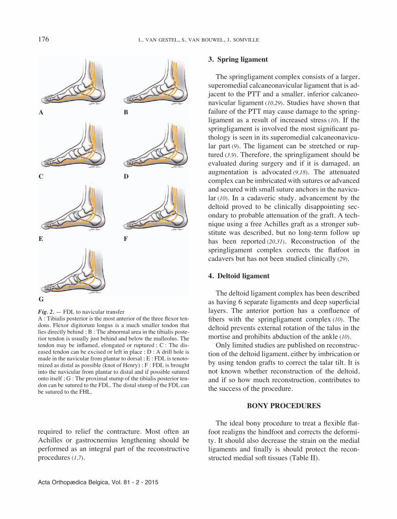
dorsal and if possible sutured onto itself. The proxi-mal part of the FDL can be tenodesed with the PTT (19,21). The FDL can also be transferred to the medial cuneiform. Although the navicular bone is the anatomic insertion of the PTT, the transfer to the medial cuneiform is believed to increase the lever arm of the transfer and thereby more efficiently in-vert the foot (19,21) (Fig. 2).
Flexor Hallucis Longus tendon
Although the FHL is much stronger than the FDL, it is only half as strong as the PTT. However, the FHL is stronger than the peroneus brevis so it will be able to correct the midfoot abduction (11,29). It is active during stance phase and the proximity to the biomechanical axis is comparable to the PTT.
The FHL is harvested at the knot of Henry, dis-tally sutured to the FDL and divided proximal to this anastomosis. The divided FHL is pulled proxi-mally through its tunnel, and then needs to be re-routed anterior to the neurovascular bundle and into the PTT sheath (11). Like the FDL, the FHL can be transferred to the navicular or the medial cuneiform. The major disadvantage in using the FHL is its proximity to the neurovascular bundle. Furthermore it can cause less push-off force of the hallux during gait (11,29).
Tibialis Anterior tendon (Cobb tendon proce-dure)
The use of the tibialis anterior to augment the PTT is described by Cobb and Helal. This proce-dure uses half of the TA harvested through a sepa-rate proximal pretibial incision at his musculo-tendinous junction. The tendon is splitted distally towards its insertion at the medial cuneiform. It is then rerouted through the medial cuneiform from anteromedial to plantar-lateral and attached to the proximal stump of the PTT. This technique seems to be more able to restore the medial arch, but has less potential in restoring the hindfoot valgus. Because only half of the tibialis anterior tendon is harvested there is no obvious weakness of ankle dorsiflexion afterwards (16).
van gestel-.indd 174 30/06/15 14:52

Acta Orthopædica Belgica, Vol. 81 - 2 - 2015
flexiBle flatfoot 175
not useful because it creates an ongoing reaction in the area where the transfer was carried out, which results in scarring and persisting synovitis. He advocates to release the diseased PTT from its in-sertion into the navicular bone and cut it proximally at the level of the medial malleolus, allowing the tendon to retract up completely out of the area of the operative field (18).
Others recommend not to remove the diseased tendon. According to a study of Valderrabano in 2004 the recovery potential of the PTT was signifi-cant even after delayed repair of a diseased tendon. They concluded that it is better not to transect the PTT (10,33) (Fig. 2C, G).
2. Achilles tendon/gastrocnemius complex
In most flatfeet, especially in the severe ones, there is a contributing shortening of the gastrosole-us complex. Tightness of the gastrosoleus should be tested pre- and peroperatively with the knee extend-ed and in 90° of flexion. If the ankle can only be dorsiflexed with the knee in flexion but not in exten-sion, a gastrocnemius recession is advised. If there is no capacity to dorsiflex the ankle, neither in ex-tension or flexion of the knee, an AP lengthening is
Peroneus brevis and peroneus longus tendon
The peroneus longus is both a first-ray plantar flexor and a major forefoot abductor (31). Its strength is comparable with the PTT, but losing its plantar flexion strength on the first metatarsal base could contribute to the flattening of the MLA. Thereby, it is less useful in flatfoot surgery (29).
The peroneus brevis tendon (a strong abductor and evertor) as a tendon transfer for the PTT would reduce the abduction deformity force on the fore-foot. However, the PB would not be able to provide sufficient inversion strength to the hindfoot because it would traverse closer to the axis of rotation of the ST joint than the PTT (29). Furthermore, because its location on the lateral side of the leg, it needs to be rerouted from lateral to medial which may damage the neurovascular bundle (29). It has its limited use in revision surgery or in case the flexors are not usable (29).
1.2.3. What to do with the diseased posterior tibial tendon in case of a tendon transfer ?
After assessing the PTT there may be a tendency to leave the PTT in place or to suture it side-to-side to the donor tendon (18). According to Mann, this is
Table I. — Tendons to augment PTT- advantages and disadvantagesadvantages disadvantages
FDL • Same distance from axis of rotation• Most expendable• Closest proximity• Easy to harvest• Opposes the abduction force of peroneus brevis• Can correct MLA and midfoot abduction• Least donor deficits
• Only 1/3 as strong as PTT
FHL • Same distance from axis to rotation• Opposes abduction force of PB better than FDL• Can correct MLA and midfoot abduction
• 50% strength og PTT• proximity to NV bundle• difficult to harvest• loss of push-off force hallux
PB • Reduces the abduction force • Risk of damaging NV bundle due to tendon rerouting lateral to medial
• Does not provide sufficient inversion forcePL • Comparable in strength with PTT • Can contribute to more flattening MLATA • Acts as a substitute for springligament
• Able to restore MLA• Non-phasic tendon: active in swing phase and not in
stance phase• Less potential in restoring hindfoot valgus
van gestel-.indd 175 30/06/15 14:52
176 l. Van Gestel, s. Van Bouwel, j. somVille
Acta Orthopædica Belgica, Vol. 81 - 2 - 2015
3. Spring ligament
The springligament complex consists of a larger, superomedial calcaneonavicular ligament that is ad-jacent to the PTT and a smaller, inferior calcaneo-navicular ligament (10,29). Studies have shown that failure of the PTT may cause damage to the spring-ligament as a result of increased stress (10). If the springligament is involved the most significant pa-thology is seen in its superomedial calcaneonavicu-lar part (9). The ligament can be stretched or rup-tured (3,9). Therefore, the springligament should be evaluated during surgery and if it is damaged, an augmentation is advocated (9,18). The attenuated complex can be imbricated with sutures or advanced and secured with small suture anchors in the navicu-lar (10). In a cadaveric study, advancement by the deltoid proved to be clinically disappointing sec-ondary to probable attenuation of the graft. A tech-nique using a free Achilles graft as a stronger sub-stitute was described, but no long-term follow up has been reported (20,31). Reconstruction of the springligament complex corrects the flatfoot in cadavers but has not been studied clinically (29).
4. Deltoid ligament
The deltoid ligament complex has been described as having 6 separate ligaments and deep superficial layers. The anterior portion has a confluence of fibers with the springligament complex (10). The deltoid prevents external rotation of the talus in the mortise and prohibits abduction of the ankle (10).
Only limited studies are published on reconstruc-tion of the deltoid ligament, either by imbrication or by using tendon grafts to correct the talar tilt. It is not known whether reconstruction of the deltoid, and if so how much reconstruction, contributes to the success of the procedure.
BONY PROCEDURES
The ideal bony procedure to treat a flexible flat-foot realigns the hindfoot and corrects the deformi-ty. It should also decrease the strain on the medial ligaments and finally is should protect the recon-structed medial soft tissues (Table II).
required to relief the contracture. Most often an Achilles or gastrocnemius lengthening should be performed as an integral part of the reconstructive procedures (1,7).
Fig. 2. — FDL to navicular transferA : Tibialis posterior is the most anterior of the three flexor ten-dons. Flexor digitorum longus is a much smaller tendon that lies directly behind ; B : The abnormal area in the tibialis poste-rior tendon is usually just behind and below the malleolus. The tendon may be inflamed, elongated or ruptured ; C : The dis-eased tendon can be excised or left in place ; D : A drill hole is made in the navicular from plantar to dorsal ; E : FDL is tenoto-mized as distal as possible (knot of Henry) ; F : FDL is brought into the navicular from plantar to distal and if possible sutured onto itself ; G : The proximal stump of the tibialis posterior ten-don can be sutured to the FDL. The distal stump of the FDL can be sutured to the FHL.
A
C
E
G
B
D
F
van gestel-.indd 176 30/06/15 14:52
Acta Orthopædica Belgica, Vol. 81 - 2 - 2015
flexiBle flatfoot 177
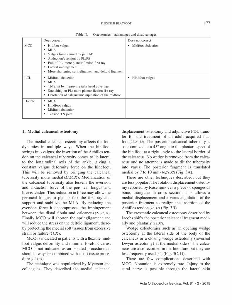
displacement osteotomy and adjunctive FDL trans-fer for the treatment of an adult acquired flat-foot (22,23,32). The posterior calcaneal tuberosity is osteotomized at a 45° angle to the plantar aspect of the hindfoot at a right angle to the lateral border of the calcaneus. No wedge is removed from the calca-neus and no attempt is made to tilt the tuberosity into varus. The posterior fragment is translated medial by 7 to 10 mm (10,21,32) (Fig. 3A).
There are other techniques described, but they are less popular. The rotation displacement osteoto-my reported by Rose removes a piece of spongeous bone, triangular in cross section. This allows a medial displacement and a varus angulation of the posterior fragment to realign the insertion of the Achilles tendon (16,32) (Fig. 3B).
The crescentic calcaneal osteotomy described by Jacobs shifts the posterior calcaneal fragment medi-ally and plantarly (12,32).
Wedge osteotomies such as an opening wedge osteotomy at the lateral side of the body of the calcaneus or a closing wedge osteotomy (reversed Dwyer osteotomy) at the medial side of the calca-neus are also recorded in the literature but they are less frequently used (32) (Fig. 3C, D).
There are few complications described with MCO. Nonunion is extremely rare. Injury to the sural nerve is possible through the lateral skin
1. Medial calcaneal osteotomy
The medial calcaneal osteotomy affects the foot dynamics in multiple ways. When the hindfoot swings into valgus, the insertion of the Achilles ten-don on the calcaneal tuberosity comes to lie lateral to the longitudinal axis of the ankle, giving a constant valgus deformity force on the hindfoot. This will be removed by bringing the calcaneal tuberosity more medial (21,26,32). Medialization of the calcaneal tuberosity also lessens the eversion and abduction force of the peroneal longus and brevis tendon. This reduction in force may allow the peroneal longus to plantar flex the first ray and support and stabilize the MLA. By reducing the eversion force it decompresses the impingement between the distal fibula and calcaneus (21,32,34). Finally MCO will shorten the springligament and will reduce the stress on the deltoid ligament, there-by protecting the medial soft tissues from excessive strain or failure (21,32).
MCO is indicated in patients with a flexible hind-foot valgus deformity and minimal forefoot varus. MCO is not indicated as an isolated procedure ; it should always be combined with a soft tissue proce-dure (1,23,34).
The technique was popularized by Myerson and colleagues. They described the medial calcaneal
Table II. — Osteotomies – advantages and disadvantagesDoes correct Does not correct
MCO • Hidfoot valgus• MLA• Valgus force caused by pull AP• Abduction/eversion by PL/PB• Pull of PL: more plantar flexion first ray• Lateral impingement• More shortening springligament and deltoid ligament
• Midfoot abduction
LCL • Midfoot abduction• MLA• TN joint by improving talar head coverage• Stretching on PL: more plantar flexion fist ray• Derotation of calcaneum: supination of the midfoot
• Hindfoot valgus
Double • MLA• Hindfoot valgus• Midfoot abduction• Tension TN joint
van gestel-.indd 177 30/06/15 14:52

178 l. Van Gestel, s. Van Bouwel, j. somVille
Acta Orthopædica Belgica, Vol. 81 - 2 - 2015
3- dimensional correction by adducting the foot at the TN joint, plantarflexing the midfoot and derotat-ing the hindfoot out of valgus (15,28,33).
The lateral column can be lengthened by a calcaneal osteotomy or by a calcaneocuboid joint arthrodesis.
Evans initially described a lateral calcaneal open-ing wedge osteotomy 1.5 cm proximal to the calca-neocuboid joint (6,8,21,26,32). There is no consensus in the literature concerning the starting point of the osteotomy. Starting positions ranging from 5 to 15 mm proximal to the CC joint are described. An anatomical study by Raines and Brage however proved that a 10 mm interval proximal to the CC joint is the optimal position : it provides the best opportunity to avoid damage to the anterior and middle facet of the subtalar joint (21,27). A more
incision, injury to the medial neurovascular struc-tures can occur when the medial cortex is cut. The sharp edge of the medialized posterior fragment can cause peroneal tendon irritations (34).
2. Lateral column lengthening
Lengthening of the lateral column (LCL) restores the MLA. Several hypotheses are proposed to explain the mechanism by which the correction occurs. With lengthening of the lateral column, the tension of the plantar fascia and peroneus longus is increased. Through a windlass effect, this will lead to elevation of the MLA and correction of the val-gus deformity (8,10,21). Other authors dispute this theory and showed that LCL results in loosening of the plantar fascia (10,21). The goal of LCL is a
Fig. 3. — Medial calcaneal osteotomyA : The posterior calcaneal tuberosity is osteotomized at a 45° angle to the plantar aspect of the hindfoot at a right angle to the lateral border of the calcaneus. The posterior fragment is translated by 7 to 10 mm ; B : The rotation displacement osteotomy. A piece of spongeous bone, triangular in cross section, is displaced medially and angu-lated into varus ; C : Opening wedge osteotomy at the lateral side of the calcaneus ; D : Closing wedge or re-versed Dwyer osteotomy.
van gestel-.indd 178 30/06/15 14:52
Acta Orthopædica Belgica, Vol. 81 - 2 - 2015
flexiBle flatfoot 179
Distraction arthrodesis of the calcaneocuboid joint has the advantages that the concern of degen-erative disease at the CC joint is eliminated (10,21). The disadvantages of this procedure are the risk for degenerative arthrosis in the adjacent joints as well as a higher non-union rate than the Evans proce-dure (21).
Lateral column lengthening is indicated in pa-tients with midfoot abduction, flexible valgus defor-mity of the hindfoot, reduced or absent strength in inversion of the foot and minimal or no subtalar arthritis (2,17,28). It is important to notice that, although LCL corrects midfoot abduction, it does not correct forefoot varus. This medial column deformity, when noted, should be addressed by a concomitant medial column stabilization (17).
3. Double osteotomy
Combining a LCL with a MCO in a so-called double osteotomy, all the components of a planoval-gus foot can be addressed. The LCL will restore the height of the MLA and the midfoot abduction whereas the MCO will restore the hindfoot valgus and reduce the valgus deformity force of the Achil-les tendon (8,21,26,32). The combination of both os-teotomies appears to reduce tension in the talona-vicular and subtalar joints, while obtaining correc-tion of bone alignment. It tends to restore the foot biomechanics towards normal, especially in combi-nation with tendon transfers (21).
4. Medial column procedures
Numerous procedures are described to address residual forefoot varus after correction of a flexible flatfoot. Although these procedures were originally used in the reconstruction of forefoot adduction and residual clubfeet in children and adolescents, they have proved their utility in the treatment of the fore-foot varus component in adult acquired flatfeet (2,30). These techniques are not indicated in isolation, but as a concomitant procedure to correct the forefoot varus in a flexible flatfoot deformity (2,21).
An opening wedge medial cuneiform osteotomy to elevate the depressed MLA in pes planus was de-scribed by Cotton. The procedure is indicated when
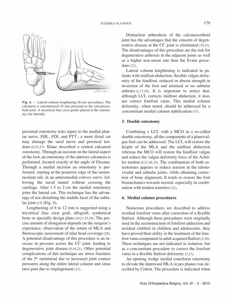
proximal osteotomy risks injury to the medial plan-tar nerve, FHL, FDL and PTT ; a more distal cut may damage the sural nerve and peroneal ten-dons (8,21,27). Klaue described a central calcaneal osteotomy. Through an incision on the lateral aspect of the foot, an osteotomy of the anterior calcaneus is performed, located exactly at the angle of Gissane. Through a medial incision an osteotomy is per-formed, starting at the posterior edge of the susten-taculum tali, in an anteromedial convex curve, fol-lowing the tarsal tunnel without crossing the cartilage. After 1.5 to 2 cm the medial osteotomy joins the lateral cut. This technique has the advan-tage of not disturbing the middle facet of the subta-lar joint (15) (Fig. 4).
Lengthening of 8 to 12 mm is suggested using a tricortical iliac crest graft, allograft, synthetical bone or specially design plate (10,21,25,28). The pre-cise amount of elongation depends on the surgeon’s experience, observation of the return of MLA and fluoroscopic assessment of talar head coverage (28). A potential disadvantage of this procedure is an in-crease in pressure across the CC joint, leading to degenerative joint disease (8,10,21). Other potential complications of this technique are stress fractures of the 5th metatarsal due to increased joint contact pressures along the entire lateral column and sinus tarsi pain due to impingement (21).
Fig. 4. — Lateral column lengthening (Evans procedure). The calcaneus is osteotomized 10 mm proximal to the calcaneocu-boid joint. A tricortical iliac crest graftis placed in the osteoto-my site laterally.
van gestel-.indd 179 30/06/15 14:52

180 l. Van Gestel, s. Van Bouwel, j. somVille
Acta Orthopædica Belgica, Vol. 81 - 2 - 2015
talus with the line along the axis of the calcaneus on lateral weight-bearing views. The normal range is 25-45°. An angle over 45° indicates hindfoot valgus (Fig. 5). The AP talocalcaneal angle is the angle be-tween the long axis of the calcaneus and the long axis of the talus. The normal range is 15-30°. An angle greater than 30° indicates hindfoot valgus (Fig. 6).
A measurement that is useful for evaluating pes planus on AP views is the lateral subluxation of the navicular on the talus, or talonavicular uncoverage, which is an indication of midfoot abduction. The ta-lonavicular coverage angel represents the degree of shift of the navicular on the talus. An angle greater than 7° indicates lateral talar subluxation (Fig. 7).
there is significant forefoot varus without degenera-tion of the first TMT joint (2,30). Typically, a 4 to 6 mm opening wedge correction with an interposi-tional graft or specially designed plate is neces-sary (2).
An arthrodesis is indicated when there is joint de-generation. Loss of the MLA may occur at the talo-navicular, naviculocuneiform, or first metatarsocu-neiform joint. Stabilization of the medial column affects the hindfoot motion : the more proximal the medial column is stabilized, the more hindfoot mo-tion is reduced. TMT 1 or NC arthrodesis will hard-ly reduce motion of the hindfoot/subtalar joints, whereas a TN arthrodesis will reduce hindfoot motion by approximately 80 to 90% (2,21,30). There-fore, a TN arthrodesis is less indicated in the treat-ment of a flexible flatfoot. A naviculocuneiform arthrodesis is done when there is residual forefoot varus secondary to severe instability or arthritis at this joint (2,14). A first tarsometatarsal arthrodesis is indicated when there is significant forefoot varus due to degenerative change in the first TMT joint (2).
DISCUSSION
Treatment of ‘a flatfoot’ starts with a thorough clinical examination. The foot should be examined in both weight-bearing and non-weight bearing po-sitions. A patient with a flexible flatfoot will have a (near-) normal arch when non-weight bearing, but will have substantial loss of height of the arch when weight bearing. The hindfoot is observed from be-hind, with the patient standing. Hindfoot alignment is determined with the midline of the calf. Normal-ly, the alignment is 5 to 10° valgus. Both midfoot and forefoot are observed in standing position, the midfoot is observed for the presence of abduction, the forefoot is observed for the presence of supina-tion. From a posterior aspect, normally only one or two lateral toes are visible. Seeing more toes (“too-many-toes” sign) is often caused by pes planus.
Standing radiographs are performed to quantita-tively describe the deformity. They allow to define the extend of the deformity.
Hindfoot valgus can be measured by the talocal-caneal angle. The lateral talocalcaneal is he angle formed by the intersection of the line bisecting the
Fig. 5. — Lateral talocalcaneal angle. A line is drawn at the plantar border of the calcaneus (or a line can be drawn bisecting the long axis of the calcaneus). The other lie is drawn through two midpoints in the talus, one at the body and one at the neck. The angle is formed by the intersection of these axes. The nor-mal range is 25-45°. An angle > 45°indicates hindfoot valgus. A : normal lateral talocalcaneal angle ; B : increased talocalca-neal angle indicating hindfoot valgus.
A
B
van gestel-.indd 180 30/06/15 14:52

Acta Orthopædica Belgica, Vol. 81 - 2 - 2015
flexiBle flatfoot 181
Fig. 6. — AP talocalcaneal angle. This angle is formed by the intersection of a line bisecting the head and neck of the talus and a line running parallel with the lateral surface of the calca-neus. The normal range is 15-30°. An angle > 30° indicates hindfoot valgus. A : normal AP talocalcaneal angle ; B : increased AP talocalca-neal angle indicating hindfoot valgus.
Fig. 8. — CYMA line. Line between talonavicular joint and calcaneocuboid joint. A : normal AP and lateral cyma line : line is smooth and continuous ; B : broken cyma line on AP and lateral view.
Fig. 7. — Talonavicular coverage angle. Two lines are drawn, one connecting the edges of the articular surface of the talus, and one connecting the edges of the articular surface of the navicular. The angle is formed by the intersection of these axes. An angle > 7° indicates lateral talar subluxation.A : normal talonavicular coverage angle ; B : increased talona-vicular coverage angle indicating midfoot abduction.
AA
BB
A B
van gestel-.indd 181 30/06/15 14:52

182 l. Van Gestel, s. Van Bouwel, j. somVille
Acta Orthopædica Belgica, Vol. 81 - 2 - 2015
The tendon that should be used to augment the PTT depends on the location of the collapse of the MLA. We feel that when the collapse is predomi-nantly located proximally, at the talonavicular joint, the FDL is best used. If the collapse is predominant-ly located distally, at the naviculocuneiform joint, the FHL is best used, because it can be harvested more distally.
Residual forefoot varus can be addressed by a medial column procedure. Depending on the affect-ed joint and the presence/absence of joint degenera-tion a medial cuneiform osteotomy, a NC arthrode-sis or a TMT 1 arthrodesis can be added to the above procedures.
CONCLUSION
The condition ‘flatfoot’ encompasses a wide range of deformities, depending on the degree of deformity in soft tissue and bony structures of the MLA, hindfoot and midfoot. Although treatment al-gorithms are usually defined for the different stages of PTTD, we would like to approach the flatfoot from a more anatomical point of view. Therefore treatment should start with a thorough clinical examination to define the deformity in each of these components.
Most authors agree that reconstruction of the soft tissues alone can temporarily resolve pain at the medial side of the foot, but does not correct the deformity (24). Soft tissue procedures therefore need to be combined with bony procedures in an attempt to give long lasting correction of both pain and deformity (4,10,21,23).
More research is needed to fully understand the significance of the forces that combine to create a flatfoot deformity. As our knowledge and experi-ence grows, more options will be available and maybe this will lead to more defined treating algo-rithms.
REFERENCES
1. Coetzee C, Castro M. The indications and biomechanical rationale for various hindfoot procedures in the treatment of posterior tibial tendon dysfunction. Foot Ankle Clin N Am 2003 ; 8 : 453-459.
Finally, pes planus can also be evaluated by the ‘cyma line’. A cyma line is an architectural term designating the union of two curve lines. A normal midtarsal joint should create a smooth cyma be-tween the talonavicular joint and calcaneocuboid joint on both the AP and lateral views. If the cyma line is broken it suggests ‘shortening’ of the calca-neus relative to the talus. This radiographic shorten-ing is due to the rotation of the talus on the calca-neus, typically seen in pes planus (Fig. 8).
Our choice of treatment is predominantly based on the clinical findings. Standing radiographs are always performed to objectify the flatfoot deformi-ty.
The preferred bony procedure, in our understand-ing, depends on the degree of hindfoot and midfoot deformity. In a pes planus, with predominantly mid-foot abduction, we prefer a lateral column lengthen-ing (LCL). In a pes planovalgus with predominantly hindfoot valgus, we prefer a medial calcaneal oste-otomy (MCO). When there is equal hindfoot valgus and midfoot abduction, both LCL and MCO can be performed (Table III).
Shortening of the Achilles tendon has to be rec-ognized at the beginning of the procedure. When the hindfoot equinus is not addressed properly, the following soft tissue and bony procedures will not fully correct the flatfoot deformity, and eventually lead to surgery failure.
Table III. — Choice of osteotomiesHINDFOOT
MIDFOOTnormal valgus
normal Normal foot MCOabduction LCL MCO + LCL
Table A : based on clinical findings
TC angle
TN coverageCYMA line
25-45° > 45°
< 7°continuous Normal foot MCO
> 7°broken LCL MCO + LCL
Table B : based on radiographic findings
van gestel-.indd 182 30/06/15 14:52
Acta Orthopædica Belgica, Vol. 81 - 2 - 2015
flexiBle flatfoot 183
18. Mann RA. Posterior tibial tendon dysfunction : treatment by flexor digitorum longus transfer. Foot Ankle Clin 2001 ; 6 : 77-87.
19. Mann RA, Thompson FM. Rupture of the posterior tibial tendon causing flatfoot. J Bone Joint Surg 1985 ; 67 (4) : 556 – 561.
20. McCormack A, Ching R, Sangeorzan B. Biomechanics of procedures used in the adult flatfoot deformity. Foot Ankle Clin 2001 ; 6 : 15-23.
21. Mosier-LaClair S, Pomeroy G, Manoli A. Operative treatment of the difficult stage 2 adult acquired flatfoot deformity. Foot Ankle Clin 2001 ; 6 : 95-119.
22. Myerson MS. Adult acquired flat foot deformity treatment of dysfunction of the posterior tibial tendon. J Bone Joint Surg 1996 ; 78(5) : 780-792.
23. Myerson M, Badekas A, Schon L. Treatment of stage II posterior tibial tendon deficiency with flexor digitorum longus tendon transfer and calcaneal osteotomy. Foot Anle Int 2004 ; 25 : 445-450.
24. Pinney S, Lin S. Current concept review : Acquired adult flatfoot deformity. Foot Ankle Int 2006 ; 27 : 66-73.
25. Pomeroy GC, Pike H, Beals T, Manoli A. Acquired flatfoot in adults due to dysfunction of the posterior tibial tendon. J Bone Joint Surgery 1999 ; 81 : 1173-1182.
26. Popovic N, Lemaire R. Acquired flatfoot deformity secondary to dysfunction of the tibialis posterior tendon. Acta Orthopædica Belgica 2003 ; 69 : 211-222.
27. Raines RA, Brage ME. Evans osteotomy in the adult foot : an anatomic study of structures at risk. Foot Ankle Int 1998 ; 19 : 743-747.
28. Sands A, Tansey J. Lateral column lengthening. Foot Ankle Clin N Am 2007 ; 12 : 301-308.
29. Sitler D, Bell J. Soft tissues procedures. Foot Ankle Clin N Am 2003 ; 8 : 503-520.
30. Sizensky J, Marks R. Medial sided bony procedures : why, what and how. Foot Ankle Clin N Am 2003 ; 8 : 539-562.
31. Thordarson D, Schmotzer H, Chon J. Reconstruction with tenodesis in an adult flatfoot model. J Bone Joint Surg 1995 ; 77 : 1557-1564.
32. Trnka H, Easley M, Myerson M. The role of calcaneal osteotomies for correction of adult flatfoot. Clin Orthop 1999 ; 365 : 50-54.
33. Valderrabano V, Hintermann B, Wischer T, Fuhr P, Dick W. Recovery of the posterior tibial muscle after late reconstruction following tendon rupture. Foot Ankle Int 2004 ; 25 : 85-95.
34. Weinfeld S. Medial slide calcaneal osteotomy : technique, patient selection and results. Foot Ankle Clin 2001 ; 6 : 89-94.
2. Cohen B, Ogden F. Medial column procedures in the acquired flatfoot deformity. Foot Ankle Clin N Am 2007 ; 12 : 287-299.
3. Deland JT. The adult acquired flatfoot and springligament complex. Pathology and implications for treatment. Foot Ankle Clin 2001 ; 6(1) : 129-135.
4. Den Hartog B. Flexor digotorum longus transfer with medial displacement calcaneal osteotomy : biomechanical rationale. Foot Ankle Clin 2001 ; 6 : 67-75.
5. Dyal C, Feder J, Deland J, Thompson F. Pes planus in patients with posterior tibial tendon insufficiency : asympto-matic versus symptomatic foot. Foot and Ankle Int 1997 ; 18 : 84-88.
6. Evans D. Calcaneo-valgus deformity. J Bone Joint Surg Br 1975 ; 75 : 270-278.
7. Fortin PT. Posterior tibial tendon dysfunction. Isolated fusion of the talonavicular joint. Foot Ankle Clin 2001 ; 6 : 137-151.
8. Gallina J, Sands A. Lateral sided bony procedures. Foot Ankle Clin N Am 2003 ; 8 : 563-567.
9. Gazdag A,Cracchiolo A. Rupture of the posterior tibial tendon. Evaluation of injury of the springligament and clinical assessment of tendon transfer and ligament repair. J Bone Joint Surg 1997 ; 79 : 675-681.
10. Giza E, Cush G, Schon LC. The flexible flatfoot in the adult. Foot and Ankle Clin N Am 2007 ; 12 : 251- 271.
11. Hockenbury RD, Sammarco GJ. Medial sliding calcaneal osteotomy with flexor halluces longus transfer for the treatment of posterior tibial tendon insufficiency. Foot Ankle Clin 2001 ; 6 : 569-581.
12. Jacobs AM, Hodson BS, Albrecht HM. Synovectomy-arthroplasty as an alternative to triple arthrodesis in the management of subtalar joint pain. CLin Podiatr Med Surg 1991 ; 3 : 485-500.
13. Johnson KA, Storm DE. Tibialis posterior tendon dysfunction. Clin Orthop 1989 ; 239 : 196-206.
14. Johnson J, Yu J. Arthrodesis techniques in the management of stage II and III acquired adult flatfoot deformity. J Bone Joint Surg 2005 ; 87 : 1866-1876.
15. Klaue K et al. Central calcaneal osteotomy for correction of flexible pes planovalgus deformity. Foot Ankle Int 2013 ; 34 : 1079-1089.
16. Madhav R, Kampa R, Singh D, Angel J. Cobb procedure and calcaneal osteotomy for the treatment of tibial posterior tendon dysfunction. Acta Orthop Belg 2009 ; 75 : 64-69.
17. Mankey MG. A classification of severity with an analysis of causative problems related to the type of treatment. Foot Ankle Clin N Am 2003 ; 8 : 461-471.
van gestel-.indd 183 30/06/15 14:52