Surgical Treatment of Median Arcuate Ligament Syndrome...
5
74 Srp Arh Celok Lek. 2015 Jan-Feb;143(1-2):74-78 DOI: 10.2298/SARH1502074K ПРИКАЗ БОЛЕСНИКА / CASE REPORT UDC: 617.55-009.7-089 : 616.13-089 Correspondence to: Vladimir DJORDJEVIĆ Clinic for Digestive Surgery First Surgical Clinic Clinical Center of Serbia Dr Koste Todorovića 6 11000 Belgrade Serbia [email protected] SUMMARY Introduction Median arcuate ligament (MAL) syndrome, also called celiac trunk compression syndrome (CACS) or Dunbar syndrome is a rare disorder caused by compression of the celiac artery by median arcuate ligament of the diaphragm, which leads to mesenteric ischemia and chronic abdominal angina. The typical clinical triad of symptoms includes postprandial epigastric pain, weight loss and vomiting. The gold standard for MAL syndrome diagnosis is selective angiography, while in symptomatic patients with angiographically verified stenosis the optimal therapy is surgical treatment. Case Outline A 40-year-old male patient was presented with epigastric pain, followed by dyspepsia and weight loss. The upper endoscopy showed gastric and duodenal distention with prominent folds of gastric mucosa and slow peristalsis. Selective angiography showed stenosis (90%) of initial segment of the celiac trunk. Adhesiolysis with the transection of the median arcuate ligament was performed. Due to repeated symptoms, the patient was reoperated on the 10 th postoperative day with performed adhesiolysis and gastrostomy for gastric nutrition. Two months later, the patient was rehospitalized for closure of gastrostomy. At five years follow-up, selective angiography showed no stenosis of the initial segment of the celiac artery. Conclusion Despite the existing controversy concerning pathophysiological mechanism, the clinical presentation and treatment modalities of patients with MAL syndrome, it is evident that careful selection and adequate surgical treatment may significantly reduce symptoms in these patients. Keywords: celiac artery; median arcuate ligament; diaphragm; arterial occulsive disease Surgical Treatment of Median Arcuate Ligament Syndrome: Case Report and Review of Literature Milutin Kotarac 1 , Nebojša Radovanović 1,2 , Nebojša Lekić 1 , Zoran Ražnatović 1 , Vladimir Djordjević 1 , Dragana Lekić 3 , Dragan Sagić 2,4 1 Clinic for Digestive Surgery, First Surgical Clinic, Clinical Center of Serbia, Belgrade, Serbia; 2 University of Belgrade, School of Medicine, Belgrade, Serbia; 3 Institute for Mother and Child Health Care of Serbia “Dr Vukan Čupić”, Belgrade, Serbia; 4 Institute for Cardiovascular Diseases “Dedinje”, Belgrade, Serbia INTRODUCTION Median arcuate ligament (MAL) syndome, also known as celiac artery compression syndrome (CACS) is a rare disorder caused by compres- sion of the celiac artery by the median arcuate ligament, a fibrous arch anterior to the aorta which is formed by a connection of the dia- phragmatic crura [1]. Sporadically, the median arcuate ligament may insert at a lower level, thus compressing and narrowing the lumen of the celiac artery, especially during expiration. In that way, subsequent mesenterial ischemia may cause a typical triad of symptoms charac- teristic for MAL syndrome: postprandial ab- dominal pain, weight loss, nausea and vomiting [2]. The first celiac trunk compression by the median arcuate ligament was observed during autopsies by Lipshutz et al. [3] in 1917. How- ever, the first clinical cases of celiac artery com- pression syndrome with angiographically vis- ible stenosis of the celiac trunk were described by Harjola [4] and Dunbar et al. [5] in 1963 and 1965, respectively, who also defined celiac artery compression syndrome (often called Dunbar syndrome). Treatment modalities for patients with a symptomatic compression of celiac artery and angiographically verified stenosis are percuta- neous transluminal angioplasty and surgical treatment. Here we report a case of a 40-year- old symptomatic patient with angiographically verified compression of celiac artery that was surgically treated and who had no symptoms during the 5-year follow-up period. CASE REPORT A 40-year-old male patient presented with chronic epigastric pain which spread along both costal margins and not related to food intake, dyspepsia, weight loss and flatulence that have been ongoing on for over a year. The patient was occasionally using analgesics which only temporarily diminished abdominal pain. Physical examination and laboratory blood tests were unremarkable with normal findings on abdominal ultrasonography. Esophagogas- troduodenoscopy showed distension of the stomach with prominent folds of gastric mu- cosa, as well as distended duodenum with slow peristalsis and rough appearance of its mucous layer. Abdominal X-ray verified air fluid levels in the small intestine with gastric distention. Selective angiography of the celiac trunk and superior mesenteric artery showed steno- sis (90%) of initial segment of the celiac artery caused by median arcuate ligament compres- sion. The stenosis was respiratory-dependent:
Transcript of Surgical Treatment of Median Arcuate Ligament Syndrome...
74
Srp Arh Celok Lek. 2015 Jan-Feb;143(1-2):74-78 DOI: 10.2298/SARH1502074K
ПРИКАЗ БОЛЕСНИКА / CASE REPORT UDC: 617.55-009.7-089 : 616.13-089
Correspondence to:
Vladimir DJORDJEVIĆ
Clinic for Digestive Surgery
First Surgical Clinic
Clinical Center of Serbia
Dr Koste Todorovića 6
11000 Belgrade
Serbia
SUMMARYIntroduction Median arcuate ligament (MAL) syndrome, also called celiac trunk compression syndrome (CACS) or Dunbar syndrome is a rare disorder caused by compression of the celiac artery by median arcuate ligament of the diaphragm, which leads to mesenteric ischemia and chronic abdominal angina. The typical clinical triad of symptoms includes postprandial epigastric pain, weight loss and vomiting. The gold standard for MAL syndrome diagnosis is selective angiography, while in symptomatic patients with angiographically verified stenosis the optimal therapy is surgical treatment.Case Outline A 40-year-old male patient was presented with epigastric pain, followed by dyspepsia and weight loss. The upper endoscopy showed gastric and duodenal distention with prominent folds of gastric mucosa and slow peristalsis. Selective angiography showed stenosis (90%) of initial segment of the celiac trunk. Adhesiolysis with the transection of the median arcuate ligament was performed. Due to repeated symptoms, the patient was reoperated on the 10th postoperative day with performed adhesiolysis and gastrostomy for gastric nutrition. Two months later, the patient was rehospitalized for closure of gastrostomy. At five years follow-up, selective angiography showed no stenosis of the initial segment of the celiac artery.Conclusion Despite the existing controversy concerning pathophysiological mechanism, the clinical presentation and treatment modalities of patients with MAL syndrome, it is evident that careful selection and adequate surgical treatment may significantly reduce symptoms in these patients.Keywords: celiac artery; median arcuate ligament; diaphragm; arterial occulsive disease
Surgical Treatment of Median Arcuate Ligament Syndrome: Case Report and Review of LiteratureMilutin Kotarac1, Nebojša Radovanović1,2, Nebojša Lekić1, Zoran Ražnatović1, Vladimir Djordjević1, Dragana Lekić3, Dragan Sagić2,4
1Clinic for Digestive Surgery, First Surgical Clinic, Clinical Center of Serbia, Belgrade, Serbia;2University of Belgrade, School of Medicine, Belgrade, Serbia;3Institute for Mother and Child Health Care of Serbia “Dr Vukan Čupić”, Belgrade, Serbia;4Institute for Cardiovascular Diseases “Dedinje”, Belgrade, Serbia
INTRODUCTION
Median arcuate ligament (MAL) syndome, also known as celiac artery compression syndrome (CACS) is a rare disorder caused by compres-sion of the celiac artery by the median arcuate ligament, a fibrous arch anterior to the aorta which is formed by a connection of the dia-phragmatic crura [1]. Sporadically, the median arcuate ligament may insert at a lower level, thus compressing and narrowing the lumen of the celiac artery, especially during expiration. In that way, subsequent mesenterial ischemia may cause a typical triad of symptoms charac-teristic for MAL syndrome: postprandial ab-dominal pain, weight loss, nausea and vomiting [2]. The first celiac trunk compression by the median arcuate ligament was observed during autopsies by Lipshutz et al. [3] in 1917. How-ever, the first clinical cases of celiac artery com-pression syndrome with angiographically vis-ible stenosis of the celiac trunk were described by Harjola [4] and Dunbar et al. [5] in 1963 and 1965, respectively, who also defined celiac artery compression syndrome (often called Dunbar syndrome).
Treatment modalities for patients with a symptomatic compression of celiac artery and angiographically verified stenosis are percuta-neous transluminal angioplasty and surgical
treatment. Here we report a case of a 40-year-old symptomatic patient with angiographically verified compression of celiac artery that was surgically treated and who had no symptoms during the 5-year follow-up period.
CASE REPORT
A 40-year-old male patient presented with chronic epigastric pain which spread along both costal margins and not related to food intake, dyspepsia, weight loss and flatulence that have been ongoing on for over a year. The patient was occasionally using analgesics which only temporarily diminished abdominal pain. Physical examination and laboratory blood tests were unremarkable with normal findings on abdominal ultrasonography. Esophagogas-troduodenoscopy showed distension of the stomach with prominent folds of gastric mu-cosa, as well as distended duodenum with slow peristalsis and rough appearance of its mucous layer. Abdominal X-ray verified air fluid levels in the small intestine with gastric distention.
Selective angiography of the celiac trunk and superior mesenteric artery showed steno-sis (90%) of initial segment of the celiac artery caused by median arcuate ligament compres-sion. The stenosis was respiratory-dependent:
75Srp Arh Celok Lek. 2015 Jan-Feb;143(1-2):74-78
www.srp-arh.rs
it was present in expiration while it disappeared during the deep inspiration.
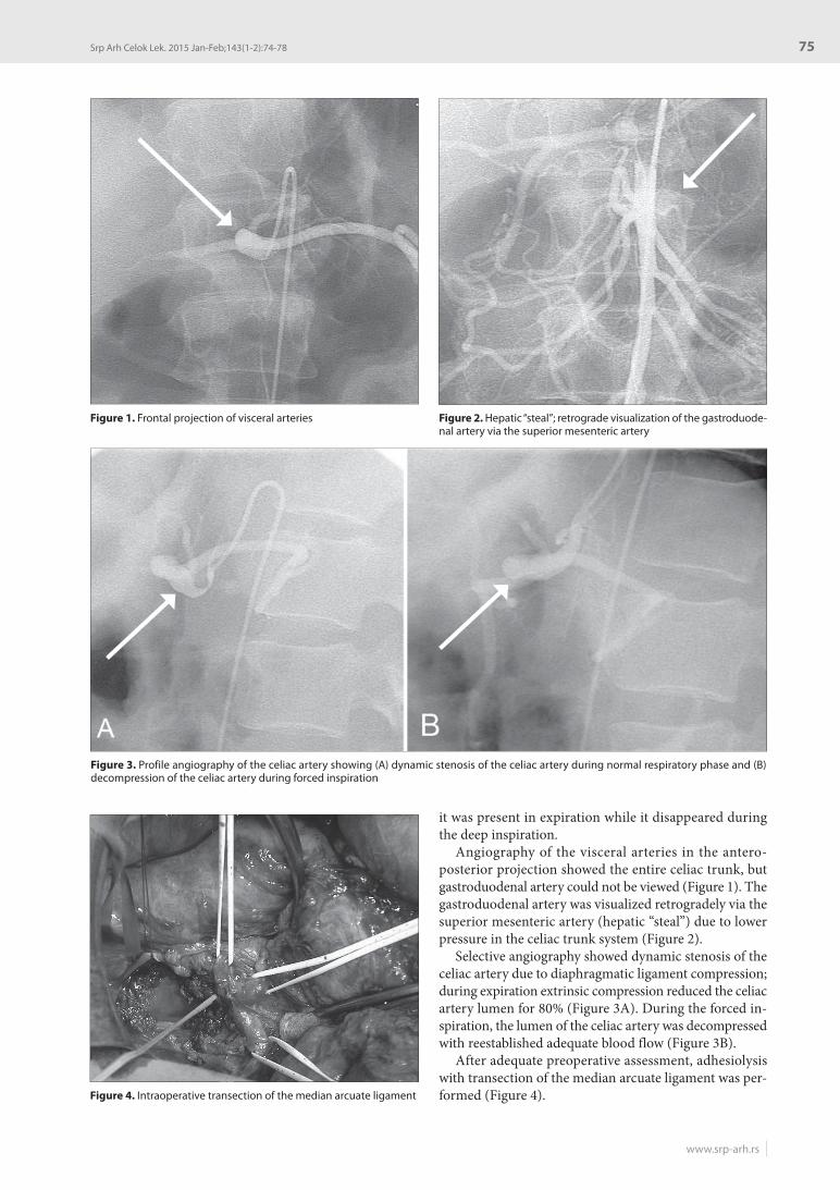
Angiography of the visceral arteries in the antero-posterior projection showed the entire celiac trunk, but gastroduodenal artery could not be viewed (Figure 1). The gastroduodenal artery was visualized retrogradely via the superior mesenteric artery (hepatic “steal”) due to lower pressure in the celiac trunk system (Figure 2).
Selective angiography showed dynamic stenosis of the celiac artery due to diaphragmatic ligament compression; during expiration extrinsic compression reduced the celiac artery lumen for 80% (Figure 3A). During the forced in-spiration, the lumen of the celiac artery was decompressed with reestablished adequate blood flow (Figure 3B).
After adequate preoperative assessment, adhesiolysis with transection of the median arcuate ligament was per-formed (Figure 4).
Figure 1. Frontal projection of visceral arteries Figure 2. Hepatic “steal”; retrograde visualization of the gastroduode-nal artery via the superior mesenteric artery
Figure 3. Profile angiography of the celiac artery showing (A) dynamic stenosis of the celiac artery during normal respiratory phase and (B) decompression of the celiac artery during forced inspiration
Figure 4. Intraoperative transection of the median arcuate ligament
76
doi: 10.2298/SARH1502074K
Kotarac M. et al. Surgical Treatment of Median Arcuate Ligament Syndrome: Case Report and Review of Literature
However, early remission was followed by repeated symptoms; abdominal pain, nausea followed by vomit-ing, and the patient refused any oral nutrition. Therefore, the patient was reoperated on the 10th postoperative day, with performed adhesiolysis and gastrostomy for gastric nutrition. Postoperative course was without complications, intrahospital stay was prolonged in order to stabilize the patient’s condition and nourishment, and the patient was discharged on the 45th postoperative day. Two months after discharge, the patient was rehospitalized for the closure of gastrostomy, and was back on oral nutrition with normal digestive functions.
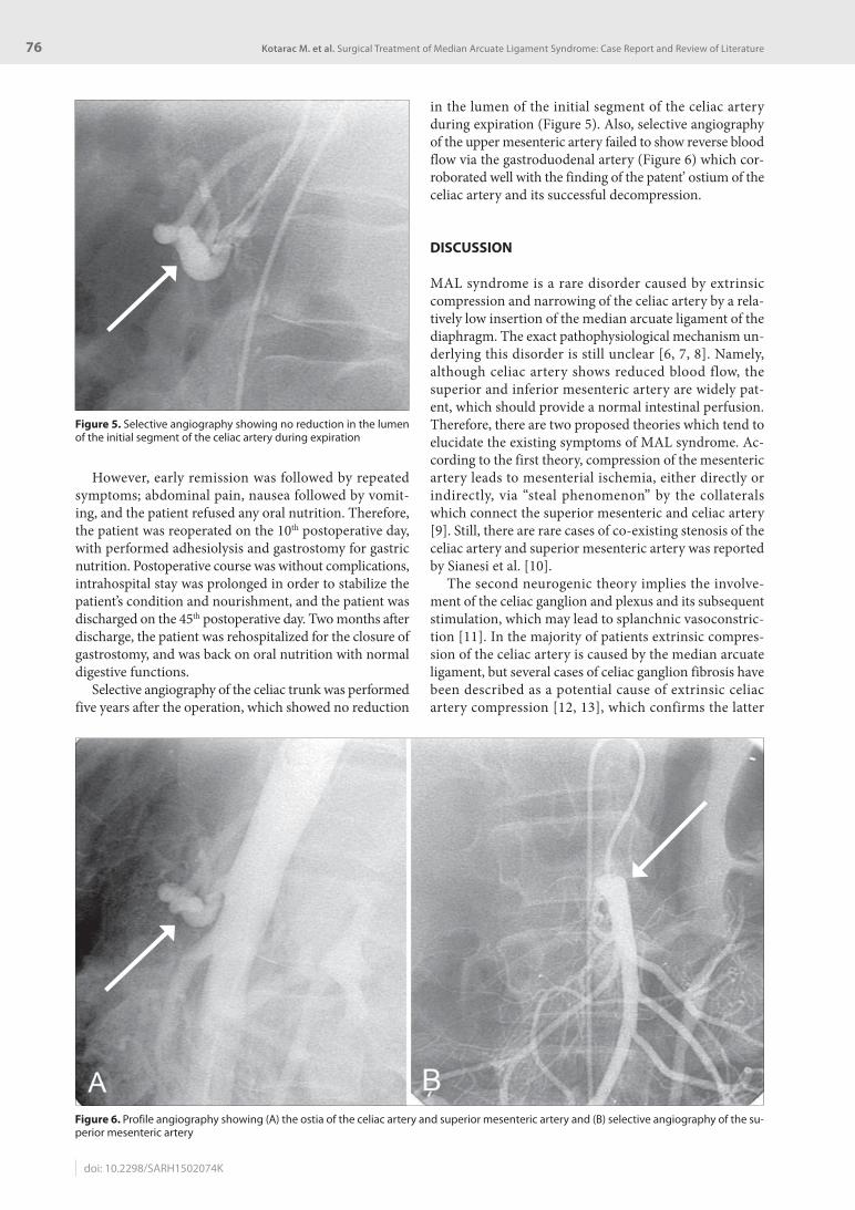
Selective angiography of the celiac trunk was performed five years after the operation, which showed no reduction
in the lumen of the initial segment of the celiac artery during expiration (Figure 5). Also, selective angiography of the upper mesenteric artery failed to show reverse blood flow via the gastroduodenal artery (Figure 6) which cor-roborated well with the finding of the patent’ ostium of the celiac artery and its successful decompression.
DISCUSSION
MAL syndrome is a rare disorder caused by extrinsic compression and narrowing of the celiac artery by a rela-tively low insertion of the median arcuate ligament of the diaphragm. The exact pathophysiological mechanism un-derlying this disorder is still unclear [6, 7, 8]. Namely, although celiac artery shows reduced blood flow, the superior and inferior mesenteric artery are widely pat-ent, which should provide a normal intestinal perfusion. Therefore, there are two proposed theories which tend to elucidate the existing symptoms of MAL syndrome. Ac-cording to the first theory, compression of the mesenteric artery leads to mesenterial ischemia, either directly or indirectly, via “steal phenomenon” by the collaterals which connect the superior mesenteric and celiac artery [9]. Still, there are rare cases of co-existing stenosis of the celiac artery and superior mesenteric artery was reported by Sianesi et al. [10].
The second neurogenic theory implies the involve-ment of the celiac ganglion and plexus and its subsequent stimulation, which may lead to splanchnic vasoconstric-tion [11]. In the majority of patients extrinsic compres-sion of the celiac artery is caused by the median arcuate ligament, but several cases of celiac ganglion fibrosis have been described as a potential cause of extrinsic celiac artery compression [12, 13], which confirms the latter
Figure 5. Selective angiography showing no reduction in the lumen of the initial segment of the celiac artery during expiration
Figure 6. Profile angiography showing (A) the ostia of the celiac artery and superior mesenteric artery and (B) selective angiography of the su-perior mesenteric artery
77Srp Arh Celok Lek. 2015 Jan-Feb;143(1-2):74-78
www.srp-arh.rs
pathogenesis theory of this syndrome. Albeit, contro-versies still exist, since it has been reported that 13-50% of healthy subjects may exhibit a certain degree of an-giographically visible compression of the celiac artery during expiration [6].
MAL syndrome is typically presented in young to mid-dle-age adults, although it has been described in infants and children [14]. It is characterized by a clinical triad of postprandial epigastric pain, weight loss and vomiting.
The typical manifestation of abdominal angina is seen only in about 40% of patients, while more than 80% of cas-es have symptoms which vary from postprandial cramp-like abdominal pain to non-specific epigastric pain [15].
The gold standard of the diagnostic method for MAL syndrome is selective angiography performed in inspira-tion as well as in expiration. Treatment modalities include endovascular and surgical procedures (open or laparo-scopic surgery). Although percutaneous transluminal an-gioplasty with stent implantation is a less invasive proce-dure, it often does not solve the problem of the underlying extrinsic compression of the celiac trunk and frequently requires surgical interventions.
Classic treatment for MAL syndrome is represented by open surgery with celiac artery decompression. The largest series of surgically treated patients with MAL syndrome included 51 patients [16]. Surgical modalities included celiac trunk decompression in 16 patients, decompres-sion and dilatation in 17 patients, and decompression and reconstruction either by primary reanastomosis or interposition of the graft in 18 patients. The best results of symptoms relief were achieved in cases of decompres-sion and certain forms of celiac revascularization (76%). Also, Grotemeyer et al. [17] in their series of 18 surgically treated patients observed that open surgical therapy in its
various forms is a safe and reliable procedure with either no mortality or low morbidity rate.
Since the beginning of laparoscopic era, there have been numerous reports of different laparoscopic techniques for treating patients with MAL syndrome [18, 19, 20]. Baccari et al. [21] (2009) have reported a successful laparoscopic decompression of the celiac artery in a group of consecu-tive patients with complete resolution and symptom-free period of 28.3 months. Despite the postoperative effective-ness of laparoscopic procedures, no long-term follow-up is still available.
Tsujimoto et al. [22] have used intraoperative Doppler ultrasound for the confirmation of the decompressed ce-liac artery after successful laparoscopic intervention in pa-tients with MAL syndrome. Recently, several authors have reported robotic-assisted treatment of MAL syndrome as a new, safe and efficacious modality treatment [23, 24, 25].
Although MAL syndrome has been described in the 1960s, controversies concerning pathophysiological mechanism, clinical presentation and treatment modali-ties still exist. Regardless of the controversial viewpoints, the majority of authors agree that symptomatic patients with angiographically confirmed celiac artery compression will benefit more from surgical treatment.
In this paper we presented a case of a 40-year-old symp-tomatic patient with angiographically verified stenosis of the celiac artery that was surgically treated. The patient had no symptoms during a 5-year follow-up period, which can be considered as a long-term follow-up.
Many questions regarding MAL syndrome have not been clarified yet, but it is evident that careful selection and adequate treatment can significantly reduce symp-toms in these patients while the choice of treatment must depend on the specific clinical situation for each patient.
1. Loukas M, Pinyard J, Vaid S, Kinsella C, Tariq A, Tubbs RS. Clinical anatomy of celiac artery compresion syndrome: a review. Clin Anat. 2007; 20:612-7.
2. Holland A, Ibach E. Long-term review of coeliac axis compression syndrome. Ann R Coll Surg Engl. 1996; 78:470-2.
3. Lipshutz B. A composite study of the celiac axis artery. Am Surg. 1917; 65:165-9.
4. Harjola PT. A rare obstruction of the coeliac artery. Ann Chir Gynaecol Fenn. 1963; 52:547-70.
5. Dunbar JD, Molnar W, Beman FF, Marable SA. Compression of the celiac trunk and abdominal angina. Am J Roentgenol Radium Ther Nucl Med. 1965; 95(3):731-44.
6. Szilagyi DE, Rian RL, Elliott JP, Smith RF. The celiac artery compression syndrome: does it exist? Surgery. 1972; 72:849-63.
7. Gloviczki P, Duncan AA. Treatment of celiac artery compression syndrome: Does it really exists? Perspect Vasc Surg Endovasc Ther. 2007; 19(3):259-63.
8. Bobadilla JL. Mesenteric ischemia. Surg Clin North Am. 2013; 93:925-40.
9. Bech FR. Celiac artery compression syndromes. Surg Clin North Am. 1997; 77:409-24.
10. Sianesi M, Soliani P, Arcuri MF, Bezer L, Iapichino G, Del Rio P. Dunbar’s syndrome and superior mesenteric artery’s syndrome: a rare association. Dig Dis Sci. 2007; 52(1):302-5.
11. Desmond CP, Roberts SK. Exercise-related abdominal pain as a manifestation of the median arcuate ligament syndrome. Scand J Gastroenterol. 2004; 39:1310-3.
12. Snyder MA, Mahoney EB, Rob CG. Symptomatic celiac artery stenosis due to constriction by the neurofibrous tissue of the celiac ganglion. Surgery. 1967; 61:372-6.
13. Van Gossun M, Morobe J, Van Laethem A, Burette A, Somerhausen J, Schockert J. Syndrome de compression isolee du tronc coeliaque (SCITC). Apropos de 8 cas. Acta Chir Belg. 1984; 84:379-83.
14. Schweizer P, Berger S, Schweizer M, Schaefer J, Beck O. Arcuate ligament vascular compression syndrome in infants and children. J Pediatr Surg. 2005; 40(10):1616-22.
15. Gander S, Mulder DJ, Jones S, Ricketts JD, Soboleski DA, Justinich CJ. Reccurent abdominal pain and weight loss in an adolescent: Celiac artery compression syndrome. Can J Gastroenterol. 2010; 24(2):91-3.
16. Reilly LM, Ammar AD, Stones RJ, Ehrenwel WK. Late results following repair for celiac artery compression syndrome. J Vasc Surg. 1985 ;2(1):79-91.
17. Grotemeyer D, Duran M, Iskandar F, Blondin D, Nguyen K, Sandmann W. Median arcuate ligament syndrome: vascular surgical therapy and follow-up of 18 patients. Langebecks Arch Surg. 2009; 394:1085-92.
18. Cienfuegos JA, Rotellar F, Valenti V, Arredondo J, Pedano N, Bueno A, et al. The celiac axis compression syndrome (CACS): critical review in the laparoscopic era. Rev Esp Enferm Dig. 2010; 102:193-201.
19. Roayaie S, Jossart G, Gitlitz D, Lamparello P, Hollier L, Gagner M. Laparoscopic release of celiac artery compression syndrome facilitated by laparoscopic ultrasound scanning to confirm restoration of flow. J Vasc Surg. 2000; 32(4):814-7.
REFERENCES
78
doi: 10.2298/SARH1502074K
20. Roseborough GS. Laparoscopic management of celiac artery compression syndrome. J Vasc Surg. 2009; 50:124-33.
21. Baccari P, Civilini E, Dordoni L, Melissano G, Nicoletti R, Chiesa R. Celiac artery compression syndrome managed by laparoscopy. J Vasc Surg. 2009; 50(1):134-9.
22. Tsujimoto H, Hiraki S, Sakamoto N, Yaguchi Y, Kumano I, Yoshida K, et al. Laparoscopic treatment for median arcuate ligament syndrome: the usefulness of intraoperative Doppler ultrasound to confirm the decompression of the celiac artery. Surg Laparosc Endosc Percutan Tech. 2012; 22(2):e71-75.
23. Meyer M, Gharagozloo F, Nguyen D, Tempesta B, Strother E, Margolis M. Robotic-assisted treatment of celiac artery compression syndrome: report of a case and review of the literature. Int J Med Robot. 2012; 8(4):379-83.
24. Relles D, Moudgill N, Rao A, Rosato F, DiMuzio P, Eisenberg J. Robotic-assisted median arcuate ligament release. J Vasc Surg. 2012; 56(2):500-3.
25. Jaik NP, Stawicki SP, Weger NS, Lukaszczyk JJ. Celiac artery compression syndrome: successful utilization of robotic-assisted laparoscopic approach. J Gastrointestin Liver Dis. 2007; 16:93-6.
КРАТАК САДРЖАЈУвод Син дром ме ди јал ног ар ку ат ног ли га мен та ди ја фраг ме, та ко ђе по знат као син дром ком пре си је це ли јач ног трун ку са или Дан ба ров (Dun bar) син дром, ре дак је по ре ме ћај узро-ко ван ком пре си јом це ли јач не ар те ри је ме ди јал ним ар ку ат-ним ли га мен том ди ја фраг ме ко ји до во ди до ме зен те рич не ис хе ми је и хро нич не ан ги не аб до ме на. Ти пич но кли нич ко трој ство симп то ма укљу чу је по стпран ди јал ни епи га стрич ни бол, гу би так те жи не и по вра ћа ње. Злат ни стан дард у ди јаг-но сти ци овог син дро ма је се лек тив на ан ги о гра фи ја, док је за бо ле сни ке ко ји има ју симп то ме и ан ги о граф ски по твр ђе-ну сте но зу оп ти мал ни трет ман хи рур шко ле че ње.При каз бо ле сни ка Му шка рац стар 40 го ди на по жа лио се на бол у епи га стри ју му пра ћен дис пеп си јом и гу бит ком те-ле сне те жи не. Ен до скоп ски пре глед гор њих ор га на ука за ла је на на ду тост же лу ца и ду о де ну ма с ис так ну тим на бо ри ма га стрич не му ко зе и спо ром пе ри стал ти ком. Се лек тив на ан-ги о гра фи ја ука за ла је на сте но зу (90%) ини ци јал ног сег мен-
та це ли јач ног трун ку са. Оба вље на је ад хе зи о ли за уз пре се-ца ње ме ди јал ног ар ку ат ног ли га мен та. Због по но вље них симп то ма, бо ле сник је опет опе ри сан де се тог да на на кон пр ве опе ра ци је, ка да је ура ђе на ад хе зи о ли за и кре и ра на ну три тив на га стро сто ми ја. Два ме се ца ка сни је бо ле сник је по но во при мљен на бол нич ко ле че ње, ка ко би се из вр ши-ло за тва ра ње ну три тив не га стро сто ми је. Пет го ди на на кон хи рур шког ле че ња кон трол ном се лек тив ном ан ги о гра фи-јом ни је утвр ђе на сте но за ини ци јал ног сег мен та це ли јач не ар те ри је.За кљу чак Упр кос опреч ним ста во ви ма у ве зи с па то ло шким ме ха ни зми ма на стан ка и кли нич ком сли ком Дан ба ро вог син дро ма, те об ли ци ма ле че ња бо ле сни ка с овим обо ље-њем, ја сно је да па жљив ода бир од го ва ра ју ћег хи рур шког при ступа мо же зна чај но ума њи ти симп то ме код ових бо-ле сни ка.Кључ не ре чи: це ли јач на ар те ри ја; ме ди јал ни ар ку ат ни ли-га мент; ди ја фраг ма; ар те риј ска оклу зив на бо лест
Хируршко лечење синдрома медијалног аркуатног лигамента дијафрагме � приказ болесника и преглед литературеМилутин Котарац1, Небојша Радовановић1,2, Небојша Лекић1, Зоран Ражнатовић1, Владимир Ђорђевић1, Драгана Лекић3, Драган Сагић2,4
1Клиника за дигестивну хирургију, Прва хируршка клиника, Клинички центар Србије, Београд, Србија;2Универзитет у Београду, Медицински факултет, Београд, Србија;3Институт за здравствену заштиту мајке и детета Србије „Др Вукан Чупић“, Београд, Србија;4Институт за кардиоваскуларне болести „Дедиње“, Београд, Србија
Примљен • Received: 10/04/2014 Прихваћен • Accepted: 03/06/2014
Kotarac M. et al. Surgical Treatment of Median Arcuate Ligament Syndrome: Case Report and Review of Literature