Surgical Technique - synthes.vo.llnwd.netsynthes.vo.llnwd.net › o16 › LLNWMB8 › US Mobile ›...
14
Surgical Technique Guide
Transcript of Surgical Technique - synthes.vo.llnwd.netsynthes.vo.llnwd.net › o16 › LLNWMB8 › US Mobile ›...

SurgicalTechnique
Guide

•IntroductIon 1
•SurgIcaltechnIqueforVertebral
bodyderotatIon(Vbd) 3
enblocVertebral bodyderotat ion
(Mult ip leapical levels ) 6
Segmental Vertebral bodyderotat ion
( Indiv idual Vertebral level ) 9
C o n t e n t s
t e C h n i q u e W r i t t e n B y :
Randal Betz, MD Philadelphia,Pa
Peter Newton, MD Sandiego,ca
Michael O’Brien, MD dallas,tX
Suken Shah, MD Wilmington,de
i n s t r u m e n t D e s i g n e r s :
Randal Betz, MD
Philadelphia,Pa
Munish Gupta, MD Sacramento,ca
Baron Lonner, MD
newyork,ny
Peter Newton, MD
Sandiego,ca
Michael O’Brien, MD
dallas,tX
Suken Shah, MD
Wilmington,de
Harry Shufflebarger, MD
Miami,fl
Dezsö J. Jeszenszky, MD
Switzerland
2

3
surgiCal teChnique for VerteBral BoDy Derotation (VBD)
using the expeDium spine system in sColiosis
ItisacceptedthatwiththeuseofpediclescrewsinScoliosiscorrection,
significantcoronalplanecorrectioncanbeconsistentlyobtained.true
axialplanecorrectioncannowbeachievedtoaddresstherotational
deformityofthespine,ribsandchestwall.themaingoalofvertebral
bodyderotation(Vbd)istoachieverotationaldeformitycorrection,
whichmaydecreasetheneedforthoracoplasty.(figure1)
the steps are as folloWs:
ontheconcaveside:InsertMonoaxial,uniplanar,orPolyaxialScrews
ateverylevel.consideralsousingPolyaxialreductionScrewsatthe
apexoftheconcavity,particularlyforseverecurves.
ontheconvexside:InsertMonoaxial,uniplanarorPolyaxialScrews
intoatleast3-4convexpediclesattheapex,aswellastheproximal
anddistalfoundations.
confirmplacementofscrewsandcheckscrewlengthwithfluoroscopy
orplainX-rayspriortorodinsertion.
f i g u r e 1
Right Thoracic Curve

4
S U R G I C A L T E C H N I Q U E G U I D E
contourtheconcaverodwithextrakyphosis(anticipatingthattherod
willbecomeflatterduringthetranslation/reductionofthescoliosis)
topulltheapicalvertebraedorsallyoutofchest,andcorrectapical
lordosis.(figure2)
contourtheconvexrodwithlessthoracickyphosistopushdownon
theconvexsideofthevertebralbodies,thusdisplacingthemanteriorly
anddecreasingtheribprominence.(figure2)
f i g u r e 3
f i g u r e 2
n o t e :
The rod’s mechanical properties will be an
important factor in kyphosis restoration
and derotation. The stronger rods in the
ExpEdium portfolio (i.e., High Strength SS,
ultra High Strength SS and CoCr Alloy rods)
will be more effective than the Standard SS
and Ti rods in axial derotation and sagittal
plane restoration, since less flattening of
the rod can be expected. The rod strength,
however, should be matched to the patient’s
bone density.
Inserttheconcaverodintothepediclescrewanchorsleavingtheset
screwsloose.(figure3)

5
therodcanengagetheanchorsviaoneorbothofthefollowing:
a.translationManeuver:Inserttherodproximallyanddistallyand
tightentheconnectorsproximallyanddistally,leavingtherodinthe
correctsagittalplane.
afterproximalanddistalfoundationsareconnectedandlocked,
apicalscrewsaretranslatedtotherodsegmentallybyusingreduction
devicesorreductionscrews.(figure4a)
b.rodrotationManeuver:Inserttherodandperformarodrotation
maneuverasintheclassiccdtechnique.Inthiscase,therodrotates
fromthemidlinescolioticpositionlaterallytotheleft.(figure4b)
duringthe90°rotationonemusthavecontrolovertheconvexribsby
pushingdowntoavoidaggravatingtheribprominence.
finally,proceedwithoneorbothoftheVertebralbodyderotation
techniquesexplainedinthefollowingsection.
f i g u r e 4 a
f i g u r e 4 B

6
S U R G I C A L T E C H N I Q U E G U I D E
en BloC VerteBral BoDy Derotation (multiple apiCal leVels)
aftertheconcaverodisengagedinallanchors,attachVbd
instruments:eXPedIuMderotationquickSticks,facilitators,flex-clip
withrotationtubeorVIPer2Screwextensions,(figure5a-d)to
apicalscrewheadsonbothconcaveandconvexsides.(figure6)
f i g u r e 5 C
A) Quick Stick
B) Facilitator
C) Flex-Clip with Rotation Tube
d) VipER 2 Screw Extension
f i g u r e 5 B f i g u r e 5 D
f i g u r e 5 a
7
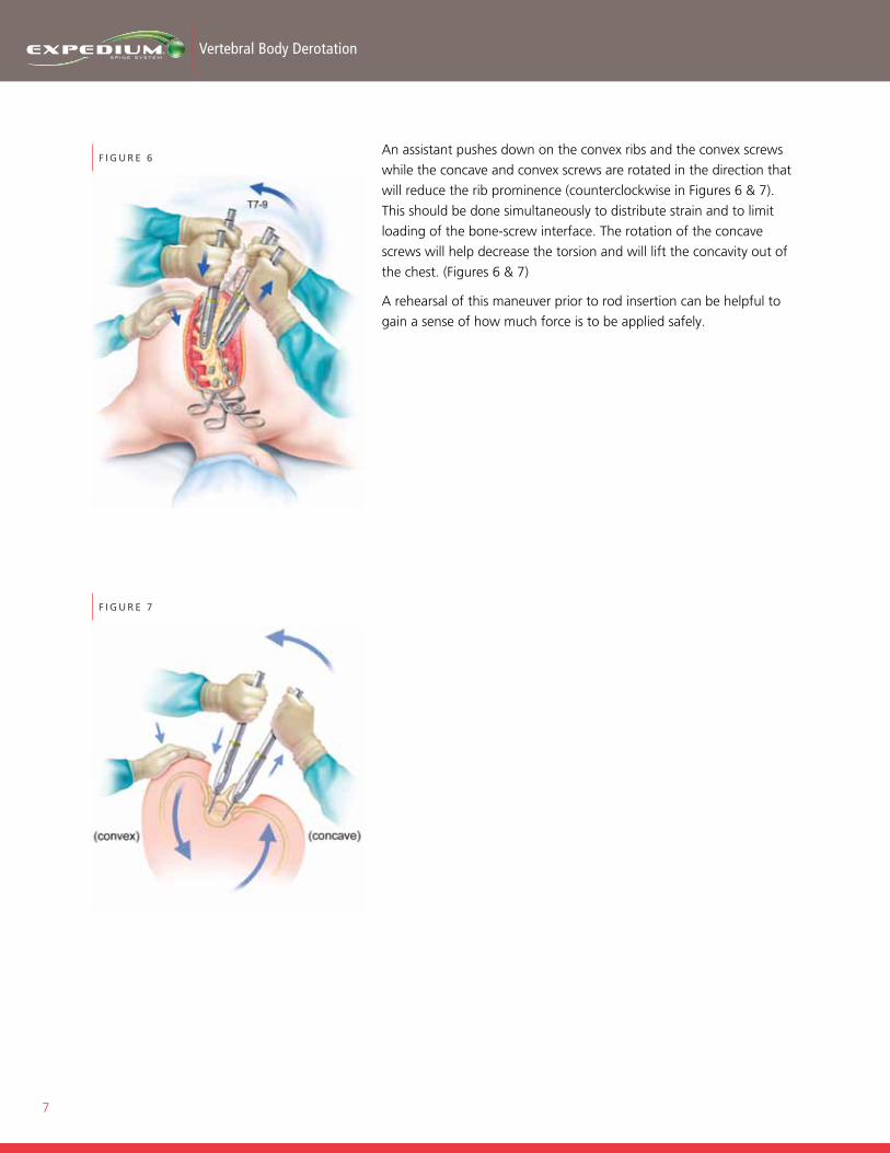
anassistantpushesdownontheconvexribsandtheconvexscrews
whiletheconcaveandconvexscrewsarerotatedinthedirectionthat
willreducetheribprominence(counterclockwiseinfigures6&7).
thisshouldbedonesimultaneouslytodistributestrainandtolimit
loadingofthebone-screwinterface.therotationoftheconcave
screwswillhelpdecreasethetorsionandwilllifttheconcavityoutof
thechest.(figures6&7)
arehearsalofthismaneuverpriortorodinsertioncanbehelpfulto
gainasenseofhowmuchforceistobeappliedsafely.
f i g u r e 6
f i g u r e 7

8
S U R G I C A L T E C H N I Q U E G U I D E
liftingtheconcavesideoutofthechestiseffectivelydonewiththe
quickStickslockedonthetoPnotchfeatureofeXPedIuMimplants.
thisfeatureprovides360°controloftheanchorpointwithoutthe
needforadditionalinstrumentation.
Inaddition,usingtheeXPedIuMderotationframeand/oralignment
forkmultiplequickStickscanbelinkedtogetherandrotatedin
unison.(figure8)
tightenthesetscrewsontheconcaverodholdingthisposition.
(figure9a)
Implanttheconvexrodandtightenthesetscrewsontheconvexside.
(figure9b)
f i g u r e 8
f i g u r e 9 a f i g u r e 9 B
n o t e :
Some surgeons believe that the VBd
maneuver with both rods in place minimizes
the loss of rotational correction that occurs
from spine “spring back” when inserting
and rotating one rod at a time. in this case,
follow the same steps described above but
with both rods already implanted.

9
segmental VerteBral BoDy Derotation (inDiViDual VerteBral leVel)
SegmentalVertebralbodyderotationcanbedoneasthesole
derotationmaneuverorinadditiontotheenblocmaneuver
describedbefore.
f i g u r e 1 0 a
f i g u r e 1 0 B
Implantbothrodsandcapturethemwiththesetscrews.Mostset
screwsshouldbeleftloosesincelengtheningofthespineisexpected
ateachlevelthatwillbesegmentallyderotated.onlythesetscrewsin
thedistalneutralvertebrashouldbetightened(e.g.,l1infigure10a).
attachtwoquickSticksinthedistalsegmenttolockthebottom
neutralvertebra(figures10a,b).thenattachquickSticksinthenext
proximal1-2vertebrae.thequickSticksonthedistalvertebramustbe
heldbyanassistanttoprovidecounter-rotationforce.

10
S U R G I C A L T E C H N I Q U E G U I D E
derotateeachproximalvertebralbodytoachieveaneutralposition
inreferencetotheneutraldistalvertebra(figure11a,b).after
derotationofeachsegment,thesetscrewsaretightened.repeatthis
process,movingalongtowardstheapex.
completeneutralderotationmaynotbeachievedattheapexrelative
toitstorsionintheaxialplaneduringthefirstpass.repeatingthe
derotationmaneuversatsomelevelscanbehelpfulduetoviscoelastic
relaxationofthespine.caremustbetakennottoloosenthebone-
screwinterfacewhileperformingthemaneuver.
f i g u r e 1 1 a
theeXPedIuMderotationquickSticks,facilitators,flex-clipwith
rotationtubeorVIPer2Screwextensions,arealleffectiveinthisstep
ofSegmentalderotation.(figure5a-d)
Inaddition,usingtheeXPedIuMderotationframe,quickSticksin
thesamevertebrallevelcanbelinkedtogetherandrotatedinunison.
(figure10b)
repeatthederotationforeachsegment,untilallvertebrallevelsnearly
matchtheneutrallyrotateddistalvertebra.
duringSegmentalSpinalderotation,segmentalcompression
(convexity)and/ordistraction(concavity)maybesimultaneously
appliedtoeffectmaximalcorrection,justbeforethesetscrews
aretightened.
f i g u r e 1 1 B

11
additionalcoronalcorrectioncanbeachievedwiththeuseof
eXPedIuMcoronalIn-situbenders.In-situcontouringismoreeasily
achievedwhenusingstandardSSrodsthanwhenusinghighand
ultraStrengthrods.(figure12)
f i g u r e 1 2

12
S U R G I C A L T E C H N I Q U E G U I D E
12
NOTES

13
IndIcatIonsthe EXPEdIUM spine system is intended to provide immobilization and stabilization of spinal segments in skeletally mature patients as an adjunct to fusion in the treatment of acute and chronic instabili-ties or deformities of the thoracic, lumbar and sacral spine.
the EXPEdIUM spine system is intended for noncervical pedicle fixation and nonpedicle fixation for the following indications: degenerative disc disease (defined as back pain of discogenic origin with de-generation of the disc confirmed by history and radiographic studies); spondylolisthesis; trauma (i.e., fracture or dislocation); spinal stenosis; curvatures (i.e., scoliosis, kyphosis, and/or lordosis); tumor, pseud-arthrosis; and failed previous fusion in skeletally mature patients.
contRaIndIcatIonsDisease conditions that have been shown to be safely and predictably managed without the use of internal fixation devices are relative contraindications to the use of these devices.
Active systemic infection or infection localized to the site of the proposed implantation are contraindications to implantation.
Severe osteoporosis is a relative contraindication because it may prevent adequate fixation of spinal anchors and thus preclude the use of this or any other spinal instrumentation system.
Any entity or condition that totally precludes the possibility of fusion, i.e., cancer, kidney dialysis, or osteopenia is a relative contraindication. Other relative contraindications include obesity, certain degenera-tive diseases, and foreign body sensitivity. In addition, the patient’s occupation or activity level or mental capacity may be relative contraindications to this surgery. Specifically, patients who because of their occupation or lifestyle, or because of conditions such as mental illness, alcoholism, or drug abuse, may place undue stresses on the implant during bony healing and may be at higher risk for implant failure.
WaRnInGs, PREcaUtIons, and PossIBLE adVERsE EFFEcts concERnInG tEMPoRaRY MEtaLLIc IntERnaL FIXatIon dEVIcEs
Following are specific warnings, precautions, and possible adverse effects that should be understood by the surgeon and explained to the patient. These warnings do not include all adverse effects that can occur with surgery in general, but are important considerations particular to metallic internal fixation devices. General surgical risks should be explained to the patient prior to surgery.
WaRnInGs
1. coRREct sELEctIon oF tHE IMPLant Is EXtREMELY IMPoRtant. The potential for satisfactory fixation is increased by the selection of the proper size, shape, and design of the implant. While proper selection can help minimize risks, the size and shape of human bones present limitations on the size, shape and strength of implants. Metallic internal fixation devices cannot withstand activity levels equal to those placed on normal healthy bone. No implant can be expected to withstand indefinitely the unsupported stress of full weight bearing.
2. IMPLants can BREaK WHEn sUBJEctEd to tHE IncREasEd LoadInG assocIatEd WItH dELaYEd UnIon oR nonUnIon. Internal fixation appliances are load-sharing devices which are used to obtain alignment until normal healing occurs. If healing is delayed, or does not occur, the implant may eventually break due to metal fatigue. The degree or success of union, loads produced by weight bearing, and activity levels will, among other conditions, dictate the longevity of the implant. Notches, scratches or bending of the implant during the course of surgery may also contribute to early failure. Patients should be fully informed of the risks of implant failure.
3. MIXInG MEtaLs can caUsE coRRosIon. There are many forms of corrosion damage and several of these occur on metals surgically implanted in humans. General or uniform corrosion is present on all implanted metals and alloys. The rate of corrosive attack on metal implant devices is usually very low due to the presence of passive surface films. Dissimilar metals in contact, such as titanium and stainless steel, accelerate the corrosion process of stainless steel and more rapid attack occurs. The presence of corrosion often accelerates fatigue fracture of implants. The amount of metal compounds released into the body system will also increase. Internal fixation devices, such as rods, hooks, etc., which come into contact with other metal objects, must be made from like or compatible metals.
4. PatIEnt sELEctIon. In selecting patients for internal fixation devices, the following factors can be of extreme importance to the eventual success of the procedure:
A. the patient’s weight. An overweight or obese patient can produce loads on the device that can lead to failure of the appliance and the operation.
B. the patient’s occupation or activity. If the patient is involved in an occupation or activity that includes heavy lifting, muscle strain, twisting, repetitive bending, stooping, running, substantial walking, or manual labor, he/she should not return to these activities until the bone is fully healed. Even with full healing, the patient may not be able to return to these activities successfully.
C. a condition of senility, mental illness, alcoholism, or drug abuse. These conditions, among others, may cause the patient to ignore certain necessary limitations and precautions in the use of the ap-pliance, leading to implant failure or other complications.
D. certain degenerative diseases. In some cases, the progression of degenerative disease may be so advanced at the time of implantation that it may substantially decrease the expected useful life of the appliance. For such cases, orthopaedic devices can only be considered a delaying technique or temporary remedy.
E. Foreign body sensitivity. The surgeon is advised that no preoperative test can completely exclude the possibility of sensitivity or allergic reaction. Patients can develop sensitivity or allergy after implants have been in the body for a period of time.
F. smoking. Patients who smoke have been observed to experience higher rates of pseudarthrosis following surgical procedures where bone graft is used. Additionally, smoking has been shown to cause diffuse degeneration of intervertebral discs. Progressive degeneration of adjacent segments caused by smoking can lead to late clinical failure (recurring pain) even after successful fusion and initial clinical improvement.
PREcaUtIons
1. sURGIcaL IMPLants MUst nEVER BE REUsEd. An explanted metal implant should never be reimplanted. Even though the device appears undamaged, it may have small defects and internal stress pat-terns which may lead to early breakage. Reuse can compromise device performance and patient safety. Reuse of single use devices can also cause cross-contamination leading to patient infection.
2. coRREct HandLInG oF tHE IMPLant Is EXtREMELY IMPoRtant. Contouring of metal implants should only be done with proper equipment. The operating surgeon should avoid any notching, scratching or reverse bending of the devices when contouring. Alterations will produce defects in surface finish and internal stresses which may become the focal point for eventual breakage of the implant. Bending of screws will significantly decrease the fatigue life and may cause failure.
3. consIdERatIons FoR REMoVaL oF tHE IMPLant aFtER HEaLInG. If the device is not removed after the completion of its intended use, any of the following complications may occur: (1) Corro-sion, with localized tissue reaction or pain; (2) Migration of implant position resulting in injury; (3) Risk of additional injury from postoperative trauma; (4) Bending, loosening, and/or breakage, which could make removal impractical or difficult; (5) Pain, discomfort, or abnormal sensations due to the presence of the device; (6) Possible increased risk of infection; and (7) Bone loss due to stress shielding. The surgeon should carefully weigh the risks versus benefits when deciding whether to remove the implant. Implant removal should be followed by adequate postoperative management to avoid refracture. If the patient is older and has a low activity level, the surgeon may choose not to remove the implant thus eliminating the risks involved with a second surgery
4. adEQUatELY InstRUct tHE PatIEnt. Postoperative care and the patient’s ability and willingness to follow instructions are among the most important aspects of successful bone healing. The patient must be made aware of the limitations of the implant, and instructed to limit and restrict physical activities, especially lifting and twisting motions and any type of sports participation. The patient should understand that a metallic implant is not as strong as normal healthy bone and could loosen, bend and/or break if excessive demands are placed on it, especially in the absence of complete bone healing. Im-plants displaced or damaged by improper activities may migrate and damage the nerves or blood vessels. An active, debilitated, or demented patient who cannot properly use weight-supporting devices may be particularly at risk during postoperative rehabilitation.
5. coRREct PLacEMEnt oF antERIoR sPInaL IMPLant. Due to the proximity of vascular and neurologic structures to the implantation site, there are risks of serious or fatal hemorrhage and risks of neurologic damage with the use of this product. Serious or fatal hemorrhage may occur if the great vessels are eroded or punctured during implantation or are subsequently damaged due to breakage of implants, migration of implants or if pulsatile erosion of the vessels occurs because of close apposition of the implants..

4.5 Spine System
5.5 Spine System
6.35 Spine System
Vertebral Body Derotation Set
SFX® Cross Connector System
Anterior Spine System
VIPER® 2 System
Limited Warranty and Disclaimer: DePuy Spine products are sold with a limited warranty to the original purchaser against defects in workmanship and materials. Any other express or implied warranties, including warranties of merchantability or fitness, are hereby disclaimed.
WARNING: In the USA, this product has labeling limitations. See package insert for complete information.
CAUTION: USA Law restricts these devices to sale by or on the order of a physician.
Not all products are currently available in all markets.
To order, call DePuy Spine Customer Service (1-800-227-6633).
©DePuy Spine, Inc. 2011. All rights reserved.
POD DF25-20-002 9/11 ADDB/AG
DePuy Spine, Inc. 325 Paramount Drive Raynham, MA 02767USATel: +1 (800) 227-6633
www.depuy.com