Surgical Removal of a Radicular Cyst in a Thirteen Year ... · PDF fileThumb forceps...
14
Surgical Removal of a Radicular Cyst in a Thirteen Year Old Border Collie Brett Beckman, DVM, FAVD, DAVDC, DAAPM Affiliated Veterinary Specialists, Orlando, Florida Florida Veterinary Dentistry and Oral Surgery, Punta Gorda, Florida Animal Emergency Center of Sandy Springs, Atlanta, GA www.veterinarydentistry.net [email protected] Introduction: Radicular cysts represent the most common type of oral cyst in humans(1,2) but have been reported infrequently in dogs and cats.(1,3) Radicular cysts are associated with inflammation of the periodontal ligament at the root apex whereas the more common dentigerous cyst is not.(4) Radicular cysts are commonly referred to as apical periodontal cysts(5). An additional periodontal cyst is described based on its location to the root and termed a lateral periodontal cyst.(6) The following is a case report describing the diagnosis and surgical excision of a large radicular cyst in a thirteen year old Border Collie. History: A thirteen year old twenty eight kilogram male neutered Border Collie presented for routine annual examination in May 2002. Dental cleaning had been performed routinely throughout the life of the patient. No other relevant history existed. Diagnostics: Physical examination of the patient was within normal limits. Oral examination revealed a gingivitis index of I, a calculus index of I, and a plaque index of I. A subtle swelling was present in the maxilla apical to the gingival margin adjacent to the maxillary left first (201) and second (202) incisors extending to the mucogingival line. (Figure 1) The swelling was firm and no pain was associated with palpation. No additional abnormalities were present. The patient was admitted for dental radiography of the swollen area and dental cleaning pending an appropriate pre-anesthetic evaluation.
Transcript of Surgical Removal of a Radicular Cyst in a Thirteen Year ... · PDF fileThumb forceps...

Surgical Removal of a Radicular Cyst in a Thirteen Year Old Border Collie
Brett Beckman, DVM, FAVD, DAVDC, DAAPM
Affiliated Veterinary Specialists, Orlando, Florida
Florida Veterinary Dentistry and Oral Surgery, Punta Gorda, Florida
Animal Emergency Center of Sandy Springs, Atlanta, GA
www.veterinarydentistry.net
Introduction:
Radicular cysts represent the most common type of oral cyst in humans(1,2) but have been
reported infrequently in dogs and cats.(1,3) Radicular cysts are associated with inflammation of
the periodontal ligament at the root apex whereas the more common dentigerous cyst is not.(4)
Radicular cysts are commonly referred to as apical periodontal cysts(5). An additional
periodontal cyst is described based on its location to the root and termed a lateral periodontal
cyst.(6) The following is a case report describing the diagnosis and surgical excision of a large
radicular cyst in a thirteen year old Border Collie.
History:
A thirteen year old twenty eight kilogram male neutered Border Collie presented for routine
annual examination in May 2002. Dental cleaning had been performed routinely throughout the
life of the patient. No other relevant history existed.
Diagnostics:
Physical examination of the patient was within normal limits. Oral examination revealed a
gingivitis index of I, a calculus index of I, and a plaque index of I. A subtle swelling was present
in the maxilla apical to the gingival margin adjacent to the maxillary left first (201) and second
(202) incisors extending to the mucogingival line. (Figure 1) The swelling was firm and no pain
was associated with palpation. No additional abnormalities were present. The patient was
admitted for dental radiography of the swollen area and dental cleaning pending an appropriate
pre-anesthetic evaluation.

Figure 1
A swelling was present in the maxilla apical to the gingival margin adjacent to teeth 201 and
202. Teeth 101, 102 and 202 show fractures in this view.
The differential diagnosis for this mass included abscess, cyst, neoplasia, fibrous bone
dysplasia and other fibrous bone lesions.(6) Abscess was least likely due to the absence of
discomfort upon palpation. Neoplasia was considered to be most likely due to the age of the
patient.
Pre-anesthetic blood testing included a complete blood count, serum chemistry profile,
urinalysis and EKG. EKGa analysis showed a normal sinus rhythm, normal complexes and a
heart rate of 120. CBC, serum chemistry profile and urinalysis were normal. Mucous membrane
color, capillary refill time, pulse and chest auscultation were all within normal limits.
Butorphanolb
(8 mg IM) was given thirty minutes pre-operatively and a twenty gauge
intravenous catheterc placed in the right cephalic vein. General anesthesia was induced with
ketamined (140 mg IV) and valium
e (5.6 mg IV). The patient was intubated with an 8.0 mm
cuffed endotracheal tubef and the cuff was gently inflated. The animal was maintained with
isofluraneg (2.0-2.5%) and oxygen (2.5 L/min) using a semi-closed anesthetic delivery system,
h
placed on a water circulated heating padi and leads for the EKG
a monitor positioned. A warmed

balanced electrolyte solutionj was administered at 10 ml/kg/hr. Temperature, respiration, pulse
and capillary refill time were regularly taken and recorded by a technician.
A complete oral examination was performed and abnormalities noted on the dental chart. In
addition to the changes discovered during the initial oral examination slight bleeding was present
upon probing the palatal aspect of 104. All pocket depths were within normal limits. A Class
VI, Stage 2 fracture (7) of the maxillary right first incisor (101) was present. The maxillary right
second (102) incisor, 201, and 202 had Class VI, Stage 1 (7) fractures. Slight mobility was
associated with 202. Owner inquiry revealed that the incisor fractures were likely induced by
Frisbee play when the patient was a young adult.
The oral cavity was thoroughly rinsed with 0.12 % chlorhexidinek solution. Complete
supragingival and subgingival scalingl was performed. The teeth were polished using a
disposable prophy anglem
on a slow speed handpiecen and polishing paste
o. The oral cavity was
rinsed thoroughly with saline followed again by 0.12 % chlorhexidinek solution.
Dental radiographsp were taken of the affected area. Radiography showed a large luceny
expanding from the maxillary bone in the mesial apical region of 201 coursing along the midline
of the rostral maxilla two centimeters in a caudal direction, rounding to course rostrally and
terminating at the distal aspect of the apex of 202.(Figure 2) A soft tissue density was present
within the silhouette of the lucency. This density was adjacent to the distal aspect of the root of
201, surrounding 202 and adjacent to the mesial aspect of the maxillary left third incisor (203).
The interradicular space between 201 and 202 was increased compared to the contralateral side.
The distal aspect of 201 and the mesial aspect of 202 showed evidence of external resorption.

Figure 2
A lucency is seen in the maxillary bone mesial to tooth 201 coursing along the midline and
terminating at the distal aspect of the apex of tooth 202. Root resorption is present on the distal
aspect of tooth 201 and the mesial aspect of tooth 202.
The above findings were relayed to the owner who gave permission for biopsy and probable
extraction of 202 to gain access to the center of the defect. Reentry and extraction of 201 and
203 pending biopsy findings were likely and discussed with the owner. The most likely causes
of this radiographic presentation were cyst, neoplasia or both.
The patient was placed in dorsal recumbency. A left infraorbital nerve block was performed
by injecting bupivicaineq (2.5 mg) using a tuberculin syringe and a 25 gauge 1 inch needle
r at the
entrance to the left infraorbital canal and holding digital pressure over the area for sixty seconds.
Aspiration to confirm that the needle was not placed intravascularly was performed prior to
injection. A 15 Bard Parker blades on a No. 7 scalpel handle
t was used to make two divergent
vertical releasing incisions starting at the line angles of 201 and the maxillary left third incisor
(203) extending one half centimeter beyond the mucogingival junction.(Figure 3) The releasing
incisions were connected by an interproximal incision incorporating the buccal sulcus of 202.
An EX-7u periosteal elevator was used to reflect the flap apically. The scalpel
blade
s was placed
in the sulcus of 202 and the periodontal attachment released around its circumference. Tooth
202 was extracted with the aid of a #3 Winged elevator.v

Figure 3
A mucoperiosteal flap with divergent releasing incisions aids in
exposure for extraction and biopsy
A small segment of bone was attached to the tooth. The tooth was partially surrounded by
granulation tissue, a fibrous tissue and a white caseous material. A cavity was present within the
maxilla that corresponded to the lucency seen on the radiographs. The elevator was used to
obtain samples of the granulation tissue, fibrous tissue and caseous material. These tissues were
sent along with 202 and adherent bone for histopathology. The edge of the gingival flap was
straightened with the scalpel blade and the periosteum incised to facilitate a tension free flap
closure. The gingiva was sutured in a simple interrupted pattern using Mayo Hagar needle
holdersw and 4-0 monocryl with a 3/8” NFS-2 cutting needle
x. Ketoprofen
y (55 mg IM) was
administered for pain control.
Post-Operative Care/Biopsy:
The patient was placed on a blanket in recovery and carefully monitored. At the first sign of
swallowing the endotracheal tubeg was deflated and removed. The patient was monitored until
sternal recumbency was achieved. Prior to discharge the IV catheterd was removed and a light
pressure bandage placed over the catheter site to aid in hemostasis. Carprofenz was dispensed

with instructions to give 100 mg every twenty four hours for three days starting twenty four
hours post-op.
Diagnosis:
Three days following discharge the owner was contacted by phone. A diagnosis of
epidermoid cyst of bone with pyogranulomatous inflammation was relayed to the owner. Due to
the proximity of the lesion to the incisors, the extent of the cavity in the maxilla, and the rare
nature of the lesion described a second opinion was recommended and accepted by the owner.
The conversation indicated that the patient showed no adverse effects from the procedure. Two
weeks later the histopathology results revealed a keratin rich radicular cyst with a foreign body
granulomatous inflammation associated with the release of keratin fragments.
Treatment Plan:
The owner was notified and surgery set for two weeks to remove the remainder of the cyst
and its components. Teeth 201 and 203 would be extracted due to significant alveolar bone
destruction.
Treatment:
The patient returned thirty three days following the original presentation for surgical removal
of the cyst and its components and extraction of 201 and 203. EKGa analysis showed a normal
sinus rhythm, normal complexes and a heart rate of 112. Mucous membrane color, capillary refill
time, pulse and chest auscultation were all within normal limits.
The induction and anesthetic protocol was identical to that of the previous surgery including a
right and left infraorbital nerve block as previously described. A 15 Bard Parker blades on a No.
7 scalpel handlet was used to raise a mucoperiosteal flap with two divergent vertical releasing
incisions starting at the mesial line angles of the maxillary left canine tooth (204) and the
maxillary right second incisor (102) extending one centimeter beyond the mucogingival junction.
The releasing incisions were connected by incising the junction of the palatal and gingival
mucosa. Visual inspection showed significant loss of alveolar bone surrounding the 201 and 203.
The blade was placed in the sulcus of 201 and 203 and the periodontal attachment released
around the circumference of each. Teeth 201 and 203 were extracted without difficulty using a
#3 Winged elevator.v
Visual examination showed no organized structure to the cyst lining which was fibrous in
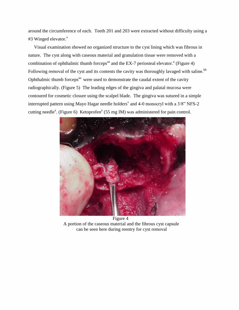
nature. The cyst along with caseous material and granulation tissue were removed with a
combination of ophthalmic thumb forcepsaa
and the EX-7 periosteal elevator.u (Figure 4)
Following removal of the cyst and its contents the cavity was thoroughly lavaged with saline.bb
Ophthalmic thumb forcepsaa
were used to demonstrate the caudal extent of the cavity
radiographically. (Figure 5) The leading edges of the gingiva and palatal mucosa were
contoured for cosmetic closure using the scalpel blade. The gingiva was sutured in a simple
interrupted pattern using Mayo Hagar needle holdersv and 4-0 monocryl with a 3/8” NFS-2
cutting needlex. (Figure 6) Ketoprofen
y (55 mg IM) was administered for pain control.
Figure 4
A portion of the caseous material and the fibrous cyst capsule
can be seen here during reentry for cyst removal

Figure 5
Thumb forceps demonstrate the caudal extent of the cavity post-op
Figure 6
The mucoperiosteal flap is closed with a simple interrupted
pattern using 4-0 Monocryl.

Post-Operative Care/Excision:
Recovery was uneventful and similar to that described with the original surgery. Carprofenz
was dispensed with instructions to give 100 mg every twenty four hours for three days starting
twenty four hours post-op.
Long Term Follow Up:
The owner was instructed to return in two weeks to examine the surgical site for healing and
to schedule a radiographic evaluation of the area to ensure complete removal in four months.
Two days after discharge the owner was contacted by phone and indicated that the patient was
doing well.
Prior to the two week recheck the owner called and cancelled citing the patient convalesced
without incident and the area looked normal with the exception of the sutures. Despite a
recommendation to keep the appointment the owner declined and acknowledged her intent to
keep the appointment for radiography four months post-op.
The patient returned four months post-op. The surgical site had healed completely. (Figure 7)
The patient showed no discomfort upon palpation of the site. No other abnormalities were noted.
Vitals and pre-anesthetic EKG were normal as in the prior anesthesia episodes. Butorphanolb
(8
mg IM) was given 25 minutes prior to sedation. Adequate sedation was achieved with ketamined
(140 mg IV) and valiume (5.6 mg IV). Radiography revealed increased bone density adjacent to
the previous post-surgical margins. A small region of lucency was present within the region of
new bone formation. (Figure 8) Although likely not of clinical significance the lucency should
be reevaluated radiographically in 6 months.

Figure 7
At six months post-op there is complete gingival healing and no
discomfort upon palpation.
Figure 8
Radiographic appearance at the six month follow up exam shows evidence of new
bone growth. A lucency is present within the bone.

Discussion:
The World Health Organization signifies radicular cyst, apical periodontal cyst and periapical
cyst as synonyms.(5) Another text further categorizes periapical cysts as periapical true cysts
and periapical pocket cysts both of inflammatory origin.(8) Periapical true cysts are enclosed
with epithelium with no communication with the root canal. Periapical pocket cysts have a
saclike epithelium with direct communication with the root canal. They cannot be differentiated
radiographically but response of each to conventional endodontics makes them clinically
relevant. A periapical pocket cyst is likely to heal following root canal therapy due to its
communication with the endodontic system.(9) This is not the case with the true cyst which fails
to resolve radiographically following root canal therapy.
Periapical or radicular cysts form as a result of chronic apical periodontitis which results from
microbial invasion of the pulp cavity.(8) Apical periodontitis is generally described as the
body’s reaction to the destruction of the pulp and microbial occupation of the root canal. The
various lesions that develop in response to this interaction of the host defense system and the
invading microbes include the radicular cysts. It is likely that the cyst described in this case
report originated with the fracture of either 201 or 202 and a resultant chronic apical
periodontitis. Not every incident of chronic apical periodontitis results in the formation of a cyst
in humans. A recent study shows that the incidence (n = 256) approximates 15%.(9) This figure
includes both true cysts and pocket cysts.
In approaching oral swellings it is important to determine the nature of the lesion
microscopically. The decision to biopsy this lesion and remove the mobile incisor was made
based upon several factors. The large area visualized radiographically involving both a lucency
and soft tissue could not definitively be deemed a cyst. Inspection of the region following
excision of the mobile tooth for better visualization revealed a mixture of fibrous, granulomatous
and caseous material not recognized in this author’s previous experience with maxillary or
mandibular cysts. Definitive recognition of the process through biopsy may be desirable prior to
attempting an involved procedure in this geriatric patient. If prognosis for removal based upon
microscopic examination was less than ideal the decision not to attempt reentry may have been
in the best interest of the patient. A decision for an attempt at total excision could be supported,
however had the surgeon’s experience revealed similar findings in past experience.
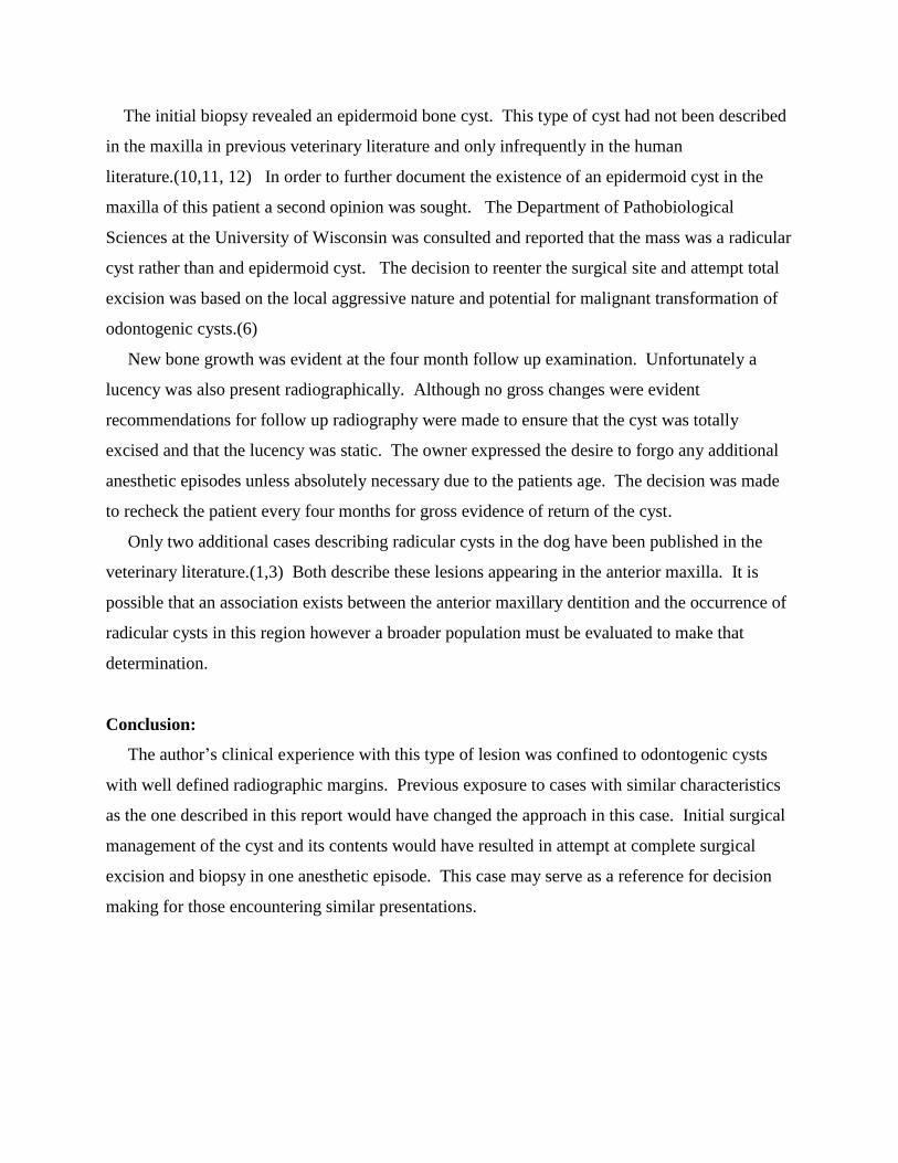
The initial biopsy revealed an epidermoid bone cyst. This type of cyst had not been described
in the maxilla in previous veterinary literature and only infrequently in the human
literature.(10,11, 12) In order to further document the existence of an epidermoid cyst in the
maxilla of this patient a second opinion was sought. The Department of Pathobiological
Sciences at the University of Wisconsin was consulted and reported that the mass was a radicular
cyst rather than and epidermoid cyst. The decision to reenter the surgical site and attempt total
excision was based on the local aggressive nature and potential for malignant transformation of
odontogenic cysts.(6)
New bone growth was evident at the four month follow up examination. Unfortunately a
lucency was also present radiographically. Although no gross changes were evident
recommendations for follow up radiography were made to ensure that the cyst was totally
excised and that the lucency was static. The owner expressed the desire to forgo any additional
anesthetic episodes unless absolutely necessary due to the patients age. The decision was made
to recheck the patient every four months for gross evidence of return of the cyst.
Only two additional cases describing radicular cysts in the dog have been published in the
veterinary literature.(1,3) Both describe these lesions appearing in the anterior maxilla. It is
possible that an association exists between the anterior maxillary dentition and the occurrence of
radicular cysts in this region however a broader population must be evaluated to make that
determination.
Conclusion:
The author’s clinical experience with this type of lesion was confined to odontogenic cysts
with well defined radiographic margins. Previous exposure to cases with similar characteristics
as the one described in this report would have changed the approach in this case. Initial surgical
management of the cyst and its contents would have resulted in attempt at complete surgical
excision and biopsy in one anesthetic episode. This case may serve as a reference for decision
making for those encountering similar presentations.

Products:
a) EKG analyzer, Vetronics, Lafayette, IN
b) Torbugesic, Fort Dodge Animal Health, Fort Dodge, IA
c) Surflo intravenous catheter, Terumo Medical Corp, Elkton, MD
d) Ketaset, Fort Dodge Animal Health, Fort Dodge, IA
e) Valium, Abbott Laboratories, N Chicago, IL
f) 5-0 Endotracheal Tube, Rusch,
g) IsoFlo, Abbott Laboratories, N Chicago, IL
h) VMS Anesthesia Machine, Matrix Medical, Inc., Orchard Park, NY
i) T Pump, Gaymar Industries, Orchard Park, NY
j) Lactated Ringer’s solution, Abbott Labs, N Chicago, IL
k) Chlorhexidine, First Priority, Elgin, IL
l) Neosonic, Amdent, Cherry Hill, NJ
m) Disposable prophy angle, Carlile Labs, Rockwell Centre, NY
n) High Speed Delivery System, Beaverstate Dental,
o) Prophy 1 Paste, Carlile Labs, Rockville Centre, NY
p) DentX Image Vet X70, AFP Imaging, Elmsford, NY
q) Bupivicaine, Abbott Laboratories, N Chicago, IL
r) Tuberculin syringe and needle, Nipro Mecical Corp, Miami, FL
s) No. 15 surgical blade, Carlile Labs, Rockville Centre, NY
t) Scalpel handle, Spectrum, Stow, OH
u) EX-7 periosteal elevator, Cislak Manufacturing Inc., Glenview, IL
v) #2 Winged elevator, Dentalaire, Fountain Valley, CA
w) Mayo-Hagar needle holders, Spectrum, Stow, OH
x) 4-0 Monocryl, Ethicon, Inc. Somerville, NJ
y) Ketofen, Fort Dodge Animal Health, Fort Dodge, IA
z) Rimadyl, Pfizer, New York, NY
aa) Ophthalmic Thumb forceps, Spectrum, Stow, OH
bb) 0.9% saline solution, Abbott Labs, N Chicago, IL
References:
1. French SL, Anthony JMG. Surgical Removal of a Radicular Odontogenic Cyst in a Four
year Old Dalmatian Dog. J Vet Dent 13(4);149-151 (1996)
2. Mosqueda-Taylor A, et al. Odontogenic cysts. Analysis of 856 cases. Med Oral Mar-
Apr;7(2):89-96 (2002)
3. Reiter AM. Periapical Cyst Formation of the Left Maxillary Third Incisor Tooth in an
Adult Standard Poodle. Presentation Proceedings from 15th
Annual Dental Forum, 149-
150 (2001)
4. Pavelic B, Plancak D, Peric B. Histologic difference in epithelial linings between
follicular and radicular cysts. Coll Antropol Dec;22 Suppl:167-71 (1998)
5. World Heath Organization: Application of the international classification of the diseases
to dentistry and stomatology. ed3, Geneva, 1995, The Organization.
6. Anderson JG, Harvey CE. Odontogenic Cysts. J Vet Dent 10(4):5-9 (1993)
7. Wiggs RB, Lobprise HB. Veterinary Dentistry Principles and Practice. Philadelphia,
Lippincott, 352-355 (1997)
8. Cohen S, Burns RC. Pathobiology of the Periapex from: Pathways of the Pulp St Louis,
Mosby (2002)
9. Nair PNR, Pajarola G, Schroeder HE. Types and incidence of human periapial lesions
obtained with extracted teeth, Oral Surg Oral Med Oral Paholol 81:93 (1996)
10. Werner A, et al. A case of epidermal cyst of the maxilla. Rev Stomatol Chir Maxillofac
79(5):407-10 (1978)
11. White AK, Jenkins HA, Coker NJ. Intradiploic epidermoid cyst of the sphenoid wing.
Arch Otolaryngol Head Neck Surg 1987 Sep;113(9):995-9.
12. Barrios JM, Garcia J, Moreno E. Malignant epidermoid cyst of the maxilla, An
Otorrinolaringol Ibero Am 10(3):281-6 (1983)