SURGICAL LITERATURE COMPENDIUM - ACell
20
SURGICAL LITERATURE COMPENDIUM Review of Selected Clinical and Pre-Clinical Research
Transcript of SURGICAL LITERATURE COMPENDIUM - ACell

SURGICAL LITERATURE COMPENDIUM
Review of Selected Clinical and Pre-Clinical Research

4

ACell’s proprietary platform technology - MatriStem UBM™ (Urinary Bladder Matrix) technology - is an extracellular matrix (ECM) derived from porcine urinary bladder and differentiated from other ECM products by its intact acellular epithelial basement membrane. MatriStem UBM technology is the only commercially available form of UBM, and is utilized in the manufacturing of the Gentrix® Surgical Matrix portfolio of devices.
UBM has been found to facilitate a host tissue remodeling response by the body that leads to the formation of biomechanically functional, site-appropriate tissue, as demonstrated in a pre-clinical animal model1,2*. As a result, ACell’s surgical devices are often used in complex cases.
UBM has a considerable breadth of research supporting its value in surgical settings. The extensive body of research includes more than 100 pre-clinical and 50 clinical peer-reviewed articles. Several of the most relevant publications in the area of surgical soft tissue reinforcement are summarized in this compendium.
SURGICAL LITERATURE COMPENDIUM
Review of Selected Clinical and Pre-Clinical Research
The publications presented in this compendium contain the opinions of and personal techniques practiced by the treating physician(s). The techniques presented herein are for informational purposes only. The decision of which techniques to use in a particular clinical application lies with the treating physician(s) based on patient profile, particular circumstances surrounding the procedure, and previous clinical experiences.
Cases presented involving Veterans Administration facilities or physicians do not reflect the opinion of the United States military or Veterans Affairs office.
The authors of certain publications presented in this compendium may make claims that are not made by ACell or its representatives. In these publications, the underlying use of the ACell surgical devices falls within the current indications for use of these devices, and therefore these publications are summarized within this compendium.
1. Young DA#, Jackson N, Ronaghan CA¶, Brathwaite CE‡, Gilbert TW§. Retrorectus repair of incisional ventral hernia with urinary bladder matrix reinforcement in a long-term porcine model. Regenerative Medicine. 2018; doi:10.2217/rme-2018-0023.
2. Young DA#, McGilvray KC, Ehrhart N, Gilbert TW§. Comparison of in vivo remodeling of urinary bladder matrix and acellular dermal matrix in an ovine model. Regenerative Medicine. 2018; doi: 10.2217/rme-2018-0091.
* Pre-clinical data may not reflect clinical results.
# ACell Employee | ¶ Consultant | § Former ACell Employee | ‡ Former Consultant | ▲ ACell Sponsored Research Agreement

6
Macrophage Phenotype TestingMacrophage phenotype cell staining at 14 days showed Group 1 ECMs to have a predominantly M1 phenotype, Group 2 to have a mix of M1 and M2 phenotypes, and Group 3 to have a mix of M1 and M2 phenotypes, but with an increased M2 phenotype at the periphery of the remodeling site. The study found a strong, statistically significant correlation between the histologic scores and macrophage phenotype (both the number of M2 cells and the ratio of M2:M1 cells) at 14 days, suggesting that early phenotype distribution stands to be a predictor for long-term remodeling outcomes.

Clinical Research
Long-term clinical, radiological, and histological follow-up after complex ventral incisional hernia repair using urinary bladder matrix graft reinforcement: a retrospective cohort study.
Sasse¶ et al. Page 1
Paraesophageal hiatal hernia repair with urinary bladder matrix graft. Howell et al. Page 2
Different biologic grafts for diaphragmatic crura reinforcement during laparoscopic repair of large hiatal hernia: a 6-year single surgeon experience.
Reznichenko Page 3
Parastomal hernia repair with urinary bladder matrix grafts: case series with 2-year follow-up and discussion.
Sasse¶ et al. Page 3
Laparoscopic rectopexy with urinary bladder xenograft reinforcement. Mehta et al. Page 4
Esophageal reinforcement with an extracellular scaffold during total gastrectomy for gastric cancer.
Afaneh et al. Page 4
Endoscopic deployment of MatriStem for treatment of a colorectal anastomotic leak. Iorio et al. Page 5
Use of scaffolding tissue biografts to bolster vesicourethral anastomosis during salvage robot-assisted prostatectomy reduces leak rates and catheter times.
Ogaya-Pinies et al. Page 6
Pre-Clinical ResearchComparison of in vivo remodeling of urinary bladder matrix and acellular dermal matrix in an ovine model.
Young# et al. Page 7
Retrorectus repair of incisional ventral hernia with urinary bladder matrix reinforcement in a long-term porcine model.
Young# et al. Page 8
Urinary bladder matrix scaffolds strengthen esophageal hiatus repair. Riganti‡ et al. Page 9, 10
Biomechanical features of reinforced esophageal hiatus repair in a porcine model Amigo et al. Page 11
Mechanical strength vs. degradation of a biologically-derived surgical mesh over time in a rodent full-thickness abdominal wall defect.
Costa et al. Page 12
Macrophage phenotype as a predictor of constructive remodeling following the implantation of biologically derived surgical mesh materials.
Brown et al. Page 13
TABLE OF CONTENTS

1
This retrospective study assessed the long-term outcomes of complex ventral incisional hernia repair using UBM graft reinforcement in patients at a single center over a five year period.
A total of 64 patients were identified that had undergone complex ventral incisional hernia repairs that were reinforced with a UBM graft (Gentrix Surgical Matrix Thick). All of the patients were classified as either moderate or major (severe) cases according to the patient severity classification published by Slater et al. Approximately 59% of the patients had a previously failed repair and 66% had concomitant procedures including bowel resection, evacuation of infection, cholecystectomy, or panniculectomy. The average BMI was 33.0 and 25% of patients had an existing stoma present.
The median time to follow up was 36 months (12-70 months). A total of nine patients required surgical repair in the cohort while a tenth patient had the recurrence managed non-surgically (15.6% recurrence rate). Median time to recurrence was 32 months. A Kaplan-Meier analysis of the data predicted a 4% recurrence rate at 24 months. The rate of seroma was 19%. There were no signs of graft infection, fistulization, or any need for graft explantation. Forty-five patients completed a Carolina Comfort Scale survey to assess quality of life after repair of their hernia with Gentrix reinforcement, with a median score of 16 out of a possible 115.
In 28 patients, abdominal ultrasound or CT scan revealed an intact fascia of the abdominal wall at least 24 months post-surgery. Three patients underwent re-exploration for unrelated conditions, and biopsies of the repaired fascia (retrorectus and intraperitoneal Gentrix placement) were histologically analyzed. In all cases, a remodeling response characterized by cell infiltration and absence of inflammatory response was observed.
The use of Gentrix reinforcement following complex ventral incisional hernia repair showed positive patient outcomes, with a low recurrence rate and minimal complications. Uniquely, this study also included histological analysis at extended time points, highlighting the functional healing response and site-appropriate remodeling of Gentrix devices.
CLINICAL RESEARCH
Sasse KC¶, Lambin JH, Gevorkian J, Elliott C, Afshar R, Gardner A, Mehta A, Lambin R, Peraza L. Hernia. 2018; doi: 10.1007/s10029-018-1830-0. Distributed under the Creative Commons Attribution 4.0 International License, http://creativecommons.org/licenses/by/4.0/.
Long-term clinical, radiological, and histological follow-up after complex ventral incisional hernia repair using urinary bladder matrix graft reinforcement: a retrospective cohort study.
H&E stains at 14 months post-retrorectus Gentrix placement (patient 1), at three years post-intraperitoneal repair with Gentrix (patient 2) and at 32 months post-retrorectus Gentrix repair (patient 3) showed incorporation of the graft and a remodeling response at the interface between the host and the graft.
Patient 1
Patient 2
Patient 3
2
The goal of this retrospective study was to evaluate the short-term outcomes of using Gentrix devices to reinforce paraesophageal hiatal hernia repairs.
The study evaluated all patients from a single center that underwent hiatal hernia repairs over a five-year period. Excluding patients that were undergoing concomitant bariatric procedures, a total of 56 patients had hernia repairs reinforced with Gentrix Surgical Matrix or Gentrix Surgical Matrix Plus (23 of the devices were cut by the physicians into a keyhole shape, 14 were cut into a U-shape, and 19 were unspecified). Another 65 patients were treated with a primary repair (without any device reinforcement). All procedures were performed either laparoscopically or robotically. Post-operative complications in patients that received Gentrix reinforcement versus patients treated with primary repair only were classified according to the Clavien-Dindo grading system.
Patient demographics were similar between the two cohorts, with the exception of age which was significantly higher in the Gentrix group. The length of stay of patients at the hospital and the 30-day readmission rate were not significantly different between patients with or without Gentrix reinforcement. The rate of post-operative complications was also similar between Gentrix-reinforced and non-reinforced (suture only) patients.
This study highlighted the safe use of Gentrix devices in laparoscopic and robotic repairs of paraesophageal hiatal hernias. While limited by the retrospective nature and limited follow-up time in this study, these results show that utilizing Gentrix devices to reinforce hiatal hernia repairs did not increase the risk of complications in a sensitive anatomy.
Howell RS, Fazzari M, Petrone P, Barkan A, Hall K, Servide MJ, Anduaga MF, Brathwaite CEM‡. Journal of the Society of Laparoendoscopic Surgeons. 2018; 22(2). doi: 10.4293/JSLS.2017.00100.
Paraesophageal hiatal hernia repair with urinary bladder matrix graft*.
* Study examined reinforcements using Gentrix Surgical Matrix and Gentrix Surgical Matrix Plus devices which were cut by surgeons into a variety of shapes at the individual surgeon's discretion. Gentrix Surgical devices do not have 510(k) clearance for a keyhole design for hiatal hernia repair.

3
CLINICAL RESEARCH (Continued)
The single-surgeon retrospective study reviewed eleven patients who underwent laparoscopic repair of large hiatal hernia in a rural community hospital using a standardized laparoscopic technique. Six patients had the reinforcement of the crura with AlloMax™ (BD Bard), one patient with Permacol™ (Medtronic), and four patients with Gentrix Surgical Matrix.
It was noted that the Gentrix Surgical Matrix devices were cut into a U-shape and secured with absorbable tacks, which were less traumatic than the titanium tacks required for use with the other grafts.
Reznichenko A. Journal of Medical Implants and Surgery. 2015;1(1).
Different biologic grafts for diaphragmatic crura reinforcement during laparoscopic repair of large hiatal hernia: a 6-year single surgeon experience*.
* Study examined reinforcements using Gentrix Surgical Matrix and Gentrix Surgical Matrix Plus devices which were cut by surgeons into a variety of shapes at the individual surgeon's discretion. Gentrix Surgical devices do not have 510(k) clearance for a keyhole design for hiatal hernia repair.
In this retrospective case series, the authors presented eight cases of parastomal hernia reinforcement with Gentrix Surgical Matrix grafts. Patients were followed for an average of 23 months.
Three of the cases were performed laparoscopically, with intraperitoneal placement of a keyhole-shaped Gentrix device. Five cases were performed through open laparotomy: one case utilized component separation and placement of the Gentrix Surgical Matrix device in the retrorectus plane; one repair utilized primary fascial closure reinforced with Gentrix Surgical Matrix Thick; and in the remaining three cases, the Gentrix Surgical Matrix device was placed in the intraperitoneal position.
All repairs had positive patient outcomes and were deemed successful, with only one late stricture requiring surgical revision six months post-op. At a median 23 months of follow-up, all patients showed intact repairs.
Sasse KC¶, Warner DL, Ackerman E, Brandt J. International Journal of Case Reports and Images. 2016; 7(2):85-91.
Parastomal hernia repair with urinary bladder matrix grafts: case series with 2-year follow-up and discussion*.
* Study examined reinforcements using Gentrix Surgical Matrix devices which were cut by surgeons into a variety of shapes at the individual surgeon's discretion.

4
This retrospective study conducted at Memorial Sloan Kettering Cancer Center compared the incidence of esophagojejunal junction (EJ) anastomotic leaks and strictures in total gastrectomy (TG) patients whose anastomoses were reinforced with Gentrix Surgical Matrix Thin, versus consecutive control patients identified from a historical database of TG surgeries at the sponsoring center who had primary EJ anastomoses without ECM reinforcement. The incidence of EJ leaks and strictures in TG cases where Gentrix was used was compared to outcomes where only primary anastomosis was performed.
The study showed a trend for reducing the incidence of EJ anastomotic leaks and strictures following TG when Gentrix was used to reinforce the anastomosis. Surgical implantation of the Gentrix device was straightforward and did not impede operative time.
EJ anastomotic leaks occurred in 3% (1/37) of Gentrix patients as compared to 12% (4/33) of the control patients, equaling a 77.5% reduction rate. Strictures occurred in 8% (n = 3) of Gentrix patients as compared to 15% (n = 5) of control patients.
The authors concluded that the 40% reduction in stricture rate and 77.5% reduction in EJ anastomotic leaks suggests that Gentrix can be helpful in reducing the incidence of complications following TG.
Afaneh C, Abelson J, Schattner M, Janjigian YY, Ilson D, Yoon SS, Strong VE. Annals of Surgical Oncology. 2015 Apr; 22 (4):1252-7.
Esophageal reinforcement with an extracellular scaffold during total gastrectomy for gastric cancer.
This retrospective study included 20 patients with severe, circumferential, full-thickness Grade V rectal prolapse per the Oxford rectal prolapse grading system. Gentrix Surgical Matrix devices were implanted via laparoscopic rectopexy repair in all patients with follow-up office examinations out to an average of 36 months post-procedure. The authors utilized a presacral placement of the graft and noted that the handling characteristics of the Gentrix material served as an attractive candidate material for anterior and posterior rectopexy repairs. During follow-up, no patients required further surgery for repair of the prolapse and zero recurrences were noted. Two patients did have small mucosal prolapse on physical examination that did not require surgery, and one patient developed an early postoperative pelvic fluid collection, which required laparoscopic drainage without graft explanation. There were no incidences of mesh erosion, mesh infection, or pain in the patients reinforced with UBM. Seventeen patients completed a survey on incontinence and constipation following the repair, with more than half of patients reporting improvement or no incidences of either. This survey also provided an average Fecal Incontinence Severity Instrument (FISI) score of eight (including eight patients who reported a score of zero), which indicated generally good bowel control amongst the patient population. Overall, the cases presented in this series showed that a laparoscopic rectopexy with Gentrix Surgical Matrix reinforcement provided durable repair without complications potentially associated with the use of a synthetic mesh.
Mehta A, Afshar R, Warner DL, Gardner A, Ackerman E, Brandt J, Sasse KC¶. JSLS. 2017; 21(1). doi: 10.4293/JSLS.2016.00106.
Laparoscopic rectopexy with urinary bladder xenograft reinforcement.
5
CLINICAL RESEARCH (Continued)
This case report describes the successful closure of a large anastomotic leak when reinforced with a Gentrix Surgical Matrix device. The patient was a 57-year-old obese male who developed an anastomotic leak five days following the reversal of a Hartmann procedure. The anastomotic leak was initially treated with pelvic drainage, a diverting ileostomy, and intravenous antibiotics before the patient underwent an outpatient procedure to close the fistula. Endoscopically, a 1.5 cm defect was identified along the linear staple line on the rectum to which a Gentrix Surgical Matrix device secured utilizing an endoscopic clip. The patient reported no complications from the procedure and healing of the anastomotic leak was confirmed via computed tomography scan at four weeks following Gentrix application. A colonoscopy confirmed that the anastomotic leak was filled with newly deposited host tissue with gross evidence of vascularization and no sign of scarring or stricture. At two months following the procedure, the patient reported normal bowel function and continence.
Iorio T, Blumberg, D. CRSLS. 2015; 19. doi: 10.4293/CRSLS.2014.00080.
Endoscopic deployment of MatriStem for treatment of a colorectal anastomotic leak.

6
In this retrospective comparative study, authors examined Gentrix Surgical Matrix Thin in the reinforcement of vesicourethral anastomoses (VUA) for robot-assisted radical prostatectomies (RARP). Outcomes from two groups of patients undergoing salvage robot-assisted radical prostatectomies (sRARP) were compared with a median follow-up of twelve months. Group 1 consisted of 15 patients who had their VUA reinforced with Gentrix while Group 2 had 45 patients who had a sRARP but no reinforcement graft reinforcement of their VUA. The authors found that the Gentrix group (Group 1) significantly reduced anastomotic disruptions in patients by 84% and median catheterization time by 6.4 days.
Ogaya-Pinies G, Kadakia Y, Palayapalayam-Ganapathi H, Woodlief T, Jenson C, Syed J, Patel V. EurUrol. 2018; 74(1):92-98. doi: 10.1016/j.eururo.2016.10.004.
Use of scaffolding tissue biografts to bolster vesicourethral anastomosis during salvage robot-assisted prostatectomy reduces leak rates and catheter times.
7
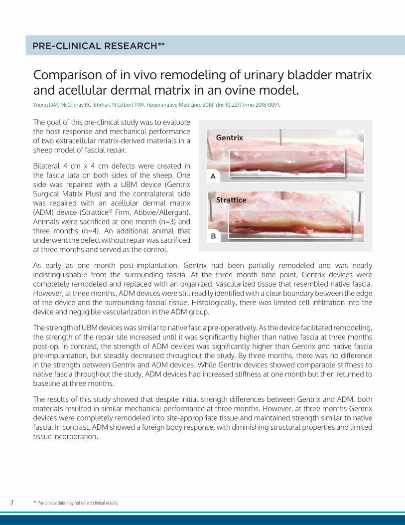
The goal of this pre-clinical study was to evaluate the host response and mechanical performance of two extracellular matrix-derived materials in a sheep model of fascial repair.
Bilateral 4 cm x 4 cm defects were created in the fascia lata on both sides of the sheep. One side was repaired with a UBM device (Gentrix Surgical Matrix Plus) and the contralateral side was repaired with an acellular dermal matrix (ADM) device (Strattice® Firm, Abbvie/Allergan). Animals were sacrificed at one month (n=3) and three months (n=4). An additional animal that underwent the defect without repair was sacrificed at three months and served as the control.
As early as one month post-implantation, Gentrix had been partially remodeled and was nearly indistinguishable from the surrounding fascia. At the three month time point, Gentrix devices were completely remodeled and replaced with an organized, vascularized tissue that resembled native fascia. However, at three months, ADM devices were still readily identified with a clear boundary between the edge of the device and the surrounding fascial tissue. Histologically, there was limited cell infiltration into the device and negligible vascularization in the ADM group.
The strength of UBM devices was similar to native fascia pre-operatively. As the device facilitated remodeling, the strength of the repair site increased until it was significantly higher than native fascia at three months post-op. In contrast, the strength of ADM devices was significantly higher than Gentrix and native fascia pre-implantation, but steadily decreased throughout the study. By three months, there was no difference in the strength between Gentrix and ADM devices. While Gentrix devices showed comparable stiffness to native fascia throughout the study, ADM devices had increased stiffness at one month but then returned to baseline at three months.
The results of this study showed that despite initial strength differences between Gentrix and ADM, both materials resulted in similar mechanical performance at three months. However, at three months Gentrix devices were completely remodeled into site-appropriate tissue and maintained strength similar to native fascia. In contrast, ADM showed a foreign body response, with diminishing structural properties and limited tissue incorporation.
Young DA#, McGilvray KC, Ehrhart N Gilbert TW§. Regenerative Medicine. 2018; doi: 10.2217/rme-2018-0091.
Comparison of in vivo remodeling of urinary bladder matrix and acellular dermal matrix in an ovine model.
PRE-CLINICAL RESEARCH**
** Pre-clinical data may not reflect clinical results.

8
This pre-clinical study evaluated the mechanical and tissue remodeling characteristics of the abdominal wall following reinforcement with UBM in a large animal ventral hernia model.
An acute midline abdominal defect model was utilized in adult female Yucatan minipigs with a classic Rives-Stoppa-Wantz retromuscular approach to simulate a hernia repair. The repairs were reinforced with either Gentrix Surgical Matrix Plus or Gentrix Surgical Matrix Thick, or left as a primary suture repair control without reinforcement. Animals were then recovered and survived for time points of three or eight months, with four animals per treatment group per time point.
Eight months post-op, animals reinforced with Gentrix devices showed remodeling of site-appropriate, biomechanically functional tissue at the defect site. Reinforcement of ventral incisional hernias with UBM-derived surgical devices prevented hernia recurrence throughout the course of observation, compared with 50% recurrence in non-reinforced controls. Furthermore, all UBM devices showed full remodeling at eight months, evidenced by the deposition of vascularized tissue mimicking that of natural, uninjured posterior fascia. This resulted in the strength of the UBM-reinforced abdominal wall being similar to that of uninjured tissue, suggesting that it was the healthy, remodeled host tissue – not the reinforcement device – that was supporting the mechanical load of the abdominal wall.
Study results provide evidence that Gentrix Surgical Matrix devices are capable of reinforcing ventral hernia repairs by facilitating site-appropriate tissue remodeling of biomechanically functional tissue that corresponds with the optimal strength of the abdominal wall pre-injury.
Young DA#, Jackson N, Ronaghan CA¶, Brathwaite CE‡, Gilbert TW§. Regenerative Medicine. 2018; doi.org/10.2217/rme-2018-0023.
Retrorectus repair of incisional ventral hernia with urinary bladder matrix reinforcement in a long-term porcine model.

9
This pre-clinical study evaluated the efficacy of Gentrix Surgical Matrix for reinforcing laparoscopic repair of surgically created hiatal hernias in an experimental porcine model.
Fourteen female domestic pigs underwent laparoscopic primary hiatal hernia repair of a simulated defect in the esophageal hiatus. Six of the hiatal repairs were reinforced with Gentrix, while the seven remaining cases served as primary repair controls. Gross morphology, histologic evaluation, and biomechanical results were evaluated.
Gross morphology at eight weeks after surgery showed animals reinforced with Gentrix Surgical Matrix (n = 7) had a robust crural repair with a smooth peritoneal-like structure, and no signs of graft erosion, esophageal fibrosis, or intrusion. The remodeled Gentrix appeared fully integrated with the host tissue and could only be identified with the securing tacks.
Histological evaluation showed that the Gentrix devices were fully integrated with surrounding native tissue and had facilitated the deposition of site-appropriate host tissue. The body of the hiatoplasty showed organized collagen fibers and neovascularization, with few signs of fibrosis. The non-reinforced control group showed thinner tissue at the site of repair, including disorganized loose connective tissue and other cellular infiltrates suggestive of scar tissue.
Biomechanical results for Gentrix showed average load to failure to be significantly stronger than the non-reinforced control group. When repaired tissues were explanted and tested for uniaxial tensile strength, samples taken from animals in the Gentrix reinforcement group withstood greater mechanical loads and had greater stiffness compared to samples taken from non-reinforced control animals.
Study results provide evidence that when used to reinforce a hiatal hernia repair, Gentrix Surgical Matrix facilitates a functional healing response that may improve the likelihood of positive outcomes. The Gentrix- reinforced group showed vascularized connective tissue within eight weeks, compared to the mechanically weaker, fibrotic tissue present at the defect site in the non-reinforced group. Interestingly, the remodeled tissue at the defect site was still mechanically stronger than the non-reinforced primary repair after Gentrix was no longer macroscopically distinguishable from host tissue.
Riganti JM‡, Ciotola F, Amenabar A, Craiem D, Graf S, Badaloni A, Gilbert TW§, Nieponice A‡. Journal of Surgical Research. 2016; 204:344-50.
Urinary bladder matrix scaffolds strengthen esophageal hiatus repair.
PRE-CLINICAL RESEARCH (Continued)

10
Host tissue remodeling response in representative histologic samples from Gentrix Surgical Matrix treated and control animals is compared at eight weeks. Hematoxylin and eosin stained histology samples at 40x and Masson’s trichrome stained samples at 200x are shown, respectively, for a treated animal (A and B) and control animal (C and D). The hiatoplasty in the treated animal shows an abundance of well organized collagen fibers and no signs of fibrosis. The control sample has clear gaps in tissue structure (C) and disorganized, loose connective tissue and a strong mononuclear infiltrate (D) indicative of fibrosis and scar tissue formation.
A B
C D
Results of hiatal closure at eight weeks: A) remodeled Gentrix Surgical Matrix-reinforced hiatoplasty site (arrow), (B) defect at hiatoplasty site in non-reinforced control animal (arrow).
A B
11
PRE-CLINICAL RESEARCH (Continued)
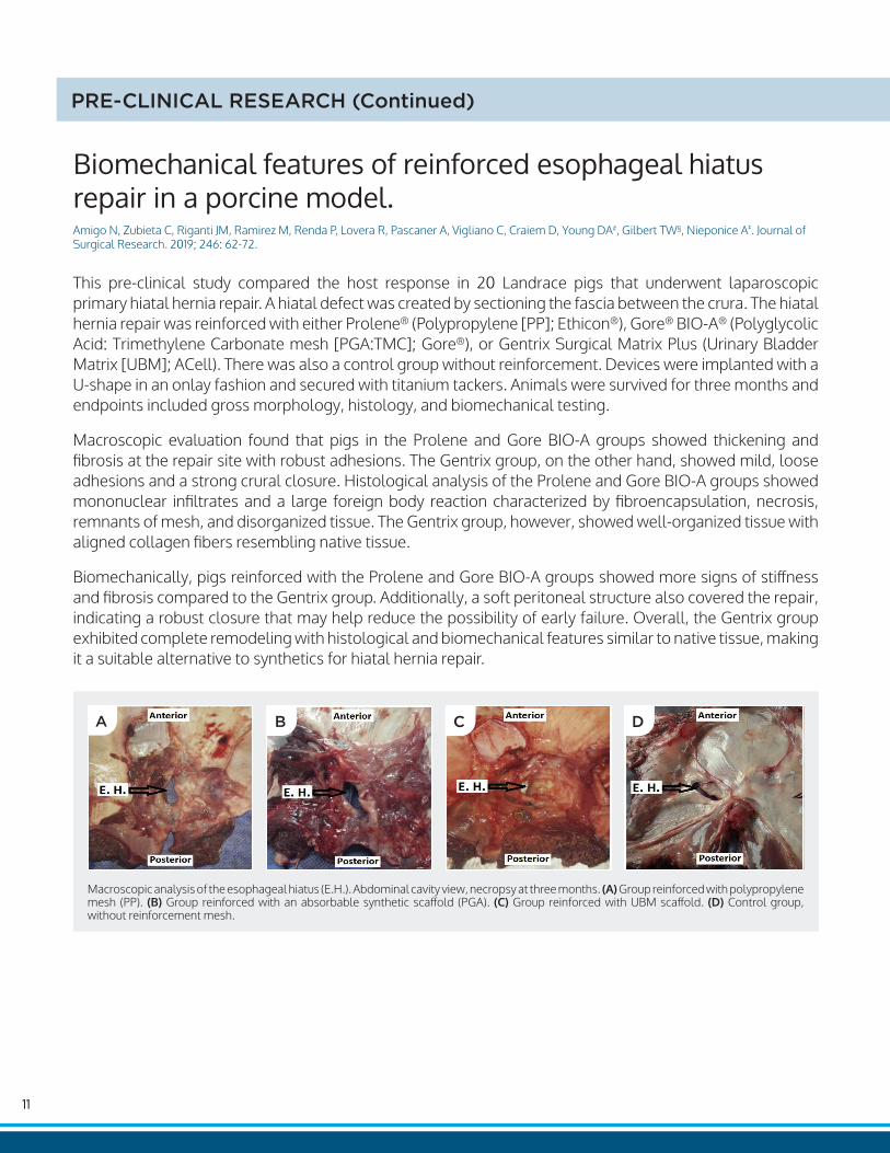
This pre-clinical study compared the host response in 20 Landrace pigs that underwent laparoscopic primary hiatal hernia repair. A hiatal defect was created by sectioning the fascia between the crura. The hiatal hernia repair was reinforced with either Prolene® (Polypropylene [PP]; Ethicon®), Gore® BIO-A® (Polyglycolic Acid: Trimethylene Carbonate mesh [PGA:TMC]; Gore®), or Gentrix Surgical Matrix Plus (Urinary Bladder Matrix [UBM]; ACell). There was also a control group without reinforcement. Devices were implanted with a U-shape in an onlay fashion and secured with titanium tackers. Animals were survived for three months and endpoints included gross morphology, histology, and biomechanical testing.
Macroscopic evaluation found that pigs in the Prolene and Gore BIO-A groups showed thickening and fibrosis at the repair site with robust adhesions. The Gentrix group, on the other hand, showed mild, loose adhesions and a strong crural closure. Histological analysis of the Prolene and Gore BIO-A groups showed mononuclear infiltrates and a large foreign body reaction characterized by fibroencapsulation, necrosis, remnants of mesh, and disorganized tissue. The Gentrix group, however, showed well-organized tissue with aligned collagen fibers resembling native tissue.
Biomechanically, pigs reinforced with the Prolene and Gore BIO-A groups showed more signs of stiffness and fibrosis compared to the Gentrix group. Additionally, a soft peritoneal structure also covered the repair, indicating a robust closure that may help reduce the possibility of early failure. Overall, the Gentrix group exhibited complete remodeling with histological and biomechanical features similar to native tissue, making it a suitable alternative to synthetics for hiatal hernia repair.
Amigo N, Zubieta C, Riganti JM, Ramirez M, Renda P, Lovera R, Pascaner A, Vigliano C, Craiem D, Young DA#, Gilbert TW§, Nieponice A‡. Journal of Surgical Research. 2019; 246: 62-72.
Biomechanical features of reinforced esophageal hiatus repair in a porcine model.
Macroscopic analysis of the esophageal hiatus (E.H.). Abdominal cavity view, necropsy at three months. (A) Group reinforced with polypropylene mesh (PP). (B) Group reinforced with an absorbable synthetic scaffold (PGA). (C) Group reinforced with UBM scaffold. (D) Control group, without reinforcement mesh.
A B C D

12
This pre-clinical study detailed the use of several forms of Urinary Bladder Matrix as a reinforcement graft for abdominal wall repair in a rodent model. In particular, a 14C radiolabeled version of UBM was created to investigate the resorption profile of UBM in vivo. One hundred thirty rats were implanted with 6-layer UBM devices, either: commercially available Gentrix Surgical Matrix (manufactured by ACell), a chemically cross-linked version of the Gentrix graft, laboratory-prepared non-crosslinked UBM devices, laboratory-prepared crosslinked UBM devices, or laboratory-prepared 14C-proline labeled UBM devices. Grafts were explanted at 7, 14, 21, 90, and 180 days post-implantation and analyzed for strength and cell infiltration.
In all subgroups, the UBM biologic meshes were 50% degraded by 16 days and completely degraded by 90 days. The UBM device, which was stronger and stiffer than the native tissue at the time of implantation, was rapidly remodeled and exhibited mechanical behavior that mimicked the native tissue, and the remodeled tissue showed increasing strength and stiffness over time despite the scaffold no longer being present. Although Crosslinked devices initially displayed an increase in mechanical strength, this strength consistently decreased over time, with limited tissue remodeling observed.
Costa A, Naranjo JD, Turner NJ, Swinehart IT, Kolich BD, Shaffiey SA, Londono R, Keane TJ, Reing JE, Johnson SA, Badylak SF‡. Biomaterials. 2016; 108:81-90.
Mechanical strength vs. degradation of a biologically-derived surgical mesh over time in a rodent full-thickness abdominal wall defect.

13
PRE-CLINICAL RESEARCH (Continued)
This pre-clinical study describes the tissue remodeling response to 14 different commercially available biologic surgical mesh devices in a rat model of abdominal wall repair. Remodeling of these ECMs was measured using a histologic assessment at 14 and 35 days after surgery, and the histologic scoring was then correlated to the macrophage phenotype. Each material elicited a distinct host tissue remodeling response that could be characterized as falling into one of three general (quantitative) groups; Encapsulation (Group 1), Integration (Group 2), or Remodeling (Group 3).
In the figure below (top), gray bars represent materials from the Encapsulation Group, blue bars represent materials from the Integration Group, and teal bars represent materials from the Remodeling Group. Higher scores are more indicative of a site-appropriate remodeling response while low scores are more indicative of a scar tissue or foreign body response. This remodeling response was correlated with an early, elevated presence of the M2 macrophage phenotype, and a higher ratio of M2:M1 macrophages, which leads to immune modulation and may be a predictor for site-appropriate tissue formation.
The figure at the bottom right of the page illustrates photomicrographs of hematoxylin and eosin stained slides that show examples of the host remodeling response of test articles in Group 1 (Collamend), Group 2 (InteXen), and Group 3 (MatriStem UBM technology) at 35 days post-implantation.
MatriStem UBM technology showed evidence of a favorable host remodeling response at both 14 and 35 days, including the presence of islands of skeletal muscle at the surgical site at 35 days. Of the 14 different commercially available ECMs tested, MatriStem UBM technology had the highest histologic score and highest M2:M1 ratio at both 14 and 35 days post-implantation.
Brown BN, Londono R, Tottey S, Zhang L, Kukla KA, Wolf MT, Daly KA, Reing JE, Badylak SF‡. Acta Biomaterialia. 2012;8:978-987.
Macrophage phenotype as a predictor of constructive remodeling following the implantation of biologically derived surgical mesh materials.

14

ACell, Inc.6640 Eli Whitney Drive Columbia, MD 21046 800-826-2926
www.acell.com
MK-0440.04 | 2020Rx ONLY Refer to IFU supplied with each device for indications, contraindications, and precautions. US Toll-Free 800-826-2926 • www.acell.com © 2020 ACell, Inc. All Rights Reserved.