Surgical and Prosthodontic Treatment of a Patient with Significant Trauma to the Middle and Lower...
12
Surgical and Prosthodontic Treatment of a Patient with Significant Trauma to the Middle and Lower Face Secondary to a Gunshot Wound: A Clinical Report Paul Kelly, DMD, MS, 1, 2 & Carl J. Drago, DDS, MS 3, 4, 5 1 Arizona Maxillofacial Surgeons PC, Mesa, AZ 2 Formerly, Chief Resident, Oral and Maxillofacial Surgery, Gundersen Lutheran Medical Center, LaCrosse, WI 3 Director of Dental Research, Biomet 3i, Palm Beach Gardens, FL 4 Adjunct Prosthodontic Faculty, Nova Southeastern University College of Dental Medicine, Ft. Lauderdale, FL 5 Formerly, Prosthodontist, Gundersen Lutheran Medical Center, LaCrosse, WI Keywords Surgical treatment; facial gunshot wound; CAD/CAM bars; maxillary implant overdenture. Correspondence Carl J. Drago, 174 Via Veracruz, Jupiter, FL 33458. E-mail: [email protected] Accepted August 19, 2008 doi: 10.1111/j.1532-849X.2009.00483.x Abstract Large defects of dentofacial structures may result from trauma, disease (including neoplasms), and congenital anomalies. The location and size of the defects are related to difficulties that patients report relative to speech, mastication, swallowing, facial esthetics, and self-image. This article reports on the evaluation and treatment of a pa- tient who suffered significant trauma to the lower and mid-face secondary to a gunshot injury. It describes the initial presentation, life-saving procedures, and subsequent bone grafts, implant placement, and prosthetic treatments required to rehabilitate the patient to a condition that closely approximated his preoperative condition. This clinical report confirms that no matter the degree of complexity involved in treating the results of significant facial trauma, successful treatment is dependent on thorough physical and radiographic examinations, development of the appropriate diagnoses, and treatment based on sound prosthodontic and surgical principles. Large defects of dentofacial structures may result from trauma, disease (including neoplasms), and congenital anomalies. The location and size of the defects are related to difficulties patients experience with speech, mastication, swallowing, facial esthet- ics, and self-image. 1-4 Patients who suffer from these facial defects often require multiple reconstructive surgeries over an extended period of time. Blood supply to the surgical sites may impact the postoperative results. Osseous and soft tissue con- tours may sometimes deviate significantly from normal. These patients may also demonstrate signs and symptoms consistent with anxiety, depression, or posttraumatic stress disorder. 5 Treatments for major facial trauma are generally divided into three categories: life-saving procedures, surgical recon- struction, and definitive prosthodontic treatment. Numerous methods have been proposed for surgical reconstruction of pan- facial trauma. 6-11 The so-called bottom-up and inside-out, or more recently, top-down and outside-in, methods have been used for treatment and debated among surgeons. 7,8 Advances in facial-trauma management, including computerized tomog- raphy (CT), have enabled surgeons to visualize and success- fully reconstruct complex panfacial injuries. Restoration of both preinjury facial esthetics and function is the goal of reconstruc- tive surgery. An organized approach to these injuries begins at the maxillary and mandibular arches with progression to the vertical mandible. The naso-orbital-ethmoidal complex should be stabilized to the cranium and bone grafted when indicated. The zygomatic complex is related medially, and orbital recon- struction is then performed. The facial architectural restoration is completed at the Lefort I level. Adherence to this protocol enables surgeons to obtain reproducibly good results, even with the most extensive facial dislocations. 7 Rigid fixation (ORIF) has also advanced in the field of oral and maxillofacial trauma reconstruction. 9 Despite excellent predicable results with various ORIF protocols, considerable debate still occurs in cases involving the traumatic avulsion of large volumes of hard and soft tissues. With severely commin- uted jaw fractures, particularly of the mandible, stripping the periosteum from fractured, small pieces of remaining bone, for ORIF, may be counterproductive to achieving continuity of the mandible. 10,11 The purpose of this article is to report on the evaluation and treatment of a patient who suffered significant trauma to the lower and mid-face secondary to a gunshot wound. This clin- ical report will briefly describe the patient’s initial emergency room presentation, triage, and treatment for life-threatening injuries. The subsequent treatments with multiple surgeries, 626 Journal of Prosthodontics 18 (2009) 626–637 c 2009 by The American College of Prosthodontists
-
Upload
paul-kelly -
Category
Documents
-
view
212 -
download
0
Transcript of Surgical and Prosthodontic Treatment of a Patient with Significant Trauma to the Middle and Lower...

Surgical and Prosthodontic Treatment of a Patient withSignificant Trauma to the Middle and Lower Face Secondaryto a Gunshot Wound: A Clinical ReportPaul Kelly, DMD, MS,1,2 & Carl J. Drago, DDS, MS3,4,5
1Arizona Maxillofacial Surgeons PC, Mesa, AZ2Formerly, Chief Resident, Oral and Maxillofacial Surgery, Gundersen Lutheran Medical Center, LaCrosse, WI3Director of Dental Research, Biomet 3i, Palm Beach Gardens, FL4Adjunct Prosthodontic Faculty, Nova Southeastern University College of Dental Medicine, Ft. Lauderdale, FL5Formerly, Prosthodontist, Gundersen Lutheran Medical Center, LaCrosse, WI
KeywordsSurgical treatment; facial gunshot wound;CAD/CAM bars; maxillary implant overdenture.
CorrespondenceCarl J. Drago, 174 Via Veracruz, Jupiter, FL33458. E-mail: [email protected]
Accepted August 19, 2008
doi: 10.1111/j.1532-849X.2009.00483.x
AbstractLarge defects of dentofacial structures may result from trauma, disease (includingneoplasms), and congenital anomalies. The location and size of the defects are relatedto difficulties that patients report relative to speech, mastication, swallowing, facialesthetics, and self-image. This article reports on the evaluation and treatment of a pa-tient who suffered significant trauma to the lower and mid-face secondary to a gunshotinjury. It describes the initial presentation, life-saving procedures, and subsequent bonegrafts, implant placement, and prosthetic treatments required to rehabilitate the patientto a condition that closely approximated his preoperative condition. This clinical reportconfirms that no matter the degree of complexity involved in treating the results ofsignificant facial trauma, successful treatment is dependent on thorough physical andradiographic examinations, development of the appropriate diagnoses, and treatmentbased on sound prosthodontic and surgical principles.
Large defects of dentofacial structures may result from trauma,disease (including neoplasms), and congenital anomalies. Thelocation and size of the defects are related to difficulties patientsexperience with speech, mastication, swallowing, facial esthet-ics, and self-image.1-4 Patients who suffer from these facialdefects often require multiple reconstructive surgeries over anextended period of time. Blood supply to the surgical sites mayimpact the postoperative results. Osseous and soft tissue con-tours may sometimes deviate significantly from normal. Thesepatients may also demonstrate signs and symptoms consistentwith anxiety, depression, or posttraumatic stress disorder.5
Treatments for major facial trauma are generally dividedinto three categories: life-saving procedures, surgical recon-struction, and definitive prosthodontic treatment. Numerousmethods have been proposed for surgical reconstruction of pan-facial trauma.6-11 The so-called bottom-up and inside-out, ormore recently, top-down and outside-in, methods have beenused for treatment and debated among surgeons.7,8 Advancesin facial-trauma management, including computerized tomog-raphy (CT), have enabled surgeons to visualize and success-fully reconstruct complex panfacial injuries. Restoration of bothpreinjury facial esthetics and function is the goal of reconstruc-tive surgery. An organized approach to these injuries begins at
the maxillary and mandibular arches with progression to thevertical mandible. The naso-orbital-ethmoidal complex shouldbe stabilized to the cranium and bone grafted when indicated.The zygomatic complex is related medially, and orbital recon-struction is then performed. The facial architectural restorationis completed at the Lefort I level. Adherence to this protocolenables surgeons to obtain reproducibly good results, even withthe most extensive facial dislocations.7
Rigid fixation (ORIF) has also advanced in the field of oraland maxillofacial trauma reconstruction.9 Despite excellentpredicable results with various ORIF protocols, considerabledebate still occurs in cases involving the traumatic avulsion oflarge volumes of hard and soft tissues. With severely commin-uted jaw fractures, particularly of the mandible, stripping theperiosteum from fractured, small pieces of remaining bone, forORIF, may be counterproductive to achieving continuity of themandible.10,11
The purpose of this article is to report on the evaluation andtreatment of a patient who suffered significant trauma to thelower and mid-face secondary to a gunshot wound. This clin-ical report will briefly describe the patient’s initial emergencyroom presentation, triage, and treatment for life-threateninginjuries. The subsequent treatments with multiple surgeries,
626 Journal of Prosthodontics 18 (2009) 626–637 c© 2009 by The American College of Prosthodontists

Kelly and Drago Surgical and Prosthodontic Treatment for a Facial Gunshot Wound
Figure 1 Preoperative photograph of the patient described in this reportafter being stabilized in the Emergency Ward. Note the extensive lossof hard and soft tissues in the middle and lower thirds of the face.
grafts, compromised nonoptimal endosseous implant place-ment, prosthodontic treatment, and plastic surgery procedureswill also be illustrated.
Clinical reportA 17-year-old malepatient presented to the Emergency Ward(EW) of the Gundersen Lutheran Medical Center, LaCrosse,WI, by helicopter, 45 minutes after extensive facial trauma froma self-inflicted gunshot wound (Fig 1). The patient’s airway wasstable, and he was not intubated at the time of EW admission.He had been hemodynamically stable during transportation. Asper initial advanced trauma and life-support guidelines (ATLS),the patient was then intubated and studies were performed.10,12
CT of the head and neck region was negative for intracra-nial penetration or other injury. The cervical spine had no ra-diographic or clinical signs of trauma. Fine cut (1 mm) axialand reconstructed coronal, sagittal, and 3D CT images wereobtained for diagnostic and treatment-planning purposes. Ra-diographic studies showed extensive middle and lower facecomminuted fractures with avulsion of multiple anterior teeth,hard palate, alveolar bone, nasal bone, nasal septum, and themaxillary and ethmoid sinuses. Zygomatic arches were intactbilaterally. The medial and lateral sides and floors of both orbitswere violated.
The patient was taken to the operating room, where a tra-cheotomy and a percutaneous endoscopic gastronomy (PEG)were performed. Comprehensive evaluation of the hard andsoft tissue injures to the face demonstrated significant avulsionof osseous and soft tissues. Given the extent of the commin-uted fractures, a decision was made to not further disrupt theblood supply by stripping the periosteum from the segmentswith rigid fixation (ORIF) bone plates. A segmental arch barand Ivy Loupes were placed to establish the occlusal verticaldimension (OVD). At this time, the patient’s posterior teethwere in occlusion bilaterally. This was considered to be con-sistent with the patient’s original OVD (Fig 2). The medialcanthal ligaments were partially avulsed along with portionsof the nasal and frontal bones. Twenty-five gauge transnasalwires were placed to reapproximate the medial canthal liga-ments bilaterally. Intraoral and extraoral soft tissues were ex-tensively explored, debrided, and closed. The patient’s woundswere closed, and the remaining segments of the comminutedfractures in the maxilla and mandible were left with intact pe-riosteum for consolidation. The patient was then taken to theintensive care unit (ICU) in a stable condition (Fig 3).
Reconstructive surgery with definitive prosthetic treatmentplanning began 15 months after the initial hospitalization. Dueto the extensive loss of bone and soft tissue, including a ma-jor portion of the upper lip, and scarring with the loss of theanterior vestibules, the patient was unable to wear transitionalremovable partial dentures (RPDs). Reconstructive surgeriesconsisted of iliac bone grafts to the maxilla and mandible, fol-lowed by osseous healing 13-19 (Fig 4). Plastic surgical proce-dures were to be completed at the conclusion of prosthodontictreatment and included auricular cartilage grafting, paramedianforehead tube pedicle flap, and nasal tip revision.
Diagnostic prosthodontic procedures were accomplished af-ter the bone grafts healed. Diagnostic casts and conventionalrecord bases were made for the diagnostic articulator mounting,prior to the construction of diagnostic wax patterns (wax/acrylicresin denture bases and denture teeth) (Figs 5 and 6).
Trial dentures were fabricated to identify the optimal loca-tion of the missing teeth for construction of surgical guidesprior to the placement of endossous implants. The OVD wasmaintained, consistent with the postoperative (surgical) con-dition. Esthetics, in terms of the amount of incisal displayduring speaking, smiling, and at rest, were not a significantconsideration in maintaining the OVD, due to the large amountof soft tissue avulsion associated with the original trauma. Itwas likely that more maxillary teeth would be visible at restfor this patient postoperatively (surgical and prosthetic) thanwould have been visible prior to the injury. The centric rela-tion record was made with some difficulty as the mandibularleft posterior segment had healed with a lingual inclination,and the mandibular left first molar had to be extracted approx-imately 5 months after the original presentation secondary toloss of attachment and mobility. The maxillary right first pre-molar was no longer in occlusion. In hindsight, this tooth shouldhave been extracted and replaced with an endosseous implant.Even with multiple surgeries and perioral scarring, there wasstill a significant amount of interocclusal clearance between thejaws (Fig 7).
Journal of Prosthodontics 18 (2009) 626–637 c© 2009 by The American College of Prosthodontists 627

Surgical and Prosthodontic Treatment for a Facial Gunshot Wound Kelly and Drago
Figure 2 Reformatted CT scan of thispatient’s facial skeleton after the initial surgicalprocedures that included stabilization of theposterior segments. Note the significant lossof bone from the anterior maxilla andmandible, as well as the numerouscomminuted mandibular segments.
Prosthodontic classificationThe American College of Prosthodontists has developed a clas-sification system for partially edentulous patients based on di-agnostic findings.20 Four classes were identified with ClassI representing uncomplicated clinical situations and Class IVrepresenting complex clinical situations. Each class was differ-entiated by specific diagnostic criteria: location and extent ofthe edentulous areas, condition of abutment teeth, occlusion,and characteristics of the residual ridges.
Location and extent of edentulous areas
Due to the significant amount of hard and soft tissue loss in theanterior maxilla and mandible, and the quality of the soft tis-sues covering the edentulous ridges, this patient was identifiedas having severely compromised edentulous areas, because itwas thought that implant placement into the preexisting eden-tulous areas would result in a nonoptimal implant placementand require a high level of patient compliance regarding oralhygiene and patient adaptation.
Condition of abutments
As a result of the trauma, this patient’s potential abutmentteeth were assessed to have poor long-term prognoses due to
their positions within the dental arches, the size/location of theedentulous ridges, and the dental/skeletal malocclusions. Theteeth themselves were not deficient in terms of the remainingtooth structure; they were deemed compromised because of theamount of bone loss suffered as a direct result of the traumaand the location of the defects.
The mandibular left first molar was originally thought to bea viable tooth. It subsequently lost a large portion of its pe-riodontal attachment, and developed acute irreversible pulpitisand mobility. It was extracted secondary to what was consideredto be a hopeless prognosis. The maxillary right first premolarwas asymptomatic and originally in a satisfactory position. Dur-ing the healing process, it moved and was noted to be slightlyabove the occlusal plane relative to the opposing mandibularpremolars. In hindsight, this tooth should have been extractedand replaced with an endosseous implant to more optimallyrestore the maxillary right posterior segment.
Occlusion
This patient was classified as having substantially compromisedocclusal characteristics, because the occlusal scheme anteriorto the mandibular right premolars had to be reestablished. TheOVD was not altered from the immediate postoperative condi-tion relative to the right posterior quadrants, because it was
628 Journal of Prosthodontics 18 (2009) 626–637 c© 2009 by The American College of Prosthodontists

Kelly and Drago Surgical and Prosthodontic Treatment for a Facial Gunshot Wound
Figure 3 Postoperative photograph of the patient as he left the operatingroom 6 hours after admission.
thought to be relatively stable. The anterior occlusion wasmissing.
Residual ridge characteristics
The edentulous ridges were characterized by the need for sur-gical revision of the supporting structures to permit adequateprosthodontic function.21 There was adequate bone volume for
Figure 4 Panoramic radiograph 26 monthsposttrauma, just prior to mandibular implantplacement.
implant placement in both jaws. The quality of the bone was un-known at this time. The quality of the soft tissues covering bothedentulous areas was poor. The soft tissues were mucosa andnonkeratinized, and the depths of the vestibules were basicallynonexistent. The vestibular tissues were coincidental with themaxillary palatal and mandibular lingual tissues, respectively.The potential for this patient to wear conventional removableprostheses was considered to be poor. There was not enoughridge height or width to resist lateral displacement for conven-tional RPDs. There were also psychosocial considerations thatneeded to be assessed relative to removable prostheses and thepatient’s self-image. The patient was adamant in his requestthat he not be treated with definitive RPDs.
Prosthodontic classification
This patient was classified as Class IV because of the severelycompromised location and extent of the edentulous areas. Theremaining natural teeth were not considered to be adequateto serve as abutments for removable or fixed partial dentures.The occlusion required rehabilitation due to the absence of theanterior teeth, and the residual edentulous ridges were severelycompromised.
Comprehensive surgical andprosthodontic treatment planDue to the extensiveness of the patient’s injuries, specificallythe amount of horizontal and vertical bone loss in the maxillaryanterior segment, the need for lip support, the lack of stableabutment teeth, and the lack of sufficient posterior occlusalcontacts on the left side, implants would be required to supportand retain a removable overdenture prosthesis in the maxilla.An overdenture was planned for the maxillary prosthesis, be-cause the viable bone for implant placement was significantlypalatal to the planned location of the maxillary teeth, and thepatient required a significant amount of lip support. Due to theamount of upper lip lost, the patient could only achieve oralcompetence with slight straining. Lip competence is typically
Journal of Prosthodontics 18 (2009) 626–637 c© 2009 by The American College of Prosthodontists 629

Surgical and Prosthodontic Treatment for a Facial Gunshot Wound Kelly and Drago
Figure 5 Maxillary diagnostic cast after osseous healing, before implantplacement. Note the lack of vestibular depth in the anterior segment.
one of the significant factors in determining vertical dimension.In this case, lip competence was not thought to be obtainablewith the patient at rest. While lip competence was desirable atthe selected OVD, the lack of lip competence as a mitigatingfactor to success in this case was not considered to be signifi-cant in determining esthetics and OVD or rest positions. If lipcompetence was considered to be essential for success in thiscase, the OVD could have been reduced by adjusting and/orrestoring the posterior teeth. A fixed implant-retained maxil-lary prosthesis was contraindicated secondary to the locationof the supporting bone, the need for lip support, phonetics,and oral hygiene procedures.22,23 For strength and additionalretention and stability, a secondary casting that fitted preciselyover the primary bar was planned for the removable maxillaryoverdenture prosthesis.24
A screw-retained, implant-supported fixed prosthesis wasplanned for the mandibular edentulous segment because therequisite bone for implant placement was much closer to nor-
Figure 6 Mandibular diagnostic cast after osseous healing, before im-plant placement. Note the lack of a definitive residual ridge and vestibularextensions. The mandibular left first molar had been extracted severalmonths prior to this photograph, secondary to the loss of attachmentand mobility.
Figure 7 Lateral cephalometric radiograph taken 18 months posttraumaprior to the extraction of the maxillary right canine and right first premo-lar. Note the relative prominence of the lower lip and the amount ofinterocclusal distance between the anterior jaws.
mal, relative to the planned prosthetic arch form, and there wasminimal need for lip support. Esthetics relative to space be-tween the inferior borders of hybrid prostheses and mandibularedentulous ridges are generally not a concern. A screw-retaineddesign was chosen for the mandibular prosthesis to permit eas-ier access to the abutments for oral hygiene procedures as wellas retrievability for prosthetic maintenance.22
The patient also suffered from a loss of tongue volume andfunction. It was estimated that approximately 10% of the ante-rior and anterior lateral tongue volumes were lost. The patientsuffered some disability in terms of phonetics, especially priorto prosthetic replacement of the missing teeth, bone, and softtissue. It also should be noted that he was not able to adapt to anytype of transitional RPDs following the trauma and surgeries.Lewandowski reported on misarticulation following surgery formalignancies in the oral cavity.25 Although the present clini-cal report illustrates the net result of a gunshot wound to themid-face, the functional results were thought to be similar topatients who lost tongue function secondary to tumors.
Lewandowski reported that the underlying cause in his caseseries was dysfunction of the tongue due to partial or totalresection, consequences of mandibular resection together withthe oral cavity floor, or dysfunction of the lower lip. Anatomicalterations revealed themselves as shifts in the points of con-tact between structures of the articulation system noticeable onpalatograms or linguograms.
In a study reported by Sun et al, the size of tongue tumors(T1, T2, T3) and the site of excision (anterior, middle, poste-rior) were responsible for significant differences between pa-tients with T1- and T3-sized tumors (p < 0.05).26 The speechintelligibilities of the patients with tumors in the anterior tonguewere significantly lower than those with tumors in the middleor posterior tongue regions (p < 0.05). Patients with preserva-tion of the tip of the tongue or floor of the mouth had higherintelligibilities (p < 0.05). They concluded that for patients af-ter glossectomy within the range of 1/2 or less of the tongue,the tumor site or excision extent of the tongue followed by thetumor size may be key factors in determining the postoperativearticulation intelligibility. In the present case, the design of the
630 Journal of Prosthodontics 18 (2009) 626–637 c© 2009 by The American College of Prosthodontists
Kelly and Drago Surgical and Prosthodontic Treatment for a Facial Gunshot Wound
Figure 8 Laboratory facial occlusal view of mandibular waxed screw-retained prosthesis. The anterior incisal plane was at the same level(horizontal) as the posterior occlusal plane.
mandibular prosthesis and the arrangement of the mandibularartificial teeth were made consistent with the preexisting archform and locations of the remaining mandibular teeth.
TreatmentFive maxillary and four mandibular implants were placed withtwo-stage surgical protocols. The mandibular implants wereplaced in the first surgical procedure; the maxillary implantswere placed in a second, later procedure. All implants inte-grated without incident. The mandibular implants were placed
Figure 9 JPEG images of the CAD/CAM design for the mandibular framework. The location of the teeth in the wax denture can be visualized andverifies that the entire framework will be contained within the confines of the mandibular prosthesis. The screw access openings were lingual to thefacial surfaces of the denture teeth in the prosthesis.
Figure 10 Maxillary intraoral occlusal image of five implants. Note thepalatal positions of the implants relative to the arch form of the remainingnatural teeth and the quality of the mobile mucosal tissues stretchedwith the lip retractors.
consistent with the planned locations of the teeth in the trial den-ture set-up and the surgical guides. The long-term prognosesof the endosseous implants in this case were thought to be lessthan prognoses that have been established for endosseous im-plants placed into healed edentulous sites.27 At the time of thisreport (3 years after implant placement), all the implants haveremained viable with less than 1 mm of bone loss noted onyearly radiographs.
The mandibular prosthesis was constructed first, since the an-terior maxilla required additional healing time due to additionalbone grafting at the time of implant placement. Abutments wereplaced (IOL Abutments, Biomet 3i, Palm Beach Gardens, FL),
Journal of Prosthodontics 18 (2009) 626–637 c© 2009 by The American College of Prosthodontists 631

Surgical and Prosthodontic Treatment for a Facial Gunshot Wound Kelly and Drago
Figure 11 JPEG images of the CAD/CAM design for the maxillary primary bar. The location of the teeth in the wax denture can be visualized andverifies that the entire primary bar will be contained within the confines of the maxillary overdenture.
and abutment level impressions were required due to the signif-icant amount of soft tissues covering/surrounding (4 to 6 mm)the mandibular implants. The mandibular anterior incisal planewas determined by identifying the level of the posterior occlusalplane (retromolar pads) and extending it anteriorly (Fig 8).Twenty-degree acrylic resin posterior teeth were used (Justi R©Blend R©, American Tooth Industries, Oxnard, CA).
Figure 12 Laboratory image of the articulated casts. The maxillary casthas both the primary bar and secondary casting in place. The milled baris offset in the bottom of the image.
The centric relation record, as defined by the Glossary ofProsthodontic Terms,28 was difficult to obtain due to trauma,surgical procedures, and residual defects the patient experi-enced. The goal of the jaw relation records was to obtain apredictable jaw relationship at the predetermined OVD.
The mandibular wax denture and master cast were scannedfor a CAD/CAM titanium alloy framework (CAM StructSUREPrecision Milled Bar, Biomet 3i) (Fig 9). The mandibular pros-thesis was finished in a conventional fashion and inserted.
The maxillary implants were placed into the viable bone inthe anterior maxilla; however, their locations were palatal tooptimal implant positions as determined by the trial denturesand the surgical guide because the quality and quantity of bonein the anterior segment was deficient despite bone grafting. Theimplants were placed approximately 8 to 10 mm apart (Fig 10).
The periimplant soft tissue depths of the maxillary implantswere normal (2 to 3 mm) compared to the mandibular peri-implant tissues, and implant level impressions were made forfabrication of the master cast and verification index. A wax trialdenture was fabricated and tried in for patient approval relativeto the overall esthetic results, including lip support and incisaldisplay at rest and smiling. At the selected OVD, lip compe-tence was obtained with effort on the patient’s part. The jawrelation record was verified. The casts and wax dentures werescanned, and a CAD/CAM framework was designed (CAMStructSURE Precision Milled Bar) (Fig 11). The maxillary pri-mary bar was designed with a 2◦ taper and machined from asolid blank of titanium alloy (Fig 12).
632 Journal of Prosthodontics 18 (2009) 626–637 c© 2009 by The American College of Prosthodontists

Kelly and Drago Surgical and Prosthodontic Treatment for a Facial Gunshot Wound
Figure 13 Laboratory images of the maxillary wax implant prosthesis prior to processing. The mandibular prosthesis (mandibular cast) was completedfirst to accommodate the increased healing time required for the second maxillary bone grafting procedures. The posterior attachment is in the locked,or “in,” position (center: anterior; lower left: right posterior segment; lower right: left posterior segment).
The bar was designed to be used with two anterior Bredent2.2 VKS-OC Stud-head screws (XPdent Corporation, Miami,FL) that screwed directly into the tapped sites prepared afterthe bar was milled. These attachments are designed for use inmultiple clinical situations and the female, plastic portions ofthe attachments are replaceable chairside.
The posterior attachment was a 6-mm SwissLoc attachment(Attachments International, Burlingame, CA). This SwissLocNG Next Generation attachment consisted of an extracoro-
Figure 14 Maxillary intraoral occlusal image with the primary bar inplace. The two anterior attachments are visible on the labial surfaceof the anterior segment.
nal locking pin/plunger designed to prevent lift-off, a commonproblem reported with bar overdentures. The design incorpo-rated positive in-and-out positions, which prevented the attach-ment from disengaging unintentionally during function. It was
Figure 15 Lateral clinical image with the maxillary primary bar in place.Note the amount of horizontal distance between the bar and the labialsurfaces of the mandibular screw-retained, implant-supported fixed pros-thesis.
Journal of Prosthodontics 18 (2009) 626–637 c© 2009 by The American College of Prosthodontists 633
Surgical and Prosthodontic Treatment for a Facial Gunshot Wound Kelly and Drago
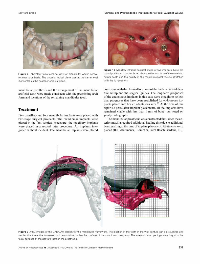
Figure 16 Anterior clinical view of centric occlusion with both prosthe-ses in place. The large horizontal discrepancy between the location ofthe maxillary implants and the optimal location of the artificial teeth hasbeen compensated for with the maxillary overdenture.
screwed directly into the preexisting tapped site in the primarybar.
The secondary casting was fabricated with type IV gold al-loy (North Shore Dental Laboratories, Lynn, MA). Frameworkswere tried in and noted to fit well. The maxillary prosthesis wasentirely implant supported and did not feature any palatal cov-erage or tooth support. The palatal portion of the maxillaryprosthesis was contoured to be consistent with the contours ofthe patient’s posterior palate. These contours were arbitrary,and the patient was advised that adjustments could be requiredto optimize his phonetics with the prosthesis in place. Themaxillary overdenture prosthesis was completed in a conven-tional fashion (Figs 13–20). When the patient returned for the2-week postinsertion clinical appointment, he reported mini-mal difficulties in adapting to the prostheses while speakingand chewing. He reported that he was extremely pleased withthe esthetic and functional results.
This patient has been followed for 3 years and is comfortablewith the prosthetic and surgical rehabilitation. He has expe-rienced no long-term significant problems regarding mastica-tion, oral hygiene, or phonetics. There has been minimal boneloss visualized around the implants in both jaws (Fig 21). His
Figure 17 Clinical image with both prostheses in place. The patient didnot have lip competence at rest.
Figure 18 Full-face image of the patient at the established OVD with aforced effort to establish lip competency.
long-term prognosis relative to the osseointegrated implantsand prosthetics is thought to be favorable.
DiscussionSurgical correction of facial defects may be hampered fromthe lack of adequate quality and quantities of hard and soft tis-sues.6,11 Trauma to the mid-face (hard palate, maxillary sinus,
Figure 19 Clinical image of the partially edentulous maxilla with theoverdenture prosthesis in place. Note the atypical contours of the palatalsoft tissues.
634 Journal of Prosthodontics 18 (2009) 626–637 c© 2009 by The American College of Prosthodontists

Kelly and Drago Surgical and Prosthodontic Treatment for a Facial Gunshot Wound
Figure 20 Profile views of the patient with the prostheses in place2 weeks postprosthesis insertion. The patient still had to strain slightlyto obtain lip competence at rest (rest vertical dimension, left; smiling,right). The anterior/posterior deficiency of the mid-face has been com-
pensated for with the labial flange and tooth positions of the maxillaryoverdenture prosthesis. The esthetic result in the right posterior max-illary quadrant was slightly compromised due to the retention of themaxillary first premolar.
nasal cavity, zygomatic processes, infraorbital rims) may resultin varying degrees of maxillary defects, depending on the veloc-ity and etiology of the injury. Due to the anatomic complexityof the mid-facial region and the almost endless potential for de-fect size, shape, and location, there are an unlimited number ofpotential disconfigurations, and there is no universally accepteddefinition of panfacial fractures in the literature. Aramany haspublished a classification of these defects.14 Markowitz andManson have also described panfacial trauma.15 Applying noone definition or classification, the patient presented in thisarticle had no frontal bone defect, yet otherwise had severelycomminuted facial fractures bordered superiorly by the naso-orbito-etmoid complex and continuing throughout the mid-faceto the inferior extent of the mandible.
Trauma to the mandible can be just as challenging to clin-icians as maxillary defects, depending on the volume and lo-
Figure 21 Panoramic radiograph 3 yearspostocclusal function. All implants have stablebone levels, without radiolucencies.
cation of the defect(s). The muscles of mastication and facialexpression attach to the mandible in an extremely complexfashion and also give form to the lower third of the face. Dis-continuity defects have the potential to impact negatively onany one of a number of critical functions associated with thesestructures. This case was particularly challenging because ofthe loss of significant amounts of bone and soft tissue, alongwith loss of vestibular depth in both jaws. This raised the levelof difficulty in providing the patient with adequate amountsof bone and soft tissue prior to definitive prosthodontic treat-ment. Another challenge was that the quality of the intraoraltissues was not ideal in that the tissues surrounding the implantabutments were quite mobile and nonkeratinized.
Debate also exists over where and when to begin treatmentin panfacial trauma.7-10 In this case, the surgical protocol con-sisted of debridement and primary closure, healing, multiple
Journal of Prosthodontics 18 (2009) 626–637 c© 2009 by The American College of Prosthodontists 635
Surgical and Prosthodontic Treatment for a Facial Gunshot Wound Kelly and Drago
bone grafts, and additional healing prior to the placement ofendosseous implants.
Authors have also differed on the methods of fixation ofsevere, comminuted mandibular fractures.16-19 With extensiveavulsion-type injuries, rigid fixation of severely comminutedfractures has been problematic. In this case, decisions weremade to minimize the risk of infection and decrease the oper-ating time by obtaining closure without rigid fixation.
This case demonstrated the importance of a team approachin comprehensive treatment planning. Initially, the foremostconcern was that the patient be stabilized. This was prior to de-ciding on the definitive surgical and prosthetic treatment plansrequired to complete the rehabilitative treatment. This patientis now approximately 5 years post-trauma and approximately3 years post-prosthodontic reconstruction. None of the attach-ments have had to be replaced. The implants and prosthetictreatments have remained stable, and the long-term prognosisfor continued success is good.
ReviewThis clinical report illustrated the devastating injuries secondaryto a self-inflicted gunshot wound to the face. The patient sur-vived the life-threatening injuries while receiving excellenttrauma and surgical care immediately posttrauma. Numeroussurgeries were required to provide satisfactory bases for long-term prosthodontic success. Endosseous implants were placedinto the available, viable maxillary and mandibular bone and be-came osseointegrated. The patient was restored with a primarymaxillary bar, retained by screws in the endosseous implants,and a maxillary overdenture prosthesis. The mandibular defectwas restored with a screw-retained, implant-supported fixedprosthesis. Both prostheses have been viable for over 3 years.
Despite the clinical success of the treatment noted above,there were several compromises that had to be taken into con-sideration: the mid-face was deficient horizontally, includinglack of soft tissue volume (lips); complete lip competence wasnot achieved without straining by the patient; an end-to-endanterior tooth arrangement was used to decrease the upper lipvolume; the maxillary primary implant-retained bar was not inthe position of the maxillary anterior teeth, but was within theanterior articulation zone of the anterior hard palate; and therewas a loss of tongue volume and tongue mobility secondary tothe original injury. Fortunately in this case, the patient proved tobe quite adept at adapting to the compromises noted above, andcontinues to function without phonetic deficits. The long-termprognosis for this patient is acceptable, both in terms of contin-ued survival of the endosseous implants and the prostheses.
AcknowledgmentsThe authors wish to acknowledge the surgical expertise of Dr.Larry Kent, former attending surgeon of Oral and MaxillofacialSurgery, Gundersen Lutheran Medical Center, LaCrosse, WI,and the laboratory assistance of Andrew Gingrasso, Departmentof Prosthodontics, Gundersen Lutheran Medical Center andThomas Peterson, CDT, MDT, President, NorthShore DentalLaboratories, Lynn, MA.
References1. Desjardins R: Early rehabilitative management of the
maxillectomy patient. J Prosthet Dent 1977;38:311-3182. Kornblith A, Zlotolow I, Gooen J, et al: Quality of life of
maxillectomy patients using an obturator prosthesis. Head Neck1996;18:323-334
3. Rogers S, Lowe D, McNally D, et al: Health-related quality oflife after maxillectomy: a comparison between prostheticobturation and free flap. J Oral Maxillofac Surg 2003;61:174-181
4. Devlin H, Barker G: Prosthetic rehabilitation of the edentulouspatient requiring a partial maxillectomy. J Prosthet Dent1992;67:223-227
5. Taylor TD (ed): Clinical Maxillofacial Prosthetics. Chicago, IL,Quintessence, 2000, p. 6
6. Vinzenz K, Schaudy C, Wuringer E: The iliac prefabricatedcomposite graft for dentoalveolar reconstruction: a clinicalprocedure. Int J Oral Maxillofac Implants 2006;21:117-123
7. Markowitz BL, Manson PN: Panfacial fractures: organization oftreatment. Clin Plast Surg 1989;16:105-114
8. Mason PN, Clark N, Robertson B, et al: Sub-unit principles inmidface fractures: the importance of sagittal buttresses, softtissue reductions and sequencing treatment of segmentalfractures. Plast Reconstr Surg 1999;103:1287-1306
9. Mercuri LG, Steinberg MJ: Sequencing of care for multiplemaxillofacial injuries. In Peterson LJ (ed): Principles of Oral andMaxillofacial Surgery. Philadelphia, PA, Lippincott, 1992, pp.615-622
10. Kelly J: War Injuries to the Jaws and Related Structures.Washington, DC, US Government Printing Office, 1978
11. Gruss JS, Phillips JH: Complex facial trauma: the evolving roleof rigid fixation and immediate bone graft reconstruction. ClinPlast Surg 1989;16:93-104
12. American College of Surgeons: Advanced Trauma and LifeSupport for Doctors (ed 7). Chicago, IL, American College ofSurgeons, 2004
13. Shockley W, Weissler M: Reconstructive alternatives followingsegmental mandibulectomy. Am J Otolaryngol 1992;13:156-165
14. Aramany M: Basic principles of obturator design for partiallyedentulous patients: Part I. Classification. J Prosthet Dent1978;40:554-561
15. Markowitz BL, Manson PN: Panfacial fractures: organizationn oftreatment. Clin Plast Surg 1989;16:105-114
16. Benson PD, Marshall MK, Engelstad ME, et al: The use ofimmediate bone graft in reconstruction of clinically infectedmandibular fractures: bone grafts in the presence of pus. J OralMaxillofac Surg 2006;64:122-126
17. Finn RA: Treatment of comminuted mandible fractures by closedreduction. J Oral Maxillofac Surg 1996;54:320-327
18. Ellis E III, Muniz O, Anand K: Treatment considerations forcomminuted mandibular fractures. J Oral Maxillofac Surg2003;61:861-870
19. Kazanjian VH: An outline of the treatment of extensivecomminuted fractures of the mandible. Am J Orthod Oral Surg1942;28:265
20. McGarry TJ, Nimmo A, Skiba JF, et al: Classification system forpartial edentulism. J Prosthodont 2002;11:181-193
21. McGarry TJ, Nimmo A, Skiba JF, et al: Classification system forcomplete edentulism. J Prosthodont 1999;8:27-39
22. Goodacre CJ, Kan JY, Rungcharassaeng K: Clinicalcomplications of osseointegrated implants. J Prosthet Dent1999;81:537-552
23. Bragger U, Aeschlimann S, Burgin W, et al: Biological andtechnical complications and failures with fixed partial dentures
636 Journal of Prosthodontics 18 (2009) 626–637 c© 2009 by The American College of Prosthodontists

Kelly and Drago Surgical and Prosthodontic Treatment for a Facial Gunshot Wound
(FPD) on implants and teeth after four to five years of function.Clin Oral Implants Res 2001;12:26-34
24. Kiener P, Oetterli M, Mericske E, et al: Effectiveness ofmaxillary overdentures supported by implants: maintenance andprosthetic complications. Int J Prosthodont 2001;14:133-140
25. Lewandowski L: Palatograms and linguograms for studyingmisarticulation following surgery for tumors of the tongue andoral cavity floor. Ann Acad Med Stetin 2006;52(Suppl 3):13-16
26. Sun J, Weng Y, Li J, et al: Analysis of determinants on speechfunction after glossectomy. J Oral Maxillofac Surg2007;65:1944-1950
27. Krennmair G, Krainhofner M, Piehslinger E: Implant-supportedmaxillary overdentures retained with milled bars: maxillaryanterior versus maxillary posterior concept—a retrospectivestudy. Int J Oral Maxillofac Implants 2008;23:343-352
28. The glossary of prosthodontic terms. J Prosthet Dent2005;94:10-92
Journal of Prosthodontics 18 (2009) 626–637 c© 2009 by The American College of Prosthodontists 637


![2019-04-27 OnTarget First Aid Training Flyer [print]...Gun Range/Gunshot Wound Basic First Aid* Topics Covered: Gunshot Wound Basics: Tactical Treatment Ballistics and the Effects](https://static.fdocuments.in/doc/165x107/5f4849042f6f1a4a393115fb/2019-04-27-ontarget-first-aid-training-flyer-print-gun-rangegunshot-wound.jpg)