
Surgery Subretinal implantation of Okayama University-type ...
8
1939 *Correspondence to: Matsuo, T.: [email protected] ©2017 The Japanese Society of Veterinary Science This is an open-access article distributed under the terms of the Creative Commons Attribution Non-Commercial No Derivatives (by-nc-nd) License. (CC-BY-NC-ND 4.0: https://creativecommons.org/licenses/by-nc-nd/4.0/) FULL PAPER Surgery Subretinal implantation of Okayama University-type retinal prosthesis (OUReP TM ) in canine eyes by vitrectomy Toshihiko MATSUO 1) *, Tetsuya UCHIDA 2) , Makoto NITTA 2) , Koichiro YAMASHITA 2) , Shigiko TAKEI 3) , Daisuke IDO 3) , Mamoru TANAKA 3) , Masao OGUCHI 3) and Toshinori FURUKAWA 4) 1) Ophthalmology, Okayama University Medical School and Graduate School of Medicine, Dentistry and Pharmaceutical Sciences, Okayama-shi, Okayama 700-8558, Japan 2) Polymer Materials Science, Okayama University Faculty of Engineering and Graduate School of Natural Science and Technology, Okayama-shi, Okayama 700-8530, Japan 3) Ina Research, Inc., Ina-shi, Nagano 399-4501, Japan 4) Kurashiki University of Science and the Arts, Kurashiki-shi, Okayama 712-8505, Japan ABSTRACT. Okayama University-type retinal prosthesis (OUReP TM ) is a photoelectric dye-coupled polyethylene film which generates electric potential in response to light and stimulates nearby neurons. This study aims to test surgical feasibility of subretinal implantation and functional durability of dye-coupled films in the subretinal space. The dye-coupled films were implanted subretinally by 25-gauge vitrectomy in the right eye of 11 normal beagle dogs: 2 dogs served for film removal after 5-month film implantation, 3 dogs for film removal after 3-month film implantation, 3 dogs for 3-month film implantation and pathological examination, and 3 dogs for sham surgery. The surface electric potential of the removed dye-coupled films in response to light was measured by the Kelvin Probe system. At surgery, rolled-up dye-coupled films in 5 × 5 mm square size could be inserted into subretinal space of retinal detachment induced by fluid injection with a 38-gauge polyimide tip. Retinal attachment was maintained by silicone oil injection in vitreous cavity. At autopsy, the retina in all dogs maintained the ganglion cell layer, inner and outer nuclear layers while it lost the outer segments in some part. All 5 sheets of removed dye-coupled films maintained the dye color. One sheet of the 5-month implanted film showed proportional increase of surface potential in response to increasing light intensity. Subretinal implantation of OUReP TM by vitrectomy was technically feasible in canine eyes, and OUReP TM maintained the function of generating light-evoked surface potential after 5 months in subretinal implantation. KEY WORDS: 38-gauge polyimide tip, vitrectomy, dog, dye-coupled thin film retinal prosthesis, photoelectric dye Blind patients with hereditary retinal diseases, such as retinitis pigmentosa, have dead photoreceptor cells, but the other retinal neurons, which send axons to the brain, remain alive. Spontaneous retinal dystrophies, similar to the disease in the human, have been also described to occur in dogs [16]. The basic concept of retinal prostheses is to replace dead photoreceptor cells with artificial devices and to exploit the function of these living neurons, and finally to send messages to the brain, following artificial stimulation in response to light [9, 12, 20]. Okayama University-type retinal prosthesis (OUReP TM ) is a new type of retinal prostheses, so called photoelectric dye- coupled thin film retinal prosthesis [1–3, 21–24]. Stable photoelectric dye molecules with absorption spectrum of visible light [8, 10, 13, 15] were chemically coupled to polyethylene film surface. The dye-coupled film generates electric potential in response to light and stimulates nearby neuronal cells to induce action potential. The dye-coupled film, as a light receiver and potential generator, implanted in subretinal space, is designed to replace the function of dead photoreceptor cells in retinal dystrophy [12, 20] and to send the signal to the brain through the living retinal ganglion cells and their axons as optic nerve fibers [9]. The main stream of retinal prosthesis utilizes a multielectrode array [7]. Camera-captured image is disintegrated to 60 pixels, and the electric current, corresponding to grayscale tone in each pixel, is outputted from 60 electrodes to stimulate the retinal living Received: 17 August 2017 Accepted: 10 October 2017 Published online in J-STAGE: 18 October 2017 J. Vet. Med. Sci. 79(12): 1939–1946, 2017 doi: 10.1292/jvms.17-0450
Transcript of Surgery Subretinal implantation of Okayama University-type ...

1939
*Correspondence to: Matsuo, T.: [email protected]©2017 The Japanese Society of Veterinary Science
This is an open-access article distributed under the terms of the Creative Commons Attribution Non-Commercial No Derivatives (by-nc-nd) License. (CC-BY-NC-ND 4.0: https://creativecommons.org/licenses/by-nc-nd/4.0/)
FULL PAPERSurgery
Subretinal implantation of Okayama University-type retinal prosthesis (OURePTM) in canine eyes by vitrectomyToshihiko MATSUO1)*, Tetsuya UCHIDA2), Makoto NITTA2), Koichiro YAMASHITA2), Shigiko TAKEI3), Daisuke IDO3), Mamoru TANAKA3), Masao OGUCHI3) and Toshinori FURUKAWA4)
1)Ophthalmology, Okayama University Medical School and Graduate School of Medicine, Dentistry and Pharmaceutical Sciences, Okayama-shi, Okayama 700-8558, Japan
2)Polymer Materials Science, Okayama University Faculty of Engineering and Graduate School of Natural Science and Technology, Okayama-shi, Okayama 700-8530, Japan
3)Ina Research, Inc., Ina-shi, Nagano 399-4501, Japan4)Kurashiki University of Science and the Arts, Kurashiki-shi, Okayama 712-8505, Japan
ABSTRACT. Okayama University-type retinal prosthesis (OURePTM) is a photoelectric dye-coupled polyethylene film which generates electric potential in response to light and stimulates nearby neurons. This study aims to test surgical feasibility of subretinal implantation and functional durability of dye-coupled films in the subretinal space. The dye-coupled films were implanted subretinally by 25-gauge vitrectomy in the right eye of 11 normal beagle dogs: 2 dogs served for film removal after 5-month film implantation, 3 dogs for film removal after 3-month film implantation, 3 dogs for 3-month film implantation and pathological examination, and 3 dogs for sham surgery. The surface electric potential of the removed dye-coupled films in response to light was measured by the Kelvin Probe system. At surgery, rolled-up dye-coupled films in 5 × 5 mm square size could be inserted into subretinal space of retinal detachment induced by fluid injection with a 38-gauge polyimide tip. Retinal attachment was maintained by silicone oil injection in vitreous cavity. At autopsy, the retina in all dogs maintained the ganglion cell layer, inner and outer nuclear layers while it lost the outer segments in some part. All 5 sheets of removed dye-coupled films maintained the dye color. One sheet of the 5-month implanted film showed proportional increase of surface potential in response to increasing light intensity. Subretinal implantation of OURePTM by vitrectomy was technically feasible in canine eyes, and OURePTM maintained the function of generating light-evoked surface potential after 5 months in subretinal implantation.
KEY WORDS: 38-gauge polyimide tip, vitrectomy, dog, dye-coupled thin film retinal prosthesis, photoelectric dye
Blind patients with hereditary retinal diseases, such as retinitis pigmentosa, have dead photoreceptor cells, but the other retinal neurons, which send axons to the brain, remain alive. Spontaneous retinal dystrophies, similar to the disease in the human, have been also described to occur in dogs [16]. The basic concept of retinal prostheses is to replace dead photoreceptor cells with artificial devices and to exploit the function of these living neurons, and finally to send messages to the brain, following artificial stimulation in response to light [9, 12, 20].
Okayama University-type retinal prosthesis (OURePTM) is a new type of retinal prostheses, so called photoelectric dye-coupled thin film retinal prosthesis [1–3, 21–24]. Stable photoelectric dye molecules with absorption spectrum of visible light [8, 10, 13, 15] were chemically coupled to polyethylene film surface. The dye-coupled film generates electric potential in response to light and stimulates nearby neuronal cells to induce action potential. The dye-coupled film, as a light receiver and potential generator, implanted in subretinal space, is designed to replace the function of dead photoreceptor cells in retinal dystrophy [12, 20] and to send the signal to the brain through the living retinal ganglion cells and their axons as optic nerve fibers [9].
The main stream of retinal prosthesis utilizes a multielectrode array [7]. Camera-captured image is disintegrated to 60 pixels, and the electric current, corresponding to grayscale tone in each pixel, is outputted from 60 electrodes to stimulate the retinal living
Received: 17 August 2017Accepted: 10 October 2017Published online in J-STAGE: 18 October 2017
J. Vet. Med. Sci. 79(12): 1939–1946, 2017doi: 10.1292/jvms.17-0450

T. MATSUO ET AL.
1940doi: 10.1292/jvms.17-0450
neurons in Argus II Retinal Prosthesis System (Second Sight Medical Products, Inc., Sylmar, CA, U.S.A.). Surgical implantation of the multielectrode array indeed requires sophisticated techniques. In contrast, the dye-coupled film is a soft and thin sheet which would be rolled up and inserted in the subretinal space by vitrectomy [14].
In this study, the dye-coupled films were implanted subretinally in normal canine eyes and were also removed by vitrectomy in order to examine the feasibility of surgical techniques. In addition, spectrophotometric absorbance and light-evoked surface electric potential was examined on the dye-coupled films which were implanted for 3 or 5 months and then removed by vitrectomy.
MATERIALS AND METHODS
Preparation of dye-coupled polyethylene filmThin films were made from polyethylene powder and exposed to fuming nitric acid to introduce carboxyl moieties on the film
surface. Photoelectric dye molecules, 2-[2-[4-(dibutylamino) phenyl]ethenyl]-3-carboxymethylbenzothiazolium bromide (NK-5962, Hayashibara, Inc., Okayama, Japan), were coupled to carboxyl moieties of the polyethylene film surface via ethylenediamine, as described previously [3, 22, 23]. The fuming nitric acid-treated only polyethylene film and the photoelectric dye-coupled polyethylene film were designated as the plain film and the dye-coupled film, respectively. Films were manufactured in quality management system at a clean-room facility in Okayama University Incubator.
AnimalsEleven male beagle dogs (Kitayama Labes Co., Ina, Japan, or Beijing Marshall Biotechnology Co., Beijing, China) at the age
ranging from 10 to 21 months (mean, 15.8 months), were used in the study: 2 dogs were assigned for 5-month dye-coupled film (OURePTM) implantation and its removal, 3 dogs for 3-month dye-coupled film implantation, 3 dogs for sham surgery, and 3 dogs for 3-month dye-coupled film implantation and its removal (Table 1). At sham surgery, all surgical procedures were done except for dye-coupled film implantation. All the surgeries were done only in the right eye. The dye-coupled films, removed by vitrectomy, were analyzed for spectrophotometric absorbance and light-evoked surface potential.
All animals were sacrificed with bleeding after overdose of intravenous thiopental (Ravonal, Mitsubishi Tanabe Pharma, Osaka, Japan), and the eyes were enucleated. Dogs with 3-month or 5-month film implantation and removal were euthanized 10 or 11 days after the second surgery for film removal. Dogs with 3-month film implantation or sham surgery were euthanized at 3 months after the initial surgery. After the cornea and iris were removed by circumferential incision of the eye ball, the posterior segment was cut meridionally to view the entire retina. The posterior segment was then fixed with phosphate-buffered 1% formaldehyde and 2.5% glutaraldehyde, stored in 10% neutral pH formalin, and embedded in paraffin. Paraffin sections were cut and stained with hematoxylin and eosin for pathological examinations. This study was approved by the Animal Care and Use Committee in Okayama University and also by the Committee at Ina Research, Inc., based on the Animal Welfare and Management Act in Japan.
Surgical proceduresDogs were sedated with intramuscular medetomidine (0.02 mg/0.02 ml/kg of body weight, Domitor, Nippon Zenyaku Kogyo
Co., Koriyama, Japan) and midazolam (0.3 mg/0.06 ml/kg of body weight, Dormicum, Astellas Pharma Inc., Tokyo, Japan), together with subcutaneous injection of meloxicam (0.5%, 0.2 mg/0.04 ml/kg of body weight, Metacam, Boehringer Ingelheim, Ingelheim am Rhein, Germany) as a non-steroidal anti-inflammatory drug. At least 15 min after the sedation, dogs were anesthetized with intravenous pentobarbital (9.72 mg/kg of body weight, Somunopentyl 0.15 ml/kg, Kyoritsu Seiyaku Co., Tokyo, Japan), and intubated to maintain the airway. Mydriasis in the right eye was induced by 1% atropine (Nitten Pharmaceutical, Nagoya, Japan) instillation a day before surgery and on the day of surgery, together with 0.5% tropicamide and 0.5% phenylephrine eye drops (Mydrin-P, Santen Pharmaceutical Co., Osaka, Japan) on the day of surgery. Intramuscular ampicillin (125 mg/body, Viccillin, Meiji Seika Pharma Co., Tokyo, Japan) was given on the day of surgery and for the following 4 days. Postoperative
Table 1. Surgical procedures and pathological findings
Dog No./Sex/Age Surgical procedures Inflammation Rolled-up retinal tissue in localized areas
Localized vitreous hemorrhage
1/Male/10 months 5-month film implantation and removal No Yes No2/Male/10 months 5-month film implantation and removal No No Yes3/Male/21 months 3-month film implantation No Yes No4/Male/19 months 3-month film implantation No Yes Yes5/Male/19 months 3-month film implantation No No No6/Male/18 months Sham surgery without film implantation No No No7/Male/21 months Sham surgery without film implantation No No No8/Male/17 months Sham surgery without film implantation No Yes No9/Male/13 months 3-month film implantation and removal No Yes No10/Male/13 months 3-month film implantation and removal No Yes Yes11/Male/13 months 3-month film implantation and removal No Yes No

VITRECTOMY FOR OUREP IN DOGS
1941doi: 10.1292/jvms.17-0450
instillation of 0.5% levofloxacin (Cravit, Santen Pharmaceutical) and 0.1% betamethasone (Rinderon, Shionogi & Co., Osaka, Japan) twice daily as well as 1% atropine once daily was continued for postoperative one month.
After disinfection with 10% povidone iodine (Negmin Solution, Pfizer Japan, Tokyo) on the haired skin around the eye and then with 40-time saline-diluted povidone iodine on the ocular surface, the head was positioned with the nose upright and covered with a surgical drape. Topical anesthesia was further obtained with 4% lidocaine (Xylocaine Ophthalmic Solution, AstraZeneka, London, U.K.). Under a surgical microscope (OPMI VISU150, Carl Zeiss Meditec, Tokyo, Japan) with a surgical machine (Constellation Vision System, Alcon Laboratories, Inc., Fort Worth, TX, U.S.A.), anterior capsulectomy was done with a 25-gauge vitreous cutter under irrigation with a 25-gauge infusion cannula through two side ports which were made at the corneal limbus with a 20-gauge knife (V-Lance Knife, Alcon). Phacoemulsification and aspiration of the lens in the capsular bag was done through a 2.4 mm-wide corneal incision made on the superior side with a disposable knife (Safety Knife, Kai Medical, Seki, Japan). The corneal incision was sutured with 8–0 Vicryl (polyglactin 910, Ethicon, Johnson & Johnson, New Brunswick, NJ, U.S.A.). Three 25-gauge trocars were inserted into the vitreous through the conjunctiva and sclera 2.5 mm from the limbus on the superior to temporal side within 120 degrees of meridian. The presence of a large vascularized nictitating membrane on the nasal side of the conjunctiva limited the surgical area used for placing trocars.
The wide-field fundus was viewed with a +128-diopter front lens by Resight 500 fundus viewing system (Carl Zeiss Meditec). Posterior capsulectomy and core vitrectomy was done under irrigation with a 25-gauge cannula placed at the middle trocar on the superior side (Fig. 1). Retinal detachment was made by infusing irrigation solution (BSS-Plus Intraocular Irrigating Solution, Alcon) into the subretinal space with a 38-gauge polyimide tip (PolyTip Cannula 25G/38G, MedOne Surgical, Inc., Sarasota, FL, U.S.A.) attached to a 10-ml syringe for the viscous fluid control (VFC) system at the setting of low intraocular pressure. A retinotomy was made by 25-gauge diathermy (Grieshaber Diathermy Probe DSP 25Ga, Alcon) at the edge of retinal detachment. After conjunctival incision, a 3 mm-wide scleral incision was placed with a microsurgery knife (Straight/Stab 22.5°, Kai Medical) 2 mm posteriorly in parallel with the corneal limbus, and wound hemostasis was done with a wet-field hemostatic eraser bipolar instrument (Beaver-Visitec International, Inc., Waltham, MA, U.S.A.). A rolled-up sheet of the dye-coupled film in 5 × 5 mm square size was grasped with a 20-gauge subretinal forceps (Synergetics 39.21S, Bausch+Lomb Retina, St. Louis, MO, U.S.A.), and inserted into the vitreous and then under the detached retina through a retinotomy. The scleral incision for film insertion was sutured with 8–0 Vicryl suture. The subretinal fluid was aspirated with a vitreous cutter, and then fluid in the vitreous was exchanged with air to reattach the retina. Laser photocoagulation was applied around the retinal tear caused by retinotomy, and silicone oil (polydimethylsiloxane, Silikon 1000, Alcon) was infused into the vitreous by the VFC syringe (Fig. 2). Trocars were removed, and the conjunctiva was sutured with 8–0 Vicryl suture.
At surgery to remove dye-coupled films implanted in the subretinal space, three 25-gauge trocars were placed and retinal detachment was induced by infusing irrigation fluid into the subretinal space by a 38-gauge tip. A retinotomy was made by diathermy and a subretinal dye-coupled film was grasped with a 25-gauge forceps (Grieshaber Revolution DSP 25Ga ILM Forceps, Alcon) and brought to the vitreous cavity. The film was brought out of the eye ball with a 25-gauge forceps through a newly made 3-mm-wide scleral incision 2 mm posterior to the corneal limbus. The scleral incision was sutured. The vitreous cavity was not refilled with silicone oil. The films were immersed in distilled water for material analysis. Microscopic surgical view was recorded with a digital processor miniature 3CCD color camera (THD-311, Ikegami Tsushinki Co., Tokyo, Japan).
SpectrophotometryAbsorption spectra of dye-coupled films were measured by an ultraviolet and visible light spectrophotometer (V-730, JASCO
Corp., Tokyo, Japan). The plain films were used to obtain baseline absorption. Absorption was measured in the wavelength ranging from 400 to 600 nm at 1 nm spectral bandwidth. The absorption of the 3-month or 5-month implanted dye-coupled films was compared with the non-implanted same lot.
Light-evoked surface potential measurementLight-evoked surface potential on dye-coupled films in the dry condition was measured by the scanning Kelvin Probe system
(SKP5050, KP Technology, Ltd., Highlands and Islands, U.K.) in the surface photovoltage mode. The dye-coupled film was fixated on a sample device. The capacitance between the probe and the sample was changed by oscillating the probe. The surface potential of the sample was measured by adjusting the bias to set electrostatic attractive force at zero. The distance between the probe and the sample was kept constant by setting the gradient at 200. To confirm the measuring system to be stable, work function at a single point on the sample was measured repeatedly 100 times until the standard deviation of work function became 10 or less. Only after a waiting time to obtain the stability, the surface potential was measured at changing light intensity with a light source (Surface Photovoltage Spectroscopy SPS040, KP Technology).
The slope of surface electric potential changes in response to increase of light intensity as well as the surface electric potential at the light intensity of dial setting 2500 (300 lux) were used as the outcome. Surface electric potential measurements on a sample were repeated several times to check the repeatability. Surface electric potential values at the light intensity of dial 2500 (300 lux) were used for comparison between the 5-month implanted film and the non-implanted same lot.

T. MATSUO ET AL.
1942doi: 10.1292/jvms.17-0450
Fig. 1. Surgical procedures (scenes 1–8) to implant retinal prosthesis, OURePTM. Scene 1. Lens anterior capsule is cut with 25G vitreous cutter under irrigation with 25G infusion cannula in the anterior chamber. Scene 2. Lens nucleus and cortex is aspirated with irrigation/aspiration (I/A) tip from the corneal incision. Scene 3. Three 25G trocars are inserted over the conjunctiva through the sclera into the vitreous at 2.5 mm from the corneal limbus: a middle trocar is connected with infusion cannula, and the other two trocars are used for vitreous cutter and light guide. Scene 4. Posterior capsule is cut with vitreous cutter. Scene 5. After vitreous gel has been cut, subretinal fluid infusion is started with 38G tip. Scene 6. Bleb retinal detachment (arrow) is made by 38G tip infusion of BSS-Plus solution. Scene 7. Bleb (arrow) is enlarged with further infusion. Scene 8. A retinal tear is made by retinal coagulation with 25G bipolar diathermy.

VITRECTOMY FOR OUREP IN DOGS
1943doi: 10.1292/jvms.17-0450
Fig. 2. Surgical procedures (scenes 9–16) to implant retinal prosthesis, OURePTM. Scene 9. Scleral incision is made with 22.5° knife after conjunctival incision. Scene 10. A rolled-up dye-coupled film is inserted through scleral incision with 20G subretinal forceps. Scene 11. Rolled-up film is inserted into the vitreous with 20G subretinal forceps. Scene 12. Rolled-up film is inserted into subretinal space through a retinal tear with 20G subretinal forceps. Scene 13. Fluid-air exchange in the vitreous is done with 25G vitreous cutter in aspiration mode. Scene 14. After retinal reattachment with air in the vitreous, laser coagulation is applied around the retinal tear. Scene 15. Silicone oil is injected in the vitreous cavity with 25G tip. Scene 16. Scleral and conjunctival incision are sutured and trocars are removed. Note subretinal dye-coupled film (arrows).

T. MATSUO ET AL.
1944doi: 10.1292/jvms.17-0450
RESULTS
Surgical feasibilityAt the general setting for eye surgery in dogs, it was impossible to expose the conjunctiva around the cornea at the entire
circumference by placing a lid speculum, as was usually possible in human eyes. Upright positioning of the head with the nose brought to the top was mandatory to put the cornea to a horizontal plane in relation with ideal viewing of the wide-field fundus by an operating microscope.
At cataract surgery (Fig. 1), anterior capsulectomy was easily accomplished by cutting with a 25G vitreous cutter inserted from a limbal side port under continuous irrigation with a 25G infusion cannula also inserted from another limbal side port, as done in congenital cataract surgery in human eyes [11]. The lens of dogs at younger ages, as used in this study, was soft enough to be aspirated by an irrigation/aspiration (I/A) tip or an ultrasound tip without phacoemulsification. One suture with absorbable 8–0 Vicryl was required to maintain the anterior chamber against the deformation of the eyeball which was caused in the process of trocar insertion.
At vitreous surgery (Figs. 1 and 2), it required greater manual power to insert 25G trocars over the conjunctiva since the sclera of dogs was thicker than the human sclera. In addition, three trocars had to be placed at superotemporal one-third circumference due to the presence of a large nictitating membrane on the nasal side. With an infusion cannula placed at the middle trocar, vitrectomy was done with a light guide inserted at a narrower angle relative to a vitreous cutter. The vitreous cavity was smaller in space, compared with human eyes. Bleb retinal detachment could be made only in the lower part of the retina since the upper part of the retina with tapetum lucidum was too rigid to make a puncture with an irrigating 38-gauge tip (Fig. 2). Scleral incision, 2 mm from the corneal limbus as a route for film insertion, led to bleeding due to more vascularization, compared with the human sclera. Scleral hemostasis required extensive coagulation with bipolar diathermy. After the film insertion into the subretinal space, fluid-air exchange, laser photocoagulation, and silicone oil injection were done as usual as in human eyes. Removal of implanted films could be accomplished with a 25G disposable forceps.
PathologyUnder the silicone oil tamponade, the retina in all 11 dogs had no infiltration with inflammatory cells (Fig. 3). No marked
proliferative tissues were noted in the vitreous or on the retinal surface. Localized vitreous hemorrhage was found in 3 dogs, and rolled-up retinal tissue in localized areas was found in 7 dogs (Table 1). At high magnification, the retina in all dogs maintained the ganglion cell layer, inner and outer nuclear layers while it lost the outer segments in some part (Fig. 3). The dye-coupled films flowed out from the subretinal space when the non-fixed eye balls were cut to take photographs (Fig. 3).
Absorbance and light-evoked surface potentialAll 5 sheets of removed dye-coupled films maintained the dye color. Absorbance could be measured in all 5 implanted samples
except for one which was implanted for 3 months. The maximum absorbance of 2 sheets of dye-coupled films implanted for 5 months was 0.287 (77.8% of the same non-implanted lot) and 0.199 (54.1%) while the absorbance of 2 sheets of dye-coupled films implanted for 3 months was 0.258 (103.4%) and 0.327 (131.1%).
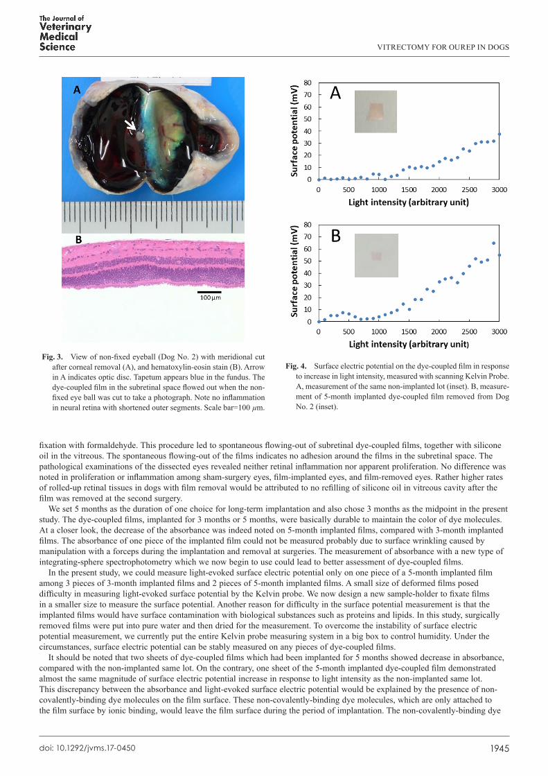
Light-evoked surface electric potential could be measured only in one film implanted for 5 months. In that 5-month implanted film, surface electric potential increased in response to the increase of light intensity (Fig. 4). A similar pattern of increase in surface electric potential was observed in the measurement of the non-implanted same lot (Fig. 4). Mean surface electric potential with standard deviation of the 5-month implanted film at the light intensity of dial 2500 (300 lux) was 27.12 ± 11.66 mV in repeat measurements of 11 times, ranging from 10.77 to 44.00 mV. Mean surface electric potential with standard deviation of the same non-implanted lot was 30.43 ± 4.05 mV in repeat measurements of 3 times, ranging from 25.76 to 32.81 mV.
DISCUSSION
The goals of this study are two folds: the first to test surgical feasibility of subretinal film implantation by vitrectomy, and the second to test the durability of implanted films. We used normal beagle dogs for this study because dogs have been frequently used in other studies for preclinical testing of medical devices such as artificial joints. In addition, there have been reports on cataract surgery in dogs [17] as well as vitrectomy to treat retinal detachment in dogs [4, 6, 18, 19] at veterinary clinical settings. An experimental study which used dogs for vitrectomy was also described [5].
A major problem in vitrectomy for dogs is the presence of a large vascularized nictitating membrane on the nasal side. In addition, many species of animals, including dogs, have a common feature which is not encountered in human eyes: the conjunctiva around the cornea cannot be exposed at the entire 360 circumference by placing a lid speculum. The limitation in conjunctival exposure naturally results in the narrow surgical field to insert three trocars for vitrectomy instruments. Surgical approaches had to be taken in the superotemporal one-third circumference where all three trocars were placed. The 25G trocars could be inserted over the conjunctiva straight through the thicker sclera into the vitreous with more magnitude of rotating and pushing force, compared with human eyes. Other feature, noted in dog surgery, is the tendency of more bleeding from the scleral incision. Bleeding could be managed by intensive bipolar coagulation. Surgical techniques designed in this study to implant dye-coupled films in canine eyes would help future application of retinal prosthesis as treatment for retinal dystrophies in dogs [16].
As for pathological examination, enucleated eyes were dissected along meridional lines to observe the entire retina before
VITRECTOMY FOR OUREP IN DOGS
1945doi: 10.1292/jvms.17-0450
fixation with formaldehyde. This procedure led to spontaneous flowing-out of subretinal dye-coupled films, together with silicone oil in the vitreous. The spontaneous flowing-out of the films indicates no adhesion around the films in the subretinal space. The pathological examinations of the dissected eyes revealed neither retinal inflammation nor apparent proliferation. No difference was noted in proliferation or inflammation among sham-surgery eyes, film-implanted eyes, and film-removed eyes. Rather higher rates of rolled-up retinal tissues in dogs with film removal would be attributed to no refilling of silicone oil in vitreous cavity after the film was removed at the second surgery.
We set 5 months as the duration of one choice for long-term implantation and also chose 3 months as the midpoint in the present study. The dye-coupled films, implanted for 3 months or 5 months, were basically durable to maintain the color of dye molecules. At a closer look, the decrease of the absorbance was indeed noted on 5-month implanted films, compared with 3-month implanted films. The absorbance of one piece of the implanted film could not be measured probably due to surface wrinkling caused by manipulation with a forceps during the implantation and removal at surgeries. The measurement of absorbance with a new type of integrating-sphere spectrophotometry which we now begin to use could lead to better assessment of dye-coupled films.
In the present study, we could measure light-evoked surface electric potential only on one piece of a 5-month implanted film among 3 pieces of 3-month implanted films and 2 pieces of 5-month implanted films. A small size of deformed films posed difficulty in measuring light-evoked surface potential by the Kelvin probe. We now design a new sample-holder to fixate films in a smaller size to measure the surface potential. Another reason for difficulty in the surface potential measurement is that the implanted films would have surface contamination with biological substances such as proteins and lipids. In this study, surgically removed films were put into pure water and then dried for the measurement. To overcome the instability of surface electric potential measurement, we currently put the entire Kelvin probe measuring system in a big box to control humidity. Under the circumstances, surface electric potential can be stably measured on any pieces of dye-coupled films.
It should be noted that two sheets of dye-coupled films which had been implanted for 5 months showed decrease in absorbance, compared with the non-implanted same lot. On the contrary, one sheet of the 5-month implanted dye-coupled film demonstrated almost the same magnitude of surface electric potential increase in response to light intensity as the non-implanted same lot. This discrepancy between the absorbance and light-evoked surface electric potential would be explained by the presence of non-covalently-binding dye molecules on the film surface. These non-covalently-binding dye molecules, which are only attached to the film surface by ionic binding, would leave the film surface during the period of implantation. The non-covalently-binding dye
Fig. 3. View of non-fixed eyeball (Dog No. 2) with meridional cut after corneal removal (A), and hematoxylin-eosin stain (B). Arrow in A indicates optic disc. Tapetum appears blue in the fundus. The dye-coupled film in the subretinal space flowed out when the non-fixed eye ball was cut to take a photograph. Note no inflammation in neural retina with shortened outer segments. Scale bar=100 µm.
Fig. 4. Surface electric potential on the dye-coupled film in response to increase in light intensity, measured with scanning Kelvin Probe. A, measurement of the same non-implanted lot (inset). B, measure-ment of 5-month implanted dye-coupled film removed from Dog No. 2 (inset).

T. MATSUO ET AL.
1946doi: 10.1292/jvms.17-0450
molecules do contribute to the absorbance of the film but do not contribute to light-evoked surface electric potential.In conclusion, dye-coupled films were implanted successfully in the subretinal space by vitrectomy in dogs. The dye-coupled
films implanted for 3 or 5 months maintained the dye color, and one piece of the 5-month implanted films showed the increase of light-evoked surface electric potential in response to increasing light intensity. Technical refinement, regarding spectrophotometry and Kelvin probe measurement, is now in progress to assess the deformed dye-coupled films in a small size which have been implanted for a while in the eye and been removed. Overall, this study proved surgical feasibility of subretinal implantation and 5-month durability of retinal prosthesis, OURePTM. The filing of a first-in-human clinical trial for OURePTM is now negotiated at Pharmaceuticals and Medical Devices Agency (PMDA) in Japan. In future, OURePTM would be also applicable to treat retinal dystrophies in dogs from the viewpoint of veterinary medicine.
ACKNOWLEDGMENTS. This study was supported by a grant for the Translational Research Network Program from the Japan Agency for Medical Research and Development (AMED). We thank Chie Matsuo, DDS, PhD, for preparation of figures.
REFERENCES
1. Alamusi, Matsuo, T., Hosoya, O. and Uchida, T. 2017. Visual evoked potential in RCS rats with Okayama University-type retinal prosthesis (OUReP™) implantation. J. Artif. Organs 20: 158–165. [Medline] [CrossRef]
2. Alamusi, Matsuo, T., Hosoya, O., Tsutsui, K. M. and Uchida, T. 2015. Vision maintenance and retinal apoptosis reduction in RCS rats with Okayama University-type retinal prosthesis (OUReP™) implantation. J. Artif. Organs 18: 264–271. [Medline] [CrossRef]
3. Alamusi, M., Matsuo, T., Hosoya, O., Tsutsui, K. M. and Uchida, T. 2013. Behavior tests and immunohistochemical retinal response analyses in RCS rats with subretinal implantation of Okayama-University-type retinal prosthesis. J. Artif. Organs 16: 343–351. [Medline] [CrossRef]
4. Grahn, B. H., Barnes, L. D., Breaux, C. B. and Sandmeyer, L. S. 2007. Chronic retinal detachment and giant retinal tears in 34 dogs: outcome comparison of no treatment, topical medical therapy, and retinal reattachment after vitrectomy. Can. Vet. J. 48: 1031–1039. [Medline]
5. Hayashi, A., Usui, S., Kawaguchi, K., Fujioka, S., Kusaka, S., Fujikado, T., Ohji, M. and Tano, Y. 2000. Retinal changes after retinal translocation surgery with scleral imbrication in dog eyes. Invest. Ophthalmol. Vis. Sci. 41: 4288–4292. [Medline]
6. Hoffman, A., Wolfer, J., Occelli, L., Lehenbauer, T. W., Sapienza, J., Novak, J. M., Combs, K. L. and Konrade, K. A. 2012. Refractive state following retinal reattachment and silicone oil tamponade in dogs. Am. J. Vet. Res. 73: 1299–1304. [Medline] [CrossRef]
7. Humayun, M. S., Dorn, J. D., da Cruz, L., Dagnelie, G., Sahel, J. A., Stanga, P. E., Cideciyan, A. V., Duncan, J. L., Eliott, D., Filley, E., Ho, A. C., Santos, A., Safran, A. B., Arditi, A., Del Priore, L. V., Greenberg R. J., Argus II Study Group 2012. Interim results from the international trial of Second Sight’s visual prosthesis. Ophthalmology 119: 779–788. [Medline] [CrossRef]
8. Liu, S., Matsuo, T., Hosoya, O. and Uchida, T. 2017. Photoelectric dye used for Okayama University-type retinal prosthesis reduces the apoptosis of photoreceptor cells. J. Ocul. Pharmacol. Ther. 33: 149–160. [Medline] [CrossRef]
9. Loewenstein, J. I., Montezuma, S. R. and Rizzo, J. F. 3rd. 2004. Outer retinal degeneration: an electronic retinal prosthesis as a treatment strategy. Arch. Ophthalmol. 122: 587–596. [Medline] [CrossRef]
10. Matsuo, T. 2003. A simple method for screening photoelectric dyes towards their use for retinal prostheses. Acta Med. Okayama 57: 257–260. [Medline]
11. Matsuo, T. 2014. Intraocular lens implantation in unilateral congenital cataract with minimal levels of persistent fetal vasculature in the first 18 months of life. Springerplus 3: 361. [Medline] [CrossRef]
12. Matsuo, T. and Morimoto, N. 2007. Visual acuity and perimacular retinal layers detected by optical coherence tomography in patients with retinitis pigmentosa. Br. J. Ophthalmol. 91: 888–890. [Medline] [CrossRef]
13. Matsuo, T., Dan-oh, Y. and Suga, S. (Inventors). 2006. Agent for inducing receptor potential. Assignee: Okayama University. United States Patent. Patent No.: US 7,101,533 B2. Date of Patent: Sep. 5, 2006.
14. Matsuo, T., Uchida, T. and Takarabe, K. 2009. Safety, efficacy, and quality control of a photoelectric dye-based retinal prosthesis (Okayama University-type retinal prosthesis) as a medical device. J. Artif. Organs 12: 213–225. [Medline] [CrossRef]
15. Okamoto, K., Matsuo, T., Tamaki, T., Uji, A. and Ohtsuki, H. 2008. Short-term biological safety of a photoelectric dye used as a component of retinal prostheses. J. Artif. Organs 11: 45–51. [Medline] [CrossRef]
16. Petersen-Jones, S. M. and Komáromy, A. M. 2015. Dog models for blinding inherited retinal dystrophies. Hum. Gene Ther. Clin. Dev. 26: 15–26. [Medline] [CrossRef]
17. Smith, P. J., Brooks, D. E., Lazarus, J. A., Kubilis, P. S. and Gelatt, K. N. 1996. Ocular hypertension following cataract surgery in dogs: 139 cases (1992-1993). J. Am. Vet. Med. Assoc. 209: 105–111. [Medline]
18. Spatola, R. A., Nadelstein, B., Leber, A. C. and Berdoulay, A. 2015. Preoperative findings and visual outcome associated with retinal reattachment surgery in dogs: 217 cases (275 eyes). Vet. Ophthalmol. 18: 485–496. [Medline] [CrossRef]
19. Steele, K. A., Sisler, S. and Gerding, P. A. 2012. Outcome of retinal reattachment surgery in dogs: a retrospective study of 145 cases. Vet. Ophthalmol. 15 Suppl 2: 35–40. [Medline] [CrossRef]
20. Tamaki, M. and Matsuo, T. 2011. Optical coherence tomographic parameters as objective signs for visual acuity in patients with retinitis pigmentosa, future candidates for retinal prostheses. J. Artif. Organs 14: 140–150. Erratum 14: 385.
21. Tamaki, T., Matsuo, T., Hosoya, O., Tsutsui, K. M., Uchida, T., Okamoto, K., Uji, A. and Ohtsuki, H. 2008. Glial reaction to photoelectric dye-based retinal prostheses implanted in the subretinal space of rats. J. Artif. Organs 11: 38–44. [Medline] [CrossRef]
22. Uchida, T., Ishimaru, S., Shimamura, K., Uji, A., Matsuo, T. and Ohtsuki, H. 2005. Immobilization of photoelectric dye on the polyethylene film surface. Mem. Fac. Eng. Okayama Univ. 39: 16–20.
23. Uji, A., Matsuo, T., Uchida, T., Shimamura, K. and Ohtsuki, H. 2006. Intracellular calcium response and adhesiveness of chick embryonic retinal neurons to photoelectric dye-coupled polyethylene films as prototypes of retinal prostheses. Artif. Organs 30: 695–703. [Medline] [CrossRef]
24. Uji, A., Matsuo, T., Ishimaru, S., Kajiura, A., Shimamura, K., Ohtsuki, H., Dan-oh, Y. and Suga, S. 2005. Photoelectric dye-coupled polyethylene film as a prototype of retinal prostheses. Artif. Organs 29: 53–57. [Medline] [CrossRef]


