Surgery 2012 v2 - scottsnotes.co.ukscottsnotes.co.uk/PDFs/Scott's Notes - Surgery 2012v2.pdf · •...
163
Surgery 2012 v2 Alasdair Scott BSc (Hons) MBBS MRCS PhD 2018 [email protected] www.scottsnotes.co.uk © Alasdair Scott, 2018
Transcript of Surgery 2012 v2 - scottsnotes.co.ukscottsnotes.co.uk/PDFs/Scott's Notes - Surgery 2012v2.pdf · •...
Surgery 2012 v2
Alasdair Scott BSc (Hons) MBBS MRCS PhD
2018 [email protected]
www.scottsnotes.co.uk
© Alasdair Scott, 2018

© Alasdair Scott, 2018

i
Table of Contents 1. Perioperative Management ..................................................................................... 1 2. Fluids and Nutrition ............................................................................................... 11 3. Trauma .................................................................................................................. 17 4. Upper GI Surgery .................................................................................................. 25 5. Hepatobiliary Surgery ............................................................................................ 35 6. Lower GI Surgery .................................................................................................. 43 7. Perianal Surgery .................................................................................................... 59 8. Hernias .................................................................................................................. 65 9. Superficial Lesions ................................................................................................ 70 10. Breast Surgery .................................................................................................... 81 11. Vascular Surgery ................................................................................................. 86 12. Urology ................................................................................................................ 95 13. Orthopaedics ..................................................................................................... 110 14. Ear, Nose and Throat ........................................................................................ 132 15. Ophthalmology .................................................................................................. 145
© Alasdair Scott, 2018

© Alasdair Scott, 2018

1
Perioperative Management
Contents Pre-Operative Assessment and Planning .............................................................................................................. 2Specific Pre-operative Complications .................................................................................................................... 3Anaesthesia ........................................................................................................................................................... 4Analgesia ............................................................................................................................................................... 4Enhanced Recovery After Surgery ........................................................................................................................ 5Surgical Complications .......................................................................................................................................... 5Post-op Complications: General ............................................................................................................................ 6Post-op Complications: Specific ............................................................................................................................ 7Post-op Pyrexia ...................................................................................................................................................... 8Deep Venous Thrombosis ..................................................................................................................................... 9Other Common Post-Operative Presentations .................................................................................................... 10
© Alasdair Scott, 2018
2
Pre-Operative Assessment and Planning Aims
• Informed consent• Assess risk vs. benefits• Optimise fitness of patient• Check anaesthesia / analgesia type c̄ anaesthetist
Pre-op Checks: OP CHECS • Operative fitness: cardiorespiratory comorbidities• Pills• Consent• History
§ MI, asthma, HTN, jaundice§ Complications of anaesthesia: DVT,
anaphylaxis• Ease of intubation: neck arthritis, dentures, loose
teeth• Clexane: DVT prophylaxis• Site: correct and marked
Drugs
Anti-coagulants • Balance risk of haemorrhage c̄ risk of thrombosis• Avoid epidural, spinal and regional blocks
AED • Give as usual• Post-op give IV or via NGT if unable to tolerate orally
OCP / HRT • Stop 4wks before major / leg surgery• Restart 2wks post-op if mobile
β-Blockers • Continue as usual
Pre-op Investigations
Bloods • Routine: FBC, U+E, G+S, clotting, glucose• Specific
§ LFTs: liver disease, EtOH, jaundice§ TFT: thyroid disease§ Se electrophoresis: Africa, West Indies, Med
• Cross-match§ Gastrectomy: 4u§ AAA: 6u
Cardiopulmonary Function • CXR: cardiorespiratory disease/symptoms, >65yrs• Echo: poor LV function, Ix murmurs• ECG: HTN, Hx of cardiac disease, >55yrs• Cardiopulmonary Exercise Testing• PFT: known pulmonary disease or obesity
Other • Lat C-spine flexion and extension views: RA, AS• MRSA swabs
Preparation
NBM • ≥2h for clear fluids, ≥6h for solids
Bowel Prep • May be needed in left-sided ops
§ Picolax: picosulfate and Mg citrate§ Klean-Prep: macrogol
• Not usually needed in right-sided procedures• Necessity is controversial as benefit of minimising
post-op infection might not outweigh risks§ Liquid bowel contents spilled during surgery§ Electrolyte disturbance§ Dehydration§ ↑ rate of post-op anastomotic leak
Prophylactic Abx • Use
§ GI surgery (20% post-op infection if elective)§ Joint replacement
• Give 15-60min before surgery• Regimens: (see local guidelines)
§ Biliary: Cef 1.5g + Met 500mg IV§ CR or appendicetomy: Cef+Met TDS§ Vascular: co-amoxiclav 1.2g IV TDS§ MRSA+ve: vancomycin
DVT Prophylaxis • Stratify pts according to patient factors and type of
surgery.• Low risk: early mobilisation• Med: early mobilisation + TEDS + 20mg enoxaparin• High: early mobilisation + TEDS + 40mg enoxaparin +
intermittent compression boots perioperatively.• Prophylaxis started @ 1800 post-op• May continue medical prophylaxis at home (up to
1mo)
ASA Grades • Normally healthy• Mild systemic disease• Severe systemic disease that limits activity• Systemic disease which is a constant threat to life• Moribund: not expected to survive 24h even c̄ op
© Alasdair Scott, 2018
3
Specific Pre-operative Complications Diabetes ↑ Risk of post-operative complications
• Surgery → stress hormones → antagonise insulin • Pts. are NBM • ↑ risk of infection • IHD and PVD
Pre-op
• Dipstick: proteinuria • Venous glucose • U+E: K+
IDDM Practical Points
• Put pt. first on list and inform surgeon and anaesthetist
• Some centres prefer to use GKI infusions • Sliding scale may not be necessary for minor ops
§ If in doubt, liaise c̄ diabetes specialist nurse Insulin
• ± stop long-acting insulin the night before • Omit AM insulin if surgery is in the morning • Start sliding scale
§ 5% Dex c̄ 20mmol KCl 125ml/hr § Infusion pump c̄ 50u actrapid § Check CPG hrly and adjust insulin rate
• Check glucose hrly: aim for 7-11mM • Post-op
§ Continue sliding-scale until tolerating food § Switch to SC regimen around a meal
NIDDM
• If glucose control poor (fasting >10mM): treat as IDDM
• Omit oral hypoglycaemics on the AM of surgery • Eating post-op: resume oral hypoglycaemics c̄ meal • No eating post-op
§ Check fasting glucose on AM of surgery § Start insulin sliding scale § Consult specialist team ore. restarting PO Rx
Diet Controlled
• Usually no problem • Pt. may be briefly insulin-dependent post-op
§ Monitor CPG Steroids Risks
• Poor wound healing • Infection • Adrenal crisis
Mx
• Need to ↑ steroid to cope c̄ stress • Consider cover if high-dose steroids w/i last yr • Major surgery: hydrocortisone 50-100mg IV c̄ pre-
med then 6-8hrly for 3d. • Minor: as for major but hydrocortisone only for 24h
Jaundice
• Best to avoid operating in jaundiced pts. • Use ERCP instead
Risks
• Pts. c̄ obstructive jaundice have ↑ risk of post-op renal failure \ need to maintain good UO.
• Coagulopathy • ↑ infection risk: may → cholangitis
Pre-op
• Avoid morphine in pre-med • Check clotting and consider pre-op vitamin K • Give 1L NS pre-op (unless CCF) → moderate diuresis • Urinary catheter to monitor UPO • Abx prophylaxis: e.g. cef+met
Intra-op
• Hrly UO monitoring • NS titrated to output
Post-op
• Intensive monitoring of fluid status • Consider CVP + frusemide if poor output despite NS
Anticoagulated Patients
• Balance risk of haemorrhage c̄ risk of thrombosis • Consult surgeon, anaesthetist and haematologist • Very minor surgery may be undertaken w/o stopping
warfarin if INR <3.5. • Avoid epidural, spinal and regional blocks if
anticoagulated, • In general, continue aspirin/clopidogrel unless risk of
bleeding is high – then stop 7d before surgery Low thromboembolic risk: e.g. AF
• Stop warfarin 5d pre-op: need INR <1.5 • Restart next day
High thromboembolic risk: valves, recurrent VTE
• Need bridging c̄ LMWH § Stop warfarin 5d pre-op and start LMWH § Stop LMWH 12-18h pre-op § Restart LMWH 6h post-op § Restart warfarin next day § Stop LMWH when INR >2
Emergency Surgery
• Discontinue warfarin • Vit K .5mg slow IV • Request FFP or PCC to cover surgery
COPD and Smoking Risks
• Basal atelectasis • Aspiration • Chest infection
Pre-op
• CXR • PFTs • Physio for breathing exercises • Quit smoking (at least 4wks prior to surgery)
© Alasdair Scott, 2018

4
Anaesthesia Principals and Practical Conduct
• Aims: hypnosis, analgesia, muscle relaxation • Induction: e.g. IV propofol • Muscle Relaxation
§ Depolarising: suxamethonium § Non-depolarising: vecuronium, atracurium
• Airway Control: ET tube, LMA • Maintenance
§ Usually volatile agent added to N2O/O2 mix § E.g. halothane, enflurane
• End of Anaesthesia § Change inspired gas to 100% O2 § Reverse paralysis: neostigmine + atropine
(prevent muscarinic side effects) Pre-medication: 7As
• Anxiolytics and Amnesia: e.g. temazepam • Analgesics: e.g. opioids, paracetamol, NSAIDs • Anti-emetics: e.g. ondansetron 4mg / metoclop 10mg • Antacids: e.g. lansoprazole • Anti-sialogue e.g. glycopyrolate (↓ secretions) • Antibiotics
Regional Anaesthesia
• May be used for minor procedures or if unsuitable for GA
• Nerve or spinal blocks § CI: local infection, clotting abnormality
• Use long-acting agents: e.g. bupivacaine Complications of Anaesthesia Propofol Induction
• Cardiorespiratory depression Intubation
• Oro-pharyngeal injury c̄ laryngoscope • Oesophageal intubation
Loss of pain sensation
• Urinary retention • Pressure necrosis • Nerve palsies
Loss of muscle power
• Corneal abrasion • No cough → atelectasis + pneumonia
Malignant Hyperpyrexia
• Rare complication ppted by halothane or suxamethonium
• AD inheritance • Rapid rise in temperature + masseter spasm • Rx: dantrolene + cooling
Anaphylaxis
• Rare • Possible triggers
§ Antibiotics § Colloid § NM blockers: e.g. vecuronium
Analgesia Necessity
• Pain → autonomic activation → arteriolar constriction → ↓ wound perfusion → impaired wound healing
• Pain → ↓ mobilisation → ↑ VTE and ↓ function • Pain → ↓ respiratory excursion and ↓ cough →
atelectasis and pneumonia • Humanitarian considerations
General Guidance
• Give regular doses at fixed intervals • Consider best route: oral when possible • PCA should be considered: morphine, fentanyl • Follow stepwise approach • Liaise c̄ Acute Pain Service
Pre-Op
• Epidural anaesthesia: e.g. c̄ bupivacaine End-Op
• Infiltrate wound edge c̄ LA • Infiltrate major regional nerves c̄ LA
Post-Op: stepwise approach
1. Non-opioid ± adjuvants § Paracetamol § NSAIDs
- Ibuprofen: 400mg/6h PO max - Diclofenac: 50mg PO / 75mg IM
2. Weak opioid + non-opioid ± adjuvants
§ Codeine § Dihydrocodeine § Tramadol
3. Strong opioid + non-opioid ± adjuvants
§ Morphine: 5-10mg/2h max § Oxycodone § Fentanyl
Spinal or Epidural Anaesthesia
• ↓ SE as drugs more localised • 1st line for major bowel resection • Caution
§ Respiratory depression § Neurogenic shock → ↓BP
© Alasdair Scott, 2018

5
Enhanced Recovery After Surgery ERAS
• Commonly employed in colorectal and orthopaedic surgery
Aims
• Optimise pre-op preparation for surgery • Avoid iatrogenic problems (e.g. ileus) • Minimise adverse physiological / immunological
responses to surgery § ↑ cortisol and ↓ insulin (absolute or relative) § Hypercoagulability § Immunosuppression
• ↑ speeded of recovery and return to function • Recognise abnormal recovery and allow early
intervention Pre-op: optimisation
• Aggressive physiological optimisation § Hydration § BP (↑ / ↓) § Anaemia § DM § Co-morbidities
• Smoking cessation: ≥4wks before surgery • Admission on day of surgery, avoidance of prolonged fast • Carb loading prior to surgery: e.g. carb drinks • Fully informed pt., encouraged to participate in recovery
Intra-op: ↓ physical stress
• Short-acting anaesthetic agents • Epidural use • Minimally invasive techniques • Avoid drains and NGTs where possible
Post-op: early return to function and mobilisation
• Aggressive Rx of pain and nausea • Early mobilisation and physiotherapy • Early resumption of oral intake (inc. carb drinks) • Early discontinuation of IV fluids • Remove drains and urinary catheters ASAP
Surgical Complications Immediate (<24h)
• Intubation → oropharyngeal trauma • Surgical trauma to local structures • Primary or reactive haemorrhage
Early (1d-1mo)
• Secondary haemorrhage • VTE • Urinary retention • Atelectasis and pneumonia • Wound infection and dehiscence • Antibiotic association colitis (AAC)
Late (>1mo)
• Scarring • Neuropathy • Failure or recurrence
© Alasdair Scott, 2018
6
Post-op Complications: General Haemorrhage Classification
• Primary: continuous bleeding starting during surgery
• Reactive § Bleeding at the end of surgery or early
post-op § 2O to ↑ CO and BP
• Secondary § Bleeding >24h post-op § Usually due to infection
Post-op Urinary Retention Causes
• Drugs: opioids, epidural/spinal, anti-AChM • Pain: sympathetic activation → sphincter
contraction • Psychogenic: hospital environment
Risk Factors
• Male • ↑ age • Neuropathy: e.g. DM, EtOH • BPH • Surgery type: hernia and anorectal
Mx
• Conservative § Privacy § Ambulation § Void to running taps or in hot bath § Analgesia
• Catheterise ± gent 2.5mg/kg IV stat • TWOC = Trial w/o Catheter
§ If failed, may be sent home c̄ silicone catheter and urology outpt. f/up.
Pulmonary Atelectasis
• Occurs after every nearly every GA • Mucus plugging + absorption of distal air →
collapse Causes
• Pre-op smoking • Anaesthetics ↑ mucus production ↓ mucociliary
clearance • Pain inhibits respiratory excursion and cough
Presentation
• w/i first 48hrs • Mild pyrexia • Dyspnoea • Dull bases c̄ ↓AE
Mx
• Good analgesia to aid coughing • Chest physiotherapy
Wound Infection
• 5-7d post-op • Organisms: S. aureus and Coliforms
Operative Classification
• Clean: incise uninfected skin w/o opening viscus • Clean/Cont: intra-op breach of viscus (not colon) • Contaminated: breach of viscus + spillage or opening of
colon • Dirty: site already contaminated – faeces, pus, trauma
Risk Factors
• Pre-operative § ↑ Age § Comorbidities: e.g. DM § Pre-existing infection: e.g. appendix perforation § Pt. colonisation: e.g. nasal MRSA
• Operative § Op classification and wound infection risk § Duration § Technical: pre-op Abx, asepsis
• Post-operative § Contamination of wound from staff
Mx
• Regular wound dressing • Abx • Abscess drainage
Wound Dehiscence Presentation
• Occurs ~10d post-op • Preceded by serosanguinous discharge from wound
Risk Factors
• Pre-Operative Factors § ↑ age § Smoking § Obesity, malnutrition, cachexia § Comorbs: e.g. BM, uraemia, chronic cough, Ca § Drugs: steroids, chemo, radio
• Operative Factors § Length and orientation of incision § Closure technique: follow Jenkin’s Rule § Suture material
• Post-operative Factors § ↑ IAP: e.g. prolonged ileus → distension § Infection § Haematoma / seroma formation
Mx
• Replace abdo contents and cover c̄ sterile soaked gauze • IV Abx: cef+met • Opioid analgesia • Call senior and arrange theatre • Repair in theatre
§ Wash bowel § Debride wound edges § Close c̄ deep non-absorbable sutures (e.g. nylon)
• May require VAC dressing or grafting
© Alasdair Scott, 2018
7
Post-op Complications: Specific General Surgery Cholecystectomy
• Conversion to open: 5% • CBD injury: 0.3% • Bile leak • Retained stones (needing ERCP) • Fat intolerance / loose stools
Inguinal Hernia Repair
• Early • Haematoma / seroma formation: 10% • Intra-abdominal injury (lap) • Infection: 1% • Urinary retention • Late
§ Recurrence: 0.5% § Ischaemic orchitis: 0.5% § Chronic groin pain / paraesthesia: 10-20%
Appendicectomy
• Abscess formation • Fallopian tube trauma • Right hemicolectomy (e.g. for carcinoid, caecal
necrosis) Colonic Surgery
• Early § Ileus § Anastomotic leak § Enterocutaneous fistulae § Abdominal or pelvic abscess
• Late § Adhesions → obstruction § Incisional hernia
Post-op Ileus
• Causes § Bowel handling § Anaesthesia § Electrolyte imbalance
• Presentation § Distension § Constipation ± vomiting § Absent bowel sounds
• Rx § IV fluids + NGT § TPN if prolonged
Anorectal Surgery
• Anal incontinence • Stenosis • Anal fissure
Small Bowel Surgery
• Short gut syndrome (≤250cm) Splenectomy
• Gastric dilatation (2O gastric ileus) § Prevent c̄ NGT
• Thrombocytosis → VTE • Infection: encapsulated organisms
Vascular Arterial Surgery
• Thrombosis and embolisation • Anastomotic leak • Graft infection
Aortic Surgery
• Gut ischaemia • Renal failure • Aorto-enteric fistula • Anterior spinal syndrome (paraplegia) • Emboli → distal ischaemia (trash foot)
Breast
• Arm lymphoedema • Skin necrosis • Seroma
Urological
• Sepsis • Uroma: extravasation of urine
Prostatectomy
• Urinary incontinence • Erectile dysfunction • Retrograde ejaculation • Prostatitis
ENT Thyroidectomy
• Wound haematoma → tracheal obstruction • Recurrent laryngeal N. trauma → hoarse voice
§ Transient in 1.5% § Permanent in 0.5% § R commonest (more medial)
• Hypoparathyroidism → hypocalcaemia • Thyroid storm • Hypothyroidism
Tracheostomy
• Stenosis • Mediastinitis • Surgical emphysema
Orthopaedic Surgery Fracture Repair
• Mal-/non-union • Osteomyelitis • Avascular necrosis • Compartment syndrome
Hip Replacement
• Deep infection • VTE • Dislocation • Nerve injury: sciatic, SGN • Leg length discrepancy
Cardiothoracic Surgery
• Pneumo-/haemo-thorax • Infection: mediastinitis, empyema
© Alasdair Scott, 2018
8
Post-op Pyrexia Causes Early: 0-5d post-op
• Blood transfusion • Physiological: SIRS 2O to trauma, 0-1d • Pulmonary atelectasis: 24-48hrs • Infection: UTI, superficial thrombophlebitis, cellulitis • Drug reaction
Delayed: >5d post-op
• Pneumonia • VTE: 5-10d • Wound infection: 5-7d • Anastomotic leak: 7d • Collection: 5-20d
Examination of Post-Op Febrile Pt.
• Observation chart, notes and drug chart • Wound • Abdo + DRE • Legs • Chest • Lines • Urine • Stool
Ix
• Urine: dip + MCS • Blood: FBC, CRP, cultures ± LFTs • Cultures: wound swabs, CVP tip for culture • CXR
Pneumonia Cause
• Anaesthesia → atelectasis • Pain → ↓ cough • Surgery → immunosuppression
Rx
• Chest physio: encouraging coughing • Good analgesia • Abx
Collection Presentation
• Malaise • Swinging fever, rigors • Localised peritonitis • Shoulder tip pain (if subphrenic)
Locations
• Pelvic • Subphrenic • Paracolic gutters • Lesser sac • Hepatorenal recess (Morrison’s space) • Small bowel (interloop spaces)
Ix
• FBC, CRP, cultures • US, CT • Diagnostic lap
Rx
• Abx • Drainage / washout
Cellulitis
• Acute infection of the subcutaneous connective tissue Cause: β-haemolytic Streps + staph. aureus Presentation
• Pain, swelling, erythema and warmth • Systemic upset • ± lymphadenopathy
Rx
• Benpen IV • Pen V and fluclox PO
© Alasdair Scott, 2018

9
Deep Venous Thrombosis Epidemiology
• DVTs occur in 25-50% of surgical patients without thromboprophylaxis
Risk Factors: Virchow’s Triad
• Blood Contents § Surgery → ↑ plats and ↑ fibrinogen § Dehydration § Malignancy § Age: ↑
• Blood Flow § Surgery § Immobility § Obesity
• Vessel Wall § Damage to veins: esp. pelvic veins § Previous VTE
Signs
• Peak incidence @ 5-10d post-op • 65% of below knee DVTs are asymptomatic • Calf warmth, tenderness, erythema, swelling • Mild pyrexia • Pitting oedema
Differential
• Cellulitis • Ruptured Baker’s cyst
Ix
• D-Dimers: sensitive but not specific • Compression US (clot will be incompressible) • Thrombophilia screen if:
§ No precipitating factors § Recurrent DVT § Family Hx
Dx
• Assess probability using Wells’ Score • Low-probability → perform D-dimers
§ Negative → excludes DVT § Positive → Compression US
• Med / High probability → Compression US Rx Anticoagulate
• Therapeutic LMWH: enoxaparin 1.5mg/kg/24h SC • Start warfarin using Tait model: 5mg OD for first 4d • Stop LMWH when INR 2.5 • Duration
§ Below knee: 6-12wks § Above knee: 3-6mo § On-going cause: indefinite
Graduated Compression Stockings
• Consider for prevention of post-phlebitic syndrome
Preventing DVT Pre-Op
• Pre-op VTE risk assessment • TED stockings • Aggressive optimisation: esp. hydration • Stop OCP 4wks pre-op
Intra-Op
• Minimise length of surgery • Use minimal access surgery where possible • Intermittent pneumatic compression boots
Post-Op
• LMWH • Early mobilisation • Good analgesia • Physio • Adequate hydration
© Alasdair Scott, 2018
10
Other Common Post-Operative Presentations Dyspnoea / Hypoxia Causes
• Previous lung disease • Atelectasis, aspiration, pneumonia • LVF • PE • Pneumothorax (e.g. due to CVP line insertion) • Pain → hypoventilation
Ix
• FBC, ABG • CXR • ECG
Rx
• Sit up, give O2, monitor SpO2 • Rx cause
Reduced Urine Output Causes
• Post-renal § Commonest cause § Blocked / malsited catheter § Acute urinary retention
• Pre-renal: hypovolaemia • Renal: NSAIDs, gentamicin • Anuria usually = blocked or malsited catheter • Oliguria usually = inadequate fluid replacement
Mx
• Information § Op Hx § Obs chart: UO § Drug chart: nephrotoxins
• Examination § Assess fluid status § Examine for palpable bladder § Inspect drips, drains, stomas, CVP
• Action § Flush c̄ 50ml NS and aspirate back § Fluid challenge
Nausea and Vomiting
• Causes § Obstruction § Ileus § Emetic drugs: e.g. opioids
• Consider NGT, AXR and ondansetron 4mg IV TDS ↓ Na
• What was pre-op level? • Common Causes:
§ S(I)ADH: pain, nausea, opioids, stress § Over administration of IV fluids
• Correct slowly § Acute: 1mM/h § Chronic:15mM/d
Hypotension Immediate Mx
• Tilt bed head down, give O2 • Assess fluid status
Causes: CHOD
• Cardiogenic § MI § Fluid overload
• Hypovolaemia § Inadequate replacement of fluid losses § Haemorrhage
• Obstructive § PE
• Distributive § Sepsis § Neurogenic shock
Mx
• Hypovolaemia → fluid challenge § 250-500ml colloid over 15-30min
• Haemorrhage → return to theatre • Sepsis → fluid challenge, start Abx • Overload → frusemide • Neurogenic → NA infusion
Hypertension
• Continue anti-hypertensives during peri-operative period
Causes
• Pain • Urinary retention • Previous HTN
Rx
• Rx cause • May use labetalol 50mg IV every 5min (200mg max)
Acute Confusional State
• Agitation, disorientation, attempts to leave hospital Common Causes: DELIRIUM
• Drugs: opiates, sedatives, L-DOPA • Eyes, ears and other sensory deficits • Low O2 states: MI, stroke, PE • Infection • Retention: stool or urine • Ictal • Under- hydration / -nutrition • Metabolic: Na, AKI, glucose, EtOH withdrawal
Mx
• May need sedation: midazolam / haldol • Nurse in well-lit environment • Rx cause
© Alasdair Scott, 2018

11
Fluids and Nutrition Contents
Fluid Homeostasis ............................................................................................................................................... 12Crystalloid ............................................................................................................................................................ 13Colloid .................................................................................................................................................................. 13Fluid Problems ..................................................................................................................................................... 14Nutrition ................................................................................................................................................................ 15Refeeding Syndrome ........................................................................................................................................... 16
© Alasdair Scott, 2018
12
Fluid Homeostasis Body Composition
• Total water: 60% of 70kg = 42L § 2/3 intracellular = 28L § 1/3 extracellular = 14L
- Plasma = 3L (21% of ECF) - Interstitial = 10L - Transcellular = 1L
Starling’s Forces Osmotic Pressure
• Pressure which needs to be applied to prevent the inflow of water across a semipermeable membrane.
• i.e. the ability of a solute to attract water. • Oncotic pressure: form of osmotic pressure exerted
by proteins. Hydrostatic Pressure
• Pressure exerted by a fluid at equilibrium due to the force of gravity.
Distribution
• Distribution between the ECF and ICF is driven by differences in osmotic pressure only.
• Distribution w/i the ECF is determined by Starling’s forces.
§ Capillary and interstitial oncotic pressure. § Capillary and interstitial hydrostatic pressure. § Filtration coefficient (capillary permeability)
3rd Space Losses → ↓ ECF
• Bowel obstruction → ↓ fluid reabsorption → 3rd space loss
• Sudden diuresis on day 2-3 post op = recovery of ileus
• Peritonitis → ascites → 3rd space loss
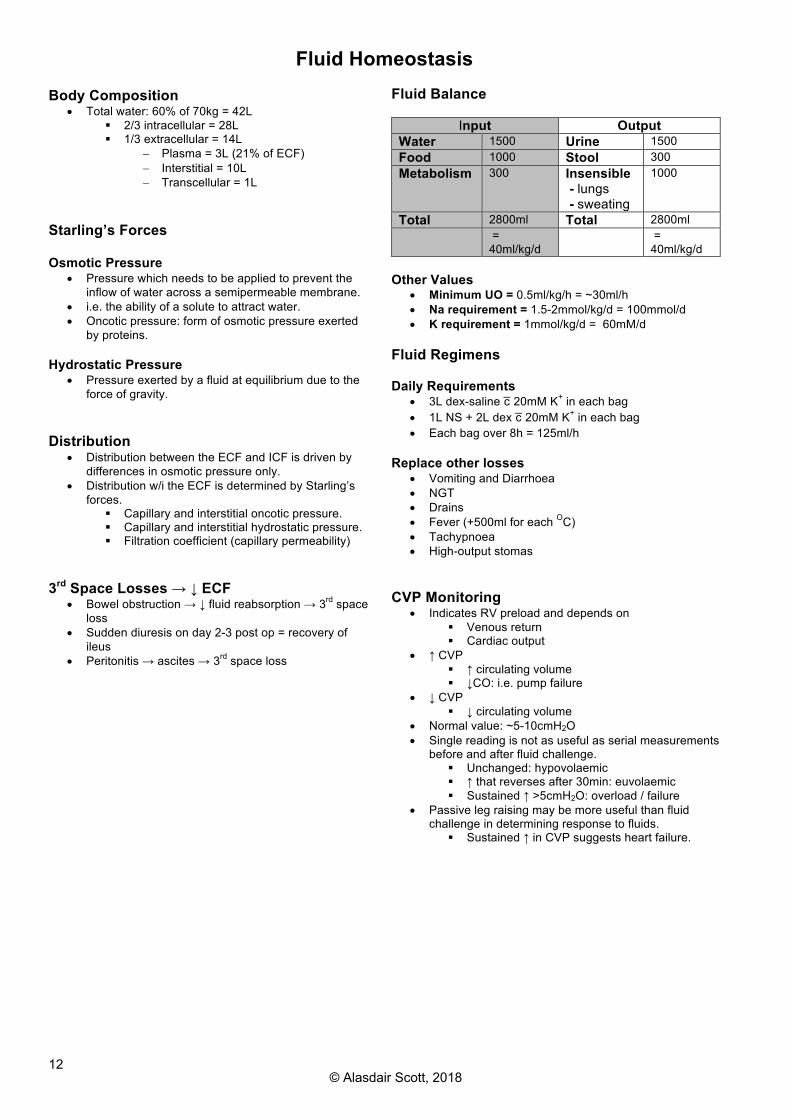
Fluid Balance
Input Output Water 1500 Urine 1500 Food 1000 Stool 300 Metabolism 300 Insensible
- lungs - sweating
1000
Total 2800ml Total 2800ml =
40ml/kg/d =
40ml/kg/d Other Values
• Minimum UO = 0.5ml/kg/h = ~30ml/h • Na requirement = 1.5-2mmol/kg/d = 100mmol/d • K requirement = 1mmol/kg/d = 60mM/d
Fluid Regimens Daily Requirements
• 3L dex-saline c̄ 20mM K+ in each bag • 1L NS + 2L dex c̄ 20mM K+ in each bag • Each bag over 8h = 125ml/h
Replace other losses
• Vomiting and Diarrhoea • NGT • Drains • Fever (+500ml for each OC) • Tachypnoea • High-output stomas
CVP Monitoring
• Indicates RV preload and depends on § Venous return § Cardiac output
• ↑ CVP § ↑ circulating volume § ↓CO: i.e. pump failure
• ↓ CVP § ↓ circulating volume
• Normal value: ~5-10cmH2O • Single reading is not as useful as serial measurements
before and after fluid challenge. § Unchanged: hypovolaemic § ↑ that reverses after 30min: euvolaemic § Sustained ↑ >5cmH2O: overload / failure
• Passive leg raising may be more useful than fluid challenge in determining response to fluids.
§ Sustained ↑ in CVP suggests heart failure.
© Alasdair Scott, 2018
13
Crystalloid Normal Saline Contents
• 0.9% NaCl = 9g/L • 154mmol NaCl
pH: 5-6 Use
• Normal daily fluid requirements + replace losses 5% Dextrose Contents
• 50g dextrose /L Use
• Normal daily fluid requirements Dextrose-Saline Contents
• 4% dextrose = 40g/L • 0.18% NaCl = 31mM NaCl
Use
• Normal daily fluid requirements Hartmann’s / Ringer’s Lactate Contents
• Na: 131mM • Cl: 111mM • K: 5mM • Ca: 2.2mM • Lactate / HCO3: 29mM
Use
• Resuscitation in trauma pts. • Parkland’s formula: 4 x wt x %burn = mL in 1st 24hrs
pH
• pH = 6.5 but Hartmann’s is an alkalinising solution • Lactate is not an acid in itself: it’s a conjugate base • Given exogenously as sodium lactate • Lactate metabolised in liver → HCO3 production • The Cori Cycle
Daily Requirements
• 3L dex-saline c̄ 20mM K+ in each bag • 1L NS + 2L dex c̄ 20mM K+ in each bag • Each bag over 8h = 125ml/h
Problems
• Give 1L NS → ~210ml remaining intravascularly • Give 1L D5W → ~70ml remaining intravascularly • Acidosis or electrolyte disturbances • Fluid overload
Colloid Physiology
• Contain large molecular wt. molecules • Gelatin • Dextrans • Preserves oncotic pressure \ remains intravascular →
preferential ↑ in intravascular volume Synthetic
• Gelofusin • Volplex • Haemaccel • Voluven
Natural
• Albumin • Blood
Use
• Fluid challenge: 250-500ml over 15-30min • Hypovolaemic shock • Mount Vernon Formula
§ (wt. x %burn)/2 = ml colloid per unit time Problems
• Anaphylaxis • Volume overload • Can interfere c̄ cross-matching therefore take blood for
x-match before using.
© Alasdair Scott, 2018

14
Fluid Problems Assessing Fluid Status
• Hx: balance chart, surgery, other losses, thirsty • Impression: drowsy, alert • Inspection: drips, drains, stomas, catheters, CVP
Examination • IV volume
§ CRT § HR § BP lying and standing § JVP
• Tissue perfusion § Skin turgor § Oedema: ankle, pulmonary, ascites § Mucus membranes
• End-organ § UO, ↑U+Cr § Consciousness § Lactate
Other Tests
• PCWP: indirect measure of left atrial pressure • CVP
Post-operative Fluids Problems
• ↑ADH, ↑aldosterone, ↑cortisol → Na +H2O conservation • ↑ K+: tissue damage, transfusion, stress hormones
Solutions
• Use UO (aim>30ml/h) to guide fluid replacement but may need to ↓ maintenance fluids to 2L first 24h post-op
• Avoid K+ supplementation for first 24h post-op Cardiac or Renal Failure Problem
• RAS activation → Na and H2O retention Solution
• Avoid fluids c̄ Na → give 5% dextrose Bowel Obstruction
• Pts. have significant third space losses c̄ loss of both water and electrolytes.
• Likely to need significantly more than standard daily requirements.
• Regimen § 0.9% NS c̄ 20-40mm KCl added to each bag § Titrate rate of fluid therapy to clinical findings on
serial examination. § Serial U+Es guide electrolyte replacement
Pancreatitis
• Inflammation → significant fluid shift into the abdomen. • Pts require aggressive fluid resuscitation and
maintenance § Insert urinary catheter and consider CVP
monitoring § 0.9% NS c̄ 20-40mm KCl added to each bag § Keep UO >30ml/h § Serial U+Es guide electrolyte replacement
Ileostomy
• Ileal fluid composition § Na: 130mM § Cl: 110mM § K: 10mM § HCO3: 30mM
• Normal output: 10-15mL/Kg/d = ~700ml/d • High output = >1000ml/d • Ileum will adapt to limit fluid and electrolyte losses • Fluids
§ 0.9% NS +KCl § Daily requirements + replaces losses, titrated to
UO § Serial U+Es guide electrolyte replacement
• High Output § Loperamide § Codeine
Reduced Urine Output Post-op Causes
• Post-renal § Commonest cause § Blocked / malsited catheter § Acute urinary retention
• Pre-renal: hypovolaemia • Renal: NSAIDs, gentamicin • Anuria usually = blocked or malsited catheter • Oliguria usually = inadequate fluid replacement
Mx
• Information § Op Hx § Obs chart: UO § Drug chart: nephrotoxins
• Examination § Assess fluid status § Examine for palpable bladder § Inspect drips, drains, stomas, CVP
• Action § Flush c̄ 50ml NS and aspirate back § Fluid challenge
Suspect Catheter Problem
• Flush c̄ 50ml NS and aspirate back Suspect Pre-renal Problem
• Fluid challenge § 250-500ml colloid bolus over 15-30min § Look for CVP or UO response w/i minutes
© Alasdair Scott, 2018

15
Nutrition Assessment Clinical
• Hx § Wt. loss § Diet
• Examination § Skin fat § Dry hair § Pressure sores § Cheilitis § Wt. and BMI (<20kg/m2)
Anthropometric
• Skin-fold thickness • Arm circumference
Ix
• Albumin • Transthyretin (prealbumin) • Phosphate
Requirements (/kg/24h)
• Calories: 20-40 Kcal • Carb: 2g • Fat: 3g • Protein: 0.5-1g • Nitrogen:0.2-0.4g
Enteral Nutrition Delivery
• PO is best § Consider semi-solid diet if risk of aspiration
• Fine bore NGT (9 Fr) • Percutaneous Endoscopic Gastrostomy • Jejunostomy • Build up feeds gradually to prevent diarrhoea
Feeds
• Oral supplements • Polymeric: e.g. osmolite, jevity
§ Intact proteins, starches and long-chain FAs • Disease-specific
§ e.g. ↓ branched chain AAs in hepatic encephalopathy
• Elemental § Simple AAs and oligo/monosaccharides § Require minimal digestion and used if abnormal
GIT: e.g. in Crohn’s Indications
• Catabolic: sepsis, burns, major surgery • Coma/ITU • Malnutrition • Dysphagia: stricture, stroke
Complications
• NGT § Nasal trauma § Malposition or tube blockage
• Feeding § Feed intolerance → diarrhoea § Electrolyte imbalance § Aspiration § Refeeding syndrome
Parenteral Nutrition
• May be “Total” or used to supplement enteral feeding • Combined c̄ H2O to deliver total daily requirements
Indications
• Prolonged obstruction or ileus (>7d) • High output fistula • Short bowel syndrome • Severe Crohn’s • Severe malnutrition • Severe pancreatitis • Unable to swallow: e.g. oesophageal Ca
Delivery
• Delivered centrally as high osmolality is toxic to veins § Short-term: CV catheter § Long-term: Hickman or PICC line
• Sterility is essential: use line only for PN Monitoring
• Standard § Wt., fluid balance and urine glucose daily § Zn, Mg weekly
• Initially § Blood glucose, FBC, U+E + PO4 3x /wk § LFTs 3x /wk
• Once stable § Blood glucose, FBC, U+E + PO4 daily § LFTs weekly
Contents
• 2000Kcal: 50% fat, 50% carb • 10-14g nitrogen • Vitamins, minerals and trace elements
Complications
• Line-related § Pneumothorax / haemothorax § Cardiac arrhythmia § Line sepsis § Central venous thrombosis → PE or SVCO
• Feed-related § Villous atrophy of GIT § Electrolyte disturbances
- Refeeding syndrome - Hypercapnoea from excessive CO2
production § Hyperglycaemia and reactive hypoglycaemia § Line sepsis: ↑ risk c̄ TPN § Vitamin and mineral deficiencies
© Alasdair Scott, 2018

16
Refeeding Syndrome Definition
• Life-threatening metabolic complication of refeeding via any route after a prolonged period of starvation.
Pathophysiology
• ↓ carbs → catabolic state c̄ ↓insulin, fat and protein catabolism and depletion of intracellular PO4
• Refeeding → ↑ insulin in response to carbs and ↑ cellular PO4 uptake.
• → hypophosphataemia § Rhabdomyolysis § Respiratory insufficiency § Arrhythmias § Shock § Seizures
Chemistry
• ↓K, ↓Mg, ↓PO4 At-risk Patients
• Malignancy • Anorexia nervosa • Alcoholism • GI surgery • Starvation
Prevention
• Identify and monitor at-risk patients • Liaise c̄ dietician
Rx
• Identify at-risk pts in advance and liaise c̄ dietician • Parenteral and oral PO4 supplementation
© Alasdair Scott, 2018

17
Trauma Contents
Primary Survey ........................................................................................................................................................... 18Secondary Survey ....................................................................................................................................................... 19Shock .......................................................................................................................................................................... 19Life-Threatening Chest Injuries ................................................................................................................................... 202O Survey Chest Injuries ............................................................................................................................................. 20Abdominal Trauma ...................................................................................................................................................... 21Head Injury .................................................................................................................................................................. 22Burns ........................................................................................................................................................................... 23Hypothermia ................................................................................................................................................................ 24
© Alasdair Scott, 2018

18
Primary Survey ADDRESS PROBLEMS IN ORDER CABDE Airway and C-Spine Airway
• Check for airway compromise § Ask pt. a question § Stridor § Orofacial injury or burns § Visualise airway and use suction if necessary
• Manoeuvres to open airway § Jaw thrust
• Adjuncts if compromise / potential compromised § NPA: gag reflex present § OPA: no gag reflex (stop tongue swallowing)
• Emergency airways § Needle cricothyroidotomy or surgical cric
• Definitive airways (no risk of aspiration) § Endotracheal tube § Tracheostomy
C-Spine
• Maintain in-line cervical support to keep neck stable • Place pt. in hard-collar and sandbags c̄ tape
Breathing
• Start 15L O2 via non-rebreathe mask (Hudson) Assessment
• SpO2 • Inspection of chest • Position of trachea • RR and chest expansion • Breath sounds, vocal resonance • Percussion • ABG
Tension Pneumothorax
• Signs § Respiratory distress § ↑JVP and ↓BP § Tracheal deviation + displaced apex § ↓ air entry and ↓ VR § Hyperresonant percussion
• Rx: immediate decompression § Insert large-bore venflon into 2nd ICS, mid-
clavicular line. § Insert ICD later
Open Sucking Chest Wounds
• Convert to closed wound by covering with damp occlusive dressing stuck down on 3 sides.
Circulation
• Two-large bore cannulae (14/16G) in each ACF • FBC, U+E, x-match (6U), clotting, VBG
Assessment
• Inspection: pale, sweaty, active bleeding • Vascular status: BP, HR, JVP, heart sounds, cardiac
mon • End-organ: consciousness, UO
Sites of Haemorrhage
• Chest • Abdomen • Pelvis: use pelvic binder • Floor
Mx
• If haemodynamic compromise give 2L warmed Hartmann’s stat.
• Consider further colloid / blood • Insert CVP and catheter (after PR) to guide resus
Response
• Assess response to fluids using UO, lactate, BP
Rapid • Usually <20% loss • Slow fluid to maintenance if haemodynamically
stable
Transient • 20-40% loss • On-going losses or inadequate resuscitation
None
• Exsanguinating haemorrhage → theatre • Consider non-haemorrhagic shock § Tamponade § Pneumothorax
Disability Assessment
• Assess consciousness using AVPU or GCS • Pupil responses
Exposure Assessment
• Completely undress pt. • Perform log-role and PR
§ Feel for high riding prostate (urethral rupture) § Look for bleeding
• Prevent hypothermia REPEAT 1O SURVEY AGAIN!
© Alasdair Scott, 2018

19
Secondary Survey History
• Allergies • Medication • PMH • Last ate / drunk • Events
Examination
• Head-to-toe examination • Examine every system
Ix
• Trauma series § C-spine: lat + peg § CXR § Pelvis
• FAST scan (Focussed Assessment c̄ Sonography in Trauma)
• CT: when pt. is stable. Assessing C-spine Radiographs
• Views § Lateral § AP § Open-mouth Peg view
• Adequacy § Must see C7-T1 junction § May need swimmer’s view c̄ abducted arm
• Alignment: 4 lines § Ant. vertebral bodies § Ant. vertebral canal § Post. vertebral canal § Tips of spinous processes
• Bones: shapes of bodies, laminae, processes • Cartilage: IV discs should be equal height • Soft tissue
§ Width of soft tissue shadow anterior to upper vertebrae should be 50% of vertebral width.
Clearing the C-Spine Clinical Clearance
• Indication: NEXUS Criteria § Fully alert and orientated § No head injury § No drugs or alcohol § No neck pain § No abnormal neurology § No distracting injury
• Method § Examine for bruising or deformity § Palpate for deformity and tenderness § Ensure pain-free active movement
Radiological Clearance
• Indications § Pt. doesn’t meet criteria for clinical clearance
• Modalities § Radiograph initially
- Clear if normal XR and clinical exam § CT C-spine if abnormal XR or clinical exam
Shock
Haemorrhagic Shock
• Circulating blood volume = 7% body mass
% ml RR HR BP UO Mental 1 0-15 750 ↔ ↔ ↔ ↔ Normal 2 15-30 750-1500 >20 >100 ↔ <30 Anxious++ 3 30-40 1500-2000 >30 >120 ↓ 5-20 Confused 4 >40 >2000 >35 >140 ↓↓ <5 Lethargic
Neurogenic Shock
• Disruption of sympathetic nervous system Causes
• Spinal anaesthesia • Hypoglycaemia • Cord injury above T5 • Closed head injuries
Presentation
• Hypotension • Bradycardia • Warm extremities
Mx
• Vasopressors: vasopressin and norad • Atropine: reverse the bradycardia
Spinal Shock
• Acute spinal cord transection • Loss of all voluntary and reflex activity below the level of injury
Presentation
• Hypotonic paralysis • Areflexia • Loss of sensation • Urinary retention
© Alasdair Scott, 2018

20
Life-Threatening Chest Injuries Differential: ATOMIC
• Airway obstruction • Tension Pneumothorax • Open pneumothorax (sucking) • Massive haemothorax • Intercostal disruption and pulmonary contusion • Cardiac Tamponade
Massive Haemothorax
• Accumulation of >1.5L of blood in chest cavity • Usually caused by disruption of hilar vessels
Presentation
• Signs of chest wall trauma • ↓BP • ↓ expansion • ↓ breath sounds and ↓VR • Stony dull percussion
Mx
• X-match 6u • Large-bore chest drain c̄ hep saline for autotransfusion • Thoracotomy if >1.5L or >200ml/h
Flail Chest
• Ant. or lat. # of ≥2 adjacent ribs in ≥2 places • Flail segment moves paradoxically c̄ respiration • ↓ Oxygenation
§ Underlying pulmonary contusion § ↓ Ventilation of affected segment
Ix
• CXR / CT chest: pulmonary contusion (white) • Serial ABGs: ↓PaO2:FiO2 ratio
Rx
• O2 • Good analgesia: PCA, epidural • Persistent respiratory failure: PPV
Cardiac Tamponade
• Disruption of myocardium or great vessels → blood in the pericardium → ↓ filling and contraction → shock
• Usually results from penetrating trauma Presentation
• Beck’s Triad § ↑ JVP / distended neck veins § ↓ BP § Muffled heart sounds
• Pulsus paradoxus: SBP fall of >10mmHg on inspiration • Kussmaul’s sign: ↑ JVP on inspiration • Intensely restless pt.
Ix
• US: FAST or transthoracic echo • CXR: enlarged pericardium • ↑CVP >12mmHg • ECG: low voltage QRS ± electrical alternans
Mx
• Pericardiocentesis: spinal needle in R subxiphoid space aiming at 45O towards the R tip of left scapula
• Thoracotomy may be needed
2O Survey Chest Injuries Rib #
• Usually 5th-9th ribs • # of upper 4 ribs = high energy trauma • Complications
§ Pneumothorax § Lacerate thoracic or abdominal viscera
• Rx: good analgesia § NSAIDs + opioids § Intrapleural analgesia § Intercostal block
Sternal #
• Usually MVA driver vs. steering wheel • Risk of mediastinal injury • Rx
§ Analgesia, admit, observe § Cardiac monitor § Troponin: rule out myocardial contusion
Pulmonary Contusion
• Usually due to rapid deceleration injury or shock waves • May → ARDS • Pres: dyspnoea, haemoptysis, respiratory failure • Ix
§ CXR: opacification § Serial ABGs: ↓ PaO2:FiO2 ratio
• Rx: O2, ventilate if necessary Myocardial Contusion
• Direct blunt trauma over precordium • Ix
§ ECG: abnormal, arrhythmias § ↑ troponin
• Rx: bed rest, cardiac monitoring, Rx arrhythmias Contained Aortic Disruption
• Rapid deceleration injury (80% immediately fatal) • Pres: initially stable but → hypotensive • Ix
§ CXR: wide mediastinum, deviation of NGT § CT
• Rx: cardiothoracic consult Diaphragmatic Injury
• Consider in penetrating injuries below 5th rib or high energy compression.
• Ix: CXR (visceral herniation), CT Oesophageal Disruption
• Usually penetrating trauma • → mediastinitis • Ix
§ CXR: pneumomediastinum, surgical emphysema
§ CT Tracheobronchial Disruption
• Presentation § Persistent pneumothorax § Pneumomediastinum
• Rx: thoracotomy
© Alasdair Scott, 2018
21
Abdominal Trauma Mechanisms
• Penetrating § All require exploration as tract may be deeper
than it appears. • Blunt
§ Have a high index of suspicion for taking to theatre.
Specific Ix Urine Dip
• Haematuria suggests injury to renal tract FAST Scan
• Replacing DPL in most centres • Check for fluid in the abdomen, pelvis and pericardium.
§ 90% sensitive for free fluid • Can be extended to look for pneumothoraces
Diagnostic Peritoneal Lavage
• Advantages and Disadvantages § 98% sensitive for intra-abdominal haemorrhage § Useful if FAST unavailable § May be better for identifying injury to hollow viscus § Unable to identify retroperitoneal injury
• Insert urinary catheter and NGT § Decompression to minimise risk of injury
• Midline incision through skin and fascia @ 1/3 distance form umbilicus to pubic symphysis (arcuate line).
• Carefully dissect to the peritoneum and insert a urinary catheter.
• Instil 10ml/kg warmed Hartmann’s • Drain fluid back into bag and send sample to lab. • +ve = >100,000 RBCs/mm3, bile/intestinal contents
Indications for Laparatomy
• Unexplained shock • Peritonism: rigid silent abdomen • Evisceration: bowel or omentum • Radiological evidence of intraperitoneal gas • Radiological evidence of ruptured diaphragm • Gunshot wounds • +ve DPL or CT
Damage Control Surgery Aim
• Early Mx of abdominal trauma should focus on “damage control” to limit physiological stress.
§ Control haemorrhage: ligation and packing § Control contamination § Stabilise in ITU
Spleen
• Kehr’s Sign § Shoulder tip pain 2O to blood in the peritoneal
cavity. § Left Kehr sign is classic symptom of ruptured
spleen • Classification
§ 1: capsular tear § 2: Tear + parenchymal injury § 3: Tear up to the hilum § 4: Complete fracture
• Mx § Haemodynamically unstable: laparotomy § Stable 1-3: observation in HDU § Stable 4: consider laparotomy
- Suture lac or partial / complete splenectomy
Liver
• Conservative if capsule is intact • Suture laceration • Partial hepatectomy • Packing
Bowel
• Resection may be required Bladder (often assoc. c̄ pelvic injury)
• Intraperitoneal rupture requires laparoscopic repair c̄ urethral and suprapubic drainage
• Extraperitoneal rupture can be treated conservatively c̄ urethral drainage.
• Give prophylactic Abx Urethra
• Classification § Anterior
- Spongy urethra (penile + bulbar) - Occur following straddling injuries or
instrumentation § Posterior
- Membranous urethra - Occur following pelvic #s
• Presentation § Often assoc. c̄ pelvic fracture § Blood in the urethral meatus or scrotum § Perineal bruising § High-riding prostate § Inability to micturate + palpable bladder
• Ix § Retrograde urethrogram
• Mx § Suprapubic catheter § Surgical repair
© Alasdair Scott, 2018
22
Head Injury Epidemiology
• Head injury, alone or in combination c̄ other injuries, is the commonest cause of trauma death (50%)
Primary Brain Injury
• Occurs at time of injury and is a result of direct or indirect injury to brain tissue.
Diffuse
• Concussion / Mild Traumatic Brain Injury § Temporary ↓ in brain function § Headache, confusion, visual symptoms, amnesia,
nausea • Diffuse Axonal Injury
§ Shearing forces disrupt axons § May → coma and persistent vegetative state § Autonomic dysfunction → fever, HTN, sweating
Focal
• Contusion § E.g. coup and contra-coup § May have focal neurological deficit
• Intracranial Haemorrhage
§ Extradural § Subdural § Subarachnoid § Parenchymal haemorrhage and laceration
Secondary Brain Injury
• Occurs after primary injury. Causes
• Hypoxia • Hypercapnoea • Hypotension • ↑ ICP • Infection
Monroe-Kelly Doctrine
• Cranium is rigid box \ total volume of intracranial contents must remain constant if ICP is not to change.
• ↑ in volume of one constituent → compensatory ↓ in another:
§ CSF § Blood (esp. venous)
• These mechanisms can compensate for a volume change of ~100ml before ICP ↑.
§ As autoregulation fails, ICP ↑ rapidly → herniation. Cerebral Blood Flow
• CBF µ CPP x radius of vessels • CPP = MABP – ICP • ↑ ICP → ↓CPP → ↓CBF
§ Autoreg → vasodilatation → ↑ volume → ↑ICP… • Prevent or attenuate this vicious circle by
§ Ventilate to normocapnoea: 4.5KPa § IV fluid to normovolaemia § Mannitol bolus acutely
Cushing Reflex: imminent herniation
• Hypertension • Bradycardia • Irregular breathing
History
• LOC • Amnesia: anterograde worse • Nausea / vomiting • Fits • Focal neurology • Mechanism • Drugs: e.g. antiplats, warfarin
Examination
• GCS: E4, V5, M6 § 3-8 = coma § 9-12 = moderate head injury § 13-15 = mild head injury
• Scalp lacerations Basal Skull #
• CSF rhinorrhoea or otorrhoea (Test: halo sign) • Battle sign: bruised mastoid • Pando sign: bilateral orbital bruising • Haemotympanum
Ix
• C-spine • CT Head
§ Basal or other skull # § Amnesia: > 30min retrograde (before event) § Neurological deficit: e.g. seizures § GCS: <13 @ scene, <15 2h later § Sick: vomiting > 1
• Bloods: FBC, U+E, glucose, clotting, EtOH level, ABG Mx
• Neurosurgical consult if +ve CT • Admit if
§ LOC >5min § Abnormalities on imaging § Difficult to assess: EtOH, post-ictal § Not returned to GCS 15 after imaging § CNS signs: persistent vomiting, severe headache
• Neuro obs: half hrly until GCS 15/15 § GCS, pupils, TPR, BP
• Analgesia: codeine phosphate 30-60mg PO/IM QDS • Suture scalp lacs • Abx: if open / base of skull #
Intubate if
• GCS ≤ 8 • PaO2 <9KPa on air / <13KPa on O2 or PCO2 >6KPa • Spontaneous hyperventilation: PCO2 <4KPa • Respiratory irregularity
Rx ↑ ICP
• Elevate bed • Good sedation, analgesia ± NM block • Neuroprotective ventilation • Mannitol or hypertonic saline
Discharge Advice
• Stay with someone for first 48hrs • Give advice card advising return on:
§ Confusion, drowsiness, LOC, fits § Visual problems § V. painful headache that won’t go away § Vomiting
© Alasdair Scott, 2018
23
Burns Risk Factors
• Age: children and elderly • Co-morbidities: epilepsy, CVA, dementia, mental illness • Occupation
Classification Superficial
• Erythema • Painful • E.g. sunburn
Partial Thickness
• Heal w/i 2-3wks if not complicated • Superficial
§ No loss of dermis § Painful § Blisters
• Deep § Loss of dermis but adnexae remain § Healing from adnexae: e.g. follicles § V. painful
Full Thickness
• Complete loss of dermis • Charred, waxy, white, skin • Anaesthetic • Heal from the edges → scar
Complications Early
• Infection: loss of barrier function, necrotic tissue, SIRS • Hypovolaemia: loss of fluid in skin + ↑ cap permeability • Metabolic disturbance: ↑↑K, ↑↑myoglobin, ↑Hb → AKI • Compartment syndrome: circumferential burns • Peptic ulcers: Curling’s ulcers • Pulmonary: laryngeal oedema, CO poisoning, ARDS • Renal and hepatic impairment
Intermediate
• VTE • Pressure sores
Late
• Scarring • Contractures • Psychological problems
Assessment Wallace Rule of 9s: % body surface area burnt
• Head and neck: 9% • Arms: 9% each • Torso: 18% front and back • Legs: 18% each • Perineum: 1% • (Palm: 1%)
NB. may also use Lund and Browder charts
Mx
• Based on ATLS principals • Specific concerns c̄ burns
§ Secure airway § Manage fluid loss § Prevent infection
Airway
• Examine for respiratory burns § Soot in oral or nasal cavity § Burnt nasal hairs § Hoarse voice, stridor
• Flexible laryngoscopy can be helpful • Consider early intubation + dexamethasone (↓ inflam)
Breathing
• 100% O2 • Exclude constricting burns • Signs of CO poisoning
§ Headache § n/v § Confusion § Cherry red appearance
• ABG § COHb level § SpO2 unreliable if CO poisoning
Circulation
• Fluid losses may be huge • 2x large-bore cannulae in each ACF • Bloods: FBC, U+E, G+S/XM • Start 2L warmed Hartmann’s immediately • Formula guide additional fluid requirements in burns pts.
Parkland Formula to guide replacement in 1st 24hrs • 4 x wt. (kg) x % burn = mL of Hartmann’s in 24h • Replace fluid from time of burn • Give half in 1st 8h • Best guide is UO: 30-50mL/h
Muir and Barclay Formula to guide fluid replacement
• (wt. x % burn)/2 = mL of Colloid per unit time • Time units: 4, 4, 4, 6, 6, 12 = 36hrs total • May need to use blood
Burn Treatments
• Analgesia: morphine • Dress partial thickness burns
§ Biological: e.g. cadaveric skin § Synthetic § Cream: e.g. Flamazine (silver sulfadiazine) +
sterile film • Full thickness burns
§ Tangential excision debridement § Split-thickness skin grafts
• Circumferential burns may require escharotomy to prevent compartment syndrome.
• Anti-tetanus toxoid (0.5ml ATT) • Consider prophylactic Abx: esp. anti-pseudommonal
© Alasdair Scott, 2018

24
Hypothermia Definition
• Core (rectal) temperature <35OC Pathophysiology
• Body heat is lost via 4 mechanisms
1. Radiation: 60% § Infra-red emissions
2. Conduction: 15%
§ Direct contact § 1O means in cold water immersion
3. Convection: 15%
§ Removes warmed air from around the body § ↑d in windy environments
4. Evaporation: 10%
§ Removal of warmed water § ↑ in dry, windy environments
Aetiological Classification
• Primary: environmental exposure • Secondary: change in temperature set-point
§ E.g.: age-related, hypothyroidism, autonomic neuropathy
Presentation Mild: 32 – 35OC
• Shivering • Tachycardia • Vasoconstriction • Apathy
Moderate: 28 – 32OC
• Dysrhythmia, bradycardia, hypotension • J waves • ↓ reflexes, dilated pupils, ↓ GCS
Severe: <28OC
• VT → VF → Cardiogenic shock • Apnoea • Non-reactive pupils • Coagulopathy • Oliguria • Pulmonary oedema
Ix
• Rectal / ear temperature • FBC, U+E, glucose • TFTs, blood gas • ECG
§ J waves: between QRS and T wave § Arrhythmias
Mx
• Cardiac monitor • Warm IVI 0.9% NS • Urinary catheter • Consider Abx for prevention of pneumonia
§ Routine if temp <32 and >65yrs Slowly Rewarm
• Reheating too quickly → peripheral vasodilatation and shock.
• Aim for 0.5OC /hr • Passive external: blankets, warm drinks • Active external: warm water or warmed air • Active internal: mediastinal lavage and CPB
§ Severe hypothermia only Complications
• Arrhythmias • Pneumonia • Coagulopathy • Acute renal failure
© Alasdair Scott, 2018
25
Upper GI Surgery Contents
Dysphagia ............................................................................................................................................................ 26Oesophageal Cancer ........................................................................................................................................... 27GORD .................................................................................................................................................................. 28Hiatus Hernia ....................................................................................................................................................... 28Peptic Ulcer Disease ............................................................................................................................................ 29Upper GI Bleeding ............................................................................................................................................... 30Perforated Peptic Ulcer ........................................................................................................................................ 31Gastric Outlet Obstruction .................................................................................................................................... 31Gastric Cancer ..................................................................................................................................................... 32Other Gastric Neoplasms ..................................................................................................................................... 33Zollinger-Ellison Syndrome .................................................................................................................................. 33Bariatric Surgery .................................................................................................................................................. 34
© Alasdair Scott, 2018
26
Dysphagia
Oesophageal Anatomy • 25cm long muscular tube (40cm from GOJ → lips)
• Starts at level of cricoid cartilage (C6)
• In the neck lies in the visceral column
• Runs in posterior mediastinum and passes through
right crus of diaphragm @ T10.
• Continues for 2-3cm before entering the cardia
• 3 locations of narrowing
§ Level of cricoid
§ Posterior to left main bronchus and aortic arch
§ LOS
• Divided into 3rds
: reflects change in musculature from
striated → mixed → smooth.
• Lined by non-keratinising squamous epithelium.
• Z-line: transition from squamous → gastric columnar
Causes
Inflammatory • Tonsillitis, pharyngitis
• Oesophagitis: GORD, candida
• Oral candidiasis
• Aphthous ulcers
Neurological / Motility Disorders • Local
§ Achalasia
§ Diffuse oesophageal spasm
§ Nutcracker oesophagus
§ Bulbar / pseudobulbar palsy (CVA, MND)
• Systemic § Systemic sclerosis / CREST
§ MG
Mechanical Obstruction • Luminal
§ FB § Large food bolus
• Mural § Benign stricture § Web (e.g. Plummer-Vinson) § Oesophagitis § Trauma (e.g. OGD) § Malignant stricture § Pharynx, oesophagus, gastric § Pharyngeal pouch
• Extra-Mural § Retrosternal goitre § Rolling hiatus hernia § Lung Ca § Mediastinal LNs (e.g. lymphoma) § Thoracic aortic aneurysm
Ix • Upper GI endoscopy
• Ba swallow
• Manometry
Achalasia
• Pathophysiology § Degeneration of myenteric plexus (Auerbach’s)
§ ↓ peristalsis
§ LOS fails to relax
• Cause § 1O / idiopathic: commonest
§ 2O: Chagas’ disease (T. cruzii)
• Presentation § Dysphagia: liquids then solids
§ Regurgitation (esp. at night)
§ Substernal cramps
§ Wt. loss
• Comps: Chronic → oesophageal SCC in 3-5%
• Ix § Ba swallow: dilated tapering oesophagus
- Bird’s beak
§ Manometry: failure of relaxation + ↓ peristalsis
§ CXR: widened mediastinum, double RH border
§ OGD: exclude malignancy
• Rx: § Med: CCBs, nitrates
§ Int: botox injection, endoscopic balloon
dilatation
§ Surg: Heller’s cardiomyotomy (typically lap)
Pharyngeal Pouch: Zenker’s Diverticulum • Outpouching between crico- and thyro-pharyngeal
components of the inf. pharyngeal constrictor.
§ Area of weakness = Killian’s dehiscence. • Defect usually occurs posteriorly but swelling usually
bulges to left side of neck.
• Food debris → pouch expansion → oesophageal
compression → dysphagia.
• Pres: Regurgitation, halitosis, gurgling sounds
• Rx: excision, endoscopic stapling
Diffuse Oesophageal Spasm • Intermittent severe chest pain ± dysphagia
• Ba swallow shows corkscrew oesophagus
Nutcracker Oesophagus • Intermittent dysphagia ± chest pain
• ↑ contraction pressure c̄ normal peristalsis
Plummer-Vinson Syndrome • Severe IDA → hyperkeratinisation of upper 3
rd of
oesophagus → web formation
• Pre-malignant: 20% risk of SCC
Oesophageal Rupture • Iatrogenic (85-90%): endoscopy, biopsy, dilatation
• Violent emesis: Boerhaave’s syndrome
• Carcinoma
• Caustic ingestion
• Trauma: surgical emphysema ± pneumothorax
Features • Odonophagia • Mediastinitis: tachypnoea, dyspnoea, fever, shock • Surgical emphysema
Mx
• Iatrogenic: PPI, NGT, Abx • Other: resus, PPI, Abx, antifungals, debridement +
formation of oesophago-cutaneous fistula c̄ T-tube
© Alasdair Scott, 2018
27
Oesophageal Cancer
Epidemiology • Incidence: 12/100,000, increasing (↑ Barrett’s)
• Age: 50-70 yrs
• Sex: M>F = 5:1
• Geo: ↑ Iran, Transkei, China
Risk Factors • EtOH
• Smoking
• Achalasia
• GORD → Barrett’s
• Plummer-Vinson
• Fatty diet
• ↓ vit A+C
• Nitrosamine exposure
Pathophysiology • 65% adenocarcinoma
§ Lower 3rd
§ GORD → Barrett’s → dysplasia → Ca
• 35% SCC § Upper and middle 3
rds
§ Assoc. c̄ EtOH and smoking
§ Commonest type worldwide
Presentation • Progressive dysphagia: solids → liquids (esp. bread)
§ Often alter dietary habit → soft food →
exacerbation of wt. loss.
• Wt. loss
• Retrosternal chest pain
• Lymphadenopathy
• Upper 3rd
:
§ Hoarseness: recurrent laryngeal N. invasion
§ Cough ± aspiration pneumonia
Spread • Direct extension, lymphatics and blood
• 75% of pts have mets @ Dx
Ix • Bloods
§ FBC: anaemia
§ LFTs: hepatic mets, albumin
• Diagnosis § Upper GI endoscopy: allows biopsy
§ Ba swallow: not often used, apple-core
stricture
• Staging: TNM § CT
§ EUS
§ Laparoscopy / mediastinoscopy: mets
Staging: TNM • Tis: carcinoma in situ
• T1: submucosa
• T2: muscularis propria (circ / long)
• T3 adventicia
• T4: adjacent structures
• N1: regional nodes
• M1: distant mets
Rx
• Discuss in an MDT § Upper GI surgeon + gastroenterologist
§ Radiologist
§ Pathologist
§ Oncologist
§ Specialist nurses
§ Macmillan nurses
§ Palliative care
Surgical: oesophagectomy • Only 25-30% have resectable tumours • May be offered neo-adjuvant chemo before surgery to
downstage tumour: e.g. cisplatin + 5FU • Approaches
§ Ivor-Lewis (2 stage): abdominal + R
thoracotomy § McKeown (3 stage): abdominal + R
thoracotomy + left neck incision § Trans-hiatal: abdominal incision
• Prognosis § Stage dependent § ~15% 5ys
Palliative
• Majority of pts. • Laser coagulation • Alcohol injection + ↓ Ascites (spiro) • Stenting and Secretion reduction (e.g. hyoscine patch) • Analgesia: e.g. fentanyl patches • Radiotherapy: external or brachytherapy
• Referral § Palliative care team
§ Macmillan nurses
• Prognosis § 5ys <5%
§ Median: 4mo
Benign Tumours
• Leiomyoma
• Lipomas
• Haemangiomas
• Benign polyps
© Alasdair Scott, 2018

28
GORD
Pathophysiology • LOS dysfunction → reflux of gastric contents →
oesophagitis.
Risk Factors • Hiatus hernia
• Smoking
• EtOH
• Obesity
• Pregnancy
• Drugs: anti-AChM, nitrates, CCB, TCAs
• Iatrogenic: Heller’s myotomy
Symptoms Oesophageal
• Retrosternal pain: heartburn § Related to meals § Worse lying down (e.g. @ night) / stooping § Relieved by antacids
• Belching • Regurgitation • Acid brash, water brash • Odonophagia
Extra-oesophageal
• Nocturnal asthma • Chronic cough • Laryngitis, sinusits
Complications
• Oesophagitis
• Ulceration: rarely → haematemesis, melaena, ↓Fe
• Benign stricture: dysphagia
• Barrett’s oesophagus
§ Intestinal metaplasia of squamous epithelium
§ Metaplasia → dysplasia → adenocarcinoma
• Oesophageal adenocarcinoma
Differential Dx • Oesophagitis
§ Infection: CMV, candida
§ IBD
§ Caustic substances / burns
• PUD
• Oesophageal Ca
Ix • Isolated symptoms don’t need Ix
• Bloods: FBC
• CXR: hiatus hernia may be seen
• OGD if:
§ >55yrs § Persistent symptoms despite Rx
§ Anaemia
§ Loss of wt.
§ Anorexia
§ Recent onset progressive symptoms
§ Melaena
§ Swallowing difficulty
§ OGD allows grading by Los Angeles
Classification
• Ba swallow: hiatus hernia, dysmotility
• 24h pH testing ± manometry
§ pH <4 for >4hrs
Rx Conservative
• Lose wt. • Raise head of bed • Small regular meals ≥ 3h before bed • Stop smoking and ↓ EtOH • Avoid hot drinks and spicy food
• Stop drugs: NSAIDs, anti-AChM, nitrates, CCB, TCAs
Medical • OTC antacids: Gaviscon, Mg trisilicate
• 1: Full-dose PPI for 1-2mo
§ Lansoprazole 30mg OD
• 2: No response → double dose PPI BD
• 3: No response: add an H2RA
§ Ranitidine 300mg nocte
• Control: low-dose acid suppression PRN
Surgical: Nissen Fundoplication • Indications: all 3 of:
§ Severe symptoms § Refractory to medical therapy § Confirmed reflux (pH monitoring)
Nissen Fundoplication
• Aim: prevent reflux, repair diaphragm • Usually laparoscopic approach • Mobilise gastric fundus and wrap around lower
oesophagus • Close any diaphragmatic hiatus • Complications
§ Gas-bloat syn.: inability to belch / vomit § Dysphagia if wrap too tight
Hiatus Hernia
Classification
Sliding (80%) • Gastro-oesophageal junction slides up into chest • Often assoc. c̄ GORD
Rolling (15%)
• Gastro-oesophageal junction remains in abdomen but
part of stomach rolls into chest alongside oesophagus • LOS remains intact so GORD uncommon • Can → strangulation
Mixed (5%) Ix
• CXR: gas bubble and fluid level in chest • Ba swallow: diagnostic • OGD: assess for oesophagitis • 24h pH + manometry: exclude dysmotility or
achalasia Rx
• Lose wt. • Rx reflux • Surgery if intractable symptoms despite medical Rx.
§ Should repair rolling hernia (even if asympto)
as it may strangulate.
© Alasdair Scott, 2018
29
Peptic Ulcer Disease Presentation
• Epigastric pain § DU
- Before meals and at night
- Relieved by eating
§ GU
- Worse on eating (→ ↓ wt.)
- Relieved by anatacids
Risk Factors • H. pylori
• NSAIDs, steroids
• Smoking, EtOH
• Stress (GU)
§ Cushing’s ulcers: head injury
§ Curling’s: ulcers: burns
Pathology • Punched out ulcers
• Usually background of chronic inflammation
• DU
§ 4x commoner cf. GU
§ 1st part of duodenum (cap)
• GU
§ Lesser curvature of gastric antrum
Complications • Haemorrhage
§ Haematemesis or melaena § Fe deficiency anaemia
• Perforation: peritonitis • Gastric Outflow Obstruction
§ Vomiting
§ Colic
§ Distension
• Malignancy § ↑ risk c̄ H. pylori infection § Actual malignant transformation probably
doesn’t occur
Ix • Bloods: FBC, urea (↑ in haemorrhage) • C
13 breath test
• OGD (stop PPIs >2wks before) § CLO / urease test for H. pylori § Biopsy all ulcers to check for malignancy
• Gastrin levels if Zollinger-Ellison suspected
Mx
Conservative • Lose wt. • Stop smoking and ↓ EtOH • Avoid hot drinks and spicy food
• Stop drugs: NSAIDs, steroids
• OTC antacids
Medical • OTC antacids: Gaviscon, Mg trisilicate
• H. pylori eradication: PAC 500 / PMC 250
• Acid suppression
§ PPIs: lansoprazole 30mg/d
§ H2RAs: ranitidine 300mg nocte
Surgery for PUD
Concepts • No acid → no ulcer • Acid secretion stimulated by gastrin (from antral G
cells) and vagus N. Vagotomy
• Truncal § ↓ acid secretion directly and via ↓ gastrin § Prevents pyloric sphincter relaxation § \ must be combined c̄ pyloroplasty (widening
of pylorus) or gastroenterostomy • Selective
§ Vagus nerve only denervated where it supplies
lower oesophagus and stomach § Nerves of Laterjet (supply pylorus) left intact
Antrectomy c̄ Vagotomy
• Distal half of stomach removed. • Anastomosis:
§ Billroth 1: directly to duodenum § Billroth 2 /Polya: to small bowel loop c̄
duodenal stump oversewn Subtotal Gastrectomy c̄ Roux-en-Y
• Occasionally performed for Zollinger-Ellison
Physical Complications • Ca: ↑ risk of gastric Ca
• Reflux or bilious vomiting (improves c̄ time)
• Abdominal fullness
• Stricture
• Stump leakage
Metabolic Complications • Dumping syndrome
§ Abdo distension, flushing, n/v, fainting,
sweating § Early: osmotic hypovolaemia § Late: reactive hypoglycaemia
• Blind loop syndrome → malabsorption, diarrhoea
§ Overgrowth of bacteria in duodenal stump
• Vitamin deficiency § ↓ parietal cells → B12 deficiency
§ Bypassing proximal SB → Fe + folate
deficiency
§ Osteoporosis
• Wt. loss: malabsorption of ↓ calories intake
© Alasdair Scott, 2018

30
Upper GI Bleeding Hx
• Previous bleeds
• Dyspepsia, known ulcers
• Liver disease or oesophageal varices
• Dysphagia, wt. loss
• Drugs and EtOH
• Co-morbidities
o/e • Signs of CLD
• PR: melaena
• Shock?
§ Cool, clammy, CRT>2s
§ ↓BP (<100) or postural hypotension (>20 drop)
§ ↓ urine output (<30ml/h)
§ Tachycardia
§ ↓GCS
Common Causes • PUD: 40% (DU commonly) • Acute erosions / gastritis:20%
• Mallory-Weiss tear: 10%
• Varices: 5%
• Oesophagitis: 5%
• Ca stomach / oesophagus:<3%
Rockall Score: (Prof T Rockall, St. Mary’s)
• Prediction of re-bleeding and mortality
• 40% of re-bleeders die
• Initial score pre-endoscopy
§ Age
§ Shock: BP, pulse
§ Comorbidities
• Final score post-endoscopy
§ Final Dx + evidence of recent haemorrhage
§ Active bleeding
§ Visible vessel
§ Adherent clot
• Initial score ≥3 or final >6 are indications for surgery
Oesophageal Varices • Portal HTN → dilated veins @ sites of porto-systemic
anastomosis: L. gastric and inferior oesophageal
veins
• 30-50% c̄ portal HTN will bleed from varices
• Overall mortality 25%: ↑ c̄ severity of liver disease.
Causes of portal HTN
• Pre-hepatic: portal vein thrombosis • Hepatic: cirrhosis (80% in UK), schisto (commonest
worldwide), sarcoidosis. • Post-hepatic: Budd-Chiari, RHF, constrict pericarditis
Bleed Prevention • 1O: β-B, repeat endoscopic banding • 2O: β-B, repeat banding, TIPSS
Transjuglar Intrahepatic Porto-Systemic Shunt (TIPSS)
• IR creates artificial channel between hepatic vein and
portal vein → ↓ portal pressure. • Colapinto needle creates tract through liver
parenchyma which is expand using a balloon and
maintained by placement of a stent. • Used prophylactically or acutely if endoscopic therapy
fails to control variceal bleeding.
Resuscitate • Head down
• 100% O2, protect airway
• 2 x 14G cannulae + IV crystalloid infusion up to 1L. • Bloods: FBC, U+E (↑ urea), LFTs, clotting, x-match 6u,
ABG, glucose
Management
Blood if remains shocked • Group specific or O- until x-matched
Maintenance • Crystalloid IVI, transfuse if necessary (keep Hb≥10) • Catheter + consider CVP (aim for >5cm H2O) • Correct coagulopathy: vit K, FFP, platelets • Thiamine if EtOH • Notify surgeons of severe bleeds
Urgent Endoscopy Haemostasis of vessel or ulcer • Adrenaline injection
• Thermal / laser coagulation
• Fibrin glue
• Endoclips
Variceal bleeding: • 2 of: banding, sclerotherapy, adrenaline, coagulation
• Balloon tamponade c̄ Sengstaken-Blakemore tube
§ Only used if exsanguinating haemorrhage or failure
of endoscopic therapy
• TIPSS if bleeding can’t be stopped endoscopically
Indications for Surgery • Re-bleeding
• Bleeding despite transfusing 6u
• Uncontrollable bleeding at endoscopy
• Initial Rockall score ≥3, or final >6.
• Open stomach, find bleeder and underrun vessel.
Variceal Bleed • Terlipressin IV (splanchnic vasopressor)
• Prophylactic Abx: e.g. ciprofloxacin 1g/24h
After endoscopy • Omeprazole IV + continuation PO (↓s re-bleeding) • Keep NBM for 24h → clear fluids → light diet @ 48h • Daily bloods: FBC, U+E, LFT, clotting • H. pylori testing and eradication • Stop NSAIDs, steroids et.c.
NA. Avoid 0.9% NS in uncompensated liver disease (worsens
ascites). Use blood/albumin for resus and 5% dex for
maintenance.
© Alasdair Scott, 2018

31
Perforated Peptic Ulcer
Pathophysiology • Perforated duodenal ulcer is commonest
§ 1st part of the duodenum: highest acid conc
§ Ant. perforation → air under diaphragm
§ Post. perforation can erode into GDA → bleed
§ ¾ of duodenum retroperitoneal \ no air under
diaphragm if perforated.
• Perforated GU
• Perforated gastric Ca
Presentation
• Sudden onset severe pain, beginning in the
epigastrium and then becoming generalised.
• Vomiting
• Peritonitis Differential
• Pancreatitis
• Acute cholecystitis
• AAA
• MI
Ix • Bloods
§ FBC, U+E, amylase, CRP, G+S, clotting
§ ABG: ? mesenteric ischaemia
• Urine dipstick • Imaging
§ Erect CXR - Must be erect for ~15min first - Air under the diaphragm seen in 70% - False +ve in Chailaditi’s sign
§ AXR - Rigler’s: air on both sides of bowel wall
Mx
Resuscitation • NBM • Aggressive fluid resuscitation
§ Urinary Catheter ± CVP line
• Analgesia: morphine 5-10mg/2h max
§ ± cyclizine
• Abx: cef and met
• NGT
Conservative • May be considered if pt. isn’t peritonitic
• Careful monitoring, fluids + Abx
• Omentum may seal perforation spontaneously
preventing operation in ~50%
Surgical: Laparotomy • DU: abdominal washout + omental patch repair • GU: excise ulcer and repair defect • Partial / gastrectomy may rarely be required
§ Send specimen for histo: exclude Ca Test and Treat
• 90% of perforated PU assoc. c̄ H. pylori
Gastric Outlet Obstruction
Cause • Late complication of PUD → fibrotic stricturing
• Gastric Ca
Presentation • Hx of bloating, early satiety and nausea
• Outlet obstruction § Copious projectile, non-bilious vomiting a few
hrs after meals.
§ Contains stale food.
§ Epigastric distension + succussion splash
Ix
• ABG: Hypochloraemic hypokalaemic met alkalosis • AXR
§ Dilated gastric air bubble, air fluid level
§ Collapsed distal bowel
• OGD • Contrast meal
Rx
• Correct metabolic abnormality: 0.9% NS + KCl • Benign
§ Endoscopic balloon dilatation § Pyloroplasty or gastroenterostomy
• Malignant § Stenting § Resection
Hypertrophic Pyloric Stenosis
Epidemiology • Sex: M>F=4:1 • Race: ↑ in Caucasians
Presentation • 6-8wks
• Projectile vomiting minutes after feeding • RUQ mass: olive • Visible peristalsis
Dx
• Test feed: palpate mass + see peristalsis • Hypochloraemic hypokalaemic metabolic alkalosis • US
Mx • Resuscitate and correct metabolic abnormality • NGT • Ramstedt pyloromyotomy: divide muscularis propria
© Alasdair Scott, 2018
32
Gastric Cancer Epidemiology
• Incidence: 23/100,000
• Age: 50s
• Sex: M>F=2:1
• Geo: ↑ in Japan, Eastern Europe, China, S. America
Risk Factors • Atrophic gastritis (→ intestinal metaplasia)
§ Pernicious anaemia / AI gastritis § H. pylori
• Diet: ↑ nitrates – smoked, pickled, salted (↑ Japan)
§ Nitrates → carcinogenic nitrosamines in GIT
• Smoking
• Blood group A
• Low SEC
• Familial: E. cadherin abnormality
• Partial gastrectomy
Pathology • Mainly adenocarcinomas
• Usually located on gastric antrum
• H. pylori may → MALToma
Classification
Depth of Invasion • Early gastric Ca: mucosa or submucosa
• Late gastric Ca: muscularis propria breached
Microscopic Appearance • Intestinal: bulky, glandular tumours, heaped
ulceration • Diffuse: infiltrative c̄ signet ring cell morphology
Borrmann Classification • Polypoid / fungating
• Excavating
• Ulcerating and raised
• Linitis plastica: leather-bottle like thickening c̄ flat
rugae
Symptoms • Usually present late
• Wt. loss + anorexia
• Dyspepsia: epigastric or retrosternal pain/discomfort
• Dysphagia
• n/v
Signs
• Anaemia
• Epigastric mass
• Jaundice
• Ascites
• Hepatomegaly
• Virchow’s node (= Troisier’s sign)
• Acanthosis nigricans
Complications • Perforation
• Upper GI bleed: haematemesis, melaena
• Gastric outlet obstruction → succession splash
Spread • w/i stomach: linitis plastica
• Direct invasion: pancreas
• Lymphatic: Virchow’s node
• Blood: liver and lung
• Transcoelomic
§ Ovaries: Krukenberg tumour (Signet ring morph)
§ Sister Mary Joseph nodule: umbilical mets
Ix • Bloods
§ FBC: anaemia
§ LFTs and clotting
• Imaging § CXR: mets
§ USS: liver mets
§ Gastroscopy + biopsy
§ Ba meal
• Staging § Endoluminal US
§ CT/MRI
§ Diagnostic laparoscopy
Mx
Medical Palliation • Analgesia: e.g. fentanyl patch
• PPI
• Secretion control
• Chemo: epirubicin, 5FU, cisplatin
• Palliative care team package
Surgical Palliation • Pyloric stenting
• Bypass procedures
Curative Surgery • EGC may be resected endoscopically • Partial or total gastrectomy c̄ roux-en-Y to prevent bile
reflux. § Spleen and part of pancreas may be removed
Prognosis
• Overall: 5ys <10%
• Surgery: 20-50% 5ys
© Alasdair Scott, 2018

33
Other Gastric Neoplasms Benign
• Benign polyps: adenomas
• Leiomyoma
• Lipomas
• Haemangiomas
• Schwannomas
Malignant
• Lymphoma
• Carcinoid
• GIST
Gastrointestinal Stromal Tumour • Commonest mesenchymal tumour of the GIT
• >50% occur in the stomach.
Epidemiology • M=F • ~60yrs • ↑ c̄ NF1
Pathology
• Arise from intestinal cells of Cajal § Located in muscularis propria § Pacemaker cells
• OGD: well-demarcated spherical mass c̄ central
punctum Presentation
• Mass effects: abdo pain, obstruction • Ulceration: → bleeding
Poor Prognosticators
• ↑ size • Extra-gastric location
• ↑ mitotic index
Mx
• Medical § Unresectable, recurrent or metastatic disease § Imatinib: kit selective tyrosine kinase inhibitor
• Surgical § Resection
Carcinoid Tumours
• Diverse group of neuroendocrine tumours of
enterochromaffin cell origin
• May secrete multiple hormones
• 10% occur in the stomach
Gastric Carcinoids • Atrophic gastritis → ↓ acid production → ↑ gastrin →
ECL hyperplasia → carcinoid tumour • Gastrinomas may also → carcinoid
Gastric Lymphoma
• Commonest site for extranodal lymphoma
• Most commonly MALToma due to chronic H. pylori
gastritis
• H. pylori eradication can be curative
Zollinger-Ellison Syndrome
Pathophysiology • Gastrin-secreting tumour (gastrinoma) most
commonly found in the small intestine or pancreas.
• ↑ Gastrin → ↑HCL→ PUD + chronic diarrhoea
§ Diarrhoea due to inactivation of panc enzymes
• ECL proliferation can → carcinoid tumours
• 60-90% of gastrinomas are malignant
• 25% assoc. c̄ MEN1
Presentation • Abdominal pain and dyspepsia
• Chronic diarrhoea / Steatorrhoea
• Refractory PUD
Ix • ↑ gastrin c̄ ↑↑ HCl (pH<2)
• MRI/CT
• Somatostatin receptor scintigraphy
Rx • High dose PPI
• Surgery
§ Tumour resection
§ May do subtotal gastrectomy c̄ Roux en Y
© Alasdair Scott, 2018
34
Bariatric Surgery Benefits
• Sustained wt. ↓ • Symptom improvement
§ Sleep apnoea § Mobility § HTN § DM
Indications
• All the criteria must be met § BMI ≥40 or ≥35 c̄ significant co-morbidities that
could improve c̄ ↓ wt. § Failure of non-surgical Mx to achieve and
maintain clinically beneficial wt. loss for 6mo. § Fit for surgery and anaesthesia § Integrated program providing guidance on diet,
physical activity, psychosocial concerns and lifelong medical monitoring
§ Well-informed and motivated pt. • If BMI >50, surgery is 1
st-line Rx
Laparoscopic Gastric Banding
• Inflatable silicone band around proximal stomach → small pre-stomach pouch.
§ Limits food intake § Slows digestion
• At 1yr 46% mean excess wt. loss Roux-en-Y Gastric Bypass
• Oesophagojejunostomy allows bypass of stomach, duodenum and proximal jejunum.
• Alters secretion of hormones influencing glucose regulation and perception of hunger / satiety.
• Greater wt. loss and lower reoperation rates. • Complications
§ Dumping syndrome § Wound infection § Hernias § Malabsorption § Diarrhoea
© Alasdair Scott, 2018

35
Hepatobiliary Surgery Contents
Gallstones ............................................................................................................................................................ 36Biliary Colic .......................................................................................................................................................... 36Acute Cholecystitis ............................................................................................................................................... 37Chronic Cholecystitis ........................................................................................................................................... 37Rarer Gallstone Disease ...................................................................................................................................... 37Obstructive Jaundice ........................................................................................................................................... 38Pancreatic Carcinoma .......................................................................................................................................... 38Acute Pancreatitis ................................................................................................................................................ 39Complications of Acute Pancreatitis .................................................................................................................... 40Chronic Pancreatitis ............................................................................................................................................. 40Pancreatic Endocrine Neoplasia .......................................................................................................................... 41Pancreatic Malformations .................................................................................................................................... 41Cholangiocarcinoma ............................................................................................................................................ 42Hydatid Cyst ......................................................................................................................................................... 42
© Alasdair Scott, 2018
36
Gallstones Epidemiology
• ~8% of the population >40yrs • Incidence ↑ over last 20yrs: western diet • Slightly ↑ incidence in females • 90% of gallstones remain asymptomatic
Formation General Composition
• Phospholipids: lecithin • Bile pigments (broken down Hb) • Cholesterol
Aetiology
• Lithogenic bile: Admirand’s Triangle • Biliary sepsis • GB hypomotility → stasis
§ Pregnancy, OCP § TPN, fasting
Cholesterol Stones: 20%
• Large • Often solitary • Formation ↑ according to Admirand’s
Trangle § ↓ bile salts § ↓ lecithin § ↑ cholesterol
• Risk factors § Female § OCP, pregnancy § ↑ age § High fat diet and obesity § Racial: e.g. American Indian tribes § Loss of terminal ileum (↓ bile salts)
Pigment Stones: 5%
• Small, black, gritty, fragile • Calcium bilirubinate • Associated c̄ haemolysis
Mixed Stones: 75%
• Often multiple • Cholesterol is the major component
Complications In the Gallbladder
• Biliary Colic • Acute cholecystitis ± empyema • Chronic cholecytsitis • Mucocele • Carcinoma • Mirizzi’s syndrome
In the CBD
• Obstructive jaundice • Pancreatitis • Cholangitis
In the Gut
• Gallstone ileus
Biliary Colic Pathogenesis
• Gallbladder spasm against a stone impacted in the neck of the gallbladder – Hartmann’s Pouch.
• Less commonly, the stone may be in the CBD Presentation
• Biliary colic § RUQ pain radiating → back (scapular region) § Assoc. c̄ sweating, pallor, n/v § Attacks may be ppted. by fatty food and last <6h
• o/e may be tenderness in right hypochondrium • ± jaundice if stones passes in to CBD
Differential
• Cholecystitis / other gallstone disease • Pancreatitis • Bowel perforation
Ix
• Same work up as cholecystitis as may be difficult to differentiate clinically.
• Urine: bilirubin, urobilinogen, Hb • Bloods: FBC, U+E, amylase, LFTs, G+S, clotting, CRP • Imaging
§ AXR: 10% of gallstones are radio-opaque § Erect CXR: look for perforation § US:
- Stones: acoustic shadow - Dilated ducts: >6mm - Inflamed GB: wall oedema
• If Dx uncertain after US § HIDA cholescintigraphy: shows failure of GB filling
(requires functioning liver) • If dilated ducts seen on US → MRCP
Rx
• Conservative § Rehydrate and NBM § Opioid analgesia: morphine 5-10mg/2h max § High recurrence rate \ surgical Rx favoured
• Surgical § Laparoscopic cholecystectomy
© Alasdair Scott, 2018
37
Acute Cholecystitis Pathogenesis
• Stone or sludge impaction in Hartmann’s pouch • → chemical and / or bacterial inflammation • 5% are acalculous: sepsis, burns, DM
Sequelae
• Resolution ± recurrence • Gangrene and rarely perforation • Chronic cholecystitis • Empyema
Presentation
• Severe RUQ pain § Continuous § Radiates to right scapula and epigastrium
• Fever • Vomiting
Examination
• Local peritonism in RUQ • Tachycardia c̄ shallow breathing • ± jaundice • Murphy’s sign
§ 2 fingers over the GB and ask pt. to breath in § → pain and breath catch. Must be –ve on the
L • Phlegmon may be palpable
§ Mass of adherent omentum and bowel • Boas’ sign
§ Hyperaesthesia below the right scapula Ix
• Urine: bilirubin, urobilinogen • Bloods
§ FBC: ↑ WCC § U+E: dehydration from vomiting § Amylase, LFTs, G+S, clotting, CRP
• Imaging § AXR: gallstone, porcelain gallbladder § Erect CXR: look for perforation § US
- Stones: acoustic shadow - Dilated ducts (>6mm) - Inflamed GB: wall oedema
• If Dx uncertain after US § HIDA cholescintigraphy: shows failure of
GB filling (requires functioning liver) • MRCP if dilated ducts seen on US
Mx
• Conservative § NBM § Fluid resuscitation § Analgesia: paracetamol, diclofenac, codeine § Abx: cefuroxime and metronidazole § 80-90% settle over 24-48h § Deterioration: perforation, empyema
• Surgical § May be elective surgery @ 6-12wks (↓
inflam) § If <72h, may perform lap chole in acute
phase • Empyema
§ High fever § RUQ mass § Percutaneous drainage: cholecystostomy
Chronic Cholecystitis Symptoms: Flatulent Dyspepsia
• Vague upper abdominal discomfort • Distension, bloating • Nausea • Flatulence, burping • Symptoms exacerbated by fatty foods
§ CCK release stimulates gallbladder Differential
• PUD • IBS • Hiatus hernia • Chronic pancreatitis
Ix
• AXR: porcelain gallbladder • US: stones, fibrotic, shrunken gallbladder • MRCP
Mx
• Medical § Bile salts (not very effective)
• Surgical § Elective cholecystectomy § ERCP first if US shows dilated ducts and stones
Rarer Gallstone Disease Mucocele
• Neck of gallbladder blocked by stone but contents remains sterile
• Can be very large → palpable mass • May become infected → empyema
Gallbladder Carcinoma
• Rare • Associated c̄ gallstones and gallbladder polyps. • Calcification of gallbladder → porcelain GB • Incidental Ca found in 0.5-1% of lap choles.
Mirizzi’s Syndrome
• Rare • Large stone in GB presses on the common hepatic duct
→ obstructive jaundice. • Stone may erode through into the ducts
Gallstone Ileus
• Large stone (>2.5cm) erodes from GB → duodenum through a cholecysto-duodenal fistula 2O to chonic inflam.
• May impact in distal ileum → obstruction • Rigler’s Triad:
§ Pneumobiliia § Small bowel obstruction § Gallstone in RLQ
• Rx: stone removal via enterotomy • NB. Bouveret’s syn. = duodenal obstruction
© Alasdair Scott, 2018
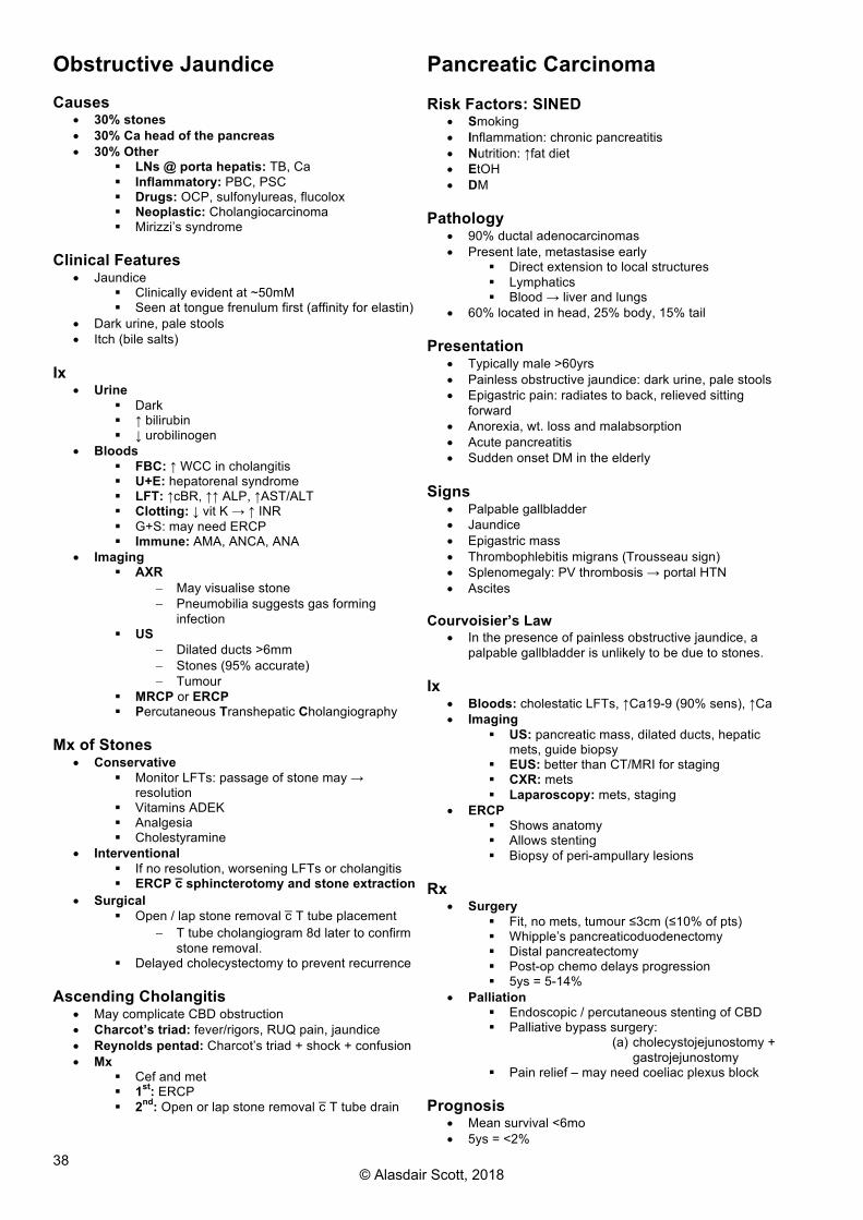
38
Obstructive Jaundice Causes
• 30% stones • 30% Ca head of the pancreas • 30% Other
§ LNs @ porta hepatis: TB, Ca § Inflammatory: PBC, PSC § Drugs: OCP, sulfonylureas, flucolox § Neoplastic: Cholangiocarcinoma § Mirizzi’s syndrome
Clinical Features
• Jaundice § Clinically evident at ~50mM § Seen at tongue frenulum first (affinity for elastin)
• Dark urine, pale stools • Itch (bile salts)
Ix
• Urine § Dark § ↑ bilirubin § ↓ urobilinogen
• Bloods § FBC: ↑ WCC in cholangitis § U+E: hepatorenal syndrome § LFT: ↑cBR, ↑↑ ALP, ↑AST/ALT § Clotting: ↓ vit K → ↑ INR § G+S: may need ERCP § Immune: AMA, ANCA, ANA
• Imaging § AXR
- May visualise stone - Pneumobilia suggests gas forming
infection § US
- Dilated ducts >6mm - Stones (95% accurate) - Tumour
§ MRCP or ERCP § Percutaneous Transhepatic Cholangiography
Mx of Stones
• Conservative § Monitor LFTs: passage of stone may →
resolution § Vitamins ADEK § Analgesia § Cholestyramine
• Interventional § If no resolution, worsening LFTs or cholangitis § ERCP c̄ sphincterotomy and stone extraction
• Surgical § Open / lap stone removal c̄ T tube placement
- T tube cholangiogram 8d later to confirm stone removal.
§ Delayed cholecystectomy to prevent recurrence Ascending Cholangitis
• May complicate CBD obstruction • Charcot’s triad: fever/rigors, RUQ pain, jaundice • Reynolds pentad: Charcot’s triad + shock + confusion • Mx
§ Cef and met § 1st: ERCP § 2nd: Open or lap stone removal c̄ T tube drain
Pancreatic Carcinoma Risk Factors: SINED
• Smoking • Inflammation: chronic pancreatitis • Nutrition: ↑fat diet • EtOH • DM
Pathology
• 90% ductal adenocarcinomas • Present late, metastasise early
§ Direct extension to local structures § Lymphatics § Blood → liver and lungs
• 60% located in head, 25% body, 15% tail Presentation
• Typically male >60yrs • Painless obstructive jaundice: dark urine, pale stools • Epigastric pain: radiates to back, relieved sitting
forward • Anorexia, wt. loss and malabsorption • Acute pancreatitis • Sudden onset DM in the elderly
Signs
• Palpable gallbladder • Jaundice • Epigastric mass • Thrombophlebitis migrans (Trousseau sign) • Splenomegaly: PV thrombosis → portal HTN • Ascites
Courvoisier’s Law
• In the presence of painless obstructive jaundice, a palpable gallbladder is unlikely to be due to stones.
Ix
• Bloods: cholestatic LFTs, ↑Ca19-9 (90% sens), ↑Ca • Imaging
§ US: pancreatic mass, dilated ducts, hepatic mets, guide biopsy
§ EUS: better than CT/MRI for staging § CXR: mets § Laparoscopy: mets, staging
• ERCP § Shows anatomy § Allows stenting § Biopsy of peri-ampullary lesions
Rx
• Surgery § Fit, no mets, tumour ≤3cm (≤10% of pts) § Whipple’s pancreaticoduodenectomy § Distal pancreatectomy § Post-op chemo delays progression § 5ys = 5-14%
• Palliation § Endoscopic / percutaneous stenting of CBD § Palliative bypass surgery:
(a) cholecystojejunostomy + gastrojejunostomy
§ Pain relief – may need coeliac plexus block Prognosis
• Mean survival <6mo • 5ys = <2%
© Alasdair Scott, 2018
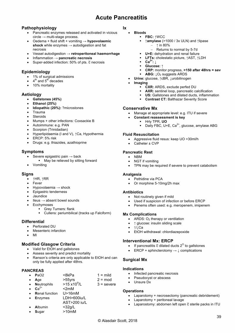
39
Acute Pancreatitis Pathophysiology
• Pancreatic enzymes released and activated in vicious circle → multi-stage process.
• Oedema + fluid shift + vomiting → hypovolaemic shock while enzymes → autodigestion and fat necrosis
• Vessel autodigestion → retroperitoneal haemorrhage • Inflammation → pancreatic necrosis • Super-added infection: 50% of pts. c̄ necrosis
Epidemiology
• 1% of surgical admissions • 4th and 5th decades • 10% mortality
Aetiology
• Gallstones (45%) • Ethanol (25%) • Idiopathic (20%): ?microstones • Trauma • Steroids • Mumps + other infections: Coxsackie B • Autoimmune: e.g. PAN • Scorpion (Trinidadian) • Hyperlipidaemia (I and V), ↑Ca, Hypothermia • ERCP: 5% risk • Drugs: e.g. thiazides, azathioprine
Symptoms
• Severe epigastric pain → back § May be relieved by sitting forward
• Vomiting Signs
• ↑HR, ↑RR • Fever • Hypovolaemia → shock • Epigastric tenderness • Jaundice • Ileus → absent bowel sounds • Ecchymoses
§ Grey Turners: flank § Cullens: periumbilical (tracks up Falciform)
Differential
• Perforated DU • Mesenteric infarction • MI
Modified Glasgow Criteria
• Valid for EtOH and gallstones • Assess severity and predict mortality • Ranson’s criteria are only applicable to EtOH and can
only be fully applied after 48hrs. PANCREAS
• PaO2 <8kPa 1 = mild 2 = mod 3 = severe
• Age >55yrs • Neutrophils >15 x109/L • Ca2+ <2mM • Renal function U>16mM • Enzymes LDH>600iu/L
AST>200 iu/L • Albumin <32g/L • Sugar >10mM
Ix
• Bloods § FBC: ↑WCC § ↑amylase (>1000 / 3x ULN) and ↑lipase
- ↑ in 80% - Returns to normal by 5-7d
§ U+E: dehydration and renal failure § LFTs: cholestatic picture, ↑AST, ↑LDH § Ca2+: ↓ § Glucose: ↑ § CRP: monitor progress, >150 after 48hrs = sev § ABG: ↓O2 suggests ARDS
• Urine: glucose, ↑cBR, ↓urobilinogen • Imaging
§ CXR: ARDS, exclude perfed DU § AXR: sentinel loop, pancreatic calcification § US: Gallstones and dilated ducts, inflammation § Contrast CT: Balthazar Severity Score
Conservative Mx
• Manage at appropriate level: e.g. ITU if severe • Constant reassessment is key
§ Hrly TPR, UO § Daily FBC, U+E, Ca2+, glucose, amylase ABG
Fluid Resuscitation
• Aggressive fluid resus: keep UO >30ml/h • Catheter ± CVP
Pancreatic Rest
• NBM • NGT if vomiting • TPN may be required if severe to prevent catabolism
Analgesia
• Pethidine via PCA • Or morphine 5-10mg/2h max
Antibiotics
• Not routinely given if mild • Used if suspicion of infection or before ERCP • Penems often used: e.g. meropenem, imipenem
Mx Complications
• ARDS: O2 therapy or ventilation • ↑ glucose: insulin sliding scale • ↑/↓Ca • EtOH withdrawal: chlordiazepoxide
Interventional Mx: ERCP
• If pancreatitis c̄ dilated ducts 2O to gallstones • ERCP + sphincterotomy → ↓ complications
Surgical Mx Indications
• Infected pancreatic necrosis • Pseudocyst or abscess • Unsure Dx
Operations
• Laparotomy + necrosectomy (pancreatic debridement) • Laparotomy + peritoneal lavage • Laparostomy: abdomen left open c̄ sterile packs in ITU
© Alasdair Scott, 2018

40
Complications of Acute Pancreatitis Early: Systemic
• Respiratory: ARDS, pleural effusion • Shock: hypovolaemic or septic • Renal failure • DIC • Metabolic
§ ↓ Ca2+ § ↑ glucose § Metabolic acidosis
Late (>1wk): Local
• Pancreatic necrosis • Pancreatic infection • Pancreatic abscess
§ May form in pseudocyst or in pancreas § Open or percutaneous drainage
• Bleeding: e.g. from splenic artery § May require embolisation
• Thrombosis § Splenic A., GDA or colic branches of SMA
- May → bowel necrosis § Portal vein → portal HTN
• Fistula formation § Pancreato-cutaneous → skin breakdown
Pancreatic Pseudocyst
• Collection of pancreatic fluid in the lesser sac surrounded by granulation tissue
• Occur in 20% (esp. in EtOH pancreatitis) • Presentation
§ 4-6wks after acute attack § Persisting abdominal pain § Epigastric mass → early satiety
• Complications § Infection → abscess § Obstruction of duodenum or CBD
• Ix § Persistently ↑ amylase ± LFTs § US / CT
• Rx § <6cm: spontaneous resolution § >6cm
- Endoscopic cyst-gastrostomy - Percutaneous drainage under US/CT
Chronic Pancreatitis Causes: AGITS
• Alcohol (70%) • Genetic
§ CF § HH
• Immune § Lymphoplasmacytic sclerosing pancreatitis
(↑IgG4) • Triglycerides ↑ • Structural
§ Obstruction by tumour § Pancreas divisum
Presentation
• Epigastric pain § Bores through to back § Relieved by sitting back or hot water bottle →
erythema ab igne § Exacerbated by fatty food or EtOH
• Steatorrhoea and wt. loss • DM: polyuria, polydipsia • Epigastric mass: pseudocyst
Ix
• ↑ glucose • ↓ faecal elastase: ↓ exocrine function • US: pseudocyst • AXR: speckled pancreatic calcifications • CT: pancreatic calcifications
Rx
• Diet § No EtOH § ↓ fat, ↑ carb
• Drugs § Analgesia: may need coeliac plexus block § Enzyme supplements: pancreatin (Creon) § ADEK vitamins § DM Rx
• Surgery § Indications
- Unremitting pain - Wt. loss - Duct blockage
§ Procedures - Distal pancreatectomy, Whipple’s - Pancreaticojejunostomy: drainage - Endoscopic stenting
Complications
• Pseudocyst • DM • Pancreatic Ca • Pancreatic swelling → biliary obstruction • Splenic vein thrombosis → splenomegaly
© Alasdair Scott, 2018

41
Pancreatic Endocrine Neoplasia Epidemiology
• 30-60yrs • ~15% assoc. c̄ MEN1
Insulinoma
• Fasting / exercise-induced hypoglycaemia • Confusion, stupor, LOC • ↑ insulin + ↑ c-peptide + ↓glucose
Gastrinoma → Zollinger-Ellison
• Hypergastrinaemia → hyperchlorhydria → PUD and chronic diarrhoea (inactivation of pancreatic enzymes)
Glucagonoma
• ↑ se glucagon → mild DM • Characteristic blistering rash
§ Necrolytic migratory erythema VIPoma / Verner-Morrison / WDHA Syn.
• Watery Diarrhoea • Hypokalaemia • Achlorhydria • Acidosis
Somatostatinoma
• Somatostatin § Inhibits glucagon and insulin release § Inhibits pancreatic enzyme secretion
• Features § DM § Steatorrhoea § Gallstones
• Usually v. malignant tumour c̄ poor prognosis
Pancreatic Malformations Ectopic Pancreas
• Meckel’s diverticulum • Small bowel
Pancreas Divisium
• Failure of fusion of dorsal and ventral buds • → bulk of pancreas drains through smaller accessory
duct. • Usually asymptomatic • May → chronic pancreatitis
Annular Pancreas
• Fusion of dorsal and ventral buds around duodenum • May present c̄ infantile duodenal obstruction.
© Alasdair Scott, 2018

42
Cholangiocarcinoma Pathology
• Rare bile duct tumour • Adenocarcinoma • Typically occur @ confluence of right and left hepatic
ducts: called “Klatskin” tumours Risk Factors
• PSC • Ulcerative colitis • Choledocholithiasis • Hep B/C • Choledochal cysts • Lynch 2 • Flukes
Presentation
• Progressive painless obstructive jaundice § Gallbladder not palpable
• Steatorrhoea • Wt. loss
Ix
• Cholestatic LFTs • CA 19-9
Rx
• Poor prognosis: no curative Rx • Palliative stenting by ERCP
Hydatid Cyst Pathophysiology
• Zoonotic infection by Echinococcus granulosus • Occurs in sheep-rearing communities • Parasite penetrates the portal system and infects the
liver → calcified cyst Presentation
• Mostly asymptomatic • Pressure effects
§ Non-specific pain § Abdominal fullness § Obstructive jaundice
• Rupture § Biliary colic § Jaundice § Urticaria § Anaphylaxis
• 2O infection Ix
• Eosinophilia • CT
Rx
• Medical: albendazole • Surgical cystectomy
§ Indicated for large cyst
© Alasdair Scott, 2018
43
Lower GI Surgery Contents
Paediatric Conditions ........................................................................................................................................... 44Small Bowel Neoplasms ...................................................................................................................................... 45Carcinoid Tumours ............................................................................................................................................... 45Acute Appendicitis ............................................................................................................................................... 46Inflammatory Bowel Disease: Pathology and Presentation ................................................................................. 47Ulcerative Colitis: Management ........................................................................................................................... 49Crohn’s Disease: Management ............................................................................................................................ 50Diverticular Disease ............................................................................................................................................. 51Bowel Obstruction: Causes and Investigation ..................................................................................................... 52Bowel Obstruction: General Management ........................................................................................................... 53Bowel Obstruction: Specific Management ........................................................................................................... 54Colorectal Carcinoma: Pathology and Presentation ............................................................................................ 55Colorectal Carcinoma: Ix, Mx and Prevention ...................................................................................................... 56Familial CRC Syndromes ..................................................................................................................................... 57GI Polyps ............................................................................................................................................................. 57Mesenteric Ischaemia .......................................................................................................................................... 58Lower GI Bleed .................................................................................................................................................... 58
© Alasdair Scott, 2018
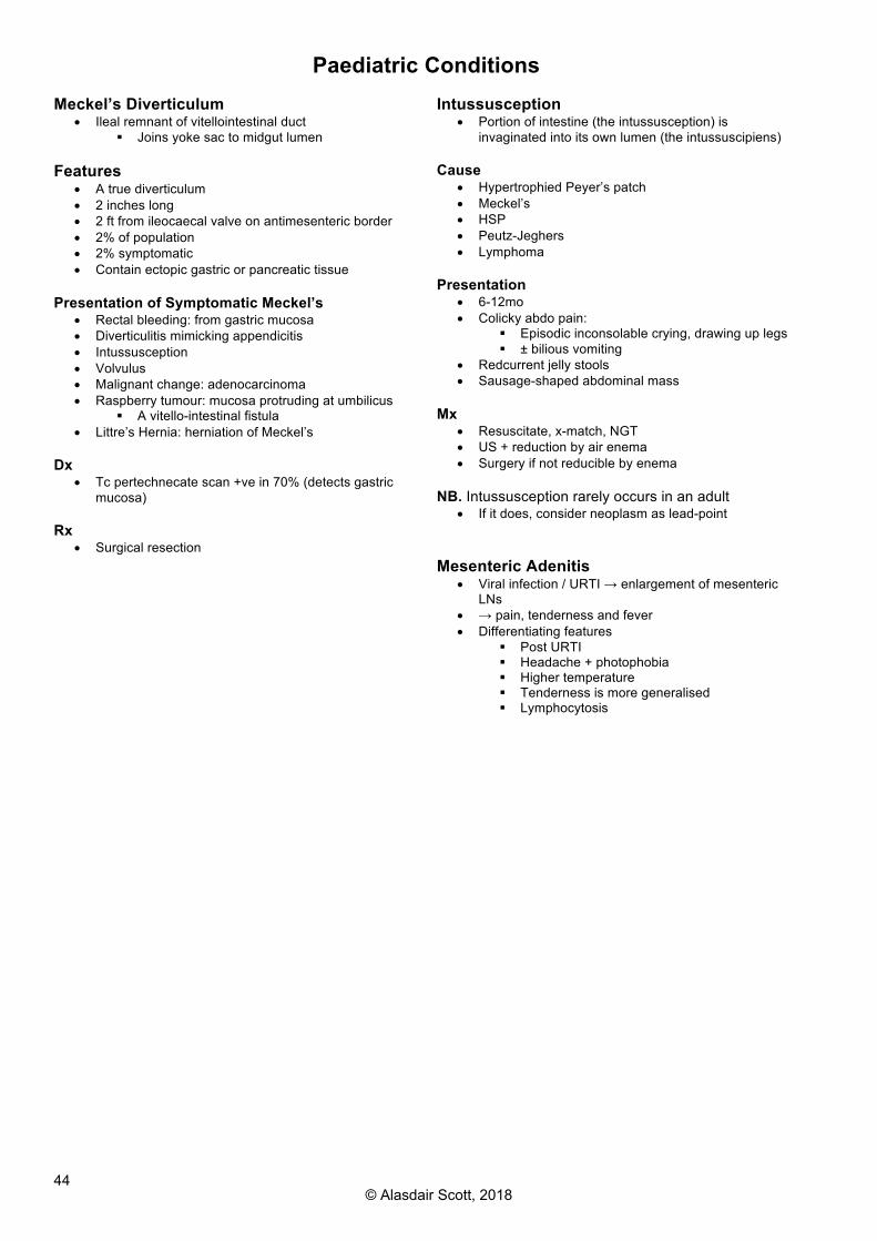
44
Paediatric Conditions Meckel’s Diverticulum
• Ileal remnant of vitellointestinal duct § Joins yoke sac to midgut lumen
Features
• A true diverticulum • 2 inches long • 2 ft from ileocaecal valve on antimesenteric border • 2% of population • 2% symptomatic • Contain ectopic gastric or pancreatic tissue
Presentation of Symptomatic Meckel’s
• Rectal bleeding: from gastric mucosa • Diverticulitis mimicking appendicitis • Intussusception • Volvulus • Malignant change: adenocarcinoma • Raspberry tumour: mucosa protruding at umbilicus
§ A vitello-intestinal fistula • Littre’s Hernia: herniation of Meckel’s
Dx
• Tc pertechnecate scan +ve in 70% (detects gastric mucosa)
Rx
• Surgical resection
Intussusception
• Portion of intestine (the intussusception) is invaginated into its own lumen (the intussuscipiens)
Cause
• Hypertrophied Peyer’s patch • Meckel’s • HSP • Peutz-Jeghers • Lymphoma
Presentation
• 6-12mo • Colicky abdo pain:
§ Episodic inconsolable crying, drawing up legs § ± bilious vomiting
• Redcurrent jelly stools • Sausage-shaped abdominal mass
Mx
• Resuscitate, x-match, NGT • US + reduction by air enema • Surgery if not reducible by enema
NB. Intussusception rarely occurs in an adult
• If it does, consider neoplasm as lead-point Mesenteric Adenitis
• Viral infection / URTI → enlargement of mesenteric LNs
• → pain, tenderness and fever • Differentiating features
§ Post URTI § Headache + photophobia § Higher temperature § Tenderness is more generalised § Lymphocytosis
© Alasdair Scott, 2018

45
Small Bowel Neoplasms Benign: 35%
• Lipoma • Leiomyoma • Neurofibroma • Haemangioma • Adenomatous polyps (FAP, Peutz-Jeghers)
Malignant: 65% (only 2% of GI malignancies)
• Adenocarcinoma (40% of malignant tumours) • Carcinoid (40% of malignant tumours) • Lymphoma (esp. c̄ Coeliac disease: EATL) • GIST
Presentation
• Often non-specific symptoms so present late • N/V, obstruction • Wt. loss and abdominal pain • Bleeding • Jaundice from biliary obstruction or liver mets.
Imaging
• AXR: SBO • CT
Endoscopy
• Push enteroscopy • Wireless capsule endoscopy
Carcinoid Tumours Pathology
• Diverse group of neuroendocrine tumours of enterochromaffin cell origin capable of producing 5HT
• May be derived from § Foregut: respiratory tract § Midgut: stomach, ileum, appendix § Hindgut: colorectum
• May secrete: 5-HT, VIP, gastrin, glucagon, insulin, ACTH § Hind gut tumours rarely secrete 5-HT
• Carcinoid syndrome suggest bypass of first-pass metabolism and is strongly assoc. c̄ metastatic disease.
• 10% part of MEN1 • Sites
§ Appendix: 45% § Ileum: 30% § Colorectum: 20% § Stomach: 10% § Elsewhere in GIT § Bronchus: 10%
• Consider all as malignant Presentation Local
• Appendicitis • Intussusception or obstruction • Abdominal pain
Carcinoid Syndrome: FIVE HT
• Flushing: paroxysmal, upper body ± wheals • Intestinal: diarrhoea • Valve fibrosis: tricuspid regurg and pulmonary stenosis • whEEze: bronchoconstriction • Hepatic involvement: bypassed 1st pass metabolism • Tryptophan deficiency → pellagra (3Ds)
Ix
• ↑ urine 5-hydroxyindoleacetic acid • ↑ plasma chromogranin A • CT/MRI: find primary
Rx
• Symptoms: octreotide or loperamide • Curative
§ Resection: tumours are v. yellow § Give octreotide to avoid carcinoid crisis
Carcinoid Crisis
• Tumour outgrows blood supply or is handled too much → massive mediator release
• Vasodilatation, hypotension, bronchoconstriction, hyperglycaemia
• Rx: high-dose octreotide Prognosis
• Median survival is 5-8yrs (~3yrs if mets present)
© Alasdair Scott, 2018

46
Acute Appendicitis Definition
• Inflammation of the vermiform appendix ranging from oedema to ischaemic necrosis and perforation.
Epidemiology
• Incidence: 6% lifetime incidence, commonest surgical emergency
• Age: rare <2yrs, maximal peak during child, ↓ thereafter
Pathogenesis
• Obstruction of the appendix § Faecolith most commonly § Lymphoid hyperplasia post-infection § Tumour (e.g. caecal Ca, carcinoid) § Worms (e.g. Ascaris lumbicoides, Schisto)
• Gut organisms → infection behind obstruction • → oedema → ischaemia → necrosis → perforation
§ Peritonitis § Abscess § Appendix mass
Pattern of Abdominal Pain Early inflammation → appendiceal irritation
• Visceral pain is not well localised cf. somatic pain. • Nociceptive info travels in the sympathetic afferent
fibres that supply the viscus • Pain referred to the dermatome corresponding to the
spinal cord entry level of these sympathetic fibres. • Append = midgut = lesser splanch (T10/11) = umb
Late inflammation → parietal peritoneum irritation
• Pain localised in RIF Symptoms
• Colicky abdo pain § Central → localised in RIF § Worse c̄ movement
• Anorexia • Nausea (vomiting is rarely prominent) • Constipation / diarrhoea
Signs
• Low-grade pyrexia: 37.5 – 38.5 • ↑HR, shallow breathing • Foetor oris • Guarding and tenderness: @ McBurney’s point
§ +ve cough / percussion tenderness • Appendix mass may be palpable in RIF • Pain PR suggests pelvic appendix.
Special Signs Rovsing’s Sign
• Pressure in LIF → more pain in RIF Psoas Sign
• Pain on extending the hip: retrocaecal appendix Cope Sign
• Flexion + internal rotation of R hip → pain § Appendix lying close to obturator internus
Differential
• Surgical § Cholecystitis § Diverticulitis § Meckel’s diverticulitis
• Gynae § Cyst accident: torsion, rupture, haemorrhage § Salpingitis / PID § Ruptured ectopic
• Medical § Mesenteric adenitis § UTI § Crohn’s
Ix
• Dx is principally clinical • Bloods: FBC, CRP, amylase, G+S, clotting • Urine
§ Sterile pyuria: may indicate bladder irritation § Ketones: anorexia § Exclude UTI § β-HCG
• Imaging § US: exclude gynae path, visualise inflamed
appendix § CT: can be used
• Diagnostic lap Mx
• Fluids • Abx: cef 1.5g + met 500g IV TDS • Analgesia: paracetamol, NSAIDs, codeine phosphate • Certain Dx → appendicectomy (open or lap) • Uncertain Dx → active observation
Complications Appendix Mass
• Inflamed appendix c̄ adherent covering of omentum and small bowel
• Dx: US or CT • Mx
§ Initially: Abx + NBM § Resolution of mass → interval appendicectomy § Exclude a colonic tumour: colonoscopy
Appendix Abscess
• Results if appendix mass doesn’t resolve • Mass enlarges, pt. deteriorates • Mx
§ Abx + NBM § CT-guided percutaneous drainage § If no resolution, surgery may involve right
hemicolectomy. Perforation
• Commoner if faecolith present and in young children (as Dx is often delayed)
• Deteriorating pt. c̄ peritonitis.
© Alasdair Scott, 2018
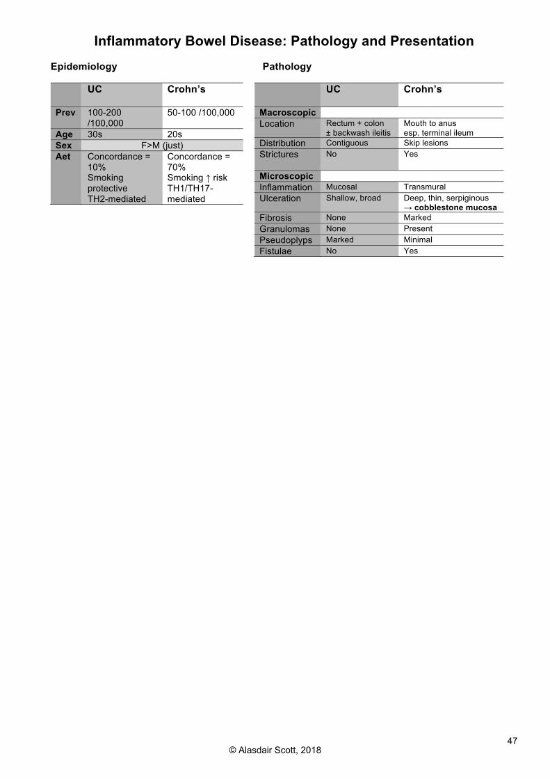
47
Inflammatory Bowel Disease: Pathology and Presentation Epidemiology
UC
Crohn’s
Prev 100-200 /100,000
50-100 /100,000
Age 30s 20s Sex F>M (just) Aet Concordance =
10% Smoking protective TH2-mediated
Concordance = 70% Smoking ↑ risk TH1/TH17-mediated
Pathology
UC
Crohn’s
Macroscopic Location Rectum + colon
± backwash ileitis Mouth to anus esp. terminal ileum
Distribution Contiguous Skip lesions Strictures
No Yes
Microscopic Inflammation Mucosal Transmural Ulceration Shallow, broad Deep, thin, serpiginous
→ cobblestone mucosa Fibrosis None Marked Granulomas None Present Pseudoplyps Marked Minimal Fistulae No Yes
© Alasdair Scott, 2018
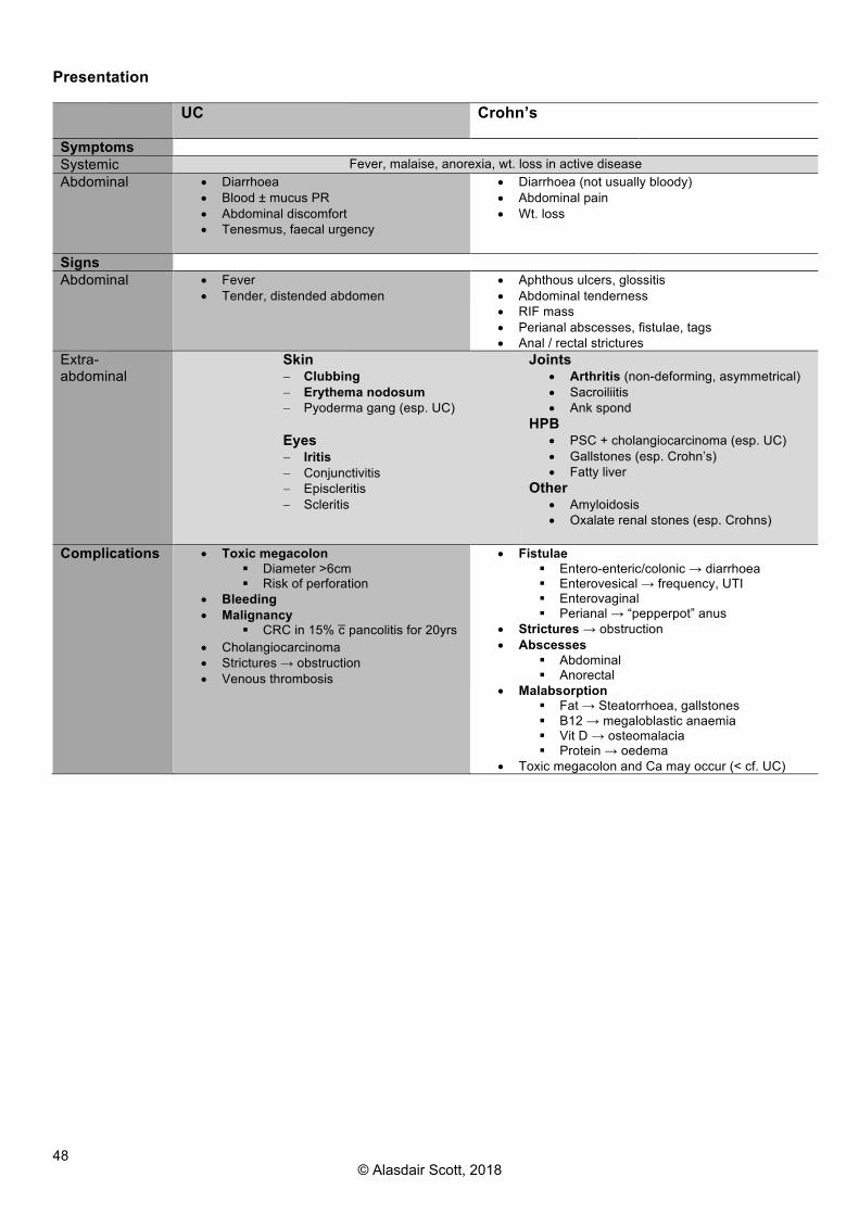
48
Presentation UC Crohn’s
Symptoms Systemic Fever, malaise, anorexia, wt. loss in active disease Abdominal • Diarrhoea
• Blood ± mucus PR • Abdominal discomfort • Tenesmus, faecal urgency
• Diarrhoea (not usually bloody) • Abdominal pain • Wt. loss
Signs Abdominal • Fever
• Tender, distended abdomen • Aphthous ulcers, glossitis • Abdominal tenderness • RIF mass • Perianal abscesses, fistulae, tags • Anal / rectal strictures
Extra-abdominal
Skin - Clubbing - Erythema nodosum - Pyoderma gang (esp. UC) Eyes - Iritis - Conjunctivitis - Episcleritis - Scleritis
Joints • Arthritis (non-deforming, asymmetrical) • Sacroiliitis • Ank spond
HPB • PSC + cholangiocarcinoma (esp. UC) • Gallstones (esp. Crohn’s) • Fatty liver
Other • Amyloidosis • Oxalate renal stones (esp. Crohns)
Complications • Toxic megacolon
§ Diameter >6cm § Risk of perforation
• Bleeding • Malignancy
§ CRC in 15% c̄ pancolitis for 20yrs • Cholangiocarcinoma • Strictures → obstruction • Venous thrombosis
• Fistulae § Entero-enteric/colonic → diarrhoea § Enterovesical → frequency, UTI § Enterovaginal § Perianal → “pepperpot” anus
• Strictures → obstruction • Abscesses
§ Abdominal § Anorectal
• Malabsorption § Fat → Steatorrhoea, gallstones § B12 → megaloblastic anaemia § Vit D → osteomalacia § Protein → oedema
• Toxic megacolon and Ca may occur (< cf. UC)
© Alasdair Scott, 2018

49
Ulcerative Colitis: Management Ix
• Bloods: § FBC: ↓Hb, ↑WCC § LFT: ↓albumin § ↑CRP/ESR § Blood cultures
• Stool § MCS: exclude Campy, Shigella, Salmonella… § CDT: C. diff may complicate or mimic
• Imaging § AXR: megacolon (>6cm), wall thickening § CXR: perforation § CT § Ba / gastrograffin enema § Lead-pipe: no haustra § Thumbprinting: mucosal thickening § Pseudopolyps: regenerating mucosal island
• Ileocolonoscopy + regional biopsy: Baron Score Severity Truelove and Witts Criteria
Mild Mod Severe Motions <4 4-6 >6 PR bleed small moderate large Temp Apyrexic 37.1-37.8 >37.8 HR <70 70-90 >90 Hb >11 10.5-11 <10.5 ESR <30 >30
Acute Severe UC
• Resus: Admit, IV hydration, NBM • Hydrocortisone: IV 100mg QDS + PR • Transfuse if required • Thromboprophylaxis: LMWH • Monitoring
§ Bloods: FBC, ESR, CRP, U+E § Vitals + stool chart § Twice daily examination § ± AXR
NB. RCTs show no benefit of Abx: not routinely recommended
• May use: megacolon, perforation, uncertain Dx Acute Complications
• Perforation • Bleeding • Toxic megacolon (>6cm) • VTE
Improvement → oral therapy
• Switch to oral pred + a 5-ASA • Taper pred after full remission
No Improvement → rescue therapy • On day 3: stool freq >8 or CRP >45
§ Predicts 85% chance of needing a colectomy during the admission
• Discussion between pt, physician and surgeon • Medical: ciclosporin, infliximab or visilizumab (anti-T
cell) • Surgical
Inducing Remission in Mild / Mod Disease • OPD-based
Oral Therapy
• 1st line: 5-ASAs • 2nd line: prednisolone
Topical Therapy: mainly left-sided disease
• Proctitis: suppositories • More proximal disease: enemas or foams • 5-ASAs ± steroids (prednisolone or budesonide)
Additional Therapy: steroid sparing
• Azathioprine or mercaptopurine • Infliximab: steroid-dependent pts
Maintaining Remission
• 1st line: 5-ASAs PO – sulfasalazine or mesalazine § Topical Rx may be used in proctitis
• 2nd line: Azathioprine or mercaptopurine § Relapsed on ASA or are steroid-dependent § Give 6-mercaptopurine if azathioprine intolerant
• 3rd line: Infliximab / adalimumab Emergency Surgery
• 20% require surgery at some stage • 30% c̄ colitis require surgery w/i 5yrs
Indications
• Toxic megacolon • Perforation • Massive haemorrhage • Failure to respond to medical Rx
Procedures
• Total / subtotal colectomy c̄ end ileostomy ± mucus fistula
• Followed after ~3mo by either § Completion proctectomy + Ileal-pouch anal
anastomosis (IPAA) or end ileostomy § Ileorectal anastomosis (IRA)
• Panproctocolectomy + permanent end ileostomy • Acute colitis op mortality: 7% (30% if perforated)
Elective Surgery Indications
• Chronic symptoms despite medical therapy • Carcinoma or high-grade dysplasia
Procedures
• Panproctocolectomy c̄ end ileostomy or IPAA • Total colectomy c̄ IRA
Surgical Complications
• Abdominal § SBO § Anastomotic stricture § Pelvic abscess
• Stoma: retraction, stenosis, prolapse, dermatitis • Pouch
§ Pouchitis (50%): metronidazole + cipro § ↓ female fertility
- Faecal leakage
© Alasdair Scott, 2018

50
Crohn’s Disease: Management Ix
• Bloods: (top 3 are severity markers) § FBC: ↓Hb, ↑WCC § LFT: ↓albumin § ↑CRP/ESR § Haematinics: Fe, B12, Folate § Blood cultures
• Stool § MCS: exclude Campy, Shigella, Salmonella… § CDT: C. diff may complicate or mimic
• Imaging § AXR: obstruction, sacroileitis § CXR: perforation § MRI § Assess pelvic disease and fistula § Assess disease severity § Small bowel follow-through or enteroclysis § Skip lesions § Rose-thorn ulcers § Cobblestoning: ulceration + mural oedema § String sign of Kantor: narrow terminal ileum
• Endoscopy § Ileocolonoscopy + regional biopsy: Ix of
choice § Wireless capsule endoscopy § Small bowel enteroscopy
Severe Attack Assessment
• ↑temp, ↑HR, ↑ESR, ↑CRP, ↑WCC, ↓albumin Management
• Resus: Admit, NBM, IV hydration • Hydrocortisone: IV + PR if rectal disease • Abx: metronidazole PO or IV • Thromboprophylaxis: LMWH • Dietician Review
§ Elemental diet - Liquid prep of amino acids, glucose and
fatty acids § Consider parenteral nutrition
• Monitoring § Vitals + stool chart § Daily examination
Improvement → oral therapy
• Switch to oral pred (40mg/d)
No Improvement → rescue therapy • Discussion between pt, physician and surgeon • Medical: methotrexate ± infliximab • Surgical
Inducing Remission in Mild / Mod Disease
• OPD treatment Supportive
• High fibre diet • Vitamin supplements
Oral Therapy
• 1st line § Ileocaecal: budesonide § Colitis: sulfasalazine
• 2nd line: prednisolone (tapering) • 3rd line: methotrexate • 4th line: infliximab or adalimumab
Perianal Disease
• Occurs in ~50% • Ix: MRI + EUA • Rx
§ Oral Abx: metronidazole § Immunosuppression ± infliximab § Local surgery ± seton insertion
Maintaining Remission
• 1st line: azathioprine or mercaptopurine • 2nd line: methotrexate • 3rd line: Infliximab / adalimumab
Surgery
• 50-80% need ≥1 operation in their life • Never curative • Should be as conservative as possible
Indications
• Emergency § Failure to respond to medical Rx § Intestinal obstruction or perforation § Massive haemorrhage
• Elective § Abscess or fistula § Perianal disease § Chronic ill health § Carcinoma
Procedures
• Limited resection: e.g. ileocaecal • Stricturoplasty • Defunction distal disease c̄ temporary loop ileostomy
Complications
• Stoma complications • Enterocutaneous fistulae • Anastomotic leak or stricture
Short gut
• <1-2m small bowel • Features
§ Steatorrhoea § ADEK and B12 malabsorption § Bile acid depletion → gallstones § Hyperoxaluria → renal stones
• Rx § Dietician § Supplements or TPN § Loperamide
© Alasdair Scott, 2018

51
Diverticular Disease Definitions
• Diverticulum = out-pouching of tubular structure • True = composed of complete wall (e.g. Meckel’s) • False = composed of mucosa only (pharyngeal,
colonic) • Diverticular disease: symptomatic diverticulosis • Diverticulitis: inflammation of diverticula
Epidemiology
• 30% of Westerner’s have diverticulosis by 60yrs • F>M
Pathophysiology
• Assoc. c̄ ↑ intraluminal pressure § Low fibre diet: no osmotic effect to keep stool
wet • Mucosa herniates through muscularis propria at points
of weakness where perforating arteries enter. • Most commonly located in sigmoid colon • Commoner in obese pts.
§ Uniting factor in Saint’s Triad? - Hiatus Hernia - Cholelithiasis - Diverticular disease
Symptoms of Diverticular Disease
• Altered bowel habit ± left-sided colic § Relieved by defecation
• Nausea • Flatulence • Rx
§ High fibre diet, mebeverine may help § Elective resection for chronic pain
Diverticulitis
• Inspissated faeces → obstruction of diverticulum • Elderly pt. c̄ prev Hx of constipation
Presentation
• Abdominal pain and tenderness § Typically LIF § Localised peritonitis
• Pyrexia Ix
• Bloods § FBC: ↑WCC § ↑CRP/ESR § Amylase § G+S/x-match
• Imaging § Erect CXR: look for perforation § AXR: fluid level / air in bowel wall § Contrast CT
• Endoscopy § Flexi Sig § Colonoscopy: not in acute attack
Hinchey Grading of Perforated Diverticulitis
1 Small confined pericolic abscesses
Surgery rarely needed
2 Large abscess extending into pelvis
May resolve w/o surgery
3 Generalised purulent peritonitis Surgery needed 4 Generalised faecal peritonitis Surgery needed
Mx of Acute Diverticulitis
• Mild Attacks § Can be treated at home c̄ bowel rest (fluids
only) and co-amoxiclav ± metronidazole • Admit if
§ Unwell § Fluids can’t be tolerated § Pain can’t be controlled
• Medical § NBM § IV fluids § Analgesia § Antibiotics: cefuroxime + metronidazole § Most cases settle
• Surgical § Indications
- Perforation - Large haemorrhage - Stricture → obstruction
§ Procedure - Hartmann’s to resect diseased bowel - May consider lap washout for Hinchey 3
Other Complications Perforation
• Sudden onset pain (± preceding diverticulitis) • Generalised peritonitis and shock • CXR: free air under diaphragm • Rx: Hartmann’s
Haemorrhage
• Sudden, painless bright red PR bleed • Ix: mesenteric angiography or colonoscopy • Rx
§ Usually stops spontaneously § May need transfusion § Colonoscopy ± diathermy / adrenaline § Embolisation § Resection
Abscess
• Walled-off perforation • Swinging fever • Localising signs: e.g. boggy rectal mass • Leukocytosis • Rx: Abx + CT/US-guided drainage
Fistulae
• Enterocolic • Colovaginal • Colovesicular: pneumaturia + intractable UTIs • Rx: resection
Strictures
• After diverticulitis, colon may heal c̄ fibrous strictures • Rx
§ Resection (usually c̄ 1O anastomosis) § Stenting
© Alasdair Scott, 2018

52
Bowel Obstruction: Causes and Investigation Classification
• Simple § 1 obstructing point + no vascular compromise § May be partial or complete
• Closed Loop § Bowel obstructed @ two points
- Left CRC c̄ competent ileocaecal valve - Volvulus
§ Gross distension → perforation • Strangulated
§ Compromised blood supply § Localised, constant pain + peritonism § Fever + ↑WCC
Commonest Causes SBO
• Adhesions: 60% • Hernia
LBO
• Colorectal Neoplasia: 60% • Diverticular stricture: 20% • Volvulus: 5%
Other Causes Non-Mechanical = Paralytic Ileus (usually SB)
• Post-op • Peritonitis • Pancreatitis or any localised inflammation • Poisons / Drugs: anti-AChM (e.g. TCAs) • Pseudo-obstruction • Metabolic: ↓K, ↓Na, ↓Mg, uraemia • Mesenteric ischaemia
Mechanical
• Intraluminal § Impacted matter: faeces, worms, bezoars § Intussusception § Gallstones
• Intramural § Benign stricture
- IBD - Surgery - Ischaemic colitis - Diverticulitis - Radiotherapy
§ Neoplasia § Congenital atresia
• Extramural § Hernia § Adhesions § Volvulus (sigmoid, caecal, gastric) § Extrinsic Compression
- Pseudocyst - Abscess - Haematoma - Tumour: e.g. ovarian - Congenital bands (e.g. Ladd’s)
Presentation
• Abdominal Pain § Colicky § Central but level depends on gut region § Constant / localised pain suggests
strangulation or impending perforation • Distension
§ ↑ c̄ lower obstructions • Vomiting
§ Early in high obstruction § Late or absent in low obstructions
• Absolute Constipation: flatus and faeces Examination
• ↑HR: hypovolaemia, strangulation • Dehydration, hypovolaemia • Fever: suggests inflammatory disease or strangulation • Surgical scars • Hernias • Mass: neoplastic or inflammatory • Bowel sounds
§ ↑: mechanical obstruction § ↓: ileus
• PR § Empty rectum § Rectal mass § Hard impacted stool § Blood from higher pathology
Ix
• Bloods § FBC: ↑WCC § U+E: dehydration, electrolyte abnormalities § Amylase: ↑↑ if strangulation/perforation § VBG: ↑ lactate in strangulation § G+S, clotting: may need surgery
• Imaging § Erect CXR § AXR: ± erect film for fluid levels § CT: can show transition point
• Gastrograffin studies § Look for mechanical obstruction: no free
flow § Follow through or enema § Follow through may relieve mild
mechanical obstruction: usually adhesional • Colonoscopy
§ Can be used in some cases § Risk of perforation § May be used therapeutically to stent
AXR Findings
SBO LBO Diameter ≥3cm ≥6cm (caecum ≥9) Location Central Peripheral Markings Valvulae coniventes
- completely across Haustra - partially across
LB Gas Absent Present - not in rectum
No. of loops Many Few Fluid levels Many, short Few, long
Ileus
• Both small and large bowel may be visible • No clear transition point
© Alasdair Scott, 2018

53
Bowel Obstruction: General Management Medical Resuscitate: “Drip and Suck”
• NBM • IV fluids: aggressive as pt. may be v. dehydrated • NGT: decompress upper GIT, stops vomiting,
prevents aspiration • Catheterise: monitor UO
Therapy
• Analgesia: may require strong opioid • Antibiotics: cef+met if strangulation or perforation • Gastrograffin study: oral or via NGT • Consider need for parenteral nutrition
Monitoring
• Regular clinical examination is necessary to ensure that the pt. is not deteriorating.
§ ↑ distension § ↑ pain or tenderness § ↑ HR/ ↑RR
• Repeat imaging and bloods • Non-operative Mx successful in ~80% of pts. c̄ SBO
w/o peritonitis • Pts. c̄ LBO are more likely to need surgery.
Surgical Indications
• Closed loop obstruction • Obstructing neoplasm • Strangulation / perforation → sepsis, peritonitis • Failure of conservative Mx (up to 72h)
Principals
• Aim to treat the cause • Typically involves resection of the obstructing lesion • Colon has not been cleansed therefore most surgeons
utilise a proximal ostomy post-resection. • Pts. c̄ substantial comorbidity or unresectable tumours
may be offered bypass procedures. • Endoscopically placed expanding metal stents offer
palliation or a bridge to surgery allowing optimisation. Procedures
• Must consent pt. for possible resection ± stoma • SBO: adhesiolysis • LBO
§ Hartmann’s § Colectomy + 1O anastomosis + on table lavage § Palliative bypass procedure § Transverse loop colostomy or loop ileostomy § Caecostomy
© Alasdair Scott, 2018
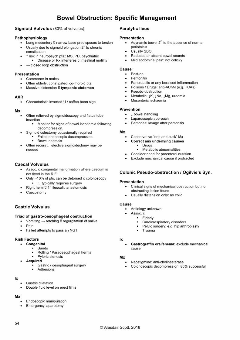
54
Bowel Obstruction: Specific Management Sigmoid Volvulus (80% of volvulus) Pathophysiology
• Long mesentery c̄ narrow base predisposes to torsion • Usually due to sigmoid elongation 2O to chronic
constipation • ↑ risk in neuropsych pts.: MS, PD, psychiatric
§ Disease or Rx interferes c̄ intestinal motility • → closed loop obstruction
Presentation
• Commoner in males • Often elderly, constipated, co-morbid pts. • Massive distension c̄ tympanic abdomen
AXR
• Characteristic inverted U / coffee bean sign Mx
• Often relieved by sigmoidoscopy and flatus tube insertion
§ Monitor for signs of bowel ischaemia following decompression.
• Sigmoid colectomy occasionally required § Failed endoscopic decompression § Bowel necrosis
• Often recurs \ elective sigmoidectomy may be needed
Caecal Volvulus
• Assoc. c̄ congenital malformation where caecum is not fixed in the RIF.
• Only ~10% of pts. can be detorsed c̄ colonoscopy § \ typically requires surgery
• Right hemi c̄ 1O ileocolic anastomosis • Caecostomy
Gastric Volvulus Triad of gastro-oesophageal obstruction
• Vomiting → retching c̄ regurgitation of saliva • Pain • Failed attempts to pass an NGT
Risk Factors
• Congenital § Bands § Rolling / Paraoesophageal hernia § Pyloric stenosis
• Acquired § Gastric / oesophageal surgery § Adhesions
Ix
• Gastric dilatation • Double fluid level on erect films
Mx
• Endoscopic manipulation • Emergency laparotomy
Paralytic Ileus Presentation
• Adynamic bowel 2O to the absence of normal peristalsis
• Usually SBO • Reduced or absent bowel sounds • Mild abdominal pain: not colicky
Cause
• Post-op • Peritonitis • Pancreatitis or any localised inflammation • Poisons / Drugs: anti-AChM (e.g. TCAs) • Pseudo-obstruction • Metabolic: ↓K, ↓Na, ↓Mg, uraemia • Mesenteric ischaemia
Prevention
• ↓ bowel handling • Laparoscopic approach • Peritoneal lavage after peritonitis
Mx
• Conservative “drip and suck” Mx • Correct any underlying causes
§ Drugs § Metabolic abnormalities
• Consider need for parenteral nutrition • Exclude mechanical cause if protracted
Colonic Pseudo-obstruction / Ogilvie’s Syn. Presentation
• Clinical signs of mechanical obstruction but no obstructing lesion found
• Usually distension only: no colic Cause
• Aetiology unknown • Assoc. c̄
§ Elderly § Cardiorespiratory disorders § Pelvic surgery: e.g. hip arthroplasty § Trauma
Ix
• Gastrograffin oral/enema: exclude mechanical cause
Mx
• Neostigmine: anti-cholinesterase • Colonoscopic decompression: 80% successful
© Alasdair Scott, 2018

55
Colorectal Carcinoma: Pathology and Presentation Epidemiology
• 3rd commonest cancer • 2nd commonest cause of cancer deaths (16,000/yr) • Age: peak in 60s • Sex: rectal Ca commoner in men • Geo: Western disease
Pathophysiology Colonic Adenomas
• Benign precursors to CRC • Characterised by dysplastic epithelium • Classification
§ Tubular: small, pedunculated, tubular glands § Villous: large, sessile, covered by villi § Tubulovillous: mixture
• Presentation § Typically asymptomatic § Large polyps can bleed → IDA § Villous adenomas can → ↓K +
hypoproteinaemia • Malignant potential
§ ↑ size § ↑ dysplasia § ↑ villous component
APC et al.
• -ve regulator of β-catenin (component of WNT pathway)
• APC binds to and promotes degradation of β-catenin. • APC mutation → ↑ β-catenin → ↑ transcription of
genes which promote cell proliferation. • Proliferation → mutation of other genes which
promote growth and prevent apoptosis § KRAS (proto-oncogene) § p53 (TSG)
Adenoma → Carcinoma Sequence (Vogelstein)
• First hit: mutation of one APC copy • Second hit: mutation of second APC copy
§ → adenoma formation • Additional mutations in adenoma → malignant
transformation: e.g KRAS, p53 Other Aetiological Factors
• Diet: ↓ fibre + ↑ animal fat / protein • IBD: CRC in 15% c̄ pancolitis for 20yrs • Familial: FAP, HNPCC, Peutz-Jeghers • Smoking • Genetics
§ No relative: 1/50 CRC risk § One 1st degree: 1/10
• NSAIDs / Aspirin (300mg/d): protective
Pathology
• 95% adenocarcinoma § Others: lymphoma, GIST, carcinoid
• Location § Rectum: 35% § Sigmoid: 25% § Caecum and ascending colon: 20% § Transverse: 10% § Descending: 5%
• Proximal tumours: sessile or polypoid • Distal tumours: annular stenosing
Spread
• Local • Lymphatic • Blood (liver, lungs) • Transcoelomic
Presentation Left
• Altered bowel habit • PR mass (60%) • Obstruction (25%) • Bleeding / mucus PR • Tenesmus
Right
• Anaemia • Wt. loss • Abdominal pain
Either
• Abdominal mass • Perforation • Haemorrhage • Fistula
Examination
• Palpable mass: per abdomen or PR • Perianal fistulae • Hepatomegaly • Anaemia • Signs of obstruction
© Alasdair Scott, 2018
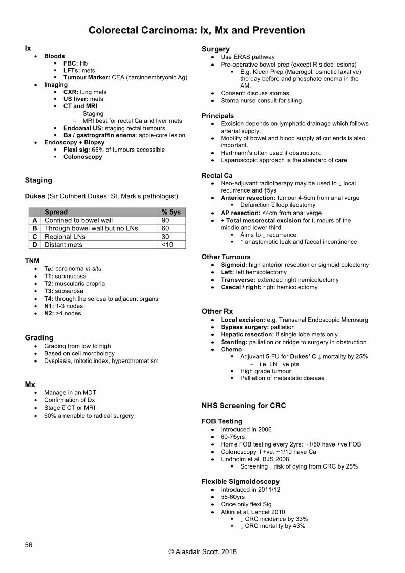
56
Colorectal Carcinoma: Ix, Mx and Prevention Ix
• Bloods § FBC: Hb § LFTs: mets § Tumour Marker: CEA (carcinoembryonic Ag)
• Imaging § CXR: lung mets § US liver: mets § CT and MRI
- Staging - MRI best for rectal Ca and liver mets
§ Endoanal US: staging rectal tumours § Ba / gastrograffin enema: apple-core lesion
• Endoscopy + Biopsy § Flexi sig: 65% of tumours accessible § Colonoscopy
Staging Dukes (Sir Cuthbert Dukes: St. Mark’s pathologist)
Spread % 5ys A Confined to bowel wall 90 B Through bowel wall but no LNs 60 C Regional LNs 30 D Distant mets <10
TNM
• TIS: carcinoma in situ • T1: submucosa • T2: muscularis propria • T3: subserosa • T4: through the serosa to adjacent organs • N1: 1-3 nodes • N2: >4 nodes
Grading
• Grading from low to high • Based on cell morphology • Dysplasia, mitotic index, hyperchromatism
Mx
• Manage in an MDT • Confirmation of Dx • Stage c̄ CT or MRI • 60% amenable to radical surgery
Surgery
• Use ERAS pathway • Pre-operative bowel prep (except R sided lesions)
§ E.g. Kleen Prep (Macrogol: osmotic laxative) the day before and phosphate enema in the AM.
• Consent: discuss stomas • Stoma nurse consult for siting
Principals
• Excision depends on lymphatic drainage which follows arterial supply.
• Mobility of bowel and blood supply at cut ends is also important.
• Hartmann’s often used if obstruction. • Laparoscopic approach is the standard of care
Rectal Ca
• Neo-adjuvant radiotherapy may be used to ↓ local recurrence and ↑5ys
• Anterior resection: tumour 4-5cm from anal verge § Defunction c̄ loop ileostomy
• AP resection: <4cm from anal verge • + Total mesorectal excision for tumours of the
middle and lower third. § Aims to ↓ recurrence § ↑ anastomotic leak and faecal incontinence
Other Tumours
• Sigmoid: high anterior resection or sigmoid colectomy • Left: left hemicolectomy • Transverse: extended right hemicolectomy • Caecal / right: right hemicolectomy
Other Rx
• Local excision: e.g. Transanal Endoscopic Microsurg • Bypass surgery: palliation • Hepatic resection: if single lobe mets only • Stenting: palliation or bridge to surgery in obstruction • Chemo
§ Adjuvant 5-FU for Dukes’ C ↓ mortality by 25% - i.e. LN +ve pts.
§ High grade tumour § Palliation of metastatic disease
NHS Screening for CRC FOB Testing
• Introduced in 2006 • 60-75yrs • Home FOB testing every 2yrs: ~1/50 have +ve FOB • Colonoscopy if +ve: ~1/10 have Ca • Lindholm et al. BJS 2008
§ Screening ↓ risk of dying from CRC by 25% Flexible Sigmoidoscopy
• Introduced in 2011/12 • 55-60yrs • Once only flexi Sig • Atkin et al. Lancet 2010
§ ↓ CRC incidence by 33% § ↓ CRC mortality by 43%
© Alasdair Scott, 2018

57
Familial CRC Syndromes Familial Adenomatous Polyposis
• Autosomal dominant • APC gene on 5q21
Presentation
• 100-1000s of adenomas by ~16yrs § Mainly in large bowel § Also stomach and duodenum (near ampulla)
• 100% develop CRC, often by ~40yrs • May be assoc. c̄ congenital hypertrophy of the retinal
pigment epithelium (CHPRE) Variants
• Attenuated FAP: <100 adenomas, later CRC (>50yrs) • Gardener’s (TODE)
§ Thyroid tumours § Osteomas of the mandible, skull and long
bones § Dental abnormalities: supernumerary teeth § Epidermal cysts
• Turcot’s: CNS tumours: medullo- and glio-blastomas Mx
• Prophylactic colectomy before 20yrs • Total colectomy + IRA
§ Requires life-long rectal stump surveillance • Proctocolectomy + IPAA • Remain @ risk of Ca in stomach and duodenum
§ Regular endoscopic screening Hereditary Non-Polyposis Colorectal Cancer
• Autosomal dominant • Mutation of mismatch repair enzymes
§ E.g. MSH2 on Chr 2p • Commonest cause of hereditary CRC: 3% of all CRC
Presentation
• Lynch 1: right sided CRC • Lynch 2: CRC + gastric, endometrial, prostate, breast
Dx: “3, 2, 1, rule”
• ≥3 family members over 2 generations c̄ one <50yrs Peutz-Jeghers Syndrome
• Autosomal dominant • STK11 mutation
Presentation
• ~ 10-15yrs • Mucocutaneous hyperpigmentation
§ Macules on palms, buccal mucosa • Multiple GI hamartomatous polyps
§ Intussusception § Haemorrhage
• ↑ Ca risk § CRC, pancreas, breast, lung, ovaries, uterus
GI Polyps Inflammatory Pseudopolyps
• Regenerating islands of mucosa in UC Hyperplastic Polyps
• Piling up of goblet cells and absorptive cells • Serrated surface architecture • No malignant potential
Hamartomatous
• Tumour-like growths composed of tissues present at site where they develop.
• Sporadic or part of familial syndromes • Juvenile polyp: solitary hamartoma in children
§ “Cherry on a stalk” Neoplastic
• Tubular or villous adenomas • Usually asymptomatic • May have blood / mucus PR, tenesmus
Other Hamartomatous Polyposis Syndromes Juvenile Polyposis
• Autosomal dominant • >10 hamartomatous polyps • ↑ CRC risk: need surveillance and polypectomy
Cowden Syndrome
• Auto dominant • Macrocephaly + skin stigmata • Intestinal hamartomas • ↑ risk of extra-intestinal Ca
© Alasdair Scott, 2018
58
Mesenteric Ischaemia Acute Causes
• Arterial: thrombotic (35%), embolic (35%) • Non-occlusive (20%)
§ Splanchnic vasoconstriction: e.g. 2O to shock • Venous thrombosis (5%) • Other: trauma, vasculitis, strangulation
Presentation
• Nearly always small bowel • Triad
§ Acute severe abdominal pain ± PR bleed § Rapid hypovolaemia → shock § No abdominal signs
• Degree of illness >> clinical signs • May be in AF
Ix
• Bloods § ↑Hb: plasma loss § ↑WCC § ↑ amylase § Persistent metabolic acidosis: ↑lactate
• Imaging § AXR: gasless abdomen § Arteriography / CT/MRI angio
Complications
• Septic peritonitis • SIRS → MODS
Mx
• Fluids • Abx: gent + met • LMWH • Laparotomy: resect necrotic bowel
Chronic Small Bowel Ischaemia
• Cause: atheroma + low flow state (e.g. LVF) • Presentation:
§ Severe, colicky post-prandial abdo pain - “gut claudication”
§ PR bleeding § Malabsorption § Wt. loss
• Mx: angioplasty Chronic Large Bowel Ischaemia
• Cause: follows low flow in IMA territory • Presentation
§ Lower, left-sided abdominal pain § Bloody diarrhoea § Pyrexia § Tachycardia
• Ix § ↑WCC § Ba enema: thumb-printing § MR angiography
• Complications § May → peritonitis and septic shock § Strictures in the long-term
• Mx § Usually conservative: fluids and Abx § Angioplasty and endovascular stenting
Lower GI Bleed Causes Common / Important
• Rectal: haemorrhoids, fissure • Diverticulitis • Neoplasm
Other
• Inflammation: IBD • Infection: shigella, campylobacter, C. diff • Polyps • Large upper GI bleed (15% of lower GI bleeds) • Angio: dysplasia, ischaemic colitis, HHT
Ix
• Bloods: FBC, U+E, LFT, x-match, clotting, amylase • Stool: MCS • Imaging
§ AXR, erect CXR § Angiography: necessary if no source on
endoscopy § Red cell scan
• Endoscopy § 1st: Rigid proctoscopy / sigmoidoscopy § 2nd: OGD § 3rd: Colonoscopy: difficult in major bleeding
Mx
• Resuscitate • Urinary catheter • Abx: if evidence of sepsis or perf • PPI: if upper GI bleed possible • Keep bed bound: need to pass stool may be large
bleed → collapse • Stool chart • Diet: keep on clear fluids (allows colonoscopy) • Surgery: only if unremitting, massive bleed
Angiodysplasia
• Submucosal AV malformations • 70-90% occur in right colon • Can affect anywhere in GIT
Presentation
• Elderly • Fresh PR bleeding
Ix
• Exclude other Dx § PR exam § Ba enema § Colonoscopy
• Mesenteric angiography or CT angiography • Tc-labelled RBC scan: identify active bleeding
Rx
• Embolisation • Endoscopic laser electrocoagulation • Resection
© Alasdair Scott, 2018
59
Perianal Surgery Contents
Key Anatomy ........................................................................................................................................................ 60Minor Anal Conditions .......................................................................................................................................... 60Haemorrhoids ...................................................................................................................................................... 61Anal Fissure ......................................................................................................................................................... 62Fistula in Ano ....................................................................................................................................................... 62Peri-anal Sepsis / Abscess .................................................................................................................................. 63Pilonidal Sinus ..................................................................................................................................................... 63Anal Carcinoma ................................................................................................................................................... 64Rectal Prolapse .................................................................................................................................................... 64
© Alasdair Scott, 2018

60
Key Anatomy Rectum
• 12cm • Sacral promontory to levator ani muscle • The 3 tenia coli fuse around the rectum to form a
continuous muscle layer. Anal Canal
• 4cm • Levator ani muscle to anal verge • Upper 2/3 of canal
§ Lined by columnar epithelium § Insensate § Sup. rectal artery and vein § Internal iliac nodes
• Lower 1/3 of canal § Lined by squamous epithelium § Sensate § Middle and inf. rectal arteries and veins § Superficial inguinal nodes
• Dentate line = squamomucosal junction • White line = where anal canal becomes true skin
Anal Sphincters Internal
• Thickening of rectal smooth muscle • Involuntary control
External
• Three rings of skeletal muscle § Deep § Superficial § Subcutaneous
• Voluntary control Anorectal Ring
• Deep segment of external sphincter which is continuous c̄ puboretalis muscle (part of levator ani)
• Palpable on PR ~5cm from the anus • Demarcates junction between anal canal and rectum. • Must be preserved to maintain continence
Minor Anal Conditions Perianal Haematoma Definition
• Subcutaneous bleeding from a burst venule caused by straining or the passage of hard stool.
• Also called an external pile: a misnomer Presentation
• Tender blue lump at the anal margin • Pain worsened by defecation or movement
Rx
• Analgesia + spontaneous resolution • Evacuation under LA
Proctalgia Fugax
• Young, anxious men • Crampy anorectal pain, worse @ night • Unrelated to defecation • Assoc. c̄ trigeminal neuralgia • Rx
§ Reassurance § GTN cream
Perineal Warts
• Commonly seen in MSM • Condylomata accuminata
§ HPV § Rx: podophyllin paint, cryo, surgical excision
• Condylomata lata § Syphilis § Rx: penicillin
Pruritis Ani
• 50% idiopathic • Poor hygiene • Haemorrhoids • Anal fissure • Anal fistula • Fungi, worms • Crohn’s • Neoplasia
© Alasdair Scott, 2018
61
Haemorrhoids Definition
• Disrupted and dilated anal cushions Pathophysiology
• Anal cushion: mass of spongy vascular tissue • Positioned @ 3, 7 and 11 O’clock
§ Where the three major arteries that feed the vascular plexuses enter the anal canal
• Gravity, straining → engorgement and enlargement of anal cushions
• Hard stool disrupts connective tissue around cushions.
• Cushions protrude and can be damaged by hard stool § → bright red (capillary) bleeding.
• Haemorrhoids arise above dentate line \ not painful • May be gripped by anal sphincter → thrombosis
§ Strangulated piles are acutely painful § May ulcerate or infarct
Causes
• Constipation c̄ prolonged straining • Venous congestion may contribute
§ Pregnancy § Abdominal tumour § Portal HTN
Classification
• 1st degree: never prolapse • 2nd: prolapse on defecation but spontaneously reduce • 3rd: prolapse on defecation but require digital
reduction • 4th: remain permanently prolapsed
Symptoms
• Fresh painless PR bleeding § Bright red § On paper, on stool, may drip into pan
• Pruritis ani • Lump in perianal area • Severe pain = thrombosis
Examination
• Full abdo exam, palpating for masses • Inspect perianal area: masses, recent bleeding • DRE: can’t palpate piles unless thrombosed • Rigid sig to identify higher rectal pathology • Proctoscopy (also allows Rx)
Differential
• Perianal haematoma • Fissure • Abscess • Tumour (must exclude in all cases)
Mx Conservative
• ↑ fibre and fluid intake • Stop straining @ stool
Medical
• Topical preparations § Anusol: hydrocortisone § Topical analgesics
• Laxatives: lactulose, fybogel Interventional
• Injection c̄ sclerosant (5% phenol in Almond oil) § Injection above dentate line § SE: impotence, prostatitis
• Banding → thrombosis and separation § SE: bleeding, infection
• Cryotherapy § SE: watery discharge post-procedure
• Infra-red coagulation Surgical: Haemorrhoidectomy
• Excision of piles + ligation of vascular pedicles • Discharge c̄ laxatives post-op • SE: bleeding, stenosis
Mx of Thrombosed Piles
• Analgesia • Ice-packs • Stool softeners • Topical lignocaine jelly • Pain usually resolves in 2-3wks • Haemorrhoidectomy is not usually necessary
© Alasdair Scott, 2018
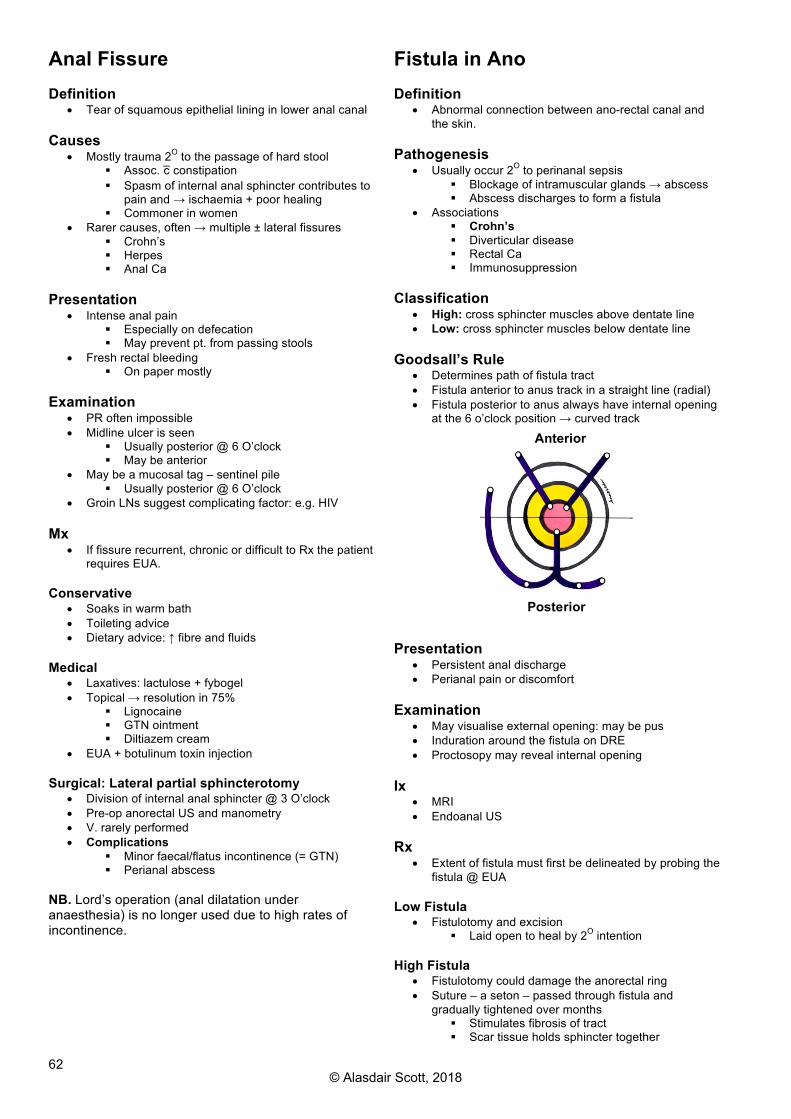
62
Anal Fissure Definition
• Tear of squamous epithelial lining in lower anal canal Causes
• Mostly trauma 2O to the passage of hard stool § Assoc. c̄ constipation § Spasm of internal anal sphincter contributes to
pain and → ischaemia + poor healing § Commoner in women
• Rarer causes, often → multiple ± lateral fissures § Crohn’s § Herpes § Anal Ca
Presentation
• Intense anal pain § Especially on defecation § May prevent pt. from passing stools
• Fresh rectal bleeding § On paper mostly
Examination
• PR often impossible • Midline ulcer is seen
§ Usually posterior @ 6 O’clock § May be anterior
• May be a mucosal tag – sentinel pile § Usually posterior @ 6 O’clock
• Groin LNs suggest complicating factor: e.g. HIV Mx
• If fissure recurrent, chronic or difficult to Rx the patient requires EUA.
Conservative
• Soaks in warm bath • Toileting advice • Dietary advice: ↑ fibre and fluids
Medical
• Laxatives: lactulose + fybogel • Topical → resolution in 75%
§ Lignocaine § GTN ointment § Diltiazem cream
• EUA + botulinum toxin injection Surgical: Lateral partial sphincterotomy
• Division of internal anal sphincter @ 3 O’clock • Pre-op anorectal US and manometry • V. rarely performed • Complications
§ Minor faecal/flatus incontinence (= GTN) § Perianal abscess
NB. Lord’s operation (anal dilatation under anaesthesia) is no longer used due to high rates of incontinence.
Fistula in Ano Definition
• Abnormal connection between ano-rectal canal and the skin.
Pathogenesis
• Usually occur 2O to perinanal sepsis § Blockage of intramuscular glands → abscess § Abscess discharges to form a fistula
• Associations § Crohn’s § Diverticular disease § Rectal Ca § Immunosuppression
Classification
• High: cross sphincter muscles above dentate line • Low: cross sphincter muscles below dentate line
Goodsall’s Rule
• Determines path of fistula tract • Fistula anterior to anus track in a straight line (radial) • Fistula posterior to anus always have internal opening
at the 6 o’clock position → curved track
Presentation
• Persistent anal discharge • Perianal pain or discomfort
Examination
• May visualise external opening: may be pus • Induration around the fistula on DRE • Proctosopy may reveal internal opening
Ix
• MRI • Endoanal US
Rx
• Extent of fistula must first be delineated by probing the fistula @ EUA
Low Fistula
• Fistulotomy and excision § Laid open to heal by 2O intention
High Fistula
• Fistulotomy could damage the anorectal ring • Suture – a seton – passed through fistula and
gradually tightened over months § Stimulates fibrosis of tract § Scar tissue holds sphincter together
Anterior
Posterior
© Alasdair Scott, 2018

63
Peri-anal Sepsis / Abscess Pathogenesis
• Anal gland blockage → infection → abscess § E.g. coli, bacteroides
• May develop from skin infections § E.g. sebaceous gland or hair follicle § Staphs
Associations
• Crohn’s • DM • Malignancy
Classification
• Perianal: 45% § Discrete local red swelling close to the anal
verge • Ischiorectal: ≤30%
§ Systemic upset § Extremely painful on DRE
• Interphincteric / intermuscular: >20% • Pelvirectal / supralevator: ~5%
§ Systemic upset § Bladder irritation
Presentation
• Throbbing perianal pain § Worse on sitting
• Occasionally a purulent anal discharge Examination
• Perianal mass or cellulitic area • Fluctuant mass on PR • Septic signs: fever, tachycardia
Rx
• Abx may suffice if Rx instigated v. early in course • Most cases require EUA c̄ I&D
§ Wound packed § Heals by 2O intention § Daily dressing for 7-10d
• Look for an anal fistula which complicates ~30% of abscesses.
Pilonidal Sinus Definition
• Pilonidal: lat “nest of hair” • Sinus: blind ending tract, lined by epithelial or
granulation tissue, which opens onto an epithelial surface.
Pathophysiology
• Hair works its way beneath skin → foreign body reaction → formation of abscess
• Usually occur in the natal cleft. Risk Factors
• M>F=4:1 • Geo: Mediterranean, Middle east, Asians • Often overweight c̄ poor personal hygiene • Occupations c̄ lots of sitting: e.g. truck drivers
Presentation
• Persistent discharge of purulent or clear fluid • Recurrent pain • Abscesses
Rx Conservative
• Hygiene advice • Shave / remove hair from affected area
Surgical
• Incision and drainage of abscesses • Elective sinus excision
§ Methylene blue to outline tract § Recurrence in 4-15%
© Alasdair Scott, 2018

64
Anal Carcinoma Epidemiology
• Relatively uncommon: 250-300 cases/yr in the UK Pathology
• 80% SCCs § Others: melanomas, adenocarcinomas
• Anal margin tumours § Well differentiated keratinising lesions § Commoner in men § Good prognosis
• Anal canal tumours § Arise above dentate line § Poorly differentiated, non-keratinising § Commoner in women § Worse prognosis
• Spread § Above dentate line → internal iliac nodes § Below dentate line → inguinal nodes
Aetiology
• HPV (16, 18, 31, 33) is important factor § ↑ incidence in MSM § ↑ incidence if perianal warts
Presentation
• Perianal pain and bleeding • Pruritis ani • Faecal incontinence
§ 70% have sphincter involvement @ presentation
• May → rectovaginal fistula Examination
• Palpable lesion in only 25% • ± palpable inguinal nodes
Ix
• ↓ Hb (ACD) • Endoanal US • Rectal EUA + biopsy • CT / MRI: assess pelvic spread
Rx
• Chemoradiotherapy: most pts § 50% 5ys
• Surgery: reserved for § Tumours that fail to respond to radiotherapy § GI obstruction § Small anal margin tumours w/o sphincter
involvement
Rectal Prolapse Definition
• Protrusion of rectal tissue through the anal canal. Classification Type 1: Mucosal prolapse
• Partial prolapse of redundant mucosa • Common in children: esp. c̄ CF • Essentially large piles \ same Rx
Type 2: Full thickness prolapse
• Commoner cf. type 1 • Usually elderly females c̄ poor O&G Hx
Presentation
• Mass extrudes from rectum on defecation § May reduce spontaneously or require manual
reduction • May become oedematous and ulcerated
§ → pain and bleeding • Faecal soiling • Assoc. c̄ vaginal prolapse and urinary incontinence
Examination
• Visible prolapse: brought out on straining • ± excoriation and ulceration • ↓ sphincter tone on PR • Assoc. uterovaginal prolapse
Ix
• Sigmoidoscopy to exclude proximal lesions • Anal manometry • Endoanal US • MRI
Rx Partial Prolapse
• Phenol injection • Rubber band ligation • Surgery: Delorme’s Procedure
Complete Prolapse
• Conservative § Pelvic floor exercises § Stool softeners
• Surgery § Abdominal Approach: Rectopexy
- Lap or open - Mobilised rectum fixed to sacrum c̄
mesh § Perineal Approach: Delorme’s Procedure
- Resect mucosa and suture the two mucosal boundaries.
© Alasdair Scott, 2018

65
Hernias Contents
Hernia Definitions ................................................................................................................................................. 66Inguinal Hernia ..................................................................................................................................................... 67Femoral Hernia .................................................................................................................................................... 68Incisional Hernia .................................................................................................................................................. 68Other Hernia Types .............................................................................................................................................. 69
© Alasdair Scott, 2018

66
Hernia Definitions Term Definition
Hernia Protrusion of a viscus or part of a viscus through the walls of its containing cavity into an abnormal position.
Reducible Sac can return to the abdominal cavity either spontaneously or c̄ manipulation.
Irreducible Sac cannot be reduced despite pressure or manipulation.
Strangulated Blood supply of contents is compromised due to pressure at the neck of the hernia.
Sliding hernia Part of the sac is formed by bowel (e.g. caecum or sigmoid): take care when excising the sac.
Maydl’s hernia Herniating double loop of bowel. Strangulated portion may reside as a single loop inside the abdomen.
Littre’s hernia Hernial sac containing strangulated Meckel’s diverticulum.
Amyand’s Inguinal hernia containing strangulated appendix
Richter’s Only part of circumference of bowel is within sac Most commonly seen with femoral hernias. Can strangulate w/o obstructing.
Pantaloon Simultaneous direct and indirect hernia.
Herniotomy Excision of hernial sac
Herniorrhaphy Suture repair of hernial defect
Hernioplasty Mesh repair of hernial defect
© Alasdair Scott, 2018
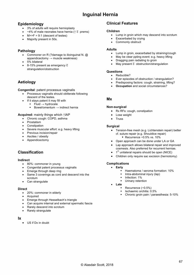
67
Inguinal Hernia Epidemiology
• 3% of adults will require hernioplasty • ~4% of male neonates have hernia (↑ c̄ prems) • M>>F = 9:1 (descent of testes) • Majority present in 50s.
Pathology
• Commoner on R (?damage to ilioinguinal N. @ appendicectomy → muscle weakness)
• 5% bilateral • 8-15% present as emergency c̄
strangulation/obstruction Aetiology Congenital: patent processus vaginalis
• Processus vaginalis should obliterate following descent of the testes.
• If it stays patent it may fill with § Fluid → hydrocele § Bowel/omentum → indirect hernia
Acquired: mainly things which ↑IAP
• Chronic cough: COPD, asthma • Prostatism • Constipation • Severe muscular effort: e.g. heavy lifting • Previous incision/repair • Ascites / obesity • Appendicectomy
Classification Indirect
• 80%: commoner in young • Congenital patent processus vaginalis • Emerge through deep ring • Same 3 coverings as cord and descend into the
scrotum • Can strangulate
Direct
• 20%: commoner in elderly • Acquired • Emerge through Hesselbach’s triangle • Can acquire internal and external spermatic fascia • Rarely descend into scrotum • Rarely strangulate
Ix
• US if Dx in doubt
Clinical Features Children
• Lump in groin which may descend into scrotum • Exacerbated by crying • Commonly obstruct
Adults
• Lump in groin, exacerbated by straining/cough • May be clear ppting event: e.g. heavy lifting • Dragging pain radiating to groin • May present c̄ obstruction/strangulation
Questions
• Reducible? • Ever episodes of obstruction / strangulation? • Predisposing factors: cough, straining, lifting? • Occupation and social circumstances?
Mx Non-surgical
• Rx RFs: cough, constipation • Lose weight • Truss
Surgical
• Tension-free mesh (e.g. Lichtenstein repair) better cf. suture repair (e.g. Shouldice repair)
§ Recurrence ~0.5% vs. 10% • Open approach can be done under LA or GA • Lap approach allows bilateral repair and improved
cosmesis. Also preferred for recurrent hernias. • 1O unilateral repairs should be open (NICE) • Children only require sac excision (herniotomy)
Complications
• Early § Haematoma / seroma formation: 10% § Intra-abdominal injury (lap) § Infection: 1% § Urinary retention
• Late § Recurrence (~0.5%) § Ischaemic orchitis: 0.5% § Chronic groin pain / paraesthesia: 5-10%
© Alasdair Scott, 2018

68
Femoral Hernia Definition
• Protrusion of viscus through the femoral canal Epidemiology
• F>M § Inguinal hernias still more common in F
• Middle aged and elderly Aetiology
• Acquired: ↑ intra-abdominal pressure • Femoral canal larger in females due to shape of pelvis
and changes in its configuration due to childbirth Clinical features Painless groin lump
• Neck inferior (and lateral) to the pubic tubercle. • Cough impulse. • Often irreducible (tight boarders)
Commonly present c̄ obstruction or strangulation
• Tender, red and hot • Abdo pain, distension, vomiting, constipation
Mx
• 50% risk of strangulation w/i 1mo • Urgent surgery
Elective: Lockwood Approach
• Low incision over hernia c̄ herniotomy and herniorrhaphy (suture ing. ligt. to pectineal ligt.)
Emergency: McEvedy Approach
• High approach in inguinal region to allow inspection and resection of non-viable bowel.
• Then herniotomy and herniorrhaphy
Incisional Hernia Definition
• Hernia arises through a previously acquired defect Epidemiology
• 6% of surgical incisions Risk Factors Pre-operative
• ↑ age • Obesity or malnutrition • Comorbidities: DM, renal failure, malignancy • Drugs: steroids, chemo, radio
Intra-operative
• Surgical technique/skill (major factor) § Too small suture bites § Inappropriate suture material
• Incision type (e.g. midline) • Placing drains through wounds
Post-operative
• ↑ IAP: chronic cough, straining, post-op ileus • Infection • Haematoma
Mx
• Surgery is not appropriate for many patients. • Must balance risk of operation and recurrence c̄ risk of
obstruction / strangulation and pt. choice. • Usually broad-necked \ low risk of strangulation
Conservative
• Manage RFs: e.g. constipation, cough • Weight loss • Elasticated corset or truss
Surgical
• Pre-op § Optimise cardiorespiratory function § Encourage wt. loss
• Mesh repair: open or lap
© Alasdair Scott, 2018

69
Other Hernia Types Umbilical Features
• Congenital • 3% of live births • Defect in the umbilical scar
Risk Factors • Afro-Caribbean • Trisomy 21 • Congenital hypothyroidism
Management • Usually resolves by 2-3yrs • Surgical repair if no closure. • Can recur in adults: pregnancy or gross
ascites
Paraumbilical Features • Acquired: middle aged obese men • Defect through linea alba just above or
below umbilicus • Small defect ® strangulation (often
omentum) Risk Factors • Chronic cough • Straining
Management • Mayo (double-breast linea alba c̄ sutures) /
mesh repair
Epigastric Features • Young M>F • Pea-sized swelling caused by defect in
linea alba above the umbilicus. • Usually contains omentum: can strangulate
Management • Mesh/suture repair
Spigelian Features • Hernia through linea semilunaris • Hernia lies between layers of abdo wall • Palpable mass more likely to be colon Ca
Obturator Features
• Old aged F>M • Sac protrudes through obturator foramen • Pain on inner aspect of thigh or knee • Frequently present obstructed / strangulated
Lumbar Features
• Middle-aged M>F • Typically follow loin incisions • Hernias through sup./inf. lumbar triangles
Sciatic Features
• Hernia through lesser sciatic foramen • Usually presents as SBO + gluteal mass
Gluteal Features • Hernia through greater sciatic foramen • Usually presents as SBO + gluteal mass
© Alasdair Scott, 2018

70
Superficial Lesions Contents
Skin Lumps .......................................................................................................................................................... 71Malignant and Pre-malignant Skin Lesions .......................................................................................................... 73Neck Lumps: General Approach .......................................................................................................................... 74Anterior Triangle Lumps ....................................................................................................................................... 74Midline Neck Lumps ............................................................................................................................................. 75Posterior Triangle Lumps ..................................................................................................................................... 75Cervical Lymphadenopathy ................................................................................................................................. 76Hypertrophic and Keloid Scars ............................................................................................................................ 76Thyroid Lumps ..................................................................................................................................................... 77Benign Thyroid Disease ....................................................................................................................................... 78Malignant Thyroid Disease .................................................................................................................................. 79Salivary Glands .................................................................................................................................................... 80
© Alasdair Scott, 2018

71
Skin Lumps
Lump Pathology Features Viva Mx Lipoma Benign tumour of mature adipocytes
Sarcomatous change probably doesn’t occur. Liposarcomas arise de novo - Older pts. - Deeper tissues of the lower limbs
Inspection
Occur anywhere fat can expand - i.e. NOT scalp or palms - inc. spermatic cord, submucosa May be a scar from recurrence
Dercum’s Disease / Adiposis dolorosa - Multiple, painful lipomas - Assoc. peripheral neuropathy - Obese postmen women Familial Multiple Lipomatosis Madelung’s Disease Bannayan-Zonana Syndrome - Multiple lipomas - Macrocephaly - Haemangiomas
Non-surgical Surgical Excision
Palpation
Soft Subcutaneous Imprecise margin Fluctuant
Sebaceous cyst
Epithelial-lined cyst containing keratin Two histological subtypes 1) Epidermal Cyst - Arise from hair follicle infundibulum 2) Trichilemmal Cyst / Wen - Arise from hair follicle epithelium - Often multiple - May be autosomal dominant
Inspection
Occur @ sites of hair growth - Scalp, face, neck, chest and back - NOT soles or palms Central Punctum
Complications - Infection: pus discharge - Ulceration - Calcification Cock’s Peculiar Tumour - Large ulcerating trichilemmal cyst on the scalp - Resemble an SCC Gardener’s Syndrome: FAP + - Thyroid tumours - Osteomas - Dental abnormalities - Epidermal cysts
Non-surgical Surgical Excision
Palpation
Firm Smooth Intradermal
Ganglion Cystic swelling related to a synovial lined structures: joint, tendon Myxoid degeneration of fibrous tissue Contain thick, gelatinous material
Inspection
Can be found anywhere 90% on dorsum of hand or wrist Dorsum of ankle May be scar from recurrence Weakly transilluminable
Differential - Bursae - Cystic protrusion from synovial cavity of arthritic joint.
Non-Surgical - Aspiration followed by 3wks of immobilisation Surgical Excision - Recurrence can be 50% - Neurovascular damage Palpation
Soft Subcutaneous May be tethered to tendon
Seborrheic keratosis
Benign hyperplasia of basal epithelial layer - Hyperkeratosis: keratin layer thickening - Acanthosis: prickle layer thickening
Stuck on appearance Dark brown Greasy
Non-surgical
© Alasdair Scott, 2018
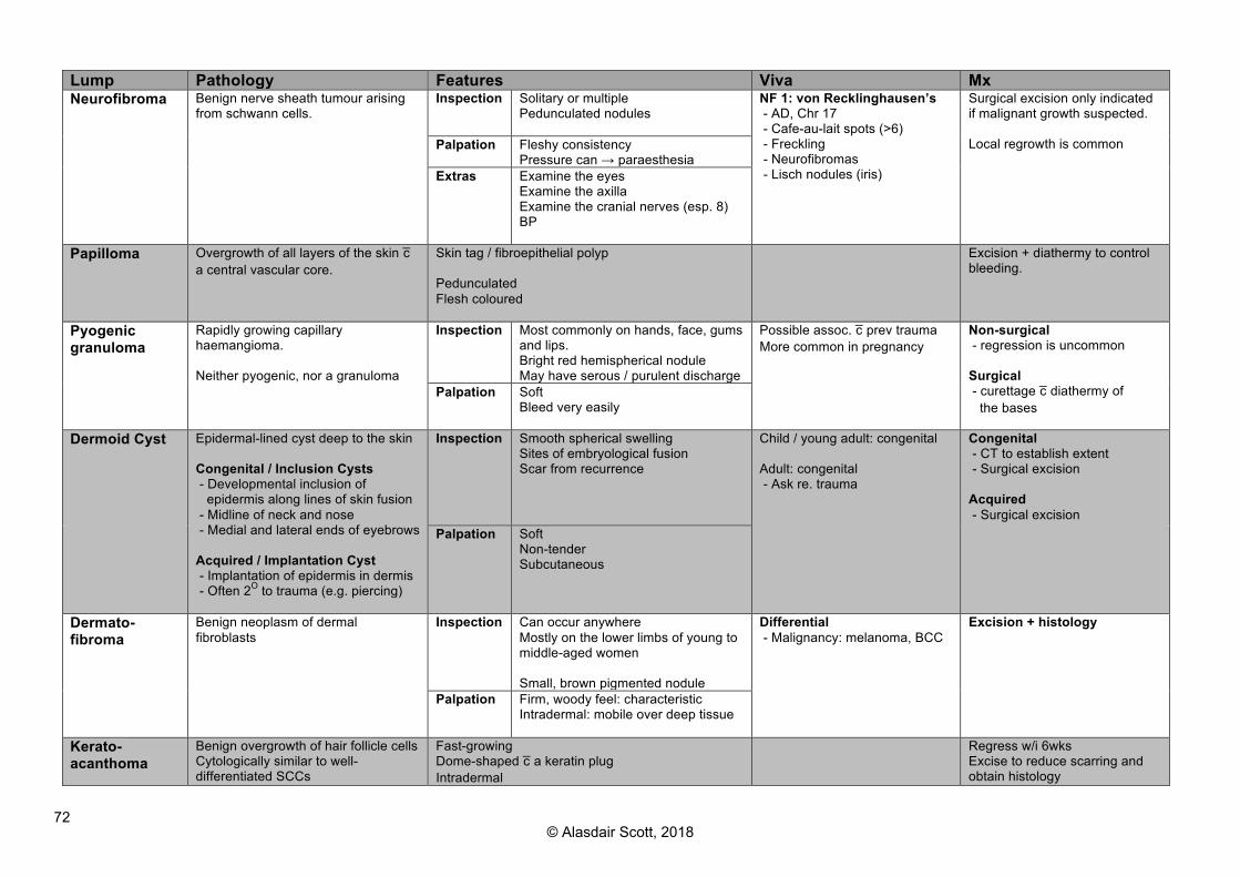
72
Lump Pathology Features Viva Mx Neurofibroma Benign nerve sheath tumour arising
from schwann cells. Inspection
Solitary or multiple Pedunculated nodules
NF 1: von Recklinghausen’s - AD, Chr 17 - Cafe-au-lait spots (>6) - Freckling - Neurofibromas - Lisch nodules (iris)
Surgical excision only indicated if malignant growth suspected. Local regrowth is common Palpation
Fleshy consistency Pressure can → paraesthesia
Extras Examine the eyes Examine the axilla Examine the cranial nerves (esp. 8) BP
Papilloma Overgrowth of all layers of the skin c̄ a central vascular core.
Skin tag / fibroepithelial polyp Pedunculated Flesh coloured
Excision + diathermy to control bleeding.
Pyogenic granuloma
Rapidly growing capillary haemangioma. Neither pyogenic, nor a granuloma
Inspection
Most commonly on hands, face, gums and lips. Bright red hemispherical nodule May have serous / purulent discharge
Possible assoc. c̄ prev trauma More common in pregnancy
Non-surgical - regression is uncommon Surgical - curettage c̄ diathermy of the bases
Palpation
Soft Bleed very easily
Dermoid Cyst Epidermal-lined cyst deep to the skin Congenital / Inclusion Cysts - Developmental inclusion of epidermis along lines of skin fusion - Midline of neck and nose - Medial and lateral ends of eyebrows Acquired / Implantation Cyst - Implantation of epidermis in dermis - Often 2O to trauma (e.g. piercing)
Inspection
Smooth spherical swelling Sites of embryological fusion Scar from recurrence
Child / young adult: congenital Adult: congenital - Ask re. trauma
Congenital - CT to establish extent - Surgical excision Acquired - Surgical excision
Palpation
Soft Non-tender Subcutaneous
Dermato-fibroma
Benign neoplasm of dermal fibroblasts
Inspection
Can occur anywhere Mostly on the lower limbs of young to middle-aged women Small, brown pigmented nodule
Differential - Malignancy: melanoma, BCC
Excision + histology
Palpation
Firm, woody feel: characteristic Intradermal: mobile over deep tissue
Kerato-acanthoma
Benign overgrowth of hair follicle cells Cytologically similar to well-differentiated SCCs
Fast-growing Dome-shaped c̄ a keratin plug Intradermal
Regress w/i 6wks Excise to reduce scarring and obtain histology
© Alasdair Scott, 2018
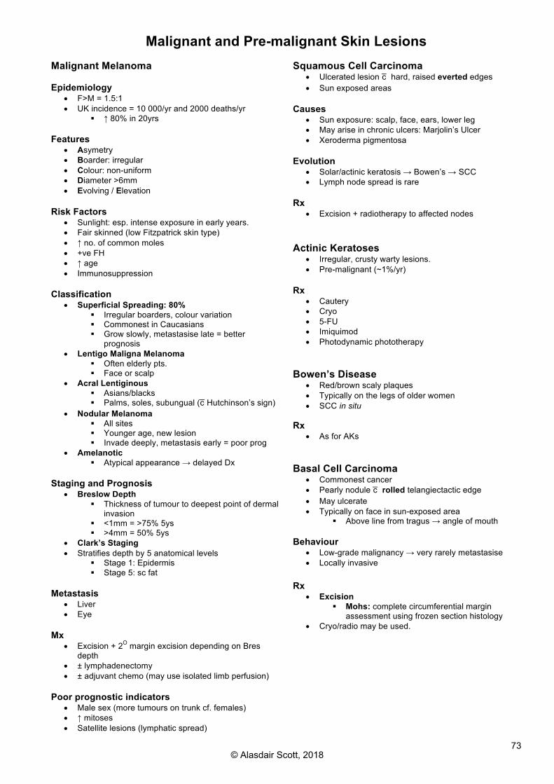
73
Malignant and Pre-malignant Skin Lesions Malignant Melanoma Epidemiology
• F>M = 1.5:1 • UK incidence = 10 000/yr and 2000 deaths/yr
§ ↑ 80% in 20yrs Features
• Asymetry • Boarder: irregular • Colour: non-uniform • Diameter >6mm • Evolving / Elevation
Risk Factors
• Sunlight: esp. intense exposure in early years. • Fair skinned (low Fitzpatrick skin type) • ↑ no. of common moles • +ve FH • ↑ age • Immunosuppression
Classification
• Superficial Spreading: 80% § Irregular boarders, colour variation § Commonest in Caucasians § Grow slowly, metastasise late = better
prognosis • Lentigo Maligna Melanoma
§ Often elderly pts. § Face or scalp
• Acral Lentiginous § Asians/blacks § Palms, soles, subungual (c̄ Hutchinson’s sign)
• Nodular Melanoma § All sites § Younger age, new lesion § Invade deeply, metastasis early = poor prog
• Amelanotic § Atypical appearance → delayed Dx
Staging and Prognosis
• Breslow Depth § Thickness of tumour to deepest point of dermal
invasion § <1mm = >75% 5ys § >4mm = 50% 5ys
• Clark’s Staging • Stratifies depth by 5 anatomical levels
§ Stage 1: Epidermis § Stage 5: sc fat
Metastasis
• Liver • Eye
Mx
• Excision + 2O margin excision depending on Bres depth
• ± lymphadenectomy • ± adjuvant chemo (may use isolated limb perfusion)
Poor prognostic indicators
• Male sex (more tumours on trunk cf. females) • ↑ mitoses • Satellite lesions (lymphatic spread)
Squamous Cell Carcinoma • Ulcerated lesion c̄ hard, raised everted edges • Sun exposed areas
Causes
• Sun exposure: scalp, face, ears, lower leg • May arise in chronic ulcers: Marjolin’s Ulcer • Xeroderma pigmentosa
Evolution
• Solar/actinic keratosis → Bowen’s → SCC • Lymph node spread is rare
Rx
• Excision + radiotherapy to affected nodes Actinic Keratoses
• Irregular, crusty warty lesions. • Pre-malignant (~1%/yr)
Rx
• Cautery • Cryo • 5-FU • Imiquimod • Photodynamic phototherapy
Bowen’s Disease
• Red/brown scaly plaques • Typically on the legs of older women • SCC in situ
Rx
• As for AKs Basal Cell Carcinoma
• Commonest cancer • Pearly nodule c̄ rolled telangiectactic edge • May ulcerate • Typically on face in sun-exposed area
§ Above line from tragus → angle of mouth Behaviour
• Low-grade malignancy → very rarely metastasise • Locally invasive
Rx
• Excision § Mohs: complete circumferential margin
assessment using frozen section histology • Cryo/radio may be used.
© Alasdair Scott, 2018

74
Neck Lumps: General Approach Diagnosis
• 85% of neck lumps are LNs: esp. if present < 3wks
§ Infection: EBV, tonsillitis, HIV § Ca: lymphoma or mets
• 8% are goitres • 7% other: e.g. sebaceous cyst or lipoma
Ix: Triple Assessment
• Clinical assessment • Imaging: US • Cyto/Histo: aspiration or biopsy
Anterior triangle
• Ant. margin of SCM • Midline • Ramus of the mandible • Roof: investing fascia • Floor: pre-vertebral fascia
Causes
• Pulsatile § Carotid artery aneurysm § Tortuous carotid artery § Carotid body tumour (chemodectoma)
• Non-pulsatile § Branchial cysts § Laryngocele § Goitre § Parotid tumour (lump in postero-superior
area) Submandibular Triangle
• Mental process • Ramus of the mandible • Line between two angles of the mandible
Causes
• Salivary stone • Sialadenitis • Salivary tumour
Posterior Triangle
• Post. margin of SCM • Ant. margin of trapezius • Mid 1/3 of clavicle.
Causes
• LNs • Cervical ribs • Pharyngeal pouch • Cystic hygromas • Pancoast’s tumour
Midline Lumps
• <20yrs § Thyroglossal cyst § Dermoid cyst
• >20yrs § Thyroid isthmus mass § Ectopic thyroid tissue
Anterior Triangle Lumps Branchial Cysts
• Embryological remnant 2nd branchial cleft Presentation
• Age <20yrs • Ant. margin of SCM at junction of upper and middle 3rd • May become infected → abscess • May be assoc. c̄ branchial fistula
Pathology
• Lined by squamous epithelium • Contain “glary” fluid c̄ cholesterol crystals
Rx
• Med § Abx for infection § Sclerotherapy c̄ OK-432 can be used
• Surgical excision § Definitive Rx § May be difficult due to proximity of carotids
Branchial sinus or fistula
• Small opening in lower 3rd of neck on ant. margin of SCM • Between tonsillar fossa and ant. border of SCM • May discharge mucus
Carotid Body Tumour: Chemodectoma
• Very rare • Carotid bodies
§ Located @ carotid bifurcation § Detect pO2, pCO2 and H+
Presentation
• Just anterior to upper 3rd of SCM. • Pulsatile • Move laterally but not vertically • May be bilateral • Pressure may → dizziness and syncope • Mostly benign (5% malignant)
Ix: Doppler or angio: splaying of bifurcation Rx: extirpation by a vascular surgeon Laryngocele
• Cystic dilatation of the laryngeal saccule • Congenital or acquired • Exacerbated by blowing
© Alasdair Scott, 2018

75
Midline Neck Lumps Dermoid Cyst
• Developmental inclusion of epidermis along lines of skin fusion.
Presentation
• Common <20yrs • Found at junctions of embryological fusion
§ Neck midline § Lateral angles of eyebrow § Under tongue
• Contains ectodermal elements § Hair follicles, sebaceous glands
Rx: excision Thyroglossal Cyst
• Cyst formed from persistent thyroglossal duct § Path of thyroid descent from base of tongue
Presentation
• Can be located anywhere between foramen caecum and the thyroid
§ Usually just inferior to the hyoid: subhyoid § Or, just above the hyoid: suprahyoid
• Fluctuant lump that moves up c̄ tongue protrusion • Can become infected → thyroglossal fistula
Rx
• Sistrunk’s Op: excision of cyst and thyroglossal duct with segment of hyoid bone
Posterior Triangle Lumps Cervical ribs
• Overdevelopment of transverse process of C7 • Occur in 1:150
Presentation
• Mostly asymptomatic • Hard swelling • ↓ radial pulse on abduction and external rotation of arm • Can → vascular symptoms
§ Compresses subclavian A § Raynaud’s § Subclavian steal § ↓ venous outflow → oedema
• Can → neurological symptoms § Compresses lower trunk of brachial plexus, T1
nerve root or stellate ganglion. § Wasting of intrinsic hand muscles § Paraesthesia along medial border of arm
Pharyngeal Pouch: Zenker’s Diverticulum
• Herniation of pharyngeal mucosa through its muscular coat at its weakest point.
§ Pulsion diverticulum • Killian’s dehiscence
§ Between thyro- and crico-pharyngeal muscles that form the inferior constrictor
Presentation
• Swelling on left side of neck • Regurgitation and aspiration • Halitosis • Gurgling sounds • Food debris → pouch expansion → oesophageal
compression → dysphagia. Ix: barium swallow Rx
• Excision and cricopharyngeal myotomy • Endoscopic stapling
Cystic Hygroma
• Congenital multiloculated lymphangioma arising from the jugular lymph sac
Presentation
• Infants • Lower part of post. triangle but may extend to axilla. • ↑ in size when child coughs/cries • Transilluminates brilliantly
Rx: excision or hypertonic saline sclerosant
• May recur
© Alasdair Scott, 2018
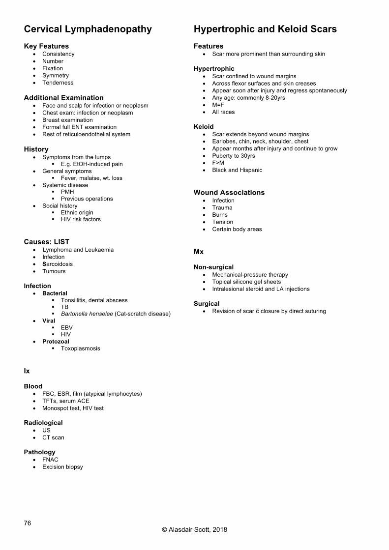
76
Cervical Lymphadenopathy Key Features
• Consistency • Number • Fixation • Symmetry • Tenderness
Additional Examination
• Face and scalp for infection or neoplasm • Chest exam: infection or neoplasm • Breast examination • Formal full ENT examination • Rest of reticuloendothelial system
History
• Symptoms from the lumps § E.g. EtOH-induced pain
• General symptoms § Fever, malaise, wt. loss
• Systemic disease § PMH § Previous operations
• Social history § Ethnic origin § HIV risk factors
Causes: LIST
• Lymphoma and Leukaemia • Infection • Sarcoidosis • Tumours
Infection
• Bacterial § Tonsillitis, dental abscess § TB § Bartonella henselae (Cat-scratch disease)
• Viral § EBV § HIV
• Protozoal § Toxoplasmosis
Ix Blood
• FBC, ESR, film (atypical lymphocytes) • TFTs, serum ACE • Monospot test, HIV test
Radiological
• US • CT scan
Pathology
• FNAC • Excision biopsy
Hypertrophic and Keloid Scars Features
• Scar more prominent than surrounding skin Hypertrophic
• Scar confined to wound margins • Across flexor surfaces and skin creases • Appear soon after injury and regress spontaneously • Any age: commonly 8-20yrs • M=F • All races
Keloid
• Scar extends beyond wound margins • Earlobes, chin, neck, shoulder, chest • Appear months after injury and continue to grow • Puberty to 30yrs • F>M • Black and Hispanic
Wound Associations
• Infection • Trauma • Burns • Tension • Certain body areas
Mx Non-surgical
• Mechanical-pressure therapy • Topical silicone gel sheets • Intralesional steroid and LA injections
Surgical
• Revision of scar c̄ closure by direct suturing
© Alasdair Scott, 2018

77
Thyroid Lumps Congenital Thyroid Disease
• Lingual thyroid • Ectopic thyroid tissue • Thyroglossal cyst
Embryology
• Thyroid migrates from its origin at the foramen caecum at the base of the tongue.
§ Passes behind the hyoid bone § Lies anterior to 3rd-4th tracheal rings in the
pretracheal fascia. • Leaves behind the thyroglossal cyst which atrophies
§ Persistence → thyroglossal cyst • Ectopic thyroid tissue can be found anywhere along
this descent Goitre Differential Diffuse
• Simple colloid goitre § Endemic: iodine deficiency § Sporadic: autoimmune, hereditary,
goitrogens (e.g. sulphonylureas) • Graves’ • Thyroiditis
§ Hashimoto’s § De Quervain’s § Subacute lymphocytic (e.g. post-partum)
• (multinodular goitre c̄ nodules too small to palpate) Multinodular
• Multinodular colloid goitre (commonest) • Multiple cysts • Multiple adenomas
Solitary nodule
• Dominant nodule in multinodular goitre • Adenoma (hot or cold) • Cyst • Malignancy
Ix Bloods
• TFTs: TSH, fT3, fT4 • Other: FBC, Ca2+, LFTs, ESR • Antibodies: anti-TPO, TSH
Imaging
• CXR: goitres and mets • High resolution US • CT • Radionucleotide (Tc or I) scan (hot vs. cold)
Histology or cytology
• FNAC (can’t distinguish adenoma vs. follicular Ca) • Biopsy
Laryngoscopy
• Pre-operative assessment of vocal cords
© Alasdair Scott, 2018

78
Benign Thyroid Disease
Disease Clinical Features Cause Management Simple goitre Diffuse painless goitre
Mass effects: - dysphagia - stridor - SVC obstruction Usually euthyroid, may →hypothyroid
Endemic - iodine deficiency Sporadic - autoimmune - goitrogens (e.g. sulphonylureas) - hereditary (dyshormogenic)
Thyroxine Total or subtotal thyroidectomy if pressure symptoms.
Multinodular goitre
Evolves from long-standing simple goitre. Mass effects. Euthyroid (or subclinical hyperthyroid)
As for simple goitre Thyroxine Total or subtotal thyroidectomy if pressure symptoms.
Plummer’s Toxic multinodular goitre
Multinodular goitre Thyrotoxicosis Uneven iodine uptake with hot nodule
Autonomous nodule develops on background multinodular goitre
Carbimazole Radioiodine Total or subtotal thyroidectomy.
Graves’ Diffuse goitre c̄ bruit Ophthalmopathy Dermopathy Thyrotoxicosis Assoc. c̄ other AI disease (T1DM, PA) ↑ uptake on radionucleotide scan
Autoimmune (T2 hypersensitivity) - anti-TSH
Propanolol Carbimazole Radioiodine Thyroidectomy
Hashimoto’s thyroiditis
Diffuse painless goitre May have transient thyrotoxicosis, then hypothyroidism Assoc. c̄ other AI disease (T1DM, PA)
Autoimmune (T2 and T4) - anti-TPO, -Tg
Levothyroxine
de Quervain’s
Diffuse painful goitre Preceding viral URTI common Thyrotoxicosis → hypo → eu ↓ iodine uptake
Viral: Coxsackie common Autoimmune
Self-limiting
Subacute lymphocytic
Diffuse painless goitre May occur post-partum Thyrotoxicosis → hypo → eu
Autoimmune Self-limiting
Riedel’s thyoiditis
Firm, fixed, irregular thyroid mass Mass effects Assoc. c̄ retroperitoneal fibrosis
Autoimmune fibrosis Conservative
Follicular adenoma
Single thyroid nodule ± thyrotoxicosis (majority are cold) May get pressure symptoms
Hemithyroidectomy
Thyroid cysts Solitary thyroid nodule Asympto or pressure symptoms Can → localised pain due to cyst bleed
Aspiration or excision
© Alasdair Scott, 2018
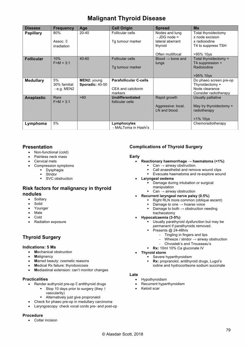
79
Malignant Thyroid Disease
Presentation
• Non-functional (cold) • Painless neck mass • Cervical mets • Compression symptoms
§ Dysphagia § Stridor § SVC obstruction
Risk factors for malignancy in thyroid nodules
• Solitary • Solid • Younger • Male • Cold • Radiation exposure
Thyroid Surgery Indications: 5 Ms
• Mechanical obstruction • Malignancy • Marred beauty: cosmetic reasons • Medical Rx failure: thyrotoxicosis • Mediastinal extension: can’t monitor changes
Practicalities
• Render euthyroid pre-op c̄ antithyroid drugs § Stop 10 days prior to surgery (they ↑
vascularity) § Alternatively just give propronalol
• Check for phaeo pre-op in medullary carcinoma • Laryngoscopy: check vocal cords pre- and post-op
Procedure
• Collar incision
Complications of Thyroid Surgery Early
• Reactionary haemorrhage → haematoma (<1%) § Can → airway obstruction. § Call anaesthetist and remove wound clips § Evacuate haematoma and re-explore wound
• Laryngeal oedema § Damage during intubation or surgical
manipulation § Can → airway obstruction
• Recurrent laryngeal nerve palsy (0.5%) § Right RLN more common (oblique ascent) § Damage to one → hoarse voice § Damage to both → obstruction needing
tracheostomy • Hypocalcaemia (3-5%)
§ Usually parathyroid dysfunction but may be permanent if parathyroids removed.
§ Presents @ 24-48hrs - Tingling in fingers and lips - Wheeze / stridor → airway obstruction - Chvostek’s and Trousseau’s
§ Rx: 10ml 10% Ca gluconate IV • Thyroid storm
§ Severe hyperthyroidism § Rx: propranolol, antithyroid drugs, Lugol’s
iodine and hydrocortisone sodium succinate Late
• Hypothyroidism • Recurrent hyperthyroidism • Keloid scar
Disease Frequency Age Cell Origin Spread Mx Papillary 80%
Assoc. c̄ irradiation
20-40 Follicular cells Tg tumour marker
Nodes and lung - JDG node = lateral aberrant thyroid Often multifocal
Total thyroidectomy ± node excision ± radioiodine T4 to suppress TSH >95% 10ys
Follicular 10% F>M = 3:1
40-60 Follicular cells Tg tumour marker
Blood → bone and lungs
Total thyroidectomy + T4 suppression + Radioiodine >95% 10ys
Medullary 5% 30% familial - e.g. MEN2
MEN2: young Sporadic: 40-50
Parafollicular C-cells CEA and calcitonin markers
Do phaeo screen pre-op Thyroidectomy + Node clearance Consider radiotherapy
Anaplastic Rare F>M = 3:1
>60 Undifferentiated follicular cells
Rapid growth Aggressive: local, LN and blood.
Usually palliative May try thyroidectomy + radiotherapy <1% 10ys
Lymphoma 5% Lymphocytes - MALToma in Hashi’s
Chemoradiotherapy
© Alasdair Scott, 2018
80
Salivary Glands History
• Swelling/pain related to food: calculi • Malaise, fever, mumps • Dry eyes/mouth: Sjogrens (Sicca, Mickulicz)
Causes of Salivary Gland Enlargement
• Whole gland § Parotitis § Sjogren’s / Sicca Syndrome § Sarcoid § Amyloid § ALL § Chronic liver disease § Anorexia or bulimia
• Localised § Tumours § Stones
Acute Parotitis
• Viral: mumps, coxsackie A, HIV • Bacterial: S. aureus
- Assoc. c̄ calculi and poor oral hygiene Calculi
• Recurrent unilateral swelling and pain • Worse on eating • Red, tender, swollen gland (80% submandibular) • Ix: plain x-ray or sialography • Rx: gland excision
Salivary Gland Neoplasms
• 80% are in the parotid (80% are superficial) • 80% are pleiomorphic adenomas • Deflection of ear outwards is classic sign • CN VII palsy = malignancy
Classification
• Benign § 1st: Pleiomorphic adenoma § 2nd: Adenolymphoma (Warthin’s)
• Malignant (CN7 palsy + fast growing) § 1st: Mucoepidermoid § 2nd: Adenoid cystic
Ix
• ENT examination • US ± CT • FNAC
Pleiomorphic Adenoma
• Commonest salivary gland neoplasm (80%) • Presentation
§ 90% occur in parotid § Occur in middle age § F>M § Benign and slow growing
• Histo: different tissue types (hence name) • Rx: superficial parotidectomy
Adenolymphoma (Warthin’s tumour)
• Benign soft cystic tumour • Older men • Rx: enucleation
Adenoid-cystic carcinoma
• One of the commonest malignant salivary tumours • Highly malignant and often incurable • Rapid growth • Hard fixed mass • Pain • Facial palsy
Surgery
• Superficial or radical parotidectomy • Facia lata face lift for facial palsy
Complications
• Facial palsy • Salivary fistula • Frey’s Syndrome (gustatory sweating)
§ Redness and sweating skin over parotid area § Occurs in relation to food (inc. thinking) § Auriculotemporal branch of CN V3 carries
sympathetic fibres to sweat glands over parotid area and parasympathetic fibres to the parotid
§ Reinervation of divided sympathetic nerves by fibres from the secretomotor branch of auriculotemporal branch of CN V3
© Alasdair Scott, 2018
81
Breast Surgery Contents
Breast Cancer: Pathology, Presentation and Assessment .................................................................................. 82Breast Cancer: Management ............................................................................................................................... 83Other Breast Disease ........................................................................................................................................... 84
© Alasdair Scott, 2018

82
Breast Cancer: Pathology, Presentation and Assessment Epidemiology
• Affects 1/10 women
• 20 000 cases/yr in UK
• Commonest cause of cancer death in females 15-54
• Second commonest cause of cancer deaths overall
Aetiology / Risk Factors
Family Hx • 10% Ca breast is familial
§ One 1st degree relative = 2x risk
• 5% assoc. c̄ BRCA mutations
§ BRCA1 (17q) → 80% breast Ca, 40% + ov Ca
§ BRCA2 (13q) → 80% breast Ca
Oestrogen Exposure
• Early menarche, late menopause • HRT, OCP (Million Women Study) • First child >35yrs
• Obesity Other Factors
• Proliferative breast disease c̄ atypia • Previous Ca breast • ↑ age (v. rare <30) • Breast feeding is protective
Pathology DCIS/LCIS
• Non-invasive pre-malignant condition • Microcalcification on mammography • 10x ↑ risk of invasive Ca
Invasive Ductal Carcinoma, NST/NOS
• Commonest: ~70% of cancers
• Feels hard (scirrhous)
Other subtypes • Invasive lobular: ~20% of cancers • Medullary: affects younger pts, feels soft • Colloid/mucinous: occur in elderly • Inflammatory: pain, erythema, swelling, peau d’orange • Papillary
Phyllodes Tumour
• Stromal tumour
• Large, non-tender mobile lump
Spread • Direct extension → muscle and/or skin
• Lymph → p’eau d’orange + arm oedema
• Blood →
§ Bones: bone pain, #, ↑Ca
§ Lungs: dyspnoea, pleural effusion
§ Liver: abdo pain, hepatic impairment
§ Brain: headache, seizures
Screening
• Every 3yrs from 47-73
• Craniocaudal and oblique views
• ↓ breast Ca deaths by 25%
• 10% false negative rate.
Presentation • Lump: commonest presentation of Ca breast
§ Usually painless
§ 50% in upper outer quadrant
§ ± axillary nodes
• Skin changes
§ Paget’s: persistent eczema
§ Peau d’orange
• Nipple
§ Discharge
§ Inversion
• Mets § Pathological # § SOB § Abdominal pain § Seizures
• May present through screening
Differential • Cysts
• Fibroadenomas
• DCIS
• Duct ectasia
Triple Assessment: any breast lump
• Hx and Clinical Examination • Radiology
§ <35yrs: US § >35ys: US + mammography
• Pathology § Solid lump: tru-cut core biopsy § Cystic lump: FNAC (green / 18G needle)
- Reassure if clear fluid - Send cytology if bloody fluid - Core biopsy residual mass - Core biopsy if +ve cytology
Other Ix
• Bloods § FBC, LFTs, ESR, bone profile
• Imaging: help staging
§ CXR § Liver US § CT scan § Breast MRI: multifocal disease or c̄ implants § Bone scan and PET-CT
• May need wire-guided excision biopsy Clinical Staging
• Stage 1: confined to breast, mobile, no LNs
• Stage 2: Stage 1 + nodes in ipsilateral axilla
• Stage 3: Stage 2 + fixation to muscle (not chest wall)
LNs matted and fixed, large skin involvement
• Stage 4: Complete fixation to chest wall + mets
TNM Staging • Tis (no palpable tumour): CIS
• T1: <2cm, no skin fixation
• T2: 2-5cm, skin fixation
• T3: 5-10cm, ulceration + pectoral fixation
• T4: >10cm, chest wall extension, skin involved
• N1: mobile nodes
• N2: fixed nodes
© Alasdair Scott, 2018

83
Breast Cancer: Management Principals
• Manage in an MDT c̄ an individual approach
§ Oncologist
§ Breast surgeon
§ Breastcare nurse
§ Radiologist
§ Histopathologist
• Try to enrol pts. in a trial
• Factors: age, fitness, wishes, clinical stage
§ 1-2: surgical
§ 3-4: chemo and palliation
Surgery • Aim = gain local control
• Two options
§ WLE + radiotherapy (80% treated like this) § Mastectomy
- Typically large tumours >4cm
- Multifocal or central tumours
- Nipple involvement
- Pt. choice
- Not radical: no longer used • Same survival, but WLE has ↑ recurrence rates
Sentinel Node Biopsy: gold standard
Rational • SN = first node that a section of breast drains to.
• If clinically –ve axillary LNs, no need for further
dissection if SN is clear.
Procedure • Blue dye / radiocolloid injected into tumour
• Visual inspection / gamma probe @ surgery to ID SN
• SN removed and sent for frozen section
• If node +ve → axillary clearance or radiotherapy
Evidence • 2 RCTs compared SNB vs. SNB + axillary clearance
• No differences in overall or disease-free survival
• ↓ morbidity c̄ SNB alone
§ Lymphoedema
§ Pain
§ Numbness
Other Axillary Options • For clinically -ve axilla options include
§ Axillary sampling
- Removal of lower nodes
- Clearance or DXT if +ve
§ Axillary clearance
- Can be done to various levels
Surgical Complications • Haematoma, seroma
• Frozen shoulder
• Long-thoracic nerve palsy
• Lymphoedema
• Upper inner arm numbness
§ Intercostobrachial nerve injury
Nottingham Prognostic Index • Predicts survival and risk of relapse
• Guides appropriate adjuvant systemic therapy
• (0.2 x tumour size) + histo grade + nodal status
§ Histo grade: Bloom-Richardson system (1-3)
Systemic Rx
Radiotherapy • Post-WLE: ↓ local recurrence • Post-mastectomy: only if high risk of local recurrence
§ Large, poorly differentiated, node +ve • Axillary: node +ve disease • Palliation: bone pain
Chemotherapy
• Pre-menopausal, node +ve, high grade or recurrent
tumours. • Neo-adjuvant chemo improves survival in large tumours • 6 x FEC: 5-FU, Epirubicin, Cyclophosphamide
• Trastuzumab (anti-Her2) is used if Her2+ve
§ SE: cardiac toxicity
Endocrine Therapy
• Used in ER or PR +ve disease: ↓ recurrence, ↑ survival • 5yrs of adjuvant therapy • Tamoxifen
§ SERM: antagonist @ breast, agonist @ uterus § SE: menopausal symptoms, endometrial Ca
• Anastrazole / Letrozole § Aromatase inhibitor → ↓ oestrogen § Better cf. tamoxifen if post-men (ATAC trial)
§ SE: menopausal symptoms • If pre-menopausal and ER+ve may consider ovarian
ablation or GnRH analogues (e.g. goserelin) Treating Advanced Disease (Stage 3-4)
• Tamoxifen if ER+ve
• Chemo for relapse
• Her2 +ve tumours may respond to trastuzumab
Supportive • Bone pain: DXT, bisphosphonates, analgesia • Brain: occasional surgery, DXT, steroids, AEDs • Lymphoedema: decongestion, compression
Reconstruction
• Offered either at 1O surgery or as delayed procedure.
Implants: silastic or saline inflatable
Lat dorsi myocutaneous flap • Pedicled flap: skin, fat, muscle and blood supply
• Supplied by thoracodorsal A. via subscapular A.
• Usually used c̄ an implant
Transverse rectus abdominis myocutaneous flap • Gold-standard • Pedicled (inf. epigastric A.) • Or free: attached to internal thoracic A • No implant necessary and combined tummy tuck • CI if poor circulation: smokers, obese, PVD, DM • Risk of abdominal hernia
Nipple Tattoo
© Alasdair Scott, 2018

84
Other Breast Disease Congenital
• Amastia: complete absence of breast and nipple
• Hypoplasia more common: some asymmetry
normal
• Accessory nipples
§ Can occur anywhere along the milk line
§ Present in 1%
Gynaecomastia • Occurs in 30% of boys at puberty
• Hormone secreting tumours: e.g. sex-cord
testicular
• Chronic liver disease: hypogonadism + ↓E2
metabolism
• Drugs: spironolactone, digoxin, cimetidine
Mastalgia Cyclical
• ~35yrs
• Pre-menstrual pain
• Relieved by menstruation
• Commonly in upper outer quadrants bilaterally
Non-cyclical
• ~45yrs • Severe lancing breast pain (often left) • May be assoc. c̄ back pain
Rx
• Reassurance + good bra for most
• 1st line: EPO (contains gamma-linoleic acid) • OCP • Topical NSAIDs (e.g. ibuprofen) • Bromocriptine
• Danazol
• Tamoxifen
Inflammatory
Disease Patient Presentation Treatment Acute Mastitis Usually lactating
Painful, red breast
May → Abscess (lump near nipple)
Fluclox alone in early stages
Fluclox + I&D if fluctuant
abscess
Fat Necrosis Associated with previous trauma
Painless, palpable, non-mobile mass
May calcify simulating Ca
Analgesia
No f/up necessary
Duct Ectasia Duct dilatation
Post-menopausal
- 50-60yrs
Slit-like nipple
Often bilateral
± peri-areolar mass
Thick white/green discharge May be calcified on mammography
Need to distinguish from Ca
Surgical duct excision if mass
present or discharge
troublesome
Close f/up
Periductal Mastitis
Smokers
30yrs
Painful, erythematous sub-areolar mass
Assoc. with inverted nipple ± purulent discharge
May → abscess or discharging fistula
Broad-spectrum Abx
Benign Epithelial Lesions
Disease Patient Presentation Treatment Benign Mammary Dysplasia
30-50yrs Pre-menstrual breast nodularity and pain
Often in upper outer quadrant
- Tender “lumpy-bumpy” breasts
Aberration of Normal Development and
Involution (ANDI) - Fibroadenosis
- Cyst formation
- Epitheliosis (hyperplasia)
- Papillomatosis
Triple assessment.
Reassurance
Analgesia
Good bra
± evening primrose oil
Danazol may occasionally be
used
Cystic Disease Peri-menopausal >40
Distinct, fluctuant round mass
Often painful
Aspiration: green-brown fluid
Persistence or blood → triple
assessment.
Duct Papilloma 40-50yrs Common cause of bloody discharge
Not usually palpable
Triple assessment.
Excise due to ↑ risk of Ca
© Alasdair Scott, 2018

85
Stromal Tumours
Disease Patient Presentation Treatment Fibroadenoma <35yrs
Rare post-menopause
↑ in Blacks
Commonest benign tumour Painless, mobile, rubbery mass
Often multiple and bilateral
Popcorn calcification
Reassurance + f/up if <2.5cm
Shell-out surgically
- >2.5cm
- FH of breast Ca
- Pt. choice
Phyllodes tumour 50s+
Large, fast growing mass
Mobile, non-tender
Epithelial and connective tissue
elements
WLE
Malignant Conditions
Disease Clinical Treatment DCIS Presents as Microcalcification on mammogram
Rarely assoc. with symptoms:
- lump
- discharge
- eczematous change = Paget’s disease
→ Ca @ 1%/yr (10x ↑ risk) in ipsilateral breast
WLE + radiotherapy
Extensive or multifocal → mastectomy + reconstruction
+ SLNB
Paget’s Unilat, scaly, erythematous, itchy
+/- palpable mass (invasive carcinoma)
Usually underlying invasive or DCIS breast cancer.
Mastectomy + radio ± chemo/endo
LCIS Incidental biopsy finding (no calcification)
Often bilat (20-40%)
Young women ↑ risk Ca risk (x10) in both breasts
Bilateral prophylactic mastectomy or close watching c̄
mammographic screening
© Alasdair Scott, 2018

86
Vascular Surgery Contents
Chronic Limb Ischaemia: Presentation and Classification ................................................................................... 87Chronic Limb Ischaemia: Investigation and Management ................................................................................... 88Acute Limb Ischaemia .......................................................................................................................................... 89Carotid Artery Disease ......................................................................................................................................... 90Aneurysms ........................................................................................................................................................... 90Popliteal Aneurysm .............................................................................................................................................. 90Abdominal Aortic Aneurysms ............................................................................................................................... 91Thoracic Aortic Dissection ................................................................................................................................... 92Gangrene ............................................................................................................................................................. 92Varicose Veins ..................................................................................................................................................... 93Leg Ulcers ............................................................................................................................................................ 94Leg Swelling ......................................................................................................................................................... 94
© Alasdair Scott, 2018

87
Chronic Limb Ischaemia: Presentation and Classification Incidence
• 5% of males >50yrs have intermittent claudication Cause
• Atherosclerosis § Typically asymptomatic until 50% stenosis
• (vasculitis and fibromuscular dysplasia are v. rare causes)
Atherosclerosis Summary
• Endothelial injury: haemodynamic, HTN, ↑ lipids • Chronic inflammation
§ Lipid-laden foam cells produce GFs, cytokines, ROS and MMPs
§ → lymphocyte and SMC recruitment • SM proliferation: conversion of fatty streak to
atherosclerotic plaque NB. Arteriosclerosis = general arterial hardening Atherosclerosis = arterial hardening specifically
due to atheroma. Atheroma Pathology
• Fibrous cap: SM cells, lymphocytes, collagen • Necrotic centre: cell debris, cholesterol, Ca, foam
cells Risk Factors Modifiable
• Smoking • BP • DM control • Hyperlipidaemia • ↓ exercise
Non-modifiable • FH and PMH • Male • ↑ age • Genetic
Associated Vascular Disease
• IHD: 90% • Carotid stenosis:15% • AAA • Renovascular disease • DM microvascular disease
Presentation Intermittent Claudication
• Cramping pain after walking a fixed distance • Pain rapidly relieved by rest • Calf pain = superficial femoral disease (commonest) • Buttock pain = iliac disease (internal or common)
Critical Limb Ischaemia: Fontaine 3 or 4
• European working group definition (1991) • Ankle pressure <50mmHg (toe <30mmHg) and
either: § Rest pain requiring analgesia for ³2 wks
- Especially @ night - Usually felt in the foot - Pt. hangs foot out of bed - Due to ↓ CO and loss of gravity help
§ Ulceration or gangrene Leriche’s Syndrome: Aortoiliac Occlusive Disease
• Atherosclerotic occlusion of abdominal aorta and iliacs • Triad
§ Buttock claudication and wasting § Erectile dysfunction § Absent femoral pulses
Buerger’s Disease: Thromboangiitis Obliterans
• Young, male, heavy smoker • Acute inflammation and thrombosis of arteries and
veins in the hands and feet → ulceration and gangrene
Signs
• Pulses: loss of pulses and ↑ CRT (norm ≤2sec) • Ulcers: painful, punched-out, on pressure points • Nail dystrophy / Onycholysis • Skin: cold, white, atrophy, absent hair • Venous guttering • Muscle atrophy • ↓ Buerger’s Angle
§ ≥90: normal § 20-30: ischaemia § <20: severe ischaemia
• +ve Buerger’s Sign § Reactive hyperaemia due to accumulation of
deoxygenated blood in dilated capillaries Clinical Classification Fontaine
• Asympto (subclinical) • Intermittent claudication
§ >200m § <200m
• Ischaemic rest pain • Ulceration / gangrene
Rutherford • Mild claudication • Moderate claudication • Severe claudication • Ischaemic rest pain • Minor tissue loss • Major tissue loss
© Alasdair Scott, 2018
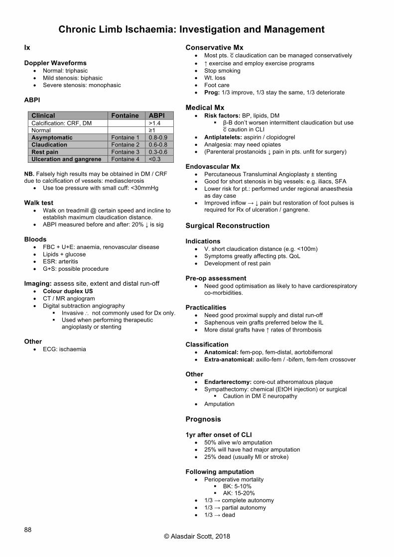
88
Chronic Limb Ischaemia: Investigation and Management Ix Doppler Waveforms
• Normal: triphasic • Mild stenosis: biphasic • Severe stenosis: monophasic
ABPI
Clinical Fontaine ABPI Calcification: CRF, DM >1.4 Normal ≥1 Asymptomatic Fontaine 1 0.8-0.9 Claudication Fontaine 2 0.6-0.8 Rest pain Fontaine 3 0.3-0.6 Ulceration and gangrene Fontaine 4 <0.3
NB. Falsely high results may be obtained in DM / CRF due to calcification of vessels: mediasclerosis
• Use toe pressure with small cuff: <30mmHg Walk test
• Walk on treadmill @ certain speed and incline to establish maximum claudication distance.
• ABPI measured before and after: 20% ↓ is sig Bloods
• FBC + U+E: anaemia, renovascular disease • Lipids + glucose • ESR: arteritis • G+S: possible procedure
Imaging: assess site, extent and distal run-off
• Colour duplex US • CT / MR angiogram • Digital subtraction angiography
§ Invasive \ not commonly used for Dx only. § Used when performing therapeutic
angioplasty or stenting Other
• ECG: ischaemia
Conservative Mx
• Most pts. c̄ claudication can be managed conservatively • ↑ exercise and employ exercise programs • Stop smoking • Wt. loss • Foot care • Prog: 1/3 improve, 1/3 stay the same, 1/3 deteriorate
Medical Mx
• Risk factors: BP, lipids, DM § β-B don’t worsen intermittent claudication but use
c̄ caution in CLI • Antiplatelets: aspirin / clopidogrel • Analgesia: may need opiates • (Parenteral prostanoids ↓ pain in pts. unfit for surgery)
Endovascular Mx
• Percutaneous Transluminal Angioplasty ± stenting • Good for short stenosis in big vessels: e.g. iliacs, SFA • Lower risk for pt.: performed under regional anaesthesia
as day case • Improved inflow → ↓ pain but restoration of foot pulses is
required for Rx of ulceration / gangrene. Surgical Reconstruction Indications
• V. short claudication distance (e.g. <100m) • Symptoms greatly affecting pts. QoL • Development of rest pain
Pre-op assessment
• Need good optimisation as likely to have cardiorespiratory co-morbidities.
Practicalities
• Need good proximal supply and distal run-off • Saphenous vein grafts preferred below the IL • More distal grafts have ↑ rates of thrombosis
Classification
• Anatomical: fem-pop, fem-distal, aortobifemoral • Extra-anatomical: axillo-fem / -bifem, fem-fem crossover
Other
• Endarterectomy: core-out atheromatous plaque • Sympathectomy: chemical (EtOH injection) or surgical
§ Caution in DM c̄ neuropathy • Amputation
Prognosis 1yr after onset of CLI
• 50% alive w/o amputation • 25% will have had major amputation • 25% dead (usually MI or stroke)
Following amputation
• Perioperative mortality § BK: 5-10% § AK: 15-20%
• 1/3 → complete autonomy • 1/3 → partial autonomy • 1/3 → dead
© Alasdair Scott, 2018

89
Acute Limb Ischaemia Definitions
• Acute: ischaemia <14d • Acute on chronic: worsening symptoms and signs
<14d • Chronic: ischaemia stable for >14d
Severity
• Incomplete: limb not threatened • Complete: limb threatened
§ Loss of limb unless intervention w/i 6hrs • Irreversible: requires amputation
Causes
• Thrombosis in situ (60%) § A previously stenosed vessel c̄ plaque rupture § Usually incomplete ischaemia
• Embolism (30%) § 80% from left atrium in AF § Valve disease § Iatrogenic from angioplasty / surgery § Cholesterol in long bone # § Paradoxical (venous via PFO) § Typically lodge at femoral bifurcation § Often complete ischaemia
• Graft / stent occlusion • Trauma • Aortic dissection
Presentation: 6Ps
• Pale • Pulseless • Perishingly cold • Painful • Paraesthesia • Paralysis
Thrombosis vs. Embolus
Thrombosis Embolus Onset Hrs-days Sudden Severity Less severe
- collaterals Profound ischaemia
Embolic source Present - often AF
Claudication Hx Present Absent Contralat pulses
Absent Present
Dx Angiography Clinical Rx Thrombolysis
Bypass surgery Embolectomy + Warfarin
Ix
• Blood § FBC, U+E, INR, G+S § CK
• ECG • Imaging
§ CXR § Duplex doppler § CT angio
General Mx
• In an acutely ischaemic limb discuss immediately c̄ a senior as time is crucial.
• NBM • Rehydration: IV fluids • Analgesia: morphine + metoclopramide • Abx: e.g. co-amoxiclav if signs of infection • Unfractionated heparin IVI: prevent extension • Complete occlusion?
§ Yes: urgent surgery: embolectomy or bypass § No: angiogram + observe for deterioration
Angiography
• Not performed if there is complete occlusion as it introduces delay: take straight to theatre.
• If incomplete occlusion, pre-op angio will guide any distal bypass.
Embolus Mx
1. Embolectomy § Under regional anaesthesia or GA § Wire fed through embolus § Fogarty catheter fed over the top § Balloon inflated and catheter withdrawn,
removing the embolism. § Adequacy confirmed by on-table angiography
2. Thrombolysis
§ Consider if embolectomy unsuccessful § E.g. local injection of TPA
3. Other options
§ Emergency reconstruction § Amputation
Post-embolectomy
• Anticoagulate: heparin IVI → warfarin • ID embolic source: ECG, echo, US aorta, fem and
pop • Complications
§ Reperfusion injury - Local swelling → compartment
syndrome - Acidosis and arrhythmia 2O to ↑K - ARDS - GI oedema → endotoxic shock
§ Chronic pain syndromes Thrombosis Mx
• Emergency reconstruction if complete occlusion • Angiography + angioplasty • Thrombolysis • Amputation
Manage Cardiovascular Risk Factors
© Alasdair Scott, 2018

90
Carotid Artery Disease Definitions
• Stroke: sudden neurological deficit of vascular origin lasting >24h
• TIA: sudden neurological deficit of vascular origin lasting <24h (usually lasts <1h) c̄ complete recovery
Pathogenesis
• Turbulent flow → ↓ shear stress @ carotid bifurcation promoting atherosclerosis and plaque formation.
• Plaque rupture → complete occlusion or distal emboli • Cause 15-25% of CVA/TIA
Presentation
• Bruit • CVA/TIA
Ix
• Duplex carotid Doppler • MRA
Mx Conservative
• Aspirin or clopidogrel • Control risk factors
Surgical: Endarterectomy
• Symptomatic (ECST, NASCET) § ≥70% (5% stroke risk per yr) § ≥50% if low risk (<3%, typically <75yrs) § Perform w/i 2wks of presentation
• Asymptomatic (ACAS, ACST) § ≥60% benefit if low risk
Complications of Endarterectomy
• Stroke or death: 3% • HTN: 60% • Haematoma • MI • Nerve injury • Hypoglossal: ipsilateral tongue deviation • Great auricular: numb ear lobe • Recurrent laryngeal: hoarse voice, bovine cough
Stenting
• Less invasive: ↓ hospital stay, ↓ infection, ↓ CN injury • There is concern over ↑ stroke risk, esp. pts. >70yrs • Meta-analysis shows no sig difference in mortality vs.
CEA @ 120d • Younger pts. have best risk / benefit ratio
Aneurysms Definition
• Abnormal dilatation of a blood vessel >50% of its normal diameter.
Classification
• True Aneurysm § Dilatation of a blood vessel involving all layers
of the wall and is >50% of its normal diameter § Two different morphologies
- Fusiform: e.g AAA - Saccular: e.g Berry aneurysm
• False Aneurysm § Collection of blood around a vessel wall that
communicates c̄ the vessel lumen. § Usually iatrogenic: puncture, cannulation
• Dissection § Vessel dilatation caused by blood splaying
apart the media to form a channel w/i the vessel wall.
Causes
• Congenital § ADPKD → Berry aneurysms § Marfan’s, Ehlers-Danlos
• Acquired § Atherosclerosis § Trauma: e.g. penetrating trauma § Inflammatory: Takayasu’s aortitis, HSP § Infection
- Mycotic: SBE - Tertiary syphilis (esp. thoracic)
Complications
• Rupture • Thrombosis • Distal embolization • Pressure: DVT, oesophagus, nutcracker syndrome • Fistula (IVC, intestine)
Popliteal Aneurysm
• Less common cf. AAA • 50% of pts. c̄ popliteal aneurysm also have AAA
Presentation
• Very easily palpable popliteal pulse • 50% bilateral • Rupture is relatively rare • Thrombosis and distal embolism is main complication
§ → acute limb ischaemia Mx
• Acute: embolectomy or fem-distal bypass • Stable
§ Elective grafting + tie off vessel § Stenting
© Alasdair Scott, 2018
91
Abdominal Aortic Aneurysms Epidemiology
• Prevalence: ~5% >50yrs • Mortality: 10,000 deaths/yr • Sex: M>F=3:1
Pathology
• Dilatation of the abdominal aorta ≥3cm • 90% infrarenal • 30% involve the iliac arteries
Presentation
• Usually asympto: discovered incidentally • May → back pain or umbilical pain radiating to groin • Acute limb ischaemia • Blue toe syndrome: distal embolisation • Acute rupture
Examination
• Expansile mass just above the umbilicus • Bruits may be heard • Tenderness + shock suggests rupture
Ix
• AXR: calcification may be seen • Abdo US: screening and monitoring • CT / MRI: gold-standard • Angiography
§ Won’t show true extent of aneurysm due to endoluminal thrombus.
§ Useful to delineate relationship of renal arteries Mx Conservative
• Manage cardiovascular risk factors: esp. BP • UK Small Aneurysm Trial suggested that AAA <5.5cm
in maximum diameter can be monitored by US (/CT) § <4cm: yearly monitoring § 4-5.5cm: 6 monthly monitoring
Surgical
• Aim to treat aneurysm before it ruptures. § Elective mortality: 5% § Emergency mortality: 50%
• Operate when risk of rupture > risk of surgery • Indications
§ Symptomatic (back pain = imminent rupture) § Diameter >5.5cm § Rapidly expanding: >1cm/yr § Causing complications: e.g. emboli
• Open or EVAR § EVAR has ↓ perioperative mortality § No ↓ mortality by 5yrs due to late endograft
failures. § EVAR not better than medical Rx in unfit pts.
Screening
• MASS trial revealed 50% ↓ aneurysm-related mortality in males aged 65-74 screened c̄ US.
• UK men offered one-time US screen @ 65yrs
AAA Rupture Rupture Rates
• <5.5cm = 1% / yr • >6cm = 25% / yr • ↑ risk if:
§ ↑BP § Smoker § Female § Strong FH
Rupture Presentation
• Sudden onset severe abdominal pain § Intermittent or continuous § Radiates to back or flanks (don’t dismiss as
colic) • Collapse → shock • Expansile abdominal mass
Mx: a surgical emergency
• High flow O2 • 2 x large bore cannulae in each ACF
§ Give fluid if shocked but keep SBP <100mmHg
§ Give O- blood if desperate § Blood: FBC, U+E, clotting, amylase, xmatch
10u • Instigate the major haemorrhage protocol • Call vascular surgeon, anaesthetist and warn theatre • Analgesia • Abx prophylaxis: cef + met • Urinary catheter + CVP line • If stable + Dx uncertain: US or CT may be feasible • Take to theatre: clamp neck, insert dacron graft
Mortality
• 100% w/o surgery • 50% c̄ surgery
© Alasdair Scott, 2018

92
Thoracic Aortic Dissection Definition
• Blood splays apart the laminar planes of the media to form a channel w/i the aortic wall.
Aetiology
• Atherosclerosis and HTN cause 90% • Minority caused by connective tissue disorder
§ Marfan’s, Ehlers Danlos § Vitamin C deficiency
Presentation
• Sudden onset, tearing chest pain § Radiates through to the back § Tachycardia and hypertension (1O +
sympathetic) • Distal propagation → sequential occlusion of branches
§ Left hemiplegia § Unequal arm pulses and BP § Paraplegia (anterior spinal A.) § Anuria
• Proximal propagation § Aortic regurgitation § Tamponade
• Rupture into pericardial, pleural or peritoneal cavities § Commonest cause of death
Stanford Classification Type A: Proximal
• 70% • Involves ascending aorta ± descending • Higher mortality due to probable cardiac involvement • Usually require surgery
Type B: Distal
• 30% • Involves descending aorta only: distal to L SC artery • Usually best managed conservatively
Ix
• ECG: exclude MI • TTE / TOE: can use in haemodynamically unstable pts • CT/MRI: not suitable for unstable pts.
Mx Resuscitate Investigate
• Bloods: x-match 10u, FBC, U+E, clotting, amylase • ECG: 20% show ischaemia due to involvement of the
coronary ostia • Imaging
§ CXR § CT/MRI: not if haemodynamically unstable § TOE: can be used if haemodynamically unstable
Treat
• Analgesia • ↓SBP
§ Labetalol or esmolol (short t½) § Keep SBP 100-110mmHg
• Type A: open repair § Acute operative mortality: <25%
• Type B: conservative initially § Surgery if persistent pain or complications § Consider TEVAR if uncomplicated
Gangrene Definition
• Death of tissue from poor vascular supply. Classification
• Wet: tissue death + infection • Dry: tissue death only • Pregangrene: tissue on the brink of gangrene
Presentation
• Black tissues ± slough • May be suppuration ± sepsis
Gas Gangrene
• Clostridium perfringes myositis • RFs: DM, trauma, malignancy • Presentation
§ Toxaemia § Haemolytic jaundice § Oedema § Crepitus from surgical emphysema § Bubbly brown pus
• Rx § Debridement (may need amputation) § Benzylpenicillin + metronidazole § Hyperbaric O2
Synergistic Gangrene
• Involves aerobes + anaerobes • Fournier’s: perineum • Meleney’s: post-op ulceration • Both progress rapidly to necrotising fasciitis + myositis
Mx
• Take cultures • Debridement (including amputation) • Benzylpenicillin ± clindamycin
© Alasdair Scott, 2018

93
Varicose Veins Definition
• Tortuous, dilated veins of the superficial venous system
Pathophysiology
• One-way flow from sup → deep maintained by valves • Valve failure → ↑ pressure in sup veins → varicosity • 3 main sites where valve incompetence occurs:
§ SFJ: 3cm below and 3cm lateral to pubic tubercle
§ SPJ: popliteal fossa § Perforators: draining GSV
- 3 medial calf perforators (Cockett’s) - 1 medial thigh perforator (Hunter’s)
Causes Primary
• Idiopathic (congenitally weak valves) § Prolonged standing § Pregnancy § Obesity § OCP § FH
• Congenital valve absence (v. rare) Secondary
• Valve destruction → reflux: DVT, thrombophlebitis • Obstruction: DVT, foetus, pelvic mass • Constipation • AVM • Overactive pumps (e.g. cyclists) • Klippel-Trenaunay
§ PWS, varicose veins, limb hypertrophy Symptoms
• Cosmetic defect • Pain, cramping, heaviness • Tingling • Bleeding: may be severe • Swelling
Signs
• Skin changes § Venous stars § Haemosiderin deposition § Venous eczema § Lipodermatosclerosis (paniculitis) § Atrophie blanche
• Ulcers: medial malleolus / gaiter area • Oedema • Thrombophlebitis
Ix
• Duplex ultrasonography § Anatomy § Presence of incompetence § Caused by obstruction or reflux
• Surgery: FBC, U+E, clotting, G+S, CXR, ECG Referral Criteria
• Bleeding • Pain • Ulceration • Superficial thrombophlebitis • Severe impact on QoL
CEAP Classification
• Chronic venous disease can be classified according to § Clinical signs (1-6 + sympto or asympto) § Etiology § Anatomy § Pathophysiology
Conservative Mx
• Treat any contributing factors § Lose weight § Relieve constipation
• Education § Avoid prolonged standing § Regular walks
• Class II Graduated Compression Stockings § 18-24mmHg § Symptomatic relief and slows progression
• Skin care § Maintain hydration c̄ emollients § Treat ulcers rapidly
Minimally Invasive Therapies Indications
• Small below knee VVs not involving GSV or SSV Techniques
• Local or GA • Injection sclerotherapy: 1% Na tetradecyl sulphate • Endovenous laser or radiofrequency ablation • Glue ablation
Post-Operatively
• Compression bandage for 24hrs • Compression stockings for 1mo
Surgical Mx Indications (rarely performed nowdays)
• SFJ incompetence • Major perforator incompetence • Symptomatic: ulceration, skin changes, pain
Procedures
• Trendelenberg: saphenofemoral ligation • SSV ligation: in the popliteal fossa • LSV stripping: no longer performed due to potential
for saphenous nerve damage. • Multiple avulsions • Perforator ligation: Cockett’s operation • Subfascial endoscopic perforator surgery (SEPS)
Post-op
• Bandage tightly • Elevate for 24h • Discharged c̄ compression stockings and instructed to
walk daily. Complications
• Haematoma (esp. groin) • Wound sepsis • Damage to cutaneous nerve (e.g. long saphenous) • Superficial thrombophlebitis • DVT • Recurrence: may approach 50%
© Alasdair Scott, 2018

94
Leg Ulcers Definition
• Interruption in the continuity of an epithelial surface Causes
• Venous: commonest • Arterial: large or small vessel • Neuropathic: EtOH, DM • Traumatic: e.g. pressure • Systemic disease: e.g. pyoderma gangrenosum • Neoplastic: SCC
Venous: 75%
• Painless, sloping, shallow ulcers • Usually on medial malleolus: “gaiter area” • Assoc. c̄ haemosiderin deposition and
lipodermatosclerosis • RFs: venous insufficiency, varicosities, DVT, obesity
Arterial: 2%
• Hx of vasculopathy and risk factors • Painful, deep, punched out lesions • Occur @ pressure points
§ Heal § Tips of, and between, toes § Metatarsal heads (esp. 5th)
• Other signs of chronic leg ischaemia Neuropathic
• Painless c̄ insensate surrounding skin • Warm foot c̄ good pulses
Complications
• Osteomyelitis • Development of SCC in the ulcer (Marjolin’s ulcer)
Ix
• ABPI if possible • Duplex ultrasonography • Biopsy may be necessary
§ Look for malignant change: Marjolin’s ulcer Mx of Venous Ulcers
• Refer to leg ulcer community clinic • Focus on prevention
§ Graduated compression stockings § Venous surgery
• Optimise risk factors: nutrition, smoking Specific Rx
• Analgesia • Bed Rest + Elevate leg • 4 layer graded compression bandage (if ABPI >0.8) • Pentoxyfylline PO
§ ↑ microcirculatory blood flow § Improves healing rates
Other Options (no proven benefit)
• Desloughing: e.g. larval therapy, hydrogel • Topical antiseptics: iodine, Manuka honey • Split-thickness skin grafting may be considered
Leg Swelling Differential Bilateral
• ↑ Venous Pressure • RHF • Venous insufficiency → stasis • Drugs: e.g. nifedipine • ↓ Oncotic Pressure • Nephrotic syndrome • Hepatic failure • Protein losing enteropathy • Lymphoedema • Myxoedema • Hyper- / hypo-thyroidism
Unilateral
• Venous insufficiency • DVT • Infection or inflammation • Lymphoedema
Lymphoedema
• Collection of interstitial fluid due to blockage or absence of lymphatics
Primary
• Congenital absence of lymphatics • May or may not be familial • Presentation
§ Congenital: evident from birth § Praecox: after birth but <35yrs § Tarda: >35yrs
Milroy’s Syndrome
§ Familial AD subtype of congenital lymphoedema
§ F>M Secondary: FIIT
• Fibrosis: e.g. post-radiotherapy • Infiltration
§ Ca: prostate, lymphoma § Filariasis: Wuchereria bancrofti
• Infection: TB • Trauma: block dissection of lymphatics
Ix
• Doppler US • Lymphoscintigraphy • CT / MRI
Mx
• Conservative § Skin care § Compression stocking § Physio § Treat or prevent comorbid infections
• Surgical: debulking operation (rarely performed)
© Alasdair Scott, 2018

95
Urology Contents
Urinary Tract Obstruction ..................................................................................................................................... 96Urethral Stricture .................................................................................................................................................. 96Obstructive Uropathy ........................................................................................................................................... 96Urinary Retention ................................................................................................................................................. 97Haematuria .......................................................................................................................................................... 98Peri-Aortitis .......................................................................................................................................................... 98Urolithiasis: Pathophysiology and Presentation ................................................................................................... 99Urolithiasis: Ix and Mx ........................................................................................................................................ 100Renal Tumours .................................................................................................................................................. 101Bladder Tumours ............................................................................................................................................... 102Benign Prostatic Hypertrophy ............................................................................................................................ 103Prostate Cancer ................................................................................................................................................. 104Prostatitis ........................................................................................................................................................... 105Urinary Incontinence .......................................................................................................................................... 105Undescended Testes ......................................................................................................................................... 106Testicular Torsion .............................................................................................................................................. 106Lumps in the Groin and Scrotum ....................................................................................................................... 107Testicular Tumours ............................................................................................................................................ 108The Penis ........................................................................................................................................................... 109
© Alasdair Scott, 2018

96
Urinary Tract Obstruction Causes Luminal
• Stones • Blood clots • Sloughed papilla
Mural
• Congenital / acquired stricture • Tumour: renal, ureteric, bladder • Neuromuscular dysfunction
Extramural
• Prostatic enlargement • Abdo / pelvic mass / tumour • Retroperitoneal fibrosis
Presentation Acute
• Upper Urinary Tract § Loin pain → groin
• Lower Urinary Tract § Bladder outflow obstruction precedes
suprapubic pain c̄ distended palpable bladder Chronic
• Upper Urinary Tract § Flank pain § Renal failure (may be polyuric)
• Lower Urinary Tract § Frequency, hesitancy, poor stream, terminal
dribbling, overflow incontinence § Distended, palpable bladder ± large prostate
PR Ix
• Bloods: FBC, U+E • Urine: dip, MC+S • Imaging
§ US: hydronephrosis or hydroureter § Anterograde / retrograde ureterograms
- Allow therapeutic drainage § Radionucleotide imaging: renal function § CT / MRI
Mx Upper Urinary Tract
• Nephrostomy • Ureteric stent
Lower Urinary Tract
• Urethral or suprapubic catheter § May be a large post-obstructive diuresis
Complications of Ureteric Stents Common
• Infection • Haematuria • Trigonal irritation • Encrustation
Rare
• Obstruction • Ureteric rupture • Stent migration
Urethral Stricture Aetiology
• Trauma § Instrumentation § Pelvic #s
• Infection: e.g. gonorrhoea • Chemotherapy • Balantitis xerotica obliterans
Presentation: voiding difficulty
• Hesitancy • Strangury • Poor stream • Terminal dribbling • Pis en deux
Examination
• PR: exclude prostatic cause • Palpate urethra through penis • Examine meatus
Ix
• Urodynamics § ↓ peak flow rate § ↑ micturition time
• Urethroscopy and cystoscopy • Retrograde urethrogram
Mx
• Internal urethrotomy • Dilatation • Stent
Obstructive Uropathy Pathogenesis
• Acute retention on a chronic background may go unnoticed for days due to lack of pain.
• Se Cr may be up to 1500uM • Renal function should return to normal over days • Some background impairment may remain.
Complications Hyperkalaemia Metabolic acidosis Post-obstructive diuresis
• Kidneys produce a lot of urine in the acute phase after relief of obstruction.
• Must keep up c̄ losses to avoid dehydration. Na and HCO3 losing nephropathy
• Diuresis may → loss of Na and HCO3 • May require replacement c̄ 1.26% NaHCO3
Infection
© Alasdair Scott, 2018
97
Urinary Retention Causes Obstructive
• Mechanical § BPH § Urethral stricture § Clots, stones § Constipation
• Dynamic: ↑ smooth muscle tone (α-adrenergic) § Post-operative pain § Drugs
Neurological
• Interruption of sensory or motor innervation § Pelvic surgery § MS § DM § Spinal injury / compression
Myogenic
• Over-distension of the bladder § Post-anaesthesia § High EtOH intake
Acute Urinary Retention (AUR) Clinical Features
• Suprapubic tenderness • Palpable bladder
§ Dull to percussion § Can’t get beneath it
• Large prostate on PR § Check anal tone and sacral sensation
• <1L drained on catheterisation Ix
• Blood: FBC, U+E, PSA (prior to PR) • Urine: dip, MC+S • Imaging
§ US: bladder volume, hydronephrosis § Pelvic XR
Mx Conservative
• Analgesia • Privacy • Walking • Running water or hot bath
Catheterise
• Use correct catheter: e.g. 3-way if clots • ± STAT gentamicin cover • Hrly UO + replace: post-obstruction diuresis • Tamsulosin: ↓ risk of recatheterisation after retention • TWOC after 24-72h
§ May d/c and f/up in OPD § More likely to be successful if predisposing
factor and lower residual volume (<1L) TURP
• Failed TWOC • Impaired renal function • Elective
Chronic Urinary Retention (CUR) Classification
• High Pressure § High detrusor pressure @ end of micturition § Typically bladder outflow obstruction § → bilateral hydronephrosis and ↓ renal function
• Low Pressure § Low detrusor pressure @ end of micturition § Large volume retention c̄ very compliant
bladder § Kidney able to excrete urine § No hydronephrosis \ normal renal function
Presentation
• Insidious as bladder capacity ↑↑ (>1.5L) • Typically painless • Overflow incontinence / nocturnal enuresis • Acute on chronic retention • Lower abdo mass • UTI • Renal failure
Mx High-Pressure
• Catheterise if § Renal impairment § Pain § Infection
• Hrly UO + replace: post-obstruction diuresis • Consider TURP before TWOC
Low-Pressure
• Avoid catheterisation if possible § Risk of introducing infection
• Early TURP § Often do poorly due to poor detrusor function § Need CISC or permanent catheter
Suprapubic Catheterisation Advantages
• ↓ UTIs • Avoids risk of urethral stricture formation • TWOC w/o catheter removal • Pt. preference: ↑ comfort • Maintain sexual function
Disadvantages
• More complex • Serious complications can occur
CI
• Known or suspected bladder carcinoma • Undiagnosed haematuria • Previous lower abdominal surgery
§ → adhesion of small bowel to abdo wall Clean Intermittent Self-Catheterisation
• Alternative to indwelling catheter in AUR and CUR • Also useful in pts. who fail to void after TURP
© Alasdair Scott, 2018
98
Haematuria False
• Beetroot • Rifampicin • Porphyria • PV bleed
True General
• HSP • Bleeding diathesis
Renal
• Infarct • Trauma: inc. stones • Infection • Neoplasm • GN • Polycystic kidneys
Ureter
• Stone • Tumour
Bladder
• Infection • Stones • Tumour • Exercise
Prostate
• BPH • Prostatitis • Tumour
Urethra
• Infection • Stones • Trauma • Tumour
Clinical Features
• Timing? § Beginning of stream: urethral § Throughout stream: renal / systemic, bladder § End of stream: bladder stone, schisto
• Painful or painless? • Obstructive symptoms? • Systemic symptoms: wt. loss, appetite
Ix
• Bloods: FBC, U+E, clotting • Urine: dip, MC+S, cytology • Imaging
§ Renal US § IVU § Flexible cystoscopy + biopsy § CT/MRI § Renal angio
Peri-Aortitis Aetiology
• Idiopathic retroperitoneal fibrosis • Inflammatory AAAs • Perianeurysmal RPF • RPF 2O to malignancy: e.g. lymphoma
Idiopathic Retroperitoneal Fibrosis
• Autoimmune vasculitis • Fibrinoid necrosis of vasa vasorum • Affects aorta and small/medium sized retroperitoneal
vessels. • Ureters are embedded in dense, fibrous tissue →
bilateral obstruction Associations
• Drugs: β-B, bromocriptine, methysergide, methyldopa • AI disease: thyroiditis, SLE, ANCA+ vasculitis • Smoking • Asbestos
Presentation
• Middle–aged male • Vague loin, back or abdo pain • ↑ BP • Chronic urinary tract obstruction
Ix
• Blood: ↑U and Cr, ↑ESR/CRP, ↓Hb • US: bilateral hydronephrosis + medial ureteric
deviation • CT/MRI: peri-aortic mass
§ Biopsy: exclude Ca Rx
• Relieve obstruction: retrograde stent placement • Ureterolysis: dissection of ureters from retroperitoneal
tissue. • ± immunosuppression
© Alasdair Scott, 2018

99
Urolithiasis: Pathophysiology and Presentation Epidemiology
• Lifetime incidence: 15% • Young men
§ Peak age: 20-40yrs § Sex: M>F=3:1
Pathophysiology
• ↑ concentration of urinary solute • ↓ urine volume • Urinary stasis
Common Anatomical Sites
• Pelviureteric junction • Crossing the iliac vessels at the pelvic brim • Under the vas or uterine artery • Vesicoureteric junction
Stone Types
• Calcium oxalate: 75% § ↑ risk in Crohn’s
• Triple phosphate (struvite): 15% § Ca Mg NH4 – phosphate § May form staghorn calculi § Assoc. c̄ proteus infection
• Urate: 5% (radiolucent) § Double if confirmed gout
• Cystine: 1% (faint) § Assoc. c̄ Fanconi Syn.
Associated Factors
• Dehydration • Hypercalcaemia: 1O HPT, immobilisation • ↑ oxalate excretion: tea, strawberries • UTIs • Hyperuricaemia: e.g. gout • Urinary tract abnormalities: e.g. bladder diverticulae • Drugs: frusemide, thiazides
Presentation Ureteric Colic
• Severe, sudden onset loin pain radiating to the groin • Assoc. c̄ n/v
• Pt. cannot lie still Bladder or Urethral Obstruction
• Bladder irritability: frequency, dysuria, haematuria • Strangury: painful urinary tenesmus • Suprapubic pain radiating → tip of penis or in labia • Pain and haematuria worse at the end of micturition
Other Possible Features
• UTI • Haematuria • Sterile pyuria • Anuria
Examination
• Usually no loin tenderness • Haematuria
© Alasdair Scott, 2018
100
Urolithiasis: Ix and Mx Urine
• Dip: haematuria • MC+S
Blood
• FBC, U+E, Ca, PO4, urate Imaging Non-contrast CT-KUB
• 99% of stones visible • Gold standard
KUB XR
• 90% of stones radio-opaque • Urate stones are radiolucent, cysteine stones are faint
USS: hydronephrosis IVU
• 600x radiation dose of KUB XR • IV contrast injected and control, immediate and serial
films taken until contrast @ level of obstruction • Abnormal findings
§ Failure of flow to the bladder § Standing column of contrast § Clubbing of the calyces: back pressure § Delayed, dense nephrogram: no flow from
kidney • CI
§ Contrast allergy § Severe asthma § Metformin § Pregnancy
Functional Scans
• DMSA: dimercaptosuccinic acid • DTPA: diethylenetriamene penta-acetic acid • MAG-3
Prevention
• Drink plenty • Treat UTIs rapidly • ↓ oxalate intake: chocolate, tea, strawberries
Initial Rx
• Analgesia § Diclofenac 75mg PO/IM or 100mg PR § Opioids if NSAIDs CI: e.g. pethidine
• Fluids: IV if unable to tolerate PO • Abx if infection: e.g. cefuroxime 1.5mg IV TDS
Conservative: <5mm in lower 1/3 of ureter
• 90-95% pass spontaneously • Can discharge pt. c̄ analgesia • Sieve urine to collect stone for OPD analysis
Medical Expulsive Therapy (MET) Indications
• Stone 5-10mm • Stone expected to pass
Drugs
• Nifedipine or tamsulosin • ± prednisolone • Most pass w/i 48h, 80% w/i 30d
Active Stone Removal Indications
• Low likelihood of spontaneous passage: e.g. >10mm • Persistent obstruction • Renal insufficiency • Infection
Extracorporeal Shockwave Lithotripsy (SWL)
• Stones <20mm in kidney or proximal ureter • SE: renal injury may → ↑BP • CI: pregnancy, AAA, bleeding diathesis
Ureterorenoscopy (URS) + Dormier Basket Removal
• Stone >10mm in distal ureter or if SWL failed • Stone >20mm in renal pelvis
Percutaneous Nephrolithotomy (PNL)
• Stone >20mm in renal pelvis • E.g. staghorn calculi: do DMSA first
Lap or Open Surgery: rare Febrile c̄ Renal Obstruction
• Surgical emergency • Percutaneous nephrostomy or ureteric stent • IV Abx: e.g. cefuroxime 1.5g IV TDS
Rx Summary
• Conservative: stone <5mm in distal ureter • MET: stone 5-10mm and expected to pass • Active: stones >10mm, persistent pain, renal
insufficiency
Location Size 1st line 2nd line Pelvis >20mm PNL or URS SWL
<20mm SWL URS, PNL Proximal ureter
>10mm URS or SWL <10mm SWL URS
Distal ureter >10mm URS SWL <10mm URS or SWL
© Alasdair Scott, 2018

101
Renal Tumours Renal Cell Carcinoma / Hypernephroma Epidemiology
• 90% of renal cancers • Age: 55yrs • Sex: M>F=2:1
Risk Factors
• Obesity • Smoking • HTN • Dialysis (15% of pts. develop RCC) • 4% heritable: e.g. VHL syndrome
Pathology
• Adenocarcinoma from proximal renal tubular epithelium • Subtypes
§ Clear Cell (glycogen): 70-80% § Papillary: 15% § Chromophobe: 5% § Collecting duct: 1%
Presentation
• 50% incidental finding • Triad: Haematuria, loin pain, loin mass • Systemic: anorexia, malaise, wt. loss, PUO • Clot retention • Invasion of L renal vein → varicocele (1%) • Cannonball mets → SOB
Paraneoplastic Features
• EPO → polycythaemia • PTHrP → ↑ Ca • Renin → HTN • ACTH → Cushing’s syn. • Amyloidosis
Spread
• Direct: renal vein • Lymph • Haematogenous: bone, liver and lung
Ix
• Blood: polycythaemia, ESR, U+E, ALP, Ca • Urine: dip, cytology • Imaging
§ CXR: cannonball mets § US: mass § IVU: filling defect § CT/MRI
Robson Staging
• Confined to kidney • Involves perinephric fat, but not Gerota’s fascia • Spread into renal vein • Spread to adjacent / distant organs
Mx
• Medical § Reserved for pts. c̄ poor prognosis § Temsirolimus (mTOR inhibitor)
• Surgical § Radical nephrectomy § Consider partial if small tumour or 1 kidney
Prognosis: 45% 5ys
Transitional Cell Carcinoma Epidemiology
• 2nd commonest renal cancer • Age: 50-80yrs • Sex: M>F=4:1
Risk Factors
• Smoking • Amine exposure (rubber industry) • Cyclophosphamide
Pathology
• Highly malignant • Locations
§ Bladder: 50% § Ureter § Renal pelvis
Presentation
• Painless haematuria • Frequency, urgency, dysuria • Urinary tract obstruction
Ix
• Urine cytology • CT/MRI • IVU: pelviceal filling defect
Mx
• Nephrouretectomy • Regular f/up: 50% develop bladder tumours
Nephroblastoma: Wilm’s Tumour
• Childhood tumour of primitive renal tubules and mesenchymal cells
• May be assoc. c̄ Chr 11 mutation • May be assoc. c̄ WAGR syndrome
§ Wilms, Aniridia, GU abnormalities, Retardation Presentation
• 2-5yrs • 5-10% bilat • Abdo mass (doesn’t cross the midline) • Haematuria • Abdo pain • HTN
Other Neoplasms Benign
• Cysts: very common • Renal papillary adenomas • Oncocytoma: eosinophilic cells c̄ numerous
mitochondria • Angiomyolipoma: seen in tuberous sclerosis
Malignant
• SCC: assoc. c̄ chronic infected staghorn calculi NB. Benign tumours commonly require nephrectomy to exclude malignancy.
© Alasdair Scott, 2018

102
Bladder Tumours Epidemiology
• Incidence: 1:5000/yr • Sex: M>F=4:1
Pathology
• Transitional cell carcinomas account for 90% • SCCs: assoc. c̄ schistosomiasis • Adenocarcinoma
Natural Hx
• Low-Grade Tumours § 80% § Non-invasive, generally not life-threatening § High rate of recurrence
• High-Grade Tumours § 20% § Invasive and life-threatening § High recurrence rates
Risk Factors
• Smoking • Amine exposure (rubber industry) • Previous renal TCC • Chronic cystitis • Schistosomiasis (SCC) • Urechal remnants (adenocarcinomas)
§ Embryological remnant of communication between umbilicus and bladder
• Pelvic irradiation Presentation
• Painless haematuria • Voiding irritability: dysuria, frequency, urgency • Recurrent UTIs • Retention and obstructive renal failure
Examination
• Anaemia • Palpable bladder mass • Palpable liver
TNM Staging
• 80% confined to mucosa • 20% penetrate muscle (↑ mortality)
TIS Carcinoma in situ Not felt at EUA Ta Confined to epithelium Not felt at EUA T1 Tumour in lamina propria Not felt at EUA T2 Superficial muscle involved Rubbery
thickness T3 Deep muscle involved Mobile mass T4 Invasion of prostate, uterus or
vagina Fixed mass
Spread
• Local → pelvic structures • Lymph → iliac and para-aortic nodes • Haem → bones, liver and lungs
Histological Classification
• Grade 1: well differentiated • Grade 2: intermediate • Grade 3: poorly differentiated
Ix
• Urine: dip (sterile pyuria), cytology • IVU: filling defects • Cystoscopy c̄ biopsy: diagnostic • Bimanual EUA: helps to assess spread • CT/MRI: helps stage
Mx
• Depends on histological grade and the presence of dissemination.
TIS, Ta and T1 (Superficial)
• 80% of all pts. • Diathermy via transurethral cystoscopy / Transurethral
Resection of Bladder Tumour (TURBT) • Intravesicular chemo: mitomycin C • Intravesicular immunotherapy: Bacille Calmette-
Guérin T2, T3 (Invasive)
• Radical cystectomy c̄ ileal conduit is gold standard
• Radiotherapy: worse 5ys but preserves bladder § Salvage cystectomy can be performed
• Adjuvant chemo: e.g. M-VAC • Neoadjuvant chemo may have a role
T4
• Palliative chemo / radiotherapy • Long-term catheterisation • Urinary diversions
Complications
• Massive bladder haemorrhage • Cystectomy → Sexual and urinary malfunction
Follow-Up
• Up to 70% of bladder tumours recur therefore intensive f/up is required.
• History, examination and regular cystoscopy • High-risk tumours: every 3mo for 2yrs, then every 6mo • Low-risk tumours: @ 9mo, then yrly
Prognosis
• Depends on age and stage • TIS, Ta and T1: 95% 5ys • T2: 40-50% 5ys • T3: 25% 5ys • T4: <1yr median survival
© Alasdair Scott, 2018
103
Benign Prostatic Hypertrophy Epidemiology
• 70% @ 60yrs • 90% @ 80yrs
Pathophysiology
• Benign nodular or diffuse hyperplasia of stromal and epithelial cells
• Affects inner (transitional) layer of prostate (cf. Ca) § → urethral compression
• DHT produced from testosterone in stromal cells by 5α-reducatase enzyme.
• DHT-induced GFs → ↑ stromal cells and ↓ epithelial cell death.
Presentation
• Storage Symptoms § Nocturia § Frequency § Urgency § Overflow incontinence
• Voiding Symptoms § Hesitancy § Straining § Poor stream/flow + terminal dribbling § Strangury (urinary tenesmus) § Incomplete emptying: pis en deux
• Bladder stones (2O to stasis) • UTI (2O to stasis)
Examination
• PR § Smoothly enlarged prostate § Definable median sulcus
• Bladder not usually palpable unless acute-on-chronic obstruction
Ix
• Blood: U+E, PSA (after PR) • Urine: dip, MC+S • Imaging
§ Transrectal US ± biopsy • Urodynamics: pressure / flow cystometry • Voiding diary
Mx Conservative
• ↓ caffeine, EtOH • Double voiding • Bladder training: hold on → ↑ time between voiding
Medical
• Useful in mild disease and while awaiting TURP • 1st: α-blockers
§ Tamsulosin, doxazosin § Relax prostate smooth muscle § SE: drowsiness, ↓BP, depression, EF, wt. ↑,
extra-pyramidal signs • 2nd: 5α-reductase inhibitors
§ Finasteride § Inhibit conversion of testosterone → DHT § Preferred if significantly enlarged prostate. § SE: excreted in semen (use condoms), ED
Surgical Mx
• Indications § Symptoms affect QoL § Complications of BPH
• TURP § Cystoscopic resection of lateral and middle
lobes § ≤14% become impotent
• Transurethral incision of prostate (TUIP) § < destruction → ↓ risk to sexual function § Similar benefits to TURP if small prostate
(<30g) • Tranurethral ElectroVaporisation of Prostate
§ Electric current → tissue vaporisation • Laser prostatectomy
§ ↓ ED and retrograde ejaculation § Similar efficacy as TURP
• Open retropubic prostatectomy § Used for very large prostates (>100g)
TURP Complications Immediate
• TUR syndrome § Absorption of large quantity of fluids → ↓Na
• Haemorrhage Early
• Haemorrhage • Infection • Clot retention: requires bladder irrigation
Late
• Retrograde ejaculation: common • ED: ~10% • Incontinence: ≤10% • Urethral stricture • Recurrence
© Alasdair Scott, 2018

104
Prostate Cancer Epidemiology
• Commonest male Ca • 3rd commonest cause of male Ca death • Prevalence: 80% of men >80yrs • Race: ↑ in Blacks
Pathology
• Adenocarcinoma • Peripheral zone of prostate
Presentation
• Usually asymptomatic • Urinary: nocturia, frequency, hesitancy, poor stream,
terminal dribbling, obstruction • Systemic: wt. loss, fatigue • Mets: bone pain
Examination
• Hard irregular prostate on PR • Loss of midline sulcus
Spread
• Local: seminal vesicles, bladder, rectum • Lymph: para-aortic nodes • Haem: sclerotic bony lesions
Ix
• Bloods: PSA, U+E, acid and alk phos, Ca • Imaging
§ XR chest and spine § Transrectal US + biopsy § Bone scan § Staging MRI
- Contrast enhancing magnetic nanoparticles ↑s detection of affected nodes.
PSA
• Proteolytic enzyme used in liquefaction of ejaculate • Not specific for prostate Ca
§ ↑ c̄ age, PR, TURP, and prostatitis • >4ng/ml: 40-90% sensitivity, 60-90% specificity
§ Only 1-in-3 will have Ca • Normal in 30% of small cancers
Gleason Grade
• Score two worst affected areas • Sum is inversely proportional to prognosis
TNM Stage
TIS Carcinoma in situ T1 Incidental finding on TURP or ↑PSA T2 Intracapsular tumour c̄ deformation of prostate T3 Extra-prostatic extension T4 Fixed to pelvis + invading neighbouring structures N1-4 1 or more lymph nodes involved M1 Distant mets, e.g. spine
Prognostic Factors
• Help determine whether to pursue radical Rx • Age • Pre-Rx PSA • Tumour stage • Tumour grade
Mx
• Difficult to know which tumours are indolent and will not → mortality before something else.
• Radical therapy assoc. c̄ significant morbidity. Conservative: Active Monitoring
• Close monitoring c̄ DRE and PSA
Radical Therapy
• Radical prostatectomy (+ goserelin if node +ve) § Performed laparoscopically c̄ robot § Only improves survival vs. active monitoring if
<75yrs • Brachytherapy: implantation of palladium seeds • SEs: ED, urinary incontinence, death (0.2-0.5%)
Medical
• Used for metastatic or node +ve disease • LHRH analogues
§ E.g. goserelin § Inhibit pituitary gonadotrophins → ↓ testosterone
• Anti-androgens § E.g. cyproterone acetate, flutamide
Symptomatic
• TURP for obstruction • Analgesia • Radiotherapy for bone mets / cord compression
Screening c̄ PSA
• Population based screening not recommended in UK • PSA not an accurate tumour marker • ERSPC trial showed small mortality benefit, PLCO trial
showed no benefit. • Must balance mortality benefit c̄ harm caused by over
diagnosis and over treatment of indolent cancers.
© Alasdair Scott, 2018

105
Prostatitis Aetiology
• S. faecalis • E. coli • Chlamydia
Presentation
• Usually >35yrs • UTI / dysuria • Pain
§ Low backache § Pain on ejaculation
• Haematospermia • Fever and rigors • Retention • Malaise
Examination
• Pyrexia • Swollen / boggy / tender prostate on PR • Examine testes to exclude epididymo-orchitis
Ix
• Blood: FBC, U+E, CRP • Urine: dip, MC+S
Rx
• Analgesia • Levofloxacin 500mg/d for 28d
Urinary Incontinence Male
• Usually caused by prostatic enlargement § Urge incontinence or dribbling may result from partial
retention. § Retention may → overflow (palpable bladder after
voiding) • TURP and pelvic surgery may weaken external urethral
sphincter. Women
• Stress Incontinence § Leakage from incompetent sphincter when IAP ↑ § Loss of small amounts of urine when coughing § Pelvic floor weakness
• Urge Incontinence / Overactive Bladder § Can’t hold urine for any length of time § May have precipitant: arriving home, running water,
coffee § Dx: urodynamic studies
Mx
• Check § PR: faecal impaction § Palpable bladder after voiding: retention c̄ overflow § UTI § DM § CNS: MS, Parkinson’s stroke, spinal trauma § Diuretics
• Stress Incontinence § Pelvic floor exercises § Ring pessary § Duloxetine § Surgery: tension-free vaginal tape
• Urge Incontinence § Bladder training § Wt. loss § Anti-AChM: tolterodine, imipramine
© Alasdair Scott, 2018

106
Undescended Testes Epidemiology
• 3% @ birth • 1% @ 1yr • Unilateral 4x commoner cf. bilateral
§ Should have genetic testing if bilateral - Noonan’s, Prader-Willi
• Commoner in prems: incidence up to 30% Normal Descent
• Testes remain in abdomen (retroperitoneal) until 7mo • Gubernaculum connects inferior pole of testis to
scrotum. • Testis descends through inguinal canal to scrotum c̄
an out-pouching of peritoneum: processus vaginalis. Classification Cryptorchidism
• Complete absence of testis from scrotum • Anorchism = absence of both testes
Retractile Testis
• Normal development but excessive cremasteric reflex • Testicle often found at external inguinal ring • Will descend: no Rx required
Maldescended Testis
• Found anywhere along normal path of descent • Testis and scrotum are usually under-developed • Often assoc. c̄ patent processus vaginalis
Ectopic Testis
• Found outside line of descent • Usually in sup. inguinal pouch (ant. to external
oblique aponeurosis) • Abdominal, perineal, penile, femoral triangle
Complications
• Infertility • 10x ↑ risk of malignancy (remains after surgery) • ↑ risk of trauma • ↑ risk of torsion • Assoc. c̄ hernias (90%) or urinary tract abnormalities
Mx
• Restores potential for spermatogenesis • Makes Ca easier to Dx
Surgical: Orchidopexy by Dartos Pouch Procedure
• Perform before 2yrs • Mobilisation of testis and cord • Removal of patent processus • Testicle brought through a hole made in the dartos
muscle to lie in a subcutaneous pouch. • Dartos prevents retraction.
Hormonal
• β-HCG may be tried if testis is in inguinal canal.
Testicular Torsion Aetiology
• Usually 2O to some exertion or minor trauma • Occurs because testicle doesn’t have a large “bare area”
to attach to scrotal wall. § Tunica vaginalis invests whole of testicle § Free-hanging “clapper bell” testicle can twist on
its mesentery. Presentation
• Usually 10-25yrs • Sudden onset severe pain in one testis • May have lower abdominal pain (testis supplied by T10) • Assoc. c̄ n/v • May be Hx of previous testicular pain or torsion
Examination
• Inflam of one testis: hot, swollen, extremely tender • Testis rides high and lies transversely
Differential
• Epididymo-orchitis § Older pt. § UTI symptoms § More gradual onset
• Torted Hydatid of Morgagni § Remnant of Mullerian duct § Younger pt. § Less pain § Tiny blue dot visible on scrotum
• Tumour • Trauma • Strangulated hernia • Appendicitis
Ix
• Doppler US may demonstrate absence of flow § Must not delay surgical exploration
Mx
• Surgical emergency § 4-6h window from onset of pain to salvage testis
• Inform senior • NBM • IV access
§ Analgesia § Bloods: FBC, U+E, G+S, clotting
• Surgery § Consent for possible orchidectomy § Bilateral orchidopexy: suture testes to scrotum
• If no torsion found and epididymo-orchitis Dx, take fluid sample from scrotum for bacteriology and Rx c̄ Abx.
© Alasdair Scott, 2018
107
Lumps in the Groin and Scrotum Differential
• Can’t get above: inguinoscrotal hernia • Separate, cystic: epididymal cyst • Separate, solid: varicocele, sperm granuloma,
epididymitis • Testicular, cystic: hydrocele • Testicular, solid: tumour, orchitis, haematocele
Epididymal Cyst
• Develop in adulthood • Contain clear or milky (spermatocele) fluid • Lie above and behind testis • Remove if symptomatic
Varicocele
• Dilated veins of pampiniform plexus • Presentation
§ Feel like bag of worms in the scrotum § May be visible dilated veins § ↓ size on lying down § Pt. may c/o dull ache § May → oligospermia (↓ fertility)
• Pathology § 1O: Left side commoner: drain into left renal vein § 2O: left renal tumour has tracked down renal
vein → testicular vein obstruction. • Mx
§ Conservative: scrotal support § Surgical: clipping the testicular vein (open or
lap) Sperm Granuloma
• Painful lump of extravasated sperm after vasectomy Hydrocele
• Collection of serous fluid w/i tunica vaginalis • Primary
§ assoc. c̄ patent processus vaginalis § Commoner, larger, tense, younger men
• Secondary § Tumour, trauma, infection § Smaller, less tense
• Ix § US testicle to exclude tumour
• Mx § May resolve spontaneously § Surgery
- Lord’s Repair: plication of the sac - Jaboulay’s Repair: eversion of the sac
§ Aspiration - Usually recur so not 1st line. - Send fluid for cytology and MC+S
Haematocele
• Blood in the tunica vaginalis • Hx of trauma • May need drainage or excision
Epididymo-Orchitis Aetiology
• STI: Chlamydia, gonorrhoea • Ascending UTI: E. coli • Mumps
Features
• Sudden onset tender swelling • Dysuria • Sweats, fever
Examination
• Tender, red, warm, swollen testis and epididymis § Elevating testicle may relieve pain
• Secondary hydrocele • Urethral discharge
Ix
• Blood: FBC, CRP • Urine: dip, MC+S (fist catch may be best) • Urethral swab and STI screen • US: may be needed to exclude abscess
Complications
• May → infertility Mx
• Bed rest • Analgesia • Scrotal support • Abx: doxycycline or cipro • Drain abscess if present
© Alasdair Scott, 2018

108
Testicular Tumours Epidemiology
• Commonest male malignancies from 15-44yrs • Whites > Blacks = 5:1
Presentation
• Painless testicular lump § Often noticed after trauma
• Haematospermia • 2O hydrocele • Mets: SOB from lung mets • Abdo mass: para-aortic lymphadenopathy • Hormones: gynaecomastia, virilisation • Contralateral tumour in 5%
Risk Factors
• Undescended testis § 10% occur in undescended testes
• Infant hernia • Infertility
Pathology Germ Cell: 95% of tumours
• Pure Seminomas: 40% § Commonest single subtype § 30-40yrs § ↑ βhCG in 15% § ↑ placental ALP in some § Very radiosensitive
• Non-seminomas (inc. mixed): 60% § Mixed: commonest NSGCT § Teratoma
- Arise from all 3 germ layers - Common and benign in children - Rare and malignant in adults: 15-30yrs - Secrete βhCG and/or AFP - Chemosensitive
§ Yolk Sac - Commonest testicular tumour in children
§ Choriocarcinoma - ↑↑ βhCG
Sex-cord Stromal
• Leydig Cell § Mostly benign § May secrete androgens or oestrogens
• Sertoli Cell § Mostly benign § May secrete oestrogens
Lymphoma / Leukaemia
• NHL: commonest malignant testicular mass >60yrs • ALL: commonest malignant testicular mass <5yrs
Staging: Royal Marsden Classification
• Disease only in testis • Para-aortic nodes involved (below diaphragm) • Supra- and infra-diaphragmatic LNs involved • Extra-lymphatic spread: lungs, liver
Ix
• Tumour markers § Useful for monitoring § ↑AFP and ↑hCG in 90% of teratomas § ↑hCG in 15% of seminomas § Normal AFP in pure seminomas
• Scrotum US • Staging
§ CXR § CT
NB. Percutaneous biopsy should not be performed as it may → seeding along needle tract Mx
• If both testes are abnormal, semen can be cryopreserved
Seminomas
• Stage 1-2: inguinal orchidectomy + radiotherapy § Groin incision allows cord clamping to prevent
seeding • Stage 3-4: as above + chemo (BEP)
§ Bleomycin, Etoposide, cisPlatin Non-seminomas / Teratomas
• Stage 1: inguinal orchidectomy + surveillance • Stage 2: orchidectomy + chemo + para-aortic LN
dissection • Stage 3: orchidectomy + chemo
Close f/up to detect relapse
• Typically w/i 18-24mo • Repeat CT scanning and tumour markers
Prognosis
• Stage 1: 98% 5ys • Stage 2: 85% 5ys • Stage 4: 60% 5ys
© Alasdair Scott, 2018

109
The Penis Balanitis
• Acute inflammation of the foreskin and glans • Cause: Strep, Staph infection, Candida (DM) • RFs: DM, young children c̄ tight foreskin • Rx: hygiene advice, Abx, circumcision
Phimosis
• Foreskin occludes the meatus • Children
§ Pres: recurrent balanitis and ballooning § Mx: Gentle retraction, steroid creams,
circumcision • Adults
§ Pres: dyspareunia, infection § Mx: circumcision § Assoc. c̄ balanitis xerotica obliterans: thickening
of foreskin and glans → phimosis + meatal narrowing
Paraphimosis
• Tight foreskin is retracted and becomes irreplaceable. • ↓ venous return → oedema and swelling of the glans
§ Can rarely → glans ischaemia • Causes: catheterisation, masturbation, intercourse • Mx:
§ Manual reduction: use ice and lignocaine jelly § May require glans aspiration or dorsal slit
Hypo- / epi-spadias
• Developmental abnormality of the position of the urethral opening
• Hypospadia: opens on ventral surface of penis • Epispadia: opens on dorsal surface
Penile Cancer Epidemiology
• Incidence: 1:100,000/yr in UK • Geo: commoner in Far East and Africa
Aetiology
• V. rare if circumcised • Risk factors
§ HPV (16, 18, 31) infection § Chronic irritation 2O to smegma
Pathology
• Erythroplasia of Querat: penile CIS • SCC
Presentation
• Chronic fungating ulcer • Bloody / purulent discharge • 50% have inguinal nodes at presentation
Mx
• Medical § Early growths c̄ no urethral involvement § DXT and iridium wires
• Surgical § Amputation required if urethral involvement § Lymph node dissection
© Alasdair Scott, 2018

110
Orthopaedics Contents
Bone and Fracture Physiology ........................................................................................................................... 111Fracture Classification ....................................................................................................................................... 111Fracture Management: 4Rs ............................................................................................................................... 112Fracture Complications ...................................................................................................................................... 113Hip Fracture ....................................................................................................................................................... 115Distal Forearm Fractures ................................................................................................................................... 116Scaphoid Fractures ............................................................................................................................................ 116Radial and Ulna Shaft Fractures ........................................................................................................................ 116The Shoulder ..................................................................................................................................................... 117Supracondylar Fractures of the Humerus .......................................................................................................... 118Femoral and Tibial Fractures ............................................................................................................................. 119Ankle Injuries ..................................................................................................................................................... 119Knee Injuries ...................................................................................................................................................... 120Osteoarthritis ...................................................................................................................................................... 121Back Pain ........................................................................................................................................................... 122Osteochondritis .................................................................................................................................................. 123Traction Apophysitis ........................................................................................................................................... 123Osteochondritis Dissecans ................................................................................................................................ 123Avascular Necrosis ............................................................................................................................................ 123The Limping Child .............................................................................................................................................. 124Acute Osteomyelitis ........................................................................................................................................... 125Septic Arthritis .................................................................................................................................................... 125Bone Tumours ................................................................................................................................................... 126Brachial Plexus Injuries ...................................................................................................................................... 128Other Nerve Injuries ........................................................................................................................................... 128Carpal Tunnel Syndrome ................................................................................................................................... 129Minor Hand Conditions ...................................................................................................................................... 130Minor Leg and Foot Conditions .......................................................................................................................... 131
© Alasdair Scott, 2018
111
Bone and Fracture Physiology Composition Cells: osteoblasts, osteoclasts, osteocytes, OPCs Matrix
• Organic = osteoid (40%) § Collagen type I § Resists tension, twisting and bending
• Inorganic (60%) § Calcium hydroxyapatite § Resists compressive forces
Classification Woven Bone
• Disorganised bone that forms the embryonic skeleton and fracture callus.
Lamellar Bone
• Mature bone that can be of two types: § Cortical/compact: dense outer layer § Cancellous/trabecular: porous central
layer
Formation Intramembranous Ossification
• Direct ossification of mesenchymal bone models formed during embryonic development.
• Skull bones, mandible and clavicle. Endochondral Ossification
• Mesenchyme → cartilage → bone • Most bones ossify this way
Fracture Healing Reactive Phase (injury – 48hrs)
• Bleeding into # site → haematoma • Inflammation → cytokine, GF and vasoactive
mediator release → recruitment of leukocytes and fibroblasts → granulation tissue
Reparative Phase (2 days – 2 wks)
• Proliferation of osteoblasts and fibroblasts → cartilage and woven bone production → callus formation.
• Consolidation (endochondral ossification) of woven bone → lamellar bone
Remodelling Phase (1wk – 7yrs)
• Remodelling of lamellar bone to cope c̄
mechanical forces applied to it (Wolff’s Law: “form follows function”)
Healing Time • Closed, paediatric, metaphyseal, upper limb: 3wks • “Complicating factor” doubles healing time
§ Adult § Lower limb § Diaphyseal § Open
Fracture Classification
Classification • Traumatic #
§ Direct: e.g. assault c̄ metal bar
§ Indirect: e.g. FOOSH → clavicle # § Avulsion
• Stress # § Bone fatigue due to repetitive strain § E.g. foot #s in marathon runners
• Pathological # § Normal forces but diseased bone
- Local: tumours - General: osteoporosis, Cushing’s, Paget’s
Describing a fracture: PAID • Radiographs must be orthogonal: request AP and lat. films. • Need images of joint above and joint below #.
• Demographics
§ Pt. details § Date radiograph taken § Orientation and content of image
• Pattern
• Transverse • Oblique • Spiral • Multifragmentary
• Crush • Greenstick • Avulsion
• Anatomical Location
• Intra- / extra-articular • Dislocation or subluxation • Deformity (distal relative to proximal)
§ Translation § Angulation or tilt § Rotation § Impaction (→shortening)
• Soft Tissues
§ Open or closed § Neurovascular status § Compartment syndrome
• ? Specific # classification/type
§ Salter-Harris § Garden § Colles’, Smith’s, Galeazzi, Monteggia
© Alasdair Scott, 2018

112
Fracture Management: 4Rs 1: Resuscitation Principles
• Follow ATLS guidelines • Trauma series in 1O survey: C-spine, chest and pelvis • # usually assessed in 2O survey • Assess neurovascular status and look for dislocations • Consider reduction and splinting before imaging
§ ↓ pain § ↓ bleeding § ↓ risk of neurovascular injury
• X-ray once stable Open fractures require urgent attention: 6As
• Analgesia: M+M • Assess: NV status, soft tissues, photograph • Antisepsis: wound swab, copious irrigation, cover with
betadine-soaked dressing. • Alignment: align # and splint • Anti-tetanus: check status (booster lasts 10yrs) • Abx
§ Fluclox 500mg IV/IM + benpen 600mg IV/IM § Or, augmentin 1.2g IV
Mx: debridement and fixation in theatre
Gustillo Classification of Open #s
• Wound <1cm in length • Wound ≥1cm c̄ minimal soft tissue damage • Extensive soft tissue damage
Clostridium perfringes
• Most dangerous complication of open # • Wound infections and gas gangrene • ± shock and renal failure • Rx: debride, benpen + clindamycin
2: Reduction Principles
• Displaced #s should be reduced § Unless no effect on outcome, e.g. ribs
• Aim for anatomical reduction (esp. if articular surfaces involved)
• Alignment is more important than opposition Methods
• Manipulation / Closed reduction § Under local, regional or general anaesthetic § Traction to disimpact § Manipulation to align
• Traction § Not typically used now. § Employed to overcome contraction of large
muscles: e.g. femoral #s § Skeletal traction vs. skin traction
• Open reduction (and internal fixation) § Accurate reduction vs. risks of surgery § Intra-articular #s § Open #s § 2 #s in 1 limb § Failed conservative Rx § Bilat identical #s
3: Restriction Principles
• Interfragmentary strain hypothesis dictates that tissue formed @ # site depends on strain it experiences.
• Fixation → ↓ strain → bone formation • Fixation also → ↓ pain, ↑ stability, ↑ ability to function.
Methods
• Non-rigid § Slings § Elastic supports
• Plaster § POP § In first 24-48h use back-slab or split cast due to
risk of compartment syndrome • Functional bracing
§ Joints free to move but bone shafts supported in cast segments.
• Continuous traction § e.g. collar-and-cuff
• Ex-Fix § Fragments held in position by pins/wires which
are then connected to an external frame. § Intervention is away from field of injury. § Useful in open #s, burns, tissue loss to allow
wound access and ↓ infection risk. § Risk of pin-site infections
• Internal fixation § Pins, plates, screws, IM nails § Usually perfect anatomical alignment § ↑ stability § Aid early mobilisation
4: Rehabilitation Principles
• Immobility → ↓ muscle and bone mass, joint stiffness • Need to maximise mobility of uninjured limbs • Quick return to function ↓s later morbidity
Methods
• Physiotherapy: exercises to improve mobility • OT: splints, mobility aids, home modification • Social services: meals on wheels, home help
© Alasdair Scott, 2018

113
Fracture Complications General Complications Tissue Damage
• Haemorrhage and shock • Infection • Muscle damage → rhabdomyolysis
Anaesthesia • Anaphylaxis • Damage to teeth • Aspiration
Prolonged Bed Rest • Chest infection, UTI • Pressure sores and muscle wasting • DVT, PE • ↓ BMD
Specific Complications Immediate
• Neurovascular damage • Visceral damage
Early • Compartment syn. • Infection (worse if assoc. c̄ metalwork) • Fat embolism → ARDS
Late • Problems c̄ union • AVN • Growth disturbance • Post-traumatic osteoarthritis • Complex regional pain syndromes • Myositis ossificans
Neurological Complications
• Severance is rare, usaully stretching over bone edge • Seddon classification describes three types of injury
Neuropraxia
• Temporary interruption of conduction w/o loss of axonal continuity.
Axonotmesis
• Disruption of nerve axon → distal Wallerian degeneration. • Connective tissue framework of nerve preserved. • Regeneration occurs and recovery is possible.
Neurotmesis
• Disruption of entire nerve fibre • Surgery required and recovery not usually complete.
Common Palsies
Injury Palsy Test/Result Ant. shoulder dislocation Humeral surgical neck
Axillary N. Numb chevron Weak abduction
# humeral shaft Radial N. Waiter’s tip Elbow dislocation Ulnar N. Claw hand Hip dislocation Sciatic N. Foot drop # neck of fibula Knee dislocation
Fibular N. Foot drop
Compartment Syndrome
• Osteofacial membranes divide limbs into muscle compartments
• Oedema following # → ↑ compartment pressure → ↓ venous drainage → ↑ compartment pressure
• Compartment pressure > capillary pressure → ischaemia • Muscle infarction →
§ Rhabdomyolysis and ATN § Fibrosis → Volkman’s ischaemic contracture
Presentation
• Pain > clinical findings • Pain on passive muscle stretching • Warm, erythematous, swollen limb • ↑ CRT and weak/absent peripheral pulses
Rx
• Elevate limb • Remove all bandages and split/remove cast • Fasciotomy
Problems with union Delayed Union: union takes longer than expected Non-union: # fails to unite Causative Factors: 5 Is
• Ischaemia: poor blood supply or AVN • Infection • ↑ interfragmentary strain • Interposition of tissue between fragments • Intercurrent disease: e.g. malignancy or malnutrition
Non-union Classification
• Hypertrophic § Bone end is rounded, dense and sclerotic
• Atrophic § Bone looks osteopenic
Management
• Optimise biology: infection, blood supply, bone graft, BMPs
• Optimise mechanics: ORIF Malunion: # healed in an imperfect position
• Poor appearance and/or function • E.g. Gunstock deformity
Avascular Necrosis
• Death of bone due to deficient blood supply. • Sites: femoral head, scaphoid, talus • Consequence: bone becomes soft and deformed →
pain, stiffness and OA. • X-ray: sclerosis and deformity.
Myositis Ossificans
• Heterotopic ossification of muscle @ sites of haematoma formation
• → restricted, painful movement • Commonly affects the elbow and quadriceps • Can be excised surgically
Pellegrini-Stieda disease
• Form of MO • Calcification of the superior attachment of MCL @
the knee following traumatic injury.
© Alasdair Scott, 2018

114
Complex Regional Pain Syndrome Type 1 = Reflex Sympathetic Dystrophy, Sudek’s Atrophy Definition
• Complex disorder of pain, sensory abnormalities, abnormal blood flow, sweating and trophic changes in superficial or deep tissues.
• No evidence of nerve injury. Causes
• Injury: #s, carpal tunnel release, ops for Dupuytren’s • Zoster, MI, Idiopathic
Presentation
• Wks – months after injury • NOT traumatised area that is affected: affects a
NEIGHBOURING area. • Lancing pain, hyperalgesia or allodynia • Vasomotor: hot and sweaty or cold and cyanosed • Skin: swollen or atrophic and shiny. • NM: weakness, hyper-reflexia, dystonia, contractures
Rx
• Usually self-limiting • Refer to pain team • Amitryptilline, gabapentin • Sympathetic nerve blocks can be tried.
CRPS Type II (= Causalgia)
• Persistent pain following injury caused by nerve lesions.
Growth Disturbance
• In children, damage to the physis (growth plate) can result in abnormal bone growth. • The Salter-Harris classification (1963) categorises growth plate injuries:
Salter Harris Classification
• Straight across • Above • Lower • Through • CRUSH
• ↑ing risk of growth plate injury • SH 1: e.g. SUFE. Normal growth c̄ good reduction. • SH 4: union across physis may interfere c̄ bone growth • SH 5: crush → physis injury → growth arrest
© Alasdair Scott, 2018
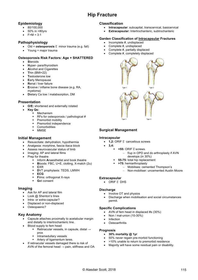
115
Hip Fracture Epidemiology
• 80/100,000 • 50% in >80yrs • F>M = 3:1
Pathophysiology
• Old = osteoporosis c̄ minor trauma (e.g. fall) • Young = major trauma
Osteoporosis Risk Factors: Age + SHATTERED
• Steroids • Hyper- para/thyroidism • Alcohol and Cigarettes • Thin (BMI<22) • Testosterone low • Early Menopause • Renal / liver failure • Erosive / inflame bone disease (e.g. RA,
myeloma) • Dietary Ca low / malabsorption, DM
Presentation
• O/E: shortened and externally rotated • Key Qs:
§ Mechanism § RFs for osteoporosis / pathological # § Premorbid mobility § Premorbid independence § Comorbidities § MMSE
Initial Management
• Resuscitate: dehydration, hypothermia • Analgesia: morphine, fascia iliaca block • Assess neurovascular status of limb • Imaging: AP and lateral films • Prep for theatre
§ Inform Anaesthetist and book theatre § Bloods: FBC, U+E, clotting, X-match (2u) § CXR § DVT prophylaxis: TEDS, LMWH § ECG § Films: orthogonal X-rays § Get consent
Imaging
• Ask for AP and lateral film • Look @ Shenton’s lines • Intra- or extra-capsular? • Displaced or non-displaced • Osteopaenic?
Key Anatomy
• Capsule attaches proximally to acetabular margin and distally to intertrochanteric line.
• Blood supply to fem head: § Retinacular vessels, in capsule, distal →
prox § Intramedullary vessels § Artery of ligamentum teres.
• If retinacular vessels damaged there is risk of AVN of the femoral head → pain, stiffness and OA
Classification
• Intracapsular: subcapital, transcervical, basicervical • Extracapsular: Intertrochanteric, subtrochanteric
Garden Classification of Intracapsular Fractures
• Incomplete #, undisplaced • Complete #, undisplaced • Complete #, partially displaced • Complete #, completely displaced
Surgical Management Intracapsular
• 1,2: ORIF c̄ cancellous screws • 3,4:
§ <55: ORIF c̄ screws. - f/up in OPD and do arthroplasty if AVN
develops (in 30%) § 55-75: total hip replacement § >75: hemiarthroplasty
- Mobilises: cemented Thompson’s - Non-mobiliser: uncemented Austin Moore
Extracapsular
• ORIF c̄ DHS Discharge
• Involve OT and physios • Discharge when mobilisation and social circumstances
permit. Specific Complications
• AVN of fem head in displaced #s (30%) • Non / mal-union (10-30%) • Infection • Osteoarthritis
Prognosis
• 30% mortality @ 1yr • 50% never regain pre-morbid functioning • >10% unable to return to premorbid residence • Majority will have some residual pain or disability.
© Alasdair Scott, 2018

116
Distal Forearm Fractures Colles’ Fracture Clinical Features
• Fall onto an outstretched hand • Most common in elderly females c̄ osteoporosis • Dinner fork deformity
Radiographic Features
• Extra-articular # of dist. radius (w/i 1.5” of joint) • Dorsal displacement of distal fragment • Dorsal angulation of distal fragment
§ Normally 11 degrees volar tilt • ↓ radial height (norm =11mm) • ↓ radial inclination (norm =22O) • ± avulsion of ulna styloid • ± impaction
Specific Management
• Examine for neurovascular injuries as median nerve and radial artery lie close.
• If much displacement → reduction § Under haematoma block, IV regional
anaesthesia (Bier’s block) or GA. § Disimpact and correct angulation. § Position: ulnar deviation + some wrist flexion § Apply dorsal backslab: provide 3-point
pressure • Re X-Ray – satisfactory position?
§ No: ortho review and consider MUA ± K wires
§ Yes: home c̄ # clinic f/up w/i 48hrs for completion of POP
• 6 wks in POP + physio • If comminuted, intra-articular or re-displaces:
§ Surgical fixation c̄ ex-fix, Kirschner-wires or ORIF and plates.
Specific Complications
• Median N. injury • Frozen shoulder / adhesive capsulitis • Tendon rupture: esp. EPL • Carpal tunnel syn. • Mal- /non-union • Sudek’s atrophy / CRPS
Other Distal Forearm Fractures Smith’s / Reverse Colles’
• Fall onto back of flexed wrist • Fracture of distal radius c̄ volar displacement and
angulation of distal fragment. • Reduce to restore anatomy and POP for 6wks
Barton’s Fracture
• Oblique intra-articular # involving the dorsal aspect of distal radius and dislocation of radio-carpal joint
• Reverse Barton’s involves the volar aspect of the radius
Scaphoid Fractures Clinical Features
• FOOSH • Pain in anatomical snuffbox • Pain on telescoping the thumb
Specific Management
• Request scaphoid x-ray view • If clinical hx and exam suggest a scaphoid #, it should
initially be treated even if the x-ray is normal. § # may become apparent after 10 days due to
localised decalcification. • Place wrist in scaphoid plaster (beer glass position) • If initial x-ray is negative, pt. returns to # clinic after 10
days for re-xray. § # visible → plaster for 6 wks § No visible # but clinically tender → plaster for 2
wks § # not visible and not clinically tender → no plaster
Specific Complications
• Main risk is AVN of the scaphoid as blood supply runs distal to proximal.
§ → stiffness and pain at the wrist Radial and Ulna Shaft Fractures Classification
• Monteggia § # of proximal 3rd of ulna shaft § Anterior dislocation of radial head at capitellum § May → palsy of deep branch of radial nerve →
weak finger extension but no sensory loss
• Galleazzi § # of radial shaft between mid and distal 3rds
§ Dislocation of distal radio-ulna joint
Specific Management
• Unstable fractures § Adults: ORIF § Children: MUA + above elbow plaster
• Fractures of forearm should be plastered in most stable position:
§ Proximal #: supination § Distal #: pronation § Mid-shaft #: neutral
© Alasdair Scott, 2018

117
The Shoulder Shoulder Dislocation Classification
• Anterior § 95% of shoulder dislocations. § Direct trauma or falling on hand § Humeral head dislocates antero-inferiorly
• Posterior § Caused by direct trauma or muscle
contraction (seen in epileptics). Associated Lesions Bankart Lesion
• Damage to anteroinferior glenoid labrum. Hill-Sachs Lesion
• Cortical depression in the posterolateral part of the humeral head following impaction against the glenoid rim during anterior dislocation.
• Occurs in 35-40% of anterior dislocations. Presentation
• Shoulder contour lost: appears square • Bulge in infraclavicular fossa: humeral head • Arm supported in opposite hand • Severe pain
Specific Management
• Assess for neurovascular deficit: esp. axillary N. § Sensation over “chevron” area before and
after reduction. § Occurs in 5%
• X-ray: AP and transcapular view • Reduction under sedation (e.g. propafol)
§ Hippocratic: Longitudinal traction c̄ arm in 30O abduction and counter traction @ the axilla
§ Kocher’s: external rotation of adducted arm, anterior movement, internal rotation
• Rest arm in a sling for 3-4wks • Physio
Complications • Recurrent dislocation
§ 90% of pts. <20yrs with traumatic dislocation • Axillary N. injury
Recurrent Shoulder Instability TUBS: Traumatic Unilateral dislocations with a Bankart lesion often require Surgery
• Mostly young patients: 15-30yrs • Surgery involves a Bankart repair
AMBRI: Atraumatic Multidirectional Bilateral shoulder dislocation is treated with Rehabilitation, but may require Inferior capsular shift
Impingement Syndrome / Painful Arc Pathology
• Entrapment of supraspinatus tendon and subacromial bursa between acromion and grater tuberosity of humerus.
• → subacromial bursitis and/or supraspinatous tendonitis Presentation
• Painful arc: 60-120O • Weakness and ↓ ROM • +ve Hawkin’s test
Ix
• Plain radiographs: may see bony spurs • US • MRI arthrogram
Rx
• Conservative § Rest § Physiotherapy
• Medical § NSAIDs § Subacromial bursa steroid ± LA injection
• Surgical § Arthroscopic acromioplasty
Differential of Painful Arc
• Impingement • Supraspinatous tear or partial tear • AC joint OA
Frozen Shoulder: Adhesive Capsulitis Presentation
• Progressive ↓ active and passive ROM § ↓ ext. rotation <30O § ↓ abduction <90O
• Shoulder pain, esp. @ night (can’t lie on affected side) Cause
• Unknown, may follow trauma in elderly • Commonly assoc. c̄ DM
Rx
• Conservative: rest, physio • Medical
§ NSAIDs § Subacromial bursa steroid ± LA injection
Rotator Cuff Tear
• 2O to degeneration or a sudden jolt or fall • Partial tears → painful arc • Complete tear
§ Shoulder tip pain § Full range of passive movement § Inability to abduct the arm § Active abduction possible following passive
abduction to 90O § Lowering the arm beneath this → sudden drop
- “drop arm” sign Rx: open or arthroscopic repair
© Alasdair Scott, 2018

118
Supracondylar Fractures of the Humerus Presentation
• Common in children after FOOSH • Elbow very swollen and held semi-flexed. • Sharp edge of proximal humerus may injure brachial
artery which lies anterior to it. Classification Extension
• Commonest type • Distal fragment displaces posteriorly • Gartland further classified extension type:
§ Type 1: non-displaced § Type 2: angulated c̄ intact posterior cortex § Type 3: displaced c̄ no cortical contact
Flexion
• Less common • Distal fragment displaces anteriorly
Specific Management
• Ensure there is no neurovascular damage § If radial pulse absent or damage to brachial
artery suspected, take urgently to theatre for reduction ± on-table angiogram.
§ Median nerve is also vulnerable
• Restore the anatomy § No displacement → flex the arm as fully as
possible and apply a collar and cuff for 3wks – triceps acts as sling to stabilise fragments.
§ Displacement → MUA + fixation with K-wires + collar and cuff with arm flexed for 3wks.
Specific Complications Neurovascular Injury
• Brachial artery • Radial nerve • Median nerve: esp. anterior interosseous branch
§ Supplies deep forearm flexors (FPL, lateral half of FDP and pronator quadratus)
Compartment syndrome
• Monitor closely during the first 24h • Pain on passive extension of the fingers (stretches
flexor compartment) is early sign. • Mx: try extension of the elbow, surgical Rx may be
needed. • Volkmann’s ischaemic contracture can result →
fibrosis of flexors → claw hand. Gunstock Deformity
• Valgus, varus and rotational deformities in the coronal plane do not remodel and → cubitus varus.
• Cubitus varus deformity is referred to as a “gunstock” deformity.
© Alasdair Scott, 2018

119
Femoral and Tibial Fractures Specific Management
• Resus and Mx life-threatening injuries first. • X-Match
§ Tibial #: 2 units § Femoral #: 4 units
• Assess neurovascular status: esp. distal pulses • If open
§ Abx and ATT § Take to theatre urgently for debridement,
washout and stabilisation • Fixation methods
§ Intramedullary nail § Ex-fix § Plates and screws § MUA c̄ fixed traction for 3-4mo
Specific Complications
• Hypovolaemic shock • Neurovascular
§ SFA: swelling and check pulses § Sciatic nerve
• Compartment syndrome • Respiratory complications
§ Fat embolism § ARDS § Pneumonia
Ankle Injuries Ligament Strains
• Typically twisting inversion injury § Strains anterior talofibular part of lateral
collateral ligament • Medial deltoid ligament strains are rare. • May be assoc. c̄ malleolar avulsion #s
Ankle Fracture Ottowa Ankle Rules
• X-ray ankle if pain in malleolar zone + in any of: § Tenderness along distal 6cm of posterior tib /
fib including posterior tip of the malleoli. § Inability to bear weight both immediately and in
ED Weber Classification
• Relation of fibula # to joint line • A: below joint line • B: at joint line • C: above joint line • Weber’s B and C represent possible injury to the
syndesmotic ligaments between tib and fib → instability
Mx
• Weber A § Boot or below-knee POP
• Non-displaced Weber B/C § Below-knee POP
• Displaced Weber B/C § Closed reduction and POP if anatomical
reduction achieved § ORIF if closed reduction fails
© Alasdair Scott, 2018

120
Knee Injuries History
• Mechanism • Swelling
§ Immediate = haemarthrosis = # or torn cruciates
§ Overnight = effusion = meniscus or other lgt • Pain / tenderness
§ Joint line = meniscal § Med/lateral margins = collateral lgts.
• Locking: meniscal tear → mechanical obstruction • Giving way: instability following lgt. injury
Knee Haemarthrosis
• 1O: spontaneous bleeding § Coagulopathy: warfarin, haemophilia
• 2O: trauma § ACL injury: 80% § Patella dislocation: 10% § Meniscal injury: 10%
- Outer third where its vascularised § Osteophyte #
Unhappy Triad of O’Donoghue
• ACL • MCL • Medial Meniscus
Mx of acutely injured knee
• Full examination of acutely swollen knee after injury is difficult.
• Take x-ray to ensure no #s § Fluid level indicates a lipohaemarthrosis and
indicates either a # or torn cruciate. • If no # → RICE + later re-examination for pathology • If meniscal or cruciate injury suspected → MRI
Arthroscopy
• Direct vision of inside of knee joint by arthroscope • Can examine knee under anaesthesia (↓ muscle tone) • Meniscal tears can be trimmed or repaired.
Mx of Ruptured ACL Conservative
• Rest • Physio to strengthen quads and hamstrings • Not enough stability for many sports
Surgical
• Gold-standard is autograft repair • Usually semitendinosus ± gracilis (can use patella
tendon) • Tendon threaded through heads of tibia and femur
and held using screws.
© Alasdair Scott, 2018

121
Osteoarthritis Definition
• Degenerative joint disorder in which there is progressive loss of hyaline cartilage and new bone formation at the joint surface and its margin.
Aetiology / Risk Factors • Age (80% > 75yrs) • Obesity • Joint abnormality
Classification • Primary: no underlying cause • Secondary: obesity, joint abnormality
Symptoms • Affects: knees, hips, DIPs, PIPs, thumb CMC • Pain: worse c̄ movement, background rest/night pain,
worse @ end of day. • Stiffness: especially after rest, lasts ~30min (e.g. AM) • Deformity • ↓ ROM
Signs • Bouchards and Heberdens nodes
§ ‘Pouchards’ (prox), ‘Heberdips’ (dist.) • Thumb CMC squaring • Fixed flexion deformity
History • Pain severity, night pain • Walking distance • Analgesic requirements • ADLs and social circumstances • Co-morbidities • Underlying causes: trauma, infection, congenital
Pathophysiology • Softening of articular cartilage → fraying and fissuring
of smooth surface → underlying bone exposure. • Subchondral bone becomes sclerotic c̄ cysts. • Proliferation and ossification of cartilage in unstressed
areas → osteophytes. • Capsular fibrosis → stiff joints.
Differential • Septic • Crystal • Trauma
X-ray Changes • Loss of joint space • Osteophytes • Subchondral cysts • Subchondral sclerosis • Deformity
Bloods • CRP may be mildly elevated • Ca, PO4 and ALP all normal
Rx MDT: GP, physio, OT, dietician, orthopod Conservative
• Lifestyle: ↓ wt., ↑ exercise • Physio: muscle strengthening • OT: walking aids, supportive footwear, home mods
Medical
• Analgesia § Paracetamol § NSAIDs: e.g. arthrotec (diclofenac +
misoprostol) § Tramadol
• Joint injection: local anaesthetic and steroids Surgical
• Arthroscopic Washout § Mainly knees § Trim cartilage § Remove loose bodies.
• Realignment Osteotomy § Small area of bone cut out § Useful in younger (<50yrs) pts. c̄ medial
knee OA § High tibial valgus osteotomy redistributes
wt. to lateral part of joint. • Arthroplasty: replacement (or excision) • Arthrodesis: last resort for pain management • Novel Techniques
§ Microfracture: stem cell release → fibro-cartilage formation
§ Autologous chondrocyte implantation
© Alasdair Scott, 2018
122
Back Pain Mechanical Pain
• Soft tissue injury → dysfunction of whole spine → muscle spasm → pain.
• May have inciting event: e.g. lifting • Younger pts. c̄ no sinister features
Mx
• Conservative § Max 2d bed rest § Education: keep active, how to lift / stoop § Physiotherapy § Psychosocial issues re. chronic pain and disability § Warmth: e.g. swimming in a warm pool
• Medical § Analgesia: paracetamol ± NSAIDs ± codeine § Muscle relaxant: low-dose diazepam (short-term)
Disc Prolapse
• Herniation of nucleus pulposus through annulus fibrosus Presentation
• L5 and S1 roots most commonly compressed by prolapse of L4/5 and L5/S1 discs.
• May present as severe pain on sneezing, coughing or twisting a few days after low back strain
• Lumbago: low back pain • Sciatica: shooting radicular pain down buttock and thigh
Signs
• Limited spinal flexion and extension • Free lateral flexion • Pain on straight-leg raise: Lesague’s Sign • Lateral herniation → radiculopathy • Central herniation → corda equina syndrome
L4/5 → L5 Root Compression
• Weak hallux extension ± foot drop § In foot drop due to L5 radiculopathy, weak
inversion (tib. post.) helps distinguish from peroneal N. palsy.
• ↓ sensation on inner dorsum of foot L5/S1 → S1 Root Compression
• Weak foot plantarflexion and eversion • Loss of ankle-jerk • Calf pain • ↓ sensation over sole of foot and back of calf
Ix: MRI (emergency if cauda equina) Rx
• Brief rest, analgesia and mobilisation effective in ≥90% • Conservative: brief rest, mobilisation/physio • Medical: analgesia, transforaminal steroid injection • Surgical: discectomy or laminectomy may be needed in
cauda-equina syndrome, continuing pain or muscle weakness.
Lumbar Microdiscectomy
• Commonest procedure for disc prolapse • Microscopic resection of the protruding nucleus pulposus • Posterior approach c̄ pt. in prone position. • May be performed endoscopically
Spondylolisthesis • Displacement of one lumbar vertebra on another
§ Usually forward § Usually L5 on S1
• May be palpable Causes
• Congenital malformation • Spondylosis • Osteoarthritis
Presentation
• Onset of pain usually in adolescence or early adulthood
§ Worse on standing • ± sciatica, hamstring tightness, abnormal gait
Dx: Plain radiography Rx
• Corset • Nerve release • Spinal fusion
Spinal Stenosis
• Developmental predisposition ± facet joint osteoarthritis → generalized narrowing of lumbar spinal canal.
Presentation
• Spinal claudication § Aching or heavy buttock and lower limb pain
on walking § Rapid onset § May c/o paraesthesiae/numbness § Pain eased by leaning forward (e.g. on bike)
• Pain on spine extension Ix: MRI Rx
• Corsets • NSAIDs • Epidural steroid injection • Canal decompression surgery
Neurosurgical Emergencies Acute Cord Compression
• Bilateral pain: back and radicular • LMN signs at compression level • UMN signs and sensory level below compression • Sphincter disturbance
Acute Cauda Equina Compression
• Alternating or bilateral radicular pain in the legs • Saddle anaesthesia • Loss of anal tone • Bladder ± bowel incontinence
Rx
• Large prolapse: laminectomy / discectomy • Tumours: radiotherapy and steroids • Abscesses: decompression
© Alasdair Scott, 2018

123
Osteochondritis • Idiopathic condition in which bony centres of
children/adolescents become temporarily softened due to osteonecrosis.
• Pressure → deformation • Bone hardens in new, deformed position
Radiography
• Initially: ↑ density / sclerosis • Then: patchy appearance
Scheuermann’s Disease
• Vertebral ring epiphyses • Auto dom • Vertebral tenderness and kyphosis • X-ray: wedge-shaped thoracic vertebra
Kohler’s Disease
• Navicular bone • Children 3-5yrs • Pain in mid-tarsal region → limp
Kienbochs Disease
• Lunate bone • Adults • Pain over lunate, esp. on active movement • Impaired grip
Friedberg’s Disease
• 2nd/3rd metatarsal heads • Around puberty • Forefoot pain worse c̄ pressure
Panner’s Disease
• Capitulum of humerus Perthes’ Disease
• Hip
Traction Apophysitis Osgood-Shlatter’s
• Tibial tuberosity apophysitis + patellar tendonitis • Children 10-14yrs, M>F=3:1 • Assoc. c̄ physical activity • Symptoms: pain below knee, esp c̄ quads contraction • X-ray: tuberosity enlargement ± fragmentation • Rx: rest, consider POP
Sinding-Larsen’s Disease
• Traction tendinopathy with calcification of proximal attachment of patellar tendon
• Children 8-10yrs Sever’s Disease
• Calcaneal apophysitis • 8-13yrs • Symptoms: pain behind heal + limping • Rx: physio
Osteochondritis Dissecans
• Piece of bone and overlying cartilage dissects off into joint space.
• Commonly knee (med. fem. condyle), also elbow, hip and ankle.
• Young adult / adolescent • Symptoms: pain, swelling, locking, ↓ ROM • X-ray: loose bodies, lucent crater • Mx: arthroscopic removal
Avascular Necrosis Causes
• # or dislocation • SCD, thalassaemia • SLE • Gaucher’s • Drugs: steroids, NSAIDs
© Alasdair Scott, 2018

124
The Limping Child Aetiology
• DDH • Transient synovitis • Septic arthritis • Perthes’ • Slipped Capital Femoral Epiphyses • JIA / Still’s Disease
DDH
• Congenital hip joint deformity in which the femoral head is or can be completely / partially displaced.
Epidemiology
• Incidence: 1/1000 • Sex: F>M
Predisposing Factors
• FH • Breach presentation • Oligohydramnios
Presentation
• Screening • Asymmetric skin folds • Limp / abnormal gait
Ix
• US is v. specific Mx: maintain abduction
• Double nappies • Pavlik harness • Plaster hip spica • Open reduction: derotation varus osteotomy
Transient Synovitis: Irritable Hip
• Commonest cause of acute hip pain in children Presentation
• 2-12yrs • Sudden onset hip pain / limp • Often following or with viral infection • Not systemically unwell
Ix
• PMN and ESR/CRP are normal • -ve blood cultures • May need joint aspiration and culture
Mx
• Rest and analgesia • Settles over 2-3d
Perthes’ Disease
• Osteochondritis of the femoral head 2O to AVN. Epidemiology
• 4-10yrs • M>F=5:1
Presentation
• Insidious onset • Hip pain initially, then painless • 10-20% bilateral
Ix
• X-rays normal initially • ↑ density of femoral head
§ Becomes fragmented and irregular § Flattening and sclerosis
• Bone scan is useful Mx
• Detected early and < half femoral head affected § Bed rest and traction
• More severe § Maintain hip in abduction c̄ plaster § Femoral or pelvic osteotomy
Slipped Capital Femoral Epiphysis
• Postero-inferior displacement of femoral head epiphysis
• 10-15yrs • Two main groups
§ Fat and sexually underdeveloped § Tall and thin
Presentation
• Slip may be acute, chronic or acute-on-chronic • Acute
§ Groin pain § Shortened, externally rotated leg § All movements painful
• 20% bilateral Ix
• Confirm Dx by x-ray Mx
• Acute: reduce and pin epiphysis • Chronic: in situ pinning
§ Epiphyseal reduction risks AVN Complications
• Chondrolysis: breakdown of articular cartilage § ↑ risk c̄ surgery
© Alasdair Scott, 2018

125
Acute Osteomyelitis Pathophysiology
• Source: local or haematogenous. • Organisms
§ Staph § Strep § E. coli § Pseudomonas § Salmonella (in SCD)
• RFs § Vascular disease § Trauma § SCD § Immunosuppression (e.g. DM) § Children
- Rich blood supply to growth plate - \ usually affects metaphysis
Symptoms and Signs
• Pain, tenderness, erythema, warmth, ↓ROM • Effusion in neighbouring joints • Signs of systemic infection
Investigations
• ↑ESR/CRP, ↑WCC • +ve blood cultures in 60% • X-ray:
§ Changes take 10-14d § Haziness + ↓ bone density § Sub-periosteal reaction § Sequestrum and involucrum
• MRI is sensitive and specific Management
• IV Abx: Vanc + cefotaxime until MCS known • Drain abscess and remove sequestra • Analgesia
Septic Arthritis Pathophysiology
• Source: local or haematogenous. • Organisms
§ Staph: 60% § Streps § Gonococcus § Gm-ve bacilli
• RFs § Joint disease (e.g. RA) § CRF § Immunosuppression (e.g. DM) § Prosthetic joints
Symptoms
• Acutely inflamed tender, swollen joint. • ↓ROM • Systemically unwell
Investigations
• Joint aspiration for MCS § ↑↑ WCC (e.g. >50,000/mm3) : mostly PMN
• ↑ESR/CRP, ↑WCC, Blood cultures • X-ray
Management
• IV Abx: vanc + cefotaxime • Consider joint washout under GA • Splint joint • Physiotherapy after infection resolved
Complications
• Osteomyelitis • Arthritis • Ankylosis: fusion
Differential
• Crystal arthropathy • Reactive arthritis
© Alasdair Scott, 2018

126
Bone Tumours
Bony Mets • Commonest bone tumours • Bronchus, thyroid, breast, kidney and prostate • Usually radiolucent (except prostate which is sclerotic) • Usually axial skeleton (contains red marrow) • Present with pain or pathological #
§ Path # → internal fixation § Pain → radiotherapy
Tumour-like Non-neoplastic Conditions
Lesion Clinical Features Location X-ray Fibrous Dysplasia 0-30
F>M ↑ # risk McCune Albright’s - Polyostotic fibrous dysplasia - Precocious puberty (females) - Café-au-lait spots
Long bones Ribs Skull Usually mono-ostotic
“Ground Glass” lytic lesion Shepherds crook deformity of prox. femur
Simple bone cyst <20 Lump migrates down shaft from original growth plate site.
Prox metaphysis of humerus or femur
Well-defined lytic lesion Cortex can fall into cyst → “fallen fragment”
Aneurysmal bone cyst <30yrs Pain
MRI shows multiple fluid levels
Fibrous cortical defect and non-ossifying fibroma
Children Non-painful, benign Spontaneously regress
Dist femur Prox tibia Often multiple
Benign Cartilaginous Neoplasms
Lesion Clinical Features Location X-ray Osteochondroma (exostosis)
10-20 M>F = 3:1 Commonest benign bone tumour May be related to previous trauma
Knee Cartilage-capped bony outgrowth
Enchondroma / Chondromas
10-40 M=F Ollier Disease = multiple enchondromas Maffucci Syn = Enchondromatosis + multiple soft-tissue haemangiomas Multiple enchondromas have risk of malignant transformation
Hands Feet
O sign - Oval lucencies c̄ radiodense rim Endosteal scalloping
Chondroblastoma 10-20 M>F = 2:1
Epiphysis Knee
Radiolucent with sclerotic border
Malignant Cartilaginous Neoplasms
Lesion Clinical Features Location X-ray Chondrosarcoma >40
Pain + lump Arise de novo or from chondromas 70% 5ys
Pelvis Axial skeleton
Lytic lesion Fluffy “popcorn calcification”
© Alasdair Scott, 2018

127
Benign Bone-forming Neoplasms Lesion Clinical Features Location X-ray Osteoma Lump
Usually solitary Multiple in Gardner syn
Skull Facial bones
Osteoid osteoma M>F = 2:1 Teens and 20s Severe nocturnal pain relieved by aspirin Hot on bone scan
Lower limb Diaphyseal cortex
Lytic lesion c̄ central nidus and sclerotic rim
Osteoblastoma Pain unresponsive to aspirin Spine Giant Cell Tumour / Osteoclastoma
20-40 (After fusion of growth plate) F>M
Knee Abut joint surface
Soap bubble appearance Solitary, expansile, lytic lesion
Malignant Bone-forming Neoplasms
Lesion Clinical Features Location X-ray Osteosarcoma Adolescents
M>F = 2:1 Commonest 1O bone tumour Pain, warm, bruit May arise 2o to Paget’s or irradiation
Knee Metaphysis
Periosteal Elevation: - Sunburst appearance - Codman’s triangle
Ewing’s Sarcoma <20 Painful, warm, enlarging mass Systemic: fever, ↑ESR, anaemia, ↑WCC
Long bone diaphysis
Lytic tumour Onion-skin periosteal reaction
© Alasdair Scott, 2018

128
Brachial Plexus Injuries Anatomy
• C5-T1 • Roots leave vertebral column between scalenus
anterior and medius. • Divisions occur under the clavicle, medial to coracoid
process. • Plexus has intimate relationship c̄ subclavian and
brachial arteries. Median N. is formed anterior to brachial artery.
Causes • Direct: e.g. shoulder girdle #, penetrating or iatrogenic
injury • Indirect: e.g. avulsion or traction injuries
Leffert Classification
• Open • Closed
§ Supraclavicular § Infraclavicular
• Radiation-induced • Obstetric
§ Upper § Lower § Mixed
High (C5/6): Erb’s Palsy
• Abductors and external rotators paralysed • Waiter’s tip position • Loss of sensation in C5/6 dermatomes
Low (C8/T1): Klumpke’s Paralysis
• Paralysis of small hand muscles • Claw hand • Loss of sensation in C8/T1 dermatomes
Other Nerve Injuries Radial Nerve (C5-T1) Low Lesions: posterior interosseous nerve
• Site: # around elbow or forearm § E.g. # head of radius
• Loss of extension of CMC joints (finger drop) • No sensory loss
High Lesions
• Site: # shaft of humerus where N. is in radial groove. • Wrist drop • Loss of sensation to dorsum of thumb root (snuff box) • Triceps functions normally
Very High Lesions
• Site: axilla – e.g. crutches or Sat night palsy • Paralysis of triceps and wrist drop
Ulnar Nerve (C8-T1) Site
• Elbow: cubital tunnel • Wrist: in Guyon’s Canal
Effects
• Intrinsic hand muscle paralysis → claw hand • Ulnar paradox: lesion at elbow has less clawing as
FDP is paralysed, decreasing flexion of 4th/5th digits. • Weakness of finger ad/abduction (interossei) • Sensory loss over little finger
Tests
• Can’t cross fingers for luck • Froment’s Sign: flexion of thumb IPJ when trying to
hold onto paper held between thumb and finger. § Indicates weak adductor policis.
Median Nerve (C5-T1) Injury Above the Antecubital Fossa
• Can’t flex index finger IPJs (e.g. on clasping hands) • Can’t flex terminal thumb phalanx (FPL) • Loss of sensation in median distribution
Injury at the Wrist
• Typically affects abductor pollicis brevis Carpal Tunnel Syndrome
C5
C6 C7 C8
T1 Roots (5)
Trunks (3) Divisions (6)
Cords (3)
Lat
Post
Med
Axillary Radial
MC
Median
Ulnar
© Alasdair Scott, 2018
129
Carpal Tunnel Syndrome Anatomy
• Carpal tunnel formed by flexor retinaculum and carpal bones.
• Contains § 4 tendons of FDS § 4 tendons of FDP § 1 tendon of FPL § Median N.
• Median N. supplies LLOAF (aBductor pollicis brevis) • Palmer cutaneous branch travels superficial to flexor
retinaculum → spares sensation over thenar area. Causes
• F>M • Primary / idiopathic • Secondary • Water: pregnancy, hypothyroidism • Radial # • Inflammation: RA, gout • Soft tissue swelling: lipomas, acromegaly, amyloidosis • Toxic: DM, EtOH
Symptoms
• Tingling / pain in thumb, index and middle fingers • Pain worse @ night or after repetitive actions • Relieved by shaking / flicking • Clumsiness
Signs
• ↓ sensation over lateral 3½ fingers • ↓ 2-point touch discrimination
§ Early sign of irreversible damage • Wasting of thenar eminence
§ Late sign of irreversible damage • Phalen’s flexing and Tinel’s tapping
Ix
• Not usually performed • Nerve conduction studies • US
Non-surgical Mx
• Mx of underlying cause • Wrist splints
§ Neutral position § Esp. @ night
• Local steroid injections Surgical Mx
• Carpal tunnel decompression by division of the flexor retinaculum
Complications
• Scar formation: high risk for hypertrophic or keloid • Scar tenderness: up to 40% • Nerve injury
§ Palmar cutaneous branch of the median nerve § Motor branch to the thenar muscles
• Failure to relieve symptoms Other Locations of Median Nerve Entrapment
• Pronator syndrome Entrapment between two heads of
pronator teres • Anterior interroseous syndrome
§ Compression of the anterior interosseous branch by the deep head of pronator teres
§ Muscle weakness only § Pronator quadratus § FPL § Radial half of FDP
© Alasdair Scott, 2018

130
Minor Hand Conditions Dupuytren’s Contracture
• Progressive, painless fibrotic thickening of palmar fascia.
The Patient
• M>F • Middle age / elderly • Skin puckering and tethering • Fixed flexion contracture of ring and little fingers • Often bilateral and symmetrical • MCP and IP joint flexion
Associations: BAD FIBERS
• Bent penis: Peyronies (3%) • AIDS • DM • FH: AD • Idiopathic: commonest • Booze: ALD • Epilepsy and epilepsy meds (phenytoin) • Reidel’s thyroiditis and other fibromatoses
§ Ledderhose disease - Fibrosis of plantar aponeurosis - 5% c̄ dupuytren’s
§ Retroperitoneal fibrosis • Smoking
Management
• Conservative: e.g. physio / exercises • Fasciectomy
§ e.g. when hand can’t be placed flat on the table.
§ Z-shaped scars: prevent contracture § Can damage ulnar nerve § Usually recurs
Differential
• Skin contracture: old laceration or burn • Tendon fibrosis, trigger finger • Ulnar N. palsy
Trigger Finger
• Tendon nodule which catches on proximal side of tendon sheath → triggering on forced extension.
• → Fixed flexion deformity • Usually ring and middle fingers • Assoc. c̄ RA • Rx: steroid injection (high recurrence) or surgery
Ganglion
• Smooth, multilocular cystic swellings • Mucoid degeneration of joint capsule or tendon sheath • May be in communication c̄ joint capsules / tendons
Presentation
• 90% located on dorsum of wrist. • Subdermal, fixed to deeper structures.
§ Limits planes of movement • May cause pain or nerve pressure symptoms
Management
• 50% disappear spontaneously • Aspiration ± steroid and hyaluronidase injection • Surgical excision
Differential
• Lipoma • Fibroma • Sebaceous cyst
© Alasdair Scott, 2018

131
Minor Leg and Foot Conditions Meralgia Paraesthetica
• Entrapment of lat. cutaneous nerve of thigh § Between ASIS and inguinal ligament
• Pain ± paraesthesia on the lateral thigh • No motor deficit • ↑ risk c̄ obesity: compression by belts, underwear
§ Relieved by sitting down • Can occasionally be damaged in lap hernia repair
Chondromalacia Patellae
• Predominantly young women • Patellar aching after prolonged sitting or climbing
stairs • Pain on patellofemoral compression: Clarke’s test • Ix: no abnormality on X-ray • Rx: vastus medialis strengthening
Baker’s Cyst
• Popliteal swelling arising between the medial head of gastrocnemius and semimembranosus muscle
• Herniation from joint synovium • Usually 2O to OA • Rupture: acute calf pain and swelling
§ DVT differential
Hallux Valgus
• Great toe deviates laterally @ MTP joint • Pressure of MTP against shoe → bunion • ↑ wt. bearing @ 2nd metatarsal head
§ → pain: “Transfer metatarsalgia” § → hammer toe
Aetiology
• Pointed shoes • Wearing high heals
Mx
• Conservative: bunion pads, plastic wedge between great and second toes.
• Surgical: metatarsal osteotomy Lesser Toe Deformities
Morton’s Metatarsalgia / Neuroma
• Pain from pressure on an interdigital neuroma between the metatarsals.
• Pain radiates to medial side of one toe and lateral side of another.
• Rx: neuroma excision
© Alasdair Scott, 2018

132
Ear, Nose and Throat Contents
Audiometry ......................................................................................................................................................... 133Otalgia ................................................................................................................................................................ 133Otitis Media ........................................................................................................................................................ 134Cholesteatoma ................................................................................................................................................... 135Tinnitus .............................................................................................................................................................. 135Vertigo ................................................................................................................................................................ 136Adult Hearing Loss ............................................................................................................................................. 137Hearing Loss in Children .................................................................................................................................... 138Miscellaneous Ear Conditions ............................................................................................................................ 138Allergic Rhinosinusitis ........................................................................................................................................ 139Sinusitis .............................................................................................................................................................. 139Nasal Polyps ...................................................................................................................................................... 140Fractured Nose .................................................................................................................................................. 140Epistaxis ............................................................................................................................................................. 141Tonsillitis ............................................................................................................................................................ 142The Larynx ......................................................................................................................................................... 143Paediatric Airway Issues .................................................................................................................................... 143Facial Nerve Palsy ............................................................................................................................................. 144
© Alasdair Scott, 2018

133
Audiometry • Quantify loss and determine its nature.
Pure tone audiometry (PTA)
• Headphones deliver tones at different frequencies and strengths in a sound-proofed room.
• Pt. indicates when sound appears and disappears.
• Mastoid vibrator → bone conduction threshold. • Threshold at different frequencies are plotted to
give an audiogram. Tympanometry
• Measures stiffness of ear drum - Evaluates middle ear function
• Flat tympanogram: mid ear fluid or perforation • Shifted tympanogram: +/- mid ear pressure
Evoked response audiometry
• Auditory stimulus c̄ measurement of elicited brain response by surface electrode.
• Used for neonatal screening (if otoacoustic emission testing negative)
Otalgia Otitis Externa Presentation
• Watery discharge • Itch • Pain and tragal tenderness
Causes
• Moisture: e.g. swimming • Trauma: e.g. fingernails • Absence of wax • Hearing aid
Organisms
• Mainly pseudomonas • Staph aureus
Management
• Aural toilet c̄ drops § Betamethasone for non-infected eczematous OE § Betamethasone c̄ neomycin § Hydrocortisone c̄ gentamicin § Acidifying drops
Malignant Otitis Externa
• Life-threatening infection which can → skull osteomyelitis • 90% of pts. are diabetic (or other immune compromise) • Presentation
§ Severe otalgia which is worse @ night § Copious otorrhoea § Granulation tissue in the canal
• Rx § Surgical debridement § Systemic Abx
Bullous Myringitis
• Painful haemorrhagic blisters on deep meatal skin and TM. • Assoc. c̄ influenza infection
TMJ Dysfunction Symptoms
• Earache (referred pain from auriculotemporal N.) • Facial pain • Joint clicking/popping • Teeth-grinding (bruxism) • Stress (assoc. c̄ depression)
Signs
• Joint tenderness exacerbated by lateral movements of an open jaw.
Investigation
• MRI Management
• NSAIDs • Stabilising orthodontic occlusal prostheses
© Alasdair Scott, 2018

134
Otitis Media Classification
• Acute: acute phase • Glue ear / OME: effusion after symptom regression • Chronic: effusion > 3mo if bilat or > 6mo if unilat • Chronic suppurative OM: Ear discharge c̄ hearing
loss and evidence of central drum perforation. Organisms
• Viral • Pneumococcus • Haemophilus • Moraxella
Acute OM Presentation
• Usually children post viral URTI • Rapid onset ear pain, tugging @ ear. • Irritability, anorexia, vomiting • Purulent discharge if drum perforates
o/e
• Bulging, red TM • Fever
Rx
• Paracetamol: 15mg/kg • Amoxicillin: may use delayed prescription
Complications
• Intratemporal § OME § Perforation of TM § Mastoiditis § Facial N. palsy
• Intracranial § Meningitis / encephalitis § Brain abscess § Sub- / epi-dural abscess
• Systemic § Bacteraemia § Septic arthritis § IE
OME Presentation
• Inattention at school • Poor speech development • Hearing impairment
o/e
• Retracted dull TM • Fluid level
Ix
• Audiometry: flat tympanogram Rx
• Usually resolves spontaneously, • Consider grommets if persistent hearing loss
§ SE: infections and tympanosclerosis
Chronic Suppurative OM Presentation
• Painless discharge and hearing loss o/e
• TM perforation Rx
• Aural toilet • Abx / Steroid ear drops
Complications
• Cholesteatoma Mastoiditis
• Middle-ear inflam → destruction of mastoid air cells and abscess formation.
Presentation
• Fever • Mastoid tenderness • Protruding auricle
Imaging: CT Rx
• IV Abx • Myringotomy ± mastoidectomy
© Alasdair Scott, 2018
135
Cholesteatoma Definition
• Locally destructive expansion of stratified squamous epithelium within the middle ear.
Classification
• Congenital • Acquired: 2O to attic perforation in chronic
suppurative OM Presentation
• Foul smelling white discharge • Headache, pain • CN Involvement
§ Vertigo § Deafness § Facial paralysis
o/e
• Appears pearly white c̄ surrounding inflammation Complications
• Deafness (ossicle destruction) • Meningitis • Cerebral abscess
Mx
• Surgery
Tinnitus • Sensation of sound w/o external sound stimulation
Causes
• Specific § Meniere’s § Acoustic neuroma § Otosclerosis § Noise-induced § Head injury § Hearing loss: e.g. presbyacusis
• General § ↑BP § ↓Hb
• Drugs § Aspirin § Aminoglycosides § Loop diuretics § EtOH
Hx
• Character: constant, pulsatile • Unilateral: acoustic neuroma • FH: otosclerosis • Alleviating/exacerbating factors: worse @ night? • Associations
§ Vertigo: Meniere’s, acoustic neuroma § Deafness: Meniere’s, acoustic neuroma
• Cause: head injury, noise, drugs, FH Examination
• Otoscopy • Tuning fork tests • Pulse and BP
Ix
• Audiometry and tympanogram • MRI if unilateral to exclude acoustic neuroma
Mx
• Treat any underlying causes • Psych support: tinnitus retraining therapy • Hypnotics @ night may help
© Alasdair Scott, 2018
136
Vertigo Definition
• The illusion of movement Causes
Peripheral / Vestibular Central • Meniere’s • Acoustic neuroma • BPV • MS • Labyrinthitis • Vertebrobasilar
insufficiency / stroke Head injury
• Inner ear syphilis
Drugs (central/ototoxic) • Gentamicin • Loop diuretics • Metronidazole • Co-trimoxazole
Hx
• Is it true vertigo or just light-headedness? § Which way are things moving?
• Timespan • Assoc. symptoms: n/v, hearing loss, tinnitus,
nystagmus Examination and Tests
• Hearing • Cranial nerves • Cerebellum and gait • Romberg’s +ve = vestibular or proprioception • Hallpike manouvre • Audiometry, calorimetry, LP, MRI
Ménière’s Disease Pathology
• Dilatation of endolymph spaces of membranous labyrinth (endolymphatic oedema)
Presentation
• Attacks occur in clusters and last up to 12h. • Progressive SNHL • Vertigo and n/v • Tinnitus • Aural fullness
Ix • Audiometry shows low-freq SNHL which fluctuates
Rx
• Medical § Vertigo: cyclizine, betahistine
• Surgical § Gentamicin instillation via grommets § Saccus decompression
Vestibular Neuronitis / Viral Labyrinthitis Presentation
• Follows febrile illness (e.g. URTI) • Sudden vomiting • Severe vertigo exacerbated by head movement
Rx
• Cyclizine • Improvement in days
Benign Positional Vertigo: BPV Pathology
• Displacement of otoliths in semicircular canals • Common after head injury.
Presentation
• Sudden rotational vertigo for <30s - Provoked by head turning
• Nystagmus Causes
• Idiopathic • Head injury • Otosclerosis • Post-viral
Dx
• Hallpike manoeuvre → upbeat-torsional nystagmus Rx
• Self-limiting • Epley manoeuvre • Betahistine: histamine analogue
© Alasdair Scott, 2018
137
Adult Hearing Loss Conductive
• Impaired conduction anywhere between auricle and round window.
External canal obstruction
• Wax • Pus • Foreign body
TM perforation
• Trauma • Infection
Ossicle defects
• Otosclerosis • Infection • Trauma
Inadequate eustachian tube ventilation of middle ear Sensorineural
• Defects of cochlea, cohlear N. or brain. Drugs
• Aminoglycosides • Vancomycin
Post-infective
• Meningitis • Measles • Mumps • Herpes
Misc.
• Meniere’s • Trauma • MS • CPA lesion (e.g. acoustic neuroma) • ↓B12
Acoustic Neuroma / Vestibular Schwannoma Pathology
• Benign, slow-growing tumour of superior vestibular N. • Acts as SOL → Cerebellopontine angle syndrome
§ 80% of CPA tumours • Assoc. c̄ NF2
Presentation
• Slow onset, unilat SNHL, tinnitus ± vertigo • Headache (↑ICP) • CN palsies: 5,7 and 8 • Cerebellar signs
Ix
• MRI of cerebellopontine angle § MRI all pts. c̄ unilateral tinnitus / deafness
• PTA Differential
• Meningioma • Cerebellar astrocytoma • Mets
Rx
• Gamma knife • Surgery (risk of hearing loss)
Otosclerosis
• AD condition characterised by fixation of stapes at the oval window.
• F>M=2:1 Presentation
• Begins in early adult life • Bilateral conductive deafness + tinnitus • HL improved in noisy places: Willis’ paracousis • Worsened by pregnancy/ menstruation/ menopause
Ix
• PTA shows dip (Caharts notch) @ 2kHz Rx
• Hearing aid or stapes implant
Presbyacussis
• Age-related hearing loss Presentation
• >65yrs • Bilateral • Slow onset • ± tinnitus
Ix: PTA Rx: hearing aid
© Alasdair Scott, 2018

138
Hearing Loss in Children Congenital Causes Conductive
• Anomalies of pinna, external auditory canal, TM or ossicles.
• Congenital cholesteatoma • Pierre-Robin
SNHL
• Autosomal Dominant § Waardenburgs: SNHL, heterochromia +
telecanthus • Autosomal recessive
§ Alport’s: SNHL + haematuria § Jewell-Lange-Nielson: SNHL + long QT
• X-linked § Alport’s
• Infections: CMV, rubella, HSV, toxo, GBS • Ototoxic drugs
Perinatal
• Anoxia • Cerebral palsy • Kernicterus • Infection: meningitis
Acquired Causes
• OM/OME • Infection: meningitis, measles • Head injury
Universal Neonatal Hearing Tests
• Detection and Mx of hearing loss before 6mo improves language.
• Tests § Otoacoustic emissions § Audiological brainstem responses.
Miscellaneous Ear Conditions Congenital Anomalies
• 1st and 2nd branchial arches form auricle while 1st branchial groove forms external auditory canal.
• Malfusion → accessory tags/auricles and preauricular pits, fistulae or sinuses.
• Sinuses may get infected, mimicking a sebaceous cyst. Pinna Haematoma
• Blunt trauma → subperichondrial haematoma. • Can → ischaemic necrosis of cartilage and subsequent
fibrosis to “cauliflower ears”. • Mx: aspiration + firm packing to auricle contour.
Exostoses
• Smooth, symmetrical bony narrowing of external canals. Path
• Bony hypertrophy due to cold exposure • e.g. from swimming / surfing
Symptoms
• Asympto unless narrowing occludes → conductive deafness.
Rx: conservative or surgical widening Wax: Cerumen Auris
• Secreted in outer 3rd of canal to prevent maceration • Wax accumulation can → conductive deafness. • Mx
§ Suction under direct vision c̄ microscope § Syringing after 1wk softening with olive oil
TM Perforation Causes
• OM • Foreign body • Barotrauma • Trauma
© Alasdair Scott, 2018
139
Allergic Rhinosinusitis Classification
• Seasonal: hay-fever (prev = 2%) • Perennial
Pathology
• T1HS IgE-mediated inflam from allergen exposure → mediator release from mast cells.
• Allergens: pollen, house dust mites (perennial) Symptoms
• Sneezing • Pruritus • Rhinorrhoea
Signs
• Swollen, pale and boggy turbinates • Nasal polyps
Ix
• Skin-prick testing to find allergens - Don’t perform if prone to eczema
• RAST tests Mx Allergen Avoidance
• Regularly washing bedding (inc. toys) on high heat or use acaricides.
• Avoid going outside when pollen count high. 1st Line
• Anti-histamines: cetirazine, desloratidine • Or, beclometasone nasal spray • Or, chromoglycate nasal spray (children)
2nd Line: intranasal steroids + anti-histamines 3rd Line: Zafirlukast 4rd Line: Immunotherapy
• Aim to induce desensitisation to allergen • OD SL grass-pollen tablets → ↑ QOL in hay-fever • Injection immunotherapy
Adjuvants
• Nasal decongestants: Pseudoephedrine, Otrivine
Sinusitis Pathophysiology
• Viruses → mucosal oedema and ↓ mucosal ciliary actions → mucus retention ± 2O bacterial infection
• Acute: Pneumococcus, Haemophilus, Moraxella • Chronic: S. aureus, anaerobes
Causes
• Majority are bacterial infection 2O to viral • 5% 2O to dental root infections • Diving / swimming in infected water • Anatomical susceptibility: deviated septum, polyps • Systemic Disease
§ PCD / Kartagener’s § Immunodeficiency
Symptoms
• Pain § Maxillary (cheek/teeth) § Ethmoidal (between eyes) § ↑ on bending / straining
• Discharge: from nose → post-nasal drip c̄ foul taste • Nasal obstruction / congestion • Anosmia or cacosmia (bad smell w/o external source) • Systemic symptoms: e.g. fever
Imaging
• Nasendoscopy ± CT Mx Acute / Single Episode
• Bed-rest, decongestants, analgesia • Nasal douching and topical steroids • Abx (e.g. clarithro) of uncertain benefit
Chronic / recurrent
• Usually a structural or drainage problem. • Stop smoking + fluticasone nasal spray • Functional Endoscopic Sinus Surgery
- If failed medical therapy Complications (rare)
• Mucoceles → pyoceles • Orbital cellulits / abscess • Osteomyelits – e.g. Staph in frontal bone • Intracranial infection
§ Meningitis, encephalitis § Abscess § Cavernous sinus thrombosis
© Alasdair Scott, 2018

140
Nasal Polyps The Patient
• Male, > 40yrs Sites
• Middle turbinates • Middle meatus • Ethmoids
Symptoms
• Watery, anterior rhinorrhoea • Purulent post-nasal drip • Nasal obstruction • Sinusitis • Headaches • Snoring
Signs
• Mobile, pale, insensitive Associations
• Allergic / non-allergic rhinitis • CF • Aspirin hypersensitivity • Asthma
Single Unilateral Polyp
• May be sign of rare but sinister pathology § Nasopharyngeal Ca § Glioma § Lymphoma § Neuroblastoma § Sarcoma
• Do CT and get histology Nasal Polyps in Children
• Rare <10yrs old • Must consider neoplasms and CF
Mx
• Drugs § Betamethasone drops for 2/7 § Short course of oral steroids
• Endoscopic polypectomy
Fractured Nose Anatomy
• Upper 3rd of nose has bony support • Lower 2/3 and septum are cartilaginous.
Hx
• Time of injury • LOC • CSF rhinorrhoea • Epistaxis • Previous nose injury • Obstruction • Consider facial #, check for
§ Teeth malocclusion § Diplopia (orbital floor #)
Ix
• Cartilaginous injury won’t show and radiographs don’t alter Mx.
Mx
• Exclude septal haematoma • Re-examine after 1wk (↓ swelling) • Reduction under GA c̄ post-op splinting best w/i 2wks
Septal haematoma
• Septal necrosis + nasal collapse if untreated § Cartilage blood supply comes from mucosa
• Boggy swelling and nasal obstruction • Needs evacuation under GA c̄ packing ± suturing.
© Alasdair Scott, 2018

141
Epistaxis Causes
• 80% unknown • Trauma: nose-picking / #s • Local infection: URTI • Pyogenic granuloma
§ Overgrowth of tissue on Little’s area due to irritation or hormonal factors.
• Osler-Weber-Rendu / HHT • Coagulopathy: Warfarin, NSAIDs, haemophilia, ↓plats,
vWD, ↑EtOH • Neoplasm
Classification
• Anterior • Posterior
Initial Mx
• Wear PPE • Assess for shock and manage accordingly • If not shocked
§ Sit up, head tilted down § Compress nasal cartilage for 15min.
• If bleeding not controlled remove clots c̄ suction or by blowing and try to visualise bleed by rhinoscopy
Anterior Epistaxis
• Usually septal haemorrhage: Little’s area / Kisselbach’s plexus
§ Ant. Ethmoidal A. § Sphenopalatine A. § Facial A.
• Insert gauze soaked in vasoconstrictor + LA § Xylometazoline + 2% lignocaine § 5min
• Bleeds can be cauterised c̄ silver nitrate sticks • Persistent bleeds should be packed with Mericel pack
§ Refer to ENT if this fails or if you can’t visualise the bleeding point.
§ They may insert a posterior pack or take pt. to theatre for endoscopic control.
Posterior / Major Epistaxis
• Posterior packing (+ anterior pack) § Pass 18/18G Foley catheter through the nose
into nasopharynx, inflate c̄ 10ml water and pull forward until it lodges.
§ Admit pt. and leave pack for ~48hrs. • Gold standard is endoscopic visualisation and direct
control: e.g. by cautery or ligation. After the Bleed
• Don’t pick nose • Sit upright, out of the sun • Avoid bending, lifting or straining • Sneeze through mouth • No hot food or drink • Avoid EtOH and tobacco
Osler-Weber-Rendu / HHT
• Autosomal dominant • 5 genetic subtypes
Features
• Telangiectasias in mucosae § Recurrent spontaneous epistaxis § GI bleed (usually painless)
• Internal telangiectasias and AVMs § Lungs § Liver § Brain
• Rarely § Pulmonary HTN § Colon polyps: may → CRC
© Alasdair Scott, 2018
142
Tonsillitis Symptoms
• Sore throat • Fever, malaise
Signs
• Lymphadenopathy: esp. jugulodigastric node • Inflamed tonsils and oropharynx • Exudates
Organisms
• Viruses are most common (consider EBV) • Group A Strep: pyogenes • Staphs • Moraxella
Mx
• Swabbing superficial bacteria is irrelevant and can → overdiagnosis.
• Analgesia: Ibuprofen / Paracetamol ± Difflam gargle
• Consider Abx only if ill: use Centor Criteria § Pen V 250mg PO QDS (125mg TDS in
children) or erythromycin for 5/7 • NOT AMOXICILLIN → MACPAP RASH IN EBV
Centor Criteria
• Guideline for admin of Abx in acute sore throat / tonsillitis / pharyngitis
1 Point for each of
• Hx of fever • Tonsillar exudates • Tender anterior cervical adenopathy • No cough
Mx
• 0-1: no Abx (risk of strep infection <10%) • 2: consider rapid Ag test + Rx if +ve • ≥3: Abx
Tonsillectomy Indications
• Recurrent tonsillitis if all the below criteria are met § Caused by tonsillitis § 5+ episodes/yr § Symptoms for >1yr § Episodes are disabling and prevent normal
functioning • Airway obstruction: e.g. OSA in children • Quinsy • Suspicion of Ca: unilateral enlargement or
ulceration Methods
• Cold steel • Cautery
Complications
• Reactive haemorrhage • Tonsillar gag may damage teeth, TMJ or posterior
pharyngeal wall. • Mortality is 1/30,000
Strep Throat Complications Peritonsillar Abscess (Quinsy)
• Typically occurs in adults • Symptoms
§ Trismus § Odonophagia: unable to swallow saliva § Halitosis
• Signs § Tonsillitis § Unilateral tonsillar enlargement § Contralateral uvula displacement § Cervical lymphadenopathy
• Rx § Admit § IV Abx § I&D under LA or tonsillectomy under GA
Retropharyngeal Abscess
• Rare • Presentation
§ Unwell child c̄ stiff, extended neck who refuses to eat or drink
§ Fails to improve c̄ IV Abx § Unilateral swelling of tonsil and neck
• Ix § Lat. neck x-rays show soft tissue swelling § CT from skull-base to diaphragm.
• Rx § IV Abx § I&D
Lemierre’s Syndrome
• IJV thrombophlebitis c̄ septic embolization most commonly affecting the lungs.
• Organism: Fusobacterium necrophorum • Rx
§ IV Abx: pen G, clinda, metro Scarlet Fever
• “Sandpaper”-like rash on chest, axillae or behind ears 12-48h after pharyngotonsillitis.
• Circumoral pallor • Strawberry tongue • Rx
§ Start Pen V/G and notify HPA. Rheumatic Fever
• Carditis • Arthritis • Subcutaneous nodules • Erythema marginatum • Sydenham’s chorea
Post-streptococcal Glomerulonephritis
• Malaise and smoky urine 1-2wks after a pharyngitis
© Alasdair Scott, 2018
143
The Larynx Functions
• Phonation • Positive thoracic pressure: inc. auto-PEEP • Respiration • Prevention of aspiration
Laryngitis
• Usually viral and self-limiting • 2O bacterial infection may develop • Symptoms: pain, hoarseness and fever • o/e: redness and swelling of the vocal cords • Rx: Supportive, Pen V if necessary
Laryngeal Papilloma
• Pedunculated vocal cord swellings 2O to HPV • Present c̄ hoarseness • Usually occur in children • Rx: laser removal
Recurrent Laryngeal N. Palsy
• Supplies all intrinsic laryngeal muscles except for cricothyroideus.
§ Ext. branch of sup laryngeal N. • Responsible for ab- and ad-uction of vocal folds
Symptoms
• Hoarseness • “Breathy” voice c̄ bovine cough • Repeated coughing from aspiration (↓ supraglottic
sensation) • Exertional dyspnoea (narrow glottis)
Causes
• 30% are cancers: larynx, thyroid, oesophagus, hypopharynx, bronchus
• 25% iatrogenic: para- / thyroidectomy, carotid endarterectomy
• Other: aortic aneurysm, bulbar / pseudobulbar palsy
Laryngeal SCC
• Incidence: 2000/yr in uk • Associations: smoking, EtOH
Presentation
• Male smoker • Progressive hoarseness → stridor • Dys-/odono-phagia • Wt. loss
Ix
• Laryngoscopy + biopsy (inc. nodes) • MRI staging
Mx
• Based on stage • Radiotherapy • Laryngectomy
After total laryngectomy
• Pts have permanent tracheostomy § Speech valve § Electrolarynx § Oesophageal speech (swallowed air)
• Regular f/up for recurrence
Paediatric Airway Issues Laryngomalacia
• Immature and floppy aryepiglottic folds and glottis → laryngeal collapse on inspiration
Presentation
• Stridor: commonest cause in children § Presents w/i first wks of life.
• Noticeable @ certain times § Lying on back, § Feeding § Excited/upset
• Problems can occur c̄ concurrent laryngeal infections or c̄ feeding.
Mx
• Usually no Rx required but severe cases may warrant surgery.
Epiglottitis Symptoms
• Sudden onset • Continuous stridor • Drooling • Toxic
Pathogens: haemophilus, Group A Strep Rx
• Don’t examine throat • Consult c̄ anaesthetists and ENT surgeons • O2 + nebulised adrenaline • IV dexamethasone • Cefotaxime • Take to theatre to secure airway by intubation
Foreign Body
• Sudden onset stridor in a previously normal child. • Back slaps and abdominal thrusts. • Needle cricothyrotomy in children • Can only exclude foreign body in bronchus by
bronchoscopy Subglottic Stenosis
• Subglottis is narrowest part of respiratory tract in children.
• Symptoms: stridor, FTT • Causes
§ Prolonged intubation § Congenital abnormalities
• Rx § Mild: conservative § Severe: Tracheostomy or partial tracheal
resection
© Alasdair Scott, 2018
144
Facial Nerve Palsy Bell’s Palsy
• Inflammatory oedema from entrapment of CNVII in narrow facial canal
• Probably of viral origin (HSV1). • 75% of facial palsy
Features • Sudden onset: e.g. overnight • Complete, unilateral facial weakness in 24-72h
§ Failure of eye closure (Bell’s Sign) → dryness and conjunctivitis
§ Drooling, speech difficulty • Numbness or pain around ear • ↓ taste (ageusia) • Hyperacusis: stapedius palsy
Ix
• Serology: Borrelia or VZV Abs • MRI: SOL, stroke, MS • LP
Mx
• Protect eye § Dark glasses § Artificial tears § Tape closed @ night
• Give prednisolone w/i 72hrs § 60mg/d PO for 5/7 followed by tapering
• Valaciclovir if zoster suspected (otherwise antivirals don’t help).
• Plastic surgery may help if no recovery Prognosis
• Incomplete paralysis usually recovers completely w/i wks.
• With complete lesions, 80% get full recovery but the remainder have delayed recovery or permanent neurological / cosmetic abnormalities.
Complications: Aberrant Neural Connections
• Synkinesis: e.g. blinking causes up-turning of mouth
• Crocodile tears: eating stimulates unilateral lacrimation, not salivation
Ramsay Hunt Syndrome
• American neurologist James Ramsay Hunt in 1907 • Reactivation of VZV in geniculate ganglion of CNVII
Features
• Preceding ear pain or stiff neck • Vesicular rash in auditory canal ± TM, pinna,
tongue, hard palate (no rash = zoster sine herpete) • Ipsilateral facial weakness, ageusia, hyperacusis, • May affect CN7 → vertigo, tinnitus, deafness
Mx
• If Dx suspected give valaciclovir and prednisolone w/i first 72h
Prognosis
• Rxed w/i 72h: 75% recovery • Otherwise: 1/3 full recovery, 1/3 partial, 1/3 poor
Other Causes of Facial Palsy May be suggested by
• Bilateral symptoms (Lyme, GBS, leukaemia, sarcoid)
• UMN signs: sparing of frontalis and orbicularis oculi • Other CN palsies (but seen in 8% of Bell’s) • Limb weakness • Rashes
Intracranial Lesions
• Vascular, MS, SOL § Motor cortex → UMN signs § Brainstem nuclei → LMN signs
• Cerebellopontine angle lesion § May be accompanied by 5th, 6th, and 8th CN
palsies Intratemporal Lesions
• Otitis media • Cholesteatoma • Ramsay Hunt Syndrome
Infratemporal
• Parotid tumours • Trauma
Systemic
• Peripheral neuropathy § Demyelinating: GBS § Axonal: DM, Lyme, HIV, Sarcoid
• Pseudopalsy: MG, botulism
© Alasdair Scott, 2018

145
Ophthalmology Contents
Pupils ................................................................................................................................................................. 146Optic Atrophy / Optic Neuropathy ...................................................................................................................... 147Red Eye: History, Examination and Differential ................................................................................................. 148Visual History ..................................................................................................................................................... 148Red Eyes: Specific Diseases ............................................................................................................................. 149Sudden Loss of Vision ....................................................................................................................................... 151Gradual Visual Loss ........................................................................................................................................... 152The Eye in DM ................................................................................................................................................... 153Cataracts ............................................................................................................................................................ 154The Retina ......................................................................................................................................................... 154The External Eye ............................................................................................................................................... 155Orbital Swellings ................................................................................................................................................ 155Refractive Errors ................................................................................................................................................ 156Strabismus / Squint / Tropia ............................................................................................................................... 156Eye Trauma ....................................................................................................................................................... 157Floaters, Flashes and Haloes ............................................................................................................................ 157Allergic Eye Disease .......................................................................................................................................... 158Tropical Eye Disease ......................................................................................................................................... 158Eye Signs in Systemic Conditions ..................................................................................................................... 159Ophthalmic Pharmacology ................................................................................................................................. 159
© Alasdair Scott, 2018

146
Pupils
Afferent Defect
Features
• No direct response but intact consensual response • Cannot initiate consensual response in contralateral
eye. • Dilatation on moving light from normal to abnormal eye
Causes • Total CN II lesion
Relative Afferent Pupillary Defect
• = Marcus-Gunn Pupil Features
• Minor constriction to direct light • Dilatation on moving light from normal to abnormal eye. • RAPD = Marcus Gunn Pupil
Causes
• Optic neuritis • Optic atrophy • Retinal disease
Efferent Defect
Feature
• Dilated pupil does not react to light • Initiates consensual response in contralateral pupil • Ophthalmoplegia + ptosis
Cause
• 3rd nerve palsy § The pupil is often spared in a vascular lesion
(e.g. DM) as pupillary fibres run in the periphery. Differential of a fixed dilated pupil
• Mydriatics: e.g. tropicamide • Iris trauma • Acute glaucoma • CN3 compression: tumour, coning
Holmes-Adie Pupil
Features
• Young woman c̄ sudden blurring of near vision • Initially unilateral and then bilateral pupil dilatation
§ Dilated pupil has no response to light and sluggish response to accommodation.
§ A “tonic” pupil
Ix
• Iris shows spontaneous wormy movements on slit-lamp examination
§ Iris streaming
Cause
• Damage to postganglionic parasympathetic fibres • Idiopathic: may have viral origin
Holmes-Adie Syndrome • Tonic pupil + absent knee/ankle jerks + ↓ BP
Horner’s Syndrome
• Johann Horner (1831-1886), Swiss ophthalmologist
Features: PEAS • Ptosis: partial (superior tarsal muscle) • Enophthalmos
• Anhydrosis
• Small pupil
Causes
• Central
§ MS
§ Wallenberg’s Lateral Medullary Syndrome
• Pre-ganglionic (neck)
§ Pancoast’s tumour: T1 nerve root lesion
§ Trauma: CVA insertion or CEA
• Post-ganglionic
§ Cavernous sinus thrombosis
§ Usually 2O to spreading facial infection via the ophthalmic veins
§ CN 3, 4, 5, 6 palsies
Argyll Robertson Pupil
Features
• Small, irregular pupils • Accommodate but doesn’t react to light • Atrophied and depigmented iris
Cause
• DM • Quaternary syphilis
© Alasdair Scott, 2018

147
Optic Atrophy / Optic Neuropathy Features
• ↓ acuity • ↓colour vision (esp. red) • Central scotoma • Pale optic disc • RAPD
Causes: CAC VISION
• Commonest: MS and glaucoma Congenital
• Leber’s hereditary optic neuropathy § Epi: mitochondrial, onset 20-30s § PC: attacks of acute visual loss, sequential in
each eye ± ataxia and cardiac defects • HMSN / CMT • Friedrich’s ataxia • DIDMOAD • Retinitis pigmentosa
Alcohol and Other Toxins
• Ethambutol • Lead
• B12 deficiency
Compression
• Neoplasia: optic glioma, pituitary adenoma • Glaucoma • Paget’s
Vascular: DM, GCA or thromboembolic Inflammatory: optic neuritis – MS, Devic’s, DM Sarcoid / other granulomatous Infection: herpes zoster, TB, syphilis Oedema: papilloedema Neoplastic infiltration: lymphoma, leukaemia
© Alasdair Scott, 2018
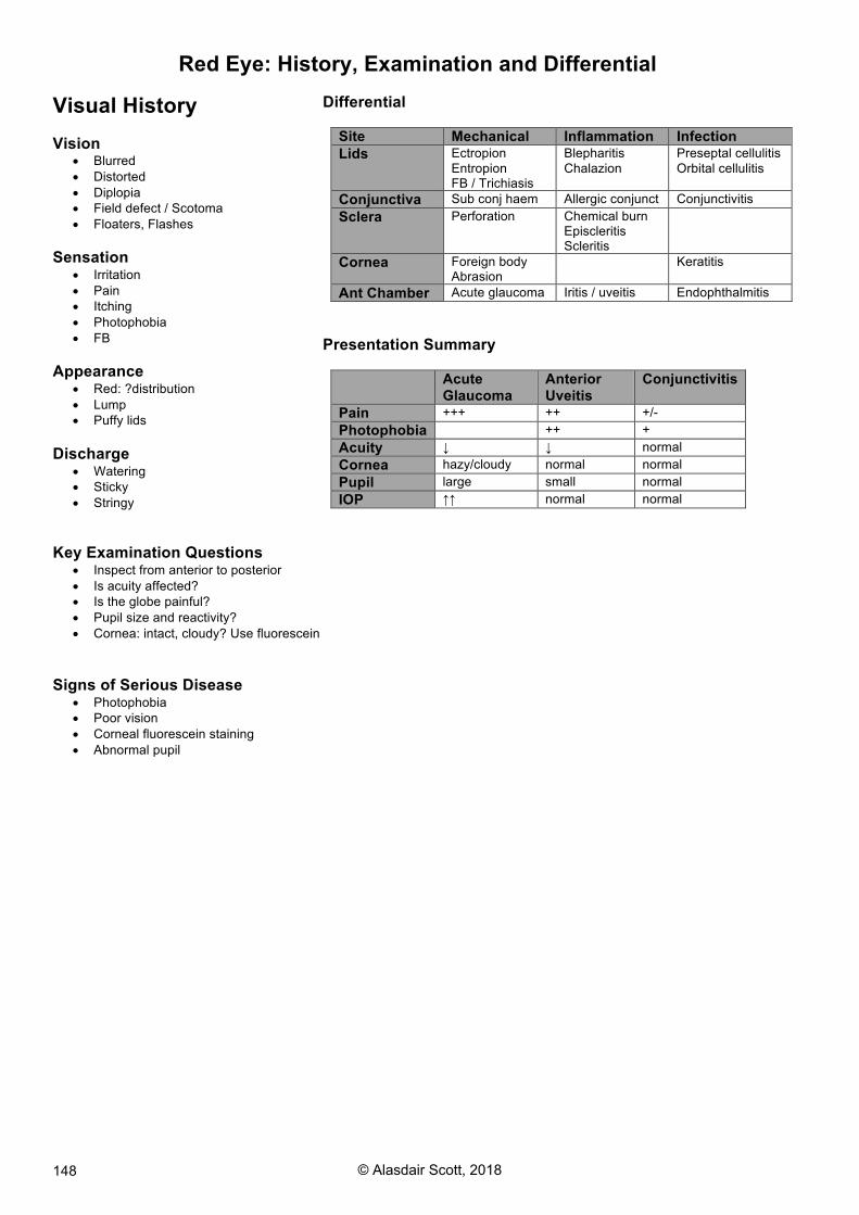
148
Red Eye: History, Examination and Differential Visual History Vision
• Blurred • Distorted • Diplopia • Field defect / Scotoma • Floaters, Flashes
Sensation
• Irritation • Pain • Itching • Photophobia • FB
Appearance
• Red: ?distribution • Lump • Puffy lids
Discharge
• Watering • Sticky • Stringy
Key Examination Questions
• Inspect from anterior to posterior • Is acuity affected? • Is the globe painful? • Pupil size and reactivity? • Cornea: intact, cloudy? Use fluorescein
Signs of Serious Disease
• Photophobia • Poor vision • Corneal fluorescein staining • Abnormal pupil
Differential
Site Mechanical Inflammation Infection
Lids Ectropion Entropion FB / Trichiasis
Blepharitis Chalazion
Preseptal cellulitis Orbital cellulitis
Conjunctiva Sub conj haem Allergic conjunct Conjunctivitis Sclera Perforation Chemical burn
Episcleritis Scleritis
Cornea Foreign body Abrasion
Keratitis
Ant Chamber Acute glaucoma Iritis / uveitis Endophthalmitis Presentation Summary
Acute
Glaucoma
Anterior
Uveitis
Conjunctivitis
Pain +++ ++ +/- Photophobia ++ + Acuity ↓ ↓ normal Cornea hazy/cloudy normal normal Pupil large small normal IOP ↑↑ normal normal
© Alasdair Scott, 2018
149
Red Eyes: Specific Diseases Acute Closed Angle Glaucoma
• Blocked drainage of aqueous from anterior chamber via the canal of Schlemm.
• Pupil dilatation (e.g. @ night) worsens the blockage. • Intraocular pressure rises from 15-20 → >60mmHg
Risk Factors
• Hypermetropia
• Shallow ant. chamber • Female
• FH
• ↑age
• Drugs
§ Anti-cholinergics
§ Sympathomimetics
§ TCAs
§ Anti-histamines
Symptoms
• Prodrome: rainbow haloes around lights at night-time. • Severe pain c̄ n/v
• ↓ acuity and blurred vision
Examination
• Cloudy cornea c̄ circumcorneal injection
• Fixed, dilated, irregular pupil
• ↑ IOP makes eye feel hard
Ix
• Tonometry: ↑↑ IOP (usually >40mmHg)
Acute Mx: Refer to Ophthalmologist
• Pilocarpine 2-4% drops stat: miosis opens blockage
• Topical β-B (e.g. timolol): ↓ aqueous formation • Acetazolamide 500mg IV stat: ↓ aqueous formation
• Analgesia and antiemetics
Subsequent Mx
• Bilat YAG peripheral iridotomy once IOP ↓ medically
Anterior Uveitis / Acute Iritis
Pathophysiology
• Uvea is pigmented part of eye and included: iris, ciliary body and choroid.
• Iris + ciliary body = anterior uvea
• Iris inflammation involves ciliary body too.
Symptoms
• Acute pain and photophobia
• Blurred vision (aqueous precipitates)
Examination
• Small pupil initially, irregular later
• Circumcorneal injection
• Hypopyon: pus in anterior chamber • White (keratic) precipitates on back of cornea
• Talbots test: ↑pain on convergence
Associations (most have no systemic associations) • Seronegative arthritis: AS, psoriatic, Reiter’s
• Still’s / JIA
• IBD
• Sarcoidosis
• Behcet’s
• Infections: TB, leprosy, syphilis, HSV, CMV, toxo
Mx
• Refer to ophthalmologist • Prednisolone drops
• Cyclopentolate drops: dilates pupil and prevents adhesions between iris and lens (synechiae)
© Alasdair Scott, 2018
150
Episcleritis
• Inflammation below conjunctiva in the episclera Presentation
• Localised reddening: can be moved over sclera • Painless / mild discomfort • Acuity preserved
Causes
• Usually idiopathic • May complicate RA or SLE
Rx: Topical or systemic NSAIDs Scleritis
• Vasculitis of the sclera Presentation
• Severe pain: worse on eye movement • Generalised scleral inflammation
§ Vessels won’t move over sclera • Conjunctival oedema (chemosis)
Causes
• Wegener’s • RA • SLE • Vasculitis
Mx • Refer to specialist • most need or corticosteroids or immunosuppressants
Complications: • Scleromalacia (thinning) → globe perforation
Conjunctivitis
Presentation
• Often bilateral c̄ purulent discharge
§ Bacterial: sticky (staph, strep, Haemophilus) § Viral: watery
• Discomfort • Conjunctival injection
§ Vessels may be moved over the sclera
• Acuity, pupil responses and cornea are unaffected.
Causes
• Viral: adenovirus
• Bacterial: staphs, chlamydia, gonococcus
• Allergic
Rx
• Bacterial: chloramphenicol 0.5% ointment • Allergic: anti-histamine drops: e.g. emedastine
Corneal Abrasion
• Epithelial breech w/o keratitis
• Cause: trauma
Symptoms
• Pain
• Photophobia
• Blurred vision
Ix
• Slit lamp: fluorescein stains defect green
Rx
• Chloramphenicol ointment for infection prophylaxis
Corneal Ulcer + Keratitis (corneal inflam)
Causes: bacteria, herpes, fungi, protozoa, RA • Dendritic ulcer = Herpes simplex • Acanthamoeba: protazoal infection affecting contact
lens wearers swimming in pools.
Presentation
• Pain, photophobia
• Conjunctival hyperaemia • ↓ acuity
• White corneal opacity
Risk factors: contact lens wearers
Ix: green c̄ fluorescein on slit lamp
Rx: refer immediately to specialist who will • Take smears and cultures • Abx drops, oral/topical aciclovir • Cycloplegics/mydriatics ease photophobia • Steroids may worsen symptoms: professionals only
Complications
• Scarring and visual loss
Ophthalmic Shingles
• Zoster of CNV1 • 20% of all Shingles (only commoner in thoracic
dermatomes) Presentation
• Pain in CNV1 dermatome precedes blistering rash • 40% → keratitis, iritis • Hutchinson’s sign
§ Nose-tip zoster due to involvement of nasociliary branch.
§ ↑ chance of globe involvement as nasociliarry nerve also supplies globe
• Ophthalmic involvement § Keratitis + corneal ulceration (fluorescein
stains) § ± iritis
© Alasdair Scott, 2018
151
Sudden Loss of Vision
Key Questions • Headache associated: GCA • Eye movements hurt: optic neuritis • Lights / flashes preceding visual loss: detached retina • Like curtain descending: TIA, GCA • Poorly controlled DM: vitreous bleed from new vessels
Anterior Ischaemic Optic Neuropathy (AION)
• Optic nerve damaged if posterior ciliary arteries blocked by inflammation or atheroma.
• Pale / swollen optic disc Causes
• Arteritic AION: Giant Cell Arteritis
• Non-arteritic AION: HTN, DM, ↑ lipids, smoking
Optic Neuritis
Symptoms • Unilateral loss of acuity over hrs – days • ↓ colour discrimination (dyschromatopsia) • Eye movements may hurt
Signs
• ↓ acuity • ↓ colour vision • Enlarged blind-spot • Optic disc may be: normal, swollen, blurred • Afferent defect
Causes
• Multiple sclerosis (45-80% over 15yrs) • DM
• Drugs: ethambutol, chloramphenicol • Vitamin deficiency
• Infection: zoster, Lyme disease
Rx
• High-dose methyl-pred IV for 72h
• Then oral pred for 11/7
Vitreous Haemorrhage
Source
• New vessels: DM
• Retinal tears / detachment / trauma
Presentation
• Small bleeds → small black dots / ring floaters
• Large bleed can obscure vision → no red reflex, retina can’t be visualised
Ix
• May use B scan US to identify cause Mx
• VH undergoes spontaneous absorption
• Vitrectomy may be performed in dense VH
Central Retinal Artery Occlusion
Presentation • Dramatic unilateral visual loss in seconds • Afferent pupil defect (may precede retinal changes) • Pale retina c̄ cherry-red macula
Causes
• GCA
• Thromboembolism: clot, infective, tumour
Rx
• If seen w/i 6h aim is to ↑ retinal blood flow by ↓ IOP § Ocular massage § Surgical removal of aqueous § Anti-hypertensives (local and systemic)
Retinal Vein Occlusion
Central
• Commoner than arterial occlusion
• Causes: arteriosclerosis, ↑BP, DM, polycythaemia
• Pres: sudden unilat visual loss c̄ RAPD
• Fundus: Stormy Sunset Appearance § Tortuous dilated vessels § Haemorrhages § Cotton wool spots
• Complications § Glaucoma § Neovascularisation
• Prognosis: possible improvement for 6mo-1yr Branch
• Presentation: unilateral visual loss • Fundus: segmental fundal changes • Comps: retinal ischaemia → VEGF release and
neovascularisation (Rx: laser photocoagulation)
Retinal Detachment
• Holes/tears in retina allow fluid to separate sensory retina from retinal pigmented epithelium
• May be 2O to cataract surgery, trauma, DM Presentation: 4 F’s
• Floaters: numerous, acute onset, “spiders-web” • Flashes • Field loss • Fall in acuity • Painless
Fundus: grey, opalescent retina, ballooning forwards
Rx
• Urgent surgery
• Vitrectomy + gas tamponade c̄ laser coagulation to secure the retina.
Causes of transient visual loss • Vascular: TIA, migraine • MS • Subacute glaucoma • Papilloedema
© Alasdair Scott, 2018

152
Gradual Visual Loss
Causes
Common
• Diabetic retinopathy • ARMD • Cataracts • Open-angle Glaucoma
Rarer
• Genetic retinal disease: retinitis pigmentosa • Hypertension • Optic atrophy
Age-Related Macular Degeneration (ARMD)
• Commonest cause of blindness >60yrs • 30% of >75yrs will have dry AMD
Risk Factors
• Smoking
• ↑ age
• Genetic factors
Presentation
• Elderly pts. • Central visual loss
Dry ARMD: Geographic Atrophy
• Drusen: fluffy white spots around macula
• Degeneration of macula
• Slow visual decline over 1-2yrs
Wet ARMD: Subretinal Neovascularisation
• Aberrant vessels grow into retina from choroid and → haemorrhage
• Rapid visual decline (sudden / days / wks) c̄ distortion
• Fundoscopy shows macular haemorrhage → scarring
• Amsler grid detects distortion
Ix
• OCT: optical coherence tomography
§ Gives high resolution images of the retina
Mx for Wet AMRD
• Photodynamic therapy • Intravitreal VEGF inhibitors
§ Bevacizumab (Avastin) § Ranibizumab (Lucentis)
• Antioxidant vitamins (C,E) + zinc may help early ARMD
Tobacco-Alcohol Amblyopia
• Due to toxic effects of cyanide radicals when combined with thiamine deficiency.
• Pres: Optic atrophy, loss of red/green discrimination, scotomata
• Rx: vitamins may help
Chronic Simple (Open-Angle) Glaucoma
Pathogenesis
• Depends on susceptibility of pt’s. retina and optic nerve to ↑ IOP damage.
• IOP >21mmHg → ↓blood flow and damage to optic nerve → optic disc atrophy (pale) + cupping
Presentation
• Peripheral visual field defect: superior nasal first • Central field is intact \ acuity maintained until late
§ Presentation delayed until optic N. damage is irreversible
Screen if High Risk
• >35yrs
• Afro-Caribbean
• FH
• Drugs: steroids
• Co-morbidities: DM, HTN, migraines
• Myopia
Ix
• Tonometry: IOP ≥21mmHg
• Fundoscopy: cupping of optic disc
• Visual field assessment: peripheral loss
Mx
• Life-long f/up Eye-drops to ↓ IOP to baseline
• 1st
line: β-blockers
§ Timolol, betaxolol § ↓ aqueous production
§ Caution in asthma, heart failure
• Prostaglandin analogues
§ Latanoprost, travoprost § ↑ uveoscleral outflow
• α-agonists
§ Brimonidine, apraclonidine
§ ↓ aqueous production and ↑ uveoscleral outflow
• Carbonic anhydrase inhibitors
§ Dorzolamide drops, acetazolamide PO
• Miotics
§ Pilocarpine
Non-medical Options
• Laser trabeculoplasty
• Surgery (trabeculectomy) is used if drugs fail § New channel allows aqueous to flow into
conjunctival bleb Commonest Causes of Blindness Worldwide
• Trachoma • Cataracts • Glaucoma • Keratomalacia: vitamin A deficiency • Onchocerciasis • Diabetic Retinopathy
© Alasdair Scott, 2018

153
The Eye in DM
The Problem • DM is leading cause of blindness up to 60yrs • 30% have ocular problems @ presentation • BP < 130/80 and normoglycaemia → ↓ diabetic
retinopathy Pathogenesis
Cataract
• DM accelerates cataract formation
• Lens absorbs glucose which is converted to sorbitol by aldose reductase.
Retinopathy
• Microangiopathy → occlusion • Occlusion → ischaemia → new vessel formation in
retina § Bleed → vitreous haemorrhage § Carry fibrous tissue c̄ them → retinal
detachment • Occlusion also → cotton wool spots (ischaemia) • Vascular leakage → oedema and lipid exudates • Rupture of microaneurysms → blot haemorrhage
Screening
• All diabetics should be screened annually • Fundus photography • Refer those c̄ maculopathy, NPDR and PDR to
ophthalmologist § 30% NPDR develop PDR in 1yr
Ix
• Fluorescein angiography Mx
• Good BP and glycaemic control • Rx concurrent disease: HTN, dyslipidaemia, renal
disease, smoking, anaemia • Laser photocoagulation
§ Maculopathy: focal or grid § Proliferative disease: pan-retinal (macula
spared) CN Palsies
• CNIII and VI palsies may occur • In diabetic CNIII palsy the pupil may be spared as its
nerve fibres run peripherally and receive blood from pial vessels.
Fundoscopy Findings
Background Retinopathy: Leakage
• Dots: microaneurysms
• Blot haemorrhages
• Hard exudates: yellow lipid patches
Pre-proliferative Retinopathy: Ischaemia
• Cotton-wool spots (infarcts) • Venous beading
• Dark Haemorrhages
• Intra-retinal microvascular abnormalities
Proliferative Retinopathy
• New vessels
• Pre-retinal or vitreous haemorrhage
• Retinal detachment Maculopathy
• Caused by macular oedema
• ↓ acuity may be only sign
• Hard exudates w/i one disc width of macula
© Alasdair Scott, 2018

154
Cataracts Presentation
• Increasing myopia • Blurred vision → gradual visual loss • Dazzling in sunshine / bright lights • Monocular diplopia
Causes
• ↑Age: 75% of >65s • DM • Steroids • Congenital
§ Idiopathic § Infection: rubella § Metabolic: Wilson’s, galactosaemia § Myotonic dystrophy
Ix
• Visual acuity • Dilated Fundoscopy • Tonometry • Blood glucose to exclude DM
Mx
Conservative
• Glasses • Mydriatic drops and sunglasses may give some relief
Surgery
• Consider if symptoms affect lifestyle or driving (<6/10) • Day-case surgery under LA
§ Phacoemulsion + lens implant • 1% risk of serious complications
§ Anterior uveitis / iritis § VH § Retinal detachment § Secondary glaucoma § Endophthalmitis (→ blindness in 0.1%)
• Post-op capsule thickening is common § Easily Rx c̄ laser capsulotomy.
• Post-op eye irritation is common and requires drops
The Retina • Outer pigmented layer in contact c̄ the choroid • Inner sensory layer in contact c̄ vitreous • At centre is fovea
Optic Disc
Colour
• Should be pale pink
• Paler in optic atrophy
Contour
• Margins blurred in papilloedema and optic neuritis
Cup
• Physiological cup lies centrally and should occupy 1/3 of disc diameter
• Cup widening and deepening in glaucoma
Retinitis Pigmentosa
• Most prevalent inherited degeneration of the macula • Various modes of inheritance
§ Mostly AR § AD has best prognosis § X-linked has worst prognosis
• Affects ~ 1/2000 Presentation
• Night blindness • ↓↓ visual fields → tunnel vision • Most are registrable blind (<3/60) by mid 30s
Fundoscopy
• Pale optic disc: optic atrophy
• Peripheral retina pigmentation: spares the macula
Associations
• Friedrich’s ataxia
• Refsum’s disease
• Kearns-Sayre Syndrome
• Usher’s Syndrome
Retinoblastoma
• Commonest intraocular tumour in children • 1:15,000 LBs
Inheritance
• Hereditary type differs from non-hereditary type • AD mutation of RB gene (a TSG) • Pts. typically have one mutant allele in every retinal
cell; if the other allele mutates → retinoblastoma. Associations
• 5% occur c̄ pineal or other tumour • ↑ risk of osteosarcoma and rhabdomyosarcoma
Signs
• Stabismus • Leukocoria (white pupil) → no red reflex
Rx
• Depends on size • Options include: chemo, radio, enucleation
© Alasdair Scott, 2018

155
The External Eye Inflammatory Lid Swellings
Stye or hordeolum externum • An abscess / infection in a lash follicle which points
outwards. • Rx: local Abx – e.g. fusidic acid
Chalazion or hordeolum internum
• Abscess of the Meibomian glands which points inwards onto conjunctiva.
§ Sebaceous glands of eyelid Blepharitis
• Chronic inflammation of eyelid • Causes: seborrhoeic dermatitis, staphs • Features
§ Red eyes § Gritty / itchy sensation § Scales on lashes. § Often assoc. c̄ rosacea
• Rx § Clean crusts of lashes c̄ warm soaks § May need fusidic acid drops
Entropion
• Lid inversion → corneal irritation • Degeneration of lower lid fascia
Ectropion • Low lid eversion → watering and exposure keratitis • Assoc. c̄ ageing and facial N. palsy
Ptosis • True ptosis is intrinsic LPS weakness • Bilateral
§ Congenital § Senile § MG § Myotonic dystrophy
• Unilateral
§ 3rd Nerve palsy § Horner’s syndrome (partial) § Mechanical: xanthelasma, trauma
Lagophthalmos
• Difficulty in lid closure over the globe which may → exposure keratitis
• Causes: exophthalmos, facial palsy, injury • Rx
§ Lubricate eyes c̄ liquid paraffin ointment § Temporary tarsorrhaphy may be needed if
corneal ulcers develop. Pinguecula
• Yellow vascular nodules either side of the cornea Pterygium
• Similar to pinguecula but grows over the cornea → ↓ vision.
• Benign growth of conjunctiva • Assoc. c̄ dusty, wind-blown life-styles, sun exposure
Orbital Swellings Orbital Cellulitis
Pathophysiology
• Infection spreads locally: e.g. from paranasal sinuses, eyelid or external eye.
• Staphs, pneumococcus, GAS
Presentation
• Usually a child c̄ inflammation of the orbit + lid swelling
• Pain and ↓ range of eye movement • Exophthalmos • Systemic signs: e.g. fever • ± tenderness over the sinuses
Rx
• IV Abx: Cefuroxime (20mg/kg/8h IV)
Complications
• Local extension → meningitis and cavernous sinus thrombosis.
• Blindness due to optic N. pressure.
Carotid-cavernous fistula • May follow carotid aneurysm rupture c̄ reflux of blood
into cavernous sinus. • Causes: spontaneous, trauma • Presentation
§ Engorgement of eye vessels § Lid and conjunctival oedema, § Pulsatile exophthalmos § Eye bruit
• Rx: endovascular treatment
Exophthalmos / Proptosis • Protrusion of one or both eyes
Common Causes
• Graves’ Disease
§ 25-50%
§ ↑ risk in smoker’s
§ Anti-TSH Abs → retro-orbital inflammation and lymphocyte infiltration → swelling
• Orbital cellulitis
• Trauma
Other Causes
• Idiopathic orbital inflammatory disease • Vasculitis: Wegener’s • Neoplasm
§ Lymphoma § Optic glioma: assoc. c̄ NF-1 § Capillary haemangioma § Mets
• Carotid-cavernous fistula
© Alasdair Scott, 2018

156
Refractive Errors Refractive errors arise from disorders of the size and shape of the eye. Myopia: Short-sightedness
The Problem
• The eye is too long • Distant objects are focussed too far forward.
Causes
• Genetic
• Excessive close work in the early decades
The Solution
• Concave lenses
Astigmatism
The Problem
• Cornea or lens doesn’t have same degree of curvature in horizontal and vertical planes.
• Image of object is distorted longitudinally or vertically
The Solution
• Correcting lenses
Hypermetropia: long-sightedness
The Problem
• Eye is too short • When eye is relaxed and not accommodating, objects
are focussed behind the retina. • Contraction of ciliary muscles to focus image →
tiredness of gaze and possibly a convergent squint in children.
The Solution
• Convex lenses
Presbyopia
• With age, lens becomes stiff and less easy to deform. • Start at about 40yrs and is complete by 60yrs. • Use convex lenses.
Strabismus / Squint / Tropia
Esotropia = convergent squint
• Commonest type in children
• May be idiopathic or due to hypermetropia
Exotropia = divergent squint
• Older children
• Often intermittent
Non-Paralytic Squint
Diagnosis
• Corneal reflection: should fall centrally and symmetrically on each cornea.
• Cover test: movement of uncovered eye to take up fixation demonstrates manifest squint.
Management: 3 O’s
• Optical: correct refractive errors
• Orthoptic: patching good eye encourages use of squinting eye.
• Operations: e.g. resection and recession of rectus muscles – help alignment and cosmesis.
Paralytic Squint
• Diplopia is most on looking in direction of pull of paralysed muscle.
• Eye won’t fixate on covering. • Cover each eye in turn: which ever eye sees the outer
image is malfunctioning CNIII
• Ptosis (LPS) • Fixed dilated pupil (no parasympathetic) • Eye looking down and out • Causes
§ Medical: DM, MS, infarction
§ Surgical: ↑ ICP, cavernous sinus thrombosis, posterior communicating artery aneurysm
CNIV
• Diplopia especially on going down stairs
• Head tilt • Test: can’t depress in adduction
• Causes
§ Peripheral: DM (30%), trauma (30%), compression
§ Central: MS, vascular, SOL
CNVI
• Eye is medially deviated and cannot abduct • Diplopia in the horizontal plane. • Causes
§ Peripheral: DM, compression, trauma
§ Central: MS, vascular, SOL
• Rx: botulinum toxin can eliminate need for surgery
© Alasdair Scott, 2018
157
Eye Trauma • Record acuity of both eyes • Take detailed Hx of event • If unable to open injured eye, instil LA (e.g. tetracaine
1%)
Foreign Bodies • X-ray orbit if metal FB suspected • Fluorescein may show corneal abrasions
Mx
• Chloramphenicol drops 0.5% prevent infection § Usually coagulase-negative Staphylococcus
• Eye patch
• Cycloplegic drops may ↓ pain
§ Tropicamide, cyclopentolate
Intra-ocular Haemorrhage
• Blood in anterior chamber = hyphaema • Small amounts clear spontaneously, but some may
need evacuation. • Complicated by corneal staining and glaucoma (pain) • Keep IOP↓ and monitor
Orbital Blowout Fracture
• Blunt injury → sudden ↑ in orbital pressure c̄ herniation of orbital contents into maxillary sinus.
Presentation
• Ophthalmoplegia + Diplopia
§ Tethering of inferior rectus and inferior oblique • Loss of sensation to lower lid skin
§ Infraorbital nerve injury • Ipsilateral epistaxis
§ Damage to anterior ethmoidal artery • ↓ acuity • Irregular pupil that reacts slowly to light
Mx
• Fracture reduction and muscle release necessary. Chemical Injury
• Alkaline solutions are particularly damaging • Mx
§ Copious irrigation § Specialist referral
Floaters, Flashes and Haloes Floaters
• Small dark spots in the visual field • Sudden showers of floaters in one eye may be due to
blood or retinal detachment
Causes • Retinal detachment • VH • Diabetic retinopathy / Hypertension • Old retinal branch vein occlusion • Syneresis (degenerative opacities in the vitreous)
Flashes (Photopsia) • Either from intraocular or intracerebral pathology • Headache, n/v: migraine • Flashes and floater: retinal detachment
Haloes
• Usually just diffractive phenomena • May be caused by hazy ocular media – cataract,
corneal oedema, acute glaucoma • Haloes + eye pain = acute glaucoma • Jagged haloes which change shape are usually
migrainous.
© Alasdair Scott, 2018

158
Allergic Eye Disease Seasonal Allergic Conjunctivitis (SAC)
• 50% of allergic eye disease • Small papillae on tarsal conjunctivae • Rx
§ Antazoline: antihistamine drops: § Cromoglycate: inhibits mast cell degranulation
Perennial Allergic Conjunctivitis (PAC)
• Symptoms all year c̄ seasonal exacerbations • Small papillae on tarsal conjunctivae • Rx: olopatadine (antihistamine and mast-cell
stabiliser) Giant Papillary Conjunctivitis
• Iatrogenic FBs: contact lenses, prostheses, sutures • Giant papillae on tarsal conjunctivae • Rx: removal of FB, mast cell stabilisers
Management of Allergic Eye Disorders
1. Remove the allergen responsible where possible
2. General measures § Cold compress § Artificial tears
3. Oral antihistamines: loratadine 10mg/d PO
4. Eye drops § Antihistamines: antazoline, azelastine § Mast cell stabilizers: cromoglycate,
lodoxamide § Steroids: dexamethasone
- Beware of inducing glaucoma § NSAIDs: diclofenac
Tropical Eye Disease Trachoma Pathophysiology
• Caused by Chlamydia trachomatis (A,B,C) • Spread by flies • Inflammatory reaction under lids → scarring → lid
distortion → entropion → eyelashes scratch cornea → ulceration → blindness
Rx
• Tetracycline 1% ointment ± PO Prevention
• Good sanitation • Face washing
Onchocerciasis (River Blindness) Pathophysiology
• Caused by microfilariae of nematode Onchocerca volvulus
• Spread by flies • Fly bites → microfilariae infection → invade the eye →
inflammation → fibrosis → corneal opacities and synechiae
Rx
• Ivermectin Xerophthalmia and Keratomalacia
• Manifestations of vitamin A deficiency Presentation
• Night blindness and dry conjunctivae (xerosis) • Corneal ulceration and perforation
Rx
• Vitamin A / palmitate reverses early corneal changes
© Alasdair Scott, 2018

159
Eye Signs in Systemic Conditions Hypertensive Retinopathy
• Keith-Wagener Classification • Tortuosity and silver wiring • AV nipping • Flame haemorrhages and soft / cotton wool spots • Papilloedema • Grades 3 and 4 = malignant hypertension
Granulomatous Disorders
• TB, sarcoid, toxo, leprosy, brucella • Uveitis (ant/post) and choroidoretinitis
Systemic Inflammatory Disease
• Conjunctivitis: SLE, reactive arthritis, IBD • Scleritis / episcleritis: RA, vasculitis, SLE, IBD • Iritis : ank spond, IBD, sarcoid • Retinopathy: dermatomyositis
Keratoconjunctivitis Sicca / Sjogren’s
• ↓ tear production (Schirmer’s: <5mm in 5min) • Dry eyes and dry mouth • 1O or 2O: SLE, RA, sarcoid • Rx: artificial tears or saliva
Vascular Occlusion
• Emboli → amaurosis fugax: GCA, carotid atheroemboli
• Microemboli → Roth spots: infective endocarditis § Boat-shaped haemorrhage c̄ pale centre
Metabolic
• Kayser-Fleischer Rings: Wilson’s • Exophthalmos: Graves’ • Corneal calcification: HPT
HIV/AIDS
• CMV retinitis: pizza-pie fundus + flames • HIV retinopathy: cotton wool spots
Ophthalmic Pharmacology Mydriatics Anti-Muscarinics
• Tropicamide § Duration: 3h
• Cyclopentolate § Duration: 24h § Preferred for paediatric use
• Pupil dilatation + loss of light reflex • Cycloplegia (ciliary paralysis) → blurred vision
Sympathomimetics
• Para-hydroxyamphetamine, phenylephrine • May be used c̄ tropicamide • Don’t affect the light reflex or accommodation
Indications
• Eye examination • Prevention of synechiae in ant. uveitis / iritis
Caution
• May → acute glaucoma if shallow anterior chamber Miotics Effect
• Constrict the pupil Pilocarpine
• Muscarinic agonist Use
• Acute closed-angle glaucoma Other Tetracaine
• Anaesthetic used to permit examination of a painful eye
Lubricants
• Hypomellose • Carbomer (viscotears)
Topical Anti-histamine
• Emedastine • Antazoline
Chronic Open-Angle Glaucoma
• 1st line: β-blockers § Timolol, betaxolol § ↓ aqueous production § Caution in asthma, heart failure
• Prostaglandin Analogues § Latanoprost, travoprost § ↑ uveoscleral outflow
• α-agonists § Brimonidine, apraclonidine § ↓ aqueous production and ↑ uveoscleral
outflow • Carbonic anhydrase inhibitors
§ Dorzolamide drops, acetazolamide PO • Miotics: Pilocarpine
© Alasdair Scott, 2018


![Plastic Surgery Post-Op [1711] General...Printed on 6/4/2020 at 4:55 PM from SUP Page 1 of 29 Plastic Surgery Post-Op [1711] General Common Present on Admission Diagnosis [ ] Acidosis](https://static.fdocuments.in/doc/165x107/60843f50021bfb1fe738e49d/plastic-surgery-post-op-1711-general-printed-on-642020-at-455-pm-from-sup.jpg)