Surface properties and hemocompatibility of - Lehigh University
11
* Corresponding author. 1 Current address: Orthogene Inc., 34700 Campus Drive, Fremont, CA 94555, USA. 2 Current address: Department of Chemical Engineering, National Cheng Kung University, Tainan, Taiwan 70101, ROC. 3 Current address: Guidant Corporation, 3200 Lakeside Drive, Santa Clara, CA 95054, USA. Biomaterials 20 (1999) 1533 } 1543 Surface properties and hemocompatibility of alkyl-siloxane monolayers supported on silicone rubber: e!ect of alkyl chain length and ionic functionality James H. Silver!,1, Jui-Che Lin!,2, Florencia Lim!,3, Vassiliki A. Tegoulia", *, Manoj K. Chaudhury#, Stuart L. Cooper$ !Department of Chemical Engineering, University of Wisconsin, Madison, WI 53706, USA "Department of Chemical Engineering, University of Delaware, Newark, DE 19716, USA #Department of Chemical Engineering, Lehigh University, Bethlehem, PA 18015, USA $Illinois Institute of Technology, Chicago, IL 60616, USA Received 1 April 1998; accepted 22 July 1998 Abstract Self-assembled monolayers of alkylsiloxanes supported on poly(dimethylsiloxane) (PDMS) rubber were used as model systems to study the relation between blood compatibility and surface composition. The inner lumen of PDMS tubes were "rst treated with an oxygen plasma. The resultant oxidized surfaces were post-derivatized by reaction with alkyltrichlorosilanes to form the monolayer "lms. The alkyl chain lengths used were slightly longer than in a previous study, and this may alter the phase-state of the monolayer from liquid-like to crystalline. The chemical properties of the monolayer were controlled by varying the chemical composition of the alkyltrichlorosilanes used. Terminal functionalities included }CH 3 , }CF 3 , }COOH, }SO 3 H and }(CH 2 CH 2 O) 4 OH. Surface derivati- zation was veri"ed with static contact angle measurements and X-ray photoelectron spectroscopy. Blood compatibility was evaluated using a canine ex vivo arterio-venous series shunt model. Surfaces grafted with hydrophobic head groups such as }CH 3 and }CF 3 were signi"cantly less thrombogenic than the surfaces composed of ionic head groups such as }COOH and }SO 3 H. Surfaces enriched in }(CH 2 CH 2 O) 4 OH had an intermediate thrombogenicity. Silastic pump grade tubing and polyethylene tubing, used as controls, were found to be the least thrombogenic of all the surfaces tested. ( 1999 Elsevier Science Ltd. All rights reserved. Keywords: Self-assembled monolayer; Surface modi"cation; Blood compatibility; Silicone rubber 1. Introduction One of the central ideas of biomaterials research is that there is a relationship between chemistry of an implanted device or material and its hemocompatibility. No study is considered complete without an evaluation of the surface properties of the biomaterial in question. For many of these materials, however, the surface chemistry is quite complex, and it is di$cult to be sure of the chemical composition at the blood}biomaterial interface. Many commercially available materials, such as Biomert, may contain surface-active additives, such as antioxidants or processing aids, which further complicates interpretation of blood}material interactions [1]. Multiphase materials such as polyurethanes may show surface reorientation between the dry and hydrated states [2], making it di$- cult to evaluate the surface composition using methods in which the polymer surface is analyzed in air or in vacuo. Recently, much interest has arisen in self-assembled monolayers (SAMs), with the goal of developing molecu- lar-level control over surface properties, both for funda- mental studies of adhesion and wettability, as well as for technological applications [3]. These "lms are typically formed by the adsorption of terminally functionalized alkanethiols (HS(CH 2 ) n X) onto gold substrates [4], or 0142-9612/99/$ - see front matter ( 1999 Elsevier Science Ltd. All rights reserved. PII: S 0 1 4 2 - 9 6 1 2 ( 9 8 ) 0 0 1 7 3 - 2
Transcript of Surface properties and hemocompatibility of - Lehigh University

*Corresponding author.1Current address: Orthogene Inc., 34700 Campus Drive, Fremont,
CA 94555, USA.2Current address: Department of Chemical Engineering, National
Cheng Kung University, Tainan, Taiwan 70101, ROC.3Current address: Guidant Corporation, 3200 Lakeside Drive, Santa
Clara, CA 95054, USA.
Biomaterials 20 (1999) 1533}1543
Surface properties and hemocompatibility of alkyl-siloxanemonolayers supported on silicone rubber:
e!ect of alkyl chain length and ionic functionality
James H. Silver!,1, Jui-Che Lin!,2, Florencia Lim!,3, Vassiliki A. Tegoulia",*,Manoj K. Chaudhury#, Stuart L. Cooper$
!Department of Chemical Engineering, University of Wisconsin, Madison, WI 53706, USA"Department of Chemical Engineering, University of Delaware, Newark, DE 19716, USA#Department of Chemical Engineering, Lehigh University, Bethlehem, PA 18015, USA
$Illinois Institute of Technology, Chicago, IL 60616, USA
Received 1 April 1998; accepted 22 July 1998
Abstract
Self-assembled monolayers of alkylsiloxanes supported on poly(dimethylsiloxane) (PDMS) rubber were used as model systems tostudy the relation between blood compatibility and surface composition. The inner lumen of PDMS tubes were "rst treated with anoxygen plasma. The resultant oxidized surfaces were post-derivatized by reaction with alkyltrichlorosilanes to form the monolayer"lms. The alkyl chain lengths used were slightly longer than in a previous study, and this may alter the phase-state of the monolayerfrom liquid-like to crystalline. The chemical properties of the monolayer were controlled by varying the chemical composition of thealkyltrichlorosilanes used. Terminal functionalities included }CH
3, }CF
3, }COOH, }SO
3H and }(CH
2CH
2O)
4OH. Surface derivati-
zation was veri"ed with static contact angle measurements and X-ray photoelectron spectroscopy. Blood compatibility was evaluatedusing a canine ex vivo arterio-venous series shunt model. Surfaces grafted with hydrophobic head groups such as }CH
3and }CF
3were signi"cantly less thrombogenic than the surfaces composed of ionic head groups such as }COOH and }SO
3H. Surfaces enriched
in }(CH2CH
2O)
4OH had an intermediate thrombogenicity. Silastic pump grade tubing and polyethylene tubing, used as controls,
were found to be the least thrombogenic of all the surfaces tested. ( 1999 Elsevier Science Ltd. All rights reserved.
Keywords: Self-assembled monolayer; Surface modi"cation; Blood compatibility; Silicone rubber
1. Introduction
One of the central ideas of biomaterials research is thatthere is a relationship between chemistry of an implanteddevice or material and its hemocompatibility. No study isconsidered complete without an evaluation of the surfaceproperties of the biomaterial in question. For many ofthese materials, however, the surface chemistry is quite
complex, and it is di$cult to be sure of the chemicalcomposition at the blood}biomaterial interface. Manycommercially available materials, such as Biomert, maycontain surface-active additives, such as antioxidants orprocessing aids, which further complicates interpretationof blood}material interactions [1]. Multiphase materialssuch as polyurethanes may show surface reorientationbetween the dry and hydrated states [2], making it di$-cult to evaluate the surface composition using methods inwhich the polymer surface is analyzed in air or in vacuo.
Recently, much interest has arisen in self-assembledmonolayers (SAMs), with the goal of developing molecu-lar-level control over surface properties, both for funda-mental studies of adhesion and wettability, as well as fortechnological applications [3]. These "lms are typicallyformed by the adsorption of terminally functionalizedalkanethiols (HS(CH
2)nX) onto gold substrates [4], or
0142-9612/99/$ - see front matter ( 1999 Elsevier Science Ltd. All rights reserved.PII: S 0 1 4 2 - 9 6 1 2 ( 9 8 ) 0 0 1 7 3 - 2
the adsorption of terminally functionalized silanes ontosilicon surfaces, glass, or fused silica [5]. Formation ofSAMs on elastomeric materials such as poly(dimethyl-siloxane) (PDMS) has recently been demonstrated as well[6}9]. The perfection of these monolayers was inferredfrom contact angle measurements while infrared dichro-ism was used to con"rm ordering in the hydrocarbonchains. The thickness of SAMs on PDMS was estimatedbased on a comparison with monolayers formed onpolished silicon wafers [8].
The interactions of these surfaces with biological mol-ecules have been under investigation. A number of re-ports have appeared which suggest that cellular adhesionon self-assembled monolayers can be controlled by thechemistry of the underlying surface [5]. Others have usedthe control provided by SAMs to direct protein adsorp-tion onto such surfaces [10]. However, the interactionsbetween larger, more complex proteins such as "bronec-tin with SAMs are still di$cult to control, and result ina number of di!erent cellular responses [11, 12]. Littleinformation is available regarding the blood compatibil-ity of such surfaces.
In order to carefully control the structure of ourblood-contacting surfaces, we have functionalized thesurface of poly(dimethylsiloxane) in a two-step process,using speci"c chemistries to form surface monolayerswith known compositions. In the "rst step, the surface isoxidized using an oxygen plasma, and in the second step,a monolayer is formed by chemisorption of alkyltrich-lorosilanes (Cl
3Si(CH
2)nR) onto the surface of the poly-
mer [6]. Previous work [6}9, 13] has shown that thesemethods create well-de"ned silane monolayers whosesurface chemistry is controlled by the head group func-tionalities (R) of the silanes. The functionalities studiedwere methyl (}CH
3), tri#uoromethyl (}CF
3), ethylene
oxide (}(OCH2CH
2)4OH), carboxylate (}COOH), and
sulfonate (}SO3H).
Previously, we have reported on the blood compatibil-ity of a related series of self-assembled monolayers sup-ported on silicone rubber [14]. In the present study, headgroups with sulfonate and carboxylate functionalities areincluded. One of the primary di!erences between the twostudies is the length of the alkyl tail in the subsurfaceregion of the monolayer. Longer alkyl chains, used in thepresent study, have been shown, by infrared spectro-scopy, to form a more extended, crystalline structure forself-assembled monolayers on PDMS. In contrast, theshorter alkyl chains, investigated in the previous studywere found to be in a more mobile, liquid-like phase [14].It has long been postulated that crystalline, less mobilesurfaces do not enhance blood compatibility. By compar-ing the thrombogenicity of these new longer monolayerswith results from the previous study we will try to testthis hypothesis.
Another hypothesis we would like to test is that #uor-ination of a surface leads to a more biocompatible sur-
face. GoreTex' vascular grafts are made from expandedpoly(tetra#uoroethylene) (e-PTFE), and a great deal ofclinical experience has been obtained with this material.Incorporation of #uorine groups by radiation graftinghas been previously attempted by Ho!man et al. [15],who found that when woven Dacron vascular grafts weremodi"ed with a tetra#uoroethylene plasma, thehemocompatibility of these materials was signi"cantlyimproved [16]. Further, they found that although less"brinogen was adsorbed on TFE-Dacron, it was lesselutable by sodium dodecyl sulfate. However, afterplasma modi"cation, the surface showed a variety offunctional groups, including CF
3, CF
2, CF}CF
n, CF, and
C}CFn, as determined from the high resolution Cls peak
in the ESCA spectrum so that the surface propertiescould be attributed to a speci"c functional group. In thepresent study, a surface exposing only CF
3functional
groups with expected high interfacial free energy [17] iscompared with more hydrophilic, lower interfacial freeenergy surfaces.
The poly(ethylene oxide) (PEO) terminated monolayerhas been included to examine if incorporation ofPEO results in surfaces with improved biocompatibility.Previous work has shown that surfaces incorporatinglong PEO chains were blood compatible [18, 19]. Onthe contrary, shorter PEO chains endlinked onpoly(glycidoxypropyl methyl-dimethyl siloxane) via theirterminal hydroxyl groups were found to enhance plateletand "brinogen deposition in an ex vivo shunt [19]. Thiswas attributed to an incomplete coverage of the surface bythe PEO chains. However, protein adsorption and celladhesion on thiol self-assembled monolayers containingonly three or six ethylene oxide groups was found to beminimal in vitro [20, 21].
A number of investigators have shown that polymersincorporating sulfonate or carboxylate groups haverather remarkable blood-contacting properties.These materials may act like heparin, a mucopolysac-charide which is clinically used as an anticoagulant.Sulfate, amino-sulfate, and carboxylate groups have beenshown to be essential to the anticoagulant activity ofheparin [22]. Jozefovicz and Jozefowicz [23] have modi-"ed polystyrenes and dextrans to incorporate these ionicgroups, and have shown that these materials possessanticoagulant, heparin-like, activity. However, it wasfound that sulfonate groups were essential for heparin-like activity, but carboxylate groups, while greatlyenhancing such activity, were non-essential. Dextranswhich contained only carboxylate groups were deter-mined to be inactive [24]. Ito et al. [25] have shownthat polyurethanes grafted with poly(vinyl sulfonate)possessed heparin-like activity, which was reducedupon incorporation of carboxylate groups, contraryto the results of Jozefovicz and Jozefowicz. Okkemaet al. found that the incorporation of carboxylategroups did not improve the hemocompatibility of
1534 J.H. Silver et al. / Biomaterials 20 (1999) 1533}1543

polyurethanes, as evaluated in a canine ex vivo model[26].
In experiments with water soluble sulfonated poly-urethanes, it was shown that these polymers prolongclotting times, inhibit the process of "brin assembly, andinhibit thrombin activity [27]. Polyurethanes graftedwith propyl sulfonate groups and containing PTMO asthe macroglycol show strong antithrombotic behavior,with low levels of platelet deposition or spreading [26].Similar results were also obtained when Biomer', a com-mercial polyurethaneurea containing PTMO as the mac-roglycol, was sulfonated [28]. Polyurethanes surfacegrafted with poly(ethylene oxide) chains which are end-terminated with sulfonate groups signi"cantly prolongocclusion times in a rabbit A}A shunt model [29]. San-terre et al. [30] have shown that sulfonated poly-urethanes interact strongly with "brinogen, and that"brinogen is not displaced from these surfaces in Vro-man-e!ect-type experiments. Thus, polymers containingsulfate or sulfonate groups have very interesting behaviorin contact with blood.
The hemocompatibility of the silicone elastomer andsurface-derivatized silicone elastomers was evaluated us-ing a canine ex vivo series shunt model [31]. PE tubingwas also used as a control surface. The monolayer forma-tion and surface composition of these elastomers aftermodi"cation was examined using X-ray photoelectronspectroscopy. Surface energetics were evaluated by con-tact angle measurements. Throughout the paper, theterm &thromboresistant' is used to refer to materialswhich show low platelet or thrombus deposition on theirsurfaces.
2. Materials and methods
2.1. Materials
Silicone rubber tubing (0.125A ID) (RX Pump Grade,Lot No. HH031678) and "ve surface-modi"ed Silastictubings were received from Dow Corning, and stored indistilled water until use. The surfaces were modi"ed tocontain }CH
3, }CF
3, }(CH
2CH
2O)
4OH, }COOH, or
}SO3H functional groups. Polyethylene tubing (0.125A
ID) was purchased from Clay-Adams (Intramedic,Parsippany, NJ, Lot d93485).
2.2. Surface modixcation of PDMS
Nine segments of PDMS (Silastic) tubing each about5.5A long (0.118A ID; 0.25A OD), were oxidized in a Har-rick plasma cleaner for 5 min in an oxygen atmosphere(0.4 Torr). The oxidized segments were joined togetherwith short polyethylene tubes, and subsequently "lledwith a silanizing solution. The silanizing solution wasprepared by dissolving alkyltrichlorosilanes in per-
#uorooctane at concentrations of 10}20 lg silane g~1 ofsolvent. After 1 h, the tubes were rinsed with fresh per-#uorooctane, followed by ethanol and then dried undera continuous stream of air. The silanes used for thesestudies were: Cl
3Si(CH
2)15
CH3
(Petrach); Cl3Si(CH
2)2
CF(CF3)CF
2CF(CF
3)2
(Dow Corning, Courtesy ofDr G. Gornowitz) and Cl
3Si(CH
2)9CH
2"CH
2(Dow
Corning). The #uorosilane contained about 15% of thefollowing isomer: Cl
3Si(CH)CH
3CF(CF
3)CF
2CF(CF
3)2.
The purity of the other two silanes was better than 99%.The ole"n-terminated PDMS were further reacted withHS(CH
2)2SO
3Na and HS(CH
2)2(OCH
2CH
2)4OH by
a free radical process in order to introduce the ethyleneoxide and sulfonate groups on its surface as shown:
PDMS09}O3Si(CH
2)9CH"CH
2#HS}R &&&&&"B%/;01)%/0/%`UV
PDMS09}O3Si(CH
2)11
SR
where R"}(CH2)2SO
3Na and }(CH
2)2(OCH
2CH
2)4OH.
In separate experiments, carboxylic acid groups wereintroduced on the surface of PDMS09}O
3Si(CH
2)9CH"
CH2, by oxidizing the ole"n groups of the monolayers by
a mixture of potassium permanganate and potassiumperiodate according to the procedure of Wasserman andWhitesides [32].
2.3. Surface property characterization
X-ray photoelectron spectroscopy (XPS) spectra wereobtained using a Perkin}Elmer Physical Electronics,PHI 5400 spectrometer. Samples of each tube were cut inhalf lengthwise, and #attened in order to hold them ontothe sample stage. A magnesium anode operating at300 W and 15 kV and photoelectron take-o! angle of 453was used. The take-o! angle is de"ned as the anglebetween the detector and the normal of the substrate.Survey spectra (0}1000 eV) were taken at a pass energy of90 eV using a 1000 lm diameter X-ray spot size. Therelative atomic percentage of each element at the surfacewas estimated from the peak areas using atomic sensitiv-ity factors speci"ed for the PHI 5400. High-resolutionspectra of the O1s peak, the N1s peak, the C1s, peak, theSi2p peak, and the F1s peak at a pass energy of 17.9 eVwere also taken.
Water-in-air static contact angles were measured onthe concave inner lumen of these surfaces using the pro-cedure of Lelah et al. [33]. Brie#y, short tubes of PDMSwere cut into hemicylindrical form. Small drops (ca.1 mm) of water were placed on the inner lumen of thesetubes, and were either photographed or analyzed bya video microscopy system. Tangent lines were drawn atthe intersection of the drop and the PDMS surface bygeometric construction directly on the photograph or ona replica of the video image to estimate the contactangles.
J.H. Silver et al. / Biomaterials 20 (1999) 1533}1543 1535

Table 1XPS surface analysis!
Material C1s O1s Si2p S2p F1s
PDMS09}O3Si(CH
2)10
COOH 46.7 29.8 23.1 0.4 *
PDMS09}O3Si(CH
2)11
S(CH2)2SO
3H 38.0 48.7 11.4 1.1 *
PDMS09}O3Si(CH
2)11
S(CH2)2(OCH
2CH
2)4OH 42.6 42.6 14.2 0.7 *
PDMS09}O3Si(CH
2)2CF(CF
3)CF
2CF(CF
3)2
38.2 44.9 12.1 1.0 3.8PDMS09}O
3Si(CH
2)15
CH3
40.9 42.5 15.4 0.7 *
Silastic pump grade tubing 44.3 36.2 18.9 0.7 *
! The standard error is on the order of 10% or less according to information provided for the PHI spectrometer by the manufacturers.
2.4. Blood-contacting properties
The blood-contacting properties of these materialswere evaluated using a canine ex vivo series shunt experi-ment with modi"cations [31]. The polymer tubings werecut into 1.5A length and assembled into shunts. Eachpolymer was present in triplicate in each shunt.
Adult mongrel dogs which were selected after hema-tological screening were injected with autologous111In-labeled platelets and 125I-labeled "brinogen. Noanticoagulant was used in this procedure. The shunts were"lled with sterile, degassed, divalent cation-free Tyrodessolution (0.2 g l~1 KCI, 8.0 g l~1 NaCI, 0.05 g l~1
NaH2PO
4}H
2O, 1.0 g l~1 dextrose, 1.0 g l~1 NaHCO
3,
pH"7.35), and hydrated overnight at 43C. The femoralartery and vein were cannulated with the shunt. A branchartery proximal to the shunt cannulation site was con-nected to a #ushing system. The blood #ow rate wascontinuously monitored during blood exposure using anelectromagnetic #ow probe. The initial #ow rate wascontrolled at 280$20 mlmin~1. Blood samples werecollected over time to determine bulk radioactivity,platelet and "brinogen concentrations, hematocrit, bloodgas and hematological function.
Three separate surgeries were performed. Shunts wererun for 1, 2, 5, 10, 15, 20, 25, 30, 45, and 60 min of bloodcontact. At the end of each blood-contact interval, thefemoral artery was clamped, and the bulk blood was#ushed out of the shunt at a #ow rate of approximately60 mlmin~1, which gives a much lower shear rate thanthat during blood contact. Immediately following #ush-ing, the test sections were removed, and the tubing con-tents were "xed with 2% glutaraldehyde. Then, test sec-tions were subdivided into sections for gamma scintilla-tion counting and for evaluation by scanning electronmicroscopy (SEM). Samples were prepared for scanningelectron microscopy using the procedure previously de-scribed [34]. These samples were examined usinga JEOL JSM-35C SEM at 15 kV accelerating voltage.
Platelet and "brinogen deposition pro"les were deter-mined by counting the segments in a gamma counter(Gamma 5500, Beckman) and converting the number ofcounts into the number of platelets or mass of "brinogen.
The average and standard deviations of the nine datapoints (three data points for each of three surgeries) wereobtained. Outlier data points among the nine points wererejected at the 99% con"dence level, using Nalimov's test[35]. Negative values (primarily "brinogen data at shorttimes) were set to zero.
3. Results and discussion
3.1. Surface characterization
The presence of each surface functionality was veri"edusing X-ray photoelectron spectroscopy (Table 1). ForPDMS09}O
3Si(CH
2)2CF(CF
3)CF
2CF(CF
3)2
a #uorinepeak on the survey spectra provides a marker for thesurface derivatization. Similarly, for the PDMS09}
O3Si(CH
2)11
S(CH2)2SO
3H, the surface concentration in
sulfur is enhanced. Incorporation of the functionalgroups can also be accounted by the increased oxygencontent for the oxygen containing functionalities. For allsurfaces but the Silastic and PDMS09}O
3Si(CH
2)15
CH3,
the high resolution C1s spectra showed more than onepeak to account for the presence of C}F, C}O and C"Obonds on the surface (data not shown). The majority ofthe materials show less C1s and more O1s than theSilastic pump grade tubing at a take-o! angle of 453. Thisincreased oxygen content and decreased carbon contentmay be due to some loss of the monolayer as the tubeswere #attened for XPS analysis and may have exposedsome of the underlying PDMS material. No measure-ment of the surface thickness on these PDMS elastomershas been made. It is also not known how homogeneousthese surfaces are, nor was information obtained aboutthe size and nature of any defects. Circular dichroism anddetailed XPS results obtained by Chaudhury and co-workers [7, 13] veri"ed the model structure of mono-layer surfaces on PDMS. However, such studies were notperformed on our samples.
Static contact angle data obtained on these surfacesare shown in Table 2. Both PDMS09}O
3Si(CH
2)15
CH3
and PDMS09}O3Si(CH
2)2CF(CF
3)CF
2CF(CF
3)2
show-ed contact angles higher than 1003 with the per#uoro-
1536 J.H. Silver et al. / Biomaterials 20 (1999) 1533}1543

Fig. 1. Platelet deposition pro"les during the initial hour of blood contact.
Table 2Water-in-air contact angles on PDMS09}O
3Si(CH
2)nX
Material Contact angle(h
H2O)
PDMS09}O3Si(CH
2)10
COOH 59$4PDMS09}O
3Si(CH
2)11
S(CH2)2SO
3H 50$2
PDMS09}O3Si(CH
2)11
S(CH2)2(OCH
2CH
2)4OH 54$3
PDMS09}O3Si(CH
2)2CF(CF
3)CF
2CF(CF
3)2
109$4PDMS09}O
3Si(CH
2)15
CH3
104$1Silastic pump grade tubing 108$1
methyl functionalized PDMS exhibiting the higher con-tact angle as expected. The remaining surfaces are foundto be much less hydrophobic, the order of hydrophilicitybeing PDMS09}O
3Si(CH
2)11
S(CH2)2SO
3H(PDMS09O
3Si(CH
2)11
S(CH2)2(OCH
2CH
2)4OH(PDMS09}O
3Si
(CH2)10
COOH. However, the di!erences in hydrophili-city in the latter group are much less than that betweenthis group and surfaces containing the methyl and per-#uoromethyl terminal units. Based on the contact angleresults as well as XPS data, we can say that the adsorbed"lms of alkylsiloxanes expose their terminal functional-ities in such a way as to a!ect the chemistry and wettabil-ity of the surface. We acknowledge the fact that thesurface density and close packing of the monolayers wasnot veri"ed in this study. However, since a considerableamount of characterization has been done by Chaudhuryand co-workers [6, 7] and the technique has been estab-lished as a successful one, we feel comfortable that the
surfaces are adequately modi"ed so that their biocom-patibility is strongly in#uenced by the terminal function-alities of the silane monolayers.
3.2. Blood-contacting properties
A material is considered to be thrombogenic if relative-ly large numbers of platelets and "brinogen/"brinmolecules adhere to it during blood contact. A surfacecoverage of approximately 70}100 platelets per 1000 lm2
represents a monolayer of platelets on the surface, al-though the degree of platelet spreading and activationcan cause this number to vary [36].
Fig. 1 shows the transient platelet deposition pro"lesfor the materials tested during the "rst hour of bloodexposure. The platelet deposition pro"les show a peakbetween 20 and 30 min of blood contact. The same trendhas been previously reported for di!erent polymeric ma-terials in similar canine series shunt experiments. Thispeak re#ects thrombus growth in competition withthrombus detachment (embolization). The RX PumpGrade tubing and PE had the lowest levels of plateletdeposition. The }(CH
2CH
2O)
4H, }CF
3, and }CH
3sur-
faces all had intermediate levels of platelet deposition,with the "rst one having the highest levels of plateletdeposition at early time points ((15 min). Samples de-rivatized with }COOH and }SO
3H both had the highest
levels of platelet deposition, with peak values of over 6000platelets per 1000 lm2 after 20 min of blood exposure.
The "brinogen/"brin deposition pro"les for these ma-terials are shown in Fig. 2. Silastic segments derivatized
J.H. Silver et al. / Biomaterials 20 (1999) 1533}1543 1537

Fig. 2. Fibrinogen/"brin deposition pro"les during the initial hour of blood contact.
Table 3Platelet and "brinogen deposition statistics relative rank
Material Platelet rank Fibrinogen rank
PDMS09}O3Si(CH
2)10
COOH 6.0$1.2 4.9$1.6PDMS09}O
3Si(CH
2)11
S(CH2)2SO
3H 5.8$0.9 5.9$1.7
PDMS09}O3Si(CH
2)11
S(CH2)2(OCH
2CH
2)4OH 5.6$1.3 5.0$1.7
PDMS09}O3Si(CH
2)2CF(CF
3)CF
2CF(CF
3)2
3.4$1.2 3.8$1.6PDMS09}O
3Si(CH
2)15
CH3
3.4$1.4 3.8$1.7Silastic pump grade tubing 2.3$1.5 2.4$1.4Polyethylene 1.7$0.8 2.2$1.4
with }COOH and }SO3H both had the highest levels of
"brinogen deposition, followed by the }(CH2CH
2O)
4H,
}CF3, and }CH
3surfaces while RX Pump Grade tubing
and PE had the lowest levels of deposition. The "brino-gen/"brin pro"les closely parallel the platelet de-position pro"les for all of the materials tested. The sim-ilarity in the trends of the platelet and "brinogen/"brindeposition pro"les indicates "brinogen incorporationinto the thrombi, possibly by binding to speci"c receptorsites on activated platelets, or by incorporation into thethrombi as "brin strands linking the platelets together[37].
Statistical evaluation of the thrombogenicity of thematerials was performed by ranking each material ateach time point from the highest to the lowest level ofplatelet deposition. This was repeated for the "brino-gen/"brin deposition data. A relative rank was assigned
(highest"7 and lowest"1) to each material at eachblood-contacting time, and the relative ranks at all bloodcontacting times were averaged for each material.Table 3 summarizes the platelet and "brinogen ranks forthe materials tested. Table 3 shows similar trends asFigs. 1 and 2, namely that the }COOH, }SO
3H and
}(CH2CH
2O)
4H surfaces were the most thrombogenic.
The }CF3
and }CH3
surfaces showed intermediate be-havior and RX Pump grade tubing and PE had thelowest levels of platelet and "brinogen deposition. Thereason that the }(CH
2CH
2O)
4H surface appears
more thrombogenic in Table 3 is due to its high throm-bogenicity at early time points. Of greater importance,however, is the maximum in platelet deposition observedbetween 20 and 30 min, so that the }(CH
2CH
2O)
4H
surface can be considered to have intermediate thrombo-genicity.
1538 J.H. Silver et al. / Biomaterials 20 (1999) 1533}1543

Fig. 3. Scanning electron micrographs for test surfaces after 15 min of blood exposure. (a) RX; (b) PE; (c) CH3; (d) CF
3; (e) (EO)
4; (f ) CO
2H and
(g) SO3H. (Scale bar equals 10 lm.)
Analysis of these surfaces using scanning electronmicroscopy at 5 min of blood exposure (data not shown)showed all surfaces to be covered with a submonolayer ofpseudopodial platelets, although the }(CH
2CH
2O)
4H
and }SO3H surfaces showed very small (5 lm) platelet
aggregates. The control RX Pump grade tubing surfacesremained largely the same after 15 min (Fig. 3). At thistime point, PE, }(CH
2CH
2O)
4H, and }CH
3showed
a few small (10 lm) platelet aggregates, and }CF3showed
slightly larger (30 lm) platelet aggregates. Among themore thrombogenic materials, }COOH showed 50 lmplatelet aggregates and adherent leukocytes. The }SO
3H
surface showed "brin and entrapped erythrocytes at15 min of blood contact.
At 30 min of blood-contacting time (Fig. 4), at or justsubsequent to the thromboembolytic peak, PE showedplatelet aggregates up to 50 lm in size, with occasionaladherent leukocytes and "brin. RX Pump grade tubing
and the }CH3, and }CF
3surfaces showed similar-sized
thrombi, with occasional leukocytes, erythrocytes, and"brin. The }(CH
2CH
2O)
4H surfaces showed similar be-
havior to PE and RX Pump grade tubing, with 50 lmplatelet thrombi, and areas of adherent, spreadleukocytes, as well as areas of "brin meshwork. The}COOH and }SO
3H surfaces showed very large
('100 lm) complex mural thrombi, consisting of plate-lets, leukocytes, "brin, and entrapped erythrocytes.
After 1 h of exposure to blood, PE showed similarbehavior as at 30 min, with 50 lm platelet thrombi, butlarger numbers of adherent, spread leukocytes. RX Pumpgrade tubing showed only platelets and adherentleukocytes, but no thrombi. The }CH
3and }CF
3surfaces
were quite similar to PE control, with 50 lm plateletthrombi, and adherent, spread leukocytes. The}(CH
2CH
2O)
4H surface showed monolayers of adher-
ent, spread leukocytes. The }COOH and }SO3H surfaces
J.H. Silver et al. / Biomaterials 20 (1999) 1533}1543 1539

Fig. 3. (continued).
remained the most thrombogenic at 60 min, as at 30 min,and showed complex mural thrombi ('100 lm), consistingof platelets, leukocytes, "brin, and entrapped erythrocytes.
The }(CH2CH
2O)
4H monolayer showed comparable
behavior to a monolayer studied earlier which containeda shorter ethylene oxide end group, }(CH
2CH
2O)
3H
[14]. It appears that a di!erence in one ethylene oxidegroup does not a!ect the thrombogenicity of the mate-rial. Both these surfaces had an intermediate throm-bogenicity. No comparisons can be done with workperformed on thiol self-assembled monolayers since thiswork was performed in vitro [20, 21].
Platelet and "brinogen adhesion seem to increaseon the PDMS09}O
3Si(CH
2)15
CH3
surface with respectto the shorter alkyl chain PDMS09}O
3Si(CH
2)9CH
3studied earlier. This can be an indication that surfacemobility (higher in the case of the shorter alkyl chain) canlead to better biocompatibility. Indeed, the biocompat-ible properties of ethylene oxide containing polymershave been attributed by some researchers to the highmobility of the chains [38].
Evaluation of the hemocompatibility of these materialsusing a canine ex vivo series shunt model showed that thesulfonated and carboxylated monolayers are extremelythrombogenic, in contrast to previous work with sul-fonated polyurethanes. This result is consistent with theobservation that under certain conditions, heparin cancause platelet activation [39]. Further, Lai et al. [40]studied the response of column-washed platelets to sul-fonated polyurethanes in vitro under static conditions,and found that platelet membranes become disrupted,although protein}surface interactions may modulate thisresponse in vivo. Takahara et al. synthesized a series ofpolyurethaneureas which contained long, hydrophilicside chains in the soft segment [41]. Sulfonate groupswere incorporated at the end of the side chains. At thelowest levels of sulfonate incorporation, there wasa slight improvement in blood compatibility, but at high-er levels of sulfonation, the materials were actually morethrombogenic than controls. Further, chromic acid-oxi-dized polyethylene, which contains numerous car-boxylate groups on the surface, has been observed to be
1540 J.H. Silver et al. / Biomaterials 20 (1999) 1533}1543
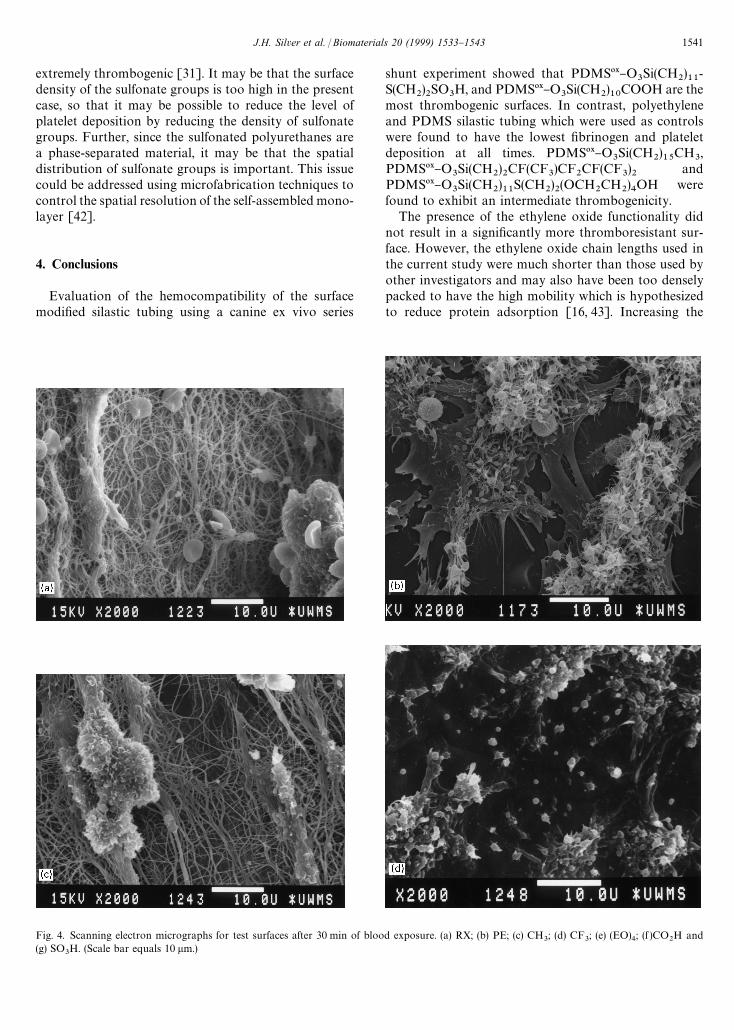
Fig. 4. Scanning electron micrographs for test surfaces after 30 min of blood exposure. (a) RX; (b) PE; (c) CH3; (d) CF
3; (e) (EO)
4; (f )CO
2H and
(g) SO3H. (Scale bar equals 10 lm.)
extremely thrombogenic [31]. It may be that the surfacedensity of the sulfonate groups is too high in the presentcase, so that it may be possible to reduce the level ofplatelet deposition by reducing the density of sulfonategroups. Further, since the sulfonated polyurethanes area phase-separated material, it may be that the spatialdistribution of sulfonate groups is important. This issuecould be addressed using microfabrication techniques tocontrol the spatial resolution of the self-assembled mono-layer [42].
4. Conclusions
Evaluation of the hemocompatibility of the surfacemodi"ed silastic tubing using a canine ex vivo series
shunt experiment showed that PDMS09}O3Si(CH
2)11
-S(CH
2)2SO
3H, and PDMS09}O
3Si(CH
2)10
COOH are themost thrombogenic surfaces. In contrast, polyethyleneand PDMS silastic tubing which were used as controlswere found to have the lowest "brinogen and plateletdeposition at all times. PDMS09}O
3Si(CH
2)15
CH3,
PDMS09}O3Si(CH
2)2CF(CF
3)CF
2CF(CF
3)2
andPDMS09}O
3Si(CH
2)11
S(CH2)2(OCH
2CH
2)4OH were
found to exhibit an intermediate thrombogenicity.The presence of the ethylene oxide functionality did
not result in a signi"cantly more thromboresistant sur-face. However, the ethylene oxide chain lengths used inthe current study were much shorter than those used byother investigators and may also have been too denselypacked to have the high mobility which is hypothesizedto reduce protein adsorption [16, 43]. Increasing the
J.H. Silver et al. / Biomaterials 20 (1999) 1533}1543 1541

Fig. 4. (continued).
ethylene oxide chain length by a single group did nota!ect the blood compatibility of these surfaces.
The }CH3
surface evaluated in this study, with analkyl chain length of 15 methylene units, was consider-ably more thrombogenic than a similar material witha shorter alkyl tail (9 methylene units) evaluated pre-viously [14]. While the present material showed a throm-boembolytic peak of approximately 2000 platelets per1000 lm2, the latter showed a maximum platelet depos-ition of only 300 platelets per 1000 lm2. Assuming thata greater degree of crystallinity has been introduced inthe present series, this would suggest that the more mo-bile, liquid-like surface created by the smaller alkyltrich-lorosilane, is more thromboresistant. The #uorinatedsurfaces showed the best behavior among the surfacemodi"ed elastomers. The control surfaces, PE and sili-cone rubber, were less thrombogenic than in the previouswork, so that animal-to-animal variation cannot accountfor some of the di!erences between the crystalline andliquid-like monolayers.
Acknowledgements
Technical assistance was provided by Larry Whitesell(animal surgery), Everett Clover (scanning electron spec-troscopy), and Arlene P. Hart (hematology). This workwas supported in part by the National Institutes ofHealth, through grants HL-24046 and HL-47179, andthe Dow Corning Corp., Midland, MI.
References
[1] Tyler BJ, Ratner BD, Castner DG, Briggs D. Variations betweenBiomer lots. I. Signi"cant di!erences in the surface chemistry oftwo lots of a commercial poly(etherurethane). J Biomed MaterRes 1992;26:273}89.
[2] Silver JH, Lewis KB, Ratner BD, Cooper SL. The e!ect of polyoltype on the surface structure of sulfonate-containing poly-urethanes. J Biomed Mater Res 1993;27:735}45.
[3] Dulsey CS, Georger JHJ, Krauthamer V, Stenger DA, Fare TL,Calvert JM. Deep UV photochemistry of chemisorbed
1542 J.H. Silver et al. / Biomaterials 20 (1999) 1533}1543
monolayers: patterned coplanar molecular assemblies. Science1991;252:551}4.
[4] Bain CD, Whitesides GM. Molecular-level control over surfaceorder in self-assembled monolayer "lms of thiols on gold. Science1988;240:62}3.
[5] Stenger DA, Georger JH, Dulcey CS, Hickman JJ, Rudolph AS,Nielsen TB, McCort SM, Calvert JM. Coplanar molecular assem-blies of amino- and per#uorinated alkylsilanes: characterizationand geometric de"nition of mammalian cell adhesion and growth.J Am Chem Soc 1992;114:8435}42.
[6] Chaudhury MK, Whitesides GM. Direct measurement of inter-facial interactions between semispherical lenses and #at sheets ofpoly(dimethylsiloxane) and their chemical derivatives. Langmuir1991;7:1343}63.
[7] Chaudhury MK, Whitesides GM. Correlation between surfacefree energy and surface constitution. Science 1992;255:1230}2.
[8] Chaudhury MK, Owen MJ. Correlation between adhesion hys-teresis and phase state of monolayer "lms. Phys Chem1993;97:5722}6.
[9] Ferguson GS, Chaudhury MK, Biebuyck HA, Whitesides GM.Monolayers on disordered substrates: self-assembly of alkyltrich-lorosilanes on surface-modi"ed polyethylene and poly(dimethyl-siloxane). Macromolecules 1993;26:5870}5.
[10] Collinson M, Bowden E, Tarlov MJ. Voltametry of covalentlyimmobilized cytochrome c on self-assembled monolayer elec-trodes. Langmuir 1992;8:1247}50.
[11] Lewandowska K, Pergament E, Sukenik CN, Culp LA. Cell-type-speci"c adhesion mechanisms mediated by "bronectin ad-sorbed to chemically derivatized substrata. J Biomed Mater Res1992;26:1343}63.
[12] Margel S, Vogler EA, Firment L, Watt T, Haynie S, Sogah DY.Peptide, protein, and cellular interactions with self-assembledmonolayer model surfaces. J Biomed Mater Res 1993;27:1463}76.
[13] Chaudhury MK. Surface free energies of alkylsiloxane mono-layers supported on elastomeric polydimethylsiloxanes. J AdhesSci Technol 1993;7:669}75.
[14] Silver JH, Hergenrother RW, Lin J-C, Lim F, Lin H-B, Okada T,Chaudhury MK, Cooper SL. Surface and blood-contacting prop-erties of alkylsiloxane monolayers supported on silicone rubber.J Biomed Mater Res 1995;29:535}48.
[15] Bonhert JL, Fowler BC, Horbett TA, Ho!man AS. Plasma gasdischarge deposited #uorocarbon polymers exhibited reducedelutability of adsorbed albumin and "brinogen. Biomater SciPolym Edn 1990;1:279.
[16] Kiaei D, Ho!man AS, Ratner BD, Horbett TA, Interaction ofblood with gas discharge treated vascular grafts. J Appl Poly Sci:Appl Polym Symp 1988;42:269}83.
[17] Yu X-H, Okkema AZ, Cooper SL. Synthesis and physical proper-ties of poly(#uoroalkyleether)-urethanes. J Appl Polym Sci 1990;1:1777}95.
[18] Brinkman E, Poot A, van der Does L, Bantjes A. Platelet depo-sition studies on copolyether-urethanes modi"ed with poly(ethy-lene oxide). Biomaterials 1990;11:200}5.
[19] Chaikof EL, Merrill EW, Callow AD, Connolly RJ, Verdon SL,Ramberg K, PEO enhancement of platelet deposition, "brinogendeposition and complement C3 activation. J Biomed Mater Res1992;26:1163}8.
[20] Prime KL, Whitesides GM. Self-assembled organic monolayers:model systems for studying adsorption of proteins at surfaces.Science 1991;252:1164}7.
[21] Ista LK, Fan H, Baca O, Lopez GP. Attachment of bacteria tomodel solid surfaces: oligo(ethylene glycol) surfaces inhibit bacter-ial attachment. FEMS Microbiol Lett 1996;142:59}63.
[22] Casu B. Structure of heparin and heparin fragments. Ann NYAcad Sci 1989;556:1}17.
[23] Jozefowitz M, Jozefonvicz J. Antithrombogenic polymers. PureAppl Chem 1984;56:1335}44.
[24] Mauzac M, Aubert N, Jozefonvicz J. Antithrombic activity ofsome polysaccharide resins. Biomaterials 1982;3:221}4.
[25] Ito Y, Iguchi Y, Kashiwagi T, Imanishi Y. Synthesis and non-thrombogenicity of polyetherurethaneurea "lm grafted withpoly(sodium vinyl sulfonate). J Biomed Mater Res 1991;25:1347}61.
[26] Okkema AZ, Visser SA, Cooper SL. Physical and blood-contact-ing properties of polyurethanes based on a sulfonic acid-contain-ing diol chain extender. J Biomed Mater Res 1991;25:1371}95.
[27] Silver JH, Hart AP, Williams EC, Cooper SL, Charef S, LabarreD, Jozefowicz M. Anticoagulant e!ects of sulfonated poly-urethanes. Biomaterials 1992;13:339}43.
[28] Okkema AZ, Yu X-H, Cooper SL. The physical and blood-con-tacting properties of propyl sulfonate grafted Biomer'. Bio-materials 1991;12:3}12.
[29] Han DK, Jeong SY, Kim YH, Min BG, Cho HI. Negative ciliaconcept for thromboresistance: synergistic e!ect of PEO andsulfonate groups grafted onto polyurethanes. J Biomed Mater Sci1991;25:561}75.
[30] Santerre JP, ten Hove P, VanderKamp NH, Brash JN. E!ect ofsulfonation of segmented polyurethanes on the transient adsorp-tion of "brinogen from plasma. Possible correlation with an-ticoagulant behavior. J Biomed Mater Sci 1992;26:39}57.
[31] Lelah MD, Lambrecht LK, Cooper SL. A canine ex vivo seriesshunt for evaluating thrombus deposition on polymer surfaces.J Biomed Mater Sci 1984;18:475}96.
[32] Wasserman SR. PhD Thesis. Harvard University, 1989.[33] Lelah D, Grasel TG, Pierce JA, Cooper SL. The measurement of
contact angles on circular tubing surfaces using the captivebubble technique. J Biomed Mater Sci 1985;19:1011}5.
[34] Park KD, Mosher DF, Cooper SL. Acute surface-induced throm-bosis in the canine ex vivo model: importance of protein composi-tion of the initial monolayer and platelet activation. J BiomedMater Res 1986;20:589}612.
[35] Zanker A. Detection of outliers by means of Nalimov's test. ChemEng 1984;91:74.
[36] Takahara A, Okkema AZ, Cooper SL, Coury SJ. E!ect of surfacehydrophilicity on ex vivo blood compatibility of segmented poly-urethanes. Biomaterials 1991;12:324}34.
[37] Grasel TG, Cooper SL. Surface properties and blood compatibil-ity of polyurethaneureas. Biomaterials 1986;7:315}28.
[38] Park KD, Okano T, Nojiri C, Kim SW. Heparin immobilizationon segmented polyurethaneurea surfaces*e!ect of hydrophilicspacers. J Biomed Mater Sci 1988;22:977}92.
[39] Brace LD, Fareed J. An objective assessment of the interaction ofheparin and its fractions with human platelets. Sem ThrombHemost 1985;11:190}8.
[40] Lai QJ, Albrecht RM, Cooper SL. E!ects of polyurethane surfacechemistry on "brinogen receptor expression on adhered plateletsunder static and #ow conditions. In: AIChE, 2nd Topical Conf onEmerging Technologies in Materials. San Fransisco, CA, 1989.
[41] Takahara A, Okkema AZ, Wabers H, Cooper SL. E!ect ofhydrophilic soft segment side chains on the surface properties andblood compatibility of segmented poly(urethaneureas). J BiomedMater Res 1991;25:1095}118.
[42] Bhatia SK, Teixeira JL, Anderson M, Shriver-Lake LC, CalvertJM, Georger J, Hickman JJ, Dulcey CS, Schoen PE, Ligler FS.Fabrication of surfaces resistant to protein adsorption and ap-plication to two dimensional protein patterning. Anal Biochem1993;208:197}205.
[43] Desai NP, Hubbell JA. Biological responses to polyethylene oxidemodi"ed polyethylene terephthalate surfaces. J Biomed MaterRes 1991;25:829}43.
J.H. Silver et al. / Biomaterials 20 (1999) 1533}1543 1543