Surface photosensitization of ZnO by ZnS to enhance the ...
13
Vol.:(0123456789) SN Applied Sciences (2021) 3:689 | https://doi.org/10.1007/s42452-021-04643-z Research Article Surface photosensitization of ZnO by ZnS to enhance the photodegradation efficiency for organic pollutants Sunaina 1,2 · Sapna Devi 1 · S. T. Nishanthi 1,4 · S. K. Mehta 2 · A. K. Ganguli 1,3 · Menaka Jha 1 Received: 20 July 2020 / Accepted: 5 May 2021 © The Author(s) 2021 OPEN Abstract It is challenging to develop a material which has low cost, high activity, good stability and recyclability under light exposure. Apart from these properties, the photocatalyst should also have good visible region absorbance and low electron-hole pair recombination rate. Keeping all this in view, we have designed a simple scalable synthesis of ZnO–ZnS heterostructures for the photocatalytic treatment of industrial waste (p-nitrophenol and methyl orange). The ZnO–ZnS heterostructures are synthesized via a solvent-free route by thermal annealing of solid-state mixture of ZnO and thiourea (a sulphur source) which results in ZnO–ZnS core shell kind of heterostructure formation. The interface formation between the ZnO–ZnS heterostructure favored the band-gap reduction in comparison to the bare ZnO and ZnS nanoparticles. Further, these ZnO–ZnS heterostructures were utilized as a photocatalyst for the degradation of toxic phenolic molecules (p-nitrophenol) and harmful organic dyes (methyl orange) present in the water under the light exposure (> 390 nm). Keywords ZnO–ZnS heterostructure · Photocatalysis mechanism · Methyl orange · P-nitrophenol · Environmental chemistry 1 Introduction Designing the metal oxide based photocatalyst for envi- ronmental pollution control has gained a lot of research interest worldwide [1–3]. Between all the various metal oxides, TiO 2 , ZnO, SnO 2 based photocatalysts are some of the most studied systems [4–6]. ZnO is one of the best candidates which has great response as a photocatalyst. However, the commercial usage of ZnO is limited because low quantum efficiency and instability in wide pH range due to photo-corrosion. This limitation has led to research oriented towards improving the ZnO properties by form- ing heterostructure or doping with other materials [7–9]. In the case of heterostructures, core-shell structures have an advantage where the shell can be a physical barrier between the optically active core and the surrounding medium. It can also modify the charge, stability, func- tionality, reactivity, and dispersive ability of core material [10–14]. Core shell heterostructures also have better elec- trical, catalytic, optical, and magnetic properties in com- parison to their bare counter parts [15–17]. Generally, the core-shell heterostructures have intermediate properties between the core and shell materials [18–20]. Khanchan- dani et al. have synthesized ZnO–CdS, ZnO–Ag 2 S and ZnO–In 2 S 3 core-shell heterostructures. They have func- tionalized the ZnO surface by using the citric acid and Supplementary Information The online version contains supplementary material available at https://doi.org/10.1007/s42452-021- 04643-z. * A. K. Ganguli, [email protected]; * Menaka Jha, [email protected] | 1 Institute of Nano Science and Technology, Mohali 140306, India. 2 Department of Chemistry, Panjab University, Chandigarh 160014, India. 3 Department of Chemistry, Indian Institute of Technology, New Delhi 110016, India. 4 ECPS Division, CSIR-Central Electrochemical Research Institute, Karaikudi, Tamil Nadu 630003, India.
Transcript of Surface photosensitization of ZnO by ZnS to enhance the ...

Vol.:(0123456789)
SN Applied Sciences (2021) 3:689 | https://doi.org/10.1007/s42452-021-04643-z
Research Article
Surface photosensitization of ZnO by ZnS to enhance the photodegradation efficiency for organic pollutants
Sunaina1,2 · Sapna Devi1 · S. T. Nishanthi1,4 · S. K. Mehta2 · A. K. Ganguli1,3 · Menaka Jha1
Received: 20 July 2020 / Accepted: 5 May 2021
© The Author(s) 2021 OPEN
AbstractIt is challenging to develop a material which has low cost, high activity, good stability and recyclability under light exposure. Apart from these properties, the photocatalyst should also have good visible region absorbance and low electron-hole pair recombination rate. Keeping all this in view, we have designed a simple scalable synthesis of ZnO–ZnS heterostructures for the photocatalytic treatment of industrial waste (p-nitrophenol and methyl orange). The ZnO–ZnS heterostructures are synthesized via a solvent-free route by thermal annealing of solid-state mixture of ZnO and thiourea (a sulphur source) which results in ZnO–ZnS core shell kind of heterostructure formation. The interface formation between the ZnO–ZnS heterostructure favored the band-gap reduction in comparison to the bare ZnO and ZnS nanoparticles. Further, these ZnO–ZnS heterostructures were utilized as a photocatalyst for the degradation of toxic phenolic molecules (p-nitrophenol) and harmful organic dyes (methyl orange) present in the water under the light exposure (> 390 nm).
Keywords ZnO–ZnS heterostructure · Photocatalysis mechanism · Methyl orange · P-nitrophenol · Environmental chemistry
1 Introduction
Designing the metal oxide based photocatalyst for envi-ronmental pollution control has gained a lot of research interest worldwide [1–3]. Between all the various metal oxides, TiO2, ZnO, SnO2 based photocatalysts are some of the most studied systems [4–6]. ZnO is one of the best candidates which has great response as a photocatalyst. However, the commercial usage of ZnO is limited because low quantum efficiency and instability in wide pH range due to photo-corrosion. This limitation has led to research oriented towards improving the ZnO properties by form-ing heterostructure or doping with other materials [7–9].
In the case of heterostructures, core-shell structures have an advantage where the shell can be a physical barrier between the optically active core and the surrounding medium. It can also modify the charge, stability, func-tionality, reactivity, and dispersive ability of core material [10–14]. Core shell heterostructures also have better elec-trical, catalytic, optical, and magnetic properties in com-parison to their bare counter parts [15–17]. Generally, the core-shell heterostructures have intermediate properties between the core and shell materials [18–20]. Khanchan-dani et al. have synthesized ZnO–CdS, ZnO–Ag2S and ZnO–In2S3 core-shell heterostructures. They have func-tionalized the ZnO surface by using the citric acid and
Supplementary Information The online version contains supplementary material available at https:// doi. org/ 10. 1007/ s42452- 021- 04643-z.
* A. K. Ganguli, [email protected]; * Menaka Jha, [email protected] | 1Institute of Nano Science and Technology, Mohali 140306, India. 2Department of Chemistry, Panjab University, Chandigarh 160014, India. 3Department of Chemistry, Indian Institute of Technology, New Delhi 110016, India. 4ECPS Division, CSIR-Central Electrochemical Research Institute, Karaikudi, Tamil Nadu 630003, India.

Vol:.(1234567890)
Research Article SN Applied Sciences (2021) 3:689 | https://doi.org/10.1007/s42452-021-04643-z
then formed CdS or Ag2S or In2S3 shell over a ZnO core by chemical route. These core/shell nanostructures show superior photocatalytic behavior for the degradation of dyes under light illumination [21–23]. Subash et al. pre-pared the Ag2S–ZnO composite via a two-step process. They first precipitated the zinc oxalate and Ag2S and then calcined the mixture at 400 °C for 12 h to obtain Ag2S–ZnO composite. The photodegradation of Acid Black 1 was per-formed by using Ag2S–ZnO composite under solar light. The composite was more capable for mineralizing the Acid Black 1 in comparison to synthesized ZnO, bulk ZnO, TiO2–P25 and TiO2 (Merck) [24]. Shi et al. have synthesized the ZnO–PbS/GO photocatalyst for hydrogen evolution from water. The multiple exciton formation in ZnO–PbS/GO is responsible for the remarkable enhanced photoac-tivity of H2 evolution reaction [25]. The above researches show that the formation of core-shell heterostructures by incorporating the metal sulfide shell on to the ZnO core enhances its absorption in the visible range which further improves the photocatalytic reaction efficiency by increasing the stability of electron-hole pairs. Zinc sul-phide (ZnS) is a wide band gap (3.7 eV) semiconductor material which is nontoxic and water insoluble [26, 27]. It can be used as a coating material for ZnO nanorods because it has shown its application in electrolumines-cent devices, sensors and lasers [28–31]. ZnO–ZnS based heterostructures have shown improved physicochemical properties in different applications [32–36]. Hence, a con-siderable efforts have been made to synthesize ZnO–ZnS based heterostructures [37–42]. Lin et al. have made an efficient ZnO–ZnS photocatalyst using template-assisted method. The photo-activity of ZnO–ZnS photocatalyst was analyzed by using degradation of rhodamine B. The surface coupling between the two semiconductors pro-poses their higher photon absorption efficiency and an enhanced exciton separation, which lead to a significant improvement in the photocatalytic efficacy in compari-son with bare ZnO [43]. Lonkar et al. have prepared the ZnS–ZnO/graphene based nano-photocatalysts. The ZnO and ZnS nanoparticles are uniformly distributed over the graphene matrix in the synthesized nanohybrids. The syn-ergic effect in ZnS–ZnO/graphene decreased the band gap in the composite system. They checked the photocatalytic efficiency of ZnS–ZnO/graphene nano–photocatalysts by a test photo-reaction with organic azo dyes and toxic phenol molecules from waste water which have harm-ful impact on the environment [44]. Based on the above literature survey, it appears that the heterojunction for-mation between the ZnO and ZnS will generate superior hybrid material having lower photoexcitation energy than the bare counter parts [32–44]. The reason behind this is ZnO–ZnS heterostructures induces a built-in electric field which improve the interfacial charge transfer and deliver
remarkable photoactivity [32–44]. Theoretically, type II band alignment formed between ZnO and ZnS results in the better photocatalytic activity in the heterostructure. Therefore, ZnO–ZnS heterostructure represents a fascinat-ing candidate for photocatalysis. Here, we have attempted to a simplistic one-pot bulk approach for synthesize of ZnO–ZnS core-shell heterostructures through solid state solvent free route. Earlier, the typical methods which are used for the synthesis of ZnO–ZnS heterostructures con-sist of solvothermal route, sol–gel process, microwave route, ion replacement etc. The large solvent usage and the complex synthetic procedure limited their usage for large scale productions. Here, we did the sulphurization of ZnO nanorods by utilizing the thiourea as sulphur source. The amount of ZnO and ZnS in the heterostructures can be controlled by varying the amount of thiourea used in the reaction. These fabricated heterostructures were thoroughly characterized using PXRD, TEM, EDX, UV-Vis spectroscopy, PL, EPR, EIS and XPS studies. Further, the as synthesized ZnO–ZnS heterostructures were utilized for removal of industrial waste water containing the organic pollutants (p-nitrophenol and methyl orange).
2 Materials and methods
2.1 Material used
For the preparation of ZnO, ZnS and ZnO–ZnS hetero-structures, zinc acetate dihydrate (Sigma-Aldrich, purity 98%) and thiourea (Merck, purity 99%) were utilized. p-nitrophenol (PNP, Fluka, > 98%) and methyl orange (MO, Sigma-Aldrich, 98%) were used as representative chemicals for industrial waste effluent treatment. Benzo-quinone (Sigma-Aldrich, purity 98%), Ammonium oxalate (Merck, purity 99%) and isopropanol (Merck, purity 99%) were used as trapping agents to identify the active spe-cies responsible for the photocatalytic activity of ZnO–ZnS heterostructures.
2.2 Synthesis procedure
ZnO nanorods were synthesized by solid state route. Briefly, zinc acetate was thermally decomposed in a muf-fle furnace for 12 h at 300 °C with a heating rate of 40 °C/h. Pure ZnS was synthesized by putting equimolar zinc ace-tate and thiourea in furnace under same condition. The ZnO–ZnS heterostructures were synthesized by taking different molar ratio of ZnO nanorods and thiourea in a mortar pestle and grounded to fine powder (Table 1). Then the resulting solid mixture was calcined in a muffle furnace under similar condition. The obtained product was washed with water and ethanol to remove unreacted thiourea.
Vol.:(0123456789)
SN Applied Sciences (2021) 3:689 | https://doi.org/10.1007/s42452-021-04643-z Research Article
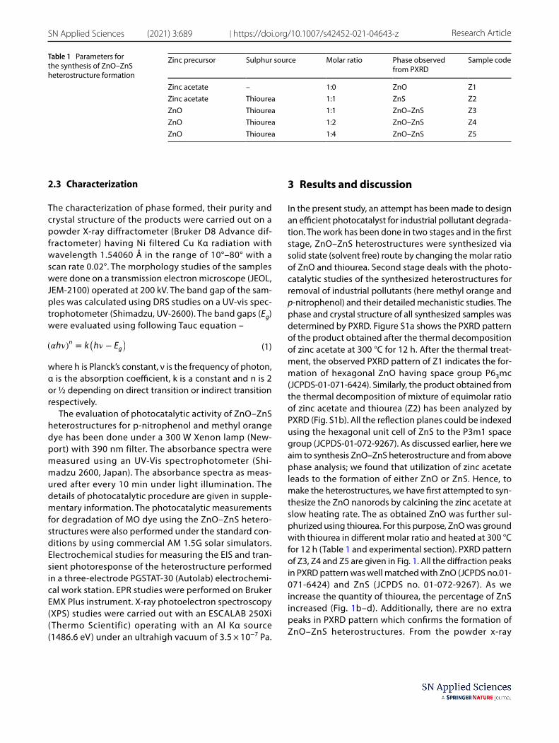
2.3 Characterization
The characterization of phase formed, their purity and crystal structure of the products were carried out on a powder X-ray diffractometer (Bruker D8 Advance dif-fractometer) having Ni filtered Cu Kα radiation with wavelength 1.54060 Å in the range of 10°–80° with a scan rate 0.02°. The morphology studies of the samples were done on a transmission electron microscope (JEOL, JEM-2100) operated at 200 kV. The band gap of the sam-ples was calculated using DRS studies on a UV-vis spec-trophotometer (Shimadzu, UV-2600). The band gaps (Eg) were evaluated using following Tauc equation –
where h is Planck’s constant, ν is the frequency of photon, α is the absorption coefficient, k is a constant and n is 2 or ½ depending on direct transition or indirect transition respectively.
The evaluation of photocatalytic activity of ZnO–ZnS heterostructures for p-nitrophenol and methyl orange dye has been done under a 300 W Xenon lamp (New-port) with 390 nm filter. The absorbance spectra were measured using an UV-Vis spectrophotometer (Shi-madzu 2600, Japan). The absorbance spectra as meas-ured after every 10 min under light illumination. The details of photocatalytic procedure are given in supple-mentary information. The photocatalytic measurements for degradation of MO dye using the ZnO–ZnS hetero-structures were also performed under the standard con-ditions by using commercial AM 1.5G solar simulators. Electrochemical studies for measuring the EIS and tran-sient photoresponse of the heterostructure performed in a three-electrode PGSTAT-30 (Autolab) electrochemi-cal work station. EPR studies were performed on Bruker EMX Plus instrument. X-ray photoelectron spectroscopy (XPS) studies were carried out with an ESCALAB 250Xi (Thermo Scientific) operating with an Al Kα source (1486.6 eV) under an ultrahigh vacuum of 3.5 × 10⁻⁷ Pa.
(1)(�h�)n = k
(
h� − Eg)
3 Results and discussion
In the present study, an attempt has been made to design an efficient photocatalyst for industrial pollutant degrada-tion. The work has been done in two stages and in the first stage, ZnO–ZnS heterostructures were synthesized via solid state (solvent free) route by changing the molar ratio of ZnO and thiourea. Second stage deals with the photo-catalytic studies of the synthesized heterostructures for removal of industrial pollutants (here methyl orange and p-nitrophenol) and their detailed mechanistic studies. The phase and crystal structure of all synthesized samples was determined by PXRD. Figure S1a shows the PXRD pattern of the product obtained after the thermal decomposition of zinc acetate at 300 °C for 12 h. After the thermal treat-ment, the observed PXRD pattern of Z1 indicates the for-mation of hexagonal ZnO having space group P63mc (JCPDS-01-071-6424). Similarly, the product obtained from the thermal decomposition of mixture of equimolar ratio of zinc acetate and thiourea (Z2) has been analyzed by PXRD (Fig. S1b). All the reflection planes could be indexed using the hexagonal unit cell of ZnS to the P3m1 space group (JCPDS-01-072-9267). As discussed earlier, here we aim to synthesis ZnO–ZnS heterostructure and from above phase analysis; we found that utilization of zinc acetate leads to the formation of either ZnO or ZnS. Hence, to make the heterostructures, we have first attempted to syn-thesize the ZnO nanorods by calcining the zinc acetate at slow heating rate. The as obtained ZnO was further sul-phurized using thiourea. For this purpose, ZnO was ground with thiourea in different molar ratio and heated at 300 °C for 12 h (Table 1 and experimental section). PXRD pattern of Z3, Z4 and Z5 are given in Fig. 1. All the diffraction peaks in PXRD pattern was well matched with ZnO (JCPDS no.01-071-6424) and ZnS (JCPDS no. 01-072-9267). As we increase the quantity of thiourea, the percentage of ZnS increased (Fig. 1b–d). Additionally, there are no extra peaks in PXRD pattern which confirms the formation of ZnO–ZnS heterostructures. From the powder x-ray
Table 1 Parameters for the synthesis of ZnO–ZnS heterostructure formation
Zinc precursor Sulphur source Molar ratio Phase observed from PXRD
Sample code
Zinc acetate – 1:0 ZnO Z1Zinc acetate Thiourea 1:1 ZnS Z2ZnO Thiourea 1:1 ZnO–ZnS Z3ZnO Thiourea 1:2 ZnO–ZnS Z4ZnO Thiourea 1:4 ZnO–ZnS Z5

Vol:.(1234567890)
Research Article SN Applied Sciences (2021) 3:689 | https://doi.org/10.1007/s42452-021-04643-z
diffraction study, it appears that when ZnO were thermally treated with thiourea, there is partial conversion of ZnO into ZnS by interaction of zinc oxide surface with thiourea at 300 °C [45]. The size and morphological characteristics of synthesized samples were analyzed under transmission electron microscopy (TEM). Figure 2 displays the TEM micrographs of the as-synthesized Z1, Z2, Z3, Z4 and Z5. In the case of Z1 (bare ZnO), the nanorods of size 25 nm × 390 nm with an aspect ratio of 14 were observed. As discussed earlier, zinc acetate and thiourea were heated
together and ZnS was formed. TEM micrograph indicates the formation of 10–15 nm particles. To make the hetero-structures, ZnO nanorods were heated with thiourea. These heterostructures have core-shell type of structure with inner core of ZnO and ZnS shell as shown in Fig. 2c,e. The TEM micrographs of the ZnO–ZnS heterostructures show the formation of nanorods of size 65 nm × 375 nm for Z3 with shell thickness 15 nm, 75 nm × 335 nm with shell thickness 20 nm, 90 nm × 300 nm for Z5 with shell thickness 23 nm. In Fig. 2, TEM micrographs of the ZnO–ZnS heterostructure shows that ZnS nanoparticles are attached on to the surface of ZnO nanorods uniformly which enables effective interfacial electron transfer, thus, an increased photocatalytic activity is expected in hetero-structures. The results of line EDX mapping (Fig. 3) show that there is co-existence of Zn, O and S elements in ZnO–ZnS core/shell heterostructures. Fig 3 demonstrates the results of TEM line-EDX mapping acquired by position-ing the electron beam across the ZnO–ZnS nanorods. Posi-tioning the electron beam across the ZnO–ZnS hetero-structure resulted in significantly intense homogeneous signals of Zn and S in the shell and weaker signals of O beam across the ZnO–ZnS nanorods respectively. This analysis confirms the composition of ZnO–ZnS hetero-structures have ZnO as core and ZnS as the shell material. Thus, the EDX analysis, gives the clear evidence of the for-mation of a core/shell heterostructure [46]. The insights of surface properties and components in the synthesized samples were obtained by XPS studies. The plots for all the samples are shown in Fig. 4 and S2. The surface O 1 s peak
Fig. 1 Powder X-ray diffraction patterns of a standard peaks for ZnO and ZnS, b Z3,c Z4 and d Z5
Fig. 2 TEM micrographs for the a Z1, b Z2, c Z3, d Z4 and e Z5

Vol.:(0123456789)
SN Applied Sciences (2021) 3:689 | https://doi.org/10.1007/s42452-021-04643-z Research Article
Fig. 3 STEM micrographs and their corresponding line mapping a, d Z3, b, e Z4 and c, f Z5
Fig. 4 a-c Fully scanned XPS spectra, high-resolution XPS spectrum of d-f Zn 2p, g-i O 1 s and j-l S 2p of Z3, Z4 and Z5 heterostructures respectively

Vol:.(1234567890)
Research Article SN Applied Sciences (2021) 3:689 | https://doi.org/10.1007/s42452-021-04643-z
contained contributions from different chemical species in relative to the processing conditions. The Zn2p3/2 and Zn2p1/2 peaks at ~ 1020 and ~ 1043 eV, respectively, are shown in Fig. 4d-f. Besides the presence of lattice oxygen located at 530.0 eV which is an indication of ZnO forma-tion, a second band centered at ~ 532 eV is assigned to the chemisorbed oxygen caused by surface hydroxyl groups. In the S2p XPS spectra (Fig. 4g–i), the S2p3/2 and S2p1/2 peaks centered at ~ 161 and ~ 163 eV, respectively, are related to Zn−S bonding, indicating that ZnS has been suc-cessfully synthesized. Compared with the pure ZnO, O 1 s peak at around 531.9 eV was detected and shifted after the deposition of ZnS nanoparticles, and the peak area at 530.0 eV of lattice oxygen decreased sharply (Fig. 4 and S2), which shows that almost no ZnO crystal exists in the ZnS shell layer [47–49]. The optical band gap determined for ZnO, ZnS and ZnO–ZnS heterostructures by using UV/vis diffuse reflectance spectroscopy (DRS) and the values calculated for Z1, Z2, Z3, Z4 and Z5 were found to be 3.3, 3.6, 3.1, 3.1 and 3.1 eV, respectively (Fig. S3). This shift in the absorption energy can be ascribed to the sensitization of ZnS. When the ZnO nanorods are covered with the ZnS shell, there is Type II kind of heterojunction formation. In a Type II heterostructure, the bandgap effectively reduced in comparison to the core and shell material. Decrease in bandgap will make the heterostructure to absorb the lower energy light to generate the charge carriers and also favors the fast charge transport across the heterojunction. This observation shows that ZnO–ZnS heterostructures can show better photocatalytic activity. BET adsorp-tion–desorption isotherms of ZnO, ZnS and ZnS–ZnO het-erostructures are shown in Fig. S3a. BET surface area of the ZnS–ZnO heterostructures are larger than those of bare ZnO, which indicates that the resulting heterostructures can provide a larger interface to enhance the contact between the dye and the active catalyst and facilitate pho-tocatalytic reaction. The BJH pore size distribution curves of all the samples are shown in Fig. S3b. The mesoporosity in the resulting heterostructures was further confirmed from the pore size distribution peaks that are centered on a 2 nm and a broad peak around 6–8 nm. The measured BET surface areas and average pore radius for ZnO, ZnS and ZnS–ZnO heterostructures are presented in Table S1. After heterojunction formation, the surface areas tend to increase due to surface decoration of ZnO nanorods with ZnS nanoparticles, this further evidence the in-situ forma-tion of nanostructured heterostructures [50]. Photocata-lytic behavior of the synthesized ZnO, ZnS and ZnO–ZnS heterostructures is evaluated in comparison to bare ZnO and ZnS through photo-degradation studies of p-nitro-phenol (PNP) and Methyl Orange (MO) dye under light exposure (> 390 nm). Before illuminating the light over catalyst containing PNP or MO solution, the solution is
kept in dark to acquire the adsorption–desorption equi-librium. The UV-vis absorption spectra for the reduction of PNP in aqueous medium using NaBH4 with addition of the catalyst (1 mg/mL) are shown in Fig. 5. The pure PNP shows a λmax at 317 nm in aqueous medium and when the NaBH4 is added to it, a red shift occurs from 317 to 403 nm due to the formation of p-nitrophenolate ions. The yellow color of the PNP solution thus disappeared as the photocatalytic reaction proceeds. In the case of bare Z1 or Z2, there was insignificant photodegradation of PNP (Fig. 5a, b), whereas, in the case of Z3, Z4 and Z5 heterostructures, there is an increased photodegradation of PNP under light illumina-tion (Fig. 5c, d). In case of Z3, Z4 and Z5, there is appear-ance of a new peak ~ 297 nm which corresponds to forma-tion of p-aminophenol [51]. Figure 5f shows that in the absence of any photocatalyst, there is no degradation of PNP under light irradiation which indicates the photocata-lytic nature of ZnO–ZnS heterostructures. The comparison and the kinetics of photodegradation of PNP in the pres-ence of heterostructures are shown in Fig. 6 and Table 2. The reaction rates of Z1, Z2, Z3, Z4 and Z5 were 0.012, 0.002, 0.037, 0.030 and 0.024 min−1 respectively. The Z3 sample is showing the best results for PNP photodegrada-tion with an efficiency of ~ 90%. Further, the photodegra-dation of MO dye has been done and mechanistic studies have been explored. Fig 7 shows a typical UV-vis spectrum of MO by Z1, Z2, Z3, Z4 and Z5 for 60 min. Methyl orangedye has an azo group which acts as chromophore-which gives its characteristic absorptionmaximum at 464 nm. The illumination of light over MO in the presence of photocatalyst causes the breakage of this azo bond hence the intensity is decreased at 464 nm with time. Fig 8 depicts the comparison of photo-catalytic activities of ZnO–ZnS heterostructures with respect to bare ZnO and ZnS under light exposure. The efficiency of bare ZnO or ZnS is comparatively lower than those of heterostructures (Fig. 8a). Z3 is showing the maximum efficiency ~ 97% for MO degradation. The rate kinetics for the photodegrada-tion of MO by all the samples follows the pseudo first order reaction (Fig. 8b and Table 3). The reaction rates were for pure Z1, Z2, Z3, Z4 and Z5 are 0.006, 0.001, 0.049, 0.039 and 0.014 min−1, respectively. Fig 8c shows the reusability of Z3 for photocatalytic degradation of MO up to three cycles. The decrease in the photocatalytic efficiency of Z3 after the 3rd cycle is from 97 to 89%. Table 4 shows the comparison of the prepared ZnO–ZnS photocatalysts with the literature. The photocatalytic measurements for deg-radation of MO dye using the ZnO–ZnS heterostructures were also performed under the standard conditions by using commercial AM 1.5G solar simulators (Fig. S5). The results obtained using commercial AM 1.5G solar simula-tors were corroborated with the results obtained from Xenon lamp experiments (Fig. S6 and Table S2). The

Vol.:(0123456789)
SN Applied Sciences (2021) 3:689 | https://doi.org/10.1007/s42452-021-04643-z Research Article
mechanism of photocatalysis in ZnO–ZnS heterostructures can be explained on the basis of band structures of the samples (Fig. 9). The type II kind of photocatalytic hetero-structure is formed and the formation of ZnS shell has resulted in a red shift in the absorption spectra of ZnO core. This results an effective smaller band gap when com-pared to core and shell materials. Under light exposure, the valence band (VB) electrons get excited to conduction band (CB) in both ZnO and ZnS. The excited electrons
Fig. 5 Photo-degradation study of p-nitrophenol under light illumination containing photocatalyst a Z1, b Z2, c Z3, d Z4, e Z5 and f no cata-lyst
Fig. 6 a Comparison and b kinetics of Photo-degradation of p–nitrophenol under light illumination containing differ-ent photocatalyst
Table 2 Comparison of photodegradation efficiency of the synthe-sized photocatalysts for p-nitrophenol
Sample code Efficiency (%) Rate constant (min−1)
R
Z1 55.7 0.012 0.96Z2 11.0 0.002 0.97Z3 90.9 0.037 0.99Z4 84.8 0.030 0.99Z5 72.0 0.024 0.98

Vol:.(1234567890)
Research Article SN Applied Sciences (2021) 3:689 | https://doi.org/10.1007/s42452-021-04643-z
move from CB (ZnS) to CB (ZnO) and generated holes from VB (ZnO) to VB (ZnS). This transfer process increases the
separation between the electrons and holes which slow-down the recombination of electron-hole pair in the het-erostructure. The reactive oxygen species are generated due to the reaction of photo-generated electron and holes in the semiconductor with surface adsorbed water mole-cules. The electrons in CB (ZnO) will combine with adsorbed O2 and produce •O2
− species. The holes accumu-lated in VB (ZnS) react with adsorbed H2O molecules and generate OH• species in the solution. These OH• and •O2
− species are very oxidative and reductive respectively and consequently convert the MO molecules into smaller degraded molecules, H2O and CO2. The reactions in the process involved are given below [2] -
Fig. 7 Photo-degradation study of methyl orange dye under light illumination containing photocatalyst a Z1, b Z2, c Z3, d Z4 and e Z5
Fig. 8 a Comparison, b kinetics of Photo-degradation of methyl orange under light illumination containing different photocata-lyst and c recyclability test of Z3 for the degradation of MO up to 3 cycles
Table 3 Comparison of photodegradation efficiency of the synthe-sized photocatalysts for methyl orange
Sample code Efficiency (%) Rate constant (min−1)
R
Z1 37.1 0.006 0.98Z2 4.7 0.001 0.98Z3 96.3 0.049 0.96Z4 83.4 0.029 0.99Z5 59.2 0.014 0.99

Vol.:(0123456789)
SN Applied Sciences (2021) 3:689 | https://doi.org/10.1007/s42452-021-04643-z Research Article
(2)ZnO − ZnS + hv → ZnO − ZnS(
h+ + e−)
(3)H2O + h+ → OH∙
(4)O−2+ e− →
∙O−2
(5)∙O2− ∕h + ∕OH∙ +MO → CO
2+ H
2O
Therefore, the heterostructure shows higher photo-degradation efficiency than that of pure ZnO and ZnS [35, 36]. The light illumination over the catalyst will gen-erate electrons (e−) and holes (h+). Further, these photo-excited electrons and holes will combine with surface adsorbed H2O and O2 and generate superoxide radical anions (•O2
−) and hydroxyl radicals (OH•). These in-situ generated four active species (e−, h+, OH• and •O2
−) are responsible for the oxidation of organic dye pollutants. For identification of the principal active species during the degradation of MO with ZnO–ZnS as photocatalyst,
Table 4 Comparison of ZnO–ZnS core shell heterostructures with literature
S. No. System and structure Light source Photocatalytic efficiency for MO
Photocatalytic efficiency for PNP
Ref
1 ZnO@ZnS hollow dumbbells–graphene composites
Mercury lamp Efficiency =94% in 40 minRate constant =0.082 min-1,
– [28]
2 Spherical ZnO/ZnS core/shell particles
UV radiation Efficiency =95% in 2 h, – [31]
3 ZnO/ZnS sheets Sunlight Efficiency =~83% in 4 h – [38]4 ZnO/ZnS nanorod array 300 W Hg lamp Efficiency=~96% in 660 min – [40]5 ZnO@ZnS/C nanofibres UV (365 nm) Efficiency =92.2 % in 75 min
Rate constant =0.0358 min-1– [41]
6 ZnO–ZnS nanowire UV-light (λ > 324 nm). Efficiency= 96% in 40 min Rate constant= 0.072 min-1
– [57]
7 ZnS-ZnO/Graphene visible light (400 W) Efficiency =97.5% for 160 minutes
Rate constant =0.011 min-1
Efficiency=96.3% in 140 min [44]
8 ZnO/ZnS nanosphere UV light Efficiency=70% in 2 h – [58]9 Hollow ZnO core/ZnS shell
structureUV lamps Efficiency=93.7% within 60
min– [59]
10 ZnO-ZnS core shell structures Visible light (>400 nm) Efficiency =96.3% in 60 minRate constant=0.049 min-1
Efficiency =90. 9% in 60 minRate constant=0.037 min-1
This work
Fig. 9 Schematics for the electron and hole transfer in the ZnO–ZnS heterostructures
Fig. 10 The scavenger studies for the detection of active species for the photodegradation of methyl orange in the presence of Z3

Vol:.(1234567890)
Research Article SN Applied Sciences (2021) 3:689 | https://doi.org/10.1007/s42452-021-04643-z
different trapping agents were added to reaction solu-tion i.e. ammonium oxalate for holes (h+), isopropanol for hydroxyl radicals (OH•) and benzoquinone for super-oxide radical anions (•O2
−) before the addition of photo-catalyst (Fig. 10). In the absence of any added trapping agent, the photocatalyst degraded MO up to ~ 97.1% in 60 min. When the benzoquinone is added to the dye solution, the maximum quenching was observed (~ 28.7%) in photocatalysis. In case of addition of iso-propanol (~ 84.3%) and ammonium oxalate (~ 95.2%), there is not much reduction in photocatalysis rate was observed. From these results, it appears that superoxide radical anions generated in solution are mainly responsi-ble for the degradation of the dyes [52]. Photolumines-cence (PL) technique is used for monitoring the recombi-nation of electron hole pair within a semiconductor. It is
known that when the rate of recombination of electron- hole is slow down, then, there is a reduction in PL peak intensity. The PL spectra of ZnO–ZnS heterostructures in comparison to bare ZnO excited at a wavelength of 350 nm are shown in Fig. 11. The emission peak for bare ZnO nanorods is obtained at ~ 390 nm. This UV emission arises from the recombination of the free electron-hole pairs via collision of exciton-exciton within the semicon-ductor [53]. The delay in the recombination of electron and hole due to their separation at the heterojunc-tion of ZnO–ZnS heterostructures result the suppres-sion in the emission signal [54]. PL also shows that Z3 has lowest intensity which indicates that it has slower recombination rate for electron and holes therefore, they can be available for photocatalytic reaction, and hence, it is showing highest catalytic activity. In order to compare the dark and light illuminated behavior of heterostructures with its bare counterparts, transient current response was measured. The typical photocur-rent density (J) vs. time plot is shown in Fig. 12a. There was negligible photo-response observed in the bare ZnS whereas photocurrent was degrading with time in case of ZnO due to fast recombination rate of excitons. In the case of ZnO–ZnS heterostructures, there was a signifi-cant enhancement in the current on light illumination which again drops gradually after the removal of light source. The EIS studies were carried out to acquire the insights of the enhanced photocatalytic properties in case of Z3 heterostructure. The EIS studies were done in the absence and presence of light. The Nyquist plot of (Fig. 12b) showed a smaller arc radius in the light illumi-nation than dark conditions. This confirmed that there is a low transfer barrier of electron and faster interfa-cial charge transfer occurs on heterostructure interface, hence, more efficient charge separation happens when
Fig. 11 Photoluminescence studies of ZnO–ZnS heterostructures in comparison to ZnO
Fig. 12 a Transient photoresponse of ZnO–ZnS heterostructures and b EIS studies for Z3 in presence and absence of light

Vol.:(0123456789)
SN Applied Sciences (2021) 3:689 | https://doi.org/10.1007/s42452-021-04643-z Research Article
it is illuminated under light compared to dark condi-tions. This observation is in closed agreement with ear-lier reports wherein SnO2/SnS2 heterostructure showed higher current density when light is switched on and it drops again when the light is off [55]. The micro-struc-tural characterization of ZnO and its heterostructures have been also done by performing EPR spectroscopy (Fig. S7). This technique is a useful tool to monitor the presence of radical species and the behavior of the pre-sent native defects, such as oxygen and/or zinc vacan-cies. EPR spectra of the bare ZnO showed two signals at g = 1.9580 and g = 2.0036. The first peak is usually attrib-uted to the shallow donor caused by the surface oxygen vacancies and interstitial Zn atoms. At the same time, the signal at g = 2.0024 is attributed to the presence of Zn vacancies. These results evidence the presence of a high concentration of defects in the ZnO nanorods. Also, a sharp decrease in the intensity of peak at g = 1.9580 has been observed for the ZnO–ZnS heterostructures with respect to the bare ZnO. This clearly indicates that the relative lower concentration of oxygen vacancies in het-erostructures, confirming that Sulphur is occupying the oxygen vacancies [56]. The synthesis of ZnO–ZnS core shell heterostructures with optimized composition can be utilized as an effective photocatalyst for the removal of toxic pollutant from the industrial aqueous waste.
4 Conclusions
ZnO–ZnS heterostructures were successfully synthesized by solid state solvent free route. Sulphurization of ZnO nanorods has been done by utilizing the thiourea as sul-phur source. The ratio of ZnO and ZnS can be controlled by varying the amount of sulphur source. Further, the as synthesized ZnO–ZnS heterostructure were utilized for removal of industrial waste (p-nitrophenol and methyl orange) from contaminated water. The Z3 has shown the maximum efficiency for PNP (~ 90%) and MO (~ 97.1%) under the light exposure. It is observed that Z3 consists of optimum amount of ZnO and ZnS in the ZnO–ZnS core–shell heterostructure. The heterostructure formation led to the effective separation of the electron and hole which in result decrease the recombination time, hence, these electrons and holes are available for photocata-lytic activity. To confirm this, PL studies of ZnO–ZnS het-erostructures has been done. We observed that Z3 shows maximum decrease in the PL intensity in comparison to bare ZnO. The details of photocatalytic activity have been explored by trapping agent studies. Thus, our studies show that ZnO–ZnS are the potential material with non-toxic nature which can work as an efficient photocatalyst for
the waste water treatment to address the environmental pollution issues.
Acknowledgements Sunaina thanks the CSIR, India for the fellowship. SD thanks INST, Mohali for the fellowship. AKG, SKM and MJ thank the Department of Science and Technology, India.
Declarations
Conflict of interest There is no conflict of interest.
Open Access This article is licensed under a Creative Commons Attri-bution 4.0 International License, which permits use, sharing, adap-tation, distribution and reproduction in any medium or format, as long as you give appropriate credit to the original author(s) and the source, provide a link to the Creative Commons licence, and indicate if changes were made. The images or other third party material in this article are included in the article’s Creative Commons licence, unless indicated otherwise in a credit line to the material. If material is not included in the article’s Creative Commons licence and your intended use is not permitted by statutory regulation or exceeds the permitted use, you will need to obtain permission directly from the copyright holder. To view a copy of this licence, visit http:// creat iveco mmons. org/ licen ses/ by/4. 0/.
References
1. Wang Q, Zhang Z, Liao Q et al (2018) Enhanced field emission properties of graphene-based cathodes fabricated by ultrasonic atomization spray. RSC Adv 8:16207–16213. https:// doi. org/ 10. 1039/ c8ra0 2154f
2. Malefane ME, Feleni U, Mafa PJ, Kuvarega AT (2020) Fabrication of direct Z-scheme Co3O4/BiOI for ibuprofen and trimetho-prim degradation under visible light irradiation. Appl Surf Sci 514:145940. https:// doi. org/ 10. 1016/j. apsusc. 2020. 145940
3. Li YS, Li TT, Song XF et al (2020) Enhanced adsorption-photocat-alytic reduction removal for Cr (VI) based on functionalized TiO2 with hydrophilic monomers by pre-radiation induced grafting-ring opening method. Appl Surf Sci 514:145789. https:// doi. org/ 10. 1016/j. apsusc. 2020. 145789
4. Bera A, Basak D (2010) Photoluminescence and photoconduc-tivity of ZnS-coated ZnO nanowires. ACS Appl Mater Interfaces 2:408–412. https:// doi. org/ 10. 1021/ am900 686c
5. Bathula B, Koutavarapu R, Shim J, Yoo K (2020) Facile one-pot synthesis of gold/tin oxide quantum dots for visible light cata-lytic degradation of methylene blue: Optimization of plasmonic effect. J Alloys Compd 812:152081. https:// doi. org/ 10. 1016/j. jallc om. 2019. 152081
6. Li M, Zhu Q, Li J, Kim B (2020) Applied surface science elongation of ZnWO4 nanocrystals for enhanced photocatalysis and the effects of Ag decoration. Appl surface sci 515:146011. https:// doi. org/ 10. 1016/j. apsusc. 2020. 146011
7. Rakibuddin M, Ananthakrishnan R (2017) Fabrication of gra-phene aerosol hybridized coordination polymer derived CdO/SnO2 heteronanostructure with improved visible light photo-catalytic performance. Sol Energy Mater Sol Cells 162:62–71. https:// doi. org/ 10. 1016/j. solmat. 2016. 12. 018
8. Cho S, Jang JW, Lee JS, Lee KH (2012) Porous ZnO-ZnSe nano-composites for visible light photocatalysis. Nanoscale 4:2066–2071. https:// doi. org/ 10. 1039/ c2nr1 1869f
9. Jung HJ, Koutavarapu R, Lee S et al (2018) Enhanced photocata-lytic activity of Au-doped Au@ZnO core-shell flower-like nano-composites. J Alloys Compd 735:2058–2066. https:// doi. org/ 10. 1016/j. jallc om. 2017. 11. 378

Vol:.(1234567890)
Research Article SN Applied Sciences (2021) 3:689 | https://doi.org/10.1007/s42452-021-04643-z
10. Sadollahkhani A, Kazeminezhad I, Lu J et al (2014) Synthesis, structural characterization and photocatalytic application of ZnO@ZnS core-shell nanoparticles. RSC Adv 4:36940–36950. https:// doi. org/ 10. 1039/ c4ra0 5247a
11. Zhang YG, He HY, Pan BC (2013) Tailoring the band gap of ZnO/MgZnO coaxial nanowires by the size and the component of Mg. Phys Chem Chem Phys 15:2932–2936. https:// doi. org/ 10. 1039/ c2cp4 4130f
12. Wang G, Li M, Chen C et al (2016) Enhanced field emission prop-erties of ZnO-Ag2S core-shell heterojunction nanowires. Dalt Trans 45:8777–8782. https:// doi. org/ 10. 1039/ c6dt0 0711b
13. Zhou Q, Li L, Xin Z et al (2020) Visible light response and het-erostructure of composite CdS@ZnS–ZnO to enhance its pho-tocatalytic activity. J Alloys Compd 813:152190. https:// doi. org/ 10. 1016/j. jallc om. 2019. 152190
14. Tie L, Yu C, Zhao Y et al (2018) Fabrication of WO3 nanorods on reduced graphene oxide sheets with augmented visible light photocatalytic activity for efficient mineralization of dye. J Alloys Compd 769:83–91. https:// doi. org/ 10. 1016/j. jallc om. 2018. 07. 176
15. Baranowska-Korczyc A, Sobczak K, Dłuzewski P et al (2015) Facile synthesis of core/shell ZnO/ZnS nanofibers by electro-spinning and gas-phase sulfidation for biosensor applications. Phys Chem Chem Phys 17:24029–24037. https:// doi. org/ 10. 1039/ c5cp0 2278a
16. Hsu YK, Chen YC, Lin YG (2015) Novel ZnO/Fe2O3 core-shell nanowires for photoelectrochemical water splitting. ACS Appl Mater Interfaces 7:14157–14162. https:// doi. org/ 10. 1021/ acsami. 5b039 21
17. Zeng F, Huang W-Q, Xiao J-H et al (2018) Isotype heterojunc-tion g-C3N4/g-C3N4 nanosheets as 2D support to highly dis-persed 0D metal oxide nanoparticles: Generalized self-assem-bly and its high photocatalytic activity. J Phys D Appl Phys 52:25501. https:// doi. org/ 10. 1088/ 1361- 6463/ aae81a
18. Xu J, Sang H, Wang X, Wang K (2015) Facile synthesis and pho-tocatalytic properties of ZnO core/ZnS-CdS solid solution shell nanorods grown vertically on reductive graphene oxide. Dalt Trans 44:9528–9537. https:// doi. org/ 10. 1039/ c5dt0 0293a
19. Meng XQ, Peng H, Gai YQ, Li J (2010) Influence of ZnS and MgO shell on the photoluminescence properties of ZnO core/shell nanowires. J Phys Chem C 114:1467–1471. https:// doi. org/ 10. 1021/ jp909 176p
20. Chen CY, Lin CA, Chen MJ et al (2009) ZnO/Al2O3 core-shell nanorod arrays: Growth, structural characterization, and luminescent properties. Nanotechn. 20(18):185605. https:// doi. org/ 10. 1088/ 0957- 4484/ 20/ 18/ 185605
21. Khanchandani S, Kundu S, Patra A, Ganguli AK (2013) Band gap tuning of ZnO/In2S3 core/shell nanorod arrays for enhanced visible-light-driven photocatalysis. J Phys Chem C 117:5558–5567. https:// doi. org/ 10. 1021/ jp310 495j
22. Khanchandani S, Kundu S, Patra A, Ganguli AK (2012) Shell thickness dependent photocatalytic properties of ZnO / CdS. J Phys Chem C 116:23653
23. Khanchandani S, Srivastava PK, Kumar S et al (2014) Band gap engineering of ZnO using core/shell morphology with. Inorg Chem 53:8912–8902. https:// doi. org/ 10. 1021/ ic500 518a
24. Subash B, Krishnakumar B, Pandiyan V et al (2012) An efficient nanostructured Ag2S-ZnO for degradation of acid black 1 dye under day light illumination. Sep Purif Technol 96:204–213. https:// doi. org/ 10. 1016/j. seppur. 2012. 06. 002
25. Shi XF, Xia XY, Cui GW et al (2015) Multiple exciton generation application of PbS quantum dots in ZnO@PbS/graphene oxide for enhanced photocatalytic activity. Appl Catal B Environ 163:123–128. https:// doi. org/ 10. 1016/j. apcatb. 2014. 07. 054
26. Longo E, Flores EM, Raubach CW, Cava S (2016) Optical and structural investigation of ZnO @ ZnS core e shell. Mater chem and phys 173:347–354
27. Liang YC, Lo YR, Wang CC, Xu NC (2018) Shell layer thickness-dependent photocatalytic activity of sputtering synthesized hexagonally structured ZnO-ZnS composite nanorods. Mater (Basel) 11(1):87. https:// doi. org/ 10. 3390/ ma110 10087
28. Yu X, Zhang G, Cao H et al (2012) ZnO@ZnS hollow dumbbells-graphene composites as high-performance photocatalysts and alcohol sensors. New J Chem 36:2593–2598. https:// doi. org/ 10. 1039/ c2nj4 0770a
29. Cao G, Yang H, Hong K et al (2015) Synthesis of long ZnO/ZnS core-shell nanowires and their optical properties. Mater Lett 161:278–281. https:// doi. org/ 10. 1016/j. matlet. 2015. 08. 127
30. Schrier J, Demchenko DO, Wang LW, Alivisatos AP (2007) Opti-cal properties of ZnO/ZnS and ZnO/ZnTe heterostructures for photovoltaic applications. Nano Lett 7:2377–2382. https:// doi. org/ 10. 1021/ nl071 027k
31. Li W, Song G, Xie F et al (2013) Preparation of spherical ZnO/ZnS core/shell particles and the photocatalytic activity for methyl orange. Mater Lett 96:221–223. https:// doi. org/ 10. 1016/j. matlet. 2013. 01. 077
32. Chen W, Ruan H, Hu Y et al (2012) One-step preparation of hol-low ZnO core/ZnS shell structures with enhanced photocatalytic properties. CrystEngComm 14:6295–6305. https:// doi. org/ 10. 1039/ c2ce2 5591j
33. Chen HM, Chen CK, Liu RS et al (2011) A new approach to solar hydrogen production: A ZnO-ZnS solid solution nanowire array photoanode. Adv Energy Mater 1:742–747. https:// doi. org/ 10. 1002/ aenm. 20110 0246
34. Liu C, Liu Z, Li J et al (2013) Cu-doping ZnO/ZnS nanorods serve as the photoanode to enhance photocurrent and conversion efficiency. Microelectron Eng 103:12–16. https:// doi. org/ 10. 1016/j. mee. 2012. 09. 006
35. Zou Z, Yang X, Zhang P et al (2019) Trace carbon-hybridized ZnS/ZnO hollow nanospheres with multi-enhanced visible-light photocatalytic performance. J Alloys Compd 775:481–489. https:// doi. org/ 10. 1016/j. jallc om. 2018. 10. 116
36. Chang YC (2016) Complex ZnO/ZnS nanocable and nanotube arrays with high performance photocatalytic activity. J Alloys Compd 664:538–546. https:// doi. org/ 10. 1016/j. jallc om. 2016. 01. 010
37. Yang L, Zhao Z, Wang H et al (2019) Synthesis of ZnO/ZnS core/shell microsphere and its photocatalytic activity for methyl-ene blue and eosin dyes degradation. J Dispers Sci Technol. 41(14):2152–2158. https:// doi. org/ 10. 1080/ 01932 691. 2019. 16537 68
38. Yu X, Wei P, Li Y (2019) Enhanced sunlight photocatalytic per-formance of ZnO/ZnS binary heterostructure sheets. Mater Lett 240:284–286. https:// doi. org/ 10. 1016/j. matlet. 2018. 12. 136
39. Ma H, Cheng X, Ma C, et al (2013). Synthesis characterization and hotocatalytic activity of N-Doped ZnO / ZnS composites
40. Liu Z, Lu X, Xie S et al (2011) Facile electrochemical synthesis of ZnO/ZnS heterostructure nanorod arrays. J Electrochem Soc 158:84–87. https:// doi. org/ 10. 1149/1. 35976 11
41. Shami Z, Sharifi-Sanjani N (2014) A well-designed three-dimen-sional ternary hierarchical co-axial ZnO@ZnS heteroarchitecture decorated electrospun carbon hollow tube nanofibrous mat: Improved ultraviolet-light photocatalytic performance. Cryst Eng Comm 16:910–921. https:// doi. org/ 10. 1039/ c3ce4 1513a
42. Sadollahkhani A, Nur O, Willander M et al (2015) A detailed optical investigation of ZnO@ZnS core-shell nanoparticles and their photocatalytic activity at different pH values. Ceram Int 41:7174–7184. https:// doi. org/ 10. 1016/j. ceram int. 2015. 02. 040
43. Lin D, Wu H, Zhang R et al (2010) Facile synthesis of hetero-structured ZnO-ZnS nanocables and enhanced photocatalytic

Vol.:(0123456789)
SN Applied Sciences (2021) 3:689 | https://doi.org/10.1007/s42452-021-04643-z Research Article
activity. J Am Ceram Soc 93:3384–3389. https:// doi. org/ 10. 1111/j. 1551- 2916. 2010. 03855.x
44. Lonkar SP, Pillai VV, Alhassan SM (2018) Facile and scalable pro-duction of heterostructured ZnS-ZnO/Graphene nano-photo-catalysts for environmental remediation. Sci Rep 8:1–14. https:// doi. org/ 10. 1038/ s41598- 018- 31539-7
45. Sunaina SM, Ghosh S, Mehta SK et al (2017) Investigation of the growth mechanism of the formation of ZnO nanorods by thermal decomposition of zinc acetate and their field. Cryst Eng Comm 19:2264–2270. https:// doi. org/ 10. 1039/ C7CE0 0073A
46. Khanchandani S, Kumar S, Ganguli AK (2016) Comparative study of TiO2/CuS core/shell and composite nanostructures for efficient visible light photocatalysis. ACS Sustain Chem Eng 4:1487–1499. https:// doi. org/ 10. 1021/ acssu schem eng. 5b014 60
47. Liu S, Wang X, Zhao W et al (2013) Synthesis, characterization and enhanced photocatalytic performance of Ag2S-coupled ZnO/ZnS core/shell nanorods. J Alloys Compd 568:84–91. https:// doi. org/ 10. 1016/j. jallc om. 2013. 03. 149
48. Liu Y, Gu Y, Yan X et al (2015) Design of sandwich-structured ZnO/ZnS/Au photoanode for enhanced efficiency of photoelec-trochemical water splitting. Nano Res 8:2891–2900. https:// doi. org/ 10. 1007/ s12274- 015- 0794-y
49. Devi MM, Sunaina SH et al (2019) New approach for the trans-formation of metallic waste into nanostructured Fe3O4 and SnO2 - Fe3O4 heterostructure and their application in treatment of organic pollutant. Waste Manag 87:719–730. https:// doi. org/ 10. 1016/j. wasman. 2019. 03. 007
50. Kaur J, Kaur K, Mehta SK, Matharu AS (2020) A novel molybde-num oxide-Starbon catalyst for wastewater remediation. J Mater Chem A 8:14519–14527. https:// doi. org/ 10. 1039/ d0ta0 5388k
51. Bhattacharjee A, Ahmaruzzaman M (2015) Photocatalytic-degradation and reduction of organic compounds using SnO2 quantum dots (via a green route) under direct sunlight. RSC Adv 5:66122–66133. https:// doi. org/ 10. 1039/ c5ra0 7578e
52. Singh J, Arora A, Basu S (2019) Synthesis of coral like WO3/g-C3N4 nanocomposites for the removal of hazardous dyes under
visible light. J Alloys Compd 808:151734. https:// doi. org/ 10. 1016/j. jallc om. 2019. 151734
53. Saikia L, Bhuyan D, Saikia M et al (2015) Photocatalytic perfor-mance of ZnO nanomaterials for self sensitized degradation of malachite green dye under solar light. Appl Catal A Gen 490:42–49. https:// doi. org/ 10. 1016/j. apcata. 2014. 10. 053
54. Shaikh AF, Arbuj SS, Tamboli MS et al (2017) ZnSe/ZnO Nano-heterostructures for enhanced solar light hydrogen generation. ChemSelect 2:9174–9180. https:// doi. org/ 10. 1002/ slct. 20170 1618
55. Sunaina YKK, Ankush et al (2020) Mechanistic insights of enhanced photocatalytic efficiency of SnO2-SnS2 heterostruc-tures derived from partial sulphurization of SnO2. Sep Purif Technol 242:116835. https:// doi. org/ 10. 1016/j. seppur. 2020. 116835
56. Papari GP, Silvestri B, Vitiello G et al (2017) Morphological, struc-tural, and charge transfer properties of F-doped ZnO: a spectro-scopic investigation. J Phys Chem C 121:16012–16020. https:// doi. org/ 10. 1021/ acs. jpcc. 7b048 21
57. Gao X, Wang J, Yu J, Xu H (2015) Novel ZnO-ZnS nanowire arrays with heterostructures and enhanced photocatalytic properties. CrystEngComm 17:6328–6337. https:// doi. org/ 10. 1039/ c5ce0 1078k
58. Wang C, Liu H, Zhou G, Li T (2018) Templated synthesis and Photocatalytic activity of ZnO/ZnS heterojunction. Russ J Phys Chem A 92:200–203
59. Yu L, Chen W, Li D et al (2015) Inhibition of photocorrosion and photoactivity enhancement for ZnO via specific hollow ZnO core/ZnS shell structure. Appl Catal B Environ 164:453–461. https:// doi. org/ 10. 1016/j. apcatb. 2014. 09. 055
Publisher’s Note Springer Nature remains neutral with regard to jurisdictional claims in published maps and institutional affiliations.