
Surface-Enhanced Raman Scattering of MEH-PPV on Gold and...
7
Research Article Surface-Enhanced Raman Scattering of MEH-PPV on Gold and Silver Nanoparticles Beatriz R. Moraes, Nathalia S. Campos, Alvaro C. C. Barra, and Celly M. S. Izumi Laboratório de Nanoestruturas Plasmônicas, Departamento de Química, Universidade Federal de Juiz de Fora, 36036-330 Juiz de Fora, MG, Brazil Correspondence should be addressed to Celly M. S. Izumi; [email protected] Received 18 December 2017; Accepted 13 February 2018; Published 13 March 2018 Academic Editor: Anatoly Frenkel Copyright © 2018 Beatriz R. Moraes et al. This is an open access article distributed under the Creative Commons Attribution License, which permits unrestricted use, distribution, and reproduction in any medium, provided the original work is properly cited. The interaction of poly[2-methoxy-5-(2-ethylhexyloxy)-1,4-phenylenevinylene] (MEH-PPV) with Au or Ag nanospheres, Au nanostars, and Ag nanoprisms was investigated using surface-enhanced Raman scattering (SERS). The SERS investigation showed that adsorption of MEH-PPV strongly depends on the nature of the nanoparticle surface. On gold nanostars that present a thick layer of capping polymer, SERS spectrum is only observed in relatively concentrated MEH-PPV solution (1 mmol L -1 ). On the other hand, Au and Ag nanospheres present SERS spectra down to 10 -6 mol L -1 and no chemical interaction of MEH-PPV and metal surface is observed. The spectra of MEH-PPV on Ag nanoprisms with PVP as stabilizing agent suggest that the capping polymer induces a planar conformation of MEH-PPV and consequently an increase of conjugation length. These results give support for the application of MEH-PPV on optoelectronics in which interfacial effects are critical in the device efficiency and stability. 1. Introduction Poly[2-methoxy-5-(2-ethylhexyloxy)-1,4-phenylenevinylene] (MEH-PPV), structure in Figure 1, is an electroluminescent conjugated polymer that presents improved processability compared to its parent polymer: poly(1,4-phenyleneviny- lene) (PPV), see also Figure 1 [1, 2]. This class of polymers has attracted interest in fundamental and applied research due to its potential application in photonic and electronic devices such as in light-emitting diodes and photovoltaic cells [3–6]. Although the growing interest in the application of MEH-PPV in optoelectronics, critical challenges concern- ing the stability and efficiency remain and they are directly related to the interfaces within these devices. In particular, the interface between metal electrodes and the conjugated polymer plays a significant role in the device performance and stability [7, 8]. Additionally, the use of plasmonic nano- particles (Ag or Au) combined with conjugated polymers has attracted great attention aiming at enhancing their lumines- cence, resulting in more efficient devices [9–12]. The performance of solar cells based on conjugated polymers can be also improved by the enhancement of solar harvesting in devices containing Au or Ag nanoparticles [13, 14]. Raman spectroscopy is an important tool for studying the structure of conducting polymers. The correct choice of exciting radiation allows the use of Raman spectroscopy to investigate different chromophoric segments or to probe modifications in the polymer chains after doping [15–17]. Theoretical and experimental vibrational studies of PPV and its oligomers are well reported in the literature describing the structure and the modifications in the neutral and conducting states (n-doped and p-doped) [18–22]. This vibrational characterization has been extended to the PPV- based polymers such as the MEH-PPV, since the polymer backbones are similar [23–26]. Surface-enhanced Raman scattering (SERS) enables the study of conducting polymers within only a few nanometers around metal nanostructures, and hence, this technique is suitable to probe interfacial effects [27]. The interface between polymer and electrode comprising MEH-PPV and thermal evaporated gold or silver was investigated by SERS, Hindawi Journal of Spectroscopy Volume 2018, Article ID 6924758, 6 pages https://doi.org/10.1155/2018/6924758
Transcript of Surface-Enhanced Raman Scattering of MEH-PPV on Gold and...

Research ArticleSurface-Enhanced Raman Scattering of MEH-PPV on Gold andSilver Nanoparticles
Beatriz R. Moraes, Nathalia S. Campos, Alvaro C. C. Barra, and Celly M. S. Izumi
Laboratório de Nanoestruturas Plasmônicas, Departamento de Química, Universidade Federal de Juiz de Fora, 36036-330 Juiz deFora, MG, Brazil
Correspondence should be addressed to Celly M. S. Izumi; [email protected]
Received 18 December 2017; Accepted 13 February 2018; Published 13 March 2018
Academic Editor: Anatoly Frenkel
Copyright © 2018 Beatriz R. Moraes et al. This is an open access article distributed under the Creative CommonsAttribution License, which permits unrestricted use, distribution, and reproduction in any medium, provided the originalwork is properly cited.
The interaction of poly[2-methoxy-5-(2-ethylhexyloxy)-1,4-phenylenevinylene] (MEH-PPV) with Au or Ag nanospheres, Aunanostars, and Ag nanoprisms was investigated using surface-enhanced Raman scattering (SERS). The SERS investigationshowed that adsorption of MEH-PPV strongly depends on the nature of the nanoparticle surface. On gold nanostars thatpresent a thick layer of capping polymer, SERS spectrum is only observed in relatively concentrated MEH-PPV solution(1mmol L−1). On the other hand, Au and Ag nanospheres present SERS spectra down to 10−6mol L−1 and no chemicalinteraction of MEH-PPV and metal surface is observed. The spectra of MEH-PPV on Ag nanoprisms with PVP as stabilizingagent suggest that the capping polymer induces a planar conformation of MEH-PPV and consequently an increase ofconjugation length. These results give support for the application of MEH-PPV on optoelectronics in which interfacial effectsare critical in the device efficiency and stability.
1. Introduction
Poly[2-methoxy-5-(2-ethylhexyloxy)-1,4-phenylenevinylene](MEH-PPV), structure in Figure 1, is an electroluminescentconjugated polymer that presents improved processabilitycompared to its parent polymer: poly(1,4-phenyleneviny-lene) (PPV), see also Figure 1 [1, 2]. This class of polymershas attracted interest in fundamental and applied researchdue to its potential application in photonic and electronicdevices such as in light-emitting diodes and photovoltaiccells [3–6].
Although the growing interest in the application ofMEH-PPV in optoelectronics, critical challenges concern-ing the stability and efficiency remain and they are directlyrelated to the interfaces within these devices. In particular,the interface between metal electrodes and the conjugatedpolymer plays a significant role in the device performanceand stability [7, 8]. Additionally, the use of plasmonic nano-particles (Ag or Au) combined with conjugated polymers hasattracted great attention aiming at enhancing their lumines-cence, resulting in more efficient devices [9–12]. The
performance of solar cells based on conjugated polymerscan be also improved by the enhancement of solar harvestingin devices containing Au or Ag nanoparticles [13, 14].
Raman spectroscopy is an important tool for studying thestructure of conducting polymers. The correct choice ofexciting radiation allows the use of Raman spectroscopy toinvestigate different chromophoric segments or to probemodifications in the polymer chains after doping [15–17].Theoretical and experimental vibrational studies of PPVand its oligomers are well reported in the literature describingthe structure and the modifications in the neutral andconducting states (n-doped and p-doped) [18–22]. Thisvibrational characterization has been extended to the PPV-based polymers such as the MEH-PPV, since the polymerbackbones are similar [23–26].
Surface-enhanced Raman scattering (SERS) enables thestudy of conducting polymers within only a few nanometersaround metal nanostructures, and hence, this technique issuitable to probe interfacial effects [27]. The interfacebetween polymer and electrode comprising MEH-PPV andthermal evaporated gold or silver was investigated by SERS,
HindawiJournal of SpectroscopyVolume 2018, Article ID 6924758, 6 pageshttps://doi.org/10.1155/2018/6924758

showing significant differences between the SERS spectra onthese metals assigned to different conformations of MEH-PPV chains [23]. SERS spectra of individual chromophoreson single MEH-PPV chains were reported showing twodifferent configurations of the chromophores associatedto the packed and loose conformation of the polymerbackbone [28].
In this work, we investigated the interaction of MEH-PPV with Ag and Au nanoparticles presenting differentmorphologies and stabilizing agents using SERS. The SERSspectra of MEH-PPV on these nanoparticles were recordedand the changes in the spectra were correlated with theinfluence of the nature of the nanoparticle in the structureof the polymer.
2. Materials and Methods
2.1. Materials and Instruments. Poly[2-methoxy-5-(2-ethyl-hexyloxy)-1,4-phenylenevinylene] (MEH-PPV) (Aldrich)Mn 40–70 kDa, chloroform (Vetec), AgNO3 (Aldrich),tetrachloroauric acid (Aldrich), polyvinylpyrrolidone (PVP)with average mol wt 40,000Da (Sigma-Aldrich), N,N-dimethylformamide (DMF) (Vetec), and sodium citrate(Sigma-Aldrich) were used as received.
UV–VIS spectra were recorded on a Shimadzu UV-1800UV–VIS spectrophotometer. Scanning electron microscopy(SEM) was performed on a FEI Magellan scanning electronmicroscope, with a field emission gun source. Emission spec-tra were obtained using a Horiba Fluorog3 and a 450WXenon lamp as excitation source.
Raman measurements were recorded on a BrukerSENTERRA Raman microscope with a He-Ne laser sourceat 632.8 nm and a diode laser at 785nm. The laser beamwas focused on the sample by a 50x lens (NA=0.51),and the laser power has always been kept at 0.2mW inorder to avoid sample degradation.
2.2. Synthesis of Silver Nanoparticles
2.2.1. Silver Nanoparticles. Silver nanospheres were preparedfollowing the well-established method described byCreighton et al. using AgNO3 as silver source and NaBH4
as reducing agent [29]. UV–VIS spectrum of colloid presentsmaximum absorption at 398nm (Supplementary material –Figure S1). This colloid presents silver nanospheres inthe range of 1 to 50 nm and borate as primary stabilizingagent [29].
Silver nanoprisms were synthesized in DMF follow-ing the procedure described by Pastoriza-Santos andLiz-Marzan [30]. Briefly, 0.160 g of PVP and 0.037 g ofAgNO3 were dissolved in 10mL of DMF and the mixturewas kept under reflux for 30min. The mixture was centri-fuged at 14000 rpm for 10min, and the solid was dispersedin DMF. UV–VIS spectrum of silver nanoprisms dispersedin DMF is characteristic of triangular nanoprisms with max-imum absorptions at 610 nm (Supplementary material –Figure S2). The nanoparticles present mean size of 45nmand PVP as stabilizing agent.
2.2.2. Gold Nanoparticles. Gold nanospheres were preparedusing the method described by Frens [31]. UV–VIS spectrumof gold colloid presentsmaximum absorption at 522nm. Typ-ically, this colloid presents spherical nanoparticles with meandiameter of 15 nm (Supplementary material – Figure S3).
Gold nanostars were synthesized following a proceduredescribed by Senthil Kumar et al. [32]. Briefly, Au Frens’ col-loid was used as seed in DMF solution containing PVP andHAuCl4; after one week, the colloidal suspension becomesblue, indicating the formation of nanostars. UV–VIS spec-trum of gold nanostars presents a broad absorption band withmaximum at 590nm (Supplementary material – Figure S4).
2.2.3. SERS Characterization. A stock solution of 1mg/mL ofMEH-PPV in chloroform was prepared; the adequate vol-ume of this solution was mixed with the metal nanoparticlessuspensions to obtain the desired polymer concentration.Silver or gold nanoparticle suspensions were incubated for12 h with MEH-PPV at a final concentration of about 10−3,10−4, 10−5, and 10−6mol L−1 in which the molarity was calcu-lated on the basis of monomeric units (C18H28O2)n. After thepolymer adsorption, the suspension was centrifuged andrinsed with solvent to remove the remaining solubilized freepolymer. The solid was deposited on a glass slide and dried atroom temperature.
3. Results and Discussion
3.1. SERS of MEH-PPV on Gold Nanoparticles. Figure 2presents Raman spectra of MEH-PPV at 633 and 785nm inthe range of 1700 to 900 cm−1, which is dominated by thebackbone modes. MEH-PPV presents a strong emission inthe range of 500 to 650nm, overlapping the Raman spectrain visible exciting radiation (Supplementary material –Figure S5). The assignment of vibrational modes was basedon previously vibrational studies of PPV and MEH-PPV[18–22, 28]. In the high frequency region, the spectrum at785 nm presents bands assigned primarily to the backbonemodes at 1623 cm−1 (vinyl ν(C=C)), 1581 cm−1 (ringν(C=C)), 1310 cm−1 (vinyl ν(C=C)+ δ(C=C–H)), 1283 cm−1
(ring ν(C=C)+ δ(C=C–H)), and 967 and 1110 cm−1
(β(C=C–H)) [18, 19, 21].
O
O nn
PPV
MEH-PPV
Figure 1: Chemical structure of poly[1,4-phenylenevinylene] (PPV)and poly[2-methoxy-5-(2-ethylhexyloxy)-1,4-phenylenevinylene](MEH-PPV).
2 Journal of Spectroscopy
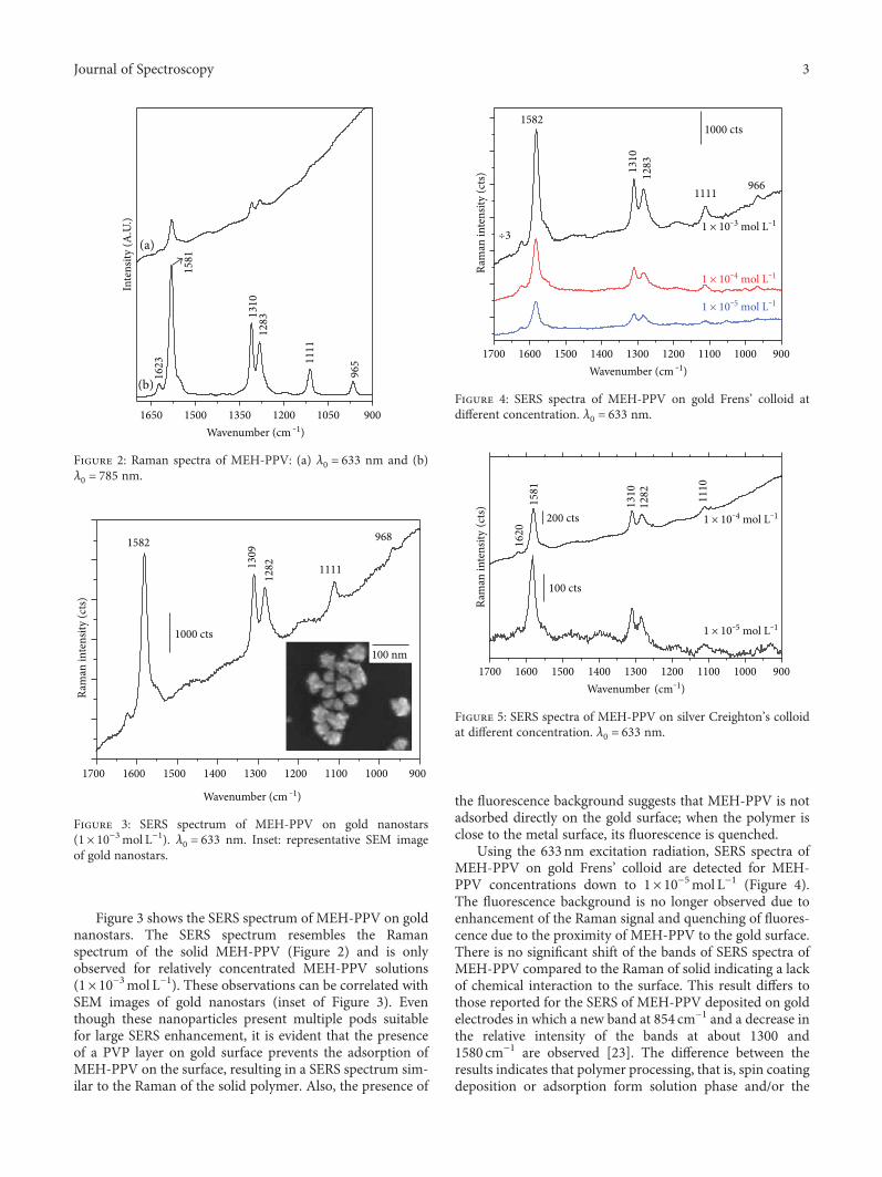
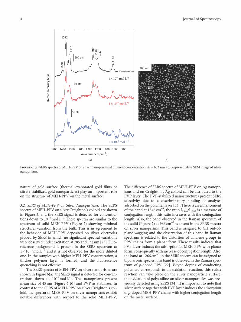
Figure 3 shows the SERS spectrum of MEH-PPV on goldnanostars. The SERS spectrum resembles the Ramanspectrum of the solid MEH-PPV (Figure 2) and is onlyobserved for relatively concentrated MEH-PPV solutions(1× 10⁻³mol L−1). These observations can be correlated withSEM images of gold nanostars (inset of Figure 3). Eventhough these nanoparticles present multiple pods suitablefor large SERS enhancement, it is evident that the presenceof a PVP layer on gold surface prevents the adsorption ofMEH-PPV on the surface, resulting in a SERS spectrum sim-ilar to the Raman of the solid polymer. Also, the presence of
the fluorescence background suggests that MEH-PPV is notadsorbed directly on the gold surface; when the polymer isclose to the metal surface, its fluorescence is quenched.
Using the 633nm excitation radiation, SERS spectra ofMEH-PPV on gold Frens’ colloid are detected for MEH-PPV concentrations down to 1× 10⁻⁵mol L−1 (Figure 4).The fluorescence background is no longer observed due toenhancement of the Raman signal and quenching of fluores-cence due to the proximity of MEH-PPV to the gold surface.There is no significant shift of the bands of SERS spectra ofMEH-PPV compared to the Raman of solid indicating a lackof chemical interaction to the surface. This result differs tothose reported for the SERS of MEH-PPV deposited on goldelectrodes in which a new band at 854 cm−1 and a decrease inthe relative intensity of the bands at about 1300 and1580 cm−1 are observed [23]. The difference between theresults indicates that polymer processing, that is, spin coatingdeposition or adsorption form solution phase and/or the
(a)
1650 1500 1350 1200 1050 900
96511
11
128313
10
Inte
nsity
(A.U
.)
Wavenumber (cm ‒1)
1623
1581
(b)
Figure 2: Raman spectra of MEH-PPV: (a) λ0 = 633 nm and (b)λ0 = 785 nm.
1700 1600 1500 1400 1300 1200 1100 1000 900
Ram
an in
tens
ity (c
ts)
Wavenumber (cm ‒1)
1000 cts
1582
1309
1282 1111
968
100 nm
Figure 3: SERS spectrum of MEH-PPV on gold nanostars(1× 10⁻³mol L−1). λ0 = 633 nm. Inset: representative SEM imageof gold nanostars.
1700 1600 1500 1400 1300 1200 1100 1000 900
1 × 10‒5 mol L‒1
1 × 10‒4 mol L‒1
9661111
1283
Ram
an in
tens
ity (c
ts)
Wavenumber (cm ‒1)
÷3
1000 cts1582
1310
1 × 10‒3 mol L‒1
Figure 4: SERS spectra of MEH-PPV on gold Frens’ colloid atdifferent concentration. λ0 = 633 nm.
1 × 10‒5 mol L‒1
1 × 10‒4 mol L‒1
100 cts
1110
1282
1581
Ram
an in
tens
ity (c
ts)
1620
1310
200 cts
1700 1600 1500 1400 1300 1200 1100 1000 900Wavenumber (cm‒1)
Figure 5: SERS spectra of MEH-PPV on silver Creighton’s colloidat different concentration. λ0 = 633 nm.
3Journal of Spectroscopy
nature of gold surface (thermal evaporated gold films orcitrate-stabilized gold nanoparticles) play an important roleon the structure of MEH-PPV on the metal surface.
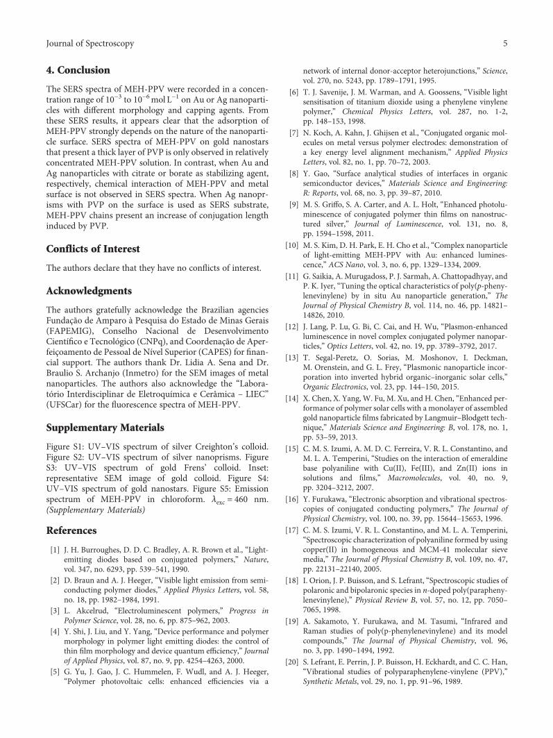
3.2. SERS of MEH-PPV on Silver Nanoparticles. The SERSspectra of MEH-PPV on silver Creighton’s colloid are shownin Figure 5, and the SERS signal is detected for concentra-tions down to 10−5mol L−1. These spectra are similar to thespectrum of solid MEH-PPV (Figure 2) showing minimalstructural variation from the bulk. This is in agreement tothe behavior of MEH-PPV deposited on silver electrodesprobed by SERS in which no significant spectral variationswere observed under excitation at 785 and 532 nm [23]. Fluo-rescence background is present in the SERS spectrum at1× 10−4mol L−1 and it is not observed for the more dilutedone. In the samples with higher MEH-PPV concentration, athicker polymer layer is formed, and the fluorescencequenching is not effective.
The SERS spectra of MEH-PPV on silver nanoprisms areshown in Figure 6(a), the SERS signal is detected for concen-trations down to 10−6mol L−1. The nanoprisms presentmean size of 45nm (Figure 6(b)) and PVP as stabilizer. Incontrast to the SERS of MEH-PPV on silver Creighton’s col-loid, the spectra of MEH-PPV on silver nanoprisms exhibitnotable differences with respect to the solid MEH-PPV.
The difference of SERS spectra of MEH-PPV on Ag nanopr-isms and on Creighton’s Ag colloid can be attributed to thePVP layer. The PVP-stabilized nanostructures present SERSselectivity due to a discriminatory binding of analytesadsorbed on the polymer layer [33]. There is an enhancementof the band at 1546 cm−1, the ratio I1546/I1582 is a measure ofconjugation length, this ratio increases with the conjugationlength. Also, the band observed in the Raman spectrum ofthe solid (Figure 2) at 966 cm−1 is absent in the SERS spectraon silver nanoprisms. This band is assigned to CH out-of-plane wagging and the observation of this band in Ramanspectrum is related to the distortion of vinylene groups inPPV chains from a planar form. These results indicate thatPVP layer induces the adsorption of MEH-PPV with planarform, consequently with increase of conjugation length. Also,the band at 1266 cm−1 in the SERS spectra can be assigned tobipolaronic species, this band is observed in the Raman spec-trum of p-doped PPV [22]. P-type doping of conductingpolymers corresponds to an oxidation reaction, this redoxreaction can take place on the silver nanoparticle surface;the oxidation of polyaniline on silver nanoparticles was pre-viously detected using SERS [34]. It is important to note thatsilver surface together with PVP layer induces the adsorptionof p-doped MEH-PPV chains with higher conjugation lengthon the metal surface.
50 cts
50 cts
1 × 10‒6 mol L‒1
1 × 10‒5 mol L‒1
1 × 10‒4 mol L‒1
1108
1582
1546
1620
1309
126612
88
200 cts
Ram
an in
tens
ity (c
ts)
1700 1600 1500 1400 1300 1200 1100 1000 900Wavenumber (cm ‒1)
(a)
200 nm
(b)
Figure 6: (a) SERS spectra of MEH-PPV on silver nanoprisms at different concentration. λ0 = 633 nm. (b) Representative SEM image of silvernanoprisms.
4 Journal of Spectroscopy
4. Conclusion
The SERS spectra of MEH-PPV were recorded in a concen-tration range of 10−3 to 10−6mol L−1 on Au or Ag nanoparti-cles with different morphology and capping agents. Fromthese SERS results, it appears clear that the adsorption ofMEH-PPV strongly depends on the nature of the nanoparti-cle surface. SERS spectra of MEH-PPV on gold nanostarsthat present a thick layer of PVP is only observed in relativelyconcentrated MEH-PPV solution. In contrast, when Au andAg nanoparticles with citrate or borate as stabilizing agent,respectively, chemical interaction of MEH-PPV and metalsurface is not observed in SERS spectra. When Ag nanopr-isms with PVP on the surface is used as SERS substrate,MEH-PPV chains present an increase of conjugation lengthinduced by PVP.
Conflicts of Interest
The authors declare that they have no conflicts of interest.
Acknowledgments
The authors gratefully acknowledge the Brazilian agenciesFundação de Amparo à Pesquisa do Estado de Minas Gerais(FAPEMIG), Conselho Nacional de DesenvolvimentoCientífico e Tecnológico (CNPq), and Coordenação de Aper-feiçoamento de Pessoal de Nível Superior (CAPES) for finan-cial support. The authors thank Dr. Lidia A. Sena and Dr.Braulio S. Archanjo (Inmetro) for the SEM images of metalnanoparticles. The authors also acknowledge the “Labora-tório Interdisciplinar de Eletroquímica e Cerâmica – LIEC”(UFSCar) for the fluorescence spectra of MEH-PPV.
Supplementary Materials
Figure S1: UV–VIS spectrum of silver Creighton’s colloid.Figure S2: UV–VIS spectrum of silver nanoprisms. FigureS3: UV–VIS spectrum of gold Frens’ colloid. Inset:representative SEM image of gold colloid. Figure S4:UV–VIS spectrum of gold nanostars. Figure S5: Emissionspectrum of MEH-PPV in chloroform. λexc = 460 nm.(Supplementary Materials)
References
[1] J. H. Burroughes, D. D. C. Bradley, A. R. Brown et al., “Light-emitting diodes based on conjugated polymers,” Nature,vol. 347, no. 6293, pp. 539–541, 1990.
[2] D. Braun and A. J. Heeger, “Visible light emission from semi-conducting polymer diodes,” Applied Physics Letters, vol. 58,no. 18, pp. 1982–1984, 1991.
[3] L. Akcelrud, “Electroluminescent polymers,” Progress inPolymer Science, vol. 28, no. 6, pp. 875–962, 2003.
[4] Y. Shi, J. Liu, and Y. Yang, “Device performance and polymermorphology in polymer light emitting diodes: the control ofthin film morphology and device quantum efficiency,” Journalof Applied Physics, vol. 87, no. 9, pp. 4254–4263, 2000.
[5] G. Yu, J. Gao, J. C. Hummelen, F. Wudl, and A. J. Heeger,“Polymer photovoltaic cells: enhanced efficiencies via a
network of internal donor-acceptor heterojunctions,” Science,vol. 270, no. 5243, pp. 1789–1791, 1995.
[6] T. J. Savenije, J. M. Warman, and A. Goossens, “Visible lightsensitisation of titanium dioxide using a phenylene vinylenepolymer,” Chemical Physics Letters, vol. 287, no. 1-2,pp. 148–153, 1998.
[7] N. Koch, A. Kahn, J. Ghijsen et al., “Conjugated organic mol-ecules on metal versus polymer electrodes: demonstration ofa key energy level alignment mechanism,” Applied PhysicsLetters, vol. 82, no. 1, pp. 70–72, 2003.
[8] Y. Gao, “Surface analytical studies of interfaces in organicsemiconductor devices,” Materials Science and Engineering:R: Reports, vol. 68, no. 3, pp. 39–87, 2010.
[9] M. S. Griffo, S. A. Carter, and A. L. Holt, “Enhanced photolu-minescence of conjugated polymer thin films on nanostruc-tured silver,” Journal of Luminescence, vol. 131, no. 8,pp. 1594–1598, 2011.
[10] M. S. Kim, D. H. Park, E. H. Cho et al., “Complex nanoparticleof light-emitting MEH-PPV with Au: enhanced lumines-cence,” ACS Nano, vol. 3, no. 6, pp. 1329–1334, 2009.
[11] G. Saikia, A. Murugadoss, P. J. Sarmah, A. Chattopadhyay, andP. K. Iyer, “Tuning the optical characteristics of poly(p-pheny-lenevinylene) by in situ Au nanoparticle generation,” TheJournal of Physical Chemistry B, vol. 114, no. 46, pp. 14821–14826, 2010.
[12] J. Lang, P. Lu, G. Bi, C. Cai, and H. Wu, “Plasmon-enhancedluminescence in novel complex conjugated polymer nanopar-ticles,” Optics Letters, vol. 42, no. 19, pp. 3789–3792, 2017.
[13] T. Segal-Peretz, O. Sorias, M. Moshonov, I. Deckman,M. Orenstein, and G. L. Frey, “Plasmonic nanoparticle incor-poration into inverted hybrid organic–inorganic solar cells,”Organic Electronics, vol. 23, pp. 144–150, 2015.
[14] X. Chen, X. Yang, W. Fu, M. Xu, and H. Chen, “Enhanced per-formance of polymer solar cells with a monolayer of assembledgold nanoparticle films fabricated by Langmuir–Blodgett tech-nique,” Materials Science and Engineering: B, vol. 178, no. 1,pp. 53–59, 2013.
[15] C. M. S. Izumi, A. M. D. C. Ferreira, V. R. L. Constantino, andM. L. A. Temperini, “Studies on the interaction of emeraldinebase polyaniline with Cu(II), Fe(III), and Zn(II) ions insolutions and films,” Macromolecules, vol. 40, no. 9,pp. 3204–3212, 2007.
[16] Y. Furukawa, “Electronic absorption and vibrational spectros-copies of conjugated conducting polymers,” The Journal ofPhysical Chemistry, vol. 100, no. 39, pp. 15644–15653, 1996.
[17] C. M. S. Izumi, V. R. L. Constantino, and M. L. A. Temperini,“Spectroscopic characterization of polyaniline formed by usingcopper(II) in homogeneous and MCM-41 molecular sievemedia,” The Journal of Physical Chemistry B, vol. 109, no. 47,pp. 22131–22140, 2005.
[18] I. Orion, J. P. Buisson, and S. Lefrant, “Spectroscopic studies ofpolaronic and bipolaronic species in n-doped poly(parapheny-lenevinylene),” Physical Review B, vol. 57, no. 12, pp. 7050–7065, 1998.
[19] A. Sakamoto, Y. Furukawa, and M. Tasumi, “Infrared andRaman studies of poly(p-phenylenevinylene) and its modelcompounds,” The Journal of Physical Chemistry, vol. 96,no. 3, pp. 1490–1494, 1992.
[20] S. Lefrant, E. Perrin, J. P. Buisson, H. Eckhardt, and C. C. Han,“Vibrational studies of polyparaphenylene-vinylene (PPV),”Synthetic Metals, vol. 29, no. 1, pp. 91–96, 1989.
5Journal of Spectroscopy

[21] E. Mulazzi, A. Ripamonti, J. Wery, B. Dulieu, and S. Lefrant,“Theoretical and experimental investigation of absorptionand Raman spectra of poly(paraphenylene vinylene),” PhysicalReview B, vol. 60, no. 24, pp. 16519–16525, 1999.
[22] M. Baıtoul, J. Wéry, J.-P. Buisson et al., “In situ resonantRaman and optical investigations of p-doped poly (p-pheny-lene vinylene),” Polymer, vol. 41, no. 18, pp. 6955–6964, 2000.
[23] D. Li, N. J. Borys, and J. M. Lupton, “Probing the electrode-polymer interface in conjugated polymer devices withsurface-enhanced Raman scattering,” Applied Physics Letters,vol. 100, no. 14, article 141907, 2012.
[24] M. Baibarac, I. Baltog, I. Smaranda et al., “Spectroelectrochem-ical properties of the poly[(2,5-bisoctyloxy)-1,4-phenylenevi-nylene]/single-walled carbon nanotube composite,” SyntheticMetals, vol. 195, pp. 276–285, 2014.
[25] A. J. Wise, M. R. Precit, A. M. Papp, and J. K. Grey, “Effect offullerene intercalation on the conformation and packing ofpoly-(2-methoxy-5-(3′-7′-dimethyloctyloxy)-1,4-phenylene-vinylene),” ACS Applied Materials & Interfaces, vol. 3, no. 8,pp. 3011–3019, 2011.
[26] V. V. Bruevich, T. S. Makhmutov, S. G. Elizarov, E. M.Nechvolodova, and D. Y. Paraschuk, “Raman spectroscopyof intermolecular charge transfer complex between a conju-gated polymer and an organic acceptor molecule,” TheJournal of Chemical Physics, vol. 127, no. 10, article104905, 2007.
[27] E. C. Le Ru and P. G. Etchegoin, “Chapter 1 – a quick overviewof surface-enhanced Raman spectroscopy,” in Principles ofSurface-Enhanced Raman Spectroscopy, pp. 1–27, Elsevier,Amsterdam, Netherlands, 2009.
[28] Z. Wang and L. J. Rothberg, “Structure and dynamics of singleconjugated polymer chromophores by surface-enhancedRaman spectroscopy,” ACS Nano, vol. 1, no. 4, pp. 299–306,2007.
[29] J. A. Creighton, C. G. Blatchford, and M. G. Albrecht, “Plasmaresonance enhancement of Raman scattering by pyridineadsorbed on silver or gold sol particles of size comparable tothe excitation wavelength,” Journal of the Chemical Society,Faraday Transactions 2: Molecular and Chemical Physics,vol. 75, p. 790, 1979.
[30] I. Pastoriza-Santos and L. M. Liz-Marzán, “Synthesis of silverNanoprisms in DMF,” Nano Letters, vol. 2, no. 8, pp. 903–905, 2002.
[31] G. Frens, “Controlled nucleation for the regulation of the par-ticle size in monodisperse gold suspensions,” Nature PhysicalScience, vol. 241, no. 105, pp. 20–22, 1973.
[32] P. Senthil Kumar, I. Pastoriza-Santos, B. Rodríguez-González,F. Javier García de Abajo, and L. M. Liz-Marzán, “High-yieldsynthesis and optical response of gold nanostars,” Nanotech-nology, vol. 19, no. 1, article 015606, 2008.
[33] P. Pinkhasova, L. Yang, Y. Zhang, S. Sukhishvili, and H. Du,“Differential SERS activity of gold and silver nanostructuresenabled by adsorbed poly(vinylpyrrolidone),” Langmuir,vol. 28, no. 5, pp. 2529–2535, 2012.
[34] C. M. S. Izumi, G. F. S. Andrade, and M. L. A. Temperini,“Surface-enhanced resonance Raman scattering of polyani-line on silver and gold colloids,” The Journal of PhysicalChemistry B, vol. 112, no. 51, pp. 16334–16340, 2008.
6 Journal of Spectroscopy

TribologyAdvances in
Hindawiwww.hindawi.com Volume 2018
Hindawiwww.hindawi.com Volume 2018
International Journal ofInternational Journal ofPhotoenergy
Hindawiwww.hindawi.com Volume 2018
Journal of
Chemistry
Hindawiwww.hindawi.com Volume 2018
Advances inPhysical Chemistry
Hindawiwww.hindawi.com
Analytical Methods in Chemistry
Journal of
Volume 2018
Bioinorganic Chemistry and ApplicationsHindawiwww.hindawi.com Volume 2018
SpectroscopyInternational Journal of
Hindawiwww.hindawi.com Volume 2018
Hindawi Publishing Corporation http://www.hindawi.com Volume 2013Hindawiwww.hindawi.com
The Scientific World Journal
Volume 2018
Medicinal ChemistryInternational Journal of
Hindawiwww.hindawi.com Volume 2018
NanotechnologyHindawiwww.hindawi.com Volume 2018
Journal of
Applied ChemistryJournal of
Hindawiwww.hindawi.com Volume 2018
Hindawiwww.hindawi.com Volume 2018
Biochemistry Research International
Hindawiwww.hindawi.com Volume 2018
Enzyme Research
Hindawiwww.hindawi.com Volume 2018
Journal of
SpectroscopyAnalytical ChemistryInternational Journal of
Hindawiwww.hindawi.com Volume 2018
MaterialsJournal of
Hindawiwww.hindawi.com Volume 2018
Hindawiwww.hindawi.com Volume 2018
BioMed Research International Electrochemistry
International Journal of
Hindawiwww.hindawi.com Volume 2018
Na
nom
ate
ria
ls
Hindawiwww.hindawi.com Volume 2018
Journal ofNanomaterials
Submit your manuscripts atwww.hindawi.com