Surface engineered and drug releasing pre-fabricated ...€¦ · 5.3. Wound repair ......
14
Surface engineered and drug releasing pre-fabricated scaffolds for tissue engineering ☆ Hyun Jung Chung, Tae Gwan Park ⁎ Department of Biological Sciences, Korea Advanced Institute of Science and Technology, Daejeon 305-701, Republic of Korea Received 30 December 2006; accepted 28 March 2007 Available online 10 April 2007 Abstract A wide range of polymeric scaffolds have been intensively studied for use as implantable and temporal devices in tissue engineering. Biodegradable and biocompatible scaffolds having a highly open porous structure and good mechanical strength are needed to provide an optimal microenvironment for cell proliferation, migration, and differentiation, and guidance for cellular in-growth from host tissue. A variety of natural and synthetic polymeric scaffolds can be fabricated in the form of a solid foam, nanofibrous matrix, microsphere, or hydrogel. Biodegradable porous scaffolds can be surface engineered to provide an extracellular matrix mimicking environment for better cell adhesion and tissue in-growth. Furthermore, scaffolds can be designed to release bioactive molecules, such as growth factors, DNA, or drugs, in a sustained manner to facilitate tissue regeneration. This paper reviews the current status of surface engineered and drug releasing scaffolds for tissue engineering. © 2007 Elsevier B.V. All rights reserved. Keywords: Scaffolds; Porous; Biomimetic; Surface modification; Drug delivery; Tissue regeneration Contents 1. Introduction ............................................................. 250 2. Materials ............................................................... 251 2.1. Naturally derived materials .................................................. 251 2.2. Scaffolds based on synthetic polymers ............................................ 251 3. Scaffold fabrication .......................................................... 251 3.1. Fiber bonding ......................................................... 251 3.2. Emulsion freeze drying .................................................... 251 3.3. Solvent casting/particulate leaching .............................................. 251 3.4. High-pressure processing ................................................... 252 3.5. Gas foaming/particulate leaching ............................................... 252 3.6. Thermally induced phase separation ............................................. 253 3.7. Electrospinning ........................................................ 253 3.8. Rapid prototyping ....................................................... 253 4. Surface engineered scaffolds ..................................................... 253 4.1. Collagen and gelatin ..................................................... 253 4.2. Cell adhesive peptides ..................................................... 254 4.3. Hyaluronic acid ........................................................ 255 Advanced Drug Delivery Reviews 59 (2007) 249 – 262 www.elsevier.com/locate/addr ☆ This review is part of the Advanced Drug Delivery Reviews theme issue on “Matrices and Scaffolds for Drug Delivery in Tissue Engineering”. ⁎ Corresponding author. Tel.: +82 42 869 2621; fax: +82 42 869 2610. E-mail address: [email protected] (T.G. Park). 0169-409X/$ - see front matter © 2007 Elsevier B.V. All rights reserved. doi:10.1016/j.addr.2007.03.015
Transcript of Surface engineered and drug releasing pre-fabricated ...€¦ · 5.3. Wound repair ......

ews 59 (2007) 249–262www.elsevier.com/locate/addr
Advanced Drug Delivery Revi
Surface engineered and drug releasing pre-fabricated scaffoldsfor tissue engineering☆
Hyun Jung Chung, Tae Gwan Park ⁎
Department of Biological Sciences, Korea Advanced Institute of Science and Technology, Daejeon 305-701, Republic of Korea
Received 30 December 2006; accepted 28 March 2007Available online 10 April 2007
Abstract
A wide range of polymeric scaffolds have been intensively studied for use as implantable and temporal devices in tissue engineering.Biodegradable and biocompatible scaffolds having a highly open porous structure and good mechanical strength are needed to provide an optimalmicroenvironment for cell proliferation, migration, and differentiation, and guidance for cellular in-growth from host tissue. A variety of naturaland synthetic polymeric scaffolds can be fabricated in the form of a solid foam, nanofibrous matrix, microsphere, or hydrogel. Biodegradableporous scaffolds can be surface engineered to provide an extracellular matrix mimicking environment for better cell adhesion and tissue in-growth.Furthermore, scaffolds can be designed to release bioactive molecules, such as growth factors, DNA, or drugs, in a sustained manner to facilitatetissue regeneration. This paper reviews the current status of surface engineered and drug releasing scaffolds for tissue engineering.© 2007 Elsevier B.V. All rights reserved.
Keywords: Scaffolds; Porous; Biomimetic; Surface modification; Drug delivery; Tissue regeneration
Contents
1. Introduction . . . . . . . . . . . . . . . . . . . . . . . . . . . . . . . . . . . . . . . . . . . . . . . . . . . . . . . . . . . . . 2502. Materials . . . . . . . . . . . . . . . . . . . . . . . . . . . . . . . . . . . . . . . . . . . . . . . . . . . . . . . . . . . . . . . 251
2.1. Naturally derived materials . . . . . . . . . . . . . . . . . . . . . . . . . . . . . . . . . . . . . . . . . . . . . . . . . . 2512.2. Scaffolds based on synthetic polymers . . . . . . . . . . . . . . . . . . . . . . . . . . . . . . . . . . . . . . . . . . . . 251
3. Scaffold fabrication . . . . . . . . . . . . . . . . . . . . . . . . . . . . . . . . . . . . . . . . . . . . . . . . . . . . . . . . . . 2513.1. Fiber bonding . . . . . . . . . . . . . . . . . . . . . . . . . . . . . . . . . . . . . . . . . . . . . . . . . . . . . . . . . 2513.2. Emulsion freeze drying . . . . . . . . . . . . . . . . . . . . . . . . . . . . . . . . . . . . . . . . . . . . . . . . . . . . 2513.3. Solvent casting/particulate leaching . . . . . . . . . . . . . . . . . . . . . . . . . . . . . . . . . . . . . . . . . . . . . . 2513.4. High-pressure processing . . . . . . . . . . . . . . . . . . . . . . . . . . . . . . . . . . . . . . . . . . . . . . . . . . . 2523.5. Gas foaming/particulate leaching . . . . . . . . . . . . . . . . . . . . . . . . . . . . . . . . . . . . . . . . . . . . . . . 2523.6. Thermally induced phase separation . . . . . . . . . . . . . . . . . . . . . . . . . . . . . . . . . . . . . . . . . . . . . 2533.7. Electrospinning . . . . . . . . . . . . . . . . . . . . . . . . . . . . . . . . . . . . . . . . . . . . . . . . . . . . . . . . 2533.8. Rapid prototyping . . . . . . . . . . . . . . . . . . . . . . . . . . . . . . . . . . . . . . . . . . . . . . . . . . . . . . . 253
4. Surface engineered scaffolds . . . . . . . . . . . . . . . . . . . . . . . . . . . . . . . . . . . . . . . . . . . . . . . . . . . . . 2534.1. Collagen and gelatin . . . . . . . . . . . . . . . . . . . . . . . . . . . . . . . . . . . . . . . . . . . . . . . . . . . . . 2534.2. Cell adhesive peptides . . . . . . . . . . . . . . . . . . . . . . . . . . . . . . . . . . . . . . . . . . . . . . . . . . . . . 2544.3. Hyaluronic acid . . . . . . . . . . . . . . . . . . . . . . . . . . . . . . . . . . . . . . . . . . . . . . . . . . . . . . . . 255
☆ This review is part of the Advanced Drug Delivery Reviews theme issue on “Matrices and Scaffolds for Drug Delivery in Tissue Engineering”.⁎ Corresponding author. Tel.: +82 42 869 2621; fax: +82 42 869 2610.
E-mail address: [email protected] (T.G. Park).
0169-409X/$ - see front matter © 2007 Elsevier B.V. All rights reserved.doi:10.1016/j.addr.2007.03.015
250 H.J. Chung, T.G. Park / Advanced Drug Delivery Reviews 59 (2007) 249–262
4.4. Galactose . . . . . . . . . . . . . . . . . . . . . . . . . . . . . . . . . . . . . . . . . . . . . . . . . . . . . . . . . . . . 2554.5. Heparin . . . . . . . . . . . . . . . . . . . . . . . . . . . . . . . . . . . . . . . . . . . . . . . . . . . . . . . . . . . . . 255
5. Scaffolds for growth factor release . . . . . . . . . . . . . . . . . . . . . . . . . . . . . . . . . . . . . . . . . . . . . . . . . . 2565.1. Angiogenesis . . . . . . . . . . . . . . . . . . . . . . . . . . . . . . . . . . . . . . . . . . . . . . . . . . . . . . . . . . 2565.2. Bone and cartilage regeneration . . . . . . . . . . . . . . . . . . . . . . . . . . . . . . . . . . . . . . . . . . . . . . . . 2565.3. Wound repair . . . . . . . . . . . . . . . . . . . . . . . . . . . . . . . . . . . . . . . . . . . . . . . . . . . . . . . . . . 2575.4. Liver regeneration . . . . . . . . . . . . . . . . . . . . . . . . . . . . . . . . . . . . . . . . . . . . . . . . . . . . . . . 2575.5. Neural tissue engineering . . . . . . . . . . . . . . . . . . . . . . . . . . . . . . . . . . . . . . . . . . . . . . . . . . . 257
6. Scaffolds for DNA and drug delivery . . . . . . . . . . . . . . . . . . . . . . . . . . . . . . . . . . . . . . . . . . . . . . . . . 2576.1. Scaffolds for DNA delivery . . . . . . . . . . . . . . . . . . . . . . . . . . . . . . . . . . . . . . . . . . . . . . . . . . 2576.2. Drug releasing scaffolds . . . . . . . . . . . . . . . . . . . . . . . . . . . . . . . . . . . . . . . . . . . . . . . . . . . . 258
7. Injectable matrices . . . . . . . . . . . . . . . . . . . . . . . . . . . . . . . . . . . . . . . . . . . . . . . . . . . . . . . . . . 2587.1. Hydrogels . . . . . . . . . . . . . . . . . . . . . . . . . . . . . . . . . . . . . . . . . . . . . . . . . . . . . . . . . . . 2587.2. Microspheres . . . . . . . . . . . . . . . . . . . . . . . . . . . . . . . . . . . . . . . . . . . . . . . . . . . . . . . . . . 259
8. Conclusions . . . . . . . . . . . . . . . . . . . . . . . . . . . . . . . . . . . . . . . . . . . . . . . . . . . . . . . . . . . . . . 259Acknowledgement . . . . . . . . . . . . . . . . . . . . . . . . . . . . . . . . . . . . . . . . . . . . . . . . . . . . . . . . . . . . . 259References . . . . . . . . . . . . . . . . . . . . . . . . . . . . . . . . . . . . . . . . . . . . . . . . . . . . . . . . . . . . . . . . . 259
1. Introduction
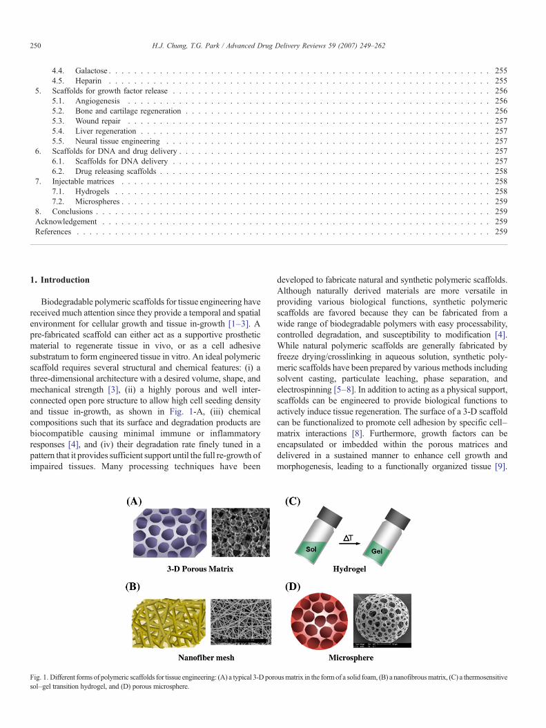
Biodegradable polymeric scaffolds for tissue engineering havereceived much attention since they provide a temporal and spatialenvironment for cellular growth and tissue in-growth [1–3]. Apre-fabricated scaffold can either act as a supportive prostheticmaterial to regenerate tissue in vivo, or as a cell adhesivesubstratum to form engineered tissue in vitro. An ideal polymericscaffold requires several structural and chemical features: (i) athree-dimensional architecture with a desired volume, shape, andmechanical strength [3], (ii) a highly porous and well inter-connected open pore structure to allow high cell seeding densityand tissue in-growth, as shown in Fig. 1-A, (iii) chemicalcompositions such that its surface and degradation products arebiocompatible causing minimal immune or inflammatoryresponses [4], and (iv) their degradation rate finely tuned in apattern that it provides sufficient support until the full re-growth ofimpaired tissues. Many processing techniques have been
Fig. 1. Different forms of polymeric scaffolds for tissue engineering: (A) a typical 3-D porosol–gel transition hydrogel, and (D) porous microsphere.
developed to fabricate natural and synthetic polymeric scaffolds.Although naturally derived materials are more versatile inproviding various biological functions, synthetic polymericscaffolds are favored because they can be fabricated from awide range of biodegradable polymers with easy processability,controlled degradation, and susceptibility to modification [4].While natural polymeric scaffolds are generally fabricated byfreeze drying/crosslinking in aqueous solution, synthetic poly-meric scaffolds have been prepared by various methods includingsolvent casting, particulate leaching, phase separation, andelectrospinning [5–8]. In addition to acting as a physical support,scaffolds can be engineered to provide biological functions toactively induce tissue regeneration. The surface of a 3-D scaffoldcan be functionalized to promote cell adhesion by specific cell–matrix interactions [8]. Furthermore, growth factors can beencapsulated or imbedded within the porous matrices anddelivered in a sustained manner to enhance cell growth andmorphogenesis, leading to a functionally organized tissue [9].
usmatrix in the formof a solid foam, (B) a nanofibrousmatrix, (C) a thermosensitive
251H.J. Chung, T.G. Park / Advanced Drug Delivery Reviews 59 (2007) 249–262
Studies on scaffolds releasing DNA encoding the growth factorhave also been suggested as an alternative approach to bypasslimitations of protein delivery [10].
Recently, 3-D matrices based on different structural char-acteristics or minimally invasive surgical methods have drawnattention for potential tissue engineering applications in the nextgeneration. A nanofibrous matrix prepared by electrospinning orself-assembly would provide a better resemblance of thephysiological environment (Fig. 1-B) [3,11]. Injectable matricessuch as hydrogels (Fig. 1-C) and micro-spheres (Fig. 1-D),which are already widely utilized as sustained protein releaseformulations, have also been applied in tissue engineering for itspotential use as a cell delivery carrier or supportive matrix[12,13]. This review will introduce previous techniques forfabricating biodegradable scaffolds, followed by surfaceengineered and drug releasing scaffolds for directing a seriesof tissue regeneration processes in a more active manner.
2. Materials
Several natural and synthetic polymers have been utilized forfabricating tissue engineering scaffolds. Requirements for thesematerials are that they must be inherently biocompatible,biodegradable, and highly cell adhesive. Additionally, they musthave a porous,mechanically stable, and three-dimensional structurewith facile manufacturing processes. Recently, hybrid polymericscaffolds combining natural and synthetic polymers have gatheredgrowing interests to mimic the extracellular matrix of a naturaltissue.
2.1. Naturally derived materials
Natural polymers widely used for tissue engineeringapplications include fibrin, collagen, gelatin, chitosan, alginate,and hyaluronic acid [14–19]. Fibrin, a major constituent ofblood clots, has been used in mixtures with thrombin to producean in-situ forming gel [14]. Type I collagen extracted fromanimal tissues and gelatin prepared from denaturation ofcollagen are capable of forming porous gel matrices which arealready commercially available as skin replacements [15].Chitosan, a cationic polymer derived from chitin, can produce ascaffold with a hydrophilic surface, and cell adhesive/differentiating characteristics, while its inherent osteoconduc-tive nature endows its potential use for bone tissue engineering[16,17]. Alginate, an anionic polysaccharide extracted frombrown algae, exhibits gel forming behavior when complexedwith divalent cations such as Ca2+ [18]. Hyaluronic acid, a non-sulfated glycosaminoglycan of repeating disaccharide units, is amajor component of the natural ECM and forms crosslinkablehydrogels with various modifications [19].
2.2. Scaffolds based on synthetic polymers
Biodegradable synthetic polymers widely used for preparingtissue engineering matrices include poly(α-hydroxyester)s,polyanhydrides, and polyorthoesters [20]. Among these, thepoly(α-hydroxyester)s such as polylactide (PLA), polyglycolide
(PGA), and its copolymers are the most extensively used forbiodegradable scaffolds in the form of a solid foam [21]. Thesepolymers which are biocompatible, biodegradable, and bior-esorbable are approved by the Food and Drug Administration(FDA), and are easily processed into various structures of 3-Dmatrices. Especially, biodegradable devices made of poly(lactide-co-glycolide) (PLGA) copolymers are advantageousdue to their controlled degradation behavior and tunablemechanical properties according to the specific requirementsfor the desired tissue. The rate of polymer degradation should becarefully controlled to synchronize with the rate of tissueformation and in-growth to achieve successful regeneration orrepair within a desired time frame. These hydrophobic polymerscan also be fabricated into microspheres as injectable matrices,or scaffolds with nanofibrous structure. However, these poly-mers degrade into acidic by-products, which might causenegative effects on cell adhesion and growth. Biodegradablehydrogel scaffolds fabricated with hydrophilic polymers such aspoly(ethylene glycol) would overcome these limitations byproviding a benign and controllable environment for cellulargrowth and differentiation, but their nonadhesive properties andlimited transport also cause some disadvantages as well [22].
3. Scaffold fabrication
A variety of techniques have been used for processing bio-degradable polymers into 3-D porous scaffolds. The conventionalmethods include fiber felts, fiber bonding, melt molding, solventcasting/particulate leaching, gas foaming/particulate leaching,phase separation, and high-pressure processing. Electrospinninghas also been utilized in producing a nanofibrous 3-D matrix, andrapid prototyping technologies have enabled solid free formfabrication directly from a computer-aided design (CAD) model.
3.1. Fiber bonding
3-D porous matrices can be constructed by bonding polymerfibers at their crosspoints using a secondary polymer. For example,PGA fibers have been bonded by embedding in PLLA solution,cooling, and subsequent removal of PLLA [23]. However,difficulties reside for this technique in controlling the porosity orchoice of solvents.
3.2. Emulsion freeze drying
Freeze drying an emulsion solution composed of a dispersedwater phase and an organic continuous phase containing bio-degradable polymer can give rise to porous scaffolds withvarious pore sizes and inter-connectivities. Using this tech-nique, Whang et. al. [24] have prepared PLGA scaffolds withporosity of up to 95% and pore sizes of up to 200 μm.
3.3. Solvent casting/particulate leaching
Solvent casting–particulate leaching is probably the mostconvenient method for preparing porous scaffolds [5]. It involvesthe casting of a polymer/salt/organic solvent mixture solution

252 H.J. Chung, T.G. Park / Advanced Drug Delivery Reviews 59 (2007) 249–262
followed by solvent evaporation and dissolution of the saltparticulates in an aqueous solution. However, this technique haslimitations; it can only produce thin membranes with a densesurface skin layer, and might contain residual salt particles usedduring the process. Efforts have resulted in the production of thinscaffolds with an open-cell morphology and porosity as high as93%. For preparing a thick 3-D scaffold, PLLA or PLGA porousmembranes were laminated into multi-layer structures withvarious anatomical shapes [25].
3.4. High-pressure processing
High-pressure processing, also known as the supercriticalfluid technology, is performed by applying a gas, such as carbondioxide, to the dry polymer at high pressure which forms asingle phase polymer/gas solution. The pressure is then reducedto create thermodynamic instability of the dissolved CO2 andresults in nucleation and growth of gas cells to generate poreswithin the polymer matrix. Mooney et al. [26] utilized thistechnique to fabricate highly porous sponges of PLGA. Soliddisks of PLGA prepared by compression molding or solventcasting were saturated with CO2 by applying high pressure andthen reducing it, to produce a macroporous structure. The mainadvantage of this method is that it excludes the use of organicsolvents. Since the technique does not include a heating process,it is also useful for incorporation of heat sensitive biomolecules.The porous structure is quite uniform because the CO2 gas ishomogeneously dissolved with the polymer and also acts as aplasticizer which induces closer packing of polymer chains,resulting in higher mechanical strength. However, this methodresults in insufficient inter-connectivity of pores within thematrix, with a non-porous surface in most cases. To overcomethis problem, Harris et al. [27] modified this technique bycombining it with the particulate leaching method. A PLGA/NaCl mixture was compression molded into solid disks,exposed to high-pressure CO2 and immersed in water to leach
Fig. 2. Fabrication of porous scaffolds by gas foaming/particulate leaching: Sieved effor solvent evaporation, immersed in water for gas foaming/salt leaching, and freeze
out the salt. The process led to the formation of a highly inter-connected porous network with no sign of non-porous surface.
3.5. Gas foaming/particulate leaching
A “gas foaming” method was developed by Park et al. [6]using an effervescent salt as a gas foaming agent. As shown inFig. 2, a binary mixture of PLA–solvent gel containingdispersed ammonium bicarbonate salt particles was cast in amold and subsequently immersed in hot water. The evolution ofammonia and carbon dioxide gas, along with the leaching out ofammonium bicarbonate particulates from the solidifyingpolymer matrix, resulted in the formation of pores with highinter-connectivity. The formed scaffolds had a macroporousopen cellular structure with pores having a uniform distributionranging from 100 to 200 μm and no sign of surface skin. Thegas foaming/salt leaching method was further improved forpreparing porous PLGA scaffolds by adding another salt, citricacid, into the aqueous solution [28]. In this case, amorphousPLGA dissolved in chloroform was first precipitated in ethanolto obtain a gel slurry. Ammonium bicarbonate salt particlesmixed with this gel paste were cast in a mold, semi-solidified atroom temperature, and immersed into aqueous citric acidsolution. Macroporous PLGA scaffolds with a porosity of over90% and pore size of about 200 μmwere obtained. The porosityand mechanical strength could be controlled by adjusting theextent of acid–base gas evolving reaction between the two salts.These scaffolds were commercialized in Korea under the tradename of Innopol-D™ for use as a temporal prosthetic device forpenile enlargement. Based on the same gas foaming principle,injectable PLGA micro-carrier scaffolds were also fabricatedusing a double emulsion solvent evaporation method [29].Highly open porous micro-spheres with a size of 200–300 μmwere prepared by incorporating ammonium bicarbonate into theinner water (W1) phase droplets, which vigorously producedgas bubbles during solvent removal. The surface pores had sizes
fervescent salt particles are dispersed in polymer gel paste, cast on a Teflon molddried.

253H.J. Chung, T.G. Park / Advanced Drug Delivery Reviews 59 (2007) 249–262
of about 30 μm, which were sufficient for cell infiltration andseeding, demonstrated by cultivation with fibroblasts.
3.6. Thermally induced phase separation
The phase separation technique is based on thermodynamicdemixing of a homogeneous polymer–solvent solution into apolymer-rich phase and a polymer-poor phase, usually by eitherexposure of the solution to another immiscible solvent or coolingthe solution to a point below the binodal solubility curve [30].Particularly, thermally induced phase separation (TIPS) usesthermal energy as the latent solvent to induce phase separation[31]. The polymer solution is quenched below the freezing pointof the solvent and subsequently freeze dried, producing a porousstructure which can be finely tuned by adjusting the variousthermodynamic and kinetic parameters. The earliest tissueengineering scaffolds prepared by the TIPSmethod had a micro-porous (1–10 μm) structure which lacked pore inter-connectiv-ity and open cellularity [32]. Park [30] used the TIPS techniqueto obtain scaffolds with a macroporous structure with an opencellular morphology. The coarsening process was used toincrease the size of phase-separated droplets, thus enlarging thepores (∼100 μm). The resultant scaffolds also had pores with auniform size distribution and porosity of over 90%. They alsoshowed that the addition of a surfactant, Pluronic F127,enhanced the pore morphology of the scaffold.
3.7. Electrospinning
Electrospinning is the most widely usedmethod for fabricationof nanofiber non-woven matrices, due to its simplicity andefficiency. A polymer solution or melt is drawn from a nozzle byapplying a force of gravity or mechanical pressure combined withan electric field of high voltage (10–20 kV). When the electriccharge overcomes the surface tension of the polymer solutiondroplet, a polymer jet is sprouted, followed by solvent evaporationwhich forms the solid nanofibers [7]. Various materials can beelectrospun into nanofiber: biodegradable polymers such asPLGA and polycaprolactone (PCL), water-soluble materialssuch as poly(ethylene oxide) (PEO), polyvinyl alcohol (PVA),and natural polymer such as collagen, silk protein, and otherpeptides [7,33–35]. The electrospinning technology has beenemployed in the polymer industry for over 70 years and wasintroduced into the tissue engineeringworld just several years ago.Li et al. [33] developed a novel PLGAnanofibrousmeshwith fiberdiameter ranging from 500 to 800 nm and pores that were wellinter-connected. Usually, the electrospinning method primarilyresults in a 2-Dmesh structure with a nanoscale pore size which isnot sufficient for cell seeding and infiltration. Therefore, macro-porous and nanofibrous 3-D hybrid scaffolds with a requiredvolume and shape are highly desirable for use as an implantabledevice for tissue regeneration.
3.8. Rapid prototyping
Rapid prototyping (RP), also known as solid free formfabrication, has recently introduced a new method in fabricating
well-designed tissue engineering scaffolds. RP utilizes acomputer-aided design model to construct a 3-D architecturein a layer-by-layer manner with precise control of morpholog-ical characteristics as well as chemical composition andmechanical properties [36]. This technique allows the produc-tion of scaffolds that are customized in size and shape accordingto specific requirements which are highly reproducible.Biodegradable porous scaffolds can be fabricated directly by amelt–dissolution deposition process using fused depositionmodeling (FDM) [37] or 3-D fiber-deposition [38], or by aparticle bonding technique such as 3-D printing (3DP) [39,40].Scaffolds can also be produced indirectly by casting in a moldand employing techniques such as melt deposition, dropletdeposition, and photo-polymerization [41].
4. Surface engineered scaffolds
Although biodegradable porous scaffolds with well inter-connected pores are good enough to permit cell infiltration andgrowth, their surface characteristics such as hydrophilicity/hydrophobicity arising from chemical composition may not besatisfactory for inducing selective cell adhesion, migration, andproliferation. In most cases, specific cellular interactions arerequired for the formation of a desired tissue. In general, surfaceproperties of implantable biomaterials dictate protein adsorptionbehaviors with concomitantly determining cellular interactions.Attempts have been made to mimic the natural extracellularmatrix by immobilizing naturally derived biomolecules on thesurface of polymer scaffolds (Fig. 3). The surface engineeredscaffolds were capable of enhanced cell adhesion and growth, orsustained release of growth factors [8], thereby providing achance to facilitate the tissue regeneration process. The surfaceof a scaffold can be functionalized either by physical adsorptionor chemical modification. Substances including poly(L-lysine),collagen, and cell adhesive proteins such as fibronectin,laminin, or vitronectin have been adsorbed onto the surface ofa polymeric matrix to promote cell attachment [42,43].However, covalent binding of functional biomolecules wouldbe necessary to provide a more stable cell adhesive stratum.Naturally derived macromolecules such as collagen, gelatin,heparin, hyaluronic acid, short peptide sequences originatingfrom cell adhesive proteins such as the arginine–glycine–aspartic acid (RGD) or YIGSR, and sugar moieties such asgalactose or lactose, have been grafted onto polymer surfaces tomodulate cell–matrix interactions [8,44].
4.1. Collagen and gelatin
Collagen is a major structural component forming the naturalECM of connective tissues and organs [45]. Surface immobili-zation of collagen is one of the most established methods forendowing cell adhesive properties to the scaffolds. PLA andPLGA scaffolds chemically grafted with collagen by plasmatreatment have shown enhanced adhesion and spreading offibroblasts [46]. Collagen modification by conjugation reactionsonto PLA scaffolds grafted with polymethacrylic acid also hasimproved cell spreading and growth for use in cartilage tissue

254 H.J. Chung, T.G. Park / Advanced Drug Delivery Reviews 59 (2007) 249–262
engineering [47]. Unfortunately, despite the popular use ofcollagen due to its various biological functions, its immunoge-nicity has limited its applications. Gelatin is a good alternative forcollagen because of its absence of antigenicity and ease ofhandling at high concentrations. Gelatin immobilized onto porousscaffolds by physical entrapment and chemical crosslinkingshowed greatly enhanced surface properties on attachment,proliferation, and ECM deposition of osteoblasts [48].
4.2. Cell adhesive peptides
Rather than immobilizing the whole protein, chemicalconjugation of short chain peptide moieties derived from thecell adhesive proteins onto the polymer surface can be a muchmore effective strategy. The surface immobilization of shortpeptides has several advantages: higher stability againstconformational change, easy controllability of surface density,and orientation more favorable for ligand–receptor interactionand cell adhesion [8,44,49]. It is also beneficial for minimizingimmune responses and infection. Many peptide sequencesinvolved in cellular interactions by receptor binding have beenidentified, including RGD, IKVAV, and YIGSR [49]. Amongthese, the RGD sequence, which was first discovered infibronectin on 1984, is probably one of the best known foruse in tissue engineering applications. Immobilization of RGDonto 3-D matrices to improve cell adhesive properties waspreviously demonstrated in collagen gels, showing enhancedadherence of murine melanoma cells [50]. RGD, along withother short peptide sequences such as IKVAV, YIGSR,
Fig. 3. Surface engineered and growth factor releasing scaffolds for tissue engineeringor encapsulated with growth factors or DNA to promote cell proliferation and morp
RNAIAEIIKDI from laminin, and HAV from N-cadherin, wasalso used for engineering of neural tissue [51]. Incorporatingthese peptides into a crosslinked 3-D fibrin network resulted ingreatly enhanced cellular outgrowth in a neurite extensionmodel. Other than promoting cell adhesion and migration, RGDpeptides have been reported to show additional functions suchas inducing differentiation and growth of osteoblasts, and thuswidely utilized in bone tissue engineering [44]. PLA scaffoldsmodified with RGD by plasma treatment not only resulted inimproved adhesion of the osteoblast-like cells, but alsosupported its growth and differentiation [52]. Other studiesshowed that osteoblasts seeded onto the RGD immobilizedscaffolds greatly enhanced mineralization and formation ofbone-like tissues [53]. Recently, a blend mixture of PLGA andamine-end-functionalized PLGAwas used to fabricate scaffoldsto allow surface immobilization of the peptide onto porousmatrices [54]. Porous PLGA scaffolds exposing functional endgroups toward the aqueous medium were prepared by a gasfoaming/salt leaching method, followed by immobilization ofGRGDY onto the surface oriented functional groups via a bi-functional crosslinking agent. It was demonstrated that seedingand cultivation of bone marrow stem cells within the GRGDYmodified scaffolds led to enhanced cell adhesion, anddifferentiation into osteoblast-like cells. The same immobiliza-tion method was also applied in an electrospinning process tofabricate RGD modified PLGA nanofibers [55]. In addition toexamining cell adhesion and spreading extents, it wasdemonstrated that specific integrin–RGD interactions led tothe alignment of the cytoskeletal components by focal adhesion.
: Scaffolds can be either immobilized with cell specific ligands for cell adhesion,hogenesis.
255H.J. Chung, T.G. Park / Advanced Drug Delivery Reviews 59 (2007) 249–262
4.3. Hyaluronic acid
Hyaluronic acid, a non-sulfated glycosaminoglycan (GAG),is a major substance of the gel-like component in the extra-cellular matrix of connective tissues. HA is capable of specificcell interaction via the CD44 receptor which promotes woundhealing and induces chondrogenesis. HA has been chemicallyand physically incorporated into various tissue engineeringscaffold matrices. Chitosan–gelatin composite scaffolds mod-ified with HA have been shown to increase the adhesion offibroblasts [56]. PLGA scaffolds modified with HA supportedthe growth of chondrocytes with maintenance of its originalphenotype, showing great potential for cartilage tissue engi-neering [57].
4.4. Galactose
The sugar galactose has been utilized in scaffolds for livertissue engineering. Galactose is recognized by mammalianhepatocytes through the asialoglycoprotein receptor leading toregulation of a degradative pathway in glycoprotein homeosta-
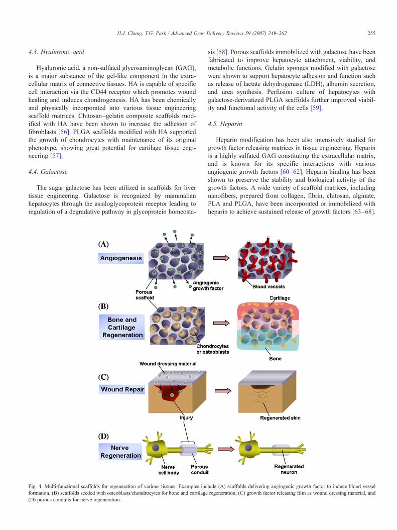
Fig. 4. Multi-functional scaffolds for regeneration of various tissues: Examples incformation, (B) scaffolds seeded with osteoblasts/chondrocytes for bone and cartilage(D) porous conduits for nerve regeneration.
sis [58]. Porous scaffolds immobilized with galactose have beenfabricated to improve hepatocyte attachment, viability, andmetabolic functions. Gelatin sponges modified with galactosewere shown to support hepatocyte adhesion and function suchas release of lactate dehydrogenase (LDH), albumin secretion,and urea synthesis. Perfusion culture of hepatocytes withgalactose-derivatized PLGA scaffolds further improved viabil-ity and functional activity of the cells [59].
4.5. Heparin
Heparin modification has been also intensively studied forgrowth factor releasing matrices in tissue engineering. Heparinis a highly sulfated GAG constituting the extracellular matrix,and is known for its specific interactions with variousangiogenic growth factors [60–62]. Heparin binding has beenshown to preserve the stability and biological activity of thegrowth factors. A wide variety of scaffold matrices, includingnanofibers, prepared from collagen, fibrin, chitosan, alginate,PLA and PLGA, have been incorporated or immobilized withheparin to achieve sustained release of growth factors [63–68].
lude (A) scaffolds delivering angiogenic growth factor to induce blood vesselregeneration, (C) growth factor releasing film as wound dressing material, and

256 H.J. Chung, T.G. Park / Advanced Drug Delivery Reviews 59 (2007) 249–262
5. Scaffolds for growth factor release
Polymeric scaffolds can be designed to function moreactively in tissue remodeling and regeneration by incorporatinggrowth factors (Fig. 3). Local and sustained delivery ofparacrine factors, either by inducing or inhibiting cellproliferation, survival, migration and/or differentiation, maygreatly enhance tissue remodeling or organogenesis (Fig. 4) [9].Growth factors can be incorporated into the scaffold matrixeither by bulk encapsulation, specific or non-specific surfaceadsorption, and adding microspheres encapsulating them.
5.1. Angiogenesis
Therapeutic angiogenesis is crucial in treating ischemic heartdiseases, wound repair, and tissue regeneration. In tissueengineering, the formation of blood vessels is essential for thesurvival of a growing tissue or organ, for it provides faciletransport of oxygen and nutrients [69]. Various angiogenicgrowth factors, such as vascular endothelial growth factor(VEGF), acidic or basic fibroblast growth factor (aFGF, bFGF),angiopoietin, and platelet-derived growth factor (PDGF), havebeen incorporated into 3-D matrices and delivered locally in asustained manner. Initially, direct incorporation of angiogenicgrowth factor was attempted by fabricating the scaffold using awater-in-oil emulsion where an aqueous protein solution wasemulsified in an organic polymer solution [70]. Later, VEGFwas embedded into the porous matrix by mixing the proteinpowder with polymer particles followed by exposing high-pressure CO2 [71]. The scaffold exhibited sustained releaseprofiles of VEGF showing bioactivity on proliferation ofendothelial cells in vitro, as well as angiogenic effects in vivo.As an alternative method, PLGA microspheres encapsulatingthe growth factor were incorporated into a 3-D scaffold [72].PLGA microspheres releasing bFGF were entrapped within analginate scaffold, which induced proliferation of cardiofibro-blasts in vitro and angiogenesis in vivo. Though the directencapsulation process provides an effective way of physicallyentrapping the therapeutic protein within a polymeric device,serious problems reside in maintaining structural integrity andbioactivity of the protein. Simple physical adsorption of growthfactors on the surface of scaffolds could partially solve thisproblem [73]. Porous PLA sponges were prepared by a gasfoaming method and subsequently surface coated with bFGF.Engraftment of hepatocytes followed by implantation resultedin improved blood vessel in-growth with increasing the extentof cell survival. However, this physical adsorption method onthe surface failed to induce angiogenesis when implanted, dueto the lack of long-term sustained release effects at the localtissue site. To achieve sustained release of angiogenic growthfactors from the scaffold, heparin immobilized scaffolds wereprepared. Heparin grafted on the surface or chemically bound tothe polymer can interact with heparin-binding angiogenicgrowth factors including VEGF and bFGF with specific bindingaffinity [60–62]. Growth factors can be released out in asustained manner due to the presence of a reversiblethermodynamic equilibrium between the immobilized heparin
and the incorporated growth factor. Collagen matrices weremodified with heparin, by a conjugation reaction betweencarboxyl groups of heparin and amino groups of collagen, forbinding and release of bFGF [63]. A similar procedure wasperformed in a later study for evaluating VEGF release [74].Both systems exhibited enhanced bioactivity of the angiogenicfactor by presence of heparin in the scaffold matrices, showinggreat angiogenic potential.
Synthetic polymeric scaffolds made of PLGA were alsoimmobilized with heparin on the surface for sustained release ofangiogenic growth factors. Park et al. fabricated macroporousPLGA scaffolds by a gas foaming/salt leaching method asmentioned earlier [68]. A blending mixture of PLGA and NH2–PEG–PLGA was used to generate surface amine groups forheparin immobilization. A carboxyl group of heparin wasconjugated to the surface amine group. bFGF binding andrelease studies showed that bFGF sustained release whileretaining its bioactivity as determined by proliferation ofendothelial cells in vitro. When bFGF loaded heparin modifiedscaffolds were implanted in vivo, significantly enhancedneovascularization was observed. More recently, porous andinjectable microspheres immobilized with heparin have beenutilized for growth factor delivery. A phase separation–porogenleaching process was used for preparing porous PLGA micro-spheres with a size of∼50 μm and pores of∼5 μm [75,76]. Theheparin immobilized microspheres released out bioactive bFGFin a sustained manner over a one month period and exhibitedpronounced angiogenic effect in an animal model.
5.2. Bone and cartilage regeneration
Formation of bone and cartilage requires stimulation byvarious growth factors, such as members of the transforminggrowth factor-β (TGF-β) superfamily, platelet-derived growthfactor (PDGF), and insulin-like growth factor (IGF) [9]. Amongthe TGF-β superfamily, TGF-β1 is the most well known factorinvolved in chondrogenic differentiation [77]. For bone tissueengineering, a family of bone morphogenic proteins (BMPs)plays important roles in osteogenesis and bone formation [78].These growth factors have been delivered by biodegradablepolymer scaffolds with a variety of intrinsic and structuralcharacteristics for engineering of bone and cartilage, and evenwound repair [79]. TGF-β1 has been incorporated into porousscaffolds, usually indirectly by pre-encapsulation into micro-spheres and subsequent loading into a scaffold matrix [80] orhydrogel [81]. Chitosan scaffolds containing TGF-β encapsu-lated chitosan microspheres were developed for in vitro cultureof chondrocytes [80]. The presence of TGF-β within thescaffold enhanced cell proliferation, and stimulated productionof GAG and collagen type II, leading to better cartilageformation. Recently, another system for cartilage repair wasstudied by combining a gelatin–chondrotin–hyaluronate com-posite scaffold and gelatin microspheres releasing TGF-β [82].To investigate chondrogenic potential, the construct was seededwith mesenchymal stem cells (MSCs) and implanted intoarticular cartilage defects, which resulted in facilitated repair ofthe tissue. BMP-2 has drawn interest in tissue engineering

257H.J. Chung, T.G. Park / Advanced Drug Delivery Reviews 59 (2007) 249–262
research due to its effectiveness and safety for treating bonedefects, which were well demonstrated in clinical trials. BMP-2releasing PLA scaffolds have been prepared by a supercriticalfluid process [83]. The osteogenic factor released from thematrix enhanced osteoprogenitor differentiation and boneformation when studied ex vivo in a chick chorioallantoicmembrane, and in vivo by implantation into subcutaneousdorsum or diffusion chambers. In another study, porous silkfibroin scaffolds adsorbed with BMP-2 were also developed forinducing osteogenesis of MSCs [84]. Scaffolds cultured withthe cells exhibited formation of bone-like tissue which wasdetermined in vitro by micro-computed tomography, and invivo in a cranial defect model. PDGF-BB, also a well knownangiogenic inducer, has been delivered for bone tissueengineering. Chitosan sponges have been incorporated withPDGF-BB by simple adsorption, showing significant regener-ative capacity in a craniotomy defect [85]. An improved systemwas developed by producing chitosan–PLA composite andchitosan coated PLA matrices, which revealed enhancedtherapeutic efficacy compared to the previous study [86].
5.3. Wound repair
Effective growth factor delivery systems for acceleratedwound healing have received much attention for treatingpatients suffering each year from burns or diabetic foot ulcers[87]. Wound dressing materials usually comprise of an inner gelor scaffold layer that directly contacts the wound site, and anouter elastic covering layer that fixes the device to the patients'tissue. A variety of growth factors including epidermal growthfactor (EGF), FGFs, TGF-β, and PDGF have been utilized forpromoting the healing process. EGF, a potent mitogen forepithelial cells, is the most widely used therapeutic agent forskin regeneration [88]. Initially, it was reported that sustainedrelease of EGF accelerated wound repair [87]. The sustainedrelease system was a polyvinyl alcohol sponge disc with EGF-containing cholesterol/methyl cellulose/lactose pellets embed-ded within it. Implantation of the device greatly enhancedformation of granulation tissue, and increased the collagencontent. Collagen was also one of the early materials used forwound repair. aFGF and TGF-β have been added to crosslinkedcollagen and administered into full-thickness skin defects[89,90]. The results showed considerably increased rates inepithelialization, contraction, and blood vessel formation. Later,sustained EGF releasing microspheres were incorporated into aporous gelatin scaffold which resulted in more effective healingof the skin wound, without any sign of foreign body reactions[91].
5.4. Liver regeneration
Growth factors known to promote survival and proliferationof hepatocytes include EGF and hepatocyte growth factor(HGF) [89]. EGF released from PGA mesh induced theaggregation of hepatocytes attached to the matrix [92]. Sinceblood supply plays a major role in liver regeneration andfunction, hepatocyte culture has often been associated with
angiogenesis. bFGF releasing PLA scaffold discs were seededwith hepatocytes and grafted in vivo, which induced vascular-ization resulting from enhanced proliferation and survival ofhepatocytes [93]. In another study, EGF, HGF and VEGF wereall co-delivered from PLGA scaffolds, which enhanced short-term survival of the transplanted hepatocytes [92].
5.5. Neural tissue engineering
Neural tissue engineering involves the implant of a poly-meric guide conduit at the site of neural injury to facilitate theregeneration process. Neurotrophic factors including nervegrowth factor (NGF), brain-derived neurotrophic factor(BDNF), neurotrophin-3 (NT-3), glial growth factor (GGF),and FGFs have been used to promote proliferation andmigration of neural cells [94]. Natural ECM derived materialshave been popularly used for delivery of these neurotrophicfactors. Examples are fibronectin grafts releasing NGF [95],fibrin gels releasing NT-3 [96], heparin containing fibrin gelswith NGF [97], and collagen tubules with BDNF [98]. Recently,synthetic materials have drawn attention for applications innerve repair. Biodegradable polymer conduit foams carryingGGF and Schwann cells [99], and those incorporating aFGF[100] showed great enhancement in repair of peripheral nerve orspinal cord, respectively.
6. Scaffolds for DNA and drug delivery
6.1. Scaffolds for DNA delivery
Growth factor releasing scaffolds have some inherentlimitations such as (i) protein instability problems encounteredduring the harsh formulation process and (ii) the very short half-life after release in the body fluid [9]. Therefore, transfection ofthe gene encoding a growth factor to target cells has beensuggested as an effective approach for enabling continuousexpression and release of the growth factor in the local tissuesite. Polymer scaffolds have been designed to continuouslyrelease the genetic material as naked DNA or in the form ofpolyplexes, thereby transfecting to seeded cells and expressingthe growth factor to stimulate morphogenesis of specific cells toform the desired tissue [10,11,101]. DNA plasmids have beendirectly encapsulated into scaffolds for sustained release invitro. For example, plasmid DNA encoding a luciferase genehas been directly incorporated into porous PLGA scaffolds bythe TIPS method. The DNAwas released in a sustained mannerwhile maintaining structural integrity, and also showedconsiderable transfection efficiency [102]. A plasmid encodingVEGF was encapsulated into PLGA scaffolds which resulted inlocal and sustained delivery of VEGF at the implant site leadingto increased blood vessel density, showing high angiogenicpotential [103]. Applying the TGF-β gene to support growth ofchondrocytes resulted in high secretion of ECM components,presenting its usefulness in cartilage engineering [104].Encapsulating the DNA within microspheres prior to incorpo-ration into 3-D scaffolds was shown to prolong its release [105].Formulation conditions for fabrication of the microspheres and

Fig. 5. Use of injectable porous scaffold microspheres for cartilage tissue engineering as an example. Primary chondrocytes are seeded within porous scaffold micro-spheres, expanded in vitro, and then injected or alternatively implanted as a cultured tissue into a cartilage defect site.
258 H.J. Chung, T.G. Park / Advanced Drug Delivery Reviews 59 (2007) 249–262
scaffolds could be varied to control the DNA release profiles[106]. Instead of delivering the naked plasmid, nanoparticulatepolyplexes composed of DNA and polycationic condensingagent have been incorporated within the scaffold for increasinggene transfection efficiency. Polycationic agents such aspolyethylenimine condense the plasmid DNA into positivelycharged nanoparticles and enhance DNA stability, intracellularuptake, and transfection efficiency. PEI/DNA polyplexes wereincorporated into PLGA scaffolds by using high-pressure gas[107]. When implanted in vivo, the seeded cells in the scaffoldwith the polyplexes exhibited much higher level of geneexpression than those with naked DNA. DNA polyplexes werealso physically immobilized onto the surface of pre-fabricatedscaffolds by adsorption to further enhance structural stability ofthe DNA through the formulation process [108].
6.2. Drug releasing scaffolds
Small molecular weight drugs that control proliferation ordifferentiation of cells can also be incorporated into biodegrad-able scaffolds to induce cellular differentiation and tissueremodeling. For example, dexamethasone, a steroidal anti-inflammatory drug, was loaded into the bulk phase of PLGAscaffolds for sustained release [109]. It was observed thatsustained release of dexamethasone effectively induced differ-entiation of bone marrow stem cells to osteoblasts orchondrocytes [110].
7. Injectable matrices
Although pre-fabricated scaffolds are most widely used fortissue regeneration purposes, they require a surgical procedurefor implantation. In-situ formed injectable scaffold materialshave received much attention recently, since they can beadministered using a syringe needle. For instance, injectable
hydrogels can be combined with the desired cells and growthfactors in a solution state prior to injection, but after injection, itimmediately becomes a temporal gel depot at the tissue defectsite while the tissue regenerates. Typically, temperature-sensitive sol–gel transition hydrogels and photo-crosslinkablehydrogels have been popularly used for cell and growth factordelivery.
7.1. Hydrogels
Early studies on hydrogels as tissue engineering scaffoldsinvolved naturally derived materials such as alginate, fibrin, andgelatin [111,112]. Recently, physical hydrogels based on syntheticpolymers exhibiting temperature dependent sol–gel phase transi-tion behavior have been widely used. They include poly(N-isopropylacrylamide, NIPAAm), Pluronic PEO–PPO–PEO tri-block copolymers, PLGA–PEO–PLGA tri-block copolymers, andpolyphosphazenes [113–116]. Above critical concentrations, thesehydrogels show a sol state at room temperature, but reversiblychange into a gel state at body temperature. For example, injectablepolyNIPAAm physical hydrogels encapsulating cells have beenprepared for cartilage and nerve regeneration [113,117]. ThePEO–PPO–PEO tri-block copolymer (Pluronic) is popularly usedas an in-situ forming gel for drug delivery [114]. Pluroniccopolymers at higher concentration above 20% (w/v) have beenused to encapsulate chondrocytes and produce engineeredcartilage [118]. However, Pluronic physical hydrogels rapidlydissolve out at the local site when injected, because they areimmediately dilutedwith the body fluid. To confer the gel stability,grafted copolymers composed of PLGA and PEG were synthe-sized. PLGA-g-PEG and PEG-g-PLGA hydrogels capable ofsustained insulin delivery and cartilage repair were synthesized[119]. In general, these physical hydrogels, when injected withcells, do not exhibit sufficient mechanical strength to hold theproliferating and differentiating cells with providing a structural
259H.J. Chung, T.G. Park / Advanced Drug Delivery Reviews 59 (2007) 249–262
support for a desired period as the tissue is regenerated. To bettermaintain the structural shape and dimension, photo-crosslinkablechemical hydrogels were introduced more recently. Hyaluronicacid (HA) hydrogels encapsulating cells have been prepared byphoto-polymerization of functionalized precursor macromers inthe presence of fibroblasts [120]. Photo-crosslinked PEG basedhydrogels have also been utilized for delivery of chondrocytes,osteoblasts, and MSCs [121–123]. Pluronic/heparin compositehydrogels delivering growth factor have also been studied toinduce angiogenesis [124]. Hydrogels mimicking the extracellularenvironment were prepared by incorporating protease sensitivepeptide sequences into photo-crosslinkable PEG hydrogels tofacilitate cell invasion and migration [125]. Quite recently, a newclass of in-situ formed scaffolds by self-assembly of syntheticpolypeptides also emerged. The peptide hydrogels could formunique secondary structures in physiological aqueous condition,which constitute a stable 3-D hydrogel structure at lowconcentrations (0.1–1%) [126,127]. Additionally, poly(aldehydeguluronate) or poly(organophosphazene) has been shown to forminjectable, biodegradable, and thermosensitive matrices useful forcell delivery, which should be further evaluated in the future[128,129].
7.2. Microspheres
Biodegradable PLGA microspheres have been studied fordelivery of chondrocytes for cartilage engineering. Non-porousPLGA microspheres could be used as (i) a microcarrier for cellexpansion in vitro [130], prior to use as an (ii) injectable carrierfor cartilage regeneration in vivo [131–133]. We recentlyreported that highly porous microspheres (∼200 μm in overalldiameter and ∼30 μm in pore diameter) could be prepared by agas foaming method [29]. These porous scaffold microspherescould be used for microcarrier suspension culture of cells, aswell as injection of the cell/microsphere constructs into a tissuedefect site (Fig. 5). These injectable and porous microsphereswould provide a great advantage for cell therapy in manyaspects. Prior to injection, the porous structure (∼30 μm) wouldallow sufficient cell seeding in and out of the matrix. Afterinjection in vivo, the porous matrix would permit infiltration ofcells and in-growth of tissue from the host, facilitating theregeneration process.
8. Conclusions
Multi-functional, biodegradable, biocompatible, and biomi-metic polymeric scaffolds have been intensively studied fordecades to provide implantable devices for tissue regeneration.Porous scaffolds can be fabricated by various methods such asfiber bonding, melt molding, solvent casting/particulate leach-ing, gas foaming/particulate leaching, phase separation, high-pressure processing, electrospinning, and rapid prototyping.These scaffolds can be in the form of a solid foam, nanofibrousmatrix, microsphere, or hydrogel. The polymeric devices areusually surface modified by immobilizing cell adhesive orgrowth factor binding moieties to improve its cell adhesivecharacteristics or actively induce cell migration, proliferation,
and differentiation. Surface engineered and drug releasingscaffolds can be rationally designed to mimic the extracellularmatrix environment, thereby facilitating the tissue regenerationprocess.
Acknowledgement
This work was supported by the National ResearchLaboratory grant from the Ministry of Science and Technology,Korea.
References
[1] R. Langer, J.P. Vacanti, Tissue engineering, Science 260 (1993) 920–926.[2] L.E. Freed, G. Vunjak-Novakovic, R.J. Biron, D.B. Eagles, D.C. Lesnoy,
S.K. Barlow, R. Langer, Biodegradable polymer scaffolds for tissueengineering, Nat. Biotechnol. 12 (1994) 689–693.
[3] D.W. Hutmacher, Scaffolds in tissue engineering bone and cartilage,Biomaterials 21 (2000) 2529–2543.
[4] S.J. Peter, M.J. Miller, A.W. Yasko, M.J. Yaszemski, A.G. Mikos,Polymer concepts in tissue engineering, J. Biomed. Mater. Res.(Appl. Biomater.) 43 (1998) 422–427.
[5] A.G. Mikos, A.J. Thorsen, L.A. Czerwonka, Y. Bao, R. Langer, D.N.Winslow, J.P. Vacanti, Preparation and characterization of poly(L-lacticacid) foams, Polymer 35 (1994) 1068–1077.
[6] Y.S. Nam, J.J. Yoon, T.G. Park, A novel fabrication method formacroporous scaffolds using gas foaming salt as porogen additive,J. Biomed. Mater. Res. (Appl. Biomater.) 53 (2000) 1–7.
[7] Z. Ma, M. Kotaki, R. Inai, S. Ramakrishna, Potential of nanofiber matrixas tissue-engineering scaffolds, Tissue Eng. 11 (2005) 101–109.
[8] M.P. Lutolf, J.A. Hubbell, Synthetic biomaterials as instructiveextracellular microenvironments for morphogenesis in tissue engineer-ing, Nat. Biotechnol. 23 (2005) 47–55.
[9] R.R. Chen, D.J. Mooney, Polymeric growth factor delivery strategies fortissue engineering, Pharm. Res. 20 (2003) 1103–1112.
[10] T.G. Park, J.H. Jeong, S.W. Kim, Current status of polymeric genedelivery systems, Adv. Drug Deliv. Rev. 58 (2006) 467–486.
[11] L.D. Shea, E. Smiley, J. Bonadio, D.J. Mooney, DNA delivery from polymermatrices for tissue engineering, Nat. Biotechnol. 17 (1999) 551–554.
[12] A.S. Hoffman, Hydrogels for biomedical applications, Adv. Drug Deliv.Rev. 43 (2002) 3–12.
[13] J.L. Drury, D.J. Mooney, Hydrogels for tissue engineering: scaffolddesign variables and applications, Biomaterials 24 (2003) 4337–4351.
[14] S.E. Sakiyama-Elbert, J.A. Hubbell, Development of fibrin derivativesfor controlled release of heparin-binding growth factors, J. Control.Release 65 (2000) 389–402.
[15] J.S. Pieper, T. Hafmans, P.B. van Wachem, M.J.A. van Luyn, L.A.Brouwer, J.H. Veerkamp, T.H. van Kuppevelt, Loading of collagen–heparan sulfate matrices with bFGF promotes angiogenesis and tissuegeneration in rats, J. Biomed. Mater. Res. 62 (2002) 185–194.
[16] D.L. Nettles, S.H. Elder, J.A. Gilbert, Potential use of chitosan as a cellscaffold material for cartilage tissue engineering, Tissue Eng. 8 (2002)1009–1016.
[17] Z. Li, H.R. Ramay, K.D. Hauch, D. Xiao, M. Zhang, Chitosan–alginatehybrid scaffolds for bone tissue engineering, Biomaterials 26 (2005)3919–3928.
[18] A. Perets, Y. Baruch, F. Weisbuch, G. Shoshany, G. Neufeld, S. Cohen,Enhancing the vascularization of three-dimensional porous alginatescaffolds by incorporating controlled release basic fibroblast growthfactor microspheres, J. Biomed. Mater. Res. 65A (2003) 489–497.
[19] J.B. Leach, K.A. Bivens, C.W. Patrick, C.E. Schmidt, Photocrosslinkedhyaluronic acid hydrogels: natural, biodegradable tissue engineeringscaffolds, Biotechnol. Bioeng. 82 (2003) 578–589.
[20] P.A. Gunatillake, R. Adhikari, Biodegradable synthetic polymers fortissue engineering, Eur. Cells Mater. 5 (2003) 1–16.

260 H.J. Chung, T.G. Park / Advanced Drug Delivery Reviews 59 (2007) 249–262
[21] G. Shi, Q. Cai, C. Wang, N. Lu, S. Wang, J. Bei, Fabrication andbiocompatibility of cell scaffolds of poly(L-lactic acid) and poly(L-lactic-co-glycolic acid), Polym. Adv. Technol. 13 (2002) 227–232.
[22] C.G. Williams, T.K. Kim, A. Taboas, A. Malik, P. Manson, J. Eisseeff, Invitro chondrogenesis of bone marrow-derived mesenchymal stem cells ina photopolymerizing hydrogel, Tissue Eng. 9 (2003) 679–688.
[23] B. Kim, D.J. Mooney, Engineering smooth muscle tissue with apredefined structure, J. Biomed. Mater. Res. 41 (1998) 322–332.
[24] K. Whang, C.H. Thomas, K.E. Healy, A novel method to fabricatebioabsorbable scaffolds, Polymer 36 (1995) 837–842.
[25] A.G. Mikos, G. Sarakinos, S.M. Leite, J.P. Vacanti, R. Langer, Laminatedthree-dimensional biodegradable foams for use in tissue engineering,Biomaterials 14 (1993) 323–330.
[26] D.J. Mooney, D.F. Baldwin, N.P. Suh, J.P. Vacanti, R. Langer, Novelapproach to fabricate porous sponges of poly(D,L-lactic-co-glycolic acid)without the use of organic solvents, Biomaterials 17 (1996) 1417–1422.
[27] L.D. Harris, B. Kim, D.J. Mooney, Open pore biodegradable matricesformed with gas foaming, J. Biomed. Mater. Res. 42 (1998) 396–402.
[28] J.J. Yoon, T.G. Park, Degradation behaviors of biodegradable macro-porous scaffolds prepared by gas foaming of effervescent salts,J. Biomed. Mater. Res. 55 (2001) 401–408.
[29] T.G. Kim, J.J. Yoon, D.S. Lee, T.G. Park, Gas foamed open porousbiodegradable polymeric microspheres, Biomaterials 27 (2006) 152–159.
[30] Y.S.Nam, T.G. Park, Porous biodegradable polymeric scaffolds prepared bythermally induced phase separation, J. Biomed.Mater. Res. 47 (1999) 8–17.
[31] Y.S. Nam, T.G. Park, Biodegradable polymeric microcellular foamsby modified thermally induced phase separation method, Biomaterials20 (1999) 1783–1790.
[32] C. Schugens, V. Maquet, C. Grandfils, R. Jerome, P. Teyssie, Polylactidemacroporous biodegradable implants for cell transplantation. 11.Preparation of polylactide foams by liquid–liquid phase separation,J. Biomed. Mater. Res. 30 (1996) 449–461.
[33] W. Li, C.T. Laurencin, E.J. Caterson, R.S. Tuan, F.K. Ko, Electrospunnanofibrous structure: a novel scaffold for tissue engineering, J. Biomed.Mater. Res. 60 (2002) 613–621.
[34] J.A. Matthews, G.E. Wnek, D.G. Simpson, G.L. Bowlin, Electrospinningof collagen nanofibers, Biomacromolecules 3 (2002) 232–238.
[35] S.H. Kim, Y.S. Nam, T.S. Lee, W.H. Park, Silk fibroin nanofiber:electrospinning, properties, and structure, Polym. J. 35 (2003) 185–190.
[36] S. Yang, K. Leong, Z. Du, C. Chua, The design of scaffolds for use intissue engineering. Part II. Rapid prototyping techniques, Tissue Eng.8 (2002) 1–11.
[37] I. Zein, D.W. Hutmacher, K.C. Tan, S.H. Teoh, Fused depositionmodeling of novel scaffold architectures for tissue engineering applica-tions, Biomaterials 23 (2002) 1169–1185.
[38] T.B. Woodfield, J. Malda, J. de Wijn, F. Peters, J. Riesle, C.A. vanBlitterswijk, Design of porous scaffolds for cartilage tissue engineering usinga three-dimensional fiber-deposition technique, Biomaterials 25 (2004)4149–4161.
[39] S.S. Kim, H. Utsunomiya, J.A. Koski, B.M. Wu, M.J. Cima, J. Sohn, K.Mukai, L.G. Griffith, J.P. Vacanti, Survival and function of hepatocyteson a novel three-dimensional synthetic biodegradable polymeric scaffoldwith an intrinsic network of channels, Ann. Surg. 228 (1998) 8–13.
[40] J. Zeltinger, J.K. Sherwood, D.A. Graham, R. Mueller, L.G. Griffith,Effect of pore size and void fraction on cellular adhesion, proliferationand matrix deposition, Tissue Eng. 7 (2001) 557–572.
[41] J.M. Taboas, R.D. Maddox, P.H. Krebsbach, S.J. Hollister, Indirect SFFfabrication of local and global porous, biomimetic and composite 3Dpolymer–ceramic scaffolds, Biomaterials 24 (2003) 181–194.
[42] C.R. Nuttelman, D.J. Mortisen, S.M. Henry, K.S. Anseth, Attachment offibronectin to poly(vinyl alcohol) hydrogels promotes NIH3T3 celladhesion, proliferation, and migration, J. Biomed. Mater. Res. 57 (2001)217–223.
[43] R.S. Bhati, D.P.Mukherjee, K.J.McCarthy, S.H. Rogers, D.F. Smith, S.W.Shalaby, The growth of chondrocytes into a fibronectin-coated biode-gradable scaffold, J. Biomed. Mater. Res. 56 (2001) 74–82.
[44] U. Hersel, C. Dahmen, H. Kessler, RGDmodified polymers: biomaterials forstimulated cell adhesion and beyond, Biomaterials 24 (2003) 4385–4415.
[45] J. Riesle, A.P. Hollander, R. Langer, L.E. Freed, G. Vunjak-Novakovic,Collagen in tissue-engineered cartilage: types, structure, and crosslinks,J. Cell. Biochem. 71 (1998) 313–327.
[46] J. Yang, Y. Wan, J. Yang, J. Bei, S. Wang, Plasma-treated, collagen-anchored polylactone: its cell affinity evaluation under shear or shear-freeconditions, J. Biomed. Mater. Res. 67A (2003) 1139–1147.
[47] Z. Ma, C. Gao, Y. Gong, J. Shen, Cartilage tissue engineering PLLAscaffold with surface immobilized collagen and basic fibroblast growthfactor, Biomaterials 26 (2005) 1253–1259.
[48] X. Liu, Y. Won, P.X. Ma, Surface modification of interconnected porousscaffolds, J. Biomed. Mater. Res. 74A (2005) 84–91.
[49] E. Ruoslahti, RGD and other recognition sequences for integrins, Annu.Rev. Cell Dev. Biol. 12 (1996) 697–715.
[50] J.L. Myles, B.T. Burgess, R.B. Dickson, Modification of the adhesiveproperties of collagen by covalent grafting with RGD peptides,J. Biomater. Sci., Polym. Ed. 11 (2000) 69–86.
[51] J.C. Schense, J. Bloch, P. Aebischer, J.A. Hubbell, Enzymaticincorporation of bioactive peptides into fibrin matrices enhances neuriteextension, Nat. Biotechnol. 18 (2000) 415–419.
[52] Y. Hu, S.R.Winn, I. Krajbich, J.O. Hollinger, Porous polymer scaffolds surface-modified with arginine–glycine–aspartic acid enhance bone cell attachment anddifferentiation in vitro, J. Biomed. Mater. Res. 64A (2003) 583–590.
[53] M. Ho, L. Hou, C. Tu, H. Hseih, J. Lai, W. Chen, D. Wang, Promotion ofcell affinity of porous PLLA scaffolds by immobilization of RGDpeptides via plasma treatment, Macromol. Biosci. 6 (2006) 90–98.
[54] J.J. Yoon, S.H. Song, D. Sung Lee, T.G. Park, Immobilization of celladhesive RGD peptide onto the surface of highly porous biodegradablepolymer scaffolds fabricated by a gas foaming/salt leaching method,Biomaterials 25 (2004) 5613–5620.
[55] T.G. Kim, T.G. Park, Biomimicking extracellular matrix: cell adhesiveRGD peptide modified electrospun poly(D,L-lactic-co-glycolic acid)nanofiber mesh, Tissue Eng. 12 (2006) 221–233.
[56] J.S. Mao, H.F. Liu, Y.J. Yin, K.D. Yao, The properties of chitosan–gelatinmembranes and scaffolds modified with hyaluronic acid by differentmethods, Biomaterials 24 (2003) 1621–1629.
[57] H.S. Yoo, E.A. Lee, J.J. Yoon, T.G. Park, Hyaluronic acid modifiedbiodegradable scaffolds for cartilage tissue engineering, Biomaterials26 (2005) 1925–1933.
[58] S.R. Hong, Y.M. Lee, T. Akaike, Evaluation of a galactose-carryinggelatin sponge for hepatocytes culture and transplantation, J. Biomed.Mater. Res. 67A (2003) 733–741.
[59] T.G. Park, Perfusion culture of hepatocytes within galactose-derivatizedbiodegradable poly(lactide-co-glycolide) scaffolds prepared by gasfoaming of effervescent salts, J. Biomed. Mater. Res. 59 (2002) 127–135.
[60] R. Padera, G. Venkataraman, D. Berry, R. Godavarti, R. Sasisekharan,FGF-2/fibroblast growth factor receptor/heparin-like glycosaminogly-can interactions: a compensation model for FGF-2 signaling, FASEB J.13 (1999) 1677–1687.
[61] L.D. Thompson, M.W. Pantoliano, B.A. Springer, Energetic characteriza-tion of the basic fibroblast growth factor–heparin interaction: identificationof the heparin binding domain, Biochemistry 33 (1994) 3831–3840.
[62] R. Raman, G. Venkataraman, S. Ernst, V. Sasisekharan, R. Sasisekharan,Structural specificity of heparin binding in the fibroblast growth factorfamily of proteins, Proc. Natl. Acad. Sci. 100 (2003) 2357–2362.
[63] M.J.B. Wissink, R. Beernink, A.A. Poot, G.H.M. Engbers, T. Beugeling,W.G. van Aken, J. Feijen, Improved endothelialization of vascular graftsby local release of growth factor from heparinized collagen matrices,J. Control. Release 64 (2000) 103–114.
[64] J.S. Pieper, T. Hafmans, P.B. van Wachem, M.J.A. van Luyn, L.A.Brouwer, J.H. Veerkamp, T.H. van Kuppevelt, Loading of collagen–heparan sulfate matrices with bFGF promotes angiogenesis and tissuegeneration in rats, J. Biomed. Mater. Res. 62 (2002) 185–194.
[65] S.E. Sakiyama-Elbert, J.A. Hubbell, Development of fibrin derivativesfor controlled release of heparin-binding growth factors, J. Control.Release 65 (2000) 389–402.
[66] J. Li, J. Pan, L. Zhang, X. Guo, Y. Yu, Culture of primary rat hepatocyteswithin porous chitosan scaffolds, J. Biomed. Mater. Res. 67A (2003)938–943.

261H.J. Chung, T.G. Park / Advanced Drug Delivery Reviews 59 (2007) 249–262
[67] A.T. Gutsche, H. Lo, J. Zurlo, J. Yager, K.W. Leong, Engineering of asugar-derivatized porous network for hepatocyte culture, Biomaterials17 (1996) 387–393.
[68] J.J. Yoon, H.J. Chung, H.J. Lee, T.G. Park, Heparin-immobilizedbiodegradable scaffolds for local and sustained release of angiogenicgrowth factor, J. Biomed. Mater. Res. 79 (2006) 934–942.
[69] H.J. Chung, H.K. Kim, J.J. Yoon, T.G. Park, Heparin immobilized porousPLGA microspheres for angiogenic growth factor delivery, Pharm. Res.23 (2006) 1835–1841.
[70] D.D. Hile, M.L. Amirpour, A. Akgerman, M.V. Pishko, Active growthfactor delivery from poly(D,L-lactide-coglycolide) foams prepared insupercritical CO2, J. Control. Release 66 (2000) 177–185.
[71] W.L. Murphy, M.C. Peters, D.H. Kohn, D.J. Mooney, Sustained release ofvascular endothelial growth factor frommineralized poly(lactide-co-glycolide)scaffolds for tissue engineering, Biomaterials 21 (2000) 2521–2527.
[72] A. Perets, Y. Baruch, F. Weisbuch, G. Shoshany, G. Neufeld, S. Cohen,Enhancing the vascularization of three-dimensional porous alginatescaffolds by incorporating controlled release basic fibroblast growthfactor microspheres, J. Biomed. Mater. Res. 65A (2003) 489–497.
[73] H. Lee, R.A. Cusick, F. Browne, T.H. Kim, P.X. Ma, H. Utsunomiya, R.Langer, J.P. Vacanti, Local delivery of basic fibroblast growth factorincreases both angiogenesis and engraftment of hepatocytes in tissue-engineered polymer devices, Transplantation 73 (2002) 1589–1593.
[74] G.C.M. Steffens, C. Yao, P. Prevel, M. Markowicz, P. Schenck, E.M.Noah, N. Pallua, Modulation of angiogenic potential of collagen matricesby covalent incorporation of heparin and loading with vascularendothelial growth factor, Tissue Eng. 10 (1994) 1502–1509.
[75] H.K. Kim, H.J. Chung, T.G. Park, Biodegradable polymeric micro-spheres with “open/closed” pores for sustained release of human growthhormone, J. Control. Release 112 (2006) 167–174.
[76] H. Lee, H.J. Chung, T.G. Park, Perspectives on: local and sustained deliveryof angiogenic growth factors, J. Bioact. Compat. Polym. 22 (2007) 89–114.
[77] J. Massague, The transforming growth factor-beta family, Annu. Rev.Cell Dev. Biol. 6 (1990) 597–641.
[78] J.M. Wozney, V. Rosen, Bone morphogenetic protein and bonemorphogenetic protein gene family in bone formation and repair, Clin.Ortop. 346 (1998) 26–37.
[79] W.A. Border, N.A. Noble, Transforming growth factorβ in tissue fibrosis,New Engl. J. Med. 331 (1994) 1286–1292.
[80] J.E. Lee, S.E. Kim, I.C. Kwon, H.J. Ahn, H. Cho, S. Lee, H.J. Kim, S.C.Seong, M.C. Lee, Effects of a chitosan scaffold containing TGF-b1encapsulated chitosan microspheres on in vitro chondrocyte culture,Artif. Organs 28 (2004) 829–839.
[81] A.J. DeFaila, C.R. Chub, N. Izzob, K.G. Marraa, Controlled release ofbioactive TGF-b1 from microspheres embedded within biodegradablehydrogels, Biomaterials 27 (2006) 1579–1585.
[82] H. Fan, Y. Hu, L. Qin, X. Li, H. Wu, R. Lv, Porous gelatin–chondroitin–hyaluronate tri-copolymer scaffold containing micro-spheres loaded with TGF-β1 induces differentiation of mesenchymalstem cells in vivo for enhancing cartilage repair, J. Biomed. Mater. Res.77A (2006) 785–794.
[83] X.B. Yang, M.J. Whitaker, W. Sebald, N. Clarke, S.M. Howdle, K.M.Shakesheff, R.O.C. Oreffo, Human osteoprogenitor bone formation usingencapsulated bone morphogenetic protein 2 in porous polymer scaffolds,Tissue Eng. 10 (2004) 1037–1045.
[84] V. Karageorgiou, M. Tomkins, R. Fajardo, L. Meinel, B. Snyder, K.Wade, J. Chen, G. Vunjak-Novakovic, D.L. Kaplan, Porous silk fibroin3-D scaffolds for delivery of bone morphogenetic protein-2 in vitro andin vivo, J. Biomed. Mater. Res. 78A (2006) 324–334.
[85] Y.J. Park, Y.M. Lee, S.N. Park, S.Y. Sheen, C.P. Chung, S.J. Lee, Plateletderived growth factor releasing chitosan sponge for periodontal boneregeneration, Biomaterials 21 (2000) 153–159.
[86] J. Lee, S. Nam, S. Im, Y. Park, Y. Lee, Y. Seol, C. Chung, S. Lee,Enhanced bone formation by controlled growth factor delivery fromchitosan-based biomaterials, J. Control. Release 78 (2002) 187–197.
[87] A. Buckley, J.M. Davidson, C.D. Kamerath, T.B. Wolt, S.C. Woodward,Sustained release of epidermal growth factor accelerates wound repair,Proc. Natl. Acad. Sci. 82 (1985) 7340–7344.
[88] M. Niall, G.B. Ryan, B. Mc, C. O'Brien, The effect of epidermal growthfactor on wound healing in mice, J. Surg. Res. 33 (1982) 164–169.
[89] A. Pandit, R. Ashar, D. Feldman, A. Thompson, Investigation of acidicfibroblast growth factor delivered through a collagen scaffold for thetreatment of full-thickness skin defects in a rabbit model, Plast. Reconstr.Surg. 101 (1998) 766–775.
[90] A. Pandit, R. Ashar, D. Feldman, The effect of TGF-b delivered through acollagen scaffold on wound healing, J. Invest. Surg. 12 (1999) 89–100.
[91] K. Ulubayram, A.N. Cakar, P. Korkusuz, C. Ertan, N. Hasirci, EGFcontaining gelatin-based wound dressings, Biomaterials 22 (2001)1345–1356.
[92] M.K. Smith, K.W. Riddle, D.J. Mooney, Delivery of hepatotrophicfactors fails to enhance longer-term survival of subcutaneouslytransplanted hepatocytes, Tissue Eng. 12 (2006) 235–244.
[93] J. Mayer, E. Karamuk, T. Akaike, E. Wintermantel, Matrices for tissueengineering-scaffold structure for a bioartificial liver support system,J. Control. Release 64 (2000) 81–90.
[94] C.E. Schmidt, J.B. Leach, Neural tissue engineering: strategies for repairand regeneration, Annu. Rev. Biomed. Eng. 5 (1993) 293–347.
[95] I.H. Whitworth, R.A. Brown, C.J. Dore, P. Anand, C.J. Green, G.Terenghi, Nerve growth factor enhances nerve regeneration throughfibronectin grafts, J. Hand Surg. [Br] 21 (1996) 514–522.
[96] S.J. Taylora, J.W. McDonald, S.E. Sakiyama-Elberta, Controlled releaseof neurotrophin-3 from fibrin gels for spinal cord injury, J. Control. Release98 (2004) 281–294.
[97] S.E. Sakiyama-Elbert, J.A. Hubbell, Controlled release of nerve growthfactor from a heparin-containing fibrin-based cell ingrowth matrix,J. Control. Release 69 (2000) 149–158.
[98] D.S. Utley, S.L. Lewin, E.T. Cheng, A.N. Verity, D. Sierra, D.J. Terris,Brain-derived neurotrophic factor and collagen tubulization enhancefunctional recovery after peripheral nerve transection and repair, Arch.Otolaryngol. Head Neck Surg. 122 (1996) 407–413.
[99] D.J. Bryan, A.H. Holway, K.Wang, A.E. Silva, D.J. Trantolo, D.Wise, I.C.Summerhayes, Influence of glial growth factor and Schwann cells in abioresorbable guidance channel on peripheral nerve regeneration, TissueEng. 6 (2000) 129–138.
[100] V. Maquet, D. Martin, F. Scholtes, R. Franzen, J. Schoenen, G. Moonen, R.Jerome, Poly(D,L-lactide) foams modified by poly(ethylene oxide)-block-poly(D,L-lactide) copolymers and a-FGF: in vitro and in vivo evaluation forspinal cord regeneration, Biomaterials 22 (2001) 1137–1146.
[101] D.M. Salvaya, L.D. Shea, Inductive tissue engineering with protein andDNA-releasing scaffolds, Mol. BioSyst. 2 (2006) 36–48.
[102] K.W. Chun, K.C. Cho, S.H. Kim, J.H. Jeong, T.G. Park, Controlledrelease of plasmid DNA from biodegradable scaffolds fabricated by athermally induced phase separation method, J. Biomater. Sci., Polym. Ed.15 (2004) 1341–1653.
[103] J. Jang, C.B. Rives, L.D. Shea, Plasmid delivery in vivo from poroustissue-engineering scaffolds: transgene expression and cellular transfec-tion, Molec. Ther. 12 (2005) 475–485.
[104] T. Guo, J. Zhao, J. Chang, Z. Ding, H. Hong, J. Chena, J. Zhang, Porouschitosan–gelatin scaffold containing plasmid DNA encoding trans-forming growth factor-b1 for chondrocytes proliferation, Biomaterials27 (2006) 1095–1103.
[105] M. Nof, L.D. Shea, Drug-releasing scaffolds fabricated from drug-loadedmicrospheres, J. Biomed. Mater. Res. 59 (2002) 349–356.
[106] J. Jang, L.D. Shea, Controllable delivery of non-viral DNA from porousscaffolds, J. Control. Release 86 (2003) 157–168.
[107] Y. Huang, K. Riddle, K.G. Rice, D.J. Mooney, Long-term in vivo geneexpression via delivery of PEI–DNA condensates from porous polymerscaffolds, Hum. Gene Ther. 16 (2005) 609–617.
[108] J. Jang, Z. Bengali, T.L. Houchin, L.D. Shea, Surface adsorption of DNAto tissue engineering scaffolds for efficient gene delivery, J. Biomed.Mater. Res. 77A (2006) 50–58.
[109] J.J. Yoon, J.H. Kim, T.G. Park, Dexamethasone releasing biodegradablepolymer scaffolds fabricated by a gas foaming/salt leaching method,Biomaterials 24 (2003) 2323–2329.
[110] J. Chen, C. Wang, S. Lu, J. Wu, X. Guo, C. Duan, L. Dong, Y. Song, J.Zhang, D. Jing, L. Wu, J. Ding, D. Li, In vivo chondrogenesis of adult

262 H.J. Chung, T.G. Park / Advanced Drug Delivery Reviews 59 (2007) 249–262
bone-marrow-derived autologous mesenchymal stem cells, Cell TissueRes. 319 (2005) 429–438.
[111] J.A. Rowley, G.Madlambayan, D.J. Mooney, Alginate hydrogels as syntheticextracellular matrix materials, Biomaterials 20 (1999) 45–53.
[112] D. Passaretti, R.P. Silverman,W. Huang, C.H. Kirchhoff, S. Ashiku,M.A.Randolph, M.J. Yaremchuk, Cultured chondrocytes produce injectabletissue-engineered cartilage in hydrogel polymer, Tissue Eng. 7 (2001)805–815.
[113] R.A. Stile, W.R. Burghardt, K.E. Healy, Synthesis and characterization ofinjectable poly(N-isopropylacrylamide)-based hydrogels that supporttissue formation in vitro, Macromolecules 32 (1999) 7370–7379.
[114] S. Fusco, A. Borzacchiello, P.A. Netti, Perspectives on: PEO–PPO–PEOtriblock copolymers and their biomedical applications, J. Bioact. Compat.Polym. 21 (2006) 149–164.
[115] K.T. Nguyen, J.L. West, Photopolymerizable hydrogels for tissueengineering applications, Biomaterials 23 (2002) 4307–4314.
[116] W.E. Hennink, C.F. van Nostrum, Novel crosslinking methods to designhydrogels, Adv. Drug Deliv. Rev. 54 (2002) 13–36.
[117] K.H. Park, K. Yun, Immobilization of Arg-Gly-Asp (RGD) sequence in athermosensitive hydrogel for cell delivery using pheochromocytoma cells(PC12), J. Biosci. Bioeng. 97 (2004) 374–377.
[118] A.B. Saim, Yilin Cao,Y.Weng,C.Chang,M.A.Vacanti, C.A.Vacanti, R.D.Eavey, Engineering autogenous cartilage in the shape of a helix using aninjectable hydrogel scaffold, Laryngoscope 110 (2000) 1694–1697.
[119] B. Jeong, K.M. Lee, A. Gutowska, Y.H. An, Thermogelling biodegrad-able copolymer aqueous solutions for injectable protein delivery andtissue engineering, Biomacromolecules 3 (2002) 865–868.
[120] X.Z. Shu, Y. Liu, F.S. Palumbo, Y. Luo, G.D. Prestwich, In situ crosslinkablehyaluronan hydrogels for tissue engineering, Biomaterials 25 (2004)1339–1348.
[121] S.J. Bryant, K.S. Anseth, Controlling the spatial distribution of ECMcomponents in degradable PEG hydrogels for tissue engineering cartilage,J. Biomed. Mater. Res. 64A (2003) 70–79.
[122] J.A. Burdicka, K.S. Ansetha, Photoencapsulation of osteoblasts ininjectable RGD-modified PEG hydrogels for bone tissue engineering,Biomaterials 23 (2002) 4315–4323.
[123] C.G. Williams, T.K. Kim, A. Taboas, A. Malik, P. Manson, J. Elisseeff, Invitro chondrogenesis of bone marrow-derived mesenchymal stem cells ina photopolymerizing hydrogel, Tissue Eng. 9 (2003) 679–688.
[124] J.J. Yoon, H. Chung, T.G. Park, Photo-crosslinkable and biodegradablePluronic/heparin hydrogels for local and sustained delivery of angiogenicgrowth factor, J. Biomed. Mater. Res., in press.
[125] B.K. Mann, A.S. Gobin, A.T. Tsai, R.H. Schmedlen, J.L. West, Smoothmuscle cell growth in photopolymerized hydrogels with cell adhesive andproteolytically degradable domains: synthetic ECM analogs for tissueengineering, Biomaterials 22 (2001) 3045–3051.
[126] J. Kisiday,M. Jin, B.Kurz, H. Hung, C. Semino, S. Zhang, A.J. Grodzinsky,Self-assembling peptide hydrogel fosters chondrocyte extracellular matrixproduction and cell division: implications for cartilage tissue repair, Proc.Natl. Acad. Sci. 99 (2002) 9996–10001.
[127] T.C. Holmes, Novel peptide-based biomaterial scaffolds for tissueengineering, Trends Biotechnol. 20 (2002) 16–21.
[128] K.Y. Lee, E. Alsberg, D.J. Mooney, Degradable and injectable poly(aldehyde guluronate) hydrogels for bone tissue engineering, J. Biomed.Mater. Res. 56 (2001) 228–233.
[129] K.H. Park, S.C. Song, Morphology of spheroidal hepatocytes withininjectable, biodegradable and thermosensitive poly(organophosphazene)hydrogel as cell delivery vehicle, J. Biosci. Bioeng. 101 (2006) 238–242.
[130] K.W. Chun, H.S. Yoo, J.J. Yoon, T.G. Park, Biodegradable PLGAmicrocarriers for injectable delivery of chondrocytes: effect of surfacemodification on cell attachment and function, Biotechnol. Prog. 20 (2004)1797–1801.
[131] N.R. Mercier, H.R. Costantino, M.A. Tracy, L.J. Bonassar, A novelinjectable approach for cartilage formation in vivo using PLG micro-spheres, Ann. Biomed. Eng. 32 (2004) 418–429.
[132] S.W. Kang, O. Jeon, B.S. Kim, Poly(lactic-co-glycolic acid) micro-spheres as an injectable scaffold for cartilage tissue engineering, TissueEng. 11 (2005) 438–447.
[133] H. Thissen, K.Y. Chang, T.A. Tebb, W.B. Tsai, V. Glattauer, J.A.M.Ramshaw, J.A. Werkmeister, Synthetic biodegradable microparticles forarticular cartilage tissue engineering, J. Biomed. Mater. Res. 77A (2006)590–598.