Sural nerve T-cell receptor Vb gene utilization in chronic ...
10
plates in alpha bungarotoxin-treated rats. J Physiol 1992;458: 487– 499. 45. Plomp JJ, Van Kempen GTH, De Baets M, Graus YMF, Kuks JBM, Molenaar PC. Acetylcholine release in myasthenia gra- vis: regulation at single end-plate level. Ann Neurol 1995;37: 627– 636. 46. Ropper AH, Wijdicks EFM, Truax BT. Guillain–Barré Syn- drome. Philadelphia, PA: FA Davis; 1991:201–204. Sural nerve T-cell receptor Vb gene utilization in chronic inflammatory demyelinating polyneuropathy and vasculitic neuropathy W.M.J. Bosboom, MD, PhD; L.H. Van den Berg, MD, PhD; I. Mollee; L.D. Sasker; J. Jansen, PhD; J.H.J. Wokke, MD, PhD; and T. Logtenberg, PhD Article abstract—Objective: To investigate the utilization of T-cell receptor (TCR) variable (V) regions in infiltrates of sural nerve biopsies of patients with chronic inflammatory demyelinating polyneuropathy (CIDP) and vasculitic neuropa- thy. Background: The presence of infiltrating T lymphocytes in sural nerve biopsies may suggest a T cell-mediated immune mechanism in the pathogenesis of CIDP and vasculitic neuropathy. Patients and methods: The utilization of TCR Vb regions in sural nerves of 13 patients with CIDP and five patients with vasculitic neuropathy was determined by immunohistochemistry, reverse-transcription PCR, and nucleotide sequence analysis. These techniques were also applied in four patients with chronic idiopathic axonal polyneuropathy (CIAP) who acted as noninflammatory controls, and in five autopsy controls. Results: The TCR Vb utilization of infiltrating T cells in sural nerves of patients with CIDP, vasculitic neuropathy, and noninflammatory controls is heterogeneous. A dominant TCR Vb utilization was not found in any of the patients or controls. Conclusion: There is no evidence for the presence of clonally expanded T cells in sural nerves of patients with CIDP and vasculitic neuropathy. NEUROLOGY 2001;56:74 –81 Evidence for a pathogenic role of T cells in chronic inflammatory demyelinating polyneuropathy (CIDP) includes an increased frequency of circulating acti- vated peripheral T cells, 1,2 and elevated serum levels of interleukine-2 (Il-2) and soluble Il-2 receptors. 3 In both CIDP and vasculitic neuropathy, increased numbers of T cells in the sural nerve biopsy speci- mens may indicate a role for T cells in the disease mechanism. 4-6 It is currently not clear whether sural nerve T cells, their numbers increased or not, are disease-specific autoreactive T cells, or whether these are attracted nonspecifically to the peripheral nervous tissue. Analysis of T-cell receptors (TCR) in affected tis- sue may show 1) a broad TCR repertoire without Vb family overrepresentation (similar to that found in peripheral blood or normal lymph nodes), indicating that the T cells have been attracted nonspecifically by a proinflammatory environment; 2) a restricted TCR repertoire with Vb family overrepresentation but without evidence of clonal expansion, indicating that there may be a superantigen-driven T cell ex- pansion; or 3) a restricted TCR repertoire with Vb family overrepresentation and clonal expansion, in- dicating that the T cells have been stimulated by specific antigens. 7 Patterns of restricted TCR Vb gene expression have been described in studies of multiple sclerosis, 8 inflammatory myopathies, 9-16 psori- asis, 17 and Sjögren’s syndrome 18 ; whereas other studies of MS, 19 rheumatoid arthritis, 20 and anti-Hu paraneo- plastic encephalomyelitis/sensory neuronopathy 7 have indicated a more heterogeneous TCR Vb utilization of T cells in situ. Additional material related to this article can be found on the Neurology Web site. Go to www.neurology.org and scroll down the Table of Con- tents for the January 9 issue to find the title link for this article. From the Department of Neurology of the Rudolf Magnus Institute for Neurosciences (Drs. Bosboom, Van den Berg, and Wokke, I. Mollee, L.D. Sasker, and J. Jansen); and the Department of Immunology (T. Logtenberg), University Medical Center Utrecht, the Netherlands. Supported by grants from the Netherlands Organization for Scientific Research, the Prinses Beatrix Fonds, and the Kröger Foundation. The research of L.H.V. was supported by a fellowship from the Royal Netherlands Academy of Arts and Sciences. Received March 30, 2000. Accepted in final form September 10, 2000. Address correspondence and reprint requests to Dr. Leonard H. van den Berg, Department of Neurology, University Medical Center Utrecht, P.O. Box 85500, 3508 GA Utrecht, the Netherlands; e-mail: [email protected] 74 Copyright © 2001 by AAN Enterprises, Inc.
Transcript of Sural nerve T-cell receptor Vb gene utilization in chronic ...

plates in alpha bungarotoxin-treated rats J Physiol 1992458487ndash499
45 Plomp JJ Van Kempen GTH De Baets M Graus YMF KuksJBM Molenaar PC Acetylcholine release in myasthenia gra-
vis regulation at single end-plate level Ann Neurol 199537627ndash636
46 Ropper AH Wijdicks EFM Truax BT GuillainndashBarreacute Syn-drome Philadelphia PA FA Davis 1991201ndash204
Sural nerve T-cell receptor Vb geneutilization in chronic inflammatorydemyelinating polyneuropathy and
vasculitic neuropathyWMJ Bosboom MD PhD LH Van den Berg MD PhD I Mollee LD Sasker J Jansen PhD
JHJ Wokke MD PhD and T Logtenberg PhD
Article abstractmdashObjective To investigate the utilization of T-cell receptor (TCR) variable (V) regions in infiltrates ofsural nerve biopsies of patients with chronic inflammatory demyelinating polyneuropathy (CIDP) and vasculitic neuropa-thy Background The presence of infiltrating T lymphocytes in sural nerve biopsies may suggest a T cell-mediatedimmune mechanism in the pathogenesis of CIDP and vasculitic neuropathy Patients and methods The utilization of TCRVb regions in sural nerves of 13 patients with CIDP and five patients with vasculitic neuropathy was determined byimmunohistochemistry reverse-transcription PCR and nucleotide sequence analysis These techniques were also appliedin four patients with chronic idiopathic axonal polyneuropathy (CIAP) who acted as noninflammatory controls and in fiveautopsy controls Results The TCR Vb utilization of infiltrating T cells in sural nerves of patients with CIDP vasculiticneuropathy and noninflammatory controls is heterogeneous A dominant TCR Vb utilization was not found in any of thepatients or controls Conclusion There is no evidence for the presence of clonally expanded T cells in sural nerves ofpatients with CIDP and vasculitic neuropathy
NEUROLOGY 20015674ndash81
Evidence for a pathogenic role of T cells in chronicinflammatory demyelinating polyneuropathy (CIDP)includes an increased frequency of circulating acti-vated peripheral T cells12 and elevated serum levelsof interleukine-2 (Il-2) and soluble Il-2 receptors3 Inboth CIDP and vasculitic neuropathy increasednumbers of T cells in the sural nerve biopsy speci-mens may indicate a role for T cells in the diseasemechanism4-6 It is currently not clear whether suralnerve T cells their numbers increased or not aredisease-specific autoreactive T cells or whetherthese are attracted nonspecifically to the peripheralnervous tissue
Analysis of T-cell receptors (TCR) in affected tis-sue may show 1) a broad TCR repertoire without Vb
family overrepresentation (similar to that found inperipheral blood or normal lymph nodes) indicatingthat the T cells have been attracted nonspecificallyby a proinflammatory environment 2) a restrictedTCR repertoire with Vb family overrepresentationbut without evidence of clonal expansion indicatingthat there may be a superantigen-driven T cell ex-pansion or 3) a restricted TCR repertoire with Vbfamily overrepresentation and clonal expansion in-dicating that the T cells have been stimulated byspecific antigens7 Patterns of restricted TCR Vbgene expression have been described in studies ofmultiple sclerosis8 inflammatory myopathies9-16 psori-asis17 and Sjoumlgrenrsquos syndrome18 whereas other studiesof MS19 rheumatoid arthritis20 and anti-Hu paraneo-plastic encephalomyelitissensory neuronopathy7 haveindicated a more heterogeneous TCR Vb utilization ofT cells in situ
Additional material related to this article can be found on the NeurologyWeb site Go to wwwneurologyorg and scroll down the Table of Con-tents for the January 9 issue to find the title link for this article
From the Department of Neurology of the Rudolf Magnus Institute for Neurosciences (Drs Bosboom Van den Berg and Wokke I Mollee LD Sasker andJ Jansen) and the Department of Immunology (T Logtenberg) University Medical Center Utrecht the NetherlandsSupported by grants from the Netherlands Organization for Scientific Research the Prinses Beatrix Fonds and the Kroumlger Foundation The research ofLHV was supported by a fellowship from the Royal Netherlands Academy of Arts and SciencesReceived March 30 2000 Accepted in final form September 10 2000Address correspondence and reprint requests to Dr Leonard H van den Berg Department of Neurology University Medical Center Utrecht PO Box 855003508 GA Utrecht the Netherlands e-mail lhvandenbergneuroazunl
74 Copyright copy 2001 by AAN Enterprises Inc
We used a panel of TCR Vb specific monoclonalantibodies on cryosections of sural nerve biopsy spec-imens from patients with CIDP or nonsystemic vas-culitic neuropathy as well as from control patientswith chronic idiopathic axonal polyneuropathy(CIAP) and autopsy controls The results were com-pared with PCR analysis of Vb gene family utilizationand matched with peripheral blood T lymphocyte pop-ulations In individual cases nucleotide sequence anal-ysis of Vb regions was performed
Patients and methods Patients We investigatedsural nerve biopsies taken between 1990 and 1997 from13 patients who fulfilled established criteria for CIDP21
and five patients with nonsystemic vasculitic neuropathyand sural nerve involvement None of the patients hadreceived treatment prior to the biopsy being taken exceptfor Patients 8 and 13 both of whom had received intrave-nous immunoglobulins a few months previously For nor-mal controls we used five sural nerves from autopsy
patients without a known peripheral nerve disease Fordisease controls we used four sural nerves from patientswith a noninflammatory chronic idiopathic axonal poly-neuropathy (CIAP) CIAP has a slowly progressive courseand during a 5-year follow-up no cause was found2223 Clin-ical data of patients are listed in table 1 Biopsy findingswere not used to assign diagnoses except in cases of vascu-litis for which well-established morphological criteria ex-ist24 Eleven patients with CIDP one patient with CIAPall patients with vasculitis and one normal control wereconsecutively included in this study Two patients withCIDP (Patients 1 and 2) three patients with CIAP (Pa-tients 19 20 21) and four autopsy controls (Patients 23through 26) were selected because of their relatively highnumber of T cells in the total sural nerve area as reportedin our previous study4
Immunohistochemistry Immunohistochemical stain-ing was performed on 6 mm-thick transverse acetone-fixedfrozen sections of sural nerves as well as on sections oftonsil as a positive control As antibodies against the
Table 1 Clinical laboratory and pathologic patient data
Patient Sex Age yNadir
moBiopsy
mo
RankinCSF protein
mgdLMNF
densitymm2Max Min
CIDP
1 F 45 9 4 4 dagger 340 6911
2 F 49 5 5 5 4 350 2456
3 M 48 3 3 3 2 100 6048
4 M 31 2 2 3 1 40 5065
5 F 47 10 10 3 2 174 508
6 M 72 15 14 3 3 NA 1434
7 M 45 15 14 3 2 80 6922
8 M 59 25 19 2 2 72 NA
9 F 54 2 2 4 1 130 4663
10 F 27 21 19 3 3 40 4880
11 M 55 19 22 2 2 80 2850
12 M 47 2 9 3 3 500 3085
13 M 53 28 28 4 1 100 4378
Vasculitis
14 F 66 NA NA NA 1881
15 F 62 NA 2 NA NA
16 F 58 8 3 NA 16
17 M 64 5 3 60 1610
18 F 78 2 3 30 1454
CIAP
19 M 67 24 2 44 3319
20 M 64 102 1 NA 3091
21 M 63 52 2 55 NA
22 M 53 180 2 NA 3954
daggerDeceased
Nadir 5 time from disease onset to maximum disability biopsy 5 time from disease onset to biopsy Rankin 5 modified Rankin scalemax 5 maximal disability score min 5 minimal disability score MNF 5 myelinated nerve fibers CIDP 5 chronic inflammatory demy-elinating polyneuropathy NA 5 not available CIAP 5 chronic idiopathic axonal polyneuropathy
January (1 of 2) 2001 NEUROLOGY 56 75
variable part of the b-chain of the T cell receptor (anti-Vb-antibodies) were not yet regularly used for immunohisto-chemistry these antibodies were first tested on tonsilmuscle and nerve with extensive infiltrates of T cells Theanti-Vb-antibodies tested were monoclonal antibodiesraised in mice and included anti-Vb2-(IgG1) Vb3-(IgG2a)Vb51-(IgG2a) Vb52-(IgG1) Vb53-(IgG1) Vb6-(IgM)Vb8-(IgG2a) Vb9-(IgG2a) Vb11-(IgG2a) Vb12-(IgG2a)Vb131-(IgG2b) Vb136-(IgG1) Vb14-(IgG1) Vb16-(IgG1)Vb17-(IgG1) Vb18-(IgG1) Vb20-(IgG) Vb21-(IgG2a)Vb22-(IgG1) and Vb23-(IgG1) antibodies (Beckman CoulterFullerton CA) The optimal dilutions were determined foreach Vb-antibody Insufficient or no staining in control tis-sues was observed with anti-Vb52- Vb53- Vb6- Vb9-Vb11- Vb131- Vb18- and Vb20-antibodies The anti-Vb3-antibody specifically stained T cells in tonsil and muscle butthe background staining in nerve was unacceptably high Theanti-Vb-antibodies which gave reliable staining and wereused for further analysis were anti-Vb2-(1200) Vb51-(1100) Vb8-(1100) Vb12-(1200) Vb136-(150) Vb14-(125) Vb16-(150) Vb17-(150) Vb21-(1200) Vb22-(150)and Vb23-(150) antibodies For further characterization ofinfiltrating cells consecutive sections were incubated withthe following primary antibodies diluted in phosphate-buffered saline (PBS) with 5 horse or goat serum rabbit-anti-CD3 (Pan-T cells 1200 DAKO Carpinteria CA)mouse-anti-CD4 (helperinducer T cells 140 DAKO)mouse-anti-CD8 (cytotoxicsuppressor T cells 1400DAKO) mouse-anti-CD20 (B cells 1200 DAKO) andmouse-anti-gd (pan-gd T cell receptor 1200 PharMingenSan Diego CA) As secondary antibody biotinylated goatanti-rabbit IgG or horse anti-mouse IgG (1220 VectorLabs Burlingame CA) was used Labeling was visualizedby the avidinbiotinylated enzyme complex (ABC) method(Vector Labs) Color was developed using diaminobenzi-dine with cobalt and nickel intensification and sectionswere counterstained with nuclear fast red Omitting theprimary antibody and incubating with some of the anti-Vbantibodies mentioned above did not produce any stainingAs CD4 can also be expressed on macrophages25 CD41
cells were examined but not quantified In order to calcu-late the percentage of Vb-positive cells in the entire T cellpopulation each section incubated with a specific Vb-antibody (or CD8-antibody or gd-antibody) was comparedwith a consecutive section incubated with the anti-CD3-antibody (figure 1)
Quantification of T cells T cells were quantified bylight microscopy (403 objective) Numbers of CD31CD81 CD201 and all Vb1 T cells were measured in thetotal endoneurial and epineurial area In the total suralnerve areas the endoneurial areas and the epineurialareas we calculated the percentages of specific Vb1 cellsas (Vbn1 cells CD31 cells) 3 100 of CD81 T cells as(CD81 cells CD31 cells) 3 100 of gd1 T cells as (gd1
cells CD31 cells) 3 100 We calculated the percentageof endoneurial CD31 cells as (endoneurial CD31 cells total CD31 cells) 3 100 To obtain reliable numbers ofVb positive cells the whole staining procedure was re-peated up to a maximum of four times until approxi-mately 100 CD31 cells in the total sural nerve area hadbeen counted for each patient
Fluorescence-activated cell sorter (FACS) analysis Pe-ripheral blood mononuclear cells of patients and four
healthy donors were isolated from heparin blood by Ficollgradient separation The TCR Vb gene products were ana-lyzed after stepwise incubation with unlabeled anti-Vbmonoclonal antibody (120 Beckman Coulter FullertonCA) fluorescein isothiocyanate (FITC)-labeled-goat-anti-mouse IgG (180 Becton-Dickinson Franklin Lakes NJ)mouse serum and phycoerythrin-labeled anti-CD3 (12Becton-Dickinson) The cells were then subjected to two-color analysis using FACScan flow cytometer (Becton-Dickinson) All results were expressed as percentage ofCD31 cells
Reverse-transcription PCR Because the panel of Vb-antibodies used in the immunohistochemical analysis cov-ered only approximately 39 of the entire T cellpopulation in blood (Beckman Coulter Fullerton CA) areverse-transcription PCR with 25 Vb-family specific prim-ers was carried out to further analyze TCR Vb gene utili-zation in the sural nerve biopsy specimens and peripheralblood lymphocytes RNA extraction and cDNA synthesiswere performed using standard methods2627 PCR amplifi-cation of cDNA was performed with a Cb reverse primer(sequence CTCCTTCCCATTCACCCACCAGCTCAGCTC)in combination with a specific forward primer for each ofthe following Vb TCR Vb1 to Vb4 Vb5a (recognizes fam-ily members 51 and 54) Vb5b (recognizes family mem-bers 52 and 53) Vb6 to Vb24 and Vb26 Control PCRamplifications for actin hypoxanthine-guanine phosphori-
Figure 1 Consecutive transverse sections of the suralnerve of Patient 3 with (A) anti-CD3-staining and (B)anti-Vb17-staining Bar 5 50 mm
76 NEUROLOGY 56 January (1 of 2) 2001
bosyltranferase (HPRT) TCR Ca and TCR Cb were per-formed with each sample to confirm cDNA integrity Anegative control reaction (no cDNA) was systematicallyrun in each experiment PCR amplification was carried outfor 35 cycles under the following conditions denaturation(94 degC for 30 seconds) annealing (60 degC for 45 seconds)and elongation (72 degC for 60 seconds) The PCR amplificationreaction was performed for a second time for each Vb withanother Cb primer (sequence CACAAACTCGGTAGTCT-TCGTCTCTACAGGGTGTGGGT) as reverse primer ThePCR products were run on 2 agarose gels blotted onto anitrocellulose membrane and hybridized with a 32p end-labeled Cb probe (sequence GAGGACCTGAAAAACGT-GTTC) The blots were exposed to x-ray film for 4 to 18 hours
Sequence analysis To establish whether the expandedTCR Vb1 T cell populations were monoclonal oligoclonalor polyclonal PCR products were subjected to nucleotidesequence analysis To that end PCR products were ligatedinto the pGEM-1 Vector (Promega Madison WI) andtransfected into the Escherichia coli strain XL1-BlueClones containing inserts of the correct size were se-quenced using the 373 DNA sequencer (Applied Biosys-tems Weiterstadt Germany)
Statistical analysis The Mann-Whitney U test wasused to compare percentages of the TCR Vb utilization ofeach Vb between patients with CIDP and autopsy controlsand between patients with vasculitic neuropathy and au-topsy controls
Results Immunohistochemical analysis of Vb utilizationin sural nerve The total number of T cells and the per-centage of T cells with a specific TCR Vb for the total suralnerve section of each patient are presented in table 2 Thepanel of Vb antibodies detected between 15 and 58 ofthe CD31 T cell population in the sural nerve biopsy spec-imens The TCR Vb utilization in the sural nerve biopsyspecimen was heterogeneous in all patients and controlsanalyzed No specific Vb family was dominant or persis-tently absent in any of the patient groups or controls anddifferences in percentages of each Vb between patientswith CIDP or vasculitic neuropathy and autopsy controlswere not significant In particular in Patients 1 through 4with CIDP who had a higher number of T cells than thenoninflammatory controls there was no overrepresenta-tion of a specific Vb family Only in Patient 14 who had avasculitic neuropathy and a highly increased number ofinfiltrating T cells did the percentage of Vb1361 T cellsappear to be increased (15) compared with percentages ofVb 136 found in peripheral blood lymphocytes (2) Inthis patient nucleotide sequence analysis on Vb13 wasperformed (see below) The distribution of TCR Vb genefamilies was similar in the epineurium and endoneuriumin all patients and controls The percentage of endoneurialT cells in patients with CIDP ranged from 8 to 47 (medi-an 22) in patients with vasculitic neuropathy from 4 to31 (median 5) in patients with CIAP from 14 to 40(median 17) and in autopsy controls from 14 to 62(median 22) The percentage of CD81 T cells in patientswith CIDP ranged from 37 to 96 (median 51) in pa-tients with vasculitic neuropathy from 33 to 43 (mean38) in patients with CIAP from 33 to 100 (median71) and in autopsy controls from 35 to 63 (median60) The percentage of gd1 T cells in patients with CIDP
ranged from 0 to 10 (median 1) in patients with vascu-litic neuropathy from 0 to 1 (median 1) in patientswith CIAP from 0 to 2 (median 0) and in autopsycontrols from 0 to 4 (median 0)
Comparison of Vb utilization in sural nerve biopsy spec-imens and peripheral blood The percentages of TCR Vbutilization of T cells in sural nerve biopsy specimen ofpatients with CIDP CIAP and vasculitic neuropathy werecompared with TCR Vb utilization in peripheral blood tofind evidence for expansion of T cells in nerves The TCRVb utilization in the sural nerve biopsies compared withthe peripheral blood lymphocytes showed only a mild in-crease of TCR Vb8 in Patient 8 (11 versus 4) and Pa-tient 12 (9 versus 3) with CIDP of Vb136 in Patient 13with CIDP (10 versus 1) and Patient 14 with vasculitis(15 versus 1) and of Vb17 in Patient 12 with CIDP(11 versus 5)
PCR analysis of Vb utilization in sural nerve and pe-ripheral blood With the Vb primers and the PCR proto-col used in our study we were able to detect expression ofall TCR Vb gene families in peripheral blood of patientsand healthy donors except for Vb20 in Patient 15 andVb23 in Patient 21 The results of the PCR analysis of thesural nerves and peripheral blood lymphocytes for patientsand controls are shown in figure 2 The Vb gene utilizationwas heterogeneous in all patients and controls We found awider array of Vb families expressed in the patients withCIDP and vasculitic neuropathy than in the controls non-inflammatory disease Compared with the immunohisto-chemical analysis the results were conflicting some Vbgenes were not used according to the PCR analysiswhereas T cells with the same TCR Vb were found in thesural nerve sections and vice versa (see table 2 and figure 2)
Nucleotide sequence analysis In the biopsy of Patient14 with a vasculitic neuropathy the percentage of Vb1361
T cells appeared to be increased To determine whetherthis increase was the result of a monoclonal expansion of Tcells RNA was extracted from the biopsy and used for firststrand cDNA synthesis and PCR The Vb13 PCR frag-ments were cloned and subjected to nucleotide sequenceanalysis Among 13 randomly sequenced inserts fourVb13 regions as evaluated by the CDR3 region wereunique three Vb13 regions were found twice and one Vb13region was found three times We conclude that the in-creased Vb136 frequency in the biopsy of this patientcould not be attributed to a monoclonal expansion of Tcells Similarly we analyzed collections of TCR Vb4 inPatient 10 and TCR Vb9 in Patient 12 based on the obser-vation that a strong band was found in the PCR analysiscompared with the other Vb bands Again no evidence formonoclonality was found as a maximum of two recurrentCDR3 regions were found in 16 and nine inserts Also in anormal control (Patient 27) we found a recurrent Vb4CDR3 region twice among seven inserts
Discussion We have characterized TCR Vb geneexpression in infiltrating T cells in sural nerve bi-opsy specimens of patients with CIDP vasculiticneuropathy CIAP and normal controls TCR Vbgene expression was determined by immunohisto-chemistry using a panel of monoclonal antibodiesspecific for different TCR Vb gene products Theseresults were compared with PCR analysis of the TCR
January (1 of 2) 2001 NEUROLOGY 56 77
Vb of T cells in these biopsies and with flow cyto-metric and PCR analysis of TCR Vb utilization inperipheral blood lymphocytes The TCR Vb utiliza-tion was heterogeneous in all patient groups and nosingle Vb gene family was consistently overrepre-sented Only the utilization of the TCR Vb136 in onepatient with vasculitic neuropathy was mildly in-creased but with nucleotide sequence analysis no
evidence of monoclonal expansion of these T cellswas found
In contrast to our results in inflammatory neurop-athy previous studies in inflammatory myopathieswere indicative for restriction of the utilization of theTCR Vb when investigated with immunohistochemi-cal methods91215 or PCR analysis11131416 Collec-tively a heterogeneous utilization of the TCR Vb
Table 2 Number of CD31 cells and percentages of T-cell receptor Vb utilization in total sural nerve sections (immunohistochemistry)
Patient CD3
Vb
2 51 8 12 136 14 16 17 21 22 23 Total
CIDP
1 519 7 3 5 3 1 3 1 5 5 6 1 40
2 440 12 3 3 1 1 1 1 3 2 3 1 29
3 387 11 3 5 1 2 1 1 4 1 3 1 34
4 101 11 3 3 2 1 1 1 5 4 4 2 37
5 40 12 3 7 2 2 0 2 4 2 5 0 39
6 25 6 0 0 0 2 0 0 5 0 0 2 15
7 25 7 1 0 7 3 0 4 4 1 0 1 28
8 25 6 3 11 1 1 0 1 1 0 3 0 28
9 24 6 5 2 0 0 0 0 2 0 2 0 17
10 22 11 3 2 7 5 5 0 3 0 5 5 46
11 20 7 0 5 3 3 3 1 7 4 3 0 36
12 20 7 4 9 3 7 1 4 11 1 7 2 58
13 17 10 8 6 6 10 1 2 2 1 3 2 49
Median 7 3 5 2 2 1 1 4 1 3 1 36
Vasculitis
14 937 16 2 3 1 15 1 1 5 2 3 0 47
15 629 8 7 4 1 1 1 1 4 1 5 1 30
16 569 10 4 7 1 5 2 1 4 3 7 1 44
17 372 10 4 3 3 6 1 1 2 1 5 1 36
18 327 7 5 2 3 2 1 1 3 5 4 0 33
Median 10 4 3 1 5 1 1 4 2 5 1 36
CIAP
19 64 5 2 2 1 2 1 0 1 3 0 1 16
20 57 5 2 2 1 1 0 0 1 0 2 1 16
21 41 3 5 2 1 2 1 2 1 2 3 1 24
22 19 10 7 3 2 1 0 0 3 1 3 1 31
Median 5 4 2 1 1 1 0 1 2 3 1 20
Normal
23 41 12 1 3 2 1 1 1 4 3 3 1 32
24 40 5 1 8 0 1 0 1 6 1 0 1 25
25 36 3 6 4 4 2 0 0 3 0 1 0 23
26 20 8 4 5 4 1 4 0 4 1 3 1 35
27 8 0 0 7 0 7 0 9 0 0 0 0 23
Median 5 1 5 2 1 0 1 4 1 1 1 25
Values are percentages
CD3 5 number of CD3 T cells in one sural nerve section Total 5 total percentage of T cells detected by all available Vb antibodiesCIDP 5 chronic inflammatory demyelinating polyneuropathy CIAP 5 chronic idiopathic axonal polyneuropathy
78 NEUROLOGY 56 January (1 of 2) 2001
was found In multiple sclerosis the TCR Vb reper-toire in brain lesions was restricted in one immuno-histochemical study8 and heterogeneous with PCRanalysis in another study19 The TCR Vb repertoireof affected tissue has never been investigated usingthe combination of immunohistochemical and PCRanalysis with all available antibodies and primersBoth the immunohistochemical and PCR approacheshave advantages and limitations With anti-TCR an-tibodies it is possible to quantify and localize TCRVb positive T cells but the panel of antibodies thatperformed well in immunohistochemical analysis ofsural nerve biopsy specimens comprised only approx-imately 39 of the Vb genes used in peripheral T cellpopulations The PCR primers comprise the entire Tcell population and the tissue specimen analyzed islarger than the sural nerve sections used in immuno-histochemical analysis but quantification and local-ization of T cells is not possible With PCR analysisnot all TCR Vb genes were positive which could be
interpreted as restriction of TCR Vb utilizationHowever in noninflammatory and normal controlseven fewer TCR Vb genes were positive which is notlikely to be due to clonal expansion of disease-relatedT cells Moreover the results of the PCR analysis donot correspond to the immunohistochemical analysisIt is unlikely that these conflicting results can beexplained by false positivity in the immunohisto-chemical analysis due to cross-reactions of theanti-Vb antibodies as the percentages of the Vb uti-lization are similar to the percentages obtained fromflow cytometric analysis of peripheral blood lympho-cytes An explanation could be a methodological fail-ure of the PCR method but the consistent detectionof the entire TCR repertoire in the peripheral bloodlymphocyte samples confirmed the integrity of theTCR primer sequences used in this assay It is stillpossible that the results of PCR analysis on T cellsare not reliable due to a factor concerning the char-acteristics of the tissue specimen or its preparation
Figure 2 Vb T-cell receptor utiliza-tion in sural nerve and peripheralblood of patients and controls (PCR)Pat 5 patient CIDP 5 chronic inflam-matory demyelinating polyneuropathyVasc 5 vasculitic neuropathy CIAP 5chronic idiopathic axonal polyneurop-athy Normal 5 autopsy control H 5healthy control
January (1 of 2) 2001 NEUROLOGY 56 79
A second explanation might be that the total numberof T cells in the nerve biopsy specimens affected thenumber of positive Vb genes detected in the PCRanalysis as positivity was highest in the vasculitisgroup and lowest in the noninflammatory controlsHowever this cannot be the only explanation as thecorrelation between number of T cells in the biopsiesand PCR Vb gene positivity within the groups ofpatients with vasculitis CIDP or CIAP was not con-sistent For example in the patient (Patient 14) withmost T cells per sural nerve section and a heteroge-neous utilization of Vb receptor in the immunohisto-chemical analysis a majority of the PCR Vb geneswere negative Similar results were obtained in aprevious study on the TCR Vb repertoire in MSplaques the Vb repertoire in normal controls seemedmore restricted compared with acute MS lesions Theauthors of this previous study concluded that thismight be due to fewer T cells or minute quantities ofTCR mRNA in the controls but in their study thenumber of T cells did not fully correlate with thenumber of positive TCR19 Moreover in anotherstudy on the TCR Vb of cerebrospinal fluid T cellsPCR analysis of the TCR Vb repertoire showed neg-ative results in some patients whereas in other pa-tients with the same number of T cells the resultswere positive28 It might be that the amount ofmRNA from nerve tissue is so minute that subopti-mal amplification of the different rearranged Vbgenes produces a negative result A final explanationmight be that TCR mRNA in the nerve tissue hasalready disappeared whereas the TCR protein wasstill detectable From these results one may concludethat restricted positive results of PCR analysis of theTCR Vb regions requires careful interpretation
The results of our study may reflect nonspecificrecruitment of T cells from peripheral blood T lym-phocytes to the peripheral nervous system duringthe course of the inflammatory response The pres-ence of nonspecific T cells in the peripheral nervetissue is in accordance with the finding that T cellsare also found in patients with noninflammatoryneuropathies and normal controls4 Activated non-specific T cells may help B cells to produce antibodiesagainst peripheral nerve components thereby induc-ing activation of either complement or antibody-dependent cytolytic cells2930 In CIDP howeverthere is not much evidence for a B cell or antibodymediated process31-33 Another function of nonspecificT cells may be to increase the bloodndashnerve barrierpermeability and recruit macrophages as has beenhypothesized in experimental model of inflammatoryneuropathy in Lewis rats3435 Local accumulation ofhigh numbers of non-neural T cells induced severeaxonal degeneration and conduction failure whilelower numbers induced conduction block and milddemyelination Neural damage could be induced bysoluble proinflammatory mediators released by Tcells and macrophages which could damage suscepti-ble Schwann cells first and then axons However itis also possible that T cells present at the time the
biopsy was taken have a suppressive function inCIDP In vasculitic neuropathy it is possible thatafter damage of the blood vessels by immune com-plex deposition or cytotoxic T cells6243637 T cells areattracted nonspecifically into the nervous tissue
Conversely it should be noted that the lack ofdetectable shifts in the TCR Vb repertoire betweenblood and nerve compartments does not necessarilyexclude the causative involvement of T cells in pe-ripheral nerve38 At least five reasons why antigen-driven selection of certain TCR are difficult to unveilcan be given 1) the majority of infiltrating T cells innerve may not recognize antigen thus masking theantigen-specific population 2) larger sized antigensmight comprise multiple epitopes that are recognizedby different TCR thus leading to the presence of amulticlonal T cell population 3) even a given epitopemight activate T cells characterized by different re-ceptors depending on the antigen presenting mole-cule(s)3839 4) at the time the biopsy was takenantigen-specific T cells may already have disap-peared and 5) the major pathology in CIDP is withinproximal portions of the peripheral nervous systemand the findings in the sural nerve at ankle levelmay not reflect the active process more proximally
The heterogeneous Vb gene utilization makes itimpossible to design a single broadly effective immu-notherapy that targets the Vb gene product Furtheranalysis of the TCR by investigating the Va chainwill have limited results because not many antibod-ies to the Va chain are available the Va chain ishomogeneous in combination with the Vb chain inclonal expansion and recognition of superantigen isindependent of the Va chain
Investigation of Vb TCR utilization by immuno-histochemical analysis reverse-transcription PCRand nucleotide sequence analysis in sural nerve andflow cytometric analysis in blood did not provide evi-dence for predominant TCR Vb utilization in CIDPor vasculitic neuropathy
AcknowledgmentThe authors thank H Veldman for laboratory assistance
References1 Taylor WA Hughes RA T lymphocyte activation antigens in
Guillain-Barreacute syndrome and chronic idiopathic demyelinat-ing polyradiculoneuropathy J Neuroimmunol 19892433ndash39
2 Van den Berg LH Mollee I Wokke JH Logtenberg T Increasedfrequencies of HPRT mutant T lymphocytes in patients withGuillainndashBarreacute syndrome and chronic inflammatory demyelinat-ing polyneuropathy further evidence for a role of T cells in theetiopathogenesis of peripheral demyelinating diseases J Neuro-immunol 19955837ndash42
3 Hartung HP Hughes RA Taylor WA Heininger K Reiners KToyka KV T cell activation in GuillainndashBarreacute syndrome andin MS elevated serum levels of soluble ILndash2 receptors Neu-rology 199040215ndash218
4 Bosboom WMJ Van den Berg LH De Boer L et al Thediagnostic value of sural nerve T cells in chronic inflammatorydemyelinating polyneuropathy Neurology 199953837ndash845
5 Engelhardt A Lorler H Neundorfer B Immunohistochemicalfindings in vasculitic neuropathies Acta Neurol Scand 199387318ndash321
80 NEUROLOGY 56 January (1 of 2) 2001
6 Kissel JT Riethman JL Omerza J Rammohan KW MendellJR Peripheral nerve vasculitis immune characterization ofthe vascular lesions [see comments] Ann Neurol 198925291ndash297
7 Voltz R Dalmau J Posner JB Rosenfeld MR T-cell receptoranalysis in anti-Hu associated paraneoplastic encephalomyeli-tis Neurology 1998511146ndash1150
8 Oksenberg JR Panzara MA Begovich AB et al Selection forT-cell receptor V beta-D beta-J beta gene rearrangementswith specificity for a myelin basic protein peptide in brainlesions of multiple sclerosis Nature 199336268ndash70
9 Bender A Behrens L Engel AG Hohlfeld R T-cell heteroge-neity in muscle lesions of inclusion body myositis J Neuroim-munol 19988486ndash91
10 Fyhr IM Moslemi AR Lindberg C Oldfors A T cell receptorbeta-chain repertoire in inclusion body myositis J Neuroim-munol 199891129ndash134
11 Fyhr IM Moslemi AR Tarkowski A Lindberg C Oldfors ALimited T-cell receptor V gene usage in inclusion body myosi-tis Scand J Immunol 199643109ndash114
12 Bender A Ernst N Iglesias A Dornmair K Wekerle HHohlfeld R T cell receptor repertoire in polymyositis clonalexpansion of autoaggressive CD81 T cells J Exp Med 19951811863ndash1868
13 OrsquoHanlon TP Dalakas MC Plotz PH Miller FW The alphabeta T-cell receptor repertoire in inclusion body myositis di-verse patterns of gene expression by muscle-infiltrating lym-phocytes J Autoimmun 19947321ndash333
14 OrsquoHanlon TP Dalakas MC Plotz PH Miller FW Predomi-nant TCR-alpha beta variable and joining gene expression bymuscle-infiltrating lymphocytes in the idiopathic inflamma-tory myopathies J Immunol 19941522569ndash2576
15 Lindberg C Oldfors A Tarkowski A Restricted use of T cellreceptor V genes in endomysial infiltrates of patients withinflammatory myopathies Eur J Immunol 1994242659ndash2663
16 Mantegazza R Andreetta F Bernasconi P et al Analysis of Tcell receptor repertoire of muscle-infiltrating T lymphocytes inpolymyositis J Clin Invest 1993912880ndash2886
17 Boehncke WH Kuenzlen C Zollner TM Mielke V Sterry WPredominant usage of distinct T-cell receptor V beta regionsby epidermotropic T cells in psoriasis Exp Dermatol 19943161ndash163
18 Murata H Kita Y Sakamoto A et al Limited TCR repertoireof infiltrating T cells in the kidneys of Sjoumlgrenrsquos syndromepatients with interstitial nephritis J Immunol 19951554084ndash4089
19 Wucherpfennig KW Newcombe J Li H Keddy C Cuzner MLHafler DA T cell receptor V alpha-V beta repertoire and cyto-kine gene expression in active multiple sclerosis lesions J ExpMed 1992175993ndash1002
20 Bucht A Oksenberg JR Lindblad S Gronberg A Steinman LKlareskog L Characterization of T-cell receptor alpha betarepertoire in synovial tissue from different temporal phases ofrheumatoid arthritis Scand J Immunol 199235159ndash165
21 Cornblath DR Asbury AK Albers JW et al Research criteriafor diagnosis of chronic inflammatory demyelinating polyneu-ropathy (CIDP) Report from an Ad Hoc Subcommittee of theAmerican Academy of Neurology AIDS Task Force Neurology199141617ndash618
22 Notermans NC Wokke JH Franssen H et al Chronic idio-pathic polyneuropathy presenting in middle or old age a clinicaland electrophysiological study of 75 patients [see comments]J Neurol Neurosurg Psychiatry 1993561066ndash1071
23 Notermans NC Wokke JH van der Graaf Y Franssen H vanDijk GW Jennekens FG Chronic idiopathic axonal polyneu-ropathy a five year follow up J Neurol Neurosurg Psychiatry1994571525ndash1527
24 Davies L Spies JM Pollard JD McLeod JG Vasculitis con-fined to peripheral nerves Brain 19961191441ndash1448
25 Griffin JW George R Ho T Macrophage systems in periph-eral nerves A review J Neuropathol Exp Neurol 199352553ndash560
26 Maniatis T Fritsch EF Sambrook J Extraction purificationand analysis of mRNA from eukaryotic cells In Maniatis TFritsch EF Sambrook J eds Molecular cloning a laboratorymanual Cold Spring Harbor NY Cold Spring Harbor Labora-tory1982187ndash209
27 Maniatis T Fritsch EF Sambrook J Synthesis and cloning ofcDNA In Maniatis T Fritsch EF Sambrook J eds Molecularcloning a laboratory manual Cold Spring Harbor NY ColdSpring Harbor Laboratory1982211ndash246
28 Lozeron P Chabas D Duprey B LyonndashCaen O Liblau R Tcell receptor V beta 5 and V beta 17 clonal diversity in cere-brospinal fluid and peripheral blood lymphocytes of multiplesclerosis patients Mult Scler 19984154ndash161
29 Hartung HP Jung S Stoll G et al Inflammatory mediatorsin demyelinating disorders of the CNS and PNS J Neuroim-munol 199240197ndash210
30 Koski CL Humoral mechanisms in immune neuropathiesNeurol Clin 199210629ndash649
31 MelendezndashVasquez C Redford J Choudhary PP et al Immu-nological investigation of chronic inflammatory demyelinatingpolyradiculoneuropathy J Neuroimmunol 199773124ndash134
32 McCombe PA Pollard JD McLeod JG Chronic inflammatorydemyelinating polyradiculoneuropathy A clinical and electro-physiological study of 92 cases Brain 19871101617ndash1630
33 Hughes RA Gray IA Gregson NA et al Immune responses tomyelin antigens in GuillainndashBarreacute syndrome J Neuroimmu-nol 19846303ndash312
34 Harvey GK Gold R Hartung HP Toyka KV Non-neural-specific T lymphocytes can orchestrate inflammatory periph-eral neuropathy Brain 19951181263ndash1272
35 Pollard JD Westland KW Harvey GK et al Activated T cellsof nonneural specificity open the bloodndashnerve barrier to circu-lating antibody Ann Neurol 199537467ndash475
36 Hawke SH Davies L Pamphlett R Guo YP Pollard JDMcLeod JG Vasculitic neuropathy A clinical and pathologicalstudy Brain 19911142175ndash2190
37 Panegyres PK Faull RJ Russ GR Appleton SL Wangel AGBlumbergs PC Endothelial cell activation in vasculitis of pe-ripheral nerve and skeletal muscle J Neurol Neurosurg Psy-chiatry 1992554ndash7
38 Steinman L A few autoreactive cells in an autoimmune infil-trate control a vast population of nonspecific cells a tale ofsmart bombs and the infantry Proc Natl Acad Sci USA 1996932253ndash2256
39 Boehncke WH Dressel D Manfras B et al T-cell-receptorrepertoire in chronic plaque-stage psoriasis is restricted andlacks enrichment of superantigen-associated V beta regionsJ Invest Dermatol 1995104725ndash728
January (1 of 2) 2001 NEUROLOGY 56 81
DOI 101212WNL5617420015674-81 Neurology
WMJ Bosboom LH Van den Berg I Mollee et al polyneuropathy and vasculitic neuropathy
gene utilization in chronic inflammatory demyelinatingβSural nerve T-cell receptor V
This information is current as of January 9 2001
Online ISSN 1526-632X1951 it is now a weekly with 48 issues per year Copyright All rights reserved Print ISSN 0028-3878
reg is the official journal of the American Academy of Neurology Published continuously sinceNeurology
ServicesUpdated Information amp
httpnneurologyorgcontent56174fullincluding high resolution figures can be found at
Supplementary Material
httpnneurologyorgcontentsuppl2000121156174DC2 httpnneurologyorgcontentsuppl2000121156174DC1
Supplementary material can be found at
References httpnneurologyorgcontent56174fullref-list-1
This article cites 36 articles 12 of which you can access for free at
Citations httpnneurologyorgcontent56174fullotherarticles
This article has been cited by 3 HighWire-hosted articles
Subspecialty Collections
httpnneurologyorgcgicollectionvasculitisVasculitis
httpnneurologyorgcgicollectionperipheral_neuropathyPeripheral neuropathy
ing_polyneuropathyhttpnneurologyorgcgicollectionchronic_inflammatory_demyelinatChronic inflammatory demyelinating polyneuropathy
httpnneurologyorgcgicollectionall_neuromuscular_diseaseAll Neuromuscular Disease
httpnneurologyorgcgicollectionall_immunologyAll Immunologyfollowing collection(s) This article along with others on similar topics appears in the
Permissions amp Licensing
httpwwwneurologyorgaboutabout_the_journalpermissionsits entirety can be found online atInformation about reproducing this article in parts (figurestables) or in
Reprints
httpnneurologyorgsubscribersadvertiseInformation about ordering reprints can be found online
Online ISSN 1526-632X1951 it is now a weekly with 48 issues per year Copyright All rights reserved Print ISSN 0028-3878
reg is the official journal of the American Academy of Neurology Published continuously sinceNeurology
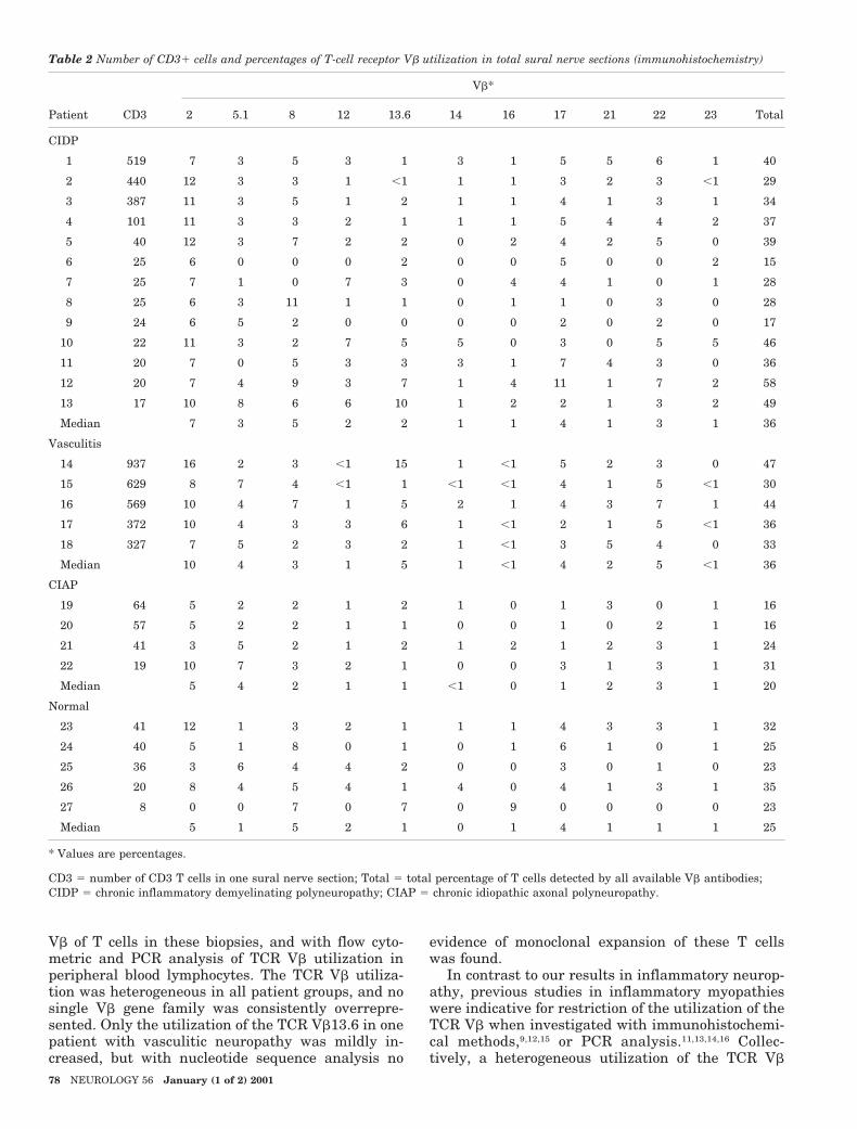


![Functional Outcomes of Multiple Sural Nerve Grafts for Facial Nerve … · 2019-08-13 · Facial Nerve Disorders Committee [12]. In addition, two of the 12 patients were assessed](https://static.fdocuments.in/doc/165x107/5edf2bbaad6a402d666a853e/functional-outcomes-of-multiple-sural-nerve-grafts-for-facial-nerve-2019-08-13.jpg)