Suppression of Pro-metastasis Phenotypes Expression in...
13
Abstract. Background: This study aimed to evaluate the impact of selective abrogation of either the MEK/ERK1/2 (UO126 or PD98059) or the PI3K/AKT (LY294002) signaling cascade on cell proliferation, motility and invasion and production of VEGF (collectively termed pro-metastasis phenotypes) in cultured malignant pleural mesothelioma (MPM) cells. Materials and Methods: Treatment-induced cytotoxicity was evaluated by MTT or Annexin V assays. Cell motility was assessed by wound healing and Matrigel invasion assays. VEGF in conditioned media of cancer cells was measured by ELISA. Results: LY294002 and UO126 significantly inhibited cell proliferation and clonogenicity of MPM cells in vitro. A substantial reduction of cell motility, Matrigel invasion as well as inhibition of basal or EGF- induced VEGF production were observed in drug-treated cells. Conclusion: The selective MEK or PI3K kinase inhibitors are equally effective in down-regulating the expression of pro- metastasis phenotypes, suggesting that MEK or PI3K are appropriate targets for the development of molecular therapeutics for malignant pleural mesothelioma. Malignant pleural mesothelioma (MPM) is a rare but deadly disease etiologically linked to asbestos exposure and possibly SV40 infection (1-3). The annual incidence of MPM in the USA (2,000 to 2,500 new cases/year) is expected to decrease in the next quarter of the century (4, 5) yet its incidence in other parts of the world, particularly in Europe, is projected to remain high in the same period of time (6, 7). MPM is characterized by unrelenting locoregional invasion causing death from encasement of contiguous intrathoracic organs. Current aggressive multimodality therapy for MPM, consisting of surgical resection, cytotoxic chemotherapy and radiation, offers survival benefit to only a small subset of patients with early stages of the disease (8). The major mode of treatment failure following ablative therapy is local/regional recurrence and/or contiguous metastasis to the peritoneal cavity. In conjunction with ongoing clinical efforts to design more effective therapy for MPM, considerable attention has been focused on the development of molecular therapy for this malignant disease by targeting the signal transduction pathways that regulate the process of tumorigenesis and metastasis formation. The development of a malignant tumor from its microscopic focus, similar to the formation of metastasis, is a multistep process. This involves acquisition of multiple cellular characteristics such as enhanced proliferation, motility and invasion through extracellular ground substance matrix, resistance to apoptosis, as well as the ability to stimulate angiogenesis (collectively termed as pro-metastasis phenotypes) (9). Substantial evidence has indicated that extracellular signals transmitted into the cells via membrane receptors with intrinsic tyrosine kinase activity, such as those 809 *The authors contributed equally to this work. **National Institutes of Health – Clinical Research Training Program, Bethesda, MD, U.S.A. Abbreviations: EGFR, epidermal growth factor receptor; MPM, malignant pleural mesothelioma; MTT, 4,5-dimethylthiazo-2-yl)- 2,5-diphenyl tetrazolium bromide; MAPK, mitogen-activated protein kinase; ERK, extracellular signal-regulated kinase; MEK, MAPK/ERK kinase; PI3K, phosphatidylinositide 3-kinase; PDK, 3-phosphoinositide-dependent kinase; VEGF, vascular endothelial cell growth factor; JAK/STAT, Janus kinase/signal transducers and activators of transcription; PLC, phospholipase C; PDGFR, platelet-derived growth factor; HGF, hepatocyte growth factor; IGFR, insulin growth factor receptor. Correspondence to: Dao M. Nguyen, MD, Clinical Research Center, Room 4W-4-3940, 10 Center Drive, Bethesda, MD 20892, U.S.A. Tel: 301-496-2127, Fax: 301-451-6934, e-mail: [email protected] Key words: EGFR, MAPK, MEK, ERK1/2, PI3K inhibitor LY294002, MEK inhibitor UO126, apoptosis, angiogenesis, prometastasis phenotypes, motility. ANTICANCER RESEARCH 26: 809-822 (2006) Suppression of Pro-metastasis Phenotypes Expression in Malignant Pleural Mesothelioma by the PI3K Inhibitor LY294002 or the MEK Inhibitor UO126 GEORGE W. COLE JR.*, ANNETTE M. ALLEVA*, JING T. ZUO*, SHAILEN S. SEHGAL**, WEN-SHUZ YEOW, DAVID S. SCHRUMP and DAO M. NGUYEN Section of Thoracic Oncology, Surgery Branch, Center for Cancer Research, National Cancer Institute, National Institutes of Health, Bethesda, MD, U.S.A. 0250-7005/2006 $2.00+.40
Transcript of Suppression of Pro-metastasis Phenotypes Expression in...

Abstract. Background: This study aimed to evaluate theimpact of selective abrogation of either the MEK/ERK1/2(UO126 or PD98059) or the PI3K/AKT (LY294002) signalingcascade on cell proliferation, motility and invasion andproduction of VEGF (collectively termed pro-metastasisphenotypes) in cultured malignant pleural mesothelioma(MPM) cells. Materials and Methods: Treatment-inducedcytotoxicity was evaluated by MTT or Annexin V assays. Cellmotility was assessed by wound healing and Matrigel invasionassays. VEGF in conditioned media of cancer cells wasmeasured by ELISA. Results: LY294002 and UO126significantly inhibited cell proliferation and clonogenicity ofMPM cells in vitro. A substantial reduction of cell motility,Matrigel invasion as well as inhibition of basal or EGF-induced VEGF production were observed in drug-treated cells.
Conclusion: The selective MEK or PI3K kinase inhibitors areequally effective in down-regulating the expression of pro-metastasis phenotypes, suggesting that MEK or PI3K areappropriate targets for the development of moleculartherapeutics for malignant pleural mesothelioma.
Malignant pleural mesothelioma (MPM) is a rare but deadlydisease etiologically linked to asbestos exposure and possiblySV40 infection (1-3). The annual incidence of MPM in theUSA (2,000 to 2,500 new cases/year) is expected to decreasein the next quarter of the century (4, 5) yet its incidence inother parts of the world, particularly in Europe, is projectedto remain high in the same period of time (6, 7). MPM ischaracterized by unrelenting locoregional invasion causingdeath from encasement of contiguous intrathoracic organs.Current aggressive multimodality therapy for MPM,consisting of surgical resection, cytotoxic chemotherapy andradiation, offers survival benefit to only a small subset ofpatients with early stages of the disease (8). The major modeof treatment failure following ablative therapy islocal/regional recurrence and/or contiguous metastasis to theperitoneal cavity. In conjunction with ongoing clinical effortsto design more effective therapy for MPM, considerableattention has been focused on the development of moleculartherapy for this malignant disease by targeting the signaltransduction pathways that regulate the process oftumorigenesis and metastasis formation.
The development of a malignant tumor from itsmicroscopic focus, similar to the formation of metastasis, isa multistep process. This involves acquisition of multiplecellular characteristics such as enhanced proliferation,motility and invasion through extracellular ground substancematrix, resistance to apoptosis, as well as the ability tostimulate angiogenesis (collectively termed as pro-metastasisphenotypes) (9). Substantial evidence has indicated thatextracellular signals transmitted into the cells via membranereceptors with intrinsic tyrosine kinase activity, such as those
809
*The authors contributed equally to this work.**National Institutes of Health – Clinical Research TrainingProgram, Bethesda, MD, U.S.A.
Abbreviations: EGFR, epidermal growth factor receptor; MPM,malignant pleural mesothelioma; MTT, 4,5-dimethylthiazo-2-yl)-2,5-diphenyl tetrazolium bromide; MAPK, mitogen-activatedprotein kinase; ERK, extracellular signal-regulated kinase; MEK,MAPK/ERK kinase; PI3K, phosphatidylinositide 3-kinase; PDK,3-phosphoinositide-dependent kinase; VEGF, vascular endothelialcell growth factor; JAK/STAT, Janus kinase/signal transducers andactivators of transcription; PLC, phospholipase C; PDGFR,platelet-derived growth factor; HGF, hepatocyte growth factor;IGFR, insulin growth factor receptor.
Correspondence to: Dao M. Nguyen, MD, Clinical Research Center,Room 4W-4-3940, 10 Center Drive, Bethesda, MD 20892, U.S.A.Tel: 301-496-2127, Fax: 301-451-6934, e-mail: [email protected]
Key words: EGFR, MAPK, MEK, ERK1/2, PI3K inhibitorLY294002, MEK inhibitor UO126, apoptosis, angiogenesis,prometastasis phenotypes, motility.
ANTICANCER RESEARCH 26: 809-822 (2006)
Suppression of Pro-metastasis Phenotypes Expression inMalignant Pleural Mesothelioma by the PI3K Inhibitor
LY294002 or the MEK Inhibitor UO126GEORGE W. COLE JR.*, ANNETTE M. ALLEVA*, JING T. ZUO*, SHAILEN S. SEHGAL**,
WEN-SHUZ YEOW, DAVID S. SCHRUMP and DAO M. NGUYEN
Section of Thoracic Oncology, Surgery Branch, Center for Cancer Research, National Cancer Institute, National Institutes of Health, Bethesda, MD, U.S.A.
0250-7005/2006 $2.00+.40

of the EGFR superfamily, PDGFR, IGFR and c-MET(HGF/SF receptor), regulate the expression of pro-metastasis phenotypes (10-17). Binding of ligands to theircognate receptors, regardless of receptor specificity, resultsin activation of the intrinsic tyrosine kinase activity of themembrane receptors, leading to autophosphorylation oftyrosine residues in their intracellular domains.Phosphorylated tyrosines within their unique amino acidmotifs provide docking sites for a variety of adaptormolecules (for example, SOS, Grb2), as well as functionalproteins (for instance PI3K p110 subunit) that lead to theactivation of parallel yet interconnecting cascades ofdownstream intracellular signaling pathways such as theRas/Raf/MEK/ERK, the PLC-Á, the PI3K/PDK/AKT andthe JAK/STAT pathways. Activated receptors can alsodirectly phosphorylate functional proteins (such as STAT orPLC-Á) (17). The biological responses from such diverseintracellular signaling include cell proliferation, survival,motility, invasion and angiogenesis. Blocking of upstream
ligand/receptors will provide a global suppression ofmultiple downstream signals, but redundancy of thereceptor tyrosine kinase repertoire may make selectivetargeting strategy less effective unless the targeted receptorsexert a dominant signal transduction effect via receptorover-expression or hyper-functioning (Figure 1).
We have previously demonstrated that MPM cells expressEGFR and yet the EGFR-selective tyrosine kinase inhibitorPD153035 mediated significant suppression of cellproliferation and of vascular endothelial growth factor(VEGF) production in a cell line-dependent fashion,particularly in those MPM cells expressing very high levelsof EGFR (18). We postulated that PD153035-mediatedinhibition of EGFR signaling may be inefficient in cell linesexpressing normal levels of EGFR due to the presence ofother growth factor receptors that, in parallel with EGFR,transduce their mitogenic signals to a similar set ofintracellular pathways. A potentially more efficient strategyis to selectively target the intracellular signal transduction
ANTICANCER RESEARCH 26: 809-822 (2006)
810
Figure 1. Multiple membrane receptor tyrosine kinases (EGFR, IGFR, PDGFR, c-Met and others) converge on distinct intracellular parallel signalingpathways (PI3K/AKT, Ras/Raf/MEK, PLC-Á/PKC, JAK/STAT) that regulate cell proliferation, motility, invasion, angiogenesis and cellular responses tocytotoxic stresses. Direct inhibition of a membrane receptor tyrosine kinase (RTK) may not result in efficient suppression of downstream intracellularsignaling pathways emanating from this particular receptor and biological responses due to multiplicity of other RTK’s. Direct inhibition of each of theintracellular pathways at the converging point may circumvent this limitation.

pathways downstream of the membrane growth factorreceptor repertoire. The theoretical advantage of targetingthe intracellular pathways, such as PI3K/PDK/AKT orRas/Raf/MEK, is the ability to collectively block inputs frommultiple upstream receptors which converge on thatparticular pathway, while the potential disadvantage of thisapproach is that this would only alter the biologicalresponses mediated by that single pathway alone. As thereare extensive "cross-talks" between pathways that aresomewhat unpredictable and cell-specific, selective down-regulation of a particular signal transduction pathway usingpharmacological inhibitors or genetic manipulations toexpress dominant negative functional proteins may affectmore than one intracellular signaling cascade. The roles ofPI3K-mediated or MEK-mediated signal transduction onthe cellular expression of pro-metastasis phenotypes havebeen well described (10, 19-25). Pharmacological inhibitorsof these kinases have been extensively evaluated inpreclinical models and, particularly in the case of the MEKinhibitor, in early phase clinical trials (26). The primaryobjective of this study, therefore, was to evaluate the effectof selectively blocking either the RTK’s/PI3K/PDK/AKTpathway using the PI3K pharmacological inhibitorLY294002, or the RTK’s/Ras/Raf/MEK/ERK1,2 pathway bythe MEK inhibitors UO126 or PD98059, on the expressionof prometastasis phenotypes in a panel of cultured MPMcells in vitro.
Materials and Methods
Cells and reagents. Cultured MPM cells (H513, H211, M28, H2373,H2595, H2052) were maintained in RPMI-1640 mediumsupplemented with 10% fetal calf serum (FCS), glutamine (2 mmol/L) and antibiotics streptomycin (100 Ìg/mL) and penicillin(100 U/mL). The PI3K inhibitor LY294002 and the MEK inhibitorsUO126 or PD98059, all purchased from Alexis (San Diego, CA,USA), were dissolved in DMSO as 100 mM stocks, aliquoted andstored at –20ÆC.
Cell proliferation, clonogenic and apoptosis assays. Cultured MPMcells were seeded in 96-well plates (3,000 cells/well) andsubsequently treated with LY294002 (5 to 80 ÌM) or UO126 (5 to100 ÌM) for 96 hours with replacement of fresh media (with orwithout PD) at 48 hours. The cell viability of the control and drug-treated cells was quantified by MTT (Sigma-Aldrich, St. Louis,MO, USA). The IC50 values of LY294002 and of UO126(concentrations of drugs that mediated 50% inhibition of cellproliferation) were extrapolated from the respective dose-responsecurves. Cell cycle analysis was performed in control (grown in 10%RPMI) and drug-treated cells (LY294002 at 20 and 40 ÌM orUO126 at 10 or 20 ÌM for 24 hours) using propidium iodine (PI)staining and flow cytometry. As UO126 creates autofluorescenceof the treated cell (27), which interferes with fluorescence-basedapoptosis assays, the MEK inhibitor PD98059 (28) was used inapoptosis experiments at drug concentrations and treatmentconditions that mediated comparable degrees of inhibition of MEK
kinase activity (reduction of phosphorylated ERK1/2 and inhibitionof clonogenicity in vitro) as UO126. LY294002- or PD98059-induced apoptosis was determined by PE-conjugated AnnexinV/7AAD staining and flow cytometry (Pharmigen-BD). Cells wereseeded in 6-well plates (200,000 cells/well) and, after an overnightincubation, they were continuously treated with either LY294002(40 ÌM) or PD98059 (50 ÌM) for 72 hours. The controls were cellsgrown in 10% RPMI. The cells were harvested and assayed forapoptosis as per protocol. A clonogenic assay was used to globallyassess the growth inhibitory effect (cell cycle arrest and cell death)of these selective kinase inhibitors. Cells were plated in 12-wellplates (500 cells/well) and, after an overnight incubation to allowcomplete attachment to the plastic surface, 2 ml of media withoutor with the respective kinase inhibitors (LY294002 at 10 or 40 ÌM,UO126 at 10 or 40 ÌM , PD98059 at 25 or 50 ÌM) were added andthe cells were incubated for 10 days. As PD98059 is not sufficientlypotent or soluble in culture media to sustain the inhibition of MEKactivation (29), PD98059-containing media were replaced daily for3 days and the cells were further incubated until clonogenicity wasevaluated 7 days later. The magnitude of colony formation of thecontrol and of UO126- or LY294002-treated cells was quantifiedby either digital photography of colonies stained with 0.1% crystalviolet or by MTT assay to quantify cell viability which was thenexpressed as percentages of untreated control cells.
Cell motility assay. Cell motility was evaluated by the in vitrowound-healing assay as previously described (30). MPM cells wereseeded in 6-well plates to obtain 90% confluence. After anovernight incubation, "wounds" were made by scratching the cellmonolayer with sterile plastic pipette tips. The cells were thentreated with LY294002 (40 ÌM) or UO126 (40 ÌM) for 24 to 48hours. Movement of cells from the wound edges into the "wounds"was indicative of cell motility. The cells were fixed with 1%paraformaldehyde, stained and the magnitude of "wound-healing"was recorded by digital microphotography.
Matrigel invasion assay. Cell migration and invasion through theMatrigel membrane was quantitated using the commerciallyavailable cell invasion kit (Chemicon International, Temecula,CA, USA). MPM cells were treated with LY294002 (40 ÌM) orUO126 (40 ÌM) for 4 hours in FCS-free media prior to beingseeded in the invasion chambers (in the presence of drug) andstimulated to migrate and invade the Matrigel-containing filterusing serum-containing media in the lower wells. Filterscontaining cells that successfully traversed the Matrigel layer werestained with the staining solution provided in the kit andphotographed with a digital camera.
VEGF assay. VEGF in conditioned media of cultured MPM cells(baseline control or following LY294002 (40 ÌM) or UO126 (20 ÌM)for 24 hours) was measured by ELISA (R&D Biosystems,Minneapolis, MN, USA). Cells were seeded in 12-well plates at 1.0or 1.5x10⁵ cells/well then, after an overnight incubation, the cellswere either incubated in 2 ml of 10% RPMI (baseline control) orin 10% RPMI with LY294002 or UO126 for 24 hours. The VEGFlevels in the conditioned media were normalized for total cellularprotein and expressed as pg VEGF/mg of cellular protein/24 h. Toevaluate the ability of either LY294002 or UO126 to inhibit EGF-mediated up-regulation of VEGF production, the cells were serum-starved for 8 hours and then pre-treated with LY294002 (40 ÌM) or
Cole et al: LY294002 or UO126 Inhibit Pro-metastasis Phenotype Expression in Cancer Cells
811
UO126 (20 ÌM) for 4 hours prior to EGF (20 ng/ml) exposure whilebeing continuously treated with the kinase inhibitors. The controlswere cells incubated in serum-free media with or without LY294002or UO126.
Western blots. The levels of total and phosphorylated EGFR, AKTand ERK1/2 in H211 and H513 cells treated with either theselective EGFR tyrosine kinase inhibitor PD153035 or LY294002or UO126 were quantified by Western blot analysis using thefollowing monoclonal antibodies: total EGFR (Santa CruzBiotechnology, Santa Cruz, CA, USA, 1:500 dilution), pY1173(Santa Cruz, 1:500 dilution), pY1068 for phosphorylated EGFR(Cell Signaling Technology, Beverly, MA, USA, 1:500 dilution),total phosphorylated tyrosine pY20 (BD TransductionLaboratories, San Jose, CA, USA, 1:2,500 dilution), phosphorylatedAKT (ser473) and total AKT (Cell Signaling Biotechnology, 1:1,000dilution), phosphorylated ERK1/2 (Thr202/Tyr204) and totalERK1/2 (Cell Signaling Biotechnology, 1:1,000 dilution) and ‚-actin(Oncogene Research Products, Cambridge, MA, USA, 1:10,000dilution). Cells were plated in 10% FCS RPMI at a density of 5x10⁶
per plate in 10-cm2 plates. The following day, the cells were treatedwith either PD153035 or LY294002 or UO126 (at concentrationsindicated in the figure legends) for 6 hours. Cell pellets were lysedby Laemmli buffer, incubated at room temperature for 15 min andheated at 95ÆC for 5 min to enhance protein extraction. Thesupernatants of the cell lysates were collected by centrifugation at14,000 rpm for 5 min and their protein concentrations weremeasured by the BCA protein assay (Pierce Biotechnology,Rockford, IL, USA). Proteins in the pre-cleared cell lysates wereresolved by electrophoresis through 4% to 20% SDS-polyacrylamide gels, transferred to nitrocellulose membrane andimmunoblotted with specific antibodies. The blots were also probedwith ‚-actin to confirm equal loading of the proteins. In order toobtain an optimal signal, the amount of loading proteins wasadjusted according to the sensitivity and specificity of theantibodies used in the study.
Data analysis. The data are expressed mean±SEM of 3 or 4independent experiments that yielded similar results. Analysis ofvariance and the Student’s t-test were used for statistical analysisand p<0.05 was considered statistically significant.
Results
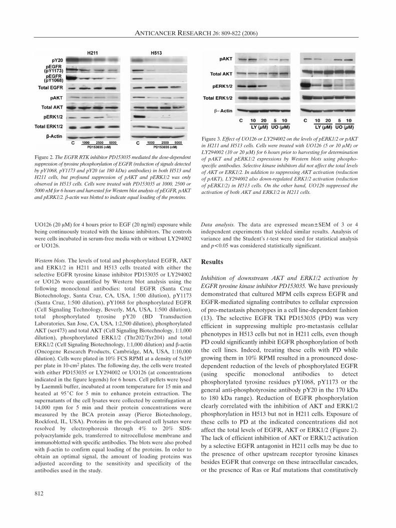
Inhibition of downstream AKT and ERK1/2 activation byEGFR tyrosine kinase inhibitor PD153035. We have previouslydemonstrated that cultured MPM cells express EGFR andEGFR-mediated signaling contributes to cellular expressionof pro-metastasis phenotypes in a cell line-dependent fashion(13). The selective EGFR TKI PD153035 (PD) was veryefficient in suppressing multiple pro-metastasis cellularphenotypes in H513 cells but not in H211 cells, even thoughPD could significantly inhibit EGFR phosphorylation of boththe cell lines. Indeed, treating these cells with PD whilegrowing them in 10% RPMI resulted in a pronounced dose-dependent reduction of the levels of phosphorylated EGFR(using specific monoclonal antibodies to detectphosphorylated tyrosine residues pY1068, pY1173 or thegeneral anti-phosphotyrosine antibody pY20 in the 170 kDato 180 kDa range). Reduction of EGFR phosphorylationclearly correlated with the inhibition of AKT and ERK1/2phosphorylation in H513 but not in H211 cells. Exposure ofthese cells to PD at the indicated concentrations did notaffect the total levels of EGFR, AKT or ERK1/2 (Figure 2).The lack of efficient inhibition of AKT or ERK1/2 activationby a selective EGFR antagonist in H211 cells may be due tothe presence of other upstream receptor tyrosine kinasesbesides EGFR that converge on these intracellular cascades,or the presence of Ras or Raf mutations that constitutively
ANTICANCER RESEARCH 26: 809-822 (2006)
812
Figure 2. The EGFR RTK inhibitor PD153035 mediated the dose-dependentsuppression of tyrosine phosphorylation of EGFR (reduction of signals detectedby pY1068, pY1173 and pY20 (at 180 kDa) antibodies) in both H513 andH211 cells, but profound suppression of pAKT and pERK1/2 was onlyobserved in H513 cells. Cells were treated with PD153035 at 1000, 2500 or5000 nM for 6 hours and harvested for Western blot analysis of pEGFR, pAKTand pERK1/2. ‚-actin was blotted to indicate equal loading of the proteins.
Figure 3. Effect of UO126 or LY294002 on the levels of pERK1/2 or pAKTin H211 and H513 cells. Cells were treated with UO126 (5 or 10 ÌM) orLY294002 (10 or 20 ÌM) for 6 hours prior to harvesting for determinationof pAKT and pERK1/2 expressions by Western blots using phospho-specific antibodies. Selective kinase inhibitors did not affect the total levelsof AKT or ERK1/2. In addition to suppressing AKT activation (reductionof pAKT), LY294002 also down-regulated ERK1/2 activation (reductionof pERK1/2) in H513 cells. On the other hand, UO126 suppressed theactivation of both AKT and ERK1/2 in H211 cells.
activate downstream signaling cascades. Direct inhibition of adownstream intracellular pathway in such an instance may bea better approach than selective inhibition of a singlemembrane receptor. This provides the impetus for furtherinvestigation into the direct inhibition of downstreampathways using currently available kinase inhibitors such asthe PI3K inhibitor LY294002 or the MEK inhibitor UO126.
Inhibition of AKT activation by LY294002 and ERK1/2activation by UO126. Treating H513 and H211 cells withUO126 or LY294002, as expected, resulted in profoundreduction of the levels of pERK1/2 (Thr202/Thr204) andpAKT (ser473), respectively, in a dose-dependent fashion(Figure 3). Near complete inhibition of ERK1/2 phos-phorylation in H153 and H211 was observed following 6-hour exposure to 10 ÌM of UO126. Similarly, LY294002 at20 ÌM for 6 hours almost completely abrogated AKTphosphorylation in both cell lines. It is interesting to notethat LY294002 also mediated the reduction of pERK1/2 inthe H513 cells, probably via interference with thePI3K/PDK/MEK axis (31). This was not observed in H211cells. Conversely, UO126, in addition to inhibiting theactivation of ERK1/2, also caused a clear reduction ofpAKT levels in H211 cells, but this was not observed inH513 cells. Such observations indicate the presence ofsignificant "cross-talks" between parallel signaling pathwaysand selective pharmacological inhibitors may not be thatspecific in blocking a single pathway in living cells.
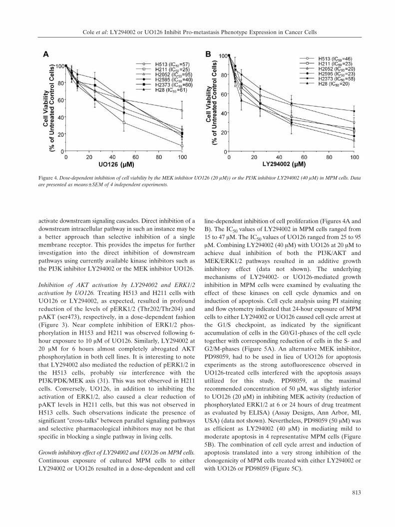
Growth inhibitory effect of LY294002 and UO126 on MPM cells.Continuous exposure of cultured MPM cells to eitherLY294002 or UO126 resulted in a dose-dependent and cell
line-dependent inhibition of cell proliferation (Figures 4A andB). The IC50 values of LY294002 in MPM cells ranged from15 to 47 ÌM. The IC50 values of UO126 ranged from 25 to 95ÌM. Combining LY294002 (40 ÌM) with UO126 at 20 ÌM toachieve dual inhibition of both the PI3K/AKT andMEK/ERK1/2 pathways resulted in an additive growthinhibitory effect (data not shown). The underlyingmechanisms of LY294002- or UO126-mediated growthinhibition in MPM cells were examined by evaluating theeffect of these kinases on cell cycle dynamics and oninduction of apoptosis. Cell cycle analysis using PI stainingand flow cytometry indicated that 24-hour exposure of MPMcells to either LY294002 or UO126 caused cell cycle arrest atthe G1/S checkpoint, as indicated by the significantaccumulation of cells in the G0/G1-phases of the cell cycletogether with corresponding reduction of cells in the S- andG2/M-phases (Figure 5A). An alternative MEK inhibitor,PD98059, had to be used in lieu of UO126 for apoptosisexperiments as the strong autofluorescence observed inUO126-treated cells interfered with the apoptosis assaysutilized for this study. PD98059, at the maximalrecommended concentration of 50 ÌM, was slightly inferiorto UO126 (20 ÌM) in inhibiting MEK activity (reduction ofphosphorylated ERK1/2 at 6 or 24 hours of drug treatmentas evaluated by ELISA) (Assay Designs, Ann Arbor, MI,USA) (data not shown). Nevertheless, PD98059 (50 ÌM) wasas efficient as LY294002 (40 ÌM) in mediating mild tomoderate apoptosis in 4 representative MPM cells (Figure5B). The combination of cell cycle arrest and induction ofapoptosis translated into a very strong inhibition of theclonogenicity of MPM cells treated with either LY294002 orwith UO126 or PD98059 (Figure 5C).
Cole et al: LY294002 or UO126 Inhibit Pro-metastasis Phenotype Expression in Cancer Cells
813
Figure 4. Dose-dependent inhibition of cell viability by the MEK inhibitor UO126 (20 ÌM)) or the PI3K inhibitor LY294002 (40 ÌM) in MPM cells. Dataare presented as means±SEM of 4 independent experiments.

LY294002- or UO126-mediated inhibition of cell motilityand invasion through Matrigel membrane. The effect ofinhibiting MEK or PI3K signaling on cell motility wasassessed by the wound-healing assay. At theconcentrations of drug that mediated >90% inhibition ofAKT or ERK1/2 phosphorylation, there was significantretardation of cell movement from the wound edge into
the denuded plastic surface created by "wounding" the cellmonolayer when compared to the reference "fresh wound’and the control "healing wound" of cells incubated indrug-free 10% RPMI culture medium (Figure 6).Similarly, LY294002 or UO126 also strongly inhibitedMPM cells to invade/migrate through the Matrigelextracellular matrix membrane (Figure 7) as determined
ANTICANCER RESEARCH 26: 809-822 (2006)
814
Figure 5. A. Treating representative MPM cells H513, H211, H2595 and H2052 with either UO126 or LY294002 resulted in cell cycle arrest at the G1/Scheckpoint with significant accumulation of cells in G1-phase and reduction of cells in S- and G2/M-phases. Representative data of 3 independentexperiments that yielded similar results are shown here. B. Significant induction of apoptosis in MPM cells following exposure to PD98059 or LY294002.The cells were treated with either the MEK inhibitor (50 ÌM) or the PI3K inhibitor (40 ÌM) for 72 hours and apoptosis was quantified using Annexin Vstaining. Data are presented as means±SEM of 3 independent experiments, *,# p<0.05 versus normal media controls. C. Suppression of clonogenicityof MPM cells H2052, H513, H2595 and H2373 by UO126, PD98059 and LY294002. Digital photographs of representative experiments and quantitativeanalysis of inhibition of clonogenicity using MTT staining of viable clones are shown in the histogram (means±SEM of 3 independent experiments).
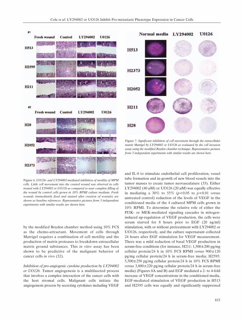
by the modified Boyden chamber method using 10% FCSas the chemo-attractant. Movement of cells throughMatrigel requires a combination of cell motility and theproduction of matrix proteases to breakdown extracellularmatrix ground substances. This in vitro assay has beenshown to be predictive of the malignant behavior ofcancer cells in vivo (32).
Inhibition of pro-angiogenic cytokine production by LY294002or UO126. Tumor angiogenesis is a multifaceted processthat involves a complex interaction of the cancer cells withthe host stromal cells. Malignant cells initiate theangiogenesis process by secreting cytokines including VEGF
and IL-8 to stimulate endothelial cell proliferation, vesseltube formation and in-growth of new blood vessels into thetumor masses to create tumor neovasculature (33). EitherLY294002 (40 ÌM) or UO126 (20 ÌM) was equally effectivein mediating a 30% to 55% (p<0.05 to p<0.01 versusuntreated control) reduction of the levels of VEGF in theconditioned media of the 4 cultured MPM cells grown in10% RPMI. To determine the relative role of either thePI3K- or MEK-mediated signaling cascades in mitogen-induced up-regulation of VEGF production, the cells wereserum starved for 8 hours prior to EGF (20 ng/ml)stimulation, with or without pretreatment with LY294002 orUO126, respectively, and the culture supernatant collected24 hours after EGF stimulation for VEGF measurement.There was a mild reduction of basal VEGF production inserum-free conditions (for instance, H211: 1,500±200 pg/mgcellular protein/24 h in 10% FCS RPMI versus 900±120pg/mg cellular protein/24 h in serum-free media; H2595:5,500±250 pg/mg cellular protein/24 h in 10% FCS RPMIversus 3,800±220 pg/mg cellular protein/24 h in serum-freemedia) (Figures 8A and B) and EGF mediated a 2- to 4-foldincrease of VEGF concentrations in the conditioned media.EGF-mediated stimulation of VEGF production in H513and H2595 cells was equally and significantly suppressed
Cole et al: LY294002 or UO126 Inhibit Pro-metastasis Phenotype Expression in Cancer Cells
815
Figure 6. UO126- and LY294002-mediated inhibition of motility of MPMcells. Little cell movement into the created wound was observed in cellstreated with LY294002 or UO126 as compared to near complete filling ofthe wound by control cells grown in 10% RPMI culture medium. Freshwounds (immediately fixed and stained after creation of wounds) areshown as baseline references. Representative pictures from 3 independentexperiments with similar results are shown here.
Figure 7. Significant inhibition of cell movement through the extracellularmatrix Matrigel by LY294002 or UO126 as evaluated by the cell invasionassay using the modified Boyden chamber technique. Representative picturesfrom 3 independent experiments with similar results are shown here.
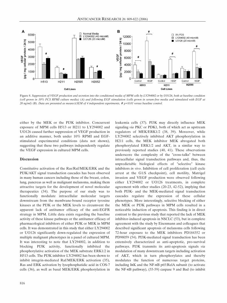
either by the MEK or the PI3K inhibitor. Concurrentexposure of MPM cells H513 or H211 to LY294002 andUO126 caused further suppression of VEGF production inan additive manner, both under 10% RPMI and EGF-stimulated experimental conditions (data not shown),suggesting that these two pathways independently regulatethe VEGF expression in cultured MPM cells.
Discussion
Constitutive activation of the Ras/Raf/MEK/ERK and thePI3K/AKT signal transduction cascades has been observedin many human cancers including those of the breast, colon,lung, pancreas as well as malignant melanoma, making themattractive targets for the development of novel moleculartherapeutics (34). The purpose of our study was tofunctionally modulate intracellular molecular targetsdownstream from the membrane-bound receptor tyrosinekinases at the PI3K or the MEK levels to circumvent theapparent lack of antitumor efficacy of the anti-EGFRstrategy in MPM. Little data exists regarding the baselineactivity of these kinase pathways or the antitumor efficacy ofpharmacological inhibitors of either PI3K or MEK in MPMcells. It was demonstrated in this study that either LY294002or UO126 significantly down-regulated the expression ofmultiple malignant phenotypes in a panel of cultured MPM.It was interesting to note that LY294002, in addition toblocking PI3K activity, functionally inhibited thephosphorylative activation of the MEK substrate ERK1/2 inH513 cells. The PI3K inhibitor LY294002 has been shown toinhibit integrin-mediated Raf/MEK/ERK activation (35),Ras and ERK activation by lysophosphatidic acid in COS-7cells (36), as well as basal MEK/ERK phosphorylation in
leukemia cells (37). PI3K may directly influence MEKsignaling via PKC or PDK1, both of which act as upstreamregulators of MEK/ERK1/2 (38, 39). Moreover, whileLY294002 selectively inhibited AKT phosphorylation inH211 cells, the MEK inhibitor MEK abrogated bothphosphorylated ERK1/2 and AKT, in a similar way topreviously reported studies (40, 41). These observationsunderscore the complexity of the "cross-talks" betweenintracellular signal transduction pathways and, thus, theunpredictable biological effects of "selective" kinaseinhibitors in vivo. Inhibition of cell proliferation (cell cyclearrest at the G1/S checkpoint), cell motility, Matrigelinvasion and VEGF production were observed followingeither LY294002 or UO126 treatments, in completeagreement with other studies (20-23, 42-52), implying thatboth PI3K- and the MEK-mediated signal transductioncascades regulate the expression of these cellularphenotypes. More interestingly, selective blocking of eitherthe MEK or PI3K pathways in MPM cells resulted in anoticeable induction of apoptosis. This finding is in directcontrast to the previous study that reported the lack of MEKinhibitor-induced apoptosis in NSCLC (53), but in completeagreement with the study by Eisenmann and colleagues thatdescribed significant apoptosis of melanoma cells following72-hour exposure to the MEK inhibitors PD184352 orPD98059 (54). PI3K-mediated signal transduction has beenextensively characterized as anti-apoptotic, pro-survivalpathways. PI3K transmits its anti-apoptosis signals viamodulation of many downstream targets including activationof AKT, which in turn phosphorylates and therebymodulates the function of numerous target proteins,including IκK and the NF-κB p65/RelA subunit (to activatethe NF-κB pathway), (55-59) caspase 9 and Bad (to inhibit
ANTICANCER RESEARCH 26: 809-822 (2006)
816
Figure 8. Suppression of VEGF production and secretion into the conditioned media of MPM cells by LY294002 or by UO126, both at baseline condition(cell grown in 10% FCS RPMI culture media) (A) and following EGF stimulation (cells grown in serum-free media and stimulated with EGF at 20 ng/ml) (B). Data are presented as means±SEM of 4 independent experiments, # p<0.01 versus baseline control.
pro-apoptotic activity), (60, 61) and p21 or p27 (to suppresscell cycle regulation activity) (62, 63). The anti-apoptoticfunction of Ras/Raf/MEK/ERK1/2 has recently been shownto involve up-regulation of Bcl2, BclXL and MCL-1,phosphorylation of Bcl2 to prevent their degradation byproteosome (64 and refs therein), as well as phosphorylationof Bad (via a p90RSK-mediated mechanism) (54) or BIM(65) to inhibit their respective pro-apoptotic functions. Thecumulative effect of cell cycle arrest and apoptosis, mediatedby either MEK or PI3K inhibitors, was evident in theclonogeneic assay in which profound suppression ofclonogenicity of the selected MPM cell lines was observed inLY294002-, UO126- or PD98059-treated cells (Figure 6Aand B). The current focus of our laboratory effort is tofurther elucidate the molecular mechanism of MEKinhibitor-induced apoptosis in a larger panel of MPM cellsand also in other cultured thoracic cancer cells in order toperform comparative analysis of the apoptosis-inducingeffect of the MEK inhibitor in different kinds of thoracicmalignancies.
In contrast to other tumor histology such as pancreatic,colon and lung cancers or malignant melanoma (66-68),mutations of K-ras or B-Raf were rare in MPM (69, 70). Itis conceivable that, without upstream constitutively activeK-ras that can activate other pathways in addition toMEK/ERK1/2, targeting MEK in this setting may be moreefficacious in mediating meaningful cytotoxicity. Indeed, ithas been shown by Wang and colleagues that K-rasmutation reduces the therapeutic efficacy of the MEKinhibitor CI-1040 in colon cancer cell lines in vitro (71).Growth factor-dependent signal transduction pathwaysemploy extensive networks of intermediaries andposttranslational modifications of these proteins to transmitthe signals to their intended targets. There is also ampleredundancy of the receptor repertoire as well as theintracellular connections with positive and negativeregulatory controls to ensure appropriate and diverseresponses to vast arrays of environmental signals. Thiselaborate system of circuitry design of the signaltransduction pathways poses opportunities as well aschallenges for the development of targeted therapy.Opportunities are the multiple signaling proteins serving aspotential targets. Targeting upstream proteins (for instance,EGFR or PDGFR) will provide a global alteration ofdownstream signaling, but the challenges arise since themultiplicity of upstream growth factor receptors may makeselective targeting strategy less effective. Targetingoverexpressed or overactive growth factor-mediatedpathways, that are partially or totally responsible for themalignat behaviors of transformed cells, may circumvent theabove-mentioned problem. One prime example is theclinical effectiveness of EGFR tyrosine kinase inhibitorssuch as Gefitinib or Erlotinib in mediating tumor regression
of tumors harboring EGFR mutations, that make themparticularly sensitive to this drug (72-74) or in patientswhose tumors overexpress EGFR (75-77). Another is theefficacy of Trastuzumab (Herceptin®) on mediating theantitumor effect on breast cancers with erbB2 geneamplification and extreme overexpression of the erbB2oncoprotein (78, 79). Similar to other carcinomas, theexpressions of IGFR, PDGFR as well as members of theEGFR superfamily have been reported in cultured MPMcell lines (14, 15, 18, 80). In many instances, multiplereceptor tyrosine kinases, some of which wereoverexpressed, were found on the same MPM cell lines (14,18). Selective targeting of a single receptor tyrosine kinasemay not be therapeutically effective, as we have observedwhen trying to modulate the expression of pro-metastasisphenotypes in MPM cells using the EGFR tyrosine kinaseinhibitor PD153035 in vitro (18). Targeting intermediateintracellular signaling proteins such as PI3K, AKT, Ras orRaf offers the advantage of influencing upstream signalsemanating from many distinct receptor/ligand interactionsat their intracellular common convergent points whichchannel signals to distinct transduction cascades. Wenoticed that significant PD153035-mediated inhibition ofcell growth or production of VEGF was only apparent incell lines that express high levels of EGFR such as H513 orH2595, but not in other MPM cells such as H211, H2052,H2373 that have slightly elevated or normal levels of EGFR(18). The lack of biological effects of EGFR TKI PD153035in MPM H211 cells was not due to its inability to suppressgrowth factor-mediated phosphorylation of EGFR,as indicated by the almost complete reduction ofphosphorylated EGFR, but due to its inability to suppressthe phosphorylative activation of AKT and ERK1/2 (Figure2). On the other hand, PD153035 was very effective indown-regulating the pro-metastasis phenotype expression inH513 since it was able to inhibit the activation of EGFR,AKT and ERK1/2 (18).
Our study specifically focused on evaluating the effect ofPI3K or MEK inhibitors on other cellular phenotypesbesides cell proliferation as cellular processes, such as cellinvasion, motility and the production of pro-angiogenesiscytokines which play crucial and equally important roles inthe development of clinically relevant tumors frommicroscopic deposits. In this context, these kinase inhibitorsmay be ideal chemopreventive or even chemotherapeuticagents for appropriate tumors in the setting of minimalresidual disease (post-resection adjuvant therapy). Even ifthese inhibitors mediate a cytostatic effect by arrestingcancer cells from progressing through the cell cycle, or mildinduction of apoptosis sufficient to counterbalance thefractions of cells undergoing division and contributing to theincrease of biomasses, they, together with their ability toinhibit the production of VEGF to suppress tumor-derived
Cole et al: LY294002 or UO126 Inhibit Pro-metastasis Phenotype Expression in Cancer Cells
817

angiogenesis, may prevent established tumors fromprogressing further to become a health burden. In thisaspect, these kinase inhibitors can not cause regression oftumor masses, but may keep them under control bymaintenance therapy as in the cases of other tyrosinekinases like Gefitinib (81) or Gleevec (82) which can besafely administered over a long period of time. Of the twoclasses of kinase inhibitors, the MEK inhibitor (CI-1040)has entered phase I and II clinical trials as a single agent forsolid tumors. CI-1040, however, was shown to be ineffectivein patients with advanced non-small cell lung, breast, colonand pancreatic cancers (26). Even though continuousadministration of CI-1040 (800 mg twice /day) resulted inplasma drug concentrations similar to those reported in theearlier phase I study, there were no complete or partialresponses and stable disease was observed in only 12% ofcases. Incomplete suppression of pERK1/2 expression, asdetermined by quantitative immuno-histochemical staining,was noted in the majority of cases with constitutivepERK1/2 expression at baseline, thus raising the possibilitythat the degree of MEK inhibition by CI-1040 may beinsufficient to mediate a meaningful tumor response (26).There appeared to be a positive correlation betweenconstitutive baseline pERK1/2 expression and stable diseasewith CI-1040 treatment. The clinical trial finding was notthat surprising given the fact that Brognard and Dennis haddemonstrated somewhat unpredictable responses ofcultured non-small cell lung cancer cells lines, wellcharacterized for their K-ras and p53 status, to MEKinhibition using either pharmacological inhibitors likeUO126 or PD98059 or dominant negative MEK (53).Pharmacological inhibitors induced cell cycle arrest but noapoptosis and little sensitization of cancer cells to paclitaxel,in stark contrast to dominant negative MEK whichefficiently induced apoptosis and chemosensitization ofcultured lung cancer cells to cytotoxic chemotherapeutics.A more recent study, however, indicated that the clinicallyrelevant MEK inhibitor CI-1040 synergistically interactedwith paclitaxel to mediate potent antitumor effect on humanheterotransplants in nude mice (83). Moreover, CI-1040,alone or in combination with paclitaxel, also significantlyinhibited VEGF production and FGF-induced angiogenesisin vivo (83), compatible with our contention that theantitumor effect of the MEK inhibitor is more than justgrowth arrest or chemosensitization but also depends on itsability to modulate other cellular phenotypes such as thepro-angiogenic activities that are necessary for tumordevelopment.
In summary, targeting either PI3K or MEK of culturedMPM cells by the pharmacological kinase inhibitors resultedin the deactivation of intended targets as well as modulationof other parallel pathways, which is most probable due tothe "cross-talks" that exist between intracellular signaling
cascades. In addition to mediating profound cytotoxicity viacell cycle arrest and induction of apoptosis, these kinaseinhibitors also down-modulated the in vitro expression ofmultiple malignant phenotypes of transformed cells, furtherenhancing their anticancer properties that can only beevaluated in animal models or in human clinical trials.There has been a recent renewal of interest in targeting theRas/Raf/MEK/ERK1/2 signal transduction pathway for thedevelopment of novel anticancer therapeutics based onexciting discoveries of the molecular mechanisms ofpathway-mediated induction of apoptosis. This forms theimpetus for our current work of evaluating MEK inhibitorsas the molecularly-targeted drugs for MPM.
Acknowledgements
This research was supported by the Intramural Research Programof the National Cancer Institute, NIH, U.S.A.
References
1 Carbone M, Kratzke RA and Testa JR: The pathogenesis ofmesothelioma. Semin Oncol 29: 2-17, 2002.
2 Mossman BT and Churg A: Mechanisms in the pathogenesis ofasbestosis and silicosis. Am J Respir Crit Care Med 157: 1666-1680, 1998.
3 Schrump DS and Waheed I: Strategies to circumvent SV40oncoprotein expression in malignant pleural mesotheliomas.Semin Cancer Biol 11: 73-80, 2001.
4 Price B and Ware A: Mesothelioma trends in the United States:an update based on Surveillance, Epidemiology, and End ResultsProgram data for 1973 through 2003. Am J Epidemiol 159: 107-112, 2004.
5 Weill H, Hughes JM and Churg AM: Changing trends in USmesothelioma incidence. Occup Environ Med 61: 438-441, 2004.
6 Hodgson JT, McElvenny DM, Darnton AJ, Price MJ and PetoJ: The expected burden of mesothelioma mortality in GreatBritain from 2002 to 2050. Br J Cancer 92: 587-593, 2005.
7 Peto J, Decarli A, La Vecchia C, Levi F and Negri E: TheEuropean mesothelioma epidemic. Br J Cancer 79: 666-672, 1999.
8 Zellos L and Sugarbaker DJ: Current surgical management ofmalignant pleural mesothelioma. Curr Oncol Rep 4: 354-360, 2002.
9 Fidler IJ: The pathogenesis of cancer metastasis: the “seed andsoil” hypothesis revisited. Nat Rev Cancer 3: 453-458, 2003.
10 Kruger JS and Reddy KB: Distinct mechanisms mediate theinitial and sustained phases of cell migration in epidermalgrowth factor receptor-overexpressing cells. Mol Cancer Res 1:801-809, 2003.
11 Aaronson SA: Growth factors and cancer. Science 254: 1146-1153, 1991.
12 Kondapaka SB, Fridman R and Reddy KB: Epidermal growthfactor and amphiregulin up-regulate matrix metalloproteinase-9(MMP-9) in human breast cancer cells. Int J Cancer 70: 722-726, 1997.
13 Chakrabarty S, Rajagopal S and Huang S: Expression of antisenseepidermal growth factor receptor RNA downmodulates themalignant behavior of human colon cancer cells. Clin ExpMetastasis 13: 191-195, 1995.
ANTICANCER RESEARCH 26: 809-822 (2006)
818
14 Hoang CD, Zhang X, Scott PD, Guillaume TJ, Maddaus MA,Yee D and Kratzke RA: Selective activation of insulin receptorsubstrate-1 and -2 in pleural mesothelioma cells: association withdistinct malignant phenotypes. Cancer Res 64: 7479-7485, 2004.
15 Langerak AW, De Laat PA, Van Der Linden-Van Beurden CA,Delahaye M, Van Der Kwast TH, Hoogsteden HC, Benner Rand Versnel MA: Expression of platelet-derived growth factor(PDGF) and PDGF receptors in human malignantmesothelioma in vitro and in vivo. J Pathol 178: 151-160, 1996.
16 Huang SM, Li J and Harari PM: Molecular inhibition ofangiogenesis and metastatic potential in human squamous cellcarcinomas after epidermal growth factor receptor blockade.Mol Cancer Ther 1: 507-514, 2002.
17 Nguyen DM and Schrump DS: Growth factor receptors astargets for lung cancer therapy. Semin Thorac Cardiovasc Surg16: 3-12, 2004.
18 Cole GW, Alleva AM, Reddy RM, Maxhimer JB, Zuo J,Schrump DS and Nguyen DM: The selective EGFR tyrosinekinase inhibitor PD153035 suppresses expression ofprometastasis phenotypes in malignant pleural mesotheliomacells in vitro. J Thorac Cardiovasc Surg. Ref Type: in press, 2005.
19 Takino T, Miyamori H, Watanabe Y, Yoshioka K, Seiki M andSato H: Membrane type 1 matrix metalloproteinase regulatescollagen-dependent mitogen-activated protein/extracellularsignal-related kinase activation and cell migration. Cancer Res64: 1044-1049, 2004.
20 Okudela K, Hayashi H, Ito T, Yazawa T, Suzuki T, Nakane Y,Sato H, Ishi H, KeQin X, Masuda A, Takahashi T andKitamura H: K-ras gene mutation enhances motility ofimmortalized airway cells and lung adenocarcinoma cells viaAkt activation: possible contribution to non-invasive expansionof lung adenocarcinoma. Am J Pathol 164: 91-100, 2004.
21 Nakamura T, Kanda S, Yamamoto K, Kohno T, Maeda K,Matsuyama T and Kanetake H: Increase in hepatocyte growthfactor receptor tyrosine kinase activity in renal carcinoma cells isassociated with increased motility partly through phosphoinositide3-kinase activation. Oncogene 20: 7610-7623, 2001.
22 Kermorgant S, Aparicio T, Dessirier V, Lewin MJ and Lehy T:Hepatocyte growth factor induces colonic cancer cellinvasiveness via enhanced motility and protease overproduction.Evidence for PI3 kinase and PKC involvement. Carcinogenesis22: 1035-1042, 2001.
23 Menu E, Kooijman R, Van Valckenborgh E, Asosingh K,Bakkus M, Van Camp B and Vanderkerken K: Specific rolesfor the PI3K and the MEK-ERK pathway in IGF-1-stimulatedchemotaxis, VEGF secretion and proliferation of multiplemyeloma cells: study in the 5T33MM model. Br J Cancer 90:1076-1083, 2004.
24 Veit C, Genze F, Menke A, Hoeffert S, Gress TM, Gierschik Pand Giehl K: Activation of phosphatidylinositol 3-kinase andextracellular signal-regulated kinase is required for glial cell line-derived neurotrophic factor-induced migration and invasion ofpancreatic carcinoma cells. Cancer Res 64: 5291-5300, 2004.
25 Ge X, Fu YM and Meadows GG: U0126, a mitogen-activatedprotein kinase kinase inhibitor, inhibits the invasion of humanA375 melanoma cells. Cancer Lett 179: 133-140, 2002.
26 Rinehart J, Adjei AA, LoRusso PM, Waterhouse D, Hecht JR,Natale RB, Hamid O, Varterasian M, Asbury P, Kaldjian EP,Gulyas S, Mitchell DY, Herrera R, Sebolt-Leopold JS andMeyer MB: Multicenter phase II study of the oral MEK
inhibitor, CI-1040, in patients with advanced non-small-celllung, breast, colon, and pancreatic cancer. J Clin Oncol 22:4456-4462, 2004.
27 Blank N, Burger R, Duerr B, Bakker F, Wohlfarth A, DumitriuI, Kalden JR and Herrmann M: MEK inhibitor U0126interferes with immunofluorescence analysis of apoptotic celldeath. Cytometry 48: 179-184, 2002.
28 Dudley DT, Pang L, Decker SJ, Bridges AJ and Saltiel AR: Asynthetic inhibitor of the mitogen-activated protein kinasecascade. Proc Natl Acad Sci USA 92: 7686-7689, 1995.
29 Cohen P: The development and therapeutic potential of proteinkinase inhibitors. Curr Opin Chem Biol 3: 459-465, 1999.
30 Alper O, Bergmann-Leitner ES, Bennett TA, Hacker NF,Stromberg K and Stetler-Stevenson WG: Epidermal growthfactor receptor signaling and the invasive phenotype of ovariancarcinoma cells. J Natl Cancer Inst 93: 1375-1384, 2001.
31 Sato S, Fujita N and Tsuruo T: Involvement of 3-phosphoinositide-dependent protein kinase-1 in theMEK/MAPK signal transduction pathway. J Biol Chem 279:33759-33767, 2004.
32 Luo J, Lubaroff DM and Hendrix MJ: Suppression of prostatecancer invasive potential and matrix metalloproteinase activityby E-cadherin transfection. Cancer Res 59: 3552-3556, 1999.
33 Hicklin DJ and Ellis LM: Role of the vascular endothelialgrowth factor pathway in tumor growth and angiogenesis. J ClinOncol 23: 1011-1027, 2005.
34 Sliva D: Signaling pathways responsible for cancer cell invasionas targets for cancer therapy. Curr Cancer Drug Targets 4: 327-336, 2004.
35 Hawes BE, Luttrell LM, van Biesen T and Lefkowitz RJ:Phosphatidylinositol 3-kinase is an early intermediate in the Gbeta gamma-mediated mitogen-activated protein kinasesignaling pathway. J Biol Chem 271: 12133-12136, 1996.
36 King WG, Mattaliano MD, Chan TO, Tsichlis PN and BruggeJS: Phosphatidylinositol 3-kinase is required for integrin-stimulated AKT and Raf-1/mitogen-activated protein kinasepathway activation. Mol Cell Biol 17: 4406-4418, 1997.
37 Rahmani M, Yu C, Reese E, Ahmed W, Hirsch K, Dent P andGrant S: Inhibition of PI-3 kinase sensitizes human leukemiccells to histone deacetylase inhibitor-mediated apoptosisthrough p44/42 MAP kinase inactivation and abrogation ofp21(CIP1/WAF1) induction rather than AKT inhibition.Oncogene 22: 6231-6242, 2003.
38 Sato S, Fujita N and Tsuruo T: Interference with PDK1-Aktsurvival signaling pathway by UCN-01 (7-hydroxystauro-sporine). Oncogene 21: 1727-1738, 2002.
39 Goekjian PG and Jirousek MR: Protein kinase C inhibitors asnovel anticancer drugs. Expert Opin Investig Drugs 10: 2117-2140, 2001.
40 Yu CF, Roshan B, Liu ZX and Cantley LG: ERK regulates thehepatocyte growth factor-mediated interaction of Gab1 and thephosphatidylinositol 3-kinase. J Biol Chem 276: 32552-32558,2001.
41 Yu CF, Liu ZX and Cantley LG: ERK negatively regulates theepidermal growth factor-mediated interaction of Gab1 and thephosphatidylinositol 3-kinase. J Biol Chem 277: 19382-19388, 2002.
42 Kawada M, Yamagoe S, Murakami Y, Suzuki K, Mizuno S andUehara Y: Induction of p27Kip1 degradation and anchorageindependence by Ras through the MAP kinase signalingpathway. Oncogene 15: 629-637, 1997.
Cole et al: LY294002 or UO126 Inhibit Pro-metastasis Phenotype Expression in Cancer Cells
819
43 Hoshino R, Tanimura S, Watanabe K, Kataoka T and KohnoM: Blockade of the extracellular signal-regulated kinasepathway induces marked G1 cell cycle arrest and apoptosis intumor cells in which the pathway is constitutively activated: up-regulation of p27(Kip1). J Biol Chem 276: 2686-2692, 2001.
44 Qi HL, Zhang Y, Ma J, Guo P, Zhang XY and Chen HL:Insulin/protein kinase B signalling pathway upregulatesmetastasis-related phenotypes and molecules in H7721 humanhepatocarcinoma cell line. Eur J Biochem 270: 3795-3805, 2003.
45 Caron RW, Yacoub A, Li M, Zhu X, Mitchell C, Hong Y,Hawkins W, Sasazuki T, Shirasawa S, Kozikowski AP, DennisPA, Hagan MP, Grant S and Dent P: Activated forms of H-RAS and K-RAS differentially regulate membraneassociation of PI3K, PDK-1, and AKT and the effect oftherapeutic kinase inhibitors on cell survival. Mol CancerTher 4: 257-270, 2005.
46 Kobayashi H, Suzuki M, Kanayama N and Terao T: Geneticdown-regulation of phosphoinositide 3-kinase by bikunincorrelates with suppression of invasion and metastasis in humanovarian cancer HRA cells. J Biol Chem 279: 6371-6379, 2004.
47 Brahmbhatt AA and Klemke RL: ERK and RhoA differentiallyregulate pseudopodia growth and retraction during chemotaxis.J Biol Chem 278: 13016-13025, 2003.
48 Sliva D, Rizzo MT and English D: Phosphatidylinositol 3-kinaseand NF-kappaB regulate motility of invasive MDA-MB-231human breast cancer cells by the secretion of urokinase-typeplasminogen activator. J Biol Chem 277: 3150-3157, 2002.
49 Krystal GW, Sulanke G and Litz J: Inhibition ofphosphatidylinositol 3-kinase-Akt signaling blocks growth,promotes apoptosis, and enhances sensitivity of small cell lungcancer cells to chemotherapy. Mol Cancer Ther 1: 913-922,2002.
50 Jemal A, Murray T, Ward E, Samuels A, Tiwari RC, GhafoorA, Feuer EJ and Thun MJ: Cancer statistics, 2005. CA CancerJ Clin 55: 10-30, 2005.
51 Skinner HD, Zheng JZ, Fang J, Agani F and Jiang BH:Vascular endothelial growth factor transcriptional activation ismediated by hypoxia-inducible factor 1alpha, HDM2, andp70S6K1 in response to phosphatidylinositol 3-kinase/AKTsignaling. J Biol Chem 279: 45643-45651, 2004.
52 Klemke RL, Cai S, Giannini AL, Gallagher PJ, de Lanerolle Pand Cheresh DA: Regulation of cell motility by mitogen-activated protein kinase. J Cell Biol 137: 481-492, 1997.
53 Brognard J and Dennis PA: Variable apoptotic response ofNSCLC cells to inhibition of the MEK/ERK pathway by smallmolecules or dominant negative mutants. Cell Death Differ 9:893-904, 2002.
54 Eisenmann KM, VanBrocklin MW, Staffend NA, Kitchen SMand Koo HM: Mitogen-activated protein kinase pathway-dependent tumor-specific survival signaling in melanoma cellsthrough inactivation of the proapoptotic protein bad. CancerRes 63: 8330-8337, 2003.
55 Madrid LV, Wang CY, Guttridge DC, Schottelius AJ, BaldwinAS Jr and Mayo MW: Akt suppresses apoptosis by stimulatingthe transactivation potential of the RelA/p65 subunit of NF-kappaB. Mol Cell Biol 20: 1626-1638, 2000.
56 Sizemore N, Leung S and Stark GR: Activation ofphosphatidylinositol 3-kinase in response to interleukin-1 leadsto phosphorylation and activation of the NF-kappaB p65/RelAsubunit. Mol Cell Biol 19: 4798-4805, 1999.
57 Sizemore N, Lerner N, Dombrowski N, Sakurai H and StarkGR: Distinct roles of the Ikappa B kinase alpha and betasubunits in liberating nuclear factor kappa B (NF-kappa B)from Ikappa B and in phosphorylating the p65 subunit of NF-kappa B. J Biol Chem 277: 3863-3869, 2002.
58 Romashkova JA and Makarov SS: NF-kappaB is a target of AKTin anti-apoptotic PDGF signalling. Nature 401: 86-90, 1999.
59 Ozes ON, Mayo LD, Gustin JA, Pfeffer SR, Pfeffer LM andDonner DB: NF-kappaB activation by tumour necrosis factorrequires the Akt serine-threonine kinase. Nature 401: 82-85, 1999.
60 Datta SR, Dudek H, Tao X, Masters S, Fu H, Gotoh Y andGreenberg ME: Akt phosphorylation of BAD couples survivalsignals to the cell-intrinsic death machinery. Cell 91: 231-241, 1997.
61 Cardone MH, Roy N, Stennicke HR, Salvesen GS, Franke TF,Stanbridge E, Frisch S and Reed JC: Regulation of cell deathprotease caspase-9 by phosphorylation. Science 282: 1318-1321, 1998.
62 Li Y, Dowbenko D and Lasky LA: AKT/PKB phosphorylationof p21Cip/WAF1 enhances protein stability of p21Cip/WAF1 andpromotes cell survival. J Biol Chem 277: 11352-11361, 2002.
63 Shin I, Yakes FM, Rojo F, Shin NY, Bakin AV, Baselga J andArteaga CL: PKB/Akt mediates cell-cycle progression byphosphorylation of p27(Kip1) at threonine 157 and modulationof its cellular localization. Nat Med 8: 1145-1152, 2002.
64 Tan TT, Degenhardt K, Nelson DA, Beaudoin B, Nieves-NeiraW, Bouillet P, Villunger A, Adams JM and White E: Key rolesof BIM-driven apoptosis in epithelial tumors and rationalchemotherapy. Cancer Cell 7: 227-238, 2005.
65 Harada H, Quearry B, Ruiz-Vela A and Korsmeyer SJ: Survivalfactor-induced extracellular signal-regulated kinasephosphorylates BIM, inhibiting its association with BAX andproapoptotic activity. Proc Natl Acad Sci USA 101: 15313-15317, 2004.
66 Brose MS, Volpe P, Feldman M, Kumar M, Rishi I, Gerrero R,Einhorn E, Herlyn M, Minna J, Nicholson A, Roth JA, AlbeldaSM, Davies H, Cox C, Brignell G, Stephens P, Futreal PA,Wooster R, Stratton MR and Weber BL: BRAF and RASmutations in human lung cancer and melanoma. Cancer Res 62:6997-7000, 2002.
67 Davies H, Bignell GR, Cox C, Stephens P, Edkins S, Clegg S,Teague J, Woffendin H, Garnett MJ, Bottomley W, Davis N,Dicks E, Ewing R, Floyd Y, Gray K, Hall S, Hawes R, HughesJ, Kosmidou V, Menzies A, Mould C, Parker A, Stevens C, WattS, Hooper S, Wilson R, Jayatilake H, Gusterson BA, Cooper C,Shipley J, Hargrave D, Pritchard-Jones K, Maitland N, Chenevix-Trench G, Riggins GJ, Bigner DD, Palmieri G, Cossu A,Flanagan A, Nicholson A, Ho JW, Leung SY, Yuen ST, WeberBL, Seigler HF, Darrow TL, Paterson H, Marais R, Marshall CJ,Wooster R, Stratton MR and Futreal PA: Mutations of theBRAF gene in human cancer. Nature 417: 949-954, 2002.
68 Barbacid M: Ras oncogenes: their role in neoplasia. Eur J ClinInvest 20: 225-235, 1990.
69 Metcalf RA, Welsh JA, Bennett WP, Seddon MB, Lehman TA,Pelin K, Linnainmaa K, Tammilehto L, Mattson K and GerwinBI: p53 and Kirsten-ras mutations in human mesothelioma celllines. Cancer Res 52: 2610-2615, 1992.
70 Dote H, Tsukuda K, Toyooka S, Yano M, Pass HI and ShimizuN: Mutation analysis of the BRAF codon 599 in malignantpleural mesothelioma by enriched PCR-RFLP. Oncol Rep 11:361-363, 2004.
ANTICANCER RESEARCH 26: 809-822 (2006)
820

71 Wang Y, Van Becelaere K, Jiang P, Przybranowski S, Omer Cand Sebolt-Leopold J: A role for K-ras in conferring resistanceto the MEK inhibitor, CI-1040. Neoplasia 7: 336-347, 2005.
72 Lynch TJ, Bell DW, Sordella R, Gurubhagavatula S, OkimotoRA, Brannigan BW, Harris PL, Haserlat SM, Supko JG,Haluska FG, Louis DN, Christiani DC, Settleman J and HaberDA: Activating mutations in the epidermal growth factorreceptor underlying responsiveness of non-small-cell lungcancer to gefitinib. N Engl J Med 350: 2129-2139, 2004.
73 Paez JG, Janne PA, Lee JC, Tracy S, Greulich H, Gabriel S,Herman P, Kaye FJ, Lindeman N, Boggon TJ, Naoki K, SasakiH, Fujii Y, Eck MJ, Sellers WR, Johnson BE and Meyerson M:EGFR mutations in lung cancer: correlation with clinicalresponse to gefitinib therapy. Science 304: 1497-1500, 2004.
74 Pao W, Miller V, Zakowski M, Doherty J, Politi K, Sarkaria I,Singh B, Heelan R, Rusch V, Fulton L, Mardis E, Kupfer D,Wilson R, Kris M and Varmus H: EGF receptor genemutations are common in lung cancers from "never smokers"and are associated with sensitivity of tumors to gefitinib anderlotinib. Proc Natl Acad Sci USA 101: 13306-13311, 2004.
75 Tsao MS, Sakurada A, Cutz JC, Zhu CQ, Kamel-Reid S, SquireJ, Lorimer I, Zhang T, Liu N, Daneshmand M, Marrano P, daCunha SG, Lagarde A, Richardson F, Seymour L, WhiteheadM, Ding K, Pater J and Shepherd FA: Erlotinib in lung cancer– molecular and clinical predictors of outcome. N Engl J Med353: 133-144, 2005.
76 Taron M, Ichinose Y, Rosell R, Mok T, Massuti B, Zamora L,Mate JL, Manegold C, Ono M, Queralt C, Jahan T, Sanchez JJ,Sanchez-Ronco M, Hsue V, Jablons D, Sanchez JM and Moran T:Activating mutations in the tyrosine kinase domain of theepidermal growth factor receptor are associated with improvedsurvival in gefitinib-treated chemorefractory lung adenocarcinomas.Clin Cancer Res 11: 5878-5885, 2005.
77 Cappuzzo F, Hirsch FR, Rossi E, Bartolini S, Ceresoli GL,Bemis L, Haney J, Witta S, Danenberg K, Domenichini I,Ludovini V, Magrini E, Gregorc V, Doglioni C, Sidoni A,Tonato M, Franklin WA, Crino L, Bunn PA Jr and Varella-Garcia M: Epidermal growth factor receptor gene and proteinand gefitinib sensitivity in non-small-cell lung cancer. J NatlCancer Inst 97: 643-655, 2005.
78 Vogel CL, Cobleigh MA, Tripathy D, Gutheil JC, Harris LN,Fehrenbacher L, Slamon DJ, Murphy M, Novotny WF,Burchmore M, Shak S, Stewart SJ and Press M: Efficacy andsafety of trastuzumab as a single agent in first-line treatment ofHER2-overexpressing metastatic breast cancer. J Clin Oncol 20:719-726, 2002.
79 Slamon DJ, Leyland-Jones B, Shak S, Fuchs H, Paton V,Bajamonde A, Fleming T, Eiermann W, Wolter J, Pegram M,Baselga J and Norton L: Use of chemotherapy plus amonoclonal antibody against HER2 for metastatic breast cancerthat overexpresses HER2. N Engl J Med 344: 783-792, 2001.
80 Janne PA, Taffaro ML, Salgia R and Johnson BE: Inhibition ofepidermal growth factor receptor signaling in malignant pleuralmesothelioma. Cancer Res 62: 5242-5247, 2002.
81 Birnbaum A and Ready N: Gefitinib therapy for non-small celllung cancer. Curr Treat Options Oncol 6: 75-81, 2005.
82 Cohen MH, Johnson JR and Pazdur R: U.S. Food and DrugAdministration Drug Approval Summary: conversion ofimatinib mesylate (STI571; Gleevec) tablets from acceleratedapproval to full approval. Clin Cancer Res 11: 12-19, 2005.
83 McDaid HM, Lopez-Barcons L, Grossman A, Lia M, Keller S,Perez-Soler R and Band HS: Enhancement of the therapeuticefficacy of Taxol by the mitogen-activated protein kinaseinhibitor CI-1040 in nude mice bearing human heterotransplants.Cancer Res 65: 2854-2860, 2005
Received January 12, 2006Accepted January 24, 2006
Cole et al: LY294002 or UO126 Inhibit Pro-metastasis Phenotype Expression in Cancer Cells
821