Supportive therapy in implant dentistry · to gingivitis and peri-implantitis, which corresponds to...
12
Supportive therapy in implant dentistry
Transcript of Supportive therapy in implant dentistry · to gingivitis and peri-implantitis, which corresponds to...

Supportive therapy in implant dentistry

2
CONTENTSTo achieve long-term implant treatment success, supportive therapy must be looked upon as an integrated part of the treatment.
This is why Dentsply Sirona Implants has produced this manual in cooperation with Professor Tord Berglundh and Professor Mariano Sanz who are authorities in the field.
Supportive therapy in implant dentistry 3
Soft and hard tissue integration 3
Peri-implant mucosa and gingiva 4
Examination of peri-implant tissues 5
Case definitions of peri-implant health and disease 5
Peri-implant health 6
Peri-implant mucositis 6
Peri-implantitis 7
Risk factors for peri-implantitis 7
Treatment of peri-implant disease 8
Guidelines for follow-up of implant-treated patients 9
Supportive therapy – prevention of peri-implant disease 9
Radiographic examination 10
Clinical examination 11
References 11
Dr. Berglundh is professor at the Department of Periodontology, The Sahlgrenska Academy at University of Gothenburg, Sweden.
Dr. Sanz is professor at the Faculty of Odontology, University Complutense of Madrid, Spain.
Clinical photos in courtesy of Tord Berglundh and Mariano Sanz.

3
Dental implants are used to support prosthetic restorations to replace missing teeth in an increasing proportion of the population. The long-term success of dental implant therapy, however, depends on the preservation of the integration between the implant and the host tissues. Microbial challenge in the oral environment may cause inflammation in peri-implant soft tissues, which may challenge the obtained tissue integration. Prevention of disease is therefore a key factor in the aim of preserving the supporting tissues around implants. For the clinician, this means that optimal long-term results in implant dentistry not only
require appropriate surgical and prosthetic procedures, but also the implementation of sufficient supportive therapy during maintenance. This also includes the role of the patient who every day needs to carry out self-performed infection control procedures. This manual provides information on how to examine patients with dental implants during follow-up and maintenance and how to use diagnostic methods to guide the clinician in detecting pathology in peri-implant tissues, such as peri-implantitis. Recommendations for prevention and treatment of such lesions are also provided.
Soft and hard tissue integrationThe integration of hard and soft tissues with implants is the result of a wound healing process. The tissue reactions that occur at recipient sites following implant placement include the formation of a blood clot that, within a few days, becomes infiltrated by vascular structures and numerous inflammatory cells to establish an early granulation tissue. The continuation of the healing process involves the substitution of the granulation tissue by organized connective tissue from which bone formation occurs that eventually results in osseointegration. Bone remodeling following implant placement is part of the healing process and may result in minor crestal bone loss during an initial 12-month period.
The healing events in the mucosal compartment include the formation of a barrier epithelium adjacent to the implant /abutment and, apical to this epithelium, a connective tissue that integrates with the implant/abutment surface and thereby prevents epithelial migration. The barrier epithelium and the connective tissue/implant interface establish the specific supra-crestal tissue attachment dimension (biological width) of the peri- implant mucosa1, 2. The integration of hard and soft tissues with the implant device is a dynamic process that requires weeks and months of healing.
Supportive therapy in implant dentistry
4
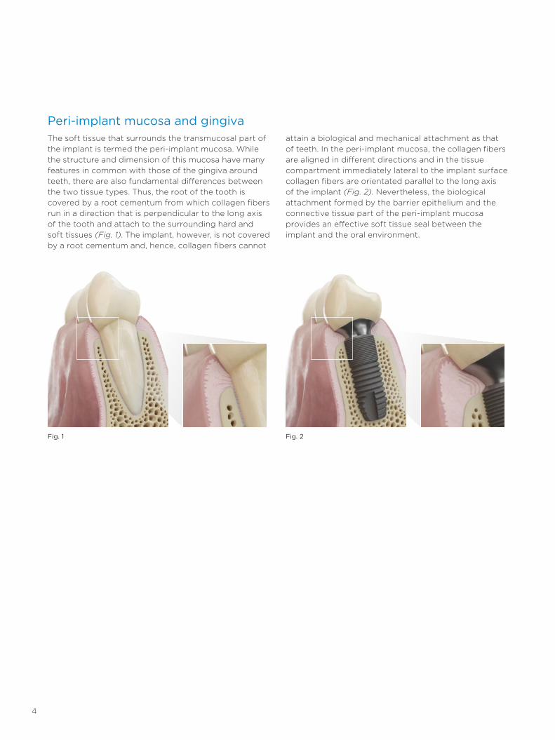
Peri-implant mucosa and gingivaThe soft tissue that surrounds the transmucosal part of the implant is termed the peri-implant mucosa. While the structure and dimension of this mucosa have many features in common with those of the gingiva around teeth, there are also fundamental differences between the two tissue types. Thus, the root of the tooth is covered by a root cementum from which collagen fibers run in a direction that is perpendicular to the long axis of the tooth and attach to the surrounding hard and soft tissues (Fig. 1). The implant, however, is not covered by a root cementum and, hence, collagen fibers cannot
attain a biological and mechanical attachment as that of teeth. In the peri-implant mucosa, the collagen fibers are aligned in different directions and in the tissue compartment immediately lateral to the implant surface collagen fibers are orientated parallel to the long axis of the implant (Fig. 2). Nevertheless, the biological attachment formed by the barrier epithelium and the connective tissue part of the peri-implant mucosa provides an effective soft tissue seal between the implant and the oral environment.
Fig. 1 Fig. 2
5
Case definitions of peri-implant health and diseasePeriodontal diseases around teeth are classified as gingivitis and periodontitis. Gingivitis refers to gingival inflammation with no signs of loss of supporting tissues, while periodontitis in addition to gingival inflammation is characterized by loss of attachment and bone5. In accordance with the classification of periodontal diseases around teeth, peri-implant disease includes
two entities: peri-implant mucositis, which corresponds to gingivitis and peri-implantitis, which corresponds to periodontitis. In the 2017 World Workshop on Classification on Periodontal and Peri-implant diseases and Conditions characteristics and case definitions were presented for peri-implant health, peri-implant mucositis and peri-implantitis6.
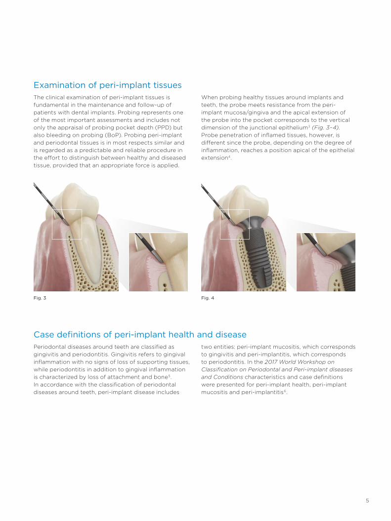
Examination of peri-implant tissuesThe clinical examination of peri-implant tissues is fundamental in the maintenance and follow-up of patients with dental implants. Probing represents one of the most important assessments and includes not only the appraisal of probing pocket depth (PPD) but also bleeding on probing (BoP). Probing peri-implant and periodontal tissues is in most respects similar and is regarded as a predictable and reliable procedure in the effort to distinguish between healthy and diseased tissue, provided that an appropriate force is applied.
When probing healthy tissues around implants and teeth, the probe meets resistance from the peri-implant mucosa/gingiva and the apical extension of the probe into the pocket corresponds to the vertical dimension of the junctional epithelium3 (Fig. 3–4). Probe penetration of inflamed tissues, however, is different since the probe, depending on the degree of inflammation, reaches a position apical of the epithelial extension4.
Fig. 3 Fig. 4
6
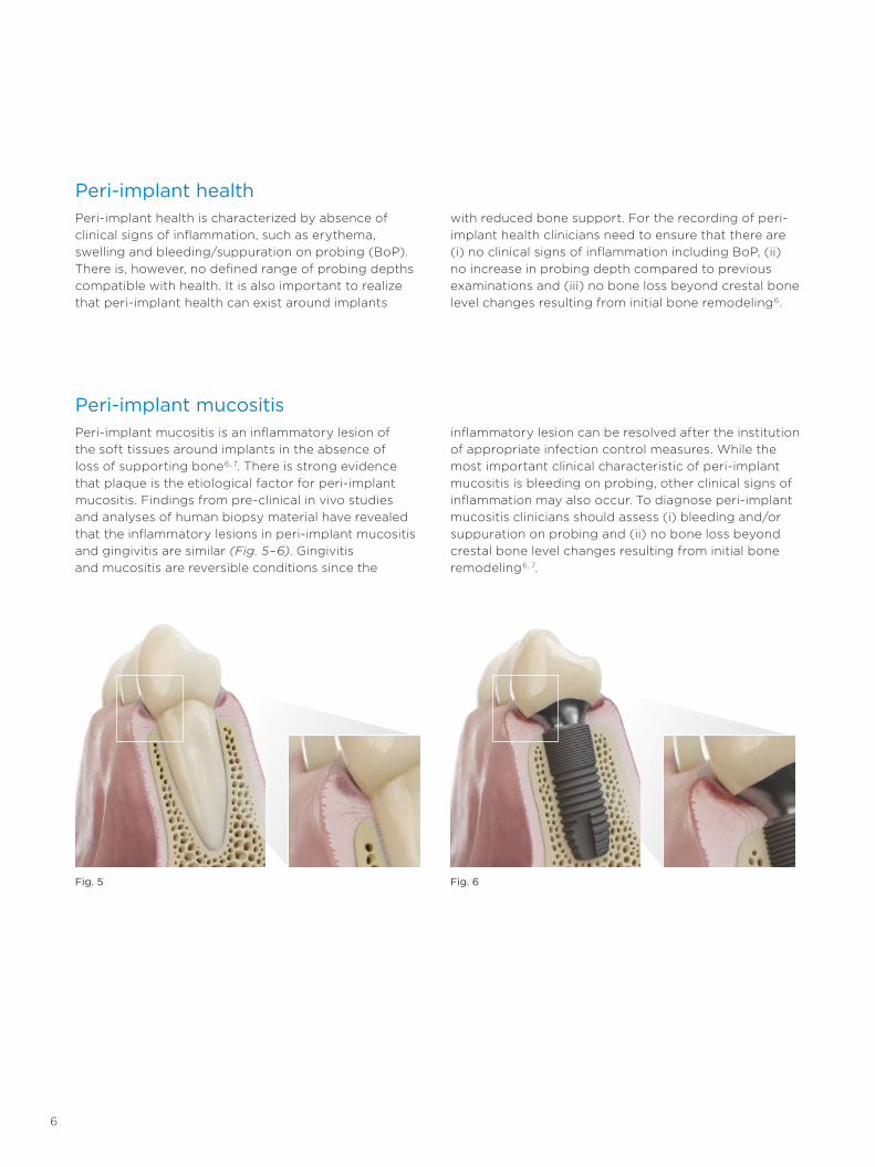
Peri-implant healthPeri-implant health is characterized by absence of clinical signs of inflammation, such as erythema, swelling and bleeding/suppuration on probing (BoP). There is, however, no defined range of probing depths compatible with health. It is also important to realize that peri-implant health can exist around implants
with reduced bone support. For the recording of peri-implant health clinicians need to ensure that there are (i) no clinical signs of inflammation including BoP, (ii) no increase in probing depth compared to previous examinations and (iii) no bone loss beyond crestal bone level changes resulting from initial bone remodeling6.
Peri-implant mucositisPeri-implant mucositis is an inflammatory lesion of the soft tissues around implants in the absence of loss of supporting bone6, 7. There is strong evidence that plaque is the etiological factor for peri-implant mucositis. Findings from pre-clinical in vivo studies and analyses of human biopsy material have revealed that the inflammatory lesions in peri-implant mucositis and gingivitis are similar (Fig. 5–6). Gingivitis and mucositis are reversible conditions since the
inflammatory lesion can be resolved after the institution of appropriate infection control measures. While the most important clinical characteristic of peri-implant mucositis is bleeding on probing, other clinical signs of inflammation may also occur. To diagnose peri-implant mucositis clinicians should assess (i) bleeding and/or suppuration on probing and (ii) no bone loss beyond crestal bone level changes resulting from initial bone remodeling6, 7.
Fig. 5 Fig. 6

7
Peri-implantitisPeri-implantitis is a plaque-associated disease occurring in tissues around dental implants. It is characterized by inflammation in the peri-implant mucosa and loss of supporting bone6, 8. Peri-implantitis lesions extend apical of the junctional/pocket epithelium and are typically larger than periodontitis lesions around teeth (Fig. 7–8).
The most important characteristics of peri-implantitis sites are clinical signs of inflammation including bleeding/suppuration on probing, increased probing depths and radiographic bone loss. It is also important to realize that progression of peri-implantitis in most
cases is faster than that observed in periodontitis and occurs in a non-linear and accelerating pattern.
To diagnose peri-implantitis clinicians should record (i) bleeding and/or suppuration on probing, (ii) increased probing depth compared to previous examinations and (iii) bone loss beyond crestal bone level changes resulting from initial bone remodeling. It is also possible to diagnose peri-implantitis in the absence of previous examination data by the combination of bleeding and/or suppuration on probing, probing depths of ≥6 mm and bone levels ≥3 mm apical of the most coronal portion of the intra-osseous part of the implant6.
Risk factors for peri-implantitis Moderate and severe forms of peri-implantitis occur in about 15 % of implant patients9. It is a generic disease-problem that may occur around all types of implants. The association between plaque and peri-implantitis is supported by evidence demonstrating that patients with poor plaque control and not attending regular maintenance therapy are at higher risk of developing peri-implantitis and that anti-infective treatment strategies are successful in arresting disease progression. There is also strong evidence that there is an increased risk for peri-implantitis in patients who have a history of severe periodontitis. Although all implant patients require sufficient follow-up and supportive therapy during maintenance, subjects with
a history of severe periodontitis should be provided with comprehensive infection control during follow-up in order to prevent peri-implant disease. Data from a 20-year prospective study on Astra Tech Implant System revealed that the implants were successfully maintained with optimal crestal bone preservation and healthy peri-implant tissues in patients with a history of severe periodontitis, given that a sufficient supportive therapy program was provided 10. The design of the implant-supported prosthesis is another risk factor for peri-implant disease. Access for infection control procedures performed by the patient and the dentist and/or dental hygienist must be provided in order to prevent inflammation in the peri-implant tissues.
Fig. 7 Fig. 8

8
Treatment of peri-implant diseasePeri-implant mucositis and peri-implantitis are caused by bacteria and, hence, treatment procedures must be aimed at eliminating the infection to resolve inflammatory lesions in peri-implant tissues. Thus, the goal in the treatment of these conditions is to achieve pocket closure and absence of bleeding on probing. The treatment should also result in prevention of further loss of supporting tissues, which is accomplished by adequate supportive therapy. Fundamental to these goals is infection control. All subjects who present any signs of peri-implant disease should be thoroughly informed about the disorder and instructed on how to carry out self- performed infection control. Whether the disease is mucositis or peri-implantitis, the initial phase of therapy must always include infection control procedures. Professional infection control procedures include the removal of hard and soft bacterial deposits on implant and suprastructure components with scalers. The instruments to be used should not damage the implant components or the surrounding tissues. In this context, it is imperative to point out that deep, “blind” instrumentation, such as “subgingival debridement” that normally is performed around teeth, is not recommended in non-surgical treatment of peri-implant disease. The reason for this difference in strategy is related to the geometry of the implant device with its threaded part and other obstacles to
access. The risk of causing injury to the inflamed tissues when performing blind instrumentation must also be emphasized.
The treatment of peri-implantitis will in most cases require surgery. The purpose of surgical therapy is to provide access for debridement and decontamination of the implant surface. After flap elevation, meticulous mechanical cleansing of the exposed implant is performed (Fig. 9). Presently, there is no documentation supporting any particular antiseptic agent to be used during cleaning being more effective than others. While peri-implantitis lesions are associated with bone loss resulting in defects of varying size and morphology, surgical treatment procedures also include strategies for the management of the hard tissue component. The common problem of lack of bone support on buccal and/or lingual aspects of bone defects associated with peri-implantitis requires resective procedures in which bone recontouring is performed to outline the bone morphology in order to facilitate soft tissue adaptation and pocket elimination (Fig 10). In other situations, when the defect morphology allows reconstructive procedures, the use of e.g. bone grafts and guided bone regeneration may be considered, provided that appropriate anti-infective measures are carried out to achieve resolution of the peri-implantitis lesion.
Fig. 9 Fig. 10

9
Supportive therapy – prevention of peri-implant diseaseFollowing the completion of the surgical and prosthetic procedures in implant therapy, it is imperative to inform the patient about how to carry out self-performed infection control procedures. Thus, depending on the design of the prosthetic reconstruction, different types
of toothbrushes and/or floss should be used to properly clean the implant and adjacent parts of the prosthesis twice a day (Fig. 11). The design of the prosthetic reconstruction must allow access for self-performed and professional infection control.
Guidelines for follow-up of implant-treated patients
The ideal implant supported prosthetic restoration is the one that reproduces the anatomy and volume of the lost teeth, with peri-implant soft tissues filling the interdental spaces and allowing a natural emergence profile, undistinguishable from natural teeth. Performing oral hygiene around such prosthetic restorations may be simple, since it involves oral
hygiene procedures identical to those applied around natural teeth, using manual or electrical toothbrushes together with interdental cleaning methods, e.g. flossing or interdental brushes (Fig. 12). Some situations may require specific oral hygiene aids (threaders, single tufted brushes, dental tape, etc.) to enable adequate oral hygiene procedures.
Fig. 11
Fig. 12
Manual/Electric Toothbrushes
Specific Brushes Interproximal Brushes Floss

10
In sites with peri-implant mucositis the patient should be instructed to use appropriate oral hygiene aids. Professional infection control, e.g. ultrasonic or air polishing devices, may be implemented to eliminate plaque and calculus from the implant/abutment surfaces. In some cases, screw-retained reconstructions may have to be removed to promote access to the implants (Fig. 13).
Patients exposed to surgical treatment of peri-implantitis require supportive therapy with frequent follow-up visits to ensure adequate self-performed infection control in order to prevent recurrence of the disease. In such situations the peri-implant tissues may be located at a more apical position and parts of the threaded portions of the implants may be exposed. Hence, the patient must be trained to handle such new situations and using individually customized brushes and interdental aids (Fig. 14).
Fig. 13 Fig. 14
Radiographic examinationRadiographic examination of the implant sites should be carried out using intra-oral radiographs at the time of the connection of the prosthesis and at the one-year follow-up. Possible marginal bone level changes documented during the first year in function of an implant may be associated with the physiological bone remodeling after implant installation. The information thereby obtained forms a baseline for subsequent
bone level evaluations. It should be pointed out, however, that the degree of physiological remodeling after implant placement may vary and that extensive peri-implant bone loss may also be reflective of the development of peri-implantitis during the remodeling phase. During the long-term follow-up period the need for radiographic examination will be determined by findings made in clinical examinations.

11
Clinical examination Baseline probing measurements should be obtained following the completion of the implant-supported prosthesis and clinical examinations should subsequently be performed at all annual follow-up visits. Besides checking the function of the prosthesis, the clinical examination should include evaluations of BoP, PPD and plaque. If the probing indicates peri-implant disease, i.e. BoP-positive and increased PPD or PPD ≥ 6 mm, these findings call for a radiographic examination to reveal possible bone loss in relation to baseline radiographs. In the absence of clinical findings of pathology in peri-implant tissues, radiographic examination should be avoided.
Following the diagnosis of either mucositis or peri- implantitis, however, appropriate infection control measures should be initiated as described above. The recall system for follow-up of implant-treated patients should be designed in accordance with evaluation of risk factors for peri-implant disease. Thus, subjects with a history of severe periodontitis require a recall program with examinations and supportive therapy carried out every 2–6 months after the delivery of the prosthesis.
1. Berglundh T, Lindhe J, Dimension of the periimplant mucosa. Biological width revisited. J Clin Periodontol 1996;23(10):971-3. Abstract
2. Jepsen S, Caton JG, Albandar JM, et al., Periodontal manifestations of systemic diseases and developmental and acquired conditions: Consensus report of workgroup 3 of the 2017 World Workshop on the Classification of Periodontal and Peri-Implant Diseases and Conditions. J Clin Periodontol 2018;45 Suppl 20:S219-S29. Abstract
3. Abrahamsson I, Soldini C, Probe penetration in periodontal and peri-implant tissues. An experimental study in the beagle dog. Clin Oral Implants Res 2006;17(6):601-5. Abstract
4. Schou S, Holmstrup P, Stoltze K, et al., Probing around implants and teeth with healthy or inflamed peri-implant mucosa/gingiva. A histologic comparison in cynomolgus monkeys (Macaca fascicularis). Clin Oral Implants Res 2002;13(2):113-26. Abstract
5. Papapanou PN, Sanz M, Buduneli N, et al., Periodontitis: Consensus report of workgroup 2 of the 2017 World Workshop on the Classification of Periodontal and Peri-Implant Diseases and Conditions. J Clin Periodontol 2018;45 Suppl 20:S162-S70. Abstract
6. Berglundh T, Armitage G, Araujo MG, et al., Peri-implant diseases and conditions: Consensus report of workgroup 4 of the 2017 World Workshop on the Classification of Periodontal and Peri-Implant Diseases and Conditions. J Clin Periodontol 2018;45 Suppl 20:S286-S91. Abstract
7. Heitz-Mayfield LJA, Salvi GE, Peri-implant mucositis. J Clin Periodontol 2018;45 Suppl 20:S237-S45. Abstract
8. Schwarz F, Derks J, Monje A, et al., Peri-implantitis. J Clin Periodontol 2018;45 Suppl 20:S246-S66. Abstract
9. Derks J, Schaller D, Hakansson J, et al., Effectiveness of Implant Therapy Analyzed in a Swedish Population: Prevalence of Peri-implantitis. J Dent Res 2016;95(1):43-9. Abstract
10. Donati M, Ekestubbe A, Lindhe J, et al., Marginal bone loss at implants with different surface characteristics - A 20-year follow-up of a randomized controlled clinical trial. Clin Oral Implants Res 2018;29(5):480-87. Abstract
References

De
nts
ply
Sir
on
a d
oes
no
t w
aive
any
rig
ht
to it
s tr
ade
mar
ks b
y n
ot
usi
ng
th
e sy
mb
ols
® o
r ™
. 3
26
7 0
152-
US
X- 1
90
5 ©
20
1 9 D
en
tsp
ly S
iro
na
. All
rig
hts
res
erv
ed
.
About Dentsply Sirona Implants
Dentsply Sirona Implants offers comprehensive solutions for all phases of implant therapy, including Ankylos®, Astra Tech Implant System® and Xive® implant lines, digital technologies, such as Atlantis® patient-specific solutions and Simplant® guided surgery, Symbios® regenerative solutions, and professional and business development programs, such as STEPPS™. Dentsply Sirona Implants creates value for dental professionals and allows for predictable and lasting implant treatment outcomes, resulting in enhanced quality of life for patients.
About Dentsply Sirona
Dentsply Sirona is the world’s largest manufacturer of professional dental products and technologies, with a 130-year history of innovation and service to the dental industry and patients worldwide. Dentsply Sirona develops, manufactures, and markets a comprehensive solutions offering including dental and oral health products as well as other consumable medical devices under a strong portfolio of world class brands. As The Dental Solutions Company™, Dentsply Sirona’s products provide innovative, high-quality and effective solutions to advance patient care and deliver better, safer and faster dentistry. Dentsply Sirona’s global headquarters is located in York, Pennsylvania, and the international headquarters is based in Salzburg, Austria. The company’s shares are listed in the United States on NASDAQ under the symbol XRAY.
Visit www.dentsplysirona.com for more information about Dentsply Sirona and its products.