Ecosystem Power Point By: Matthew Burkhardt Period 4 Science Mr. Sunesara.
Supporting InformationBurkhardt et al. 10.1073/pnas.1106189108SI Experimental ProceduresStructure Determination and Refinement. Diffraction data wereintegrated and scaled with XDS (1). The structure was solved bymolecular replacement using Phaser (2). The search model wasgenerated by pruning nonconserved residues of the neuronalMunc18-1/syntaxin 1a complex (PDB ID code 3C98; ref. 3) toalanines with Chainsaw (4). The asymmetric unit contains oneMunc18/syntaxin 1 complex. The model was refined by usingsimulated annealing, gradient minimization, and individual iso-tropic B-factor refinement as implemented in Phenix (5) alter-nated by rebuilding cycles using the program Coot (6). In a finalstep, tensor elements describing the anisotropic displacement offour individual domains of Munc18 and four regions of syntaxin1 were refined by using the Translation/Libration/Screw (TLS)implementation of Phenix. The final R factor is 0.188 with a Rfreeof 0.258. The final model comprises Munc18 residues 1–509 and561–616, syntaxin 1 residues 2–15, 39–192, and 210–261, and 48water molecules (Table S1). Figures were generated with theprogram Pymol (7). The coordinates have been deposited in theRCSB Protein Data Bank (ID code 2XHE) and will be releasedupon publication.
Phylogeny. Phylogenetic reconstruction was essentially done asdescribed in ref. 8. To gain insights into the phylogeneticplacement of the core factors of the secretory machinery fromthe choanoflagellate M. brevicollis, we included sequences from15 selected animal species, 7 selected fungal species, 6 selectedplant species, and 3 protists for the construction of the phylo-genetic trees. The sequences of the secretory SNARE proteins(i.e., type IV) were downloaded from our SNARE databasehttp://bioinformatics.mpibpc.mpg.de/snare. The sequences ofthe secretory SM proteins were gathered from the nr-database atNational Center for Biotechnology Information and few genomeprojects from the Department of Energy Joint Genome Institute.The species list and abbreviations and the sequence identity
numbers of the used sequences can be found in Table S2 andTable S3. We then aligned each factor by using muscle (9).Phylogenetic reconstruction was composed of two different
analytical approaches. The first approach used Important QuartetPuzzling and Nearest Neighbor Interchange (IQPNNI) (10) toconstruct phylogenetic trees from the curated alignments. Weused a gamma distribution as a model for rate heterogeneity withfour rate categories for the estimation of the gamma distributionparameter. The proportion of invariable sites was estimatedfrom the data and the Jones, Taylor, and Thornton (JTT) dis-tance matrix (11) served as a substitution matrix. We used thestopping rule of IQPNNI, but the calculation had to run for atleast the suggested number of iterations. The default values wereused for the remaining parameter. In addition, likelihood map-ping was applied to determine the confidence of the edges in thecalculated trees. The second approach used the phylip package(12) to apply a distance-based bootstrap analysis with 1,000 rep-licates to each of the curated alignments. Standard settings wereused for seqboot, the JTT distance matrix, and also a gammadistribution (with parameter approximation from tree puzzle) forprotdist, as were standard options for neighbor. If required, therandom seed was set to nine. We used the almost unbiased (AU)test (13) to address the systematically biased bootstrap values. Weobtained the sitewise log-likelihoods needed for the AU test byusing a modified version of phyml (14), and the test was per-formed by using consel (15). The reconstructed IQPNNI treesserved as starting points to join the results of both calculations.The inner edges of the trees were labeled with their likelihoodmapping and corrected bootstrap support values.
Electrophoretic Procedures. SDS resistance of ternary SNAREcomplexes in polyacrylamide gels (16) was tested as described(17) with the modification that the complexes were visualizedby the incorporation of synaptobrevin (1-75) labeled with thefluorescent dye Texas red at cysteine 58.
1. Kabsch W (1993) Automatic processing of rotation diffraction data from crystals ofinitially unknown symmetry and cell constants. J Appl Cryst 26:795e800.
2. McCoy AJ, Grosse-Kunstleve RW, Storoni LC, Read RJ (2005) Likelihood-enhanced fasttranslation functions. Acta Crystallogr D Biol Crystallogr 61:458e464.
3. Burkhardt P, Hattendorf DA, Weis WI, Fasshauer D (2008) Munc18a controls SNAREassembly through its interaction with the syntaxin N-peptide. EMBO J 27:923e933.
4. Stein N (2008) CHAINSAW: A program for mutating pdb files used as templates inmolecular replacement. J Appl Cryst 41:641e643.
5. Adams PD, et al. (2004) Recent developments in the PHENIX software for automatedcrystallographic structure determination. J Synchrotron Radiat 11:53e55.
6. Emsley P, Cowtan K (2004) Coot: Model-building tools for molecular graphics. ActaCrystallogr D Biol Crystallogr 60:2126e2132.
7. DeLano WL (2002) The PyMOL Molecular Graphics System (DeLano Scientific, PaloAlto, CA).
8. Kloepper TH, Kienle CN, Fasshauer D (2007) An elaborate classification of SNAREproteins sheds light on the conservation of the eukaryotic endomembrane system.Mol Biol Cell 18:3463e3471.
9. Edgar RC (2004) MUSCLE: A multiple sequence alignment method with reduced timeand space complexity. BMC Bioinformatics 5:113.
10. Vinh S, Von Haeseler A (2004) IQPNNI: Moving fast through tree space and stoppingin time. Mol Biol Evol 21:1565e1571.
11. Jones DT, Taylor WR, Thornton JM (1992) The rapid generation of mutation datamatrices from protein sequences. Comput Appl Biosci 8:275e282.
12. Felsenstein J (1998) PHYLIP - Phylogeny inference package (Version 3.2). Cladistics 5:164e166.
13. Shimodaira H (2002) An approximately unbiased test of phylogenetic tree selection.Syst Biol 51:492e508.
14. Guindon S, Gascuel O (2003) A simple, fast, and accurate algorithm to estimate largephylogenies by maximum likelihood. Syst Biol 52:696e704.
15. Shimodaira H, Hasegawa M (2001) CONSEL: For assessing the confidence ofphylogenetic tree selection. Bioinformatics 17:1246e1247.
16. Hayashi T, et al. (1994) Synaptic vesicle membrane fusion complex: Action ofclostridial neurotoxins on assembly. EMBO J 13:5051e5061.
17. Fasshauer D, Antonin W, Margittai M, Pabst S, Jahn R (1999) Mixed and non-cognateSNARE complexes. Characterization of assembly and biophysical properties. J BiolChem 274:15440e15446.
18. Dulubova I, et al. (1999) A conformational switch in syntaxin during exocytosis: Roleof munc18. EMBO J 18:4372e4382.
19. Misura KM, Scheller RH, Weis WI (2000) Three-dimensional structure of the neuronal-Sec1-syntaxin 1a complex. Nature 404:355e362.
20. Munson M, Chen X, Cocina AE, Schultz SM, Hughson FM (2000) Interactions withinthe yeast t-SNARE Sso1p that control SNARE complex assembly. Nat Struct Biol 7:894e902.
21. Margittai M, et al. (2003) Single-molecule fluorescence resonance energy transferreveals a dynamic equilibrium between closed and open conformations of syntaxin 1.Proc Natl Acad Sci USA 100:15516e15521.
22. Weimer RM, et al. (2006) UNC-13 and UNC-10/rim localize synaptic vesicles to specificmembrane domains. J Neurosci 26:8040e8047.
23. Gerber SH, et al. (2008) Conformational switch of syntaxin-1 controls synaptic vesiclefusion. Science 321:1507e1510.
24. Hu SH, Latham CF, Gee CL, James DE, Martin JL (2007) Structure of theMunc18c/Syntaxin4 N-peptide complex defines universal features of the N-peptidebinding mode of Sec1/Munc18 proteins. Proc Natl Acad Sci USA 104:8773e8778.
25. Hu SH, et al. (2011) Possible roles for Munc18-1 domain 3a and Syntaxin1 N-peptideand C-terminal anchor in SNARE complex formation. Proc Natl Acad Sci USA 108:1040e1045.
26. Peng RW, Guetg C, Abellan E, Fussenegger M (2010) Munc18b regulates core SNAREcomplex assembly and constitutive exocytosis by interacting with the N-peptide andthe closed-conformation C-terminus of syntaxin 3. Biochem J 431:353e361.
27. Lovell SC, et al. (2003) Structure validation by Calpha geometry: Phi,psi and Cbetadeviation. Proteins 50:437e450.
Burkhardt et al. www.pnas.org/cgi/content/short/1106189108 1 of 10
.64/.70
.47/.35
.84/.66
.43/.69
.44/.83
.93/.83
.84/.83
DaRe_SN47MuMu_SN47
HoSa_SN47GaGa_SN47
BrFl_SN47
NeVe_SN47
HyMa_SN47
LoGi_SN47
TrAd_SN47p
ChRe_SN29VoCa_SN29
ArTh_SN33
ArTh_SN29ArTh_SN30
OsTa_SN29BrFl_SN25likeCiIn_SN25
GaGa_SN23MuMu_SN23HoSa_SN23DaRe_SN23
DaRe_SN25bDaRe_SN25a
GaGa_SN25bMuMu_SN25bHoSa_SN25bHoSa_SN25a
MuMu_SN25aTrAd_SN25TrAd_SN25-2
CaEl_SN25HeRo_SN25-2
CaSp_SN25
HeRo_SN25LoGi_SN25
DrMe_SN25DrMe_SN24 BrFl_SN25
NeVe_SN25
MoBr_SN25
AmQu_SN25UsMa_Sec9
ShPo_Sec9SaCe_Sec9
YaLi_Sec9
MaGr_Sec9
EnDi_SNpCaEl_SN29
CaSp_SN29LoGi_SN29
HeRo_SN29
HyMa_SN29NeVe_SN29
BrFl_SN29
CiIn_SN29DaRe_SN29
GaGa_SN29MuMu_SN29HoSa_SN29
.83/.83Fungi
Viridiplantae
Metazoa
SaCe_Spo20
BSNAP-29
SNAP-47
SNAP-25
Metazoa +
Choanoflagellata
.66/.85
.94/.85
.96/.85
MiSt_Sec1
ChRe_Sec1VoCa_Sec1
ChSp_Sec1ArTh_Sec1_4
ArTh_Sec1_3ArTh_Sec1ArTh_Sec1_2
TrBr_Sec1
EnHi_Sec1EnDi_Sec1p
ScPo_Sec1
NeCr_Sec1MaGr_Sec1
UsMa_Sec1LaBi_Sec1
YaLi_Sec1
SaCe_Sec1
MoBr_Munc18
AmQu_Unc18HyMa_Unc18
NeVe_Unc18_2
NeVe_Unc18TrAd_Unc18
CiIn_Unc18
CaEl_Unc18DrMe_Unc18LoGi_Unc18HeRo_Unc18_2HeRo_Unc18CaSp_Unc18
BrFl_Unc18
DaRe_Munc18_3MuMu_Munc18_3
HoSa_Munc18_3GaGa_Munc18_3DaRe_Munc18_2
HoSa_Munc18_2MuMu_Munc18_2
DaRe_Munc18_1
GaGa_Munc18_1
HoSa_Munc18_1MuMu_Munc18_1
Fungi
Viridiplantae
Metazoa +
Choanoflagellata
A
.47/.94
1.00/.94
1.00/.94.96/.94
.90/.94.86/.94
.99/.94
.81/.94
.78/.94
DaRe_Syx11
DaRe_Syx11-likeDaRe_Syx11-like2
MuMu_Syx11HoSa_Syx11
GaGa_Syx11
HoSa_Syx19MuMu_Syx19GaGa_Syx19
DaRe_Syx19
MuMu_Syx21NAmQu_Syx1
TrAd_Syx3TrAd_Syx2
TrAd_Syx1HeRo_Syx1
DaRe_Syx4MuMu_Syx4HoSa_Syx4
HoSa_Syx2MuMu_Syx2
GaGa_Syx2GaGa_Syx3MuMu_Syx3HoSa_Syx3DaRe_Syx3
GaGa_Syx1bHoSa_Syx1bMuMu_Syx1b
DaRe_Syx1b
GaGa_Syx1aMuMu_Syx1aHoSa_Syx1a
HeRo_Syx2LoGi_Syx1 CaSp_Syx1
HeRo_Syx3
DrMe_Syx1 CaEl_Unc64CiIn_Syx2BrFl_Syx1 NeVe_Syx1
TrAd_Syx4
SaCe_Sso2SaCe_Sso1YaLi_SsoCYaLi_SsoA
MaGr_SsoBUsMa_Sso
ShPo_Sso
ArTh_Syx112ArTh_Syx111
ArTh_Syx125ArTh_Syx124
ArTh_Syx123
ArTh_Syx121ArTh_Syx122
VoCa_Syx1ChRe_Syx1
ChSp_Syx1 ArTh_Syx131ArTh_Syx132
OsTa_Syx1
EnDi_Syx1EnHi-SyxPM3
EnDi_SyxPM4EnHi_SyxPM2
CaEl_Syx4CaEl_Syx3
CaEl_Syx2DrMe_Syx4
CiIn_Syx3
Fungi
Viridiplantae
MoBr_Syx1
.95/.94
C Metazoa +
Choanoflagellata
SNAP-25
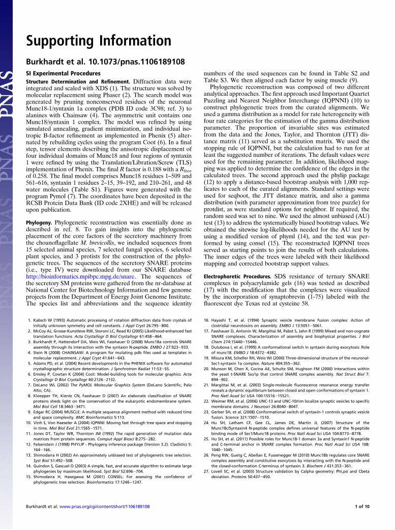
Fig. S1. Detailed versions of the phylogenetic trees of the SM protein Munc18 (A), the Qbc-SNARE SNAP-25 (B), and secretory syntaxins (C) (type Qa.IV) shownin Fig. 1. The major phylogenetic lineages are indicated by different colors. The labels on the major branches represent the likelihood mapping (left) andalmost unbiased (AU) support values (right). The species abbreviations and sequences used are given in Table S2 and Table S3. Whereas M. brevicollis possessesonly one factor each, duplications occurred recurrently in different animal lineages. A noteworthy expansion of Munc18-like factors occurred in vertebrates,giving rise to the three isoforms Munc18-1, -2, and -3. A more prominent expansion took place in secretory syntaxins in vertebrates (8). Interestingly, the SNAP-25–like protein found in M. brevicollis is closely related to the neuronal SNAP-25 of animals (8), whereas the sequences of the other two Qbc-SNAREs animalhomologs SNAP-29 and SNAP-47, have diverged substantially.
Burkhardt et al. www.pnas.org/cgi/content/short/1106189108 2 of 10
0
1x10⁴
2x10⁵
3x10⁵
4x10⁵
5x10⁵
6x10⁵
300 350 400 450
Trp-
fluor
esce
nce
(cps
)
wavelenght (nm)
+ Syx1 (1-279)
+ Syx1 (20-279)
Munc18
Syntaxin 1
A B
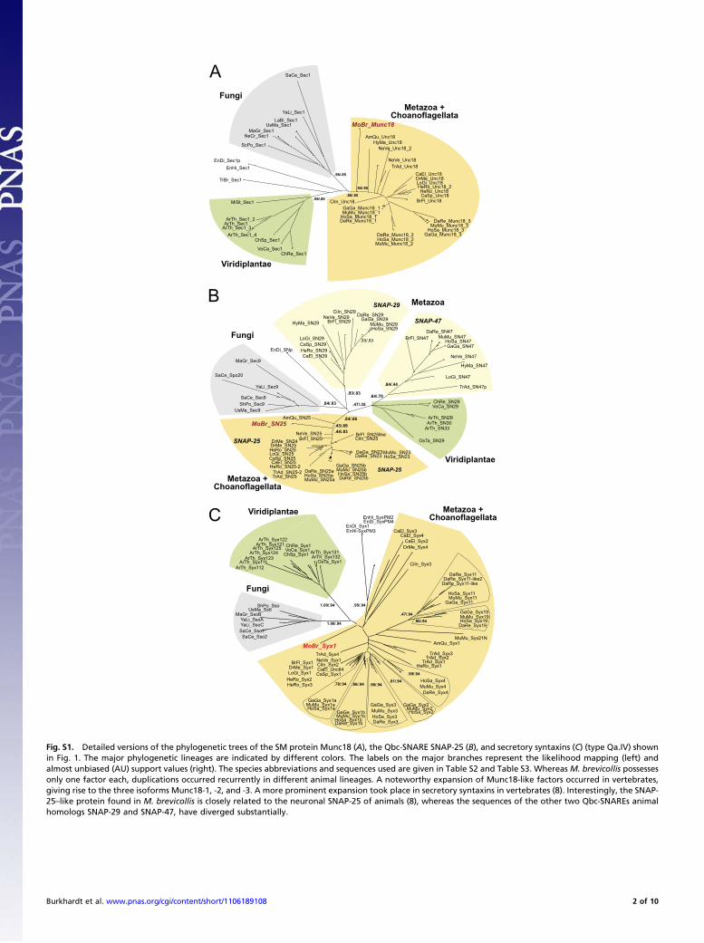
Fig. S2. Change of intrinsic fluorescence upon binding of Munc18 to syntaxin 1 (A) and overlay of the Munc18/Syx1 complexes (B). (A) Baseline correctedtryptophan fluorescence emission spectra of Syx1 (1-279), Munc18, Munc18 mixed with Syx1 (1-279), and Munc18 mixed with Syx1 (20-279) (500 nM each) afterexcitation at 295 nm. Upon addition of Syx1 (1-279) or Syx1 (20-279) to Munc18-1, an increase in fluorescence was monitored. Note that syntaxin 1 does notposses a tryptophan. Interestingly, the increase in tryptophan fluorescence in the presence of Syx1 (20-279) was somewhat less pronounced than in thepresence of Syx1 (1-279). A similar difference in the fluorescence change was observed for the rat homologs. This change in fluorescence suggests that bothsyntaxin variants adopt a slightly different conformation in complex with Munc18. It seems likely that the increase in tryptophan fluorescence is caused by closeproximity of Trp-24 of M. brevicollis Munc18 (Trp-28 of rat Munc18-1) and Phe-46 and Phe-47 of M. brevicollis syntaxin 1 (Phe-33 and Phe-34 of rat Syx1a). (B)Overlay of the Munc18/Syx1 complexes from M. brevicollis (Munc18, blue; Syx1, red) and from R. norvegicus (Munc18-1, light blue; Syx1a, salmon; PDB ID code3C98) reveals their overall similarity.
Burkhardt et al. www.pnas.org/cgi/content/short/1106189108 3 of 10
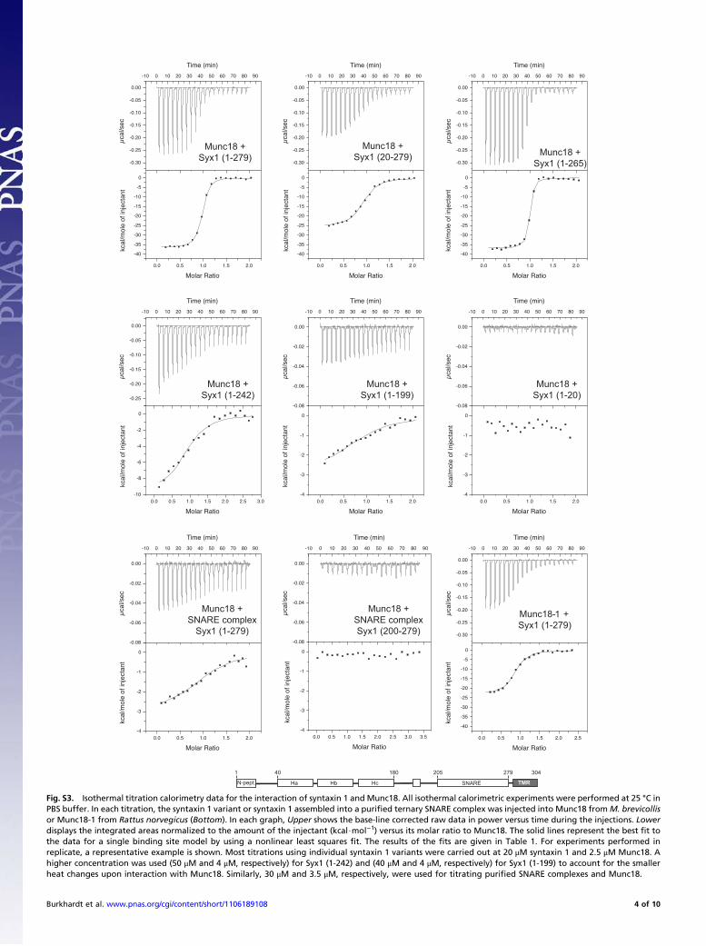
Fig. S3. Isothermal titration calorimetry data for the interaction of syntaxin 1 and Munc18. All isothermal calorimetric experiments were performed at 25 °C inPBS buffer. In each titration, the syntaxin 1 variant or syntaxin 1 assembled into a purified ternary SNARE complex was injected into Munc18 fromM. brevicollisor Munc18-1 from Rattus norvegicus (Bottom). In each graph, Upper shows the base-line corrected raw data in power versus time during the injections. Lowerdisplays the integrated areas normalized to the amount of the injectant (kcal$mol−1) versus its molar ratio to Munc18. The solid lines represent the best fit tothe data for a single binding site model by using a nonlinear least squares fit. The results of the fits are given in Table 1. For experiments performed inreplicate, a representative example is shown. Most titrations using individual syntaxin 1 variants were carried out at 20 μM syntaxin 1 and 2.5 μM Munc18. Ahigher concentration was used (50 μM and 4 μM, respectively) for Syx1 (1-242) and (40 μM and 4 μM, respectively) for Syx1 (1-199) to account for the smallerheat changes upon interaction with Munc18. Similarly, 30 μM and 3.5 μM, respectively, were used for titrating purified SNARE complexes and Munc18.
Burkhardt et al. www.pnas.org/cgi/content/short/1106189108 4 of 10
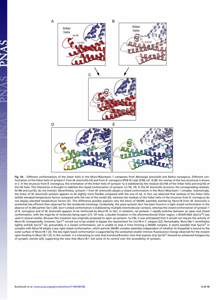
Fig. S4. Different conformations of the linker helix in the Munc18/syntaxin 1 complexes from Monosiga brevicollis and Rattus norvegicus. Different con-formation of the linker helix of syntaxin1 fromM. brevicollis (A) and from R. norvegicus (PDB ID code 3C98; ref. 3) (B). An overlay of the two structures is shownin C. In the structure from R. norvegicus, the orientation of the linker helix of syntaxin 1a is stabilized by the residues Glu166 of the linker helix and Arg142 ofthe Hb helix. This interaction is thought to stabilize the closed conformation of syntaxin 1a (18, 19). In the M. brevicollis structure, the corresponding residues,Ile186 and Lys162, do not interact. Nevertheless, syntaxin 1 from M. brevicollis adopts a closed conformation in the Munc18/syntaxin 1 complex. Interestingly,the linker of M. brevicollis syntaxin appears to be slightly more flexible compared with the one of rat. In fact, we observed that residues of the linker helixexhibit elevated temperature factors compared with the rest of the model (D), whereas the residues of the linker helix of the structure from R. norvegicus donot display elevated temperature factors (E). This difference possibly explains why the block of SNARE assembly exerted by Munc18 from M. brevicollis issomewhat less efficient than observed for the vertebrate homologs. Incidentally, the yeast syntaxin Sso1 has been found in a tight closed conformation in theabsence of its SM partner Sec1 (20). Sso1’s closed conformation is stabilized by multiple intermolecular contacts, whereas the closed conformation of syntaxin 1of R. norvegicus and of M. brevicollis appears to be reinforced by Munc18. In fact, in isolation, rat syntaxin 1 rapidly switches between an open and closedconformation, with the majority of molecules being open (21). Of note, a double mutation in the aforementioned linker region, L165A/E166A (Syx1aLE) wasused in several studies. Because this mutation was originally proposed to open up syntaxin 1a (18), it was anticipated that it would not require the activity ofMunc18. Unexpectedly, however, Syx1LE turned out to be unable to bypass the requirement for Unc18 in C. elegans (22). Remarkably, Munc18a-1 nonethelesstightly enfolds Syx1aLE (3), presumably in a closed conformation, yet is unable to stop it from forming a SNARE complex. It seems possible that Syx1aLE incomplex with Munc18 adopts a less rigid closed conformation, which permits SNARE complex assembly independent of whether its N-peptide is bound to theouter surface of Munc18-1 (3). The less rigid closed conformation is supported by the somewhat smaller intrinsic fluorescence change observed for the mutantupon binding to Munc18-1 (3). In this context. it is interesting to note that knockout/knockin mice that express only Syx1bLE showed an enhanced fusogenicityof synaptic vesicles (23), supporting the view that Munc18-1 lost some of its control over the accessibility of syntaxin.
Burkhardt et al. www.pnas.org/cgi/content/short/1106189108 5 of 10
Gln15Gln13
Gln9 Ser5 Asp2
Arg3
Leu7Ala11
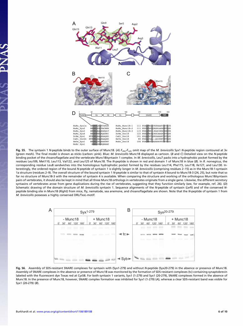
Fig. S5. The syntaxin 1 N-peptide binds to the outer surface of Munc18. (A) Fobs-Fcalc omit map of the M. brevicollis Syx1 N-peptide region contoured at 3σ(green mesh). The final model is shown as sticks (carbon: pink). Blue: M. brevicollis Munc18 displayed as cartoon. (B and C) Detailed view on the N-peptidebinding pocket of the choanoflagellate and the vertebrate Munc18/syntaxin 1 complex. In M. brevicollis, Leu7 packs into a hydrophobic pocket formed by theresidues Leu109, Met110, Leu113, Val122, and Leu125 of Munc18. The N-peptide is shown in red and domain 1 of Munc18 in blue (B). In R. norvegicus, thecorresponding residue Leu8 sandwiches into the homologous hydrophobic pocket formed by the residues Leu114, Phe115, Leu118, Ile127, and Leu130. In-terestingly, the ordered region of the bound N-peptide of syntaxin 1 is slightly longer in M. brevicollis (comprising residues 2–15) as in the Munc18-1-syntaxin1a structure (residues 2–9). The overall structure of the bound syntaxin 1 N-peptide is similar to that of syntaxin 4 bound to Munc18-3 (24, 25), but note that sofar no structure of Munc18-3 with the remainder of syntaxin 4 is available. When comparing the structure and working of the orthologous Munc18/syntaxinpairs of vertebrates, it should also be kept in mind that all three Munc18 orthologs in vertebrates originate from a single gene. Likewise, the different secretorysyntaxins of vertebrates arose from gene duplications during the rise of vertebrates, suggesting that they function similarly (see, for example, ref. 26). (D)Schematic drawing of the domain structure of M. brevicollis syntaxin 1. Sequence alignments of the N-peptide of syntaxin (Left) and of the conserved N-peptide binding site in Munc18 (Right) from mice, fly, nematode, sea anemone, and choanoflagellate are shown. Note that the N-peptide of syntaxin 1 fromM. brevicollis possesses a highly conserved DRL/TxxL-motif.
BA
Fig. S6. Assembly of SDS-resistant SNARE complexes for syntaxin with (Syx1-279) and without N-peptide (Syx20-279) in the absence or presence of Munc18.Assembly of SNARE complexes in the absence or presence of Munc18 was monitored by the formation of SDS-resistant complexes (tc) containing synaptobrevinlabeled with the fluorescent dye Texas red at Cys58. For both syntaxin 1 variants, Syx1 (1-279) and Syx1 (20-279), SNARE complexes formed in the absence ofMunc18. In the presence of Munc18, however, SNARE complex formation was inhibited for Syx1 (1-279) (A), whereas a clear SDS-resistant band was visible forSyx1 (20-279) (B).
Burkhardt et al. www.pnas.org/cgi/content/short/1106189108 6 of 10
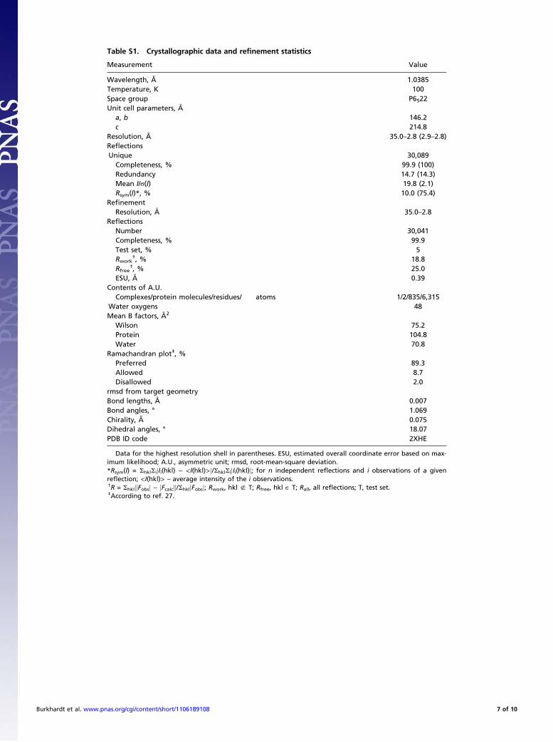
Table S1. Crystallographic data and refinement statistics
Measurement Value
Wavelength, Å 1.0385Temperature, K 100Space group P6522Unit cell parameters, Å
a, b 146.2c 214.8
Resolution, Å 35.0–2.8 (2.9–2.8)ReflectionsUnique 30,089Completeness, % 99.9 (100)Redundancy 14.7 (14.3)Mean I/σ(I) 19.8 (2.1)Rsym(I)*, % 10.0 (75.4)
RefinementResolution, Å 35.0–2.8
ReflectionsNumber 30,041Completeness, % 99.9Test set, % 5Rwork
†, % 18.8Rfree
†, % 25.0ESU, Å 0.39
Contents of A.U.Complexes/protein molecules/residues/ atoms 1/2/835/6,315
Water oxygens 48Mean B factors, Å2
Wilson 75.2Protein 104.8Water 70.8
Ramachandran plot‡, %Preferred 89.3Allowed 8.7Disallowed 2.0
rmsd from target geometryBond lengths, Å 0.007Bond angles, ° 1.069Chirality, Å 0.075Dihedral angles, ° 18.07PDB ID code 2XHE
Data for the highest resolution shell in parentheses. ESU, estimated overall coordinate error based on max-imum likelihood; A.U., asymmetric unit; rmsd, root-mean-square deviation.*Rsym(I) = ShklSijIi(hkl) − <I(hkl)>j/ShklSijIi(hkl)j; for n independent reflections and i observations of a givenreflection; <I(hkl)> – average intensity of the i observations.†R = ShklkFobsj − jFcalck/ShkljFobsj; Rwork, hkl ; T; Rfree, hkl ˛ T; Rall, all reflections; T, test set.‡According to ref. 27.
Burkhardt et al. www.pnas.org/cgi/content/short/1106189108 7 of 10
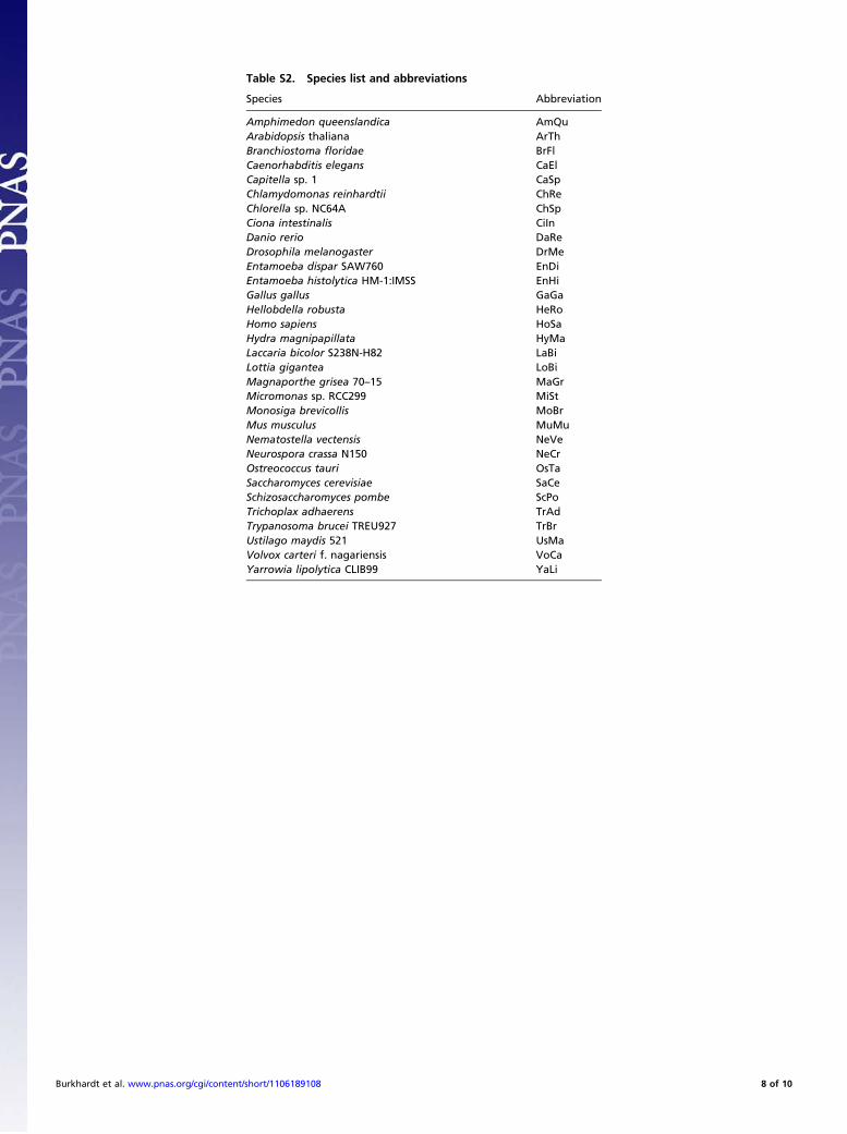
Table S2. Species list and abbreviations
Species Abbreviation
Amphimedon queenslandica AmQuArabidopsis thaliana ArThBranchiostoma floridae BrFlCaenorhabditis elegans CaElCapitella sp. 1 CaSpChlamydomonas reinhardtii ChReChlorella sp. NC64A ChSpCiona intestinalis CiInDanio rerio DaReDrosophila melanogaster DrMeEntamoeba dispar SAW760 EnDiEntamoeba histolytica HM-1:IMSS EnHiGallus gallus GaGaHellobdella robusta HeRoHomo sapiens HoSaHydra magnipapillata HyMaLaccaria bicolor S238N-H82 LaBiLottia gigantea LoBiMagnaporthe grisea 70–15 MaGrMicromonas sp. RCC299 MiStMonosiga brevicollis MoBrMus musculus MuMuNematostella vectensis NeVeNeurospora crassa N150 NeCrOstreococcus tauri OsTaSaccharomyces cerevisiae SaCeSchizosaccharomyces pombe ScPoTrichoplax adhaerens TrAdTrypanosoma brucei TREU927 TrBrUstilago maydis 521 UsMaVolvox carteri f. nagariensis VoCaYarrowia lipolytica CLIB99 YaLi
Burkhardt et al. www.pnas.org/cgi/content/short/1106189108 8 of 10
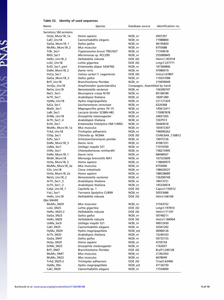
Table S3. Identity of used sequences
Name Species Database source Identification no.
Secretory SM proteinsHoSa_Munc18_1a Homo sapiens NCBI_nr 4507297CaEl_Unc18 Caenorhabditis elegans NCBI_nr 71988800GaGa_Munc18_1 Gallus gallus NCBI_nr 46195820MuMu_Munc18_2 Mus musculus NCBI_nr 6755688TrBr_Sec1 Trypanosoma brucei TREU927 NCBI_nr 71744018MiSt_Sec1 Micromonas sp. RCC299 NCBI_nr 255089845HeRo_Unc18_2 Hellobdella robusta DOE JGI Helro1j187018LoGi_Unc18 Lottia gigantea DOE JGI Lotgi1j237771EnDi_Sec1_part Entamoeba dispar SAW760 NCBI_nr 167384806DaRe_Munc18_2 Danio rerio NCBI_nr 47086919VoCa_Sec1 Volvox carteri f. nagariensis DOE JGI Volca1j67807GaGa_Munc18_3 Gallus gallus NCBI_nr 119331098BrFl_Unc18 Branchiostoma floridae NCBI_nr 219459049AmQu_Unc18 Amphimedon queenslandica Compagen, Assembled by handNeVe_Unc18 Nematostella vectensis NCBI_nr 156390747NeCr_Sec1 Neurospora crassa N150 NCBI_nr 85108189ArTh_Sec1 Arabidopsis thaliana NCBI_nr 18391384HyMa_Unc18 Hydra magnipapillata NCBI_nr 221121424SaCe_Sec1 Saccharomyces cerevisiae NCBI_nr 6320368MaGr_Sec1 Magnaporthe grisea 70–15 NCBI_nr 145612411LaBi_Sec1 Laccaria bicolor S238N-H82 NCBI_nr 170087878DrMe_Unc18 Drosophila melanogaster NCBI_nr 24657265ArTh_Sec1_4 Arabidopsis thaliana NCBI_nr 7267913EnHi_Sec1 Entamoeba histolytica HM-1:IMSS NCBI_nr 56464018MuMu_Munc18_1a Mus musculus NCBI_nr 165972307TrAd_Unc18 Trichoplax adhaerens NCBI_nr 196000262ChSp_Sec1 Chlorella sp. NC64A DOE JGI ChlNC64A_1j56812ScPo_Sec1 Schizosaccharomyces pombe NCBI_nr 19075726DaRe_Munc18_3 Danio rerio NCBI_nr 47087331UsMa_Sec1 Ustilago maydis 521 NCBI_nr 71019769ChRe_Sec1 Chlamydomonas reinhardtii NCBI_nr 158273495DaRe_Munc18_1 Danio rerio NCBI_nr 68448507MoBr_Munc18 Monosiga brevicollis MX1 NCBI_nr 167523609HoSa_Munc18_3 Homo sapiens NCBI_nr 118600975MuMu_Munc18_3a Mus musculus NCBI_nr 6755690CiIn_Unc18 Ciona intestinalis NCBI_nr 198429537HoSa_Munc18_2a Homo sapiens NCBI_nr 188528689NeVe_Unc18_2 Nematostella vectensis NCBI_nr 156390749ArTh_Sec1_3 Arabidopsis thaliana NCBI_nr 18413751ArTh_Sec1_2 Arabidopsis thaliana NCBI_nr 145334974CaSp_Unc18_1 Capitella sp. 1 DOE JGI Capca1j150412YaLi_Sec1 Yarrowia lipolytica CLIB99 NCBI_nr 50553686HeRo_Unc18 Hellobdella robusta DOE JGI Helro1j66166
Qbc-SNAREMuMu_SN29 Mus musculus NCBI_nr 31543752LoGi_SN25 Lottia gigantea DOE JGI Lotgi1j197810HeRo_SN25-2 Hellobdella robusta DOE JGI Helro1j71107GaGa_SN23 Gallus gallus NCBI_nr 50748211HeRo_SN29 Hellobdella robusta DOE JGI Helro1j184904UsMa_Sec9 Ustilago maydis 521 NCBI_nr 49072430CaEl_SN25 Caenorhabditis elegans NCBI_nr 32567202HyMa_SN29 Hydra magnipapillata NCBI_est 46968126ArTh_SN33 Arabidopsis thaliana NCBI_nr 15240163GaGa_SN47 Gallus gallus NCBI_nr 50732155HoSa_SN29 Homo sapiens NCBI_nr 4759154DrMe_SN25 Drosophila melanogaster NCBI_nr 1763657BrFl_SN47 Branchiostoma floridae DOE JGI Brafl1j240128MuMu_SN47 Mus musculus NCBI_nr 21362303MuMu_SN23 Mus musculus NCBI_nr 6678049TrAd_SN25-2 Trichoplax adhaerens DOE JGI Triad1j63490HyMa_SNx Hydra magnipapillata NCBI_est 47136750CaEl_SN29 Caenorhabditis elegans NCBI_nr 17554000
Burkhardt et al. www.pnas.org/cgi/content/short/1106189108 9 of 10
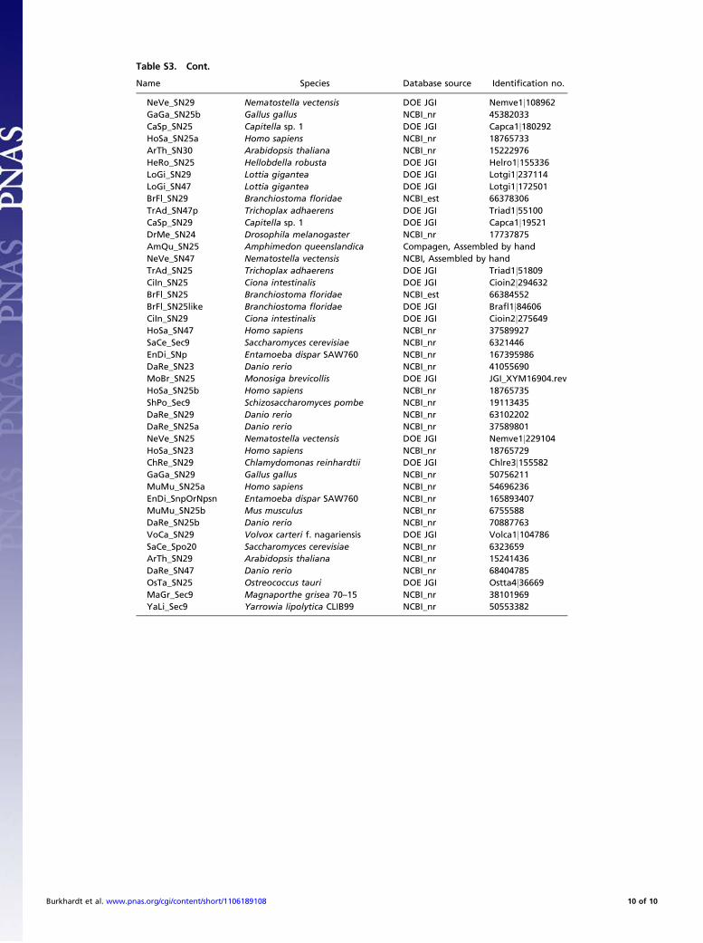
Table S3. Cont.
Name Species Database source Identification no.
NeVe_SN29 Nematostella vectensis DOE JGI Nemve1j108962GaGa_SN25b Gallus gallus NCBI_nr 45382033CaSp_SN25 Capitella sp. 1 DOE JGI Capca1j180292HoSa_SN25a Homo sapiens NCBI_nr 18765733ArTh_SN30 Arabidopsis thaliana NCBI_nr 15222976HeRo_SN25 Hellobdella robusta DOE JGI Helro1j155336LoGi_SN29 Lottia gigantea DOE JGI Lotgi1j237114LoGi_SN47 Lottia gigantea DOE JGI Lotgi1j172501BrFl_SN29 Branchiostoma floridae NCBI_est 66378306TrAd_SN47p Trichoplax adhaerens DOE JGI Triad1j55100CaSp_SN29 Capitella sp. 1 DOE JGI Capca1j19521DrMe_SN24 Drosophila melanogaster NCBI_nr 17737875AmQu_SN25 Amphimedon queenslandica Compagen, Assembled by handNeVe_SN47 Nematostella vectensis NCBI, Assembled by handTrAd_SN25 Trichoplax adhaerens DOE JGI Triad1j51809CiIn_SN25 Ciona intestinalis DOE JGI Cioin2j294632BrFl_SN25 Branchiostoma floridae NCBI_est 66384552BrFl_SN25like Branchiostoma floridae DOE JGI Brafl1j84606CiIn_SN29 Ciona intestinalis DOE JGI Cioin2j275649HoSa_SN47 Homo sapiens NCBI_nr 37589927SaCe_Sec9 Saccharomyces cerevisiae NCBI_nr 6321446EnDi_SNp Entamoeba dispar SAW760 NCBI_nr 167395986DaRe_SN23 Danio rerio NCBI_nr 41055690MoBr_SN25 Monosiga brevicollis DOE JGI JGI_XYM16904.revHoSa_SN25b Homo sapiens NCBI_nr 18765735ShPo_Sec9 Schizosaccharomyces pombe NCBI_nr 19113435DaRe_SN29 Danio rerio NCBI_nr 63102202DaRe_SN25a Danio rerio NCBI_nr 37589801NeVe_SN25 Nematostella vectensis DOE JGI Nemve1j229104HoSa_SN23 Homo sapiens NCBI_nr 18765729ChRe_SN29 Chlamydomonas reinhardtii DOE JGI Chlre3j155582GaGa_SN29 Gallus gallus NCBI_nr 50756211MuMu_SN25a Homo sapiens NCBI_nr 54696236EnDi_SnpOrNpsn Entamoeba dispar SAW760 NCBI_nr 165893407MuMu_SN25b Mus musculus NCBI_nr 6755588DaRe_SN25b Danio rerio NCBI_nr 70887763VoCa_SN29 Volvox carteri f. nagariensis DOE JGI Volca1j104786SaCe_Spo20 Saccharomyces cerevisiae NCBI_nr 6323659ArTh_SN29 Arabidopsis thaliana NCBI_nr 15241436DaRe_SN47 Danio rerio NCBI_nr 68404785OsTa_SN25 Ostreococcus tauri DOE JGI Ostta4j36669MaGr_Sec9 Magnaporthe grisea 70–15 NCBI_nr 38101969YaLi_Sec9 Yarrowia lipolytica CLIB99 NCBI_nr 50553382
Burkhardt et al. www.pnas.org/cgi/content/short/1106189108 10 of 10
![Shane E. Burkhardt, AICPdvqlxo2m2q99q.cloudfront.net/.../shane-burkhardt-select-projects-20… · Shane E. Burkhardt, AICP [Page 2] improved multi-modal access to the area through](https://static.fdocuments.in/doc/165x107/5fb0c214f3acfa69b35638e0/shane-e-burkhardt-shane-e-burkhardt-aicp-page-2-improved-multi-modal-access.jpg)