Supporting Information - pnas.org€¦ · Supporting Information ... examination by electron...
17
Supporting Information Han et al. 10.1073/pnas.0906545106 SI Materials and Methods Mice. Mice with striated-muscle-specific dystroglycan (DG) de- ficiency (MCK-cre/Dag1 flox/flox ) (1) and integrin 7-null (2) mice were described previously. For a direct comparison of DG- deficient and integrin 7-null skeletal muscle in the same mouse line, these 2 mouse lines were crossed to one another. MCK-Cre male mice bearing the floxed dystroglycan allele were mated to integrin 7 heterozygous females. F1 and F2 offspring were mated to produce F2- and F3-generation mice, respectively. Identification of the mutant mice was performed by PCR genotyping of genomic DNA prepared from mouse tail snips. The Large myd colony was originally obtained from Jackson Laboratories. Mice were maintained at The University of Iowa Animal Care Unit in accordance with animal use guidelines. All animal studies were authorized by the Animal Care Use and Review Committee of The University of Iowa. For treadmill exercise, mice (5 weeks old) were placed on an endless conveyor-type belt with a shock grid at the end (AccuPacer Treadmill, AccuScan Instruments) and exercised on a down-hill grade at 15 m/min for 20 min. Immediately after the exercise, mice were euthanized and quadriceps muscles were prepared for examination by electron microscopy or immunofluorescence. Lectin Affinity Chromatography and Sucrose Gradient Fractionation. Total muscle homogenates in TBS (50 mM Tris-Cl pH 7.4, 150 mM NaCl) were solubilized with 1% digitonin. After centrifu- gation at 140,000 g for 37 min, solubilized proteins in the supernatant were mixed with wheat germ agglutinin (WGA)- agarose beads (Vector Laboratories) and rotated end-over-end at 4 °C for 2 h. WGA-bound proteins were eluted with TBS containing 0.3 M N-acetyl-D-glucosamine and 0.1% digitonin. The eluant was applied to a 5–30% sucrose gradient and centrifuged at 215,000 g for 90 min. Fractions (1 mL) were collected from the top of the gradient and analyzed by SDS/ PAGE. Measurement of Contractile Properties. Muscle mass, fiber length, and maximum force were measured on 6 EDL muscles from 6- to 7-month-old Large myd , MCK-cre/Dag1 flox/flox , integrin 7-null, and WT littermate control mice. Mice were anesthetized and muscles isolated and stimulated to provide maximum isometric tetanic force (P o ). The susceptibility of muscles to contraction- induced injury was assessed by 2 lengthening contractions with a strain of 30% of fiber length. Total cross-sectional area (CSA, cm 2 ) and specific P o (kN/m 2 ) were determined (3). The differ- ences between the experimental and WT samples were assessed by a 1-tailed Student’s t test, with the assumption of 2-sample equal variance. Mouse Behavior Analysis. Locomotor activity was monitored by using Digiscan Animal Activity Monitoring System running Versamax Windows software (Accuscan Instruments). The Ver- samax Windows software uses a mathematical algorithm to compute total distance traveled (in cm) and rearing number. All mice were tested for 12 h starting from 6PM. Membrane Damage Assay. The membrane damage assay was performed on skeletal muscle fibers of 6- to 8-week-old mice from Large myd , integrin 7-null, and WT littermate control groups. The whole foot was cut off and the skin was removed. The connective tissues and blood vessels were trimmed off to completely expose the muscle fibers. This preparation was placed in a glass-bottom culture dish filled with Tyrode solution containing 1.8 mM Ca 2 . Individual fibers were selected for the assay. Regenerating muscle fibers (centrally nucleated or with small diameters) were carefully excluded from the assay. Mem- brane damage was induced in the presence of 2.5 M FM 1-43 dye (Molecular Probes) with a 2-photon confocal laser-scanning microscope (LSM 510; Zeiss) coupled to a 10-W Argon/ Ti:sapphire laser. After we scanned images predamage, a 7.9-m x 4.4-m area of the sarcolemma on the surface of the muscle fiber was irradiated at full power for 1.29 seconds. Fluorescence images were captured at 10-second intervals for 10 min after the initial damage. The fluorescence intensities at the damaged site were semiquantified by using ImageJ software. Production of Recombinant Glycosylated -DG. A stable HEK293F cell line (Invitrogen) expressing both of -dystroglycan and Large was generated to produce the recombinant -DG that bound LG domain proteins with high affinity. The recombinant protein was enriched from the SFMII media (Invitrogen) of this cell line by agarose-bound WGA (Vector laboratories). Injection of Purified Recombinant -DG into Large myd Muscles. Before the injection to Large myd mice, the buffer was changed to sterile 0.9% saline by Amicon Ultra (Milipore). The calf, tibial anterior, and paw muscles of Large myd mice were injected with 50, 30, and 10 L of the purified recombinant -DG (200 g/mL) or saline, respectively. The muscles were excised 5 days after injection and were analyzed by immunofluorescence staining or membrane damage assay. Laminin Overlay Assay. Laminin overlay assays were performed on PVDF membranes by using mouse Engelbreth–Hol–Swarm (EHS) laminin as previously described (4). Briefly, PVDF membranes were blocked in laminin-binding buffer (LBB: 10 mM triethanolamine, 140 mM NaCl, 1 mM MgCl 2 , 1 mM CaCl 2 , pH 7.6) containing 5% BSA followed by incubation with laminin overnight at 4 °C in LBB. Membranes were washed and incu- bated with anti-laminin (Sigma) followed by anti-rabbit IgG– HRP. Blots were developed by enhanced chemiluminescence. LCMV Treatment of WT Muscle. The WT mouse foot preparation was incubated with or without the UV-inactivated LCMV clone 13 (10 7 pfu/mL) in ice-cold Ca 2 /Mg 2 -free Tyrode solution for 2 h. The preparation was then washed twice with ice-cold normal Ca 2 /Mg 2 -containing Tyrode solution, and warmed up to 37 °C. The membrane damage assay was then conducted on these samples as described above. Electron Microscopy. Mice were anesthetized with ketamine (87.5 mg/kg body weight), and a bilateral sternum incision was per- formed to expose the left atrium. Mice were perfused with PBS and then with 2% paraformaldehyde in PBS. Quadriceps muscle blocks were dissected into pieces (1 mm x 3 mm) and fixed by using Karnowsky’s fixative (2.5% glutaraldehyde and 2% para- formaldehyde in 0.1M cacodylate buffer, pH 7.4) for 2 h at 4 °C. Tissue blocks were washed in 0.1 M cacodylate buffer (2 5 min), processed through a 6-hour routine EM processing sched- ule, and then infiltrated with epon/alardite resin (Electron Microscopy Sciences) on a Leica EM TP automatic tissue processor. Tissues were embedded, oriented longitudinally and transversely, placed in a vacuum-infiltrating oven, and then polymerized at 60 °C for 24 h. Multiple 1-m thick sections were Han et al. www.pnas.org/cgi/content/short/0906545106 1 of 17
Transcript of Supporting Information - pnas.org€¦ · Supporting Information ... examination by electron...

Supporting InformationHan et al. 10.1073/pnas.0906545106SI Materials and MethodsMice. Mice with striated-muscle-specific dystroglycan (DG) de-ficiency (MCK-cre/Dag1flox/flox) (1) and integrin �7-null (2) micewere described previously. For a direct comparison of DG-deficient and integrin �7-null skeletal muscle in the same mouseline, these 2 mouse lines were crossed to one another. MCK-Cremale mice bearing the floxed dystroglycan allele were mated tointegrin �7 heterozygous females. F1 and F2 offspring weremated to produce F2- and F3-generation mice, respectively.Identification of the mutant mice was performed by PCRgenotyping of genomic DNA prepared from mouse tail snips.The Largemyd colony was originally obtained from JacksonLaboratories. Mice were maintained at The University of IowaAnimal Care Unit in accordance with animal use guidelines. Allanimal studies were authorized by the Animal Care Use andReview Committee of The University of Iowa. For treadmillexercise, mice (�5 weeks old) were placed on an endlessconveyor-type belt with a shock grid at the end (AccuPacerTreadmill, AccuScan Instruments) and exercised on a down-hillgrade at 15 m/min for 20 min. Immediately after the exercise,mice were euthanized and quadriceps muscles were prepared forexamination by electron microscopy or immunofluorescence.
Lectin Affinity Chromatography and Sucrose Gradient Fractionation.Total muscle homogenates in TBS (50 mM Tris-Cl pH 7.4, 150mM NaCl) were solubilized with 1% digitonin. After centrifu-gation at 140,000 � g for 37 min, solubilized proteins in thesupernatant were mixed with wheat germ agglutinin (WGA)-agarose beads (Vector Laboratories) and rotated end-over-endat 4 °C for 2 h. WGA-bound proteins were eluted with TBScontaining 0.3 M N-acetyl-D-glucosamine and 0.1% digitonin.The eluant was applied to a 5–30% sucrose gradient andcentrifuged at 215,000 � g for 90 min. Fractions (1 mL) werecollected from the top of the gradient and analyzed by SDS/PAGE.
Measurement of Contractile Properties. Muscle mass, fiber length,and maximum force were measured on 6 EDL muscles from 6-to 7-month-old Largemyd, MCK-cre/Dag1flox/flox, integrin �7-null,and WT littermate control mice. Mice were anesthetized andmuscles isolated and stimulated to provide maximum isometrictetanic force (Po). The susceptibility of muscles to contraction-induced injury was assessed by 2 lengthening contractions witha strain of 30% of fiber length. Total cross-sectional area (CSA,cm2) and specific Po (kN/m2) were determined (3). The differ-ences between the experimental and WT samples were assessedby a 1-tailed Student’s t test, with the assumption of 2-sampleequal variance.
Mouse Behavior Analysis. Locomotor activity was monitored byusing Digiscan Animal Activity Monitoring System runningVersamax Windows software (Accuscan Instruments). The Ver-samax Windows software uses a mathematical algorithm tocompute total distance traveled (in cm) and rearing number. Allmice were tested for 12 h starting from 6PM.
Membrane Damage Assay. The membrane damage assay wasperformed on skeletal muscle fibers of 6- to 8-week-old micefrom Largemyd, integrin �7-null, and WT littermate controlgroups. The whole foot was cut off and the skin was removed.The connective tissues and blood vessels were trimmed off tocompletely expose the muscle fibers. This preparation was
placed in a glass-bottom culture dish filled with Tyrode solutioncontaining 1.8 mM Ca2�. Individual fibers were selected for theassay. Regenerating muscle fibers (centrally nucleated or withsmall diameters) were carefully excluded from the assay. Mem-brane damage was induced in the presence of 2.5 �M FM 1-43dye (Molecular Probes) with a 2-photon confocal laser-scanningmicroscope (LSM 510; Zeiss) coupled to a 10-W Argon/Ti:sapphire laser. After we scanned images predamage, a 7.9-�mx 4.4-�m area of the sarcolemma on the surface of the musclefiber was irradiated at full power for 1.29 seconds. Fluorescenceimages were captured at 10-second intervals for 10 min after theinitial damage. The fluorescence intensities at the damaged sitewere semiquantified by using ImageJ software.
Production of Recombinant Glycosylated �-DG. A stable HEK293Fcell line (Invitrogen) expressing both of �-dystroglycan and Largewas generated to produce the recombinant �-DG that bound LGdomain proteins with high affinity. The recombinant protein wasenriched from the SFMII media (Invitrogen) of this cell line byagarose-bound WGA (Vector laboratories).
Injection of Purified Recombinant �-DG into Largemyd Muscles. Beforethe injection to Largemyd mice, the buffer was changed to sterile0.9% saline by Amicon Ultra (Milipore). The calf, tibial anterior,and paw muscles of Largemyd mice were injected with 50, 30, and10 �L of the purified recombinant �-DG (200 �g/mL) or saline,respectively. The muscles were excised 5 days after injection andwere analyzed by immunofluorescence staining or membranedamage assay.
Laminin Overlay Assay. Laminin overlay assays were performed onPVDF membranes by using mouse Engelbreth–Hol–Swarm(EHS) laminin as previously described (4). Briefly, PVDFmembranes were blocked in laminin-binding buffer (LBB: 10mM triethanolamine, 140 mM NaCl, 1 mM MgCl2, 1 mM CaCl2,pH 7.6) containing 5% BSA followed by incubation with lamininovernight at 4 °C in LBB. Membranes were washed and incu-bated with anti-laminin (Sigma) followed by anti-rabbit IgG–HRP. Blots were developed by enhanced chemiluminescence.
LCMV Treatment of WT Muscle. The WT mouse foot preparationwas incubated with or without the UV-inactivated LCMV clone13 (107 pfu/mL) in ice-cold Ca2�/Mg2�-free Tyrode solution for2 h. The preparation was then washed twice with ice-cold normalCa2�/Mg2�-containing Tyrode solution, and warmed up to37 °C. The membrane damage assay was then conducted on thesesamples as described above.
Electron Microscopy. Mice were anesthetized with ketamine (87.5mg/kg body weight), and a bilateral sternum incision was per-formed to expose the left atrium. Mice were perfused with PBSand then with 2% paraformaldehyde in PBS. Quadriceps muscleblocks were dissected into pieces (1 mm x 3 mm) and fixed byusing Karnowsky’s fixative (2.5% glutaraldehyde and 2% para-formaldehyde in 0.1M cacodylate buffer, pH 7.4) for 2 h at 4 °C.Tissue blocks were washed in 0.1 M cacodylate buffer (2 � 5min), processed through a 6-hour routine EM processing sched-ule, and then infiltrated with epon/alardite resin (ElectronMicroscopy Sciences) on a Leica EM TP automatic tissueprocessor. Tissues were embedded, oriented longitudinally andtransversely, placed in a vacuum-infiltrating oven, and thenpolymerized at 60 °C for 24 h. Multiple 1-�m thick sections were
Han et al. www.pnas.org/cgi/content/short/0906545106 1 of 17

stained with 1% toluidine blue in 1% borax. Representativeareas were selected, ultrasectioned at 70 nm (silver sections),mounted on 200 mesh athene copper grids, double stained with
Reynolds lead citrate and uranyl acetate, and then examined byusing a Zeiss 906E electron microscope. Representative digitalimages were taken by using SIS Keenview camera and software.
1. Cohn RD, et al. (2002) Disruption of DAG1 in differentiated skeletal muscle reveals arole for dystroglycan in muscle regeneration. Cell 110:639–648.
2. Mayer U, et al. (1997) Absence of integrin alpha7 causes a novel form of musculardystrophy. Nat Genet 17:318–323.
3. Brooks SV, Faulkner JA (1988) Contractile properties of skeletal muscles from young,adult and aged mice. J Physiol 404:71–82.
4. Michele DE, et al. (2002) Post-translational disruption of dystroglycan–ligand interac-tions in congenital muscular dystrophies. Nature 418:417–422.
Han et al. www.pnas.org/cgi/content/short/0906545106 2 of 17

Fig. S1. Characterization of skeletal muscle DG and integrin �7 complexes. (A–D) Sucrose gradient fractionation of the DGC and the integrin �7 complex fromWT (A and C), DG-null (B), and integrin �7-null (D) skeletal muscle solubilized with digitonin. Glycoprotein preparations enriched by WGA-chromatography werefractionated by sucrose gradient centrifugation. Equal volumes of fractions were loaded on an SDS/PAGE gel. The blot generated from this was probed withantibodies against: integrin �7 (�7), integrin �1 (�1), �-DG (�-DG), �-sarcoglycan (�-SG), �-sarcoglycan (�-SG), and �-sarcoglycan (�-SG). Numbers at the bottomof the blot indicate the sucrose gradient fraction number, from top to bottom.
Han et al. www.pnas.org/cgi/content/short/0906545106 3 of 17
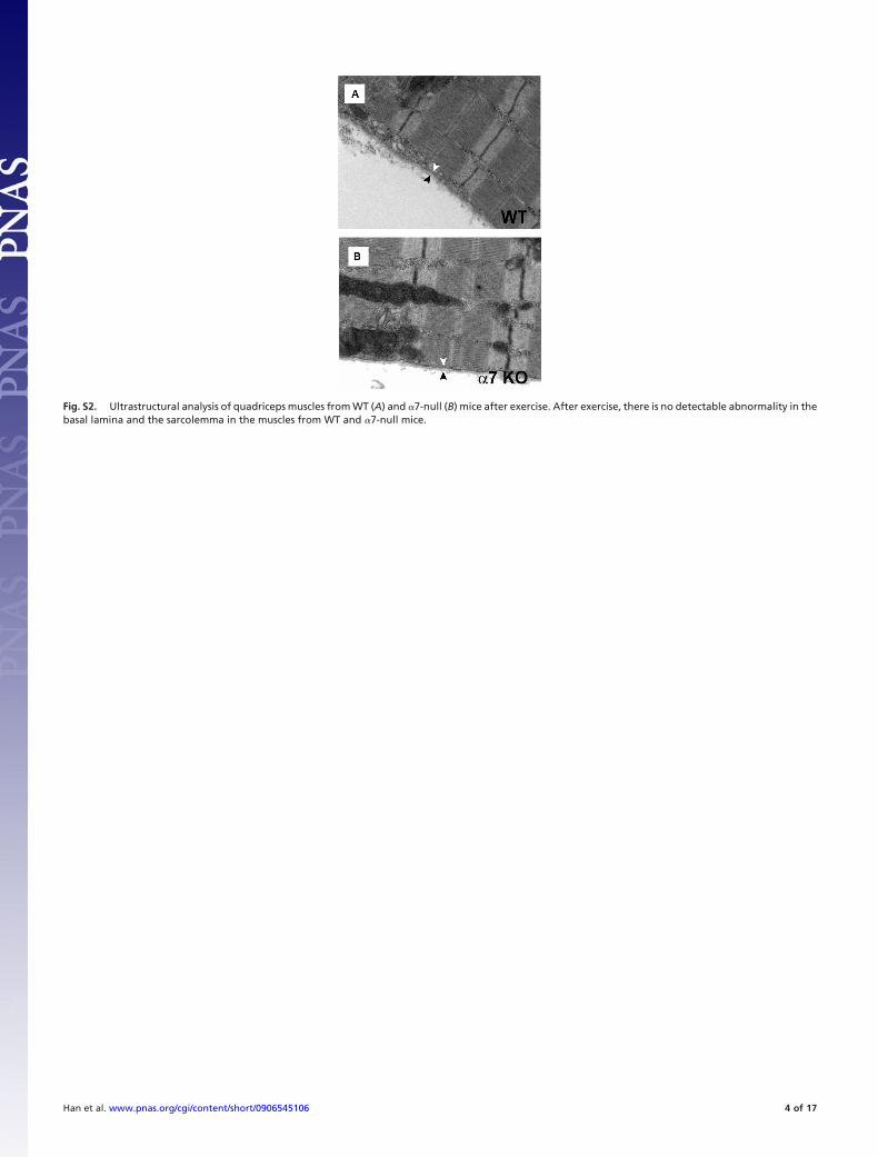
Fig. S2. Ultrastructural analysis of quadriceps muscles from WT (A) and �7-null (B) mice after exercise. After exercise, there is no detectable abnormality in thebasal lamina and the sarcolemma in the muscles from WT and �7-null mice.
Han et al. www.pnas.org/cgi/content/short/0906545106 4 of 17

Fig. S3. Exercise-induced dissociation of the basal lamina and the sarcolemma in the DG KO muscle. (A) Immunostaining of longitudinal fibers after exercise.Five-week-old mice (WT, DG KO, and �7 KO) were subjected to treadmill-exercise (15° downhill for 20 min). Immediately after the exercise, quadriceps muscleswere taken. Longitudinal cryosections were immunostained with laminin and caveolin-3. Arrow, breakage of laminin- or caveolin-3-staining; arrowhead,separation of laminin- and caveolin-3-staining; asterisk, fiber with remaining laminin deposition. (B) Acute damage of DG-null muscle after the exercise.Cryosections of DG KO postexercised quadriceps muscle were coimmunostained with laminin and caveolin-3. Serial section was stained with hematoxylin andeosin (H&E). White asterisk, fiber with remaining laminin deposition; black asterisk, necrotic fibers.
Han et al. www.pnas.org/cgi/content/short/0906545106 5 of 17

Fig. S4. Disrupted expression of DG and �7 in DG/�7 double mutant (DKO) mice. Western blotting (A) and immunofluorescence staining (B) analysis showedloss of the DG and �7 expression in DG/�7 DKO muscle. It is of note that the DG expression in DKO is higher than in the DG-null muscle due to the greaterregeneration.
Han et al. www.pnas.org/cgi/content/short/0906545106 6 of 17

Fig. S5. Schematic models of the DGC in skeletal muscle of different mouse models. (Left) WT; (Center) Largemyd; (Right) MCK DG-null.
Han et al. www.pnas.org/cgi/content/short/0906545106 7 of 17

Fig. S6. Immunofluorescence staining of dysferlin in skeletal muscle of Largemyd mice. Quadriceps muscle sections from WT C57BL/6 and Largemyd mice werestained with the anti-dysferlin antibody Hamlet.
Han et al. www.pnas.org/cgi/content/short/0906545106 8 of 17

Fig. S7. Damage assay on skeletal muscle of �7-null mice. Plot of FM 1-43 fluorescence intensity against time in integrin �7-null (open square, n � 5) musclefibers. The dashed curve represents mean fluorescence intensity in the membrane damage assay, in wild-type muscle from Fig. 4E.
Han et al. www.pnas.org/cgi/content/short/0906545106 9 of 17

Fig. S8. Stability of recombinant �-DG on the sarcolemma of DG-null muscle fibers DG-null. DG-null muscles injected with recombinant �-DG/L (�-DG/L injected)or saline (Mock) were stained with the glycosylated �-DG antibody (IIH6). Recombinant �-DG failed to stay on the sarcolemma of DG-null muscles, suggestingthat �-DG is required for securing the recombinant �-DG on the sarcolemma.
Han et al. www.pnas.org/cgi/content/short/0906545106 10 of 17

Table S1. Severe loss of body weight and muscle mass in DG/�7 DKO mice
WT DG KO �7 KO DKO
Body weight, g 23.6 � 0.4 21.7 � 3.1 20.8 � 1.0* 11.2 � 0.9***Gastrocnemius, mg 121.4 � 5.5 125.1 � 8.3 122.8 � 8.2 37.5 � 0.6***TA, mg 34.9 � 5.5 33.5 � 1.5 35.0 � 1.6 10.1 � 2.4***Triceps, mg 72.0 � 11.6 74.0 � 21.3 62.3 � 2.1 22.0 � 3.2***Quadriceps, mg 107.4 � 2.5 126.3 � 12.5 116.5 � 17.6 40.6 � 9.6***Heart, mg 115.1 � 14 128.5 � 34.2 113.9 � 19.5 71.9 � 4.8 **Shin length, cm 2.2 � 0 2.2 � 0.1 2.1 � 0.1 2.1 � 0.1
*, P � 0.05; **, P � 0.01; ***, P � 0.001; n � 3.
Han et al. www.pnas.org/cgi/content/short/0906545106 11 of 17

Movie S1A (AVI)
Movie S1A. In situ membrane damage assay in C57BL/6 WT muscle.
Han et al. www.pnas.org/cgi/content/short/0906545106 12 of 17

Movie S1B (AVI)
Movie S1B. In situ membrane damage assay of Largemyd muscle in regular buffer.
Han et al. www.pnas.org/cgi/content/short/0906545106 13 of 17

Movie S1C (AVI)
Movie S1C. In situ membrane damage assay of Largemyd muscle in hyperosmotic buffer.
Han et al. www.pnas.org/cgi/content/short/0906545106 14 of 17

Movie S2A (AVI)
Movie S2A. In situ membrane damage assay of C57BL/6 WT muscle subjected to mock treatment.
Han et al. www.pnas.org/cgi/content/short/0906545106 15 of 17

Movie S2B (AVI)
Movie S2B. In situ membrane damage assay of C57BL/6 WT muscle subjected to LCMV treatment.
Han et al. www.pnas.org/cgi/content/short/0906545106 16 of 17

Movie S3 (WMV)
Movie S3. Balloon movie.
Han et al. www.pnas.org/cgi/content/short/0906545106 17 of 17