Supporting information for Controlled Display of Enzyme ... · PDF fileFor HRP, 10 mg of . o...
5
Supporting information for Controlled Display of Enzyme Activity with a Stretchable Hydrogel Yifei Zhang, a Qile Chen, a Jun Ge, * , a and Zheng Liu* , a Department of Chemical Engineering, Tsinghua University, Beijing 100084, P. R. China Experimental Preparing protein-incorporated hydrogel. For preparing protein-incorporated hydrogel, 6.22 g of acrylamide, 780 mg of alginate, 3.76 mg N,N’-methylenebisacrylamide, and a given amount of protein were dissolved in 28 mL of deionized water. 50 mg of ammonium persulphate (in 5 mL deionized water), 20 μL of N, N, N’, N’-tetramethylethylenediamine and 45 mg of CaSO 4 slurry (in 10 mL deionized water) were sequentially added to above solution. The mixture was then poured into an acrylic mould measuring 100 × 100 × 3 mm 3 covering with a glass plate, followed by polymerization at 30 o C for 20 h. Staining protein gels with Coomassie blue. The protein-incorporated hydrogel cylinder was incubated in a solution of 40% methanol / 10% acetic acid / 0.1% Coomassie Brilliant Blue R-250 for 24 h, followed by washing with a solution composed of 10% methanol / 10% acetic acid / 80% distilled water over 24 h until the solution became colorless. Finally the cylinder was washed with 100 mM calcium chloride solution for 48 h. Determination of the enzymatic activity. For HRP, 10 mg of o-phenylenediamine and 5 mL of 30% H 2 O 2 solution were dissolved in 95 mL of citric acid buffer (pH 5.5). A piece of HRP-incorporated hydrogel (cut into square of 3 cm × 3 cm) was stretched to different sizes and immersed in 100 mL of the substrate buffer at room temperature. The increase in the absorbance at 450 nm was determined in 5 min using a UV/Vis spectrophotometer (Shimadzu UV2550). For Electronic Supplementary Material (ESI) for Chemical Communications This journal is © The Royal Society of Chemistry 2013
Transcript of Supporting information for Controlled Display of Enzyme ... · PDF fileFor HRP, 10 mg of . o...
Supporting information for
Controlled Display of Enzyme Activity with a Stretchable Hydrogel
Yifei Zhang, a Qile Chen, a Jun Ge, *, a and Zheng Liu*, a Department of Chemical Engineering, Tsinghua University, Beijing 100084, P. R. China
Experimental
Preparing protein-incorporated hydrogel. For preparing protein-incorporated hydrogel, 6.22
g of acrylamide, 780 mg of alginate, 3.76 mg N,N’-methylenebisacrylamide, and a given amount
of protein were dissolved in 28 mL of deionized water. 50 mg of ammonium persulphate (in 5 mL
deionized water), 20 μL of N, N, N’, N’-tetramethylethylenediamine and 45 mg of CaSO4 slurry
(in 10 mL deionized water) were sequentially added to above solution. The mixture was then
poured into an acrylic mould measuring 100 × 100 × 3 mm3 covering with a glass plate, followed
by polymerization at 30 oC for 20 h.
Staining protein gels with Coomassie blue. The protein-incorporated hydrogel cylinder was
incubated in a solution of 40% methanol / 10% acetic acid / 0.1% Coomassie Brilliant Blue R-250
for 24 h, followed by washing with a solution composed of 10% methanol / 10% acetic acid / 80%
distilled water over 24 h until the solution became colorless. Finally the cylinder was washed with
100 mM calcium chloride solution for 48 h.
Determination of the enzymatic activity. For HRP, 10 mg of o-phenylenediamine and 5 mL of
30% H2O2 solution were dissolved in 95 mL of citric acid buffer (pH 5.5). A piece of
HRP-incorporated hydrogel (cut into square of 3 cm × 3 cm) was stretched to different sizes and
immersed in 100 mL of the substrate buffer at room temperature. The increase in the absorbance
at 450 nm was determined in 5 min using a UV/Vis spectrophotometer (Shimadzu UV2550). For
Electronic Supplementary Material (ESI) for Chemical CommunicationsThis journal is © The Royal Society of Chemistry 2013
CALB, the assay was referred to the standard method1 as follows: 34.8 μL of p-nitrophenyl
butyrate (PNPB) was dissolved in 1 mL of acetonitrile, and then added to 99 mL of phosphate
buffer (50 mM, pH 7.2). A piece of CALB-incorporated hydrogel (3 cm × 3 cm) was stretched to
different dimensions and immersed in the substrate solution. The increase of the absorbance at 400
nm was measured in 5 min. For the stability, the enzyme-incorporated hydrogel was cut into same
pieces and stored at room temperature by packaging them in individual plastic wraps. For the
measurement, one piece of gel was immersed in solution and compared the activity with free
enzyme incubated in solution.
Scanning electron microscopy. A piece of hydrogel (3 cm × 3 cm) was stretched to different
sizes and then immediately frozen in liquid nitrogen, followed by vacuum freeze drying. The dry
gels were broke off to make fresh fracture sections for SEM (FEI Quanta 200F).
Laser scanning confocal microscopy. Fluorescence images of the hydrogels were taken on a
Zeiss LSM-710 NLO laser scanning confocal system equipped with EC Plan-Neofluar 20×/0.5
M27, 40×/1.30 oil DIC M27 objectives. Samples to be tested were supported on thin glass slide
for observation by exciting at 488 nm. The microscope was operated under “linear mode”, the
laser intensity was fixed at 30% and the master gain value was fixed at 800.
Electronic Supplementary Material (ESI) for Chemical CommunicationsThis journal is © The Royal Society of Chemistry 2013
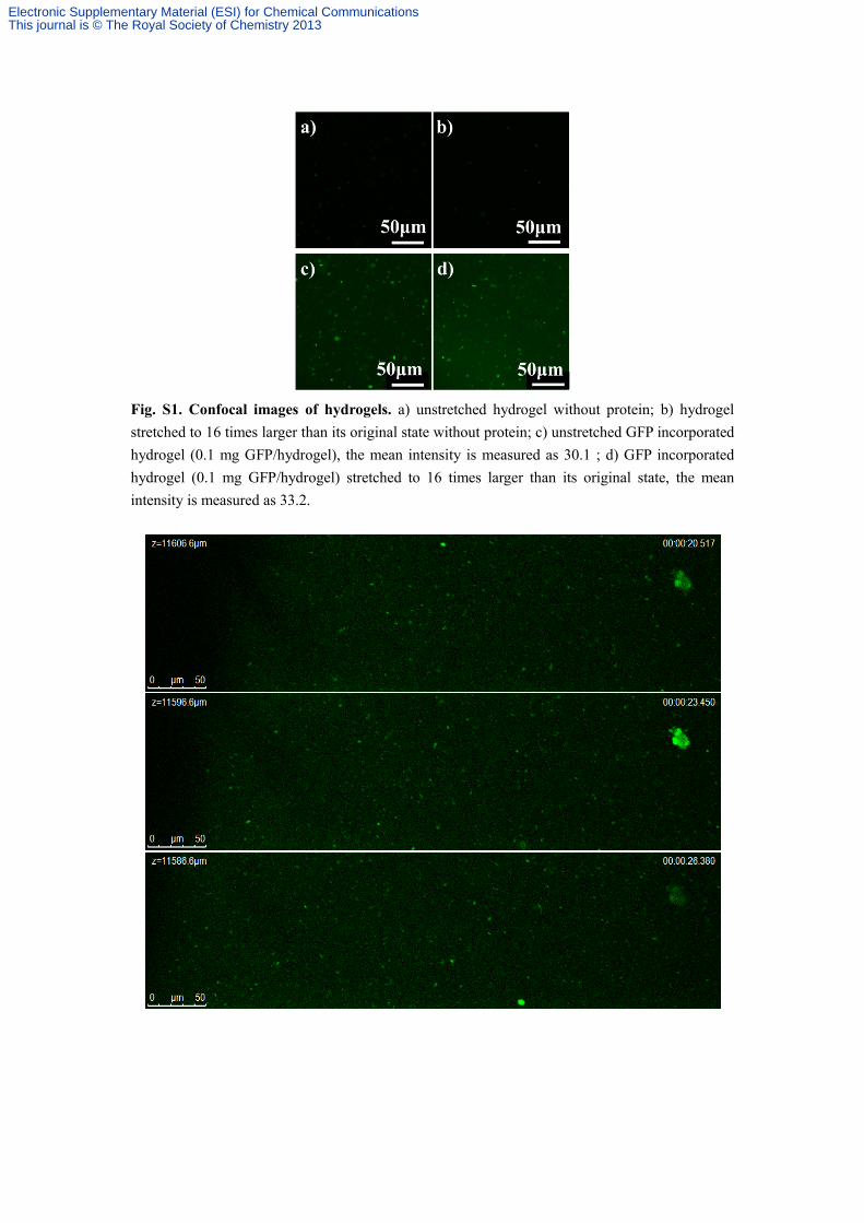
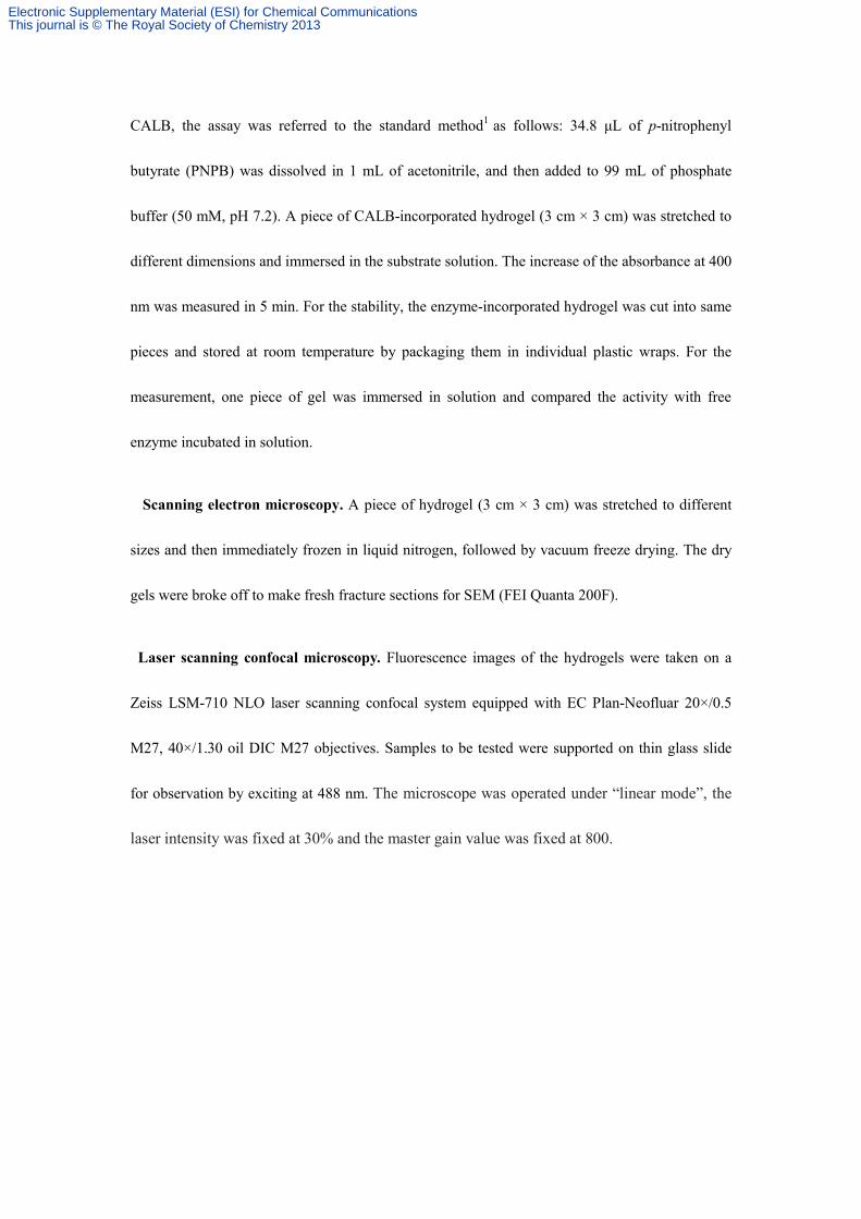
Fig. S1. Confocal images of hydrogels. a) unstretched hydrogel without protein; b) hydrogel stretched to 16 times larger than its original state without protein; c) unstretched GFP incorporated hydrogel (0.1 mg GFP/hydrogel), the mean intensity is measured as 30.1 ; d) GFP incorporated hydrogel (0.1 mg GFP/hydrogel) stretched to 16 times larger than its original state, the mean intensity is measured as 33.2.
Electronic Supplementary Material (ESI) for Chemical CommunicationsThis journal is © The Royal Society of Chemistry 2013

Fig. S2. Z-scanning of FITC-labeled HRP incorporated hydrogels (scanning depth is 60 μm, 10 μm /step). The green areas in these images show a uniform dispersion of fluorescent molecules inside the hydrogel.
Electronic Supplementary Material (ESI) for Chemical CommunicationsThis journal is © The Royal Society of Chemistry 2013
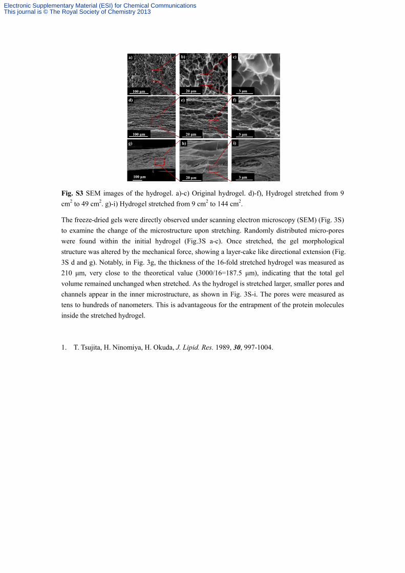
Fig. S3 SEM images of the hydrogel. a)-c) Original hydrogel. d)-f), Hydrogel stretched from 9 cm2 to 49 cm2. g)-i) Hydrogel stretched from 9 cm2 to 144 cm2.
The freeze-dried gels were directly observed under scanning electron microscopy (SEM) (Fig. 3S) to examine the change of the microstructure upon stretching. Randomly distributed micro-pores were found within the initial hydrogel (Fig.3S a-c). Once stretched, the gel morphological structure was altered by the mechanical force, showing a layer-cake like directional extension (Fig. 3S d and g). Notably, in Fig. 3g, the thickness of the 16-fold stretched hydrogel was measured as 210 μm, very close to the theoretical value (3000/16=187.5 μm), indicating that the total gel volume remained unchanged when stretched. As the hydrogel is stretched larger, smaller pores and channels appear in the inner microstructure, as shown in Fig. 3S-i. The pores were measured as tens to hundreds of nanometers. This is advantageous for the entrapment of the protein molecules inside the stretched hydrogel. 1. T. Tsujita, H. Ninomiya, H. Okuda, J. Lipid. Res. 1989, 30, 997-1004.
Electronic Supplementary Material (ESI) for Chemical CommunicationsThis journal is © The Royal Society of Chemistry 2013