Embase - Supporting Evidence Based Medicine - Webinar 24 Oct 2012
Upload
nguyenkietCategory
view
244download
4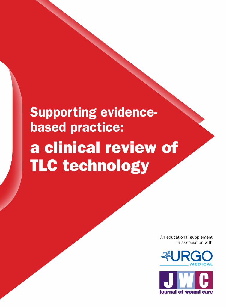
Supporting evidence- based practice:
a clinical review of TLC technology
An educational supplement in association with
2 Journal of Wound Care / Urgo Evidence Review, March 2011
ContentsForeword� 3
Evidence-based�dressing�selection� 4
Pre-clinical�evidence� 9
The�clinical�evidence�for�dressings�with�TLC�technology�11
Dressings�with�TLC-Ag�technology� 22
Dressings�with�TLC-NOSF�technology� 27
References� 33
© 2011 MA Healthcare Ltd
All rights reserved. No reproduction, transmission or copying of this publication is allowed without written permission. No part of this publication may be reproduced, stored in a retrieval system, or transmitted in any form or by any means, mechanical, electronic, photocopying, recording, or otherwise, without the prior written permission of MA Healthcare Ltd or in accordance with the relevant copyright legislation.
Although the editor and MA Healthcare Ltd have taken great care to ensure accuracy, MA Healthcare Ltd will not be liable for any errors of omission or inaccuracies in this publication.
Published on behalf of Urgo by MA Healthcare Ltd. This supplement was not subject to peer review.
To reference this document, cite: White, R., Cowan, T., Glover, D. Supporting evidence-based practice: a clinical review of TLC technology. MA Healthcare Ltd, London, 2011.
Publisher: Anthony KerrAssociate Publisher: Tracy CowanAuthors: White, R., Cowan, T., Glover, D.Designer: Louise CowburnPrinted by: Pensord, Blackwood, Newport, Wales, UK
Published by: MA Healthcare Ltd, St Jude’s Church, Dulwich Road, London SE24 0PB, UK
Tel: +44 (0)20 7738 5454 Email: [email protected] Web: www.markallengroup.com
Journal of Wound Care / Urgo Evidence Review, March 2011 3
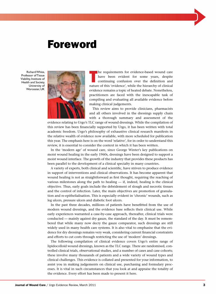
Foreword
The requirements for evidence-based wound care have been evident for some years, despite continuing confusion over the definition and
nature of this ‘evidence’, while the hierarchy of clinical evidence remains a topic of heated debate. Nonetheless, practitioners are faced with the inescapable task of compiling and evaluating all available evidence before making clinical judgements.
This review aims to provide clinicians, pharmacists and all others involved in the dressings supply chain with a thorough summary and assessment of the
evidence relating to Urgo’s TLC range of wound dressings. While the compilation of this review has been financially supported by Urgo, it has been written with total academic freedom. Urgo’s philosophy of exhaustive clinical research manifests in the relative wealth of evidence now available, with more scheduled for publication this year. The emphasis here is on the word ‘relative’, for in order to understand this review, it is essential to consider the context in which it has been written.
In the ‘modern age’ of wound care, since George Winter’s key publications on moist wound healing in the early 1960s, dressings have been designed to support a moist wound interface. The growth of the industry that provides these products has been parallel to the development of a clinical specialty in many countries.
A variety of experts, both clinical and scientific, have striven to produce evidence in support of interventions and clinical observations. It has become apparent that wound healing is not as straightforward as first thought, requiring the reaching of various milestones along the path to healing — if, indeed, healing is the clinical objective. Thus, early goals include the debridement of slough and necrotic tissues and the control of infection. Later, the main objectives are promotion of granula-tion and re-epithelialisation. This is especially evident in ‘chronic’ wounds, such as leg ulcers, pressure ulcers and diabetic foot ulcers.
In the past three decades, millions of patients have benefitted from the use of modern wound dressings, and the evidence base reflects their clinical use. While early experiences warranted a case-by-case approach, thereafter, clinical trials were conducted — mainly against dry gauze, the standard of the day. It must be remem-bered that while many now decry the gauze comparator, such dressings are still widely used in many health care systems. It is also vital to emphasise that the evi-dence for dry dressings remains very weak, considering current financial constraints and efforts to cut costs through restricting the use of ‘modern’ dressings.
The following compilation of clinical evidence covers Urgo’s entire range of lipidocolloid wound dressings, known as the TLC range. There are randomised, con-trolled clinical trials, observational studies, and a number of cases and case cohorts; these involve many thousands of patients and a wide variety of wound types and clinical challenges. This evidence is collated and presented for your information, to assist you in making judgements on clinical use, purchasing and formulary proc-esses. It is vital in such circumstances that you look at and appraise the totality of the evidence. Every effort has been made to present it here.
Richard White, Professor of Tissue Viability, Institute of Health and Society,
University of Worcester, UK
4 Journal of Wound Care / Urgo Evidence Review, March 2011
When faced with a plethora of dressings, how does the clinician decide which product to use? Clearly, clinical
knowledge, based on experience complemented by evidence from the literature, will be the largest factor influencing the decision. The clinician will start by assessing both the wound (type, duration, size, exudate level, pain, presence of malodour, condition of the surrounding skin) and the patient (age, medical history, comorbidities, psychosocial factors) and will then consider the potential effectiveness of the selected product, evidenced by clinical outcomes and demonstrated cost-effectiveness. Naturally, the type/nature of the wound and stage of healing will also influence product selection and management options.
Identifying a chronic woundThe phases of the wound healing process are well known. For the purposes of this supplement, it is assumed that acute wounds such as minor burns, surgical wounds, lacerations and other traumatic injuries generally follow the three key stages of the healing trajectory — inflammation, proliferation and maturation — with little deviation.
Chronic wounds, in contrast, do not progress through these phases in an orderly and timely sequence1 and generally fail to heal in 4–6 weeks despite the provision of standard care. Chronic wounds become ‘stuck’ in the inflammatory stage. In acute wounds, the inflammatory process serves to limit blood loss, kill invading bacteria, dispose of devitalised tissue and promote an environment that is conducive to tissue growth.2 In addition, matrix metalloproteinases (MMPs), which are protein-digesting enzymes, help remove dead and devital-ised tissue. Their number is kept in check by tissue inhibitors of metalloproteinases (TIMPs), thereby preventing undue protease damage to healthy
tissue. In chronic wounds, however, such anti-pro-tease activity is diminished, resulting in significantly elevated levels of proteases. Studies have shown that chronic wounds can contain up to 65 times more proteases than acute wounds.3 The ensuing exces-sive MMP activity effectively stalls the healing proc-ess. Proliferation of keratinocytes, fibroblasts and endothelial cells is slowed or blocked. The MMPs degrade key components of the extracellular matrix (fibronectin, fibrin and collagen) as well as the viable marginal tissue, and impair the expression of growth factors. Similarly, the release of reactive oxygen spe-cies is much higher in chronic than acute wounds. This not only results in the degradation of viable tissue but also local ischaemia.4,5 In this way, inflam-mation is prolonged and healing delayed, with potentially devastating consequences for the patient, and huge resource implications for the indi-vidual, the health service and employers (Box 1).
Patient risk factors that may predispose a wound to become chronic include incontinence, under-mining, peri-wound maceration, infection, oedema, chronic venous insufficiency or arterial disease of the lower leg, diabetes, chemotherapy, steroids and the presence of a wound biofilm. If any of these risk factors are present, the clinician must ensure that prevention measures are taken
Box 1. Impact of chronic wounds9,178
• Financialimpact: chronic wounds are estimated to cost the NHS at least £1 billion per annum179–181
• Nursingresource: community nurses spend up to 50% of their time managing chronic wounds182
• Socialisolationofthepatient: due to malodour and/or exudate
• Patientlostworkingdays: while waiting for treatment and/or because of malodour and exudate problems and potential complications
• Reducedqualityoflife: as a result of the above reasons
Evidence-based dressing selectionA wide range of considerations determine dressing selection. Taking into account the wound type, the patient’s holistic needs and his or her own clinical experience, the practitioner will then look to the literature for guidance. To support practitioners with their decision-making, this supplement provides an in-depth review of the evidence on Urgo’s Technology Lipido-Colloid range of dressings. Its objective is to summarise the literature, as opposed to critiquing it
Richard White, Professor of Tissue Viability, Institute of Health and Society, University of Worcester. Tracy Cowan, Editor, Journal of Wound Care. Deborah Glover, Independent Editorial Consultant
Journal of Wound Care / Urgo Evidence Review, March 2011 5
and the factors that may cause the wound to become chronic are addressed. This is of particular relevance to the management of MMPs, where effective ‘rebalancing’ of MMPs in the wound bed will ‘kick-start’ healing in a chronic wound.
So how can the clinician determine if the dress-ing is having this desired effect? One indicator that a treatment is effective is a >40% reduction in area in the first 2–3 weeks of treatment as this indicates that the wound is healing.6 In fact, a 20–40% wound area reduction at 3–4 weeks has been dem-onstrated to be highly predictive of complete clo-sure at 20–24 weeks in leg ulcers.7,8 Thus, if a wound (treated or not) has not shown signs of progression from the inflammatory stage to the proliferative stage within this time frame, it could have become chronic.9
Management of chronic woundsEvidence has shown that, for many patients, man-aging the symptoms of a wound is as important, if not more so, than promoting healing. The results of a multinational, multicentre trial undertaken in 200810 showed that wound pain, both ongoing and/or during dressing removal/procedures, was the most distressing and stressful aspect of having a wound. Impaired mobility, difficulties with bath-ing, leakage, malodour, bandage/dressing slippage and skin trauma were also considered important. These findings support those of other studies.11-14
Therefore, the objective of treatment is not only to treat the underlying aetiology of the wound (with pressure-redistributing equipment for pres-sure ulcers, offloading for diabetic foot ulcers or compression therapy for venous and mixed aetiol-ogy ulcers), but also to select a dressing that will promote a wound environment that is conducive to healing and acceptable to the patient. To achieve this, a dressing will need to:• Maintain a good moisture balance at the wound/
dressing interface• Allow gaseous exchange• Provide thermal insulation• Form a barrier to bacteria• Be non-toxic and non-irritant• Not cause pain or trauma at removal • Require minimal disturbance or replacement.15,16
Other desired properties include the ability to remove or inactivate proteolytic enzymes, remove excess exudate and devitalised tissue, have an anti-microbial effect and control malodour.
Inevitably, no one dressing has all of these prop-
Box 2. Hierarchy and source of clinical evidence25
• Meta-analyses of well-designed randomised controlled trials• Randomised controlled trials • Evidence from well-designed, non-randomised controlled trials, such as
cohort studies• Case control studies• Case series studies• Expert opinion
erties and, as the patient progresses to the stated goals (healing or symptom management), the functions that will determine dressing selection, such as moisture balance, pain relief, management of infection or wound bed preparation, will change. When selecting a dressing, the clinician will therefore seek to identify one that can best meet the patient’s individual needs at that particu-lar time. As stated above, the selection will be based on clinical knowledge, clinical experience and appraisal of the published evidence. Only in this way can it be claimed to be evidence based.
Evidence-based dressing selection Published evidence for the efficacy of an interven-tion can be found through a number of resources:• National Institute for Health and Clinical Excel-
lence (NICE) • Professional bodies — for example, the World
Union of World Healing Societies (WUWHS) and the European Wound Management Association (EWMA)
• Health databases (CINAHL, PubMed) • Cochrane database for systematic reviews, York
Centre for Review and Dissemination.Received wisdom on clinical evidence is that it
falls into a hierarchy depending on the type of study undertaken.17 The hierarchy of evidence and associ-ated grading recommendations relate to the strength of the literature (Box 2). Meta-analyses of RCTs, fol-lowed by the randomised controlled trial (RCT), are considered to provide the best evidence for the effi-cacy of a treatment intervention. However, there is an ongoing debate about our apparent over-reliance on RCTs for constructing the evidence base in wound care, with some arguing that the narrow inclusion/exclusion criteria used mean the findings are not necessarily applicable to the ‘real-life events that lie beyond the study confines’ and that other levels of evidence should also be used to inform practice.18
While no one denies that a well-conducted meta-analysis (or RCT) can produce robust results, or argues that studies at all levels of the evidence
6 Journal of Wound Care / Urgo Evidence Review, March 2011
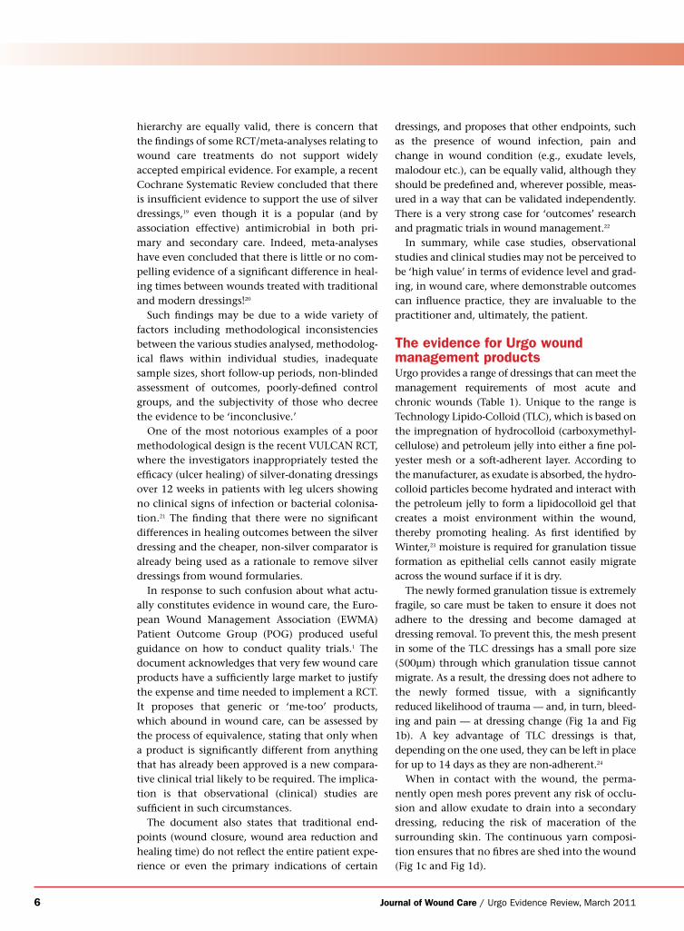
hierarchy are equally valid, there is concern that the findings of some RCT/meta-analyses relating to wound care treatments do not support widely accepted empirical evidence. For example, a recent Cochrane Systematic Review concluded that there is insufficient evidence to support the use of silver dressings,19 even though it is a popular (and by association effective) antimicrobial in both pri-mary and secondary care. Indeed, meta-analyses have even concluded that there is little or no com-pelling evidence of a significant difference in heal-ing times between wounds treated with traditional and modern dressings!20
Such findings may be due to a wide variety of factors including methodological inconsistencies between the various studies analysed, methodolog-ical flaws within individual studies, inadequate sample sizes, short follow-up periods, non-blinded assessment of outcomes, poorly-defined control groups, and the subjectivity of those who decree the evidence to be ‘inconclusive.’
One of the most notorious examples of a poor methodological design is the recent VULCAN RCT, where the investigators inappropriately tested the efficacy (ulcer healing) of silver-donating dressings over 12 weeks in patients with leg ulcers showing no clinical signs of infection or bacterial colonisa-tion.21 The finding that there were no significant differences in healing outcomes between the silver dressing and the cheaper, non-silver comparator is already being used as a rationale to remove silver dressings from wound formularies.
In response to such confusion about what actu-ally constitutes evidence in wound care, the Euro-pean Wound Management Association (EWMA) Patient Outcome Group (POG) produced useful guidance on how to conduct quality trials.1 The document acknowledges that very few wound care products have a sufficiently large market to justify the expense and time needed to implement a RCT. It proposes that generic or ‘me-too’ products, which abound in wound care, can be assessed by the process of equivalence, stating that only when a product is significantly different from anything that has already been approved is a new compara-tive clinical trial likely to be required. The implica-tion is that observational (clinical) studies are sufficient in such circumstances.
The document also states that traditional end-points (wound closure, wound area reduction and healing time) do not reflect the entire patient expe-rience or even the primary indications of certain
dressings, and proposes that other endpoints, such as the presence of wound infection, pain and change in wound condition (e.g., exudate levels, malodour etc.), can be equally valid, although they should be predefined and, wherever possible, meas-ured in a way that can be validated independently. There is a very strong case for ‘outcomes’ research and pragmatic trials in wound management.22
In summary, while case studies, observational studies and clinical studies may not be perceived to be ‘high value’ in terms of evidence level and grad-ing, in wound care, where demonstrable outcomes can influence practice, they are invaluable to the practitioner and, ultimately, the patient.
The evidence for Urgo wound management productsUrgo provides a range of dressings that can meet the management requirements of most acute and chronic wounds (Table 1). Unique to the range is Technology Lipido-Colloid (TLC), which is based on the impregnation of hydrocolloid (carboxymethyl-cellulose) and petroleum jelly into either a fine pol-yester mesh or a soft-adherent layer. According to the manufacturer, as exudate is absorbed, the hydro-colloid particles become hydrated and interact with the petroleum jelly to form a lipidocolloid gel that creates a moist environment within the wound, thereby promoting healing. As first identified by Winter,23 moisture is required for granulation tissue formation as epithelial cells cannot easily migrate across the wound surface if it is dry.
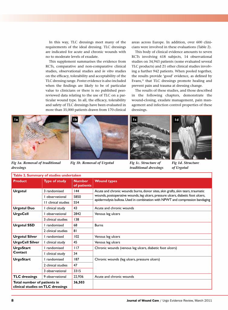
The newly formed granulation tissue is extremely fragile, so care must be taken to ensure it does not adhere to the dressing and become damaged at dressing removal. To prevent this, the mesh present in some of the TLC dressings has a small pore size (500µm) through which granulation tissue cannot migrate. As a result, the dressing does not adhere to the newly formed tissue, with a significantly reduced likelihood of trauma — and, in turn, bleed-ing and pain — at dressing change (Fig 1a and Fig 1b). A key advantage of TLC dressings is that, depending on the one used, they can be left in place for up to 14 days as they are non-adherent.24
When in contact with the wound, the perma-nently open mesh pores prevent any risk of occlu-sion and allow exudate to drain into a secondary dressing, reducing the risk of maceration of the surrounding skin. The continuous yarn composi-tion ensures that no fibres are shed into the wound (Fig 1c and Fig 1d).
Journal of Wound Care / Urgo Evidence Review, March 2011 7
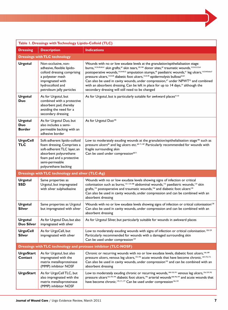
Table 1. Dressings with Technology Lipido-Colloid (TLC)
Dressing Description Indications
Dressings with TLC technology
Urgotul Non-occlusive, non-adhesive, flexible lipido-colloid dressing comprising a polyester mesh impregnated with hydrocolloid and petroleum jelly particles
Wounds with no or low exudate levels at the granulation/epithelialisation stage: burns,14,24,40,60,61 skin grafts,60 skin tears,183,184 donor sites,60 traumatic wounds,14,24,52,53,61 postoperative wounds,14,24,40,52 amputation stumps,40 paediatric wounds,61 leg ulcers,14,24,40,66,67 pressure ulcers,14,24,40 diabetic foot ulcers,14,24,78 epidermolysis bullosa63,64
Can also be used in cavity wounds, under compression,67 under NPWT55 and combined with an absorbent dressing. Can be left in place for up to 14 days,24 although the secondary dressing will still need to be changed
Urgotul Duo
As for Urgotul, but combined with a protective absorbent pad, thereby avoiding the need for a secondary dressing
As for Urgotul, but is particularly suitable for awkward places41,43
Urgotul Duo Border
As for Urgotul Duo, but also includes a semi-permeable backing with an adhesive border
As for Urgotul Duo185
UrgoCell TLC
Soft-adherent lipido-colloid foam dressing. Comprises a soft-adherent TLC layer, an absorbent polyurethane foam pad and a protective semi-permeable polyurethane backing
Low to moderately exuding wounds at the granulation/epithelialisation stage186 such as pressure ulcers85 and leg ulcers etc.68–71,187 Particularly recommended for wounds with fragile surrounding skin,
Can be used under compression68,71
Dressings with TLC technology and silver (TLC-Ag)
Urgotul SSD
Same properties as Urgotul, but impregnated with silver sulphadiazine
Wounds with no or low exudate levels showing signs of infection or critical colonisation such as burns,122–124,188 abdominal wounds,132 paediatric wounds,124 skin grafts,125 postoperative and traumatic wounds,106 and diabetic foot ulcers130
Can also be used in cavity wounds, under compression and can be combined with an absorbent dressing
Urgotul Silver
Same properties as Urgotul but impregnated with silver
Wounds with no or low exudate levels showing signs of infection or critical colonisation128
Can also be used in cavity wounds, under compression and can be combined with an absorbent dressing
Urgotul Duo Silver
As for Urgotul Duo, but also impregnated with silver
As for Urgotul Silver, but particularly suitable for wounds in awkward places
UrgoCell Silver
As for UrgoCell, but impregnated with silver
Low to moderately exuding wounds with signs of infection or critical colonisation.128,129 Particularly recommended for wounds with a damaged surrounding skinCan be used under compression129
Dressings with TLC technology and protease inhibitor (TLC-NOSF)
UrgoStart Contact
As for Urgotul, but also impregnated with the matrix metalloproteinase (MMP) inhibitor NOSF
Chronic or recurring wounds with no or low exudate levels, diabetic foot ulcers,166,189 pressure ulcers, venous leg ulcers,155,190 acute wounds that have become chronic.169,170,172 Can also be used in cavity wounds, under compression155 and can be combined with an absorbent dressing
UrgoStart As for UrgoCell TLC, but also impregnated with the matrix metalloproteinase (MMP) inhibitor NOSF
Low to moderately exuding chronic or recurring wounds,148-150,191 venous leg ulcers,156,159,192 pressure ulcers162,193,194 diabetic foot ulcers,195 arterial wounds168,196,197 and acute wounds that have become chronic.170,171,177 Can be used under compression156,159
8 Journal of Wound Care / Urgo Evidence Review, March 2011
In this way, TLC dressings meet many of the requirements of the ideal dressing. TLC dressings are indicated for acute and chronic wounds with no to moderate levels of exudate.
This supplement summarises the evidence from RCTs, comparative and non-comparative clinical studies, observational studies and in vitro studies on the efficacy, tolerability and acceptability of the TLC dressing range. Poster evidence is also included when the findings are likely to be of particular value to clinicians or there is no published peer-reviewed data relating to the use of TLC on a par-ticular wound type. In all, the efficacy, tolerability and safety of TLC dressings have been evaluated in more than 35,000 patients drawn from 170 clinical
areas across Europe. In addition, over 600 clini-cians were involved in these evaluations (Table 2).
This body of clinical evidence amounts to seven RCTs involving 618 subjects, 14 observational studies on 34,943 patients (some evaluated several TLC products) and 21 other clinical studies involv-ing a further 942 patients. When pooled together, the results provide ‘good’ evidence, as defined by Evans,25 that TLC dressings promote healing and prevent pain and trauma at dressing change.
The results of these studies, and those described in the following chapters, demonstrate the wound-closing, exudate management, pain man-agement and infection control properties of these dressings.
Table 2. Summary of studies undertaken
Product Type of study Number of patients
Wound types
Urgotul 3 randomised 144 Acute and chronic wounds: burns, donor sites, skin grafts, skin tears, traumatic wounds, postoperative wounds, leg ulcers, pressure ulcers, diabetic foot ulcers, epidermolysis bullosa. Used in combination with NPWT and compression bandaging
1 observational 5850
11 clinical studies 554
Urgotul Duo 1 clinical study 43 Acute and chronic wounds
UrgoCell 1 observational 2842 Venous leg ulcers
3 clinical studies 138
Urgotul SSD 1 randomised 68 Burns
2 clinical studies 81
Urgotul Silver 1 randomised 102 Venous leg ulcers
UrgoCell Silver 1 clinical study 45 Venous leg ulcers
UrgoStart Contact
1 randomised 117 Chronic wounds (venous leg ulcers, diabetic foot ulcers)
1 clinical study 34
UrgoStart 1 randomised 187 Chronic wounds (leg ulcers, pressure ulcers)
2 clinical studies 47
3 observational 3315
TLC dressings 9 observational 22,936 Acute and chronic wounds
Total number of patients in clinical studies on TLC dressings
36,503
Fig 1a. Removal of traditional dressings
Fig 1b. Removal of Urgotul Fig 1c. Structure of traditional dressings
Fig 1d. Structure of Urgotul
1a 1c1b 1d
Journal of Wound Care / Urgo Evidence Review, March 2011 9
Pre-clinical evidence
L ong before any wound dressing prototype is put onto a patient, many in vitro tests are conducted in the laboratory. Each test is
designed to evaluate a particular performance characteristic — for example, exudate handling,26 the bacterial barrier properties,27 the toxicity of dressing constituents and the full formulation,28 plus the dressing’s antimicrobial activity.29 As a result, anyone with experience in wound care will have encountered and, in many cases be guided by, the evidence available from in vitro tests.
Fibrogenesis is an important mechanism in wound repair. Fibroblasts (a dermal cell type) play a key role in producing extracellular matrix com-ponents that are vital for granulation tissue and, later, wound closure and remodelling.30 In chronic wounds, where prolonged inflammation has dis-rupted normal healing, altered fibroblast functions can lead to fibrosis, oxidative stress and impaired closure.31 It follows that any intervention that is likely to ‘normalise’ fibroblast function will elicit noticeable clinical responses, which suggests that a dressing that can stimulate fibroblast proliferation will promote wound healing. In vitro experiments have investigated the effects of TLC dressings on fibroblast viability and proliferation. The results show that they simulate these activities.
Effects on fibroblastsThe first in vitro study to assess the effect of TLC dressings on fibroblasts looked specifically at whether or not it modified their behaviour. The effects of Urgotul and five other non-adhesive wound-contact dressings — Adaptic (then pro-duced by Johnson & Johnson), tulle gras Lumiere (Solvay Pharma), Mepitel (Mölnlycke Healthcare), Ialuset (Genevrier Laboratories) and Physiotulle (Coloplast) — on cultured human fibroblast were evaluated. Fibroblasts were taken from healthy volunteers aged 12, 32 and 51 years. The MTT assay was used to assess fibroblast viability. (MTT is a colourimetric assay that assesses the overall activ-ity of cells.) Cultures in monolayer were used to study fibroblast morphology and growth. To char-acterise the effects of the dressings on cell pheno-type, fibroblasts were seeded within collagen gels and labelled for alpha-SM and F-actin, which are markers of myofibroblast differentiation. (During
the wound healing process, fibroblasts transform into myofibroblasts, which have contractile prop-erties that facilitate wound closure.) Fibroblast cells were exposed to the dressing samples for 1 and 3 days.
The results demonstrated that, for all skin ages, Urgotul, Mepitel, Physiotulle and tulle gras had no significant effects on cell growth on day 3, whereas cell proliferation was significantly reduced with Adaptic and Ialuset (p<0.05). Changes in cell mor-phology were noted with these two dressings, with the fibroblasts appearing round in shape on day 3, which is indicative of cell death. Cells in contact with Urgotul, Mepitel, Physiotulle and tulle gras demonstrated the same bipolar and elongated morphology as did the controls, again indicating that the dressing did not have any cytotoxic effects. However, only fibroblasts exposed to Urgotul exhibited long stress fibres, which is a pre-cursor to transformation into myofibroblasts.32
The next step was to assess the effect of Urgotul on fibroblast proliferation. An in vitro study there-fore compared the level of fibroblast proliferation achieved with Urgotul with that of two similar comparators: soft-silicone wound-contact dressing (Mepitel) and tulle gras. Proliferation was deter-mined by whether or not there was an increase in tritiated thymidine incorporation in the DNA of replicating normal human dermal fibroblasts (a validated assay for evaluating the rate of fibroblast proliferation.) In addition, cell viability/cytotoxic-ity was assessed using the MTT assay. Finally, fol-lowing contact with the dressings, fibroblasts were also visualised using confocal laser microscopy.
In terms of cell proliferation, of all the dressings Urgotul was associated with the highest levels of thymidine incorporation at all time points tested (24, 48, 76 and 96 hours). The difference was most marked at 48 hours (p<0.01), when it was 45% greater than in the controls (cultures with no dress-ings). Mepitel was associated with an overall non-significant tendency to reduce cell proliferation, although this became significant when the medium was not changed every 24 hours (Fig 2).
The MTT results confirmed Viennet et al.’s results, showing that none of the test dressings sig-nificantly modified the overall metabolic activity of the fibroblast culture. However, visual observa-
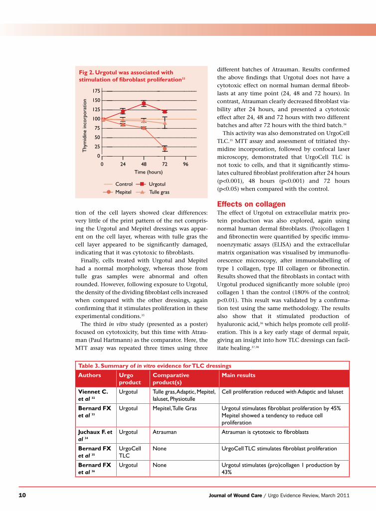
10 Journal of Wound Care / Urgo Evidence Review, March 2011
tion of the cell layers showed clear differences: very little of the print pattern of the net compris-ing the Urgotul and Mepitel dressings was appar-ent on the cell layer, whereas with tulle gras the cell layer appeared to be significantly damaged, indicating that it was cytotoxic to fibroblasts.
Finally, cells treated with Urgotul and Mepitel had a normal morphology, whereas those from tulle gras samples were abnormal and often rounded. However, following exposure to Urgotul, the density of the dividing fibroblast cells increased when compared with the other dressings, again confirming that it stimulates proliferation in these experimental conditions.33
The third in vitro study (presented as a poster) focused on cytotoxicity, but this time with Atrau-man (Paul Hartmann) as the comparator. Here, the MTT assay was repeated three times using three
different batches of Atrauman. Results confirmed the above findings that Urgotul does not have a cytotoxic effect on normal human dermal fibrob-lasts at any time point (24, 48 and 72 hours). In contrast, Atrauman clearly decreased fibroblast via-bility after 24 hours, and presented a cytotoxic effect after 24, 48 and 72 hours with two different batches and after 72 hours with the third batch.34
This activity was also demonstrated on UrgoCell TLC.35 MTT assay and assessment of tritiated thy-midine incorporation, followed by confocal laser microscopy, demonstrated that UrgoCell TLC is not toxic to cells, and that it significantly stimu-lates cultured fibroblast proliferation after 24 hours (p<0.001), 48 hours (p<0.001) and 72 hours (p<0.05) when compared with the control.
Effects on collagenThe effect of Urgotul on extracellular matrix pro-tein production was also explored, again using normal human dermal fibroblasts. (Pro)collagen 1 and fibronectin were quantified by specific immu-noenzymatic assays (ELISA) and the extracellular matrix organisation was visualised by immunoflu-orescence microscopy, after immunolabelling of type 1 collagen, type III collagen or fibronectin. Results showed that the fibroblasts in contact with Urgotul produced significantly more soluble (pro)collagen 1 than the control (180% of the control; p<0.01). This result was validated by a confirma-tion test using the same methodology. The results also show that it stimulated production of hyaluronic acid,36 which helps promote cell prolif-eration. This is a key early stage of dermal repair, giving an insight into how TLC dressings can facil-itate healing.37,38
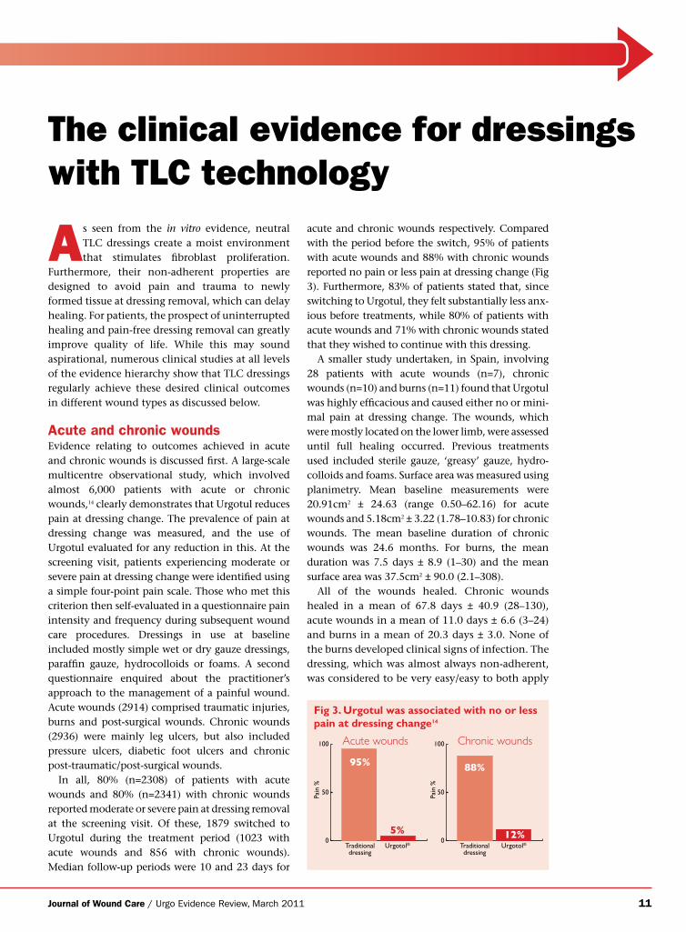
Table 3. Summary of in vitro evidence for TLC dressings
Authors Urgo product
Comparative product(s)
Main results
Viennet C. et al 32
Urgotul Tulle gras, Adaptic, Mepitel, Ialuset, Physiotulle
Cell proliferation reduced with Adaptic and Ialuset
Bernard FX et al 33
Urgotul Mepitel, Tulle Gras Urgotul stimulates fibroblast proliferation by 45%Mepitel showed a tendency to reduce cell proliferation
Juchaux F. et al 34
Urgotul Atrauman Atrauman is cytotoxic to fibroblasts
Bernard FX et al 35
UrgoCell TLC
None UrgoCell TLC stimulates fibroblast proliferation
Bernard FX et al 36
Urgotul None Urgotul stimulates (pro)collagen 1 production by 43%
Fig 2. Urgotul was associated with stimulation of fibroblast proliferation33
175 –––––––––––––––––––––––––––––––––––––––
150 –––––––––––––––––––––––––––––––––––––––
125 –––––––––––––––––––––––––––––––––––––––
100 –––––––––––––––––––––––––––––––––––––––
75 –––––––––––––––––––––––––––––––––––––––
50 –––––––––––––––––––––––––––––––––––––––
25 –––––––––––––––––––––––––––––––––––––––
0 –––––––––––––––––––––––––––––––––––––––
Thy
mid
ine
inco
rpor
atio
n
10
124
148
172
196
n
n
n
n
Time (hours)
ll
l
l
▲▲▲▲
Control n Urgotul
l Mepitel ▲ Tulle gras
Journal of Wound Care / Urgo Evidence Review, March 2011 11
0
50
100
0
50
100
Pain
%
Pain
%
95% 88%
12%5%
Acute wounds Chronic wounds
Traditionaldressing
Urgotol® Urgotol®Traditionaldressing
The clinical evidence for dressings with TLC technology
As seen from the in vitro evidence, neutral TLC dressings create a moist environment that stimulates fibroblast proliferation.
Furthermore, their non-adherent properties are designed to avoid pain and trauma to newly formed tissue at dressing removal, which can delay healing. For patients, the prospect of uninterrupted healing and pain-free dressing removal can greatly improve quality of life. While this may sound aspirational, numerous clinical studies at all levels of the evidence hierarchy show that TLC dressings regularly achieve these desired clinical outcomes in different wound types as discussed below.
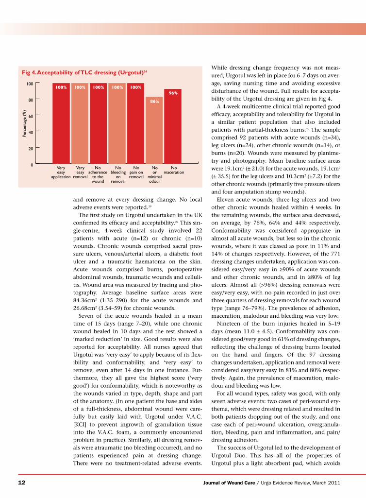
Acute and chronic woundsEvidence relating to outcomes achieved in acute and chronic wounds is discussed first. A large-scale multicentre observational study, which involved almost 6,000 patients with acute or chronic wounds,14 clearly demonstrates that Urgotul reduces pain at dressing change. The prevalence of pain at dressing change was measured, and the use of Urgotul evaluated for any reduction in this. At the screening visit, patients experiencing moderate or severe pain at dressing change were identified using a simple four-point pain scale. Those who met this criterion then self-evaluated in a questionnaire pain intensity and frequency during subsequent wound care procedures. Dressings in use at baseline included mostly simple wet or dry gauze dressings, paraffin gauze, hydrocolloids or foams. A second questionnaire enquired about the practitioner’s approach to the management of a painful wound. Acute wounds (2914) comprised traumatic injuries, burns and post-surgical wounds. Chronic wounds (2936) were mainly leg ulcers, but also included pressure ulcers, diabetic foot ulcers and chronic post-traumatic/post-surgical wounds.
In all, 80% (n=2308) of patients with acute wounds and 80% (n=2341) with chronic wounds reported moderate or severe pain at dressing removal at the screening visit. Of these, 1879 switched to Urgotul during the treatment period (1023 with acute wounds and 856 with chronic wounds). Median follow-up periods were 10 and 23 days for
acute and chronic wounds respectively. Compared with the period before the switch, 95% of patients with acute wounds and 88% with chronic wounds reported no pain or less pain at dressing change (Fig 3). Furthermore, 83% of patients stated that, since switching to Urgotul, they felt substantially less anx-ious before treatments, while 80% of patients with acute wounds and 71% with chronic wounds stated that they wished to continue with this dressing.
A smaller study undertaken, in Spain, involving 28 patients with acute wounds (n=7), chronic wounds (n=10) and burns (n=11) found that Urgotul was highly efficacious and caused either no or mini-mal pain at dressing change. The wounds, which were mostly located on the lower limb, were assessed until full healing occurred. Previous treatments used included sterile gauze, ‘greasy’ gauze, hydro-colloids and foams. Surface area was measured using planimetry. Mean baseline measurements were 20.91cm2 ± 24.63 (range 0.50–62.16) for acute wounds and 5.18cm2 ± 3.22 (1.78–10.83) for chronic wounds. The mean baseline duration of chronic wounds was 24.6 months. For burns, the mean duration was 7.5 days ± 8.9 (1–30) and the mean surface area was 37.5cm2 ± 90.0 (2.1–308).
All of the wounds healed. Chronic wounds healed in a mean of 67.8 days ± 40.9 (28–130), acute wounds in a mean of 11.0 days ± 6.6 (3–24) and burns in a mean of 20.3 days ± 3.0. None of the burns developed clinical signs of infection. The dressing, which was almost always non-adherent, was considered to be very easy/easy to both apply
Fig 3. Urgotul was associated with no or less pain at dressing change14
12 Journal of Wound Care / Urgo Evidence Review, March 2011
and remove at every dressing change. No local adverse events were reported.39
The first study on Urgotul undertaken in the UK confirmed its efficacy and acceptability.24 This sin-gle-centre, 4-week clinical study involved 22 patients with acute (n=12) or chronic (n=10) wounds. Chronic wounds comprised sacral pres-sure ulcers, venous/arterial ulcers, a diabetic foot ulcer and a traumatic haematoma on the skin. Acute wounds comprised burns, postoperative abdominal wounds, traumatic wounds and celluli-tis. Wound area was measured by tracing and pho-tography. Average baseline surface areas were 84.36cm2 (1.35–290) for the acute wounds and 26.68cm2 (3.54–59) for chronic wounds.
Seven of the acute wounds healed in a mean time of 15 days (range 7–20), while one chronic wound healed in 10 days and the rest showed a ‘marked reduction’ in size. Good results were also reported for acceptability. All nurses agreed that Urgotul was ‘very easy’ to apply because of its flex-ibility and conformability, and ‘very easy’ to remove, even after 14 days in one instance. Fur-thermore, they all gave the highest score (‘very good’) for conformability, which is noteworthy as the wounds varied in type, depth, shape and part of the anatomy. (In one patient the base and sides of a full-thickness, abdominal wound were care-fully but easily laid with Urgotul under V.A.C. [KCI] to prevent ingrowth of granulation tissue into the V.A.C. foam, a commonly encountered problem in practice). Similarly, all dressing remov-als were atraumatic (no bleeding occurred), and no patients experienced pain at dressing change. There were no treatment-related adverse events.
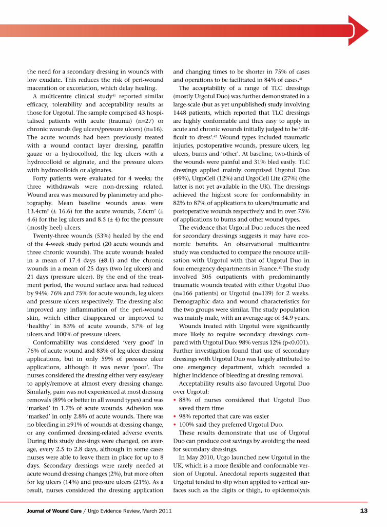
While dressing change frequency was not meas-ured, Urgotul was left in place for 6–7 days on aver-age, saving nursing time and avoiding excessive disturbance of the wound. Full results for accepta-bility of the Urgotul dressing are given in Fig 4.
A 4-week multicentre clinical trial reported good efficacy, acceptability and tolerability for Urgotul in a similar patient population that also included patients with partial-thickness burns.40 The sample comprised 92 patients with acute wounds (n=34), leg ulcers (n=24), other chronic wounds (n=14), or burns (n=20). Wounds were measured by planime-try and photography. Mean baseline surface areas were 19.1cm2 (± 21.0) for the acute wounds, 19.1cm2 (± 35.5) for the leg ulcers and 10.3cm2 (±7.2) for the other chronic wounds (primarily five pressure ulcers and four amputation stump wounds).
Eleven acute wounds, three leg ulcers and two other chronic wounds healed within 4 weeks. In the remaining wounds, the surface area decreased, on average, by 76%, 64% and 44% respectively. Conformability was considered appropriate in almost all acute wounds, but less so in the chronic wounds, where it was classed as poor in 11% and 14% of changes respectively. However, of the 771 dressing changes undertaken, application was con-sidered easy/very easy in ≥90% of acute wounds and other chronic wounds, and in ≥80% of leg ulcers. Almost all (>96%) dressing removals were easy/very easy, with no pain recorded in just over three quarters of dressing removals for each wound type (range 76–79%). The prevalence of adhesion, maceration, malodour and bleeding was very low.
Nineteen of the burn injuries healed in 5–19 days (mean 11.0 ± 4.5). Conformability was con-sidered good/very good in 61% of dressing changes, reflecting the challenge of dressing burns located on the hand and fingers. Of the 97 dressing changes undertaken, application and removal were considered easy/very easy in 81% and 80% respec-tively. Again, the prevalence of maceration, malo-dour and bleeding was low.
For all wound types, safety was good, with only seven adverse events: two cases of peri-wound ery-thema, which were dressing related and resulted in both patients dropping out of the study, and one case each of peri-wound ulceration, overgranula-tion, bleeding, pain and inflammation, and pain/dressing adhesion.
The success of Urgotul led to the development of Urgotul Duo. This has all of the properties of Urgotul plus a light absorbent pad, which avoids
Veryeasy
removal
Noadherence
to thewound
Nobleeding
onremoval
Nopain onremoval
Noor
minimalodour
Nomaceration
Veryeasy
application
0
20
40
60
80
100
Perc
enta
ge (
%)
100% 100% 100% 100% 100%
86%
96%
Fig 4. Acceptability of TLC dressing (Urgotul)24
Journal of Wound Care / Urgo Evidence Review, March 2011 13
the need for a secondary dressing in wounds with low exudate. This reduces the risk of peri-wound maceration or excoriation, which delay healing.
A multicentre clinical study41 reported similar efficacy, tolerability and acceptability results as those for Urgotul. The sample comprised 43 hospi-talised patients with acute (trauma) (n=27) or chronic wounds (leg ulcers/pressure ulcers) (n=16). The acute wounds had been previously treated with a wound contact layer dressing, paraffin gauze or a hydrocolloid, the leg ulcers with a hydrocolloid or alginate, and the pressure ulcers with hydrocolloids or alginates.
Forty patients were evaluated for 4 weeks; the three withdrawals were non-dressing related. Wound area was measured by planimetry and pho-tography. Mean baseline wounds areas were 13.4cm2 (± 16.6) for the acute wounds, 7.6cm2 (± 4.6) for the leg ulcers and 8.5 (± 4) for the pressure (mostly heel) ulcers.
Twenty-three wounds (53%) healed by the end of the 4-week study period (20 acute wounds and three chronic wounds). The acute wounds healed in a mean of 17.4 days (±8.1) and the chronic wounds in a mean of 25 days (two leg ulcers) and 21 days (pressure ulcer). By the end of the treat-ment period, the wound surface area had reduced by 94%, 76% and 75% for acute wounds, leg ulcers and pressure ulcers respectively. The dressing also improved any inflammation of the peri-wound skin, which either disappeared or improved to ‘healthy’ in 83% of acute wounds, 57% of leg ulcers and 100% of pressure ulcers.
Conformability was considered ‘very good’ in 76% of acute wound and 83% of leg ulcer dressing applications, but in only 59% of pressure ulcer applications, although it was never ‘poor’. The nurses considered the dressing either very easy/easy to apply/remove at almost every dressing change. Similarly, pain was not experienced at most dressing removals (89% or better in all wound types) and was ‘marked’ in 1.7% of acute wounds. Adhesion was ‘marked’ in only 2.8% of acute wounds. There was no bleeding in ≥91% of wounds at dressing change, or any confirmed dressing-related adverse events. During this study dressings were changed, on aver-age, every 2.5 to 2.8 days, although in some cases nurses were able to leave them in place for up to 8 days. Secondary dressings were rarely needed at acute wound dressing changes (2%), but more often for leg ulcers (14%) and pressure ulcers (21%). As a result, nurses considered the dressing application
and changing times to be shorter in 75% of cases and operations to be facilitated in 84% of cases.41
The acceptability of a range of TLC dressings (mostly Urgotul Duo) was further demonstrated in a large-scale (but as yet unpublished) study involving 1448 patients, which reported that TLC dressings are highly conformable and thus easy to apply in acute and chronic wounds initially judged to be ‘dif-ficult to dress’.42 Wound types included traumatic injuries, postoperative wounds, pressure ulcers, leg ulcers, burns and ‘other’. At baseline, two-thirds of the wounds were painful and 31% bled easily. TLC dressings applied mainly comprised Urgotul Duo (49%), UrgoCell (12%) and UrgoCell Lite (27%) (the latter is not yet available in the UK). The dressings achieved the highest score for conformability in 82% to 87% of applications to ulcers/traumatic and postoperative wounds respectively and in over 75% of applications to burns and other wound types.
The evidence that Urgotul Duo reduces the need for secondary dressings suggests it may have eco-nomic benefits. An observational multicentre study was conducted to compare the resource utili-sation with Urgotul with that of Urgotul Duo in four emergency departments in France.43 The study involved 305 outpatients with predominantly traumatic wounds treated with either Urgotul Duo (n=166 patients) or Urgotul (n=139) for 2 weeks. Demographic data and wound characteristics for the two groups were similar. The study population was mainly male, with an average age of 34.9 years.
Wounds treated with Urgotul were significantly more likely to require secondary dressings com-pared with Urgotul Duo: 98% versus 12% (p<0.001). Further investigation found that use of secondary dressings with Urgotul Duo was largely attributed to one emergency department, which recorded a higher incidence of bleeding at dressing removal.
Acceptability results also favoured Urgotul Duo over Urgotul: • 88% of nurses considered that Urgotul Duo
saved them time• 98% reported that care was easier• 100% said they preferred Urgotul Duo.
These results demonstrate that use of Urgotul Duo can produce cost savings by avoiding the need for secondary dressings.
In May 2010, Urgo launched new Urgotul in the UK, which is a more flexible and conformable ver-sion of Urgotul. Anecdotal reports suggested that Urgotul tended to slip when applied to vertical sur-faces such as the digits or thigh, to epidermolysis
14 Journal of Wound Care / Urgo Evidence Review, March 2011
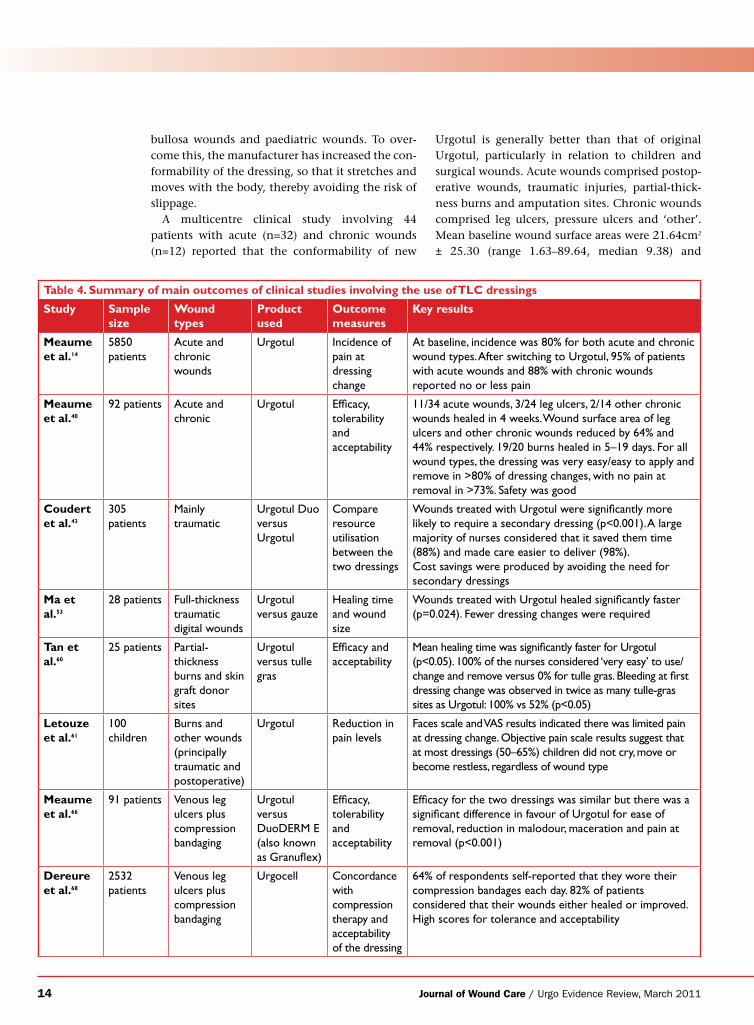
bullosa wounds and paediatric wounds. To over-come this, the manufacturer has increased the con-formability of the dressing, so that it stretches and moves with the body, thereby avoiding the risk of slippage.
A multicentre clinical study involving 44 patients with acute (n=32) and chronic wounds (n=12) reported that the conformability of new
Urgotul is generally better than that of original Urgotul, particularly in relation to children and surgical wounds. Acute wounds comprised postop-erative wounds, traumatic injuries, partial-thick-ness burns and amputation sites. Chronic wounds comprised leg ulcers, pressure ulcers and ‘other’. Mean baseline wound surface areas were 21.64cm2 ± 25.30 (range 1.63–89.64, median 9.38) and
Table 4. Summary of main outcomes of clinical studies involving the use of TLC dressings
Study Sample size
Wound types
Product used
Outcome measures
Key results
Meaume et al.14
5850 patients
Acute and chronic wounds
Urgotul Incidence of pain at dressing change
At baseline, incidence was 80% for both acute and chronic wound types. After switching to Urgotul, 95% of patients with acute wounds and 88% with chronic wounds reported no or less pain
Meaume et al.40
92 patients Acute and chronic
Urgotul Efficacy, tolerability and acceptability
11/34 acute wounds, 3/24 leg ulcers, 2/14 other chronic wounds healed in 4 weeks. Wound surface area of leg ulcers and other chronic wounds reduced by 64% and 44% respectively. 19/20 burns healed in 5–19 days. For all wound types, the dressing was very easy/easy to apply and remove in >80% of dressing changes, with no pain at removal in >73%. Safety was good
Coudert et al.43
305 patients
Mainly traumatic
Urgotul Duo versus Urgotul
Compare resource utilisation between the two dressings
Wounds treated with Urgotul were significantly more likely to require a secondary dressing (p<0.001). A large majority of nurses considered that it saved them time (88%) and made care easier to deliver (98%).Cost savings were produced by avoiding the need for secondary dressings
Ma et al.53
28 patients Full-thickness traumatic digital wounds
Urgotul versus gauze
Healing time and wound size
Wounds treated with Urgotul healed significantly faster (p=0.024). Fewer dressing changes were required
Tan et al.60
25 patients Partial-thickness burns and skin graft donor sites
Urgotul versus tulle gras
Efficacy and acceptability
Mean healing time was significantly faster for Urgotul (p<0.05). 100% of the nurses considered ‘very easy’ to use/change and remove versus 0% for tulle gras. Bleeding at first dressing change was observed in twice as many tulle-gras sites as Urgotul: 100% vs 52% (p<0.05)
Letouze et al.61
100 children
Burns and other wounds (principally traumatic and postoperative)
Urgotul Reduction in pain levels
Faces scale and VAS results indicated there was limited pain at dressing change. Objective pain scale results suggest that at most dressings (50–65%) children did not cry, move or become restless, regardless of wound type
Meaume et al.66
91 patients Venous leg ulcers plus compression bandaging
Urgotul versus DuoDERM E (also known as Granuflex)
Efficacy, tolerability and acceptability
Efficacy for the two dressings was similar but there was a significant difference in favour of Urgotul for ease of removal, reduction in malodour, maceration and pain at removal (p<0.001)
Dereure et al.68
2532 patients
Venous leg ulcers plus compression bandaging
Urgocell Concordance with compression therapy and acceptability of the dressing
64% of respondents self-reported that they wore their compression bandages each day. 82% of patients considered that their wounds either healed or improved. High scores for tolerance and acceptability
Journal of Wound Care / Urgo Evidence Review, March 2011 15
6.61cm2 (± 3.12) (range 2.19–13.40, median 6.05) for acute and chronic wounds respectively. Previ-ous treatments included hydrocolloids, foams, contact layers, alginates, greasy gauze or ‘other’ dressings. Wounds were measured by planimetry and photography.
Efficacy of new Urgotul was good, although it should be noted that 10 patients with acute wounds were excluded from the efficacy analysis, primarily due to lack of planimetric data. Seven-teen acute wounds and three chronic wounds healed. The mean healings times were 14.2 days (± 7.7) and 26.3 days (± 2.9) respectively. Acute wounds reduced by an average of 78% and chronic wounds by average of 42% during the 4-week study period. Two adverse events (infection and infected necrotic tissue) were reported in patients with acute wounds only, one of which resulted in dis-continuation with treatment.
Conformability was reported to be good/very good in 93% of acute wound dressing changes and 85% of chronic wound dressing changes, while the dressing was considered to have stayed in place at 89% of evaluations. In addition, the clinicians con-sidered new Urgotul to be more conformable than Urgotul at 73% of dressing changes respectively in all patients who had used it before entry into the study. There was no pain in 87% of dressing changes, with moderate bleeding being reported in only 8%.44
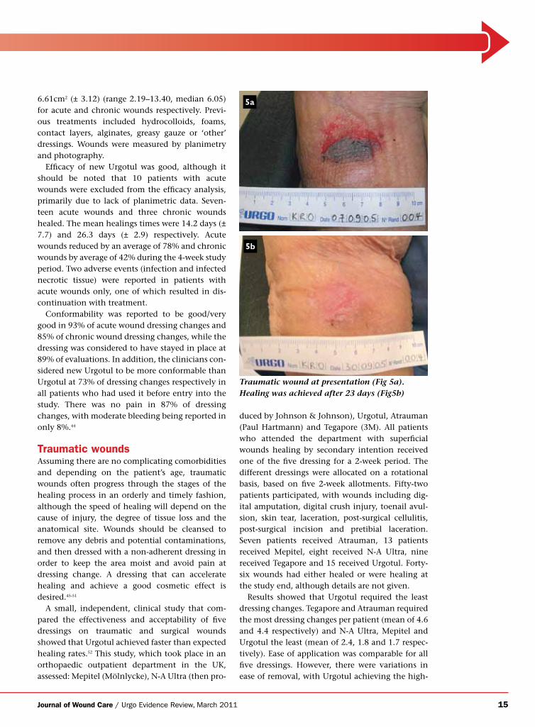
Traumatic woundsAssuming there are no complicating comorbidities and depending on the patient’s age, traumatic wounds often progress through the stages of the healing process in an orderly and timely fashion, although the speed of healing will depend on the cause of injury, the degree of tissue loss and the anatomical site. Wounds should be cleansed to remove any debris and potential contaminations, and then dressed with a non-adherent dressing in order to keep the area moist and avoid pain at dressing change. A dressing that can accelerate healing and achieve a good cosmetic effect is desired.45-51
A small, independent, clinical study that com-pared the effectiveness and acceptability of five dressings on traumatic and surgical wounds showed that Urgotul achieved faster than expected healing rates.52 This study, which took place in an orthopaedic outpatient department in the UK, assessed: Mepitel (Mölnlycke), N-A Ultra (then pro-
Traumatic wound at presentation (Fig 5a). Healing was achieved after 23 days (Fig5b)
5a
5b
duced by Johnson & Johnson), Urgotul, Atrauman (Paul Hartmann) and Tegapore (3M). All patients who attended the department with superficial wounds healing by secondary intention received one of the five dressing for a 2-week period. The different dressings were allocated on a rotational basis, based on five 2-week allotments. Fifty-two patients participated, with wounds including dig-ital amputation, digital crush injury, toenail avul-sion, skin tear, laceration, post-surgical cellulitis, post-surgical incision and pretibial laceration. Seven patients received Atrauman, 13 patients received Mepitel, eight received N-A Ultra, nine received Tegapore and 15 received Urgotul. Forty-six wounds had either healed or were healing at the study end, although details are not given.
Results showed that Urgotul required the least dressing changes. Tegapore and Atrauman required the most dressing changes per patient (mean of 4.6 and 4.4 respectively) and N-A Ultra, Mepitel and Urgotul the least (mean of 2.4, 1.8 and 1.7 respec-tively). Ease of application was comparable for all five dressings. However, there were variations in ease of removal, with Urgotul achieving the high-
16 Journal of Wound Care / Urgo Evidence Review, March 2011
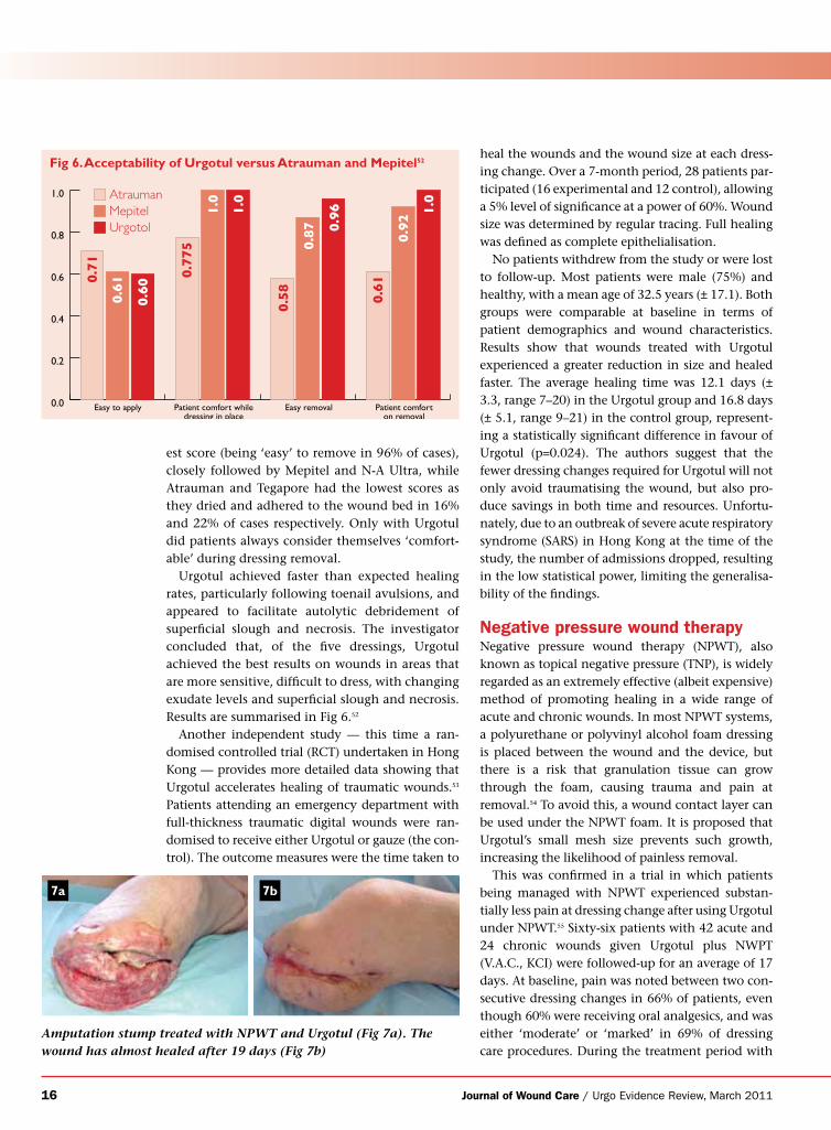
0.0
0.2
0.4
0.6
0.8
1.0
Easy to apply Patient comfort whiledressing in place
Easy removal Patient comforton removal
0.71
0.775
0.58
0.61
0.61
0.60
1.0
0.87 0.961.0
1.0
0.92
UrgotolMepitelAtrauman
est score (being ‘easy’ to remove in 96% of cases), closely followed by Mepitel and N-A Ultra, while Atrauman and Tegapore had the lowest scores as they dried and adhered to the wound bed in 16% and 22% of cases respectively. Only with Urgotul did patients always consider themselves ‘comfort-able’ during dressing removal.
Urgotul achieved faster than expected healing rates, particularly following toenail avulsions, and appeared to facilitate autolytic debridement of superficial slough and necrosis. The investigator concluded that, of the five dressings, Urgotul achieved the best results on wounds in areas that are more sensitive, difficult to dress, with changing exudate levels and superficial slough and necrosis. Results are summarised in Fig 6.52
Another independent study — this time a ran-domised controlled trial (RCT) undertaken in Hong Kong — provides more detailed data showing that Urgotul accelerates healing of traumatic wounds.53 Patients attending an emergency department with full-thickness traumatic digital wounds were ran-domised to receive either Urgotul or gauze (the con-trol). The outcome measures were the time taken to
heal the wounds and the wound size at each dress-ing change. Over a 7-month period, 28 patients par-ticipated (16 experimental and 12 control), allowing a 5% level of significance at a power of 60%. Wound size was determined by regular tracing. Full healing was defined as complete epithelialisation.
No patients withdrew from the study or were lost to follow-up. Most patients were male (75%) and healthy, with a mean age of 32.5 years (± 17.1). Both groups were comparable at baseline in terms of patient demographics and wound characteristics. Results show that wounds treated with Urgotul experienced a greater reduction in size and healed faster. The average healing time was 12.1 days (± 3.3, range 7–20) in the Urgotul group and 16.8 days (± 5.1, range 9–21) in the control group, represent-ing a statistically significant difference in favour of Urgotul (p=0.024). The authors suggest that the fewer dressing changes required for Urgotul will not only avoid traumatising the wound, but also pro-duce savings in both time and resources. Unfortu-nately, due to an outbreak of severe acute respiratory syndrome (SARS) in Hong Kong at the time of the study, the number of admissions dropped, resulting in the low statistical power, limiting the generalisa-bility of the findings.
Negative pressure wound therapyNegative pressure wound therapy (NPWT), also known as topical negative pressure (TNP), is widely regarded as an extremely effective (albeit expensive) method of promoting healing in a wide range of acute and chronic wounds. In most NPWT systems, a polyurethane or polyvinyl alcohol foam dressing is placed between the wound and the device, but there is a risk that granulation tissue can grow through the foam, causing trauma and pain at removal.54 To avoid this, a wound contact layer can be used under the NPWT foam. It is proposed that Urgotul’s small mesh size prevents such growth, increasing the likelihood of painless removal.
This was confirmed in a trial in which patients being managed with NPWT experienced substan-tially less pain at dressing change after using Urgotul under NPWT.55 Sixty-six patients with 42 acute and 24 chronic wounds given Urgotul plus NWPT (V.A.C., KCI) were followed-up for an average of 17 days. At baseline, pain was noted between two con-secutive dressing changes in 66% of patients, even though 60% were receiving oral analgesics, and was either ‘moderate’ or ‘marked’ in 69% of dressing care procedures. During the treatment period with
Fig 6. Acceptability of Urgotul versus Atrauman and Mepitel52
Amputation stump treated with NPWT and Urgotul (Fig 7a). The wound has almost healed after 19 days (Fig 7b)
7a 7b
Journal of Wound Care / Urgo Evidence Review, March 2011 17
Urgotul plus NPWT these percentages fell to 34% and 13% respectively. Patients considered the com-bination easy/very easy to remove in 95% of dress-ing changes, and there was no adherence in 88% of removals. No or minor bleeding was reported in 91% of dressing changes. Finally, the appearance of the surrounding skin improved from inflamed, oedematous, eczematous or macerated to ‘healthy’ in 18 patients. No Urgotul-related adverse events were reported. It appears, therefore, that dressing changes were less painful when NPWT is used with Urgotul because granulation tissue did not adhere to the wound bed.
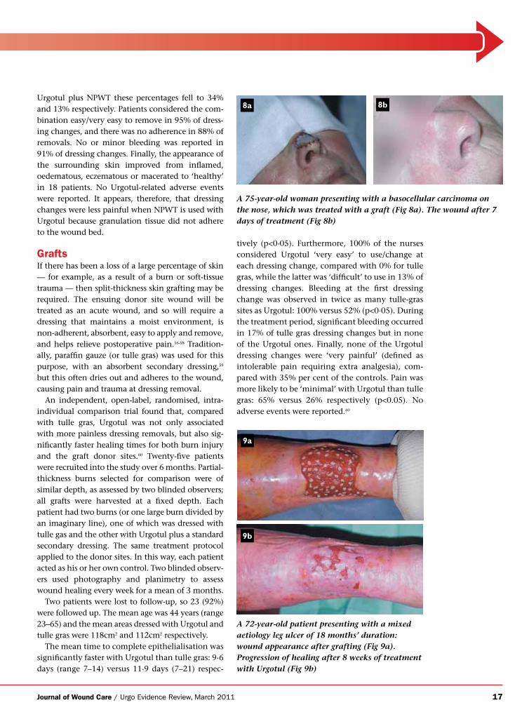
GraftsIf there has been a loss of a large percentage of skin — for example, as a result of a burn or soft-tissue trauma — then split-thickness skin grafting may be required. The ensuing donor site wound will be treated as an acute wound, and so will require a dressing that maintains a moist environment, is non-adherent, absorbent, easy to apply and remove, and helps relieve postoperative pain.56-58 Tradition-ally, paraffin gauze (or tulle gras) was used for this purpose, with an absorbent secondary dressing,59 but this often dries out and adheres to the wound, causing pain and trauma at dressing removal.
An independent, open-label, randomised, intra-individual comparison trial found that, compared with tulle gras, Urgotul was not only associated with more painless dressing removals, but also sig-nificantly faster healing times for both burn injury and the graft donor sites.60 Twenty-five patients were recruited into the study over 6 months. Partial-thickness burns selected for comparison were of similar depth, as assessed by two blinded observers; all grafts were harvested at a fixed depth. Each patient had two burns (or one large burn divided by an imaginary line), one of which was dressed with tulle gas and the other with Urgotul plus a standard secondary dressing. The same treatment protocol applied to the donor sites. In this way, each patient acted as his or her own control. Two blinded observ-ers used photography and planimetry to assess wound healing every week for a mean of 3 months.
Two patients were lost to follow-up, so 23 (92%) were followed up. The mean age was 44 years (range 23–65) and the mean areas dressed with Urgotul and tulle gras were 118cm2 and 112cm2 respectively.
The mean time to complete epithelialisation was significantly faster with Urgotul than tulle gras: 9·6 days (range 7–14) versus 11·9 days (7–21) respec-
tively (p<0·05). Furthermore, 100% of the nurses considered Urgotul ‘very easy’ to use/change at each dressing change, compared with 0% for tulle gras, while the latter was ‘difficult’ to use in 13% of dressing changes. Bleeding at the first dressing change was observed in twice as many tulle-gras sites as Urgotul: 100% versus 52% (p<0·05). During the treatment period, significant bleeding occurred in 17% of tulle gras dressing changes but in none of the Urgotul ones. Finally, none of the Urgotul dressing changes were ‘very painful’ (defined as intolerable pain requiring extra analgesia), com-pared with 35% per cent of the controls. Pain was more likely to be ‘minimal’ with Urgotul than tulle gras: 65% versus 26% respectively (p<0.05). No adverse events were reported.60
A 75-year-old woman presenting with a basocellular carcinoma on the nose, which was treated with a graft (Fig 8a). The wound after 7 days of treatment (Fig 8b)
8a 8b
A 72-year-old patient presenting with a mixed aetiology leg ulcer of 18 months’ duration: wound appearance after grafting (Fig 9a). Progression of healing after 8 weeks of treatment with Urgotul (Fig 9b)
9a
9b
18 Journal of Wound Care / Urgo Evidence Review, March 2011
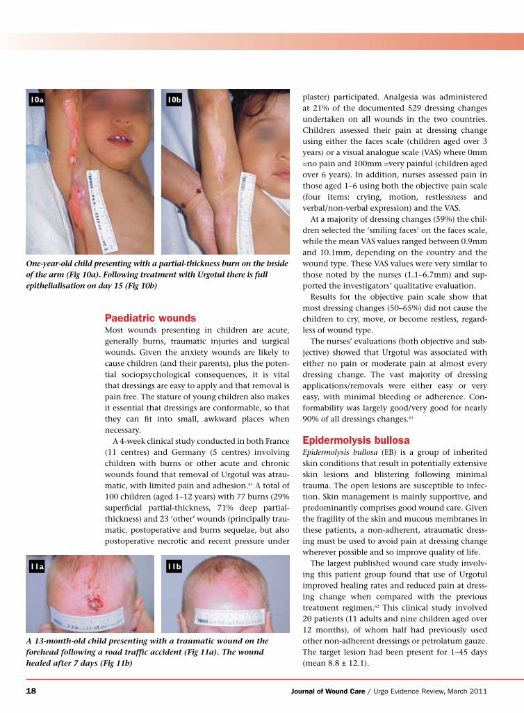
One-year-old child presenting with a partial-thickness burn on the inside of the arm (Fig 10a). Following treatment with Urgotul there is full epithelialisation on day 15 (Fig 10b)
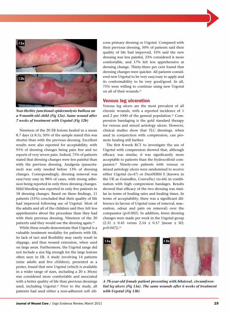
A 13-month-old child presenting with a traumatic wound on the forehead following a road traffic accident (Fig 11a). The wound healed after 7 days (Fig 11b)
Paediatric woundsMost wounds presenting in children are acute, generally burns, traumatic injuries and surgical wounds. Given the anxiety wounds are likely to cause children (and their parents), plus the poten-tial sociopsychological consequences, it is vital that dressings are easy to apply and that removal is pain free. The stature of young children also makes it essential that dressings are conformable, so that they can fit into small, awkward places when necessary.
A 4-week clinical study conducted in both France (11 centres) and Germany (5 centres) involving children with burns or other acute and chronic wounds found that removal of Urgotul was atrau-matic, with limited pain and adhesion.61 A total of 100 children (aged 1–12 years) with 77 burns (29% superficial partial-thickness, 71% deep partial-thickness) and 23 ‘other’ wounds (principally trau-matic, postoperative and burns sequelae, but also postoperative necrotic and recent pressure under
10a
11a
10b
11b
plaster) participated. Analgesia was administered at 21% of the documented 529 dressing changes undertaken on all wounds in the two countries. Children assessed their pain at dressing change using either the faces scale (children aged over 3 years) or a visual analogue scale (VAS) where 0mm =no pain and 100mm =very painful (children aged over 6 years). In addition, nurses assessed pain in those aged 1–6 using both the objective pain scale (four items: crying, motion, restlessness and verbal/non-verbal expression) and the VAS.
At a majority of dressing changes (59%) the chil-dren selected the ‘smiling faces’ on the faces scale, while the mean VAS values ranged between 0.9mm and 10.1mm, depending on the country and the wound type. These VAS values were very similar to those noted by the nurses (1.1–6.7mm) and sup-ported the investigators’ qualitative evaluation.
Results for the objective pain scale show that most dressing changes (50–65%) did not cause the children to cry, move, or become restless, regard-less of wound type.
The nurses’ evaluations (both objective and sub-jective) showed that Urgotul was associated with either no pain or moderate pain at almost every dressing change. The vast majority of dressing applications/removals were either easy or very easy, with minimal bleeding or adherence. Con-formability was largely good/very good for nearly 90% of all dressings changes.61
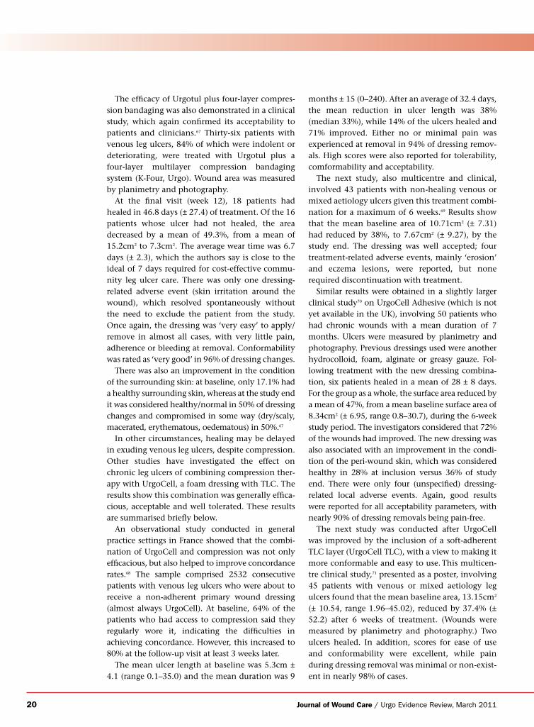
Epidermolysis bullosaEpidermolysis bullosa (EB) is a group of inherited skin conditions that result in potentially extensive skin lesions and blistering following minimal trauma. The open lesions are susceptible to infec-tion. Skin management is mainly supportive, and predominantly comprises good wound care. Given the fragility of the skin and mucous membranes in these patients, a non-adherent, atraumatic dress-ing must be used to avoid pain at dressing change wherever possible and so improve quality of life.
The largest published wound care study involv-ing this patient group found that use of Urgotul improved healing rates and reduced pain at dress-ing change when compared with the previous treatment regimen.62 This clinical study involved 20 patients (11 adults and nine children aged over 12 months), of whom half had previously used other non-adherent dressings or petrolatum gauze. The target lesion had been present for 1–45 days (mean 8.8 ± 12.1).
Journal of Wound Care / Urgo Evidence Review, March 2011 19
Nineteen of the 20 EB lesions healed in a mean 8.7 days (± 8.5); 50% of the sample stated this was shorter than with the previous dressing. Excellent results were also reported for acceptability, with 91% of dressing changes being pain free and no reports of very severe pain. Indeed, 75% of patients stated that dressing changes were less painful than with the previous dressing. Analgesia (paraceta-mol) was only needed before 13% of dressing changes. Correspondingly, dressing removal was easy/very easy in 98% of cases, with strong adhe-sion being reported in only three dressing changes. Mild bleeding was reported in only five patients in 18 dressing changes. Based on these findings, 11 patients (55%) concluded that their quality of life had improved following use of Urgotul. Most of the adults and all of the children said they felt less apprehensive about the procedure than they had with their previous dressing. Nineteen of the 20 patients said they would use the dressing again.63
While these results demonstrate that Urgotul is a valuable treatment modality for patients with EB, its lack of tact and flexibility may rarely result in slippage, and thus wound extension, when used on large areas. Furthermore, the Urgotul range did not include a size big enough for the large lesions often seen in EB. A study involving 14 patients (nine adults and five children), presented as a poster, found that new Urgotul (which is available in a wider range of sizes, including a 20 x 30cm) was considered more comfortable and associated with a better quality of life than previous dressings used, including Urgotul.64 Prior to the study, all patients had used either a non-adherent soft sili-
cone primary dressing or Urgotul. Compared with their previous dressing, 50% of patients said their quality of life had improved, 33% said the new dressing was less painful, 33% considered it more comfortable, and 17% felt less apprehensive at dressing change. Thirty-three per cent found that dressing changes were quicker. All patients consid-ered new Urgotul to be very easy/easy to apply and its conformability to be very good/good. In all, 75% were willing to continue using new Urgotul on all of their wounds.64
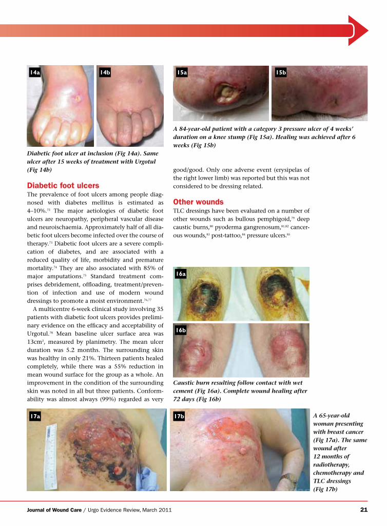
Venous leg ulcerationVenous leg ulcers are the most prevalent of all chronic wounds, with a reported incidence of 1 and 2 per 1000 of the general population.65 Com-pression bandaging is the gold standard therapy for venous and mixed aetiology ulcers. However, clinical studies show that TLC dressings, when used in conjunction with compression, can pro-mote healing still further.
The first 8-week RCT to investigate the use of Urgotul with compression showed that, although efficacy was similar, it was significantly more acceptable to patients than the hydrocolloid com-parator.66 Ninety-one patients with venous or mixed aetiology ulcers were randomised to receive either Urgotul (n=47) or DuoDERM E (known in the UK as Granuflex, ConvaTec) (n=44) in combi-nation with high compression bandages. Results showed that efficacy of the two dressing was simi-lar in terms of healing rates and healing times. In terms of acceptability, there was a significant dif-ference in favour of Urgotul (ease of removal, mac-eration, odour and pain on removal) over the comparator (p<0.002). In addition, fewer dressing changes were made per week in the Urgotul group (2.31 ± 0.45 versus 2.54 ± 0.57 [mean ± SD; p=0.047]).66
Non-Herlitz junctional epidermolysis bullosa on a 9-month-old child (Fig 12a). Same wound after 7 weeks of treatment with Urgotul (Fig 12b)
12a
12b
A 78-year-old female patient presenting with bilateral, circumferen-tial leg ulcers (Fig 13a). The same wounds after 6 weeks of treatment with Urgotul (Fig 13b)
13a 13b
20 Journal of Wound Care / Urgo Evidence Review, March 2011
The efficacy of Urgotul plus four-layer compres-sion bandaging was also demonstrated in a clinical study, which again confirmed its acceptability to patients and clinicians.67 Thirty-six patients with venous leg ulcers, 84% of which were indolent or deteriorating, were treated with Urgotul plus a four-layer multilayer compression bandaging system (K-Four, Urgo). Wound area was measured by planimetry and photography.
At the final visit (week 12), 18 patients had healed in 46.8 days (± 27.4) of treatment. Of the 16 patients whose ulcer had not healed, the area decreased by a mean of 49.3%, from a mean of 15.2cm2 to 7.3cm2. The average wear time was 6.7 days (± 2.3), which the authors say is close to the ideal of 7 days required for cost-effective commu-nity leg ulcer care. There was only one dressing-related adverse event (skin irritation around the wound), which resolved spontaneously without the need to exclude the patient from the study. Once again, the dressing was ‘very easy’ to apply/remove in almost all cases, with very little pain, adherence or bleeding at removal. Conformability was rated as ‘very good’ in 96% of dressing changes.
There was also an improvement in the condition of the surrounding skin: at baseline, only 17.1% had a healthy surrounding skin, whereas at the study end it was considered healthy/normal in 50% of dressing changes and compromised in some way (dry/scaly, macerated, erythematous, oedematous) in 50%.67
In other circumstances, healing may be delayed in exuding venous leg ulcers, despite compression. Other studies have investigated the effect on chronic leg ulcers of combining compression ther-apy with UrgoCell, a foam dressing with TLC. The results show this combination was generally effica-cious, acceptable and well tolerated. These results are summarised briefly below.
An observational study conducted in general practice settings in France showed that the combi-nation of UrgoCell and compression was not only efficacious, but also helped to improve concordance rates.68 The sample comprised 2532 consecutive patients with venous leg ulcers who were about to receive a non-adherent primary wound dressing (almost always UrgoCell). At baseline, 64% of the patients who had access to compression said they regularly wore it, indicating the difficulties in achieving concordance. However, this increased to 80% at the follow-up visit at least 3 weeks later.
The mean ulcer length at baseline was 5.3cm ± 4.1 (range 0.1–35.0) and the mean duration was 9
months ± 15 (0–240). After an average of 32.4 days, the mean reduction in ulcer length was 38% (median 33%), while 14% of the ulcers healed and 71% improved. Either no or minimal pain was experienced at removal in 94% of dressing remov-als. High scores were also reported for tolerability, comformability and acceptability.
The next study, also multicentre and clinical, involved 43 patients with non-healing venous or mixed aetiology ulcers given this treatment combi-nation for a maximum of 6 weeks.69 Results show that the mean baseline area of 10.71cm2 (± 7.31) had reduced by 38%, to 7.67cm2 (± 9.27), by the study end. The dressing was well accepted; four treatment-related adverse events, mainly ‘erosion’ and eczema lesions, were reported, but none required discontinuation with treatment.
Similar results were obtained in a slightly larger clinical study70 on UrgoCell Adhesive (which is not yet available in the UK), involving 50 patients who had chronic wounds with a mean duration of 7 months. Ulcers were measured by planimetry and photography. Previous dressings used were another hydrocolloid, foam, alginate or greasy gauze. Fol-lowing treatment with the new dressing combina-tion, six patients healed in a mean of 28 ± 8 days. For the group as a whole, the surface area reduced by a mean of 47%, from a mean baseline surface area of 8.34cm2 (± 6.95, range 0.8–30.7), during the 6-week study period. The investigators considered that 72% of the wounds had improved. The new dressing was also associated with an improvement in the condi-tion of the peri-wound skin, which was considered healthy in 28% at inclusion versus 36% of study end. There were only four (unspecified) dressing-related local adverse events. Again, good results were reported for all acceptability parameters, with nearly 90% of dressing removals being pain-free.
The next study was conducted after UrgoCell was improved by the inclusion of a soft-adherent TLC layer (UrgoCell TLC), with a view to making it more conformable and easy to use. This multicen-tre clinical study,71 presented as a poster, involving 45 patients with venous or mixed aetiology leg ulcers found that the mean baseline area, 13.15cm2 (± 10.54, range 1.96–45.02), reduced by 37.4% (± 52.2) after 6 weeks of treatment. (Wounds were measured by planimetry and photography.) Two ulcers healed. In addition, scores for ease of use and conformability were excellent, while pain during dressing removal was minimal or non-exist-ent in nearly 98% of cases.
Journal of Wound Care / Urgo Evidence Review, March 2011 21
Diabetic foot ulcersThe prevalence of foot ulcers among people diag-nosed with diabetes mellitus is estimated as 4–10%.72 The major aetiologies of diabetic foot ulcers are neuropathy, peripheral vascular disease and neuroischaemia. Approximately half of all dia-betic foot ulcers become infected over the course of therapy.73 Diabetic foot ulcers are a severe compli-cation of diabetes, and are associated with a reduced quality of life, morbidity and premature mortality.74 They are also associated with 85% of major amputations.75 Standard treatment com-prises debridement, offloading, treatment/preven-tion of infection and use of modern wound dressings to promote a moist environment.76,77
A multicentre 6-week clinical study involving 35 patients with diabetic foot ulcers provides prelimi-nary evidence on the efficacy and acceptability of Urgotul.78 Mean baseline ulcer surface area was 13cm2, measured by planimetry. The mean ulcer duration was 5.2 months. The surrounding skin was healthy in only 21%. Thirteen patients healed completely, while there was a 55% reduction in mean wound surface for the group as a whole. An improvement in the condition of the surrounding skin was noted in all but three patients. Conform-ability was almost always (99%) regarded as very
good/good. Only one adverse event (erysipelas of the right lower limb) was reported but this was not considered to be dressing related.
Other woundsTLC dressings have been evaluated on a number of other wounds such as bullous pemphigoid,79 deep caustic burns,80 pyoderma gangrenosum,81,82 cancer-ous wounds,83 post-tattoo,84 pressure ulcers.85
Diabetic foot ulcer at inclusion (Fig 14a). Same ulcer after 15 weeks of treatment with Urgotul (Fig 14b)
14a 14b
A 84-year-old patient with a category 3 pressure ulcer of 4 weeks’ duration on a knee stump (Fig 15a). Healing was achieved after 6 weeks (Fig 15b)
A 65-year-old woman presenting with breast cancer (Fig 17a). The same wound after 12 months of radiotherapy, chemotherapy and TLC dressings (Fig 17b)
15a 15b
17a 17b
Caustic burn resulting follow contact with wet cement (Fig 16a). Complete wound healing after 72 days (Fig 16b)
16a
16b
22 Journal of Wound Care / Urgo Evidence Review, March 2011
Dressings with TLC-Ag technology
A ll wounds — acute and chronic — contain microorganisms, but this does not mean that they are infected.86 In chronic wounds,
the presence of devitalised tissue can result in colonisation by a variety of microorganisms, but this is only regarded as problematic if there is a host reaction (characterised by the clinical symptoms of critical colonisation or infection), which will delay healing.87 While the number of bacterial species present in the wound is not necessarily an indicator of infection, the presence of four or more groups increases the risk,88 as does, of course, host susceptibility89 and the presence of certain pathogens and pathogenic species, such as Staphylococcus aureus, Streptococcus species, anaerobes and Pseudomonas aeruginosa.90
The concepts of critical colonisation and infec-tion are based on the level of host response to the pathogens present in the wound. While there is no universally agreed definition of critical colonisa-tion, it is proposed that a wound is critically colo-nised when the bacteria delay healing with subtle signs of a host reaction or inflammation but no overt signs of infection — for example, when the wound margins fail to contract, exudate levels increase, the wound bed is predominantly sloughy and, possibly, granulation is friable and bleeds easily at dressing change. In contrast, infection is associated with damage to the host, with the clas-sic signs including pain, erythema, heat, oedema, malodour, cellulitis, delayed healing and wound breakdown.91,92
More recently, it has been proposed that all chronic wounds contain biofilms,93 although this has yet to be proven. Most bacteria grow when attached to a surface, such as the host tissue, and the numbers and species of bacteria multiply to become a stable, self-sustaining, polymicrobial community (the biofilm) that is encased within a matrix of extracellular polymers. A host inflamma-tory response then occurs, resulting in the recruit-ment of excess neutrophils, pro-inflammatory cytokines and excessive host-derived proteases, which provide the biofilm with a constant source of nutrients.94 The biofilm has an evolutionary ability to adapt to its environment and avoid the immune system and resist antibiotics, thereby ensuring its survival. As time passes, the aerobic
and anaerobic bacteria in the biofilm increase their pathogenic effect, potentially causing infection and delaying healing still further.95
Topical antimicrobials can be used to prevent and treat infection in high-risk patients. In addi-tion, the various antiseptics contained within most of these dressings will reduce the bioburden, thereby helping the host to regain control. Topical antimicrobials have an advantage over systemic antibiotics in that they are not associated with resistance, and have a broader antimicrobial spec-trum and much lower sensitisation rates.96 While best practice guidelines are still in development, it is prudent to restrict use to a maximum of 3–4 weeks before re-assessing the patient, depending on local protocols. Topical antimicrobials should only be used while the signs of critical colonisation or local infection are resolving. If the infection becomes systemic, then antibiotics must be applied.
Silver has a broad antimicrobial effect against both Gram-positive and Gram-negative bacteria, including Pseudomonas aeruginosa, meticillin-resist-ant Staphylococcus aureus (MRSA) and vancomycin-resistant enterococcus (VRE), as well as some fungi, viruses and protozoa.97 It is thought to bind to and damage microbial cell walls, inhibit replication and reduce metabolism and growth.
The safety and tolerability profile of silver is good. While there is evidence that some silver dressings are cytotoxic to keratinocytes in vitro,98,99
this effect is less marked in vivo and ex vivo as the silver is dispersed by the blood circulation and deposited in the liver and kidney, from where it is presumably eliminated from the body in the urine.100,101 The potential for bacterial resistance to silver has also been raised,102,103 but recorded occur-rences are very rare.104
Silver has therefore been incorporated into TLC dressings to give them antimicrobial properties. Silver sulphadiazine (3.75%) has been added to Urgotul to produce Urgotul SSD, and silver ion (3.5%) to Urgotul to produce Urgotul Silver. Both are indicated for the treatment of non to low exud-ing acute (burns, skin abrasions, traumatic injuries, second-degree burns) and chronic wounds show-ing signs of infection or critical colonisation. They can also be used on more heavily exuding wounds
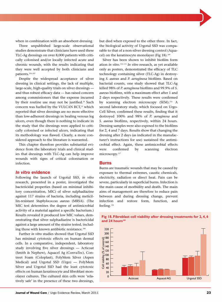
Journal of Wound Care / Urgo Evidence Review, March 2011 23
when in combination with an absorbent dressing.Three unpublished large-scale observational
studies demonstrate that clinicians have used these TLC-Ag dressings on over 8,000 patients with criti-cally colonised and/or locally infected acute and chronic wounds, with the results indicating that they were well accepted by both clinicians and patients.105–107
Despite the widespread acceptance of silver dressing in clinical settings, the lack of multiple, large-scale, high-quality trials on silver dressings — and thus robust efficacy data — has raised concern among commissioners that the expense incurred by their routine use may not be justified.19 Such concern was fuelled by the VULCAN RCT,21 which reported that silver dressings are no more effective than low-adherent dressings in healing venous leg ulcers, even though there is nothing to indicate in the study that the dressings were applied to criti-cally colonised or infected ulcers, indicating that its methodology was flawed. Clearly, a more con-sidered approach to the literature is warranted.
This chapter therefore provides substantial evi-dence from the laboratory trials and clinical stud-ies that dressings with TLC-Ag can help improve wounds with signs of critical colonisation or infection.
In vitro evidenceFollowing the launch of Urgotul SSD, in vitro research, presented in a poster, investigated the bactericidal properties (based on minimal inhibi-tory concentration, MIC) of silver sulphadiazine against 117 strains of bacteria, including meticil-lin-resistant Staphylococcus aureus (MRSA). (The MIC test determines the degree of antimicrobial activity of a material against a specific bacterium.) Results revealed it produced low MIC values, dem-onstrating that silver sulphadiazine is bactericidal against a large amount of the strains tested, includ-ing those with known antibiotic resistance.108
Further in vitro studies showed that Urgotul SSD has minimal cytotoxic effects on human dermal cells. In a comparative, independent, laboratory study involving five silver dressings — Acticoat (Smith & Nephew), Aquacel Ag (ConvaTec), Con-treet Foam (Coloplast), PolyMem Silver (Aspen Medical) and Urgotul SSD (Urgo) — PolyMem Silver and Urgotul SSD had the least cytotoxic effects on human keratinocyte and fibroblast mon-olayer cultures. The cultured skin cells were ‘rela-tively safe’ in the presence of these two dressings,
but died when exposed to the other three. In fact, the biological activity of Urgotul SSD was compa-rable to that of a non-silver dressing control (Aqua-cel) on the keratinocyte monolayer (Fig 18).109
Silver has been shown to inhibit biofilm form ation in vitro.110–115 In vitro research, as yet available only as posters, demonstrated the efficacy of TLC technology containing silver (TLC-Ag) in destroy-ing S. aureus and P. aeruginosa biofilms. Based on bacterial counts, one study showed that TLC-Ag killed 98% of P. aeruginosa biofilms and 99.9% of S. aureus biofilms, with a maximum effect after 1 and 2 days respectively. These results were confirmed by scanning electron microscopy (SEM).116 A second laboratory study, which focused on Urgo-Cell Silver, confirmed these results, finding that it destroyed 100% and 98% of P. aeruginosa and S. aureus biofilms, respectively, within 24 hours. Dressing samples were also exposed to the biofilms for 2, 4 and 7 days. Results show that changing the dressing after 2 days (as indicated in the manufac-turer’s instructions for use) sustained the antimi-crobial effect. Again, these antimicrobial effects were confirmed by scanning electron microscopy.117
BurnsBurns are traumatic wounds that may be caused by exposure to thermal extremes, caustic chemicals, electricity, radiation or direct heat. Pain can be severe, particularly in superficial burns. Infection is the main cause of morbidity and death. The main aims of management are therefore to reduce pain between and during dressing change, prevent infection and restore form, function, and feeling.118
0
10
20
30
40
50
60
70
80
90
100
Acticoat Aquacel AG
Cel
l via
bilit
y (%
of c
ontr
ol)
0
20
40
60
80
100
120
140
160
180
200
220
Acticoat Aquacel AG Urgotul SSD
Cel
l via
bilit
y (%
of c
ontr
ol)
Keratinocyte – FBS
Fig 18. Fibroblast cell viability after dressing treatments for 2, 4, 6 and 24 hours109
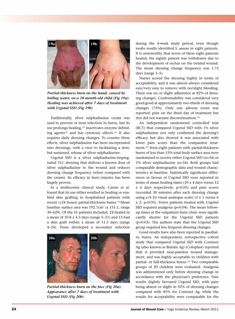
24 Journal of Wound Care / Urgo Evidence Review, March 2011
Traditionally, silver sulphadiazine cream was used to prevent or treat infection in burns, but its use prolongs healing,119 inactivates enzyme debrid-ing agents120 and has cytotoxic effects.121 It also requires daily dressing changes. To counter these effects, silver sulphadiazine has been incorporated into dressings, with a view to facilitating a slow, but sustained, release of silver sulphadiazine.
Urgotul SSD is a silver sulphadiazine-impreg-nated TLC dressing that delivers a known dose of silver sulphadiazine to the wound and reduces dressing change frequency (when compared with the cream). Its efficacy in burn injuries has been largely proven.
In a multicentre clinical study, Carsin et al. found that its use either resulted in healing or ena-bled skin grafting in hospitalised patients with recent (<24 hours) partial-thickness burns.122 Mean baseline surface area was 192.7cm2 (± 151.1, range 30–629). Of the 41 patients included, 24 healed in a mean of 10.8 ± 4.3 days (range 5–21) and 13 had a skin graft within a mean of 11.5 days (range 4–24). None developed a secondary infection
during the 4-week study period, even though swabs results identified S. aureus in eight patients. It is noteworthy that seven of these eight patients healed; the eighth patient was withdrawn due to the development of eschar on the treated wound. The mean dressing change frequency was 1.73 days (range 1–5).
Nurses scored the dressing highly in terms of acceptability, and it was almost always considered easy/very easy to remove with no/slight bleeding. There was no or slight adherence at 82% of dress-ing changes. Conformability was considered very good/good at approximately two-thirds of dressing changes (73%). Only one adverse event was reported: pain on the third day of treatment but this did not warrant discontinuation.122
An independent randomised controlled trial (RCT) that compared Urgotul SSD with 1% silver sulphadiazine not only confirmed the dressing’s efficacy but also showed it was associated with lower pain scores than the comparator treat-ment.123 Sixty-eight patients with partial-thickness burns of less than 15% total body surface area were randomised to receive either Urgotul SSD (n=34) or 1% silver sulphadiazine (n=34). Both groups had comparable demographic data and wound charac-teristics at baseline. Statistically significant differ-ences in favour of Urgotul SSD were reported in terms of mean healing times (10 ± 4 days versus 12 ± 6 days respectively, p<0.05) and pain scores (recorded 30 minutes after each dressing change using a 0–10 visual analogue scale) (3 ± 1 versus 6 ± 2, p<0.05). Fewer patients treated with Urgotul SSD required analgesia (p=0.04). The mean follow-up times at the outpatient burn clinic were signifi-cantly shorter for the Urgotul SSD patients (p=0.03). The authors state that the Urgotul SSD group required less frequent dressing changes.
Good results have also been reported in paediat-ric burns. An independent, retrospective cohort study that compared Urgotul SSD with Contreet Ag (also known as Biatain Ag) (Coloplast) reported that it provided near-painless wound manage-ment, and was highly acceptable to children with partial- or full-thickness burns.124 Two comparable groups of 20 children were evaluated. Analgesia was administered only before dressing change in accordance with the physician’s preference. Pain results slightly favoured Urgotul SSD, with pain being absent or slight in 92% of dressing changes compared with 85% for Contreet Ag, while the results for acceptability were comparable for the
Partial-thickness burn on the face (Fig 20a). Appearance after 7 days of treatment with Urgotul SSD (Fig 20b)
Partial-thickness burn on the hand, caused by boiling water, on a 18-month-old child (Fig 19a). Healing was achieved after 7 days of treatment with Urgotul SSD (Fig 19b)
19a 19b
20a 20b
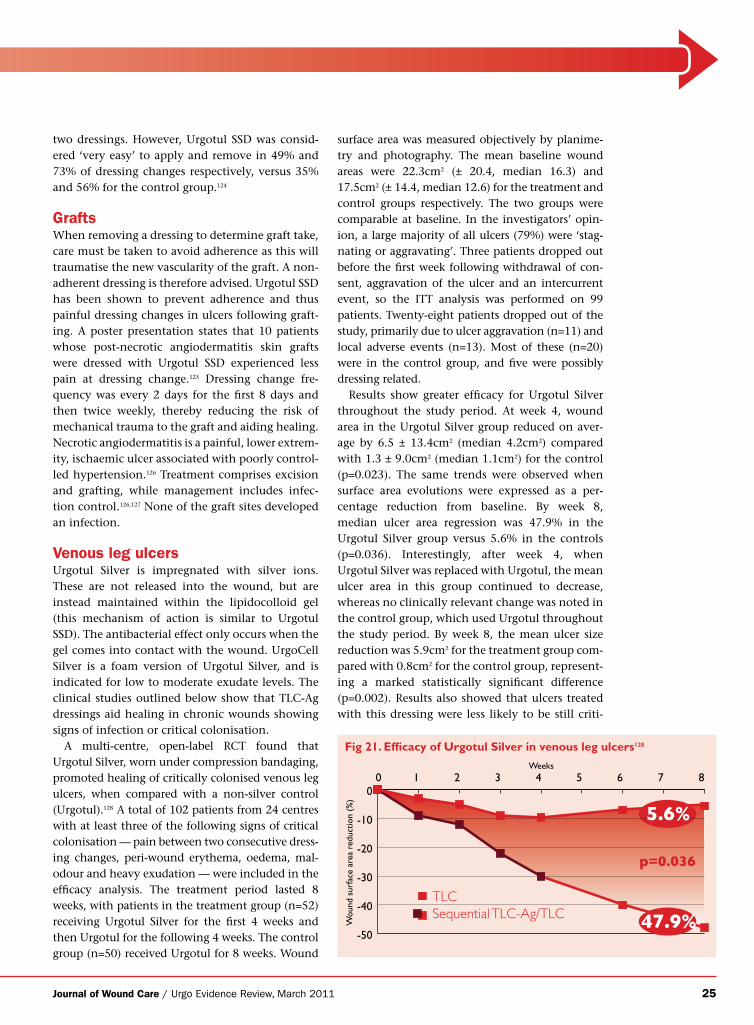
Journal of Wound Care / Urgo Evidence Review, March 2011 25
two dressings. However, Urgotul SSD was consid-ered ‘very easy’ to apply and remove in 49% and 73% of dressing changes respectively, versus 35% and 56% for the control group.124
GraftsWhen removing a dressing to determine graft take, care must be taken to avoid adherence as this will traumatise the new vascularity of the graft. A non-adherent dressing is therefore advised. Urgotul SSD has been shown to prevent adherence and thus painful dressing changes in ulcers following graft-ing. A poster presentation states that 10 patients whose post-necrotic angiodermatitis skin grafts were dressed with Urgotul SSD experienced less pain at dressing change.125 Dressing change fre-quency was every 2 days for the first 8 days and then twice weekly, thereby reducing the risk of mechanical trauma to the graft and aiding healing. Necrotic angiodermatitis is a painful, lower extrem-ity, ischaemic ulcer associated with poorly control-led hypertension.126 Treatment comprises excision and grafting, while management includes infec-tion control.126,127 None of the graft sites developed an infection.
Venous leg ulcersUrgotul Silver is impregnated with silver ions. These are not released into the wound, but are instead maintained within the lipidocolloid gel (this mechanism of action is similar to Urgotul SSD). The antibacterial effect only occurs when the gel comes into contact with the wound. UrgoCell Silver is a foam version of Urgotul Silver, and is indicated for low to moderate exudate levels. The clinical studies outlined below show that TLC-Ag dressings aid healing in chronic wounds showing signs of infection or critical colonisation.
A multi-centre, open-label RCT found that Urgotul Silver, worn under compression bandaging, promoted healing of critically colonised venous leg ulcers, when compared with a non-silver control (Urgotul).128 A total of 102 patients from 24 centres with at least three of the following signs of critical colonisation — pain between two consecutive dress-ing changes, peri-wound erythema, oedema, mal-odour and heavy exudation — were included in the efficacy analysis. The treatment period lasted 8 weeks, with patients in the treatment group (n=52) receiving Urgotul Silver for the first 4 weeks and then Urgotul for the following 4 weeks. The control group (n=50) received Urgotul for 8 weeks. Wound
surface area was measured objectively by planime-try and photography. The mean baseline wound areas were 22.3cm2 (± 20.4, median 16.3) and 17.5cm2 (± 14.4, median 12.6) for the treatment and control groups respectively. The two groups were comparable at baseline. In the investigators’ opin-ion, a large majority of all ulcers (79%) were ‘stag-nating or aggravating’. Three patients dropped out before the first week following withdrawal of con-sent, aggravation of the ulcer and an intercurrent event, so the ITT analysis was performed on 99 patients. Twenty-eight patients dropped out of the study, primarily due to ulcer aggravation (n=11) and local adverse events (n=13). Most of these (n=20) were in the control group, and five were possibly dressing related.
Results show greater efficacy for Urgotul Silver throughout the study period. At week 4, wound area in the Urgotul Silver group reduced on aver-age by 6.5 ± 13.4cm2 (median 4.2cm2) compared with 1.3 ± 9.0cm2 (median 1.1cm2) for the control (p=0.023). The same trends were observed when surface area evolutions were expressed as a per-centage reduction from baseline. By week 8, median ulcer area regression was 47.9% in the Urgotul Silver group versus 5.6% in the controls (p=0.036). Interestingly, after week 4, when Urgotul Silver was replaced with Urgotul, the mean ulcer area in this group continued to decrease, whereas no clinically relevant change was noted in the control group, which used Urgotul throughout the study period. By week 8, the mean ulcer size reduction was 5.9cm2 for the treatment group com-pared with 0.8cm2 for the control group, represent-ing a marked statistically significant difference (p=0.002). Results also showed that ulcers treated with this dressing were less likely to be still criti-
0 1 2 3 4 5 6 7 8Weeks
Wou
nd s
urfa
ce a
rea
redu
ctio
n (%
)
-50
-40
-30
-20
-10
0
p=0.036
TLCSequential TLC-Ag/TLC
5.6%
47.9%
Fig 21. Efficacy of Urgotul Silver in venous leg ulcers128
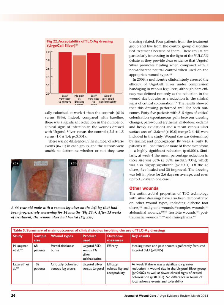
26 Journal of Wound Care / Urgo Evidence Review, March 2011
cally colonised at week 4 than the controls (61% versus 83%). Indeed, compared with baseline, there was a significant reduction in the number of clinical signs of infection in the wounds dressed with Urgotul Silver versus the control (-2.5 ± 1.5 versus -1.0 ± 1.4; p<0.001).
There was no difference in the number of adverse events (n=11) in each group, and the authors were unable to determine whether or not they were
dressing related. Four patients from the treatment group and five from the control group discontin-ued treatment because of them. These results are particularly interesting in the light of the VULCAN debate as they provide clear evidence that Urgotul Silver promotes healing when compared with a non-adherent neutral control when used on the appropriate wound types.128
In 2006, a multicentre clinical study assessed the efficacy of UrgoCell Silver under compression bandaging in venous leg ulcers, although here effi-cacy was defined not only as the reduction in the wound size but also as a reduction in the clinical signs of critical colonisation.129 The results showed that this dressing performed well for both out-comes. Forty-five patients with 3–5 signs of critical colonisation (spontaneous pain between dressing changes, peri-wound erythema, malodour, oedema and heavy exudation) and a mean venous ulcer surface area of 12.6cm2 (± 10.0) (range 2.6–48) were included in the study. Wound size was determined by tracing and photography. By week 4, only 10 patients still had three or more of these symptoms — a highly significant reduction (p<0.001). Simi-larly, at week 4 the mean percentage reduction in ulcer size was 35% (± 58%, median 33%), which was also highly significant (p<0.001). Of the 45 ulcers, five healed and 30 improved. The dressing was left in place for 2.6 days on average, and even up to 13 days in one case.
Other woundsThe antimicrobial properties of TLC technology with silver dressings have also been demonstrated on other wound types, including diabetic foot ulcers,130 malignant wounds,83 complex wounds,131 abdominal wounds,132,133 frostbite wounds,134 post-traumatic wounds,135,136 and rhinophyma.137
0.0
0.2
0.4
0.6
0.8
1.0
Easy/very easyto remove
No painat
dressingchange
Easy/very easy
toapply
Good/very good
conformability
Fig 22. Acceptability of TLC-Ag dressing (UrgoCell Silver)129
A 66-year-old male with a venous leg ulcer on the left leg that had been progressively worsening for 14 months (Fig 23a). After 15 weeks of treatment, the venous ulcer had healed (Fig 23b)
Table 5. Summary of main outcomes of clinical studies involving the use of TLC-Ag dressings
Study Sample size
Wound types Product used
Outcome measures
Key results
Muangman et al.123
68 patients
Partial-thickness burns
Urgotul SSD versus 1% silver sulphadiazine
Efficacy Healing times and pain scores significantly favoured Urgotul SSD (p<0.05)
Lazareth et al.128
102 patients
Critically colonised venous leg ulcers
Urgotul Silver versus Urgotul
Efficacy, tolerability and acceptability
At week 8, there was a significantly greater reduction in wound size in the Urgotul Silver group (p=0.002) as well as fewer clinical signs of critical colonisation (p<0.001). No difference in terms of local adverse events and tolerability
23a 23b
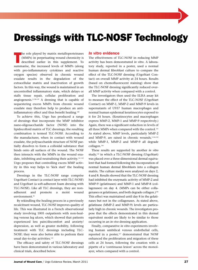
Journal of Wound Care / Urgo Evidence Review, March 2011 27
Dressings with TLC-NOSF Technology
The role played by matrix metalloproteinases (MMPs) in perpetuating wound chronicity is described earlier in this supplement. To
summarise, the increased levels of MMPs (along with pro-inflammatory cytokines and reactive oxygen species) observed in chronic wound exudate results in the degradation of the extracellular matrix and inactivation of growth factors. In this way, the wound is maintained in an uncontrolled inflammatory state, which delays or stalls tissue repair, cellular proliferation and angiogenesis.3,138,139 A dressing that is capable of sequestering excess MMPs from chronic wound exudate may therefore help to produce an anti-inflammatory effect and thus benefit healing.140
To achieve this, Urgo has produced a range of dressings that incorporate the MMP inhibitor nano-oligosaccharide factor (NOSF) into the lipido colloid matrix of TLC dressings; the resulting combination is termed TLC-NOSF. According to the manufacturer, when in contact with wound exudate, the polysaccharide structure of NOSF par-tially dissolves to form a colloidal substance that binds onto all surfaces of the wound. The NOSF then interacts with the MMPs in the wound exu-date, inhibiting and neutralising their activity.141,142 Urgo proposes that controlling excess MMP activ-ity in this way helps to ‘kick start’ the healing process.
Dressings in the TLC-NOSF range comprise UrgoStart Contact (a contact layer with TLC-NOSF) and UrgoStart (a soft-adherent foam dressing with TLC-NOSF). Like all TLC dressings, they are non-adherent and promote a moist wound environment.24
By rekindling the healing process in a previously recalcitrant wound, TLC-NOSF improves quality of life. This was illustrated in a French observational study involving 1005 outpatients with non-heal-ing venous leg ulcers, which showed that patients experienced less pain/discomfort and anxiety/depression, as well as greater mobility, following treatment with TLC dressings including TLC-NOSF; they were also better able to perform their usual day-to-day activities.143
The efficacy and safety of TLC-NOSF dressings have been demonstrated in various laboratory and clinical trials, described below.
In vitro evidenceThe effectiveness of TLC-NOSF in reducing MMP activity has been demonstrated in vitro. A labora-tory study, reported in a poster, used a normal human dermal fibroblast culture to compare the effect of the TLC-NOSF dressing (UrgoStart Con-tact) on overall MMP activity at 24 hours. Results (based on chemofluorescent staining) show that the TLC-NOSF dressing significantly reduced over-all MMP activity when compared with a control.
The investigators then used the ELISA assay kit to measure the effect of the TLC-NOSF (UrgoStart Contact) on MMP-1, MMP-2 and MMP-9 levels in supernatants of U937 human macrophages and normal human epidermal keratinocytes exposed to it for 24 hours. (Keratinocytes and macrophages express MMP-2, MMP-1 and MMP-9 respectively.) Again, there was a significant reduction in levels of all three MMPs when compared with the control.141
As stated above, MMP levels, particularly MMP-2 and MMP-9, are raised in chronic wounds,3,144,145 while MMP-1, MMP-2 and MMP-9 all degrade collagen.146
These results are supported by another in vitro study,142 in which a TLC-NOSF dressing (UrgoStart) was placed over a three-dimensional dermal equiva-lent that had formed following the incorporation of normal human dermal fibroblasts into a collagen matrix. The culture media were analysed on days 2, 4 and 8. Results showed that the TLC-NOSF dressing had inhibited the enzymatic activity of MMP-2 and MMP-9 (gelatinases) and MMP-1 and MMP-8 (col-lagenases) on day 4. (MMPs can be either colla-genases or gelatinases, and both degrade collagen.)146 This effect was maintained until day 8 in the gelati-nases but not in the collagenases. As stated above, gelatinase (MMP-2 and MMP-9) levels are particu-larly high in chronic wounds. The investigators pro-pose that the effects demonstrated in this dermal equivalent model are likely to be similar to those occurring in an in vivo dressing application.
Finally, comparative in vitro experiments involv-ing human umbilical venous endothelial cells, reported in a poster,147 demonstrated that NOSF enhanced the proliferation and migration of these cells at 24 hours, following the creation with a pipette of a ‘continuous lesion’ across the monol-ayer, when compared with a control.
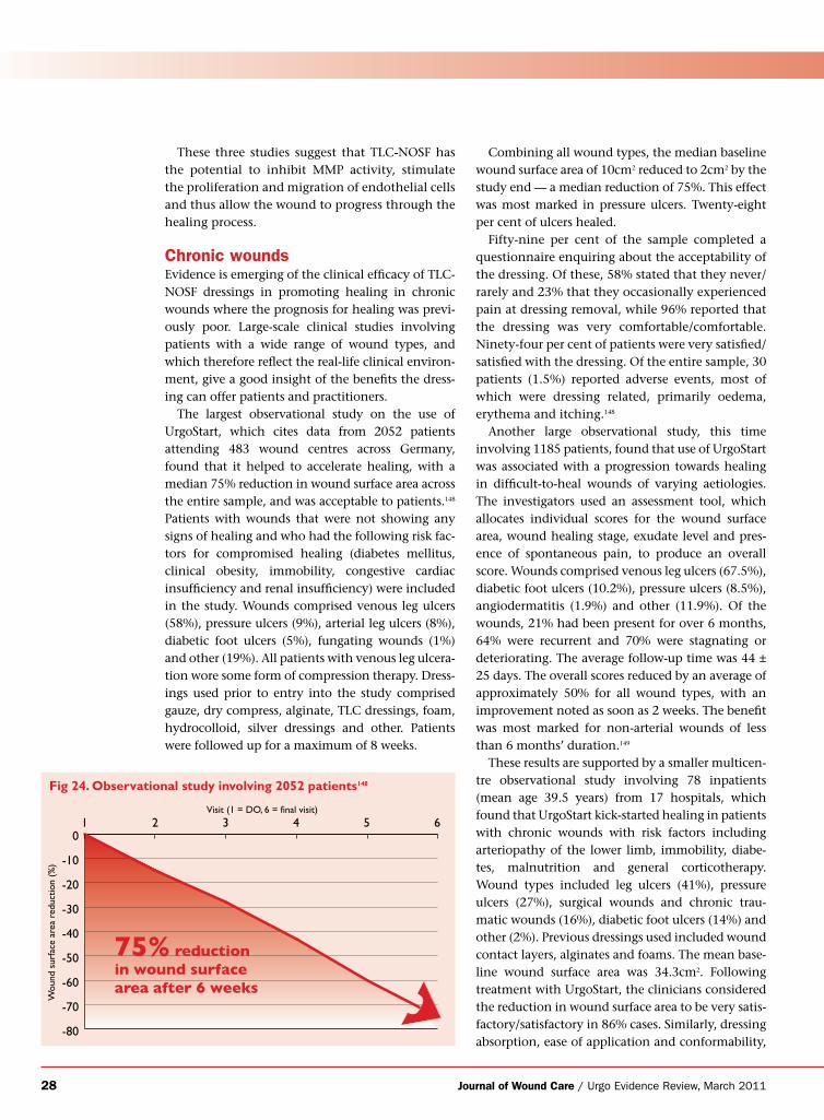
28 Journal of Wound Care / Urgo Evidence Review, March 2011
These three studies suggest that TLC-NOSF has the potential to inhibit MMP activity, stimulate the proliferation and migration of endothelial cells and thus allow the wound to progress through the healing process.
Chronic woundsEvidence is emerging of the clinical efficacy of TLC-NOSF dressings in promoting healing in chronic wounds where the prognosis for healing was previ-ously poor. Large-scale clinical studies involving patients with a wide range of wound types, and which therefore reflect the real-life clinical environ-ment, give a good insight of the benefits the dress-ing can offer patients and practitioners.
The largest observational study on the use of UrgoStart, which cites data from 2052 patients attending 483 wound centres across Germany, found that it helped to accelerate healing, with a median 75% reduction in wound surface area across the entire sample, and was acceptable to patients.148 Patients with wounds that were not showing any signs of healing and who had the following risk fac-tors for compromised healing (diabetes mellitus, clinical obesity, immobility, congestive cardiac insufficiency and renal insufficiency) were included in the study. Wounds comprised venous leg ulcers (58%), pressure ulcers (9%), arterial leg ulcers (8%), diabetic foot ulcers (5%), fungating wounds (1%) and other (19%). All patients with venous leg ulcera-tion wore some form of compression therapy. Dress-ings used prior to entry into the study comprised gauze, dry compress, alginate, TLC dressings, foam, hydrocolloid, silver dressings and other. Patients were followed up for a maximum of 8 weeks.
Combining all wound types, the median baseline wound surface area of 10cm2 reduced to 2cm2 by the study end — a median reduction of 75%. This effect was most marked in pressure ulcers. Twenty-eight per cent of ulcers healed.
Fifty-nine per cent of the sample completed a questionnaire enquiring about the acceptability of the dressing. Of these, 58% stated that they never/rarely and 23% that they occasionally experienced pain at dressing removal, while 96% reported that the dressing was very comfortable/comfortable. Ninety-four per cent of patients were very satisfied/satisfied with the dressing. Of the entire sample, 30 patients (1.5%) reported adverse events, most of which were dressing related, primarily oedema, erythema and itching.148
Another large observational study, this time involving 1185 patients, found that use of UrgoStart was associated with a progression towards healing in difficult-to-heal wounds of varying aetiologies. The investigators used an assessment tool, which allocates individual scores for the wound surface area, wound healing stage, exudate level and pres-ence of spontaneous pain, to produce an overall score. Wounds comprised venous leg ulcers (67.5%), diabetic foot ulcers (10.2%), pressure ulcers (8.5%), angiodermatitis (1.9%) and other (11.9%). Of the wounds, 21% had been present for over 6 months, 64% were recurrent and 70% were stagnating or deteriorating. The average follow-up time was 44 ± 25 days. The overall scores reduced by an average of approximately 50% for all wound types, with an improvement noted as soon as 2 weeks. The benefit was most marked for non-arterial wounds of less than 6 months’ duration.149
These results are supported by a smaller multicen-tre observational study involving 78 inpatients (mean age 39.5 years) from 17 hospitals, which found that UrgoStart kick-started healing in patients with chronic wounds with risk factors including arteriopathy of the lower limb, immobility, diabe-tes, malnutrition and general corticotherapy. Wound types included leg ulcers (41%), pressure ulcers (27%), surgical wounds and chronic trau-matic wounds (16%), diabetic foot ulcers (14%) and other (2%). Previous dressings used included wound contact layers, alginates and foams. The mean base-line wound surface area was 34.3cm2. Following treatment with UrgoStart, the clinicians considered the reduction in wound surface area to be very satis-factory/satisfactory in 86% cases. Similarly, dressing absorption, ease of application and conformability,
-80
-70
-60
-50
-40
-30
-20
-10
01 2 3 4 5 6
Visit (1 = DO, 6 = final visit)
Wou
nd s
urfa
ce a
rea
redu
ctio
n (%
)
75% reductionin wound surfacearea after 6 weeks
Fig 24. Observational study involving 2052 patients148
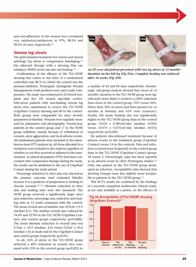
Journal of Wound Care / Urgo Evidence Review, March 2011 29
and non-adherence to the wound were considered very satisfactory/satisfactory in 97%, 98.5% and 98.2% of cases, respectively.150
Venous leg ulcersThe gold standard treatment for venous and mixed aetiology leg ulcers is compression bandaging,151
but adjuvant therapy with a dressing that can rebalance MMPs levels can also aid healing.152
Confirmation of the efficacy of the TLC-NOSF dressing first comes in the form of a randomised controlled trial (RCT) in which the control was the protease-inhibitor, Promogran (Systagenix Wound Management); both products were used under com-pression. The study was conducted in 22 French hos-pitals and five UK wound specialist centres. Fifty-seven patients with non-healing venous leg ulcers were randomised to receive the TLC-NOSF (UrgoStart Contact) dressing and 60 to the control. Both groups were comparable for ulcer severity parameters at baseline. Wounds were regularly meas-ured by planimetry and photography. Twenty-four patients in the control group and 17 in the NOSF group withdrew, mainly because of withdrawal of consent, ulcer aggravation, and local adverse events. All randomised patients were included in the inten-tion-to-treat (ITT) analysis (ie, all those allocated to a treatment were included in the analysis regardless of whether or not they received or adhered to the inter-vention). As almost all patients (93%) had been con-cordant with compression therapy during the study, the results can be attributed to the use of UrgoStart Contact during the study period.
Percentage reduction in ulcer area was selected as the primary outcome (and evaluated blindly) because it is a predictor of progression to healing in chronic wounds.8,153,154 Absolute reduction in ulcer area and healing rates were also measured. The NOSF group achieved a significantly larger ulcer area reduction, percentage area reduction and heal-ing rates at 12 weeks compared with the control. The mean wound area at baseline was 10.9cm2 ± 9.3 (median 8.1). The median wound area reduced by 54.4% and 12.9% in the TLC-NOSF (UrgoStart Con-tact) and control groups respectively (p=0.0286). The mean absolute reduction in wound area was 2.3cm2 ± 10.2 (median: 4.2) versus 0.2cm2 ± 10.4 (median 1.0) at study end for the UrgoStart Contact and control groups respectively (p=0.01).
In all, 56% of ulcers in the TLC-NOSF group achieved a 40% reduction in wound area com-pared with 35% in the control group (p=0.022) in
An 87-year-old patient presented with two leg ulcers of 12 months’ duration on her left leg (Fig 25a). Complete healing was achieved after 16 weeks (Fig 25b)
a median of 42 and 84 days respectively. Interest-ingly, sub-group analysis showed that ulcers of >6 months’ duration in the TLC-NOSF group were sig-nificantly more likely to achieve a ≥40% reduction than those in the control group: 55% versus 26%. (More than 56% of ulcers had been present for >6 months at baseline and 61% were recurrent.) Finally, the mean healing rate was significantly higher in the TLC-NOSF group than in the control group: -0.016 ± 0.285cm2/day (median -0.056) versus +0.075 ± 0.475cm2/day (median -0.015) respectively (p=0.029).
Six patients discontinued treatment because of adverse events in the treatment group (UrgoStart Contact) versus 14 in the controls. Pain and infec-tion occurred more frequently in the control group than in the TLC-NOSF (UrgoStart Contact) group: 18 versus 5. Interestingly, pain has been reported as an adverse event in other Promogran studies.152 Only one patient in the TLC-NOSF group devel-oped an infection. Acceptability data showed that dressing changes were also slightly more accepta-ble to patients in the TLC-NOSF group.155
This RCT’s results are confirmed by the findings of a recently completed multicentre clinical study, as yet only available as a poster, on the efficacy of
25a 25b
0.0
0.2
0.4
0.6
0.8
1.0
Easy toremove
Noadherence
to thewound
Nobleeding
at removal
No painat
dressingchange
Fig 26. Acceptability of TLC-NOSF dressing (UrgoStart Contact)155
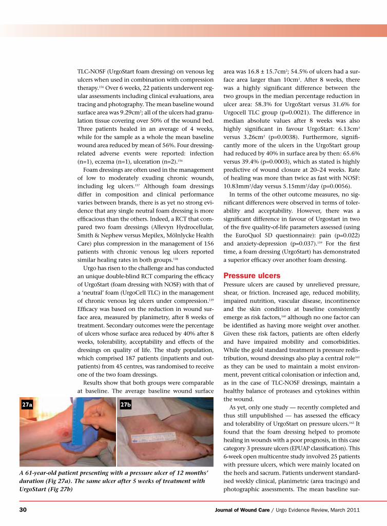
30 Journal of Wound Care / Urgo Evidence Review, March 2011
TLC-NOSF (UrgoStart foam dressing) on venous leg ulcers when used in combination with compression therapy.156 Over 6 weeks, 22 patients underwent reg-ular assessments including clinical evaluations, area tracing and photography. The mean baseline wound surface area was 9.29cm2; all of the ulcers had granu-lation tissue covering over 50% of the wound bed. Three patients healed in an average of 4 weeks, while for the sample as a whole the mean baseline wound area reduced by mean of 56%. Four dressing-related adverse events were reported: infection (n=1), eczema (n=1), ulceration (n=2).156
Foam dressings are often used in the management of low to moderately exuding chronic wounds, including leg ulcers.157 Although foam dressings differ in composition and clinical performance varies between brands, there is as yet no strong evi-dence that any single neutral foam dressing is more efficacious than the others. Indeed, a RCT that com-pared two foam dressings (Allevyn Hydrocellular, Smith & Nephew versus Mepilex, Mölnlycke Health Care) plus compression in the management of 156 patients with chronic venous leg ulcers reported similar healing rates in both groups.158
Urgo has risen to the challenge and has conducted an unique double-blind RCT comparing the efficacy of UrgoStart (foam dressing with NOSF) with that of a ‘neutral’ foam (UrgoCell TLC) in the management of chronic venous leg ulcers under compression.159 Efficacy was based on the reduction in wound sur-face area, measured by planimetry, after 8 weeks of treatment. Secondary outcomes were the percentage of ulcers whose surface area reduced by 40% after 8 weeks, tolerability, acceptability and effects of the dressings on quality of life. The study population, which comprised 187 patients (inpatients and out-patients) from 45 centres, was randomised to receive one of the two foam dressings.
Results show that both groups were comparable at baseline. The average baseline wound surface
area was 16.8 ± 15.7cm2; 54.5% of ulcers had a sur-face area larger than 10cm2. After 8 weeks, there was a highly significant difference between the two groups in the median percentage reduction in ulcer area: 58.3% for UrgoStart versus 31.6% for Urgocell TLC group (p=0.0021). The difference in median absolute values after 8 weeks was also highly significant in favour UrgoStart: 6.13cm2 versus 3.26cm2 (p=0.0038). Furthermore, signifi-cantly more of the ulcers in the UrgoStart group had reduced by 40% in surface area by then: 65.6% versus 39.4% (p=0.0003), which as stated is highly predictive of wound closure at 20–24 weeks. Rate of healing was more than twice as fast with NOSF: 10.83mm2/day versus 5.15mm2/day (p=0.0056).
In terms of the other outcome measures, no sig-nificant differences were observed in terms of toler-ability and acceptability. However, there was a significant difference in favour of Urgostart in two of the five quality-of-life parameters assessed (using the EuroQuol 5D questionnaire): pain (p=0.022) and anxiety-depression (p=0.037).159 For the first time, a foam dressing (UrgoStart) has demonstrated a superior efficacy over another foam dressing.
Pressure ulcersPressure ulcers are caused by unrelieved pressure, shear, or friction. Increased age, reduced mobility, impaired nutrition, vascular disease, incontinence and the skin condition at baseline consistently emerge as risk factors,160 although no one factor can be identified as having more weight over another. Given these risk factors, patients are often elderly and have impaired mobility and comorbidities. While the gold standard treatment is pressure redis-tribution, wound dressings also play a central role161 as they can be used to maintain a moist environ-ment, prevent critical colonisation or infection and, as in the case of TLC-NOSF dressings, maintain a healthy balance of proteases and cytokines within the wound.
As yet, only one study — recently completed and thus still unpublished — has assessed the efficacy and tolerability of UrgoStart on pressure ulcers.162 It found that the foam dressing helped to promote healing in wounds with a poor prognosis, in this case category 3 pressure ulcers (EPUAP classification). This 6-week open multi centre study involved 25 patients with pressure ulcers, which were mainly located on the heels and sacrum. Patients underwent standard-ised weekly clinical, planimetric (area tracings) and photographic assessments. The mean baseline sur-
A 61-year-old patient presenting with a pressure ulcer of 12 months’ duration (Fig 27a). The same ulcer after 5 weeks of treatment with UrgoStart (Fig 27b)
27a 27b
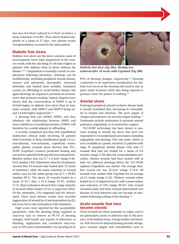
Journal of Wound Care / Urgo Evidence Review, March 2011 31
face area of 6.56cm2 reduced to 4.19cm2 at week 6, a mean reduction of 43.8%. Three ulcers healed com-pletely in a mean of 27 days. Two adverse events (overgranulation) occurred in the same patient.
Diabetic foot ulcersDiabetic foot ulcers are the most common cause of non-traumatic lower limb amputation in the west-ern world, with the risk being 15–46 times higher in patients with diabetes than in those without the disease.163,164 Amputation is normally a result of com-plications following ulceration. Aetiology can be multifactorial, including peripheral arterial disease, sensory and autonomic neuropathy, structural deformity and limited joint mobility. Treatment centres on offloading to avoid further trauma, but again dressings are required to promote an environ-ment that promotes healing. Indeed, biopsies have shown that the concentration of MMPs is up to 65-fold higher in diabetic foot ulcers than in trau-matic wounds, with MMP-2 and MMP-9 being six- and 14-fold higher respectively.165
A dressing that can inhibit MMPs, and thus rebalance the relationship between MMPs and tissue inhibitors of metalloproteinases (TIMPs) will therefore be a useful adjunct to offloading.
A recently completed and thus still unpublished multicentre clinical study involving 34 patients with University of Texas classification grade 1A (i.e. non-infected, non-ischaemic, superficial) neuro-pathic plantar wounds ulcers showed that TLC-NOSF (UrgoStart Contact) promoted healing and was well accepted by both practitioners and patients. Baseline surface area was 2.7 ± 2.4cm2 (range 0.46–8.63, median 1.85). Planimetric data (for 32 patients) showed that 10 wounds had healed after 12 weeks of treatment, while the mean reduction in wound surface area for the entire group was 62.7 ± 49.8% (median 82.7). The above 10 wounds healed in a mean of 61.7 days ± 21.4 (range 31–91, median 57.5). Final evaluation showed that a large majority of wounds either healed (31%) or improved (50%). Of the remainder, 13% stagnated and 6% deterio-rated. Three local adverse events were recorded (aggravation of wound [n=1] and maceration [n=2]), but none led to discontinuation with treatment.
High scores were reported for the acceptability parameters, with the dressing being regarded as easy/very easy to remove at 99.7% of dressing changes, with hardly any reports of adherence or bleeding. Application was considered easy/very easy in 96% and conformability very good/good in
85% of dressing changes, respectively.166 Dressing conformity is an important consideration for dia-betic foot ulcers as the dressing will need to stay in place under footwear while also being exposed to pressure when the patient is walking.166
Arterial ulcersProlonged peripheral arterial occlusive disease leads to poorly nourished skin, increasing its vulnerabil-ity to trauma and infection. The poor supply of oxygen and nutrients can severely impair healing.167 Treatments include medication to promote arterial perfusion, angioplasty or reconstructive surgery.
TLC-NOSF technology has been shown to pro-mote healing in arterial leg ulcers that have not responded to revascularisation procedures including angioplasty and stenting. This case series, which as yet is available as a poster, involved 15 patients with stage IV peripheral arterial disease who had 17 wounds that had not healed for a mean of 9.8 months (range 2–34) after the revascularisation pro-cedure. Sixteen wounds had been treated with at least two different dressings before the TLC-NOSF regimen (UrgoStart) was started. The average base-line wound size was 22.5cm2 (range 3–74.6) All wounds were treated with UrgoStart for an average of 6.5 weeks (range 2–14). Thirteen wounds either healed (n=7) or improved (n=6), with a mean surface area reduction of 72% (range 49–91). One wound remained static and three wounds deteriorated: two because of local infection and one because of mac-eration following cutting of the dressing.168
Acute wounds that have become chronicThese wounds are often traumatic in origin, and so are particularly prone to infection due to the pres-ence of devitalised tissue, foreign bodies and bacte-ria. Risk factors for delayed healing include old age, poor vascular supply and comorbidities such as
Diabetic foot ulcer (Fig 28a). Healing was achieved after 10 weeks with UrgoStart (Fig 28b)
28a 28b
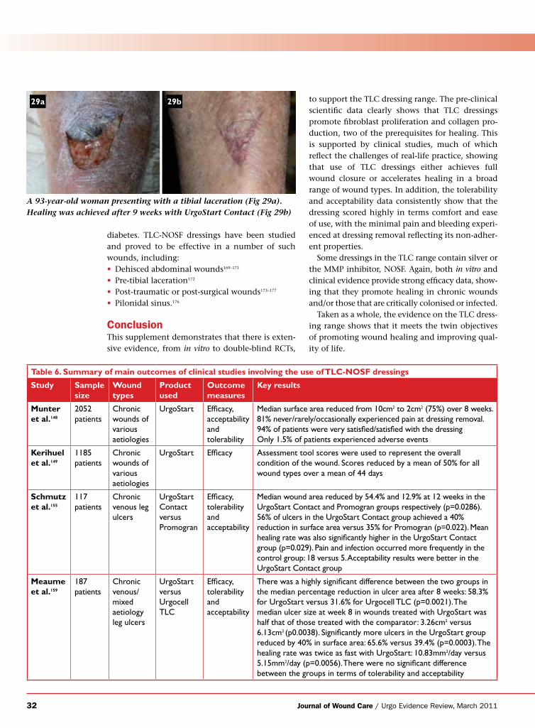
32 Journal of Wound Care / Urgo Evidence Review, March 2011
diabetes. TLC-NOSF dressings have been studied and proved to be effective in a number of such wounds, including:• Dehisced abdominal wounds169–171 • Pre-tibial laceration172
• Post-traumatic or post-surgical wounds173–177
• Pilonidal sinus.176
ConclusionThis supplement demonstrates that there is exten-sive evidence, from in vitro to double-blind RCTs,
to support the TLC dressing range. The pre-clinical scientific data clearly shows that TLC dressings promote fibroblast proliferation and collagen pro-duction, two of the prerequisites for healing. This is supported by clinical studies, much of which reflect the challenges of real-life practice, showing that use of TLC dressings either achieves full wound closure or accelerates healing in a broad range of wound types. In addition, the tolerability and acceptability data consistently show that the dressing scored highly in terms comfort and ease of use, with the minimal pain and bleeding experi-enced at dressing removal reflecting its non-adher-ent properties.
Some dressings in the TLC range contain silver or the MMP inhibitor, NOSF. Again, both in vitro and clinical evidence provide strong efficacy data, show-ing that they promote healing in chronic wounds and/or those that are critically colonised or infected.
Taken as a whole, the evidence on the TLC dress-ing range shows that it meets the twin objectives of promoting wound healing and improving qual-ity of life.
A 93-year-old woman presenting with a tibial laceration (Fig 29a). Healing was achieved after 9 weeks with UrgoStart Contact (Fig 29b)
Table 6. Summary of main outcomes of clinical studies involving the use of TLC-NOSF dressings
Study Sample size
Wound types
Product used
Outcome measures
Key results
Munter et al.148
2052 patients
Chronic wounds of various aetiologies
UrgoStart Efficacy, acceptability and tolerability
Median surface area reduced from 10cm2 to 2cm2 (75%) over 8 weeks. 81% never/rarely/occasionally experienced pain at dressing removal. 94% of patients were very satisfied/satisfied with the dressingOnly 1.5% of patients experienced adverse events
Kerihuel et al.149
1185 patients
Chronic wounds of various aetiologies
UrgoStart Efficacy Assessment tool scores were used to represent the overall condition of the wound. Scores reduced by a mean of 50% for all wound types over a mean of 44 days
Schmutz et al.155
117 patients
Chronic venous leg ulcers
UrgoStart Contact versus Promogran
Efficacy, tolerability and acceptability
Median wound area reduced by 54.4% and 12.9% at 12 weeks in the UrgoStart Contact and Promogran groups respectively (p=0.0286). 56% of ulcers in the UrgoStart Contact group achieved a 40% reduction in surface area versus 35% for Promogran (p=0.022). Mean healing rate was also significantly higher in the UrgoStart Contact group (p=0.029). Pain and infection occurred more frequently in the control group: 18 versus 5. Acceptability results were better in the UrgoStart Contact group
Meaume et al.159
187 patients
Chronic venous/mixed aetiology leg ulcers
UrgoStart versus Urgocell TLC
Efficacy, tolerability and acceptability
There was a highly significant difference between the two groups in the median percentage reduction in ulcer area after 8 weeks: 58.3% for UrgoStart versus 31.6% for Urgocell TLC (p=0.0021). The median ulcer size at week 8 in wounds treated with UrgoStart was half that of those treated with the comparator: 3.26cm2 versus 6.13cm2 (p0.0038). Significantly more ulcers in the UrgoStart group reduced by 40% in surface area: 65.6% versus 39.4% (p=0.0003). The healing rate was twice as fast with UrgoStart: 10.83mm2/day versus 5.15mm2/day (p=0.0056). There were no significant difference between the groups in terms of tolerability and acceptability
29a 29b
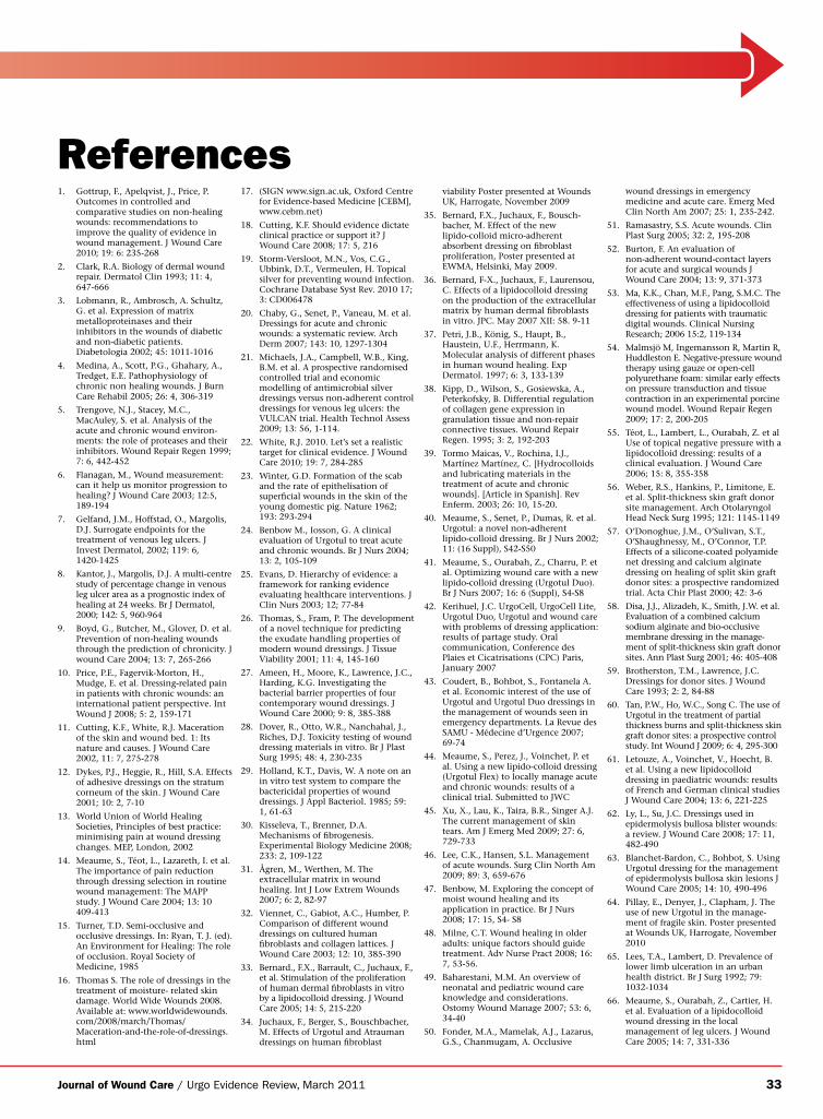
Journal of Wound Care / Urgo Evidence Review, March 2011 33
References1. Gottrup, F., Apelqvist, J., Price, P.
Outcomes in controlled and comparative studies on non-healing wounds: recommendations to improve the quality of evidence in wound management. J Wound Care 2010; 19: 6: 235-268
2. Clark, R.A. Biology of dermal wound repair. Dermatol Clin 1993; 11: 4, 647-666
3. Lobmann, R., Ambrosch, A. Schultz, G. et al. Expression of matrix metalloproteinases and their inhibitors in the wounds of diabetic and non-diabetic patients. Diabetologia 2002; 45: 1011-1016
4. Medina, A., Scott, P.G., Ghahary, A., Tredget, E.E. Pathophysiology of chronic non healing wounds. J Burn Care Rehabil 2005; 26: 4, 306-319
5. Trengove, N.J., Stacey, M.C., MacAuley, S. et al. Analysis of the acute and chronic wound environ-ments: the role of proteases and their inhibitors. Wound Repair Regen 1999; 7: 6, 442-452
6. Flanagan, M., Wound measurement: can it help us monitor progression to healing? J Wound Care 2003; 12:5, 189-194
7. Gelfand, J.M., Hoffstad, O., Margolis, D.J. Surrogate endpoints for the treatment of venous leg ulcers. J Invest Dermatol, 2002; 119: 6, 1420-1425
8. Kantor, J., Margolis, D.J. A multi-centre study of percentage change in venous leg ulcer area as a prognostic index of healing at 24 weeks. Br J Dermatol, 2000; 142: 5, 960-964
9. Boyd, G., Butcher, M., Glover, D. et al. Prevention of non-healing wounds through the prediction of chronicity. J wound Care 2004; 13: 7, 265-266
10. Price, P.E., Fagervik-Morton, H., Mudge, E. et al. Dressing-related pain in patients with chronic wounds: an international patient perspective. Int Wound J 2008; 5: 2, 159-171
11. Cutting, K.F., White, R.J. Maceration of the skin and wound bed. 1: Its nature and causes. J Wound Care 2002, 11: 7, 275-278
12. Dykes, P.J., Heggie, R., Hill, S.A. Effects of adhesive dressings on the stratum corneum of the skin. J Wound Care 2001; 10: 2, 7-10
13. World Union of World Healing Societies, Principles of best practice: minimising pain at wound dressing changes. MEP, London, 2002
14. Meaume, S., Téot, L., Lazareth, I. et al. The importance of pain reduction through dressing selection in routine wound management: The MAPP study. J Wound Care 2004; 13: 10 409-413
15. Turner, T.D. Semi-occlusive and occlusive dressings. In: Ryan, T. J. (ed). An Environment for Healing: The role of occlusion. Royal Society of Medicine, 1985
16. Thomas S. The role of dressings in the treatment of moisture- related skin damage. World Wide Wounds 2008. Available at: www.worldwidewounds.com/2008/march/Thomas/Maceration-and-the-role-of-dressings.html
17. (SIGN www.sign.ac.uk, Oxford Centre for Evidence-based Medicine [CEBM], www.cebm.net)
18. Cutting, K.F. Should evidence dictate clinical practice or support it? J Wound Care 2008; 17: 5, 216
19. Storm-Versloot, M.N., Vos, C.G., Ubbink, D.T., Vermeulen, H. Topical silver for preventing wound infection. Cochrane Database Syst Rev. 2010 17; 3: CD006478
20. Chaby, G., Senet, P., Vaneau, M. et al. Dressings for acute and chronic wounds: a systematic review. Arch Derm 2007; 143: 10, 1297-1304
21. Michaels, J.A., Campbell, W.B., King, B.M. et al. A prospective randomised controlled trial and economic modelling of antimicrobial silver dressings versus non-adherent control dressings for venous leg ulcers: the VULCAN trial. Health Technol Assess 2009; 13: 56, 1-114.
22. White, R.J. 2010. Let’s set a realistic target for clinical evidence. J Wound Care 2010; 19: 7, 284-285
23. Winter, G.D. Formation of the scab and the rate of epithelisation of superficial wounds in the skin of the young domestic pig. Nature 1962; 193: 293-294
24. Benbow M., Iosson, G. A clinical evaluation of Urgotul to treat acute and chronic wounds. Br J Nurs 2004; 13: 2, 105-109
25. Evans, D. Hierarchy of evidence: a framework for ranking evidence evaluating healthcare interventions. J Clin Nurs 2003; 12; 77-84
26. Thomas, S., Fram, P. The development of a novel technique for predicting the exudate handling properties of modern wound dressings. J Tissue Viability 2001; 11: 4, 145-160
27. Ameen, H., Moore, K., Lawrence, J.C., Harding, K.G. Investigating the bacterial barrier properties of four contemporary wound dressings. J Wound Care 2000; 9: 8, 385-388
28. Dover, R., Otto, W.R., Nanchahal, J., Riches, D.J. Toxicity testing of wound dressing materials in vitro. Br J Plast Surg 1995; 48: 4, 230-235
29. Holland, K.T., Davis, W. A note on an in vitro test system to compare the bactericidal properties of wound dressings. J Appl Bacteriol. 1985; 59: 1, 61-63
30. Kisseleva, T., Brenner, D.A. Mechanisms of fibrogenesis. Experimental Biology Medicine 2008; 233: 2, 109-122
31. Ågren, M., Werthen, M. The extracellular matrix in wound healing. Int J Low Extrem Wounds 2007; 6: 2, 82-97
32. Viennet, C., Gabiot, A.C., Humber, P. Comparison of different wound dressings on cultured human fibroblasts and collagen lattices. J Wound Care 2003; 12: 10, 385-390
33. Bernard., F.X., Barrault, C., Juchaux, F., et al. Stimulation of the proliferation of human dermal fibroblasts in vitro by a lipidocolloid dressing. J Wound Care 2005; 14: 5, 215-220
34. Juchaux, F., Berger, S., Bouschbacher, M. Effects of Urgotul and Atrauman dressings on human fibroblast
viability Poster presented at Wounds UK, Harrogate, November 2009
35. Bernard, F.X., Juchaux, F., Bousch-bacher, M. Effect of the new lipido-colloid micro-adherent absorbent dressing on fibroblast proliferation, Poster presented at EWMA, Helsinki, May 2009.
36. Bernard, F-X., Juchaux, F., Laurensou, C. Effects of a lipidocolloid dressing on the production of the extracellular matrix by human dermal fibroblasts in vitro. JPC. May 2007 XII: 58. 9-11
37. Petri, J.B., König, S., Haupt, B., Haustein, U.F., Herrmann, K. Molecular analysis of different phases in human wound healing. Exp Dermatol. 1997; 6: 3, 133-139
38. Kipp, D., Wilson, S., Gosiewska, A., Peterkofsky, B. Differential regulation of collagen gene expression in granulation tissue and non-repair connective tissues. Wound Repair Regen. 1995; 3: 2, 192-203
39. Tormo Maicas, V., Rochina, I.J., Martínez Martínez, C. [Hydrocolloids and lubricating materials in the treatment of acute and chronic wounds]. [Article in Spanish]. Rev Enferm. 2003; 26: 10, 15-20.
40. Meaume, S., Senet, P., Dumas, R. et al. Urgotul: a novel non-adherent lipido-colloid dressing. Br J Nurs 2002; 11: (16 Suppl), S42-S50
41. Meaume, S., Ourabah, Z., Charru, P. et al. Optimizing wound care with a new lipido-colloid dressing (Urgotul Duo). Br J Nurs 2007; 16: 6 (Suppl), S4-S8
42. Kerihuel, J.C. UrgoCell, UrgoCell Lite, Urgotul Duo, Urgotul and wound care with problems of dressing application: results of partage study. Oral communication, Conference des Plaies et Cicatrisations (CPC) Paris, January 2007
43. Coudert, B., Bohbot, S., Fontanela A. et al. Economic interest of the use of Urgotul and Urgotul Duo dressings in the management of wounds seen in emergency departments. La Revue des SAMU - Médecine d’Urgence 2007; 69-74
44. Meaume, S., Perez, J., Voinchet, P. et al. Using a new lipido-colloid dressing (Urgotul Flex) to locally manage acute and chronic wounds: results of a clinical trial. Submitted to JWC
45. Xu, X., Lau, K., Taira, B.R., Singer A.J. The current management of skin tears. Am J Emerg Med 2009; 27: 6, 729-733
46. Lee, C.K., Hansen, S.L. Management of acute wounds. Surg Clin North Am 2009; 89: 3, 659-676
47. Benbow, M. Exploring the concept of moist wound healing and its application in practice. Br J Nurs 2008; 17: 15, S4- S8
48. Milne, C.T. Wound healing in older adults: unique factors should guide treatment. Adv Nurse Pract 2008; 16: 7, 53-56.
49. Baharestani, M.M. An overview of neonatal and pediatric wound care knowledge and considerations. Ostomy Wound Manage 2007; 53: 6, 34-40
50. Fonder, M.A., Mamelak, A.J., Lazarus, G.S., Chanmugam, A. Occlusive
wound dressings in emergency medicine and acute care. Emerg Med Clin North Am 2007; 25: 1, 235-242.
51. Ramasastry, S.S. Acute wounds. Clin Plast Surg 2005; 32: 2, 195-208
52. Burton, F. An evaluation of non-adherent wound-contact layers for acute and surgical wounds J Wound Care 2004; 13: 9, 371-373
53. Ma, K.K., Chan, M.F., Pang, S.M.C. The effectiveness of using a lipidocolloid dressing for patients with traumatic digital wounds. Clinical Nursing Research; 2006 15:2, 119-134
54. Malmsjö M, Ingemansson R, Martin R, Huddleston E. Negative-pressure wound therapy using gauze or open-cell polyurethane foam: similar early effects on pressure transduction and tissue contraction in an experimental porcine wound model. Wound Repair Regen 2009; 17: 2, 200-205
55. Téot, L., Lambert, L., Ourabah, Z. et al Use of topical negative pressure with a lipidocolloid dressing: results of a clinical evaluation. J Wound Care 2006; 15: 8, 355-358
56. Weber, R.S., Hankins, P., Limitone, E. et al. Split-thickness skin graft donor site management. Arch Otolaryngol Head Neck Surg 1995; 121: 1145-1149
57. O’Donoghue, J.M., O’Sulivan, S.T., O’Shaughnessy, M., O’Connor, T.P. Effects of a silicone-coated polyamide net dressing and calcium alginate dressing on healing of split skin graft donor sites: a prospective randomized trial. Acta Chir Plast 2000; 42: 3-6
58. Disa, J.J., Alizadeh, K., Smith, J.W. et al. Evaluation of a combined calcium sodium alginate and bio-occlusive membrane dressing in the manage-ment of split-thickness skin graft donor sites. Ann Plast Surg 2001; 46: 405-408
59. Brotherston, T.M., Lawrence, J.C. Dressings for donor sites. J Wound Care 1993; 2: 2, 84-88
60. Tan, P.W., Ho, W.C., Song C. The use of Urgotul in the treatment of partial thickness burns and split-thickness skin graft donor sites: a prospective control study. Int Wound J 2009; 6: 4, 295-300
61. Letouze, A., Voinchet, V., Hoecht, B. et al. Using a new lipidocolloid dressing in paediatric wounds: results of French and German clinical studies J Wound Care 2004; 13: 6, 221-225
62. Ly, L., Su, J.C. Dressings used in epidermolysis bullosa blister wounds: a review. J Wound Care 2008; 17: 11, 482-490
63. Blanchet-Bardon, C., Bohbot, S. Using Urgotul dressing for the management of epidermolysis bullosa skin lesions J Wound Care 2005; 14: 10, 490-496
64. Pillay, E., Denyer, J., Clapham, J. The use of new Urgotul in the manage-ment of fragile skin. Poster presented at Wounds UK, Harrogate, November 2010
65. Lees, T.A., Lambert, D. Prevalence of lower limb ulceration in an urban health district. Br J Surg 1992; 79: 1032-1034
66. Meaume, S., Ourabah, Z., Cartier, H. et al. Evaluation of a lipidocolloid wound dressing in the local management of leg ulcers. J Wound Care 2005; 14: 7, 331-336
34 Journal of Wound Care / Urgo Evidence Review, March 2011
67. Smith, J., Hill, J., Barrett, S. et al Evaluation of Urgotul plus K-Four compression for venous leg ulcers. Br J Nurs 2004; 13: (Suppl. 6), S20-S28
68. Dereure, O., Vin, F., Lazareth, I., Bohbot, S.J. Compression in peri-ulcer skin in outpatients’ venous leg ulcers: results of a French Survey. J Wound Care 2005; 14: 6, 265-271
69. Fays, S., Schmutz, J.L., Vin, F. et al. Leg ulcers and the UrgoCell Non-Adhesive wound dressing. Br J Nurs 2005; 14: (Suppl. 11), S15–S20
70. Maillard, H. et al. Management of venous or mixed leg ulcers: Efficacy and tolerance of the new Urgocell Adhesive dressing. Abstract Dermatologie, December 2005, number 506
71. Schmutz, J.L., Gobin, J.P., Cartier, H. et al. Leg ulcers and the new micro-adherent absorbent dressing: results of the clinical qualification study. Poster presented at EWMA, Helsinki, May 2009
72. Singh, N., Armstrong, D.G., Lipsky, B.A. Preventing foot ulcers in patients with diabetes. JAMA 2005; 293: 2, 217-228
73. Lavery, L.A., Armstrong, D.G., Murdoch, D.P. et al. Validation of the infectious Diseases Society of America’s diabetic foot infection classification system. Clin Infect Dis 2007; 44: 4, 562-565
74. International Diabetes Federation. Time to Act: Diabetes and foot care. IDF, 2005
75. Frykberg, R.G., Armstrong, D.G., Giurini, J. et al. Diabetic foot disorders: a clinical practice guideline. For the American College of Foot and Ankle Surgeons and the American College of Foot and Ankle Orthopedics and Medicine. J Foot Ankle Surg 2000; 45: (Suppl. 5), S1-60
76. Queen, D., Queen, D., Orsted, H., Sanada,H., Sussman, G. A dressing history. Int Wound J 2004; 1: 1, 59-77
77. Sibbald, R.G., Torrance, G., Hux, M., et al. Cost-effectiveness of becaplermin for non healing neuropathic diabetic foot ulcers. Ostomy Wound Manage 2003; 49: 11, 76-84.
78. Ehrler, S., Salomon, C. [Critical analysis of an observational evaluation of diabetic foot wounds treated with Urgotul]. [Article in French]. Journal des Plaies et Cicatrisations 2007; 13: 58, 114-118
79. Neuwirth, F., Drouot, F., Olsson, N-O. Localised pemphigoid bullosa imitative of a bilateral leg ulcer. Poster presented at 4th Wound Healing Society, Singapore, August 2007
80. Neuwirth, F. Huber, C. Controlled wound healing of an outpatient with a deep caustic burn. Poster presented at 4th Wound Healing Society, Singapore, August 2007
81. Cendras, J., Sparsa, A., Peyrot, I. et al. Use of a contact layer in the treatment of an extensive pyoderma gangrenosum. Poster presented at Conference des Plaies et Cicatrisations (CPC), Paris, January 2005.
82. Steinhoff, M., Ma, H.L., Hensen, P., Luger, T.A. Case reports: Cellosorb and Urgotul in the treatment of pyoderma gangrenosum and chronic venous ulcers. Poster presented at World Union of Wound Healing Societies, Paris, July 2004
83. Padiglione, S., Artaud, F., Fromantin, I. Management of an ulcerated tumour. Poster presented at
Conference des Plaies et Cicatrisa-tions, Paris, January, 2005
84. Bitoun, C., Thirion, V., Vives, F., Bohbot, S. Healing of post-tattoo wounds with the lipidocolloid dressing Urgotul. Poster presented at Conference des Plaies et Cicatrisations (CPC) Paris, January 2006
85. Fenwick, K. Use of the new UrgoCell TLC in the treatment of a degenerat-ing pressure ulcer. Poster presented at Wounds UK, Harrogate, November 2009
86. Dow, G., Browne, A., Sibbald, R.G. Infection in chronic wounds: controversies in diagnosis and treatment. Ostomy Wound Manage 1999; 45: 8, 23-40
87. Casadevall, A., Pirofski, L.A. Host-pathogen interactions: redefining the basic concepts of virulence and pathogenicity. Infect Immun 1999; 67: 8, 3703-3713
88. Trengrove, N.J., Stacey, M.C., McGechie, D.F., Mata, S. Qualitative bacteriology and leg ulcer healing. J Wound Care 1996; 5: 6, 277-280
89. Heinzelmann, M., Scott, M., Lam, T. Factors predisposing to bacterial invasion and infection. Am J Surg 2002; 183: 2, 179-190
90. Cooper, R. Understanding wound infection. In: Cutting, K., Gilchrist, B., Gottrup, F. et al. (eds). Identifying Criteria for Wound Infection. EWMA Position Document, 2005
91. Cutting, K.F., Harding, K.G. Criteria for identifying wound infection. J Wound Care 1994; 3: 4, 196-201;
92. Cutting, K.F., White, R.J., Mahoney, P., Harding, K.G. Clinical identification of wound infection: a Delphi approach. Identifying Criteria for Wound Infection. EWMA Position Document, 2005
93. Wolcott, R.D., Rhoads, D.D., Bennett, M.E. et al. Chronic wounds and the medical biofilm paradigm. J Wound Care 2010; 19: 2, 45-53
94. Wolcott, R.D., Rhoads, D.D., Dowd, S.E. Biofilms and chronic wound inflammation. J Wound Care 2008; 17: 8, 333-341
95. Percival, S., Bowler, P. Biofilms and their potential role in wound healing. Wounds 2004; 16: 7. Available at: http://www.woundsresearch.com/article/2870
96. Drosou, A., Falabella, A., Kirsner, R. Antiseptics on wounds: an area of controversy. Wounds 2003; 15: 5, 149-166
97. Lansdown, A.B. Silver 1: its antibacterial properties and mechanism of action. J Wound Care 2002; 11: 4, 125-130
98. Fraser, J.F., Cuttle, L., Kempf, M., Kimble, R.M. Cytotoxicity of topical antimicrobial agents used in burn wounds in Australia. ANZ J Surg 2004; 74: 3, 139-142
99. Paddle-Ledinek, J.E., Nasa, Z., Cleland, H.J. Effect of different wound dressings on cell viability and proliferation. Plast Reconstr Surg 2006; 117: (Suppl. 7), 110S-118S
100. Lansdown, A.B. Silver 2: toxicity in mammals and how its products aid wound repair. J Wound Care 2002; 11: 5, 173-177
101. White, R., Cutting, K. Exploring the effects of silver in wound manage-ment: what is optimal? Wounds 2006; 18: 11, 307-314
102. Silver, S., Phung, le T. A bacterial view of the periodic table: genes and proteins for toxic inorganic ions. J Ind Microbiol Biotechnol 2005; 32: 11-12, 587-605
103. Percival, S.L., Bowler, P.G., Russell, D. Bacterial resistance to silver in wound care. J Hosp Infect 2005; 60: 1, 1-7
104. Lansdown, A.B., Williams, A. Bacterial resistance to silver in wound care and medical devices. J Wound Care 2007; 16: 1, 15-19
105. Age, B., Condaminet, B., Josse, Ph., et al. IMAg survey: use of a lipido-colloid silver sulphadiazine contact layer and silver absorbent wound dressing. Poster presented at EWMA, Glasgow, May 2007
106. Kerihuel, J.C. How are lipido-colloid dressings used in daily practice? Oral presentation Conference des Plaies et Cicatrisations (CPC) Congress, Paris, January 2006.
107. Kerihuel, J.C. Prevalence of use of silver dressings in the community. Oral communication at Conference des Plaies et Cicatrisations (CPC), Paris, January 2007
108. Neuwirth, C., Martin, M. Is sulphadiazine still active? – results of a bacteriological study and interest of the new lipidocolloid dressing Urgotul SSD. Poster presented at EWMA, Stuttgart, September 2005
109. Burd, A., Kwok, C.H., Hung, S.C. et al. A comparative study of the cytotoxicity of silver-based dressings in monolayer cell, tissue explant, and animal models. Wound Repair Regen 2007; 15: 1, 94–104
110. Kalishwaralal, K., BarathManiKanth, S., Pandian, S.R Silver nanoparticles impeded the biofilm formation by Pseudomonas aeruginosa and Staphylococcus epidermidis. Colloids Surf B Biointerfaces 2010; 79: 2, 340-344
111. Thorn, R.M., Austin, A.J., Greenman, J. et al. In vitro comparison of antimicrobial activity of iodine and silver dressings against biofilms. J Wound Care 2009; 18: 8, 343-346
112. Percival, S.L., Bowler, P., Woods, E.J. Assessing the effect of an antimicrobial wound dressing on biofilms. Wound Repair Regen 2008; 16: 1, 52-57
113. Bjarnsholt, T., Kirketerp-Moller, K., Kristiansen, S. et al. Silver against Pseudomonas aeruginosa biofilms. APMIS 2007; 115: 8, 921-928
114. Percival, S.L., Bowler, P.G., Dolman, J. Antimicrobial activity of silver-con-taining dressings on wound microorganisms using an in vitro biofilm model. Int Wound J 2007; 4: 2, 186-191
115. Akiyama, H., Yamasaki, O., Kanzaki, H. et al. Effects of sucrose and silver on Staphylococcus aureus biofilms. J Antimicrob Chemother 1998; 42: 5, 629-634
116. Quatravaux, S., Rodrigues, S., Desroche, N. et al. Comparison of two methods for the quantification of a bacterial biofilm in the presence of bactericidal agents. Poster presented at EWMA, Lisbon, May 2008.
117. Desroche, N., Rodrigues, S., Quatravaux, S. et al Bactericidal and destructuring effect of antibacterial absorbent dressing on biofilms of S. aureus and P. aeruginosa. Poster presented at EWMA, Helsinki, May 2009
118. Hettiaratchy, S. Dziewulski, P. ABC of Burns. BMJ. 2004; 5: 328, 7452, 1366-1368
119. Muller, M.J., Hollyoak, M.A., Moaveni, Z. et al. Retardation of wound healing by silver sulfadiazine is reversed by aloe vera and nystatin. Burns 2003; 29: 8, 834-836
120. Sweetman, S. (ed). Silver sulfasalazine. In: Martindale: The complete drug reference (33rd edn). Pharmaceutical Press, 2002
121. Duc, Q., Breetveld, M., Middelkoop. E. et al. A cytotoxic analysis of antiseptic medication on skin substitutes and autograft. Br J Dermatol 2007; 157: 1, 33-40
122. Carsin, H., Wasserman, D., Pannier, M. et al. A silver suphadiazine-impreg-nated lipidocolloid dressing to treat second degree burns. J Wound Care 2004; 13: 4, 145-148
123. Muangman, P., Muangman, S., Keorochana, K. et al. Benefit of hydrocolloid SSD dressing in the outpatients management of partial thickness burns. J Med Ass Thai 2009; 92: 10, 1300-1305
124. Jester, I., Böhn, I., Hannmann, T. et al. Comparison of two silver dressings for wound management in pediatric burns. Wounds 2008; 20: 11, 303-308
125. Cendras, J., Sparsa A., Peyrot, I. et al. Interest of Urgotul SSD in skin grafts on necrotic angiodermatitis. Poster presented at World Conference des Plaies et Cicatrisations, Paris, July, 2004
126. Lazareth, I., Priollet, P. Necrotic angiodermatitis: treatment by early cutaneous grafts. Ann Dermatol Venereol 1995; 122: 9, 575-578
127. El Khatib, K., Danino, A., Rzin, A., Malka, G. [Necrotic angiodermatitis: evaluation of an early skin graft treatment]. [Article in French] Ann Chir Plast Esthet 2009; 54: 6, 567-570
128. Lazareth, I., Meaume, S., Sigal-Grin-berg, M. et al. The role of a silver releasing lipido-colloid contact layer in venous leg ulcers presenting inflammatory signs suggesting heavy bacterial colonisation: results of a randomised controlled study. Wounds 2008; 20: 6, 158-166
129. Lazareth, I., Ourabah, Z., Senet, P. et al. Evaluation of a new silver foam dressing in patients with critically colonised venous leg ulcers. J Wound Care 2007; 16: 3, 129-132
130. Giraud, A., Amougay, H., Duprez, D. et al. Care of the wounds in the diabetic foot: experience with local use of Urgotul SSD dressing. Poster presented at Wounds UK, Harrogate, November 2004.
131. Fromantin, I. Observation of Urgocell Silver efficacy in complex wounds. Poster presented at Conference des Plaies et Cicatrisations (CPC), Paris, January 2007
132. Hayes, W. The use of Urgotul SSD on a problematic abdominal wound. Poster presented at Wounds UK, Harrogate, November 2004
133. Thomas, K. Clinical case report illustrating the benefits in treatment with an antibacterial lipido-colloid contact layer and an absorbent dressing. Poster presented at EWMA, Lisbon, May 2008
134. Cauchy, E. Management of frostbite in the extremities. Poster presented at Conference des Plaies et Cicatrisations (CPC), Paris, January 2006
Journal of Wound Care / Urgo Evidence Review, March 2011 35
135. Pourreyron, D. Laigle, M.M., Mauve, C. et al. Use of an absorbent dressing impregnated with silver salts in the treatment of complex, delayed healing post-traumatic wounds. Poster presented at EWMA, Lisbon, May 2008
136. Grenier de Cardenal, D., Salomon, C. Experience of the treatment of post-traumatic necrosis with the hydrocellular absorbent dressing. Poster presented at EWMA, Lisbon, May 2008
137. Neuwirth, F. Use of Urgotul SSD contact layer after treatment of rhinophyma by CO2 laser. Poster presented at 4th Wound Healing Society, Singapore, August 2007
138. Lauer, G., Sollberg, S., Cole, M. Expression and proteolysis of vascular endothelial growth factor is increased in chronic wounds. J Invest Dermatol 2000; 115: 1, 12-18
139. Chen, S.M., Ward, S.I., Olutoye, O.O. et al. Ability of chronic wound fluids to degrade peptide growth factors is associated with increased levels of elastase activity and diminished levels of proteinase inhibitors. Wound Repair Regen 1997; 5: 1, 23-32
140. Veves, A., Sheehan, P., Pham H.T. A randomized, controlled trial of Promogran (a collagen/oxidized regenerated cellulose dressing) vs standard treatment in the management of diabetic foot ulcers. Arch Surg 2002; 137: 7, 822-877
141. Bernard, F.X., Juchaux, F., Laurensou, C. Et al. In vitro effect of a new metalloproteinase inhibitor. Poster presented at EWMA, Lisbon, May 2008
142. Coulomb, B., Couty, L., Fournier, B. et al. Evaluation of the matrix impregnated with NOSF (Nano Oligo Saccharide Factor) in an in vitro dermal reconstruction model. JPC 2008; 63: 8, 54-57
143. Zagnoli, A., Kerihuel, J.C., Dompmartin, A., Thirion, C. Venous leg ulcers regarded as ‘difficult to heal’ and their impact on patient QoL: results of the TRAJECTOIRE Survey, JPC 2008; 8: 63, 34-37
144. Rayment, E.A., Upton, Z., Shooter, G.K. Increased matrix metalloprotein-ase-9 (MMP-9) activity observed in chronic wound fluid is related to the clinical severity of the ulcer. Br J Dermatol 2008; 158: 5, 951-961
145. Ladwig, G.P., Robson, M.C., Liu, R. et al. Ratios of activated matrix metalloproteinase-9 to tissue inhibitor of matrix metalloproteinase-1 in wound fluids are inversely correlated with healing of pressure ulcers. Wound Repair Regen 2002; 10: 1, 26-37
146. Visse, R., Nagase, H. Matrix metalloproteinases and tissue inhibitors of metalloproteinases: structure, function and biochemistry. Circ Res 2003; 92: 8, 627-639
147. Marconi, A., Delmau, C., Gilbert, J.E. et al. Effect of Nano-OligoSaccharide Factor (NOSF) on endothelial cell activity in vitro. Poster presented at EWMA, Helsinki, May 2009
148. Munter, K-C. Urgocell Anwendung-sergebnisse aus ambulanten Einrichtungen. Medizin & Praxis Spezial: Wundheilungsstörungen, 2008, pp. 6-9
149. Kerihuel, J-C. Results of various difficult to heal wounds with UrgoCell Start in daily practice. Oral communication at Conference des Plaies et Cicatrisations (CPC), Paris, January 2009
150. Faucher., N., Genet, M., Philippe A. Prospective trial on the use of UrgoCell Start. Poster presented at Conference des Plaies et Cicatrisations (CPC), Paris, January 2010
151. Fletcher, A., Cullum, N., Sheldon, T.A. A systematic review of compression treatment for venous leg ulcers. BMJ 1997; 315: 7108, 576-580
152. Vin, F., Téot, L., Meaume, S. The healing properties of Promogran in venous leg ulcers. J Wound Care 2002; 11: 9, 335-341
153. Margolis, D.J., Berlin, J.A., Strom, B.L. Which venous leg ulcers will heal with limb compression bandages? Am J Med 2000; 109: 15-19
154. Phillips, T.J., Machado, F., Trout, R. et al. Prognostic indicators of venous ulcers. J Am Acad Dermatol 2000; 43: 4, 627-630
155. Schmutz, J-L., Meaume, S., Fays, S. et al, Evaluation of the nano-oligosac-charide factor lipidocolloid matrix in the local management of venous leg ulcer: results of a randomised, controlled trial Int Wound J 2008; 5: 2, 172-182
156. Leger, P., Vin, F., Paradis, H. et al. Management of venous leg ulcers with a lipidocolloid matrix impregnated with NOSF (nano oligosaccharide factor): results of a clinical study. Poster presented at EWMA, Geneva, May 2010
157. Andersen, K.E., Franken, C.P., Pernille, G.A. et al. A randomized controlled study to compare the effectiveness of two foam dressings in the management of lower leg ulcers. Ostomy Wound Manage 2002; 48: 8, 34-41
158. Franks, P.J., Moody, M., Moffatt, C.J. Randomized trail of two foam dressings in the management of chronic venous ulceration. Wound Rep Reg 2007; 15: 197-202
159. Meaume, S., Cambazard, F., Lok, C. et al. Oral presentation. Conference des Plaies et Cicatrisations (CPC), Paris, January 2011.
160. Cullum, N., Petherick, E. Pressure ulcers. Clinical Evidence. BMJ online 19 Mar 2008. [Available at: clinicalevidence.bmj.com/ceweb/conditions/wnd/1901/1901_back-ground.jsp Last accessed: 12/7/10]
161. European Pressure Ulcer Advisory Panel and National Pressure Ulcer Advisory Panel. Treatment of pressure ulcers: Quick Reference Guide. National Pressure Ulcer Advisory Panel, 2009. [Available at: http://www.epuap.org/guidelines/Final_Quick_Treatment.pdf Last accessed: 8/2/11]
162. Rethore, V., Chikhi, R. Noury, H. et al. Effect of a new metalloprotease inhibitor (NOSF) in the local management of pressure ulcers: results of a clinical study. Poster presented at EWMA, Geneva, May 2010
163. Lavery, L.A., Ashry, H.R., van Houtum, W. et al. Variation in the incidence and proportion of diabetes-related amputation in minorities. Diabetes Care 1996; 19: 48-52
164. Armstrong, D.G., Lavery, L.A., Quebedeaux, T.L. et al. Surgical morbidity and the risk of amputation due to infected puncture wounds in diabetic versus nondiabetic adults. South Med J 1997; 90: 384-389
165. Lobmann, R., Ambrosch, A., Schultz, G. et al. Expression of matrix-metallo-
proteinases and their inhibitors in the wounds of diabetic and non-diabetic patients. Diabetologia 2002; 45: 7, 1011-1016
166. Richard, J.L., Leger, P. Evaluation of the efficacy and safety of a TLC matrix impregnated with NOSF (nano-oli-gosaccharide factor) in the local treatment of diabetic foot ulcers. Oral presentation EWMA, Geneva, 2010
167. Moffatt, C., Martin, R., Smithdale, R. Leg Ulcer Management. Blackwell, 2007
168. Allain, A-A., Lucas, A., Cardon, A. Benefit of the NOSF absorbent lipido-colloid dressing in the treatment of arterial ulcers that are stagnant despite a revascularisation procedure Poster presented at EWMA, Helsinki, May 2009
169. Mason, S. Healing of a dehisced abdominal wound with Urgotul START while improving the patient’s quality of life Poster presented at Wounds UK, Harrogate, November 2009
170. Belcher, J.L. Treatment of a non-healing abdominal wound using Urgotul Start. Poster presented at Wounds UK, Harrogate, November 2010
171. Timmons, J. Evaluating a new foam dressing with a healing accelerator. Wounds UK 2010; 6: 3, 88-92
172. Keane, H. Proactive management of a traumatic injury in an elderly patient with Urgotul START Poster presented at Wounds UK, Harrogate, November 2009
173. Grenier de Cardenal, D. Treatment of delayed healing wounds following a surgical procedure in a plastic and reconstructive surgery department. Poster presented at EWMA, Helsinki, May 2009
174. Grenier de Cadenal, D., Rifler, J-P., Salomon, C. Healing post traumatic wounds with UrgoCell Start. J Comm Nurs 2009; 23: 2, 18-19
175. Masseron, C., Leaute-Labreze, C., Pelissier, P. et al. Local treatment of delayed healing wounds in the wound healing unit Poster presented at EWMA, Helsinki, May 2009
176. Powell, G. The new Start dressing range: Urgotul Start, UrgoCell Start. BJN 2009; 18: 6, S30-S36
177. Rifler, J-P. Management of delayed healing post-traumatic wounds that have become chronic. Poster presented at EWMA, Helsinki, May 2009
178. Harding, K., Morris, H., Patel, G. Healing chronic wounds Br Med J 2002; 324: 160-163
179. Thomas, S. Cost of managing chronic wounds in the UK, with particular emphasis on maggot debridement therapy. J Wound Care 2006; 15: 10, 465-469.
180. Harding, K.G. The future of wound healing. In: Leaper, D.J., Harding, K.J. (eds) Wounds: biology and management. Oxford University Press, 1998
181. White, R. Tissue viability in tomorrow’s NHS. J Wound Care 2008; 17: 3, 97-99
182. Bale, S. Using different designs in wound healing research. Nurse Res 2004; 11: 4, 42-53.
183. Binks, J. Use of new Urgotul in the local treatment of graft sites, donor sites and skin tears. Poster presented at Wounds UK, Harrogate, November 2010
184. Hutchcox, S., Williams, H. Use of new Urgotul in the local treatment of skin tears. Poster presented at Wounds UK, Harrogate, November 2010
185. Rifler, J.P. Emergency department treatment of low-exuding traumatic wounds, sutured or otherwise, with a ready-to-apply lipido-colloid dressing. Poster presented at EWMA, Helsinki, May 2009
186. Hailon, S. Healing prognostic wounds: self-treatment by patients. Poster presented at Conference des Plaies et Cicatrisations (CPC) Paris, January 2007.
187. Cartier, H., Granel-Brocard, F., Schmutz, J.L. Leg ulcers and UrgoCell dressing: results of a multicentre clinical study. Journal des Plaies et Cicatrisations 2003: 8: 38, 37-42.
188. Khan, C., Ailleres, P., Marie, M. Clinical experience of Urgotul SSD: ambulatory treatment of superficial burns in adults in an emergency department. Poster presented Conference des Plaies et Cicatrisations (CPC), Paris, January 2006
189. Candito, M., Sansey, A., Reihes, L. et al. Interest in a lipido-colloid contact layer impregnated with NOSF (inhibitor of matricial metallopro-teases) in the local treatment of diabetic foot sores. Poster presented at EWMA, Geneva, May 2010
190. Gardner, S. The use of a new two-in-one treatment (K Two Start) in a venous leg ulcer. Br J Comm Nurs 2009; 14: (Suppl. 12), S22-S25.
191. Le Guyadec, T., Bagan, P. Benefit of the new NOSF absorbent lipido-col-loid dressing in the management of chronic skin ulcerations with a delayed healing prognosis. Poster presented at EWMA, Helsinki, May 2009.
192. Tadej, M., Hampton, S., Nicot, D. Treatment of leg ulcers with a new Soft-Adherent foam dressing with TLC-NOSF (UrgoCell Start TLC). Poster presented at EWMA, Geneva, May 2010.
193. Hoffman, J.J. Local treatment of pressure ulcers in a specialist centre for multiple sclerosis patients: experience with the NOSF absorbent lipidocolloid dressing. Poster presented at EWMA, Helsinki, May 2009
194. Martin, E., Mojallal, A., Lambert, A., Braye, F. Experience of the NOSF absorbent lipido-colloid dressing in the local management of pressure ulcers. Poster presented at EWMA, Helsinki, May 2009.
195. Gaston, J., Durox, H., Sparsa, A. Benefit of the NOSF absorbent lipido-colloid dressing in the treatment of perforating ulcers of the foot. Poster presented at EWMA, Helsinki, May 2009
196. Peistel, F. et al. Treatment of delayed healing arterial ulcers with new NOSF lipidocolloid dressing. Poster presented at EWMA, Lisbon, May 2008.
197. Charlate, F. et al. Treatment of arterial wounds in amputee patients undergoing physiatrics and rehabilitation using the NOSF absorbent lipidocolloid dressing. Poster presented at EWMA, Geneva, 2010.

Copyright licensed to Urgo Medical Ltd for 12 months from
8th March 2011 to 7th March 2012
MA Healthcare Ltd, St Jude’s Church, Dulwich Road, London SE24 0PB, UK
Tel: +44 (0)20 7738 5454 Web: www.markallengroup.com