Supplementary Information · clearly identified in the genomes of lobed fin fish (Sarcopterigii as...
21
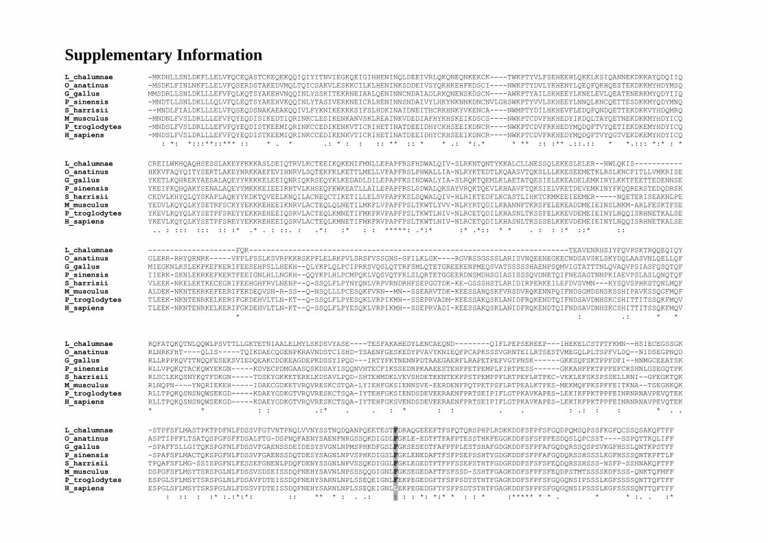
Supplementary Information L_chalumnae -MKDHLLSNLDKFLLELVFQCEQASTCKEQEKQQIQIYITNVIEGKQEIGIHHENINQLDEEIVRLQKQNEQNKEKCK----TWKPTYVLFSEHEEHLQKELKSIQANNEKDKKAYQDQIIQ O_anatinus -MSDKLFINLNKFLLELVFQSERDSTAKEDVMQLTQICSAKVLESKKCILKLHENINKSDDEIVSYQKHKEHFKDSCI----NWKPTYDVLYKHENYLQEQFQKHQESTEKDKKMYHDYMSQ G_gallus MMSDRLLSNLDKLLLEFVFQLKQTSYAKEHVNQQINLYSSKITEKRNEIARLQENINNCNDAIADLRKQNENSKDSCN----AWKPTYAILSKHEEYLKNELEVLQEATENERKMYQDYIIQ P_sinensis -MNDTLLSNLDKLLLQLVFQLEQTSYAKEHVKQQINLYTASIVERKNEICRLHENINNSNDAIVYLHKYNKNNKDNCNVLGHSWKPTYVVLSKHEEYLNNQLKNCQETTESDKKMYQDYMNQ S_harrisii --MNDLFIALDKLLLELVFQSEQDSNAKAEAKQQIVLFYKNIKEKKKSIFSLHDKINAIDNEITHCRKHNKYVKENCA----NWMPTYDILHKHEVFLEDQFQNDQETTEKDKKVYHDQMRQ M_musculus -MNDNLFVSLDRLLLEFVFQYEQDISIKEDTIQRINKCLESIKENKANVSKLREAINKVDEDIAFHYKHSKEIKDSCS----NWKPTCDVFHKHEDYIKDQLTAYQETNEKDKKMYHDYICQ P_troglodytes -MNDSLFVSLDRLLLEFVFQYEQDISTKEEMIQRINKCCEDIKENKVTICRIHETINATDEEIDHYCKHSEEIKDNCR----NWKPTCDVFRKHEDYMQDQFTVYQETIEKDKEMYHDYICQ H_sapiens -MNDSLFVSLDRLLLEFVFQYEQDISTKEEMIQRINKCCEDIKENKVTICRIHETINATDEEIDHYCKHSEEIKDNCR----NWKPTCDVFRKHEDYMQDQFTVYQGTVEKDKEMYHDYICQ : *: *:::**::*** :: * . * .: * : : :: ** :: * * .: *:.* * ** :: :** .::.:: * *.::: *:* : * L_chalumnae CREILWKHQAQHSESSLAKEYFKKKASLDEIQTRVLKCTEEIKQKENIFMNLLEPAPFRSFHDWALQIV-SLRKNTQNTYKKALCLLNESSQLEKESLELER--NWLQKIS----------- O_anatinus HKKVFAQYQITYSERTLAKEYNRKKAEFEVIHNRVLSQTEKFKLKETTLMELLVPAPFRSLPHWALLIA-NLRYKTEDTLKQAASVTQKSLLLKKESEEMETKLRSLKNCFITLLVMKRISE G_gallus YKETLKQHREKYAESALAQEYYKKKKELEEIQNRIQKRSEQYKLKEDADLDILEPAPFKSINDWALYIA-SLRQKTQEMLKLAETATQESIELEKEADELEMKINYLKKTFEETTEDENNSE P_sinensis YKEIFKQHQAKYSENALAQEYYMKKKEIEEIRNTVLKHSEQFKWKEATLLAILEPAPFRSLSDWALQKSAYVRQKTQEVLKHAAVFTQKSIELVKETDEVEMKINYFKQQRERSTEDQDRSK S_harrisii CKDVLKHYQLQYSKAPLAQKYYKDKTQVEELKNQILACNEQCTIKETILLELSVPAPFKSLSQWALQIV-HLRIKTEDFLKCASTLIHKTCKMKEEIEEMER-----NQETERISEAKNLPE M_musculus YEDVLKQYQLKYSETRFSCKYYEKKKEHEEIKNRVLACTEQLQLNETILMKFLVPAPFPSLTKWTLYVV-NLRYRTQDILKRANNFTKRSFELEKEADDMEIEINSLNKM-ARLFESKTFSE P_troglodytes YKEVLKQYQLKYSETPFSREYYEKKREHEEIQSRVLACTEQLKMNETIFMKFRVPAPFPSLTKWTLNIV-NLRCETQDILKHASNLTKSSFELKKEVDEMEIEINYLNQQISRHNETKALSE H_sapiens YKEVLKQYQLKYSETPFSREYYEKKREHEEIQSRVLACTEQLKMNETIFMKFRVPAPFPSLTKWTLNIV-NLRCETQDILKHASNLTKSSSELKKEVDEMEIEINYLNQQISRHNETKALSE .. : ::: ::: :: :* .* . : ::. : .*: :* : : *****: .*:* :* .*:: * * . : : :* ::* :: L_chalumnae --------------------FQK-------------------------------------------------------------------------TEAVENRHSIYFQVPSKTRQQEQIQY O_anatinus GLERR-RRYQRNRK-----VFPLFSSLKSVRPKKRSKPFLELRKPVLSRSFVSSGNS-GFILKLGK----RGVRSSGSSSLARISVNQEENEGKECNDSAVSKLSKYDQLAASVNLQELLQF G_gallus MIEGKNLKSLEKPKEFKERIFEESEHPSLLHEKH--QLYKPLQLPCIPRKSVQSLQTTRFSMLQTETGREEKENPMEQSVATSSSSSHAENPSQMVIGTATTTNLQVAQVPSIASFQSQTQF P_sinensis IIERK-SKNLEKRKEFKERTFEEIGNLHLLNGKH--QQYKPLHLPCMPQKLVQSVQTFRLSLQRTETGGEERDNSMDHSGIASISSSQVDNETQIFNESAGTNNPKIAEVPSLASLQNQTQF S_harrisii VLEEK-NKELEKTKECEGRIFEEHGHFRVLNENP--Q-SSQLFLPYNYQNLVRPVRNDRHFSEPGGTDK-KE-GSSSHSTLARIDIRPERKEILEFDVSVMN---KYSQVSPHKSTQNLMQF M_musculus ALDEK-NKNTEKRKEFEERIFEKDEQVSN-R-SS--Q-NSQLLLPCESQKFVRN--MN--SSEARVTDK-KEESSANQSKFVRSDVRQKENNPQIFNDSGMDSNSKSSHIPAVKSSQGFMQF P_troglodytes TLEEK-NKNTENRKELKERIFGKDEHVLTLN-KT--Q-SSQLFLPYESQKLVRPIKMH--SSEPRVADM-KEESSAKQSKLANIDFRQKENDTQIFNDSAVDNHSKCSHITTITSSQKFMQV H_sapiens TLEEK-NKNTENRKELKERIFGKDEHVLTLN-KT--Q-SSQLFLPYESQKLVRPIKMH--SSEPRVADI-KEESSAKQSKLANIDFRQKENDTQIFNDSAVDNHSKCSHITTITSSQKFMQV * : .: * * L_chalumnae RQFATQKQTNLQQWLPSVTTLLGKTETNIAALELMYLSKDSVYASE----TESFAKAHEDYLENCAEQND--------QIFLPEPSEREEP---IHEKELCSTPTFKMN--HSIECEGSSGK O_anatinus RLNRKFHT----QLIS-----TQIKDAECQDENPKRAVNDSTCISHD-TSAENFGESKEDYPVAVYKNIEQFPCAPKSSSVGRNTEILRTSESTVMEGQLPLTSPFVLDQ--NIDSEGPNQD G_gallus RLLRPPKQVTTNQQFESEKSVIEDQEAKCDDKEAGDEPKDSSYIPQD---IRTYFKTNENNPDTAAEGAERFLRAPETPEFVGTPNSK------GKKSQFSKTPPFDFI--HNMGCEEATSK P_sinensis RLLVPQKQTACKQWYEKGN-----KDVECPDMGAASQSKDSAYISQQNVHTECFIKSSEDNPKAAEESTEHFPETPEMPLFIRTPESS------GKKAHFFKTPPFEFCRSHNLGSEGQTPK S_harrisii RLSCLEKQSNYKQTFDKGN-----TDSEYGKKETERELKDSAVLPQD-SHTEHMDKLVEVSHDETEENTEKFPSTPEMPPFLRTPEFLRTPEC-VEKLEFSKSPSSELLRNI--GFEGKTQK M_musculus RLNQPN----YNQRIEKEH-----IDAECGDKETVRQVRESKCSTQA-LYIEHFGKSIENNSVE-EERDENFPQTPETPSFLRTPEALKTPES-MEKMQFPKSPFFEITKNA--TSEGHKQK P_troglodytes RLLTPQKQSNSNQWSEKGD-----KDAEYGDKGTVRQVRESKCTSQA-IYTEHFGKSIENDSDEVEERAENFPRTSEIPIFLGTPKAVKAPES-LEKIKFPKTPPFEINRNRNAVPEVQTEK H_sapiens RLLTPQKQSNSNQWSEKGD-----KDAEYGDKGTVRQVRESKCTSQA-IYTEHFGKSVENDSDEVEERAENFPRTSEIPIFLGTPKAVKAPES-LEKIKFPKTPPFEINRNRNAVPEVQTEK * * : : .:* . . : * : : . . : .: : : * . . L_chalumnae -STPFSFLMASTPKTPDFNLFDSSVFGTVNTPNQLVVNYSSTNQDQANPQEETESTFDRAQGEEEFTFSFQTQRSPHPLRDKKDDFSFPFSFGQDPQMSQPSSFKGFQCSSQSAKQFTFF O_anatinus ASPTIPFFLTSATQSPGFSFFDSALFTG-DSPNQFAENYSAENFNRGSSQKDIGDLFGKLE-EDTFTFAFPTESSTHKFEGGKDDFSFSFPFESDQSLQPCSST----SSPQTTKQLIFF G_gallus -SPAFFSLLGITQKSPGFNLFDSSVFGAENSSDEIDESYSVGNLNPMSPHKDFGSLFGKSESEDTFAFPFPLESTSHAFGDGKDDFSFPFAFGQDQRSSQSPSVKGFHSSLQNTKPSTFF P_sinensis -SPAFSFLMACTQKSPGFNLFDSSVFGAENSSDQTDESYSAGNLNPVSPHKDIGSLFGKLENEDAFTFSFPSEPSSHTYGDGKDDFSFPFAFGQDQRSSHSSSLKGFHSSSQNTKPFTLF S_harrisii TPQAFSFLMG-SSISPGFNLFESSEFGNENLPDQFDENYSSGNLNPVSSQKDIGGLFGKLEGEDTFTFPFSSEPSTHTFGDGRDDFSFSFSFEQDQRSSHSSS-NSFP-SSHNAKQFTFF M_musculus DSPGFSFLMSYTSRSPGLNLFDSSVSDSEISSDQFNEHYSAVNLNPSSSQQGIGNLFGKSEGEDAFTFSFSSD-SSHTFGAGKDDFSFPFSFEQDPSTMTSSSSKDFSSS-QNKTQFMFF P_troglodytes ESPGLSFLMSYTSRSPGLNLFDSAVFDTEISSDQFNEHYSARNLNPLSSEQEIGNLFEKPEGEDGFTFSFPSDTSTHTFGAGKDDFSFPFSFGQGQNSIPSSSLKGFSSSSQNTTQFTFF H_sapiens ESPGLSFLMSYTSRSPGLNLFDSSVFDTEISSDQFNEHYSARNLNPLSSEQEIGNLLEKPEGEDGFTFSFPSDTSTHTFGAGKDDFSFPFSFGQGQNSIPSSSLKGFSSSSQNTTQFTFF : :: : :* :.:*:*: :: ** * : . .: : : : *: *:* * : : * :***** * * . * * :. . :*
Transcript of Supplementary Information · clearly identified in the genomes of lobed fin fish (Sarcopterigii as...
Supplementary Information
L_chalumnae -MKDHLLSNLDKFLLELVFQCEQASTCKEQEKQQIQIYITNVIEGKQEIGIHHENINQLDEEIVRLQKQNEQNKEKCK----TWKPTYVLFSEHEEHLQKELKSIQANNEKDKKAYQDQIIQ
O_anatinus -MSDKLFINLNKFLLELVFQSERDSTAKEDVMQLTQICSAKVLESKKCILKLHENINKSDDEIVSYQKHKEHFKDSCI----NWKPTYDVLYKHENYLQEQFQKHQESTEKDKKMYHDYMSQ
G_gallus MMSDRLLSNLDKLLLEFVFQLKQTSYAKEHVNQQINLYSSKITEKRNEIARLQENINNCNDAIADLRKQNENSKDSCN----AWKPTYAILSKHEEYLKNELEVLQEATENERKMYQDYIIQ
P_sinensis -MNDTLLSNLDKLLLQLVFQLEQTSYAKEHVKQQINLYTASIVERKNEICRLHENINNSNDAIVYLHKYNKNNKDNCNVLGHSWKPTYVVLSKHEEYLNNQLKNCQETTESDKKMYQDYMNQ
S_harrisii --MNDLFIALDKLLLELVFQSEQDSNAKAEAKQQIVLFYKNIKEKKKSIFSLHDKINAIDNEITHCRKHNKYVKENCA----NWMPTYDILHKHEVFLEDQFQNDQETTEKDKKVYHDQMRQ
M_musculus -MNDNLFVSLDRLLLEFVFQYEQDISIKEDTIQRINKCLESIKENKANVSKLREAINKVDEDIAFHYKHSKEIKDSCS----NWKPTCDVFHKHEDYIKDQLTAYQETNEKDKKMYHDYICQ
P_troglodytes -MNDSLFVSLDRLLLEFVFQYEQDISTKEEMIQRINKCCEDIKENKVTICRIHETINATDEEIDHYCKHSEEIKDNCR----NWKPTCDVFRKHEDYMQDQFTVYQETIEKDKEMYHDYICQ
H_sapiens -MNDSLFVSLDRLLLEFVFQYEQDISTKEEMIQRINKCCEDIKENKVTICRIHETINATDEEIDHYCKHSEEIKDNCR----NWKPTCDVFRKHEDYMQDQFTVYQGTVEKDKEMYHDYICQ
: *: *:::**::*** :: * . * .: * : : :: ** :: * * .: *:.* * ** :: :** .::.:: * *.::: *:* : *
L_chalumnae CREILWKHQAQHSESSLAKEYFKKKASLDEIQTRVLKCTEEIKQKENIFMNLLEPAPFRSFHDWALQIV-SLRKNTQNTYKKALCLLNESSQLEKESLELER--NWLQKIS-----------
O_anatinus HKKVFAQYQITYSERTLAKEYNRKKAEFEVIHNRVLSQTEKFKLKETTLMELLVPAPFRSLPHWALLIA-NLRYKTEDTLKQAASVTQKSLLLKKESEEMETKLRSLKNCFITLLVMKRISE
G_gallus YKETLKQHREKYAESALAQEYYKKKKELEEIQNRIQKRSEQYKLKEDADLDILEPAPFKSINDWALYIA-SLRQKTQEMLKLAETATQESIELEKEADELEMKINYLKKTFEETTEDENNSE
P_sinensis YKEIFKQHQAKYSENALAQEYYMKKKEIEEIRNTVLKHSEQFKWKEATLLAILEPAPFRSLSDWALQKSAYVRQKTQEVLKHAAVFTQKSIELVKETDEVEMKINYFKQQRERSTEDQDRSK
S_harrisii CKDVLKHYQLQYSKAPLAQKYYKDKTQVEELKNQILACNEQCTIKETILLELSVPAPFKSLSQWALQIV-HLRIKTEDFLKCASTLIHKTCKMKEEIEEMER-----NQETERISEAKNLPE
M_musculus YEDVLKQYQLKYSETRFSCKYYEKKKEHEEIKNRVLACTEQLQLNETILMKFLVPAPFPSLTKWTLYVV-NLRYRTQDILKRANNFTKRSFELEKEADDMEIEINSLNKM-ARLFESKTFSE
P_troglodytes YKEVLKQYQLKYSETPFSREYYEKKREHEEIQSRVLACTEQLKMNETIFMKFRVPAPFPSLTKWTLNIV-NLRCETQDILKHASNLTKSSFELKKEVDEMEIEINYLNQQISRHNETKALSE
H_sapiens YKEVLKQYQLKYSETPFSREYYEKKREHEEIQSRVLACTEQLKMNETIFMKFRVPAPFPSLTKWTLNIV-NLRCETQDILKHASNLTKSSSELKKEVDEMEIEINYLNQQISRHNETKALSE
.. : ::: ::: :: :* .* . : ::. : .*: :* : : *****: .*:* :* .*:: * * . : : :* ::* ::
L_chalumnae --------------------FQK-------------------------------------------------------------------------TEAVENRHSIYFQVPSKTRQQEQIQY
O_anatinus GLERR-RRYQRNRK-----VFPLFSSLKSVRPKKRSKPFLELRKPVLSRSFVSSGNS-GFILKLGK----RGVRSSGSSSLARISVNQEENEGKECNDSAVSKLSKYDQLAASVNLQELLQF
G_gallus MIEGKNLKSLEKPKEFKERIFEESEHPSLLHEKH--QLYKPLQLPCIPRKSVQSLQTTRFSMLQTETGREEKENPMEQSVATSSSSSHAENPSQMVIGTATTTNLQVAQVPSIASFQSQTQF
P_sinensis IIERK-SKNLEKRKEFKERTFEEIGNLHLLNGKH--QQYKPLHLPCMPQKLVQSVQTFRLSLQRTETGGEERDNSMDHSGIASISSSQVDNETQIFNESAGTNNPKIAEVPSLASLQNQTQF
S_harrisii VLEEK-NKELEKTKECEGRIFEEHGHFRVLNENP--Q-SSQLFLPYNYQNLVRPVRNDRHFSEPGGTDK-KE-GSSSHSTLARIDIRPERKEILEFDVSVMN---KYSQVSPHKSTQNLMQF
M_musculus ALDEK-NKNTEKRKEFEERIFEKDEQVSN-R-SS--Q-NSQLLLPCESQKFVRN--MN--SSEARVTDK-KEESSANQSKFVRSDVRQKENNPQIFNDSGMDSNSKSSHIPAVKSSQGFMQF
P_troglodytes TLEEK-NKNTENRKELKERIFGKDEHVLTLN-KT--Q-SSQLFLPYESQKLVRPIKMH--SSEPRVADM-KEESSAKQSKLANIDFRQKENDTQIFNDSAVDNHSKCSHITTITSSQKFMQV
H_sapiens TLEEK-NKNTENRKELKERIFGKDEHVLTLN-KT--Q-SSQLFLPYESQKLVRPIKMH--SSEPRVADI-KEESSAKQSKLANIDFRQKENDTQIFNDSAVDNHSKCSHITTITSSQKFMQV
* : .: * *
L_chalumnae RQFATQKQTNLQQWLPSVTTLLGKTETNIAALELMYLSKDSVYASE----TESFAKAHEDYLENCAEQND--------QIFLPEPSEREEP---IHEKELCSTPTFKMN--HSIECEGSSGK
O_anatinus RLNRKFHT----QLIS-----TQIKDAECQDENPKRAVNDSTCISHD-TSAENFGESKEDYPVAVYKNIEQFPCAPKSSSVGRNTEILRTSESTVMEGQLPLTSPFVLDQ--NIDSEGPNQD
G_gallus RLLRPPKQVTTNQQFESEKSVIEDQEAKCDDKEAGDEPKDSSYIPQD---IRTYFKTNENNPDTAAEGAERFLRAPETPEFVGTPNSK------GKKSQFSKTPPFDFI--HNMGCEEATSK
P_sinensis RLLVPQKQTACKQWYEKGN-----KDVECPDMGAASQSKDSAYISQQNVHTECFIKSSEDNPKAAEESTEHFPETPEMPLFIRTPESS------GKKAHFFKTPPFEFCRSHNLGSEGQTPK
S_harrisii RLSCLEKQSNYKQTFDKGN-----TDSEYGKKETERELKDSAVLPQD-SHTEHMDKLVEVSHDETEENTEKFPSTPEMPPFLRTPEFLRTPEC-VEKLEFSKSPSSELLRNI--GFEGKTQK
M_musculus RLNQPN----YNQRIEKEH-----IDAECGDKETVRQVRESKCSTQA-LYIEHFGKSIENNSVE-EERDENFPQTPETPSFLRTPEALKTPES-MEKMQFPKSPFFEITKNA--TSEGHKQK
P_troglodytes RLLTPQKQSNSNQWSEKGD-----KDAEYGDKGTVRQVRESKCTSQA-IYTEHFGKSIENDSDEVEERAENFPRTSEIPIFLGTPKAVKAPES-LEKIKFPKTPPFEINRNRNAVPEVQTEK
H_sapiens RLLTPQKQSNSNQWSEKGD-----KDAEYGDKGTVRQVRESKCTSQA-IYTEHFGKSVENDSDEVEERAENFPRTSEIPIFLGTPKAVKAPES-LEKIKFPKTPPFEINRNRNAVPEVQTEK
* * : : .:* . . : * : : . . : .: : : * . .
L_chalumnae -STPFSFLMASTPKTPDFNLFDSSVFGTVNTPNQLVVNYSSTNQDQANPQEETESTFDRAQGEEEFTFSFQTQRSPHPLRDKKDDFSFPFSFGQDPQMSQPSSFKGFQCSSQSAKQFTFF
O_anatinus ASPTIPFFLTSATQSPGFSFFDSALFTG-DSPNQFAENYSAENFNRGSSQKDIGDLFGKLE-EDTFTFAFPTESSTHKFEGGKDDFSFSFPFESDQSLQPCSST----SSPQTTKQLIFF
G_gallus -SPAFFSLLGITQKSPGFNLFDSSVFGAENSSDEIDESYSVGNLNPMSPHKDFGSLFGKSESEDTFAFPFPLESTSHAFGDGKDDFSFPFAFGQDQRSSQSPSVKGFHSSLQNTKPSTFF
P_sinensis -SPAFSFLMACTQKSPGFNLFDSSVFGAENSSDQTDESYSAGNLNPVSPHKDIGSLFGKLENEDAFTFSFPSEPSSHTYGDGKDDFSFPFAFGQDQRSSHSSSLKGFHSSSQNTKPFTLF
S_harrisii TPQAFSFLMG-SSISPGFNLFESSEFGNENLPDQFDENYSSGNLNPVSSQKDIGGLFGKLEGEDTFTFPFSSEPSTHTFGDGRDDFSFSFSFEQDQRSSHSSS-NSFP-SSHNAKQFTFF
M_musculus DSPGFSFLMSYTSRSPGLNLFDSSVSDSEISSDQFNEHYSAVNLNPSSSQQGIGNLFGKSEGEDAFTFSFSSD-SSHTFGAGKDDFSFPFSFEQDPSTMTSSSSKDFSSS-QNKTQFMFF
P_troglodytes ESPGLSFLMSYTSRSPGLNLFDSAVFDTEISSDQFNEHYSARNLNPLSSEQEIGNLFEKPEGEDGFTFSFPSDTSTHTFGAGKDDFSFPFSFGQGQNSIPSSSLKGFSSSSQNTTQFTFF
H_sapiens ESPGLSFLMSYTSRSPGLNLFDSSVFDTEISSDQFNEHYSARNLNPLSSEQEIGNLLEKPEGEDGFTFSFPSDTSTHTFGAGKDDFSFPFSFGQGQNSIPSSSLKGFSSSSQNTTQFTFF
: :: : :* :.:*:*: :: ** * : . .: : : : *: *:* * : : * :***** * * . * * :. . :*
Supplementary Figure 1. Sequence alignment. Sequence alignment of SIX6OS1
homologues in vertebrates. Amino acid sequences of H. sapiens (human, Q8N1H7), M.
musculus (mouse, NP_083381), P. troglodytes (Chimp, H2Q8E6), S. charissii
(Tasmnaina devil, G3WQS7), O. anatinus (Latypus, F6ZZ02), P. sinensis (Chinese
turtle, K7GAG2), G. gallus (Chick, E1C952) and L. chalumnae (West india coelacanth,
M3XIB0) are derived from the UniProt database. Mouse data are derived from cDNA
clone (4930447C04Rik). The protein is conserved among most vertebrates (with the
exceptions of Amphibia, Reptilia and Actinopterygii). SIX6OS1 orthologues were
identified by BLASTP and/or UniProt server. Phylogenetic analysis through genome
databases indicated that SIX6OS1 is a unique gene without paralogues that seems to
appear firstly in the genomes of cartilaginous fish (absent in ray-finned fish) and can be
clearly identified in the genomes of lobed fin fish (Sarcopterigii as coelacantus),
Sauropsida (birds and turtles but not in lizards and amphibians) and mammals. When no
orthologues were found deposited in databases (i.e. boney fish, and reptiles), we
verified its presence/absence by intensive tBLASTN search against their genomic
sequences. Amino acid alignments were performed with ClustalW, using the default
settings. No hits were found against the recent sequenced genome of Spotted gar
(Lepisosteus oculatus, unduplicated genome from the sister lineage of teleost named
Holostei) when using as a probe the sequence of the west india coelacanth. However, a
small piece of homology was found in the genome of the shark elephant (scaffold_114
from position 2479341 to 945433 at http://esharkgenome.imcb.a-star.edu.sg/blast/ or
http://skatebase.org/ skateBLAST) covering the conserved AKEYFKKK sequence and
flanking residues. This recent evolutionary origin of SIX6OS1 is in concordance with
the evolutionary origins of SYCE1 and SYCE3 (billateria and vertebrates, respectively)
and in contrast to the more ancestral origin of the proteins SYCE2, TEX12 and SYCP1-
31,2
. The variant rs1254319 (p.Leu524Phe) is indicated (grey).
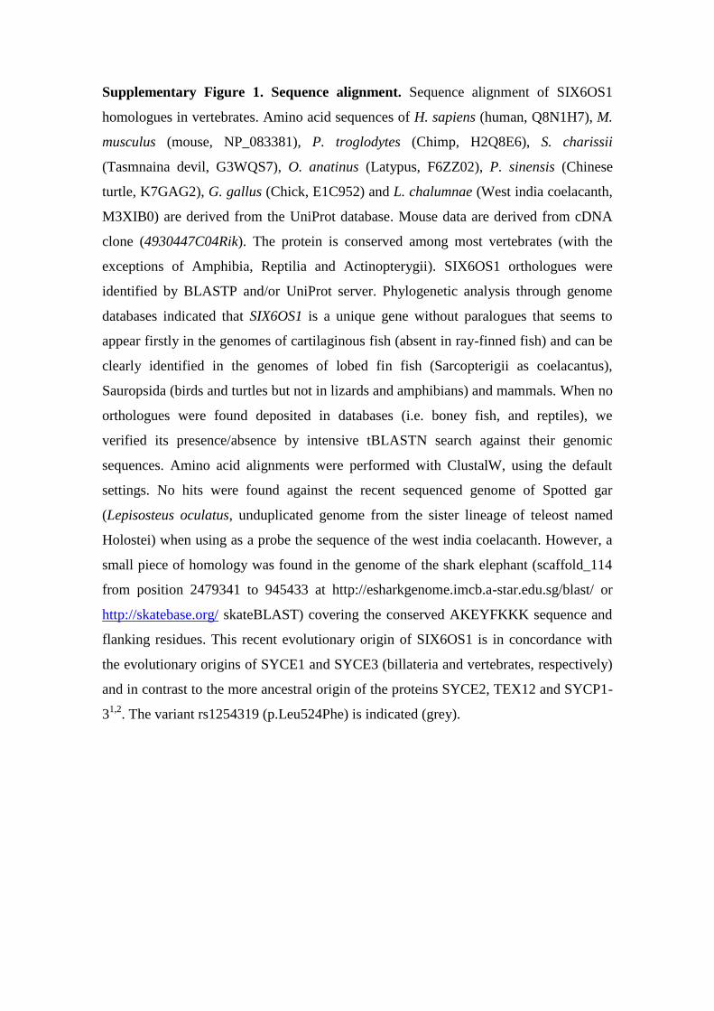
Supplementary Figure 2. Sequence analysis of SIX6OS1. (a) Secondary structure
analysis of mouse SIX6OS1 in which the α-helical, β-sheet, unstructured and coiled-coil
predictions are plotted on the basis of their confidence as calculated by JPred43. (b)
Conservation of the SIX6OS1 sequence, based on an alignment of all full length
sequences produced in MUSCLE4, plotted as the per residue conservation scores
calculated in Jalview 25. (c) Schematic diagram of the predicted SIX6OS1 structure, in
which an N-terminal α-helical domain is linked to an unstructured protein-protein
interaction module in the C-terminus through a flexible linker sequence. The putative
protein-protein interaction region of the C-terminus includes multiple predicted
phosphorylation sites, including four SP/TP potential CDK sites between amino acids
426-472, which may function in the dynamic regulation of interactions.
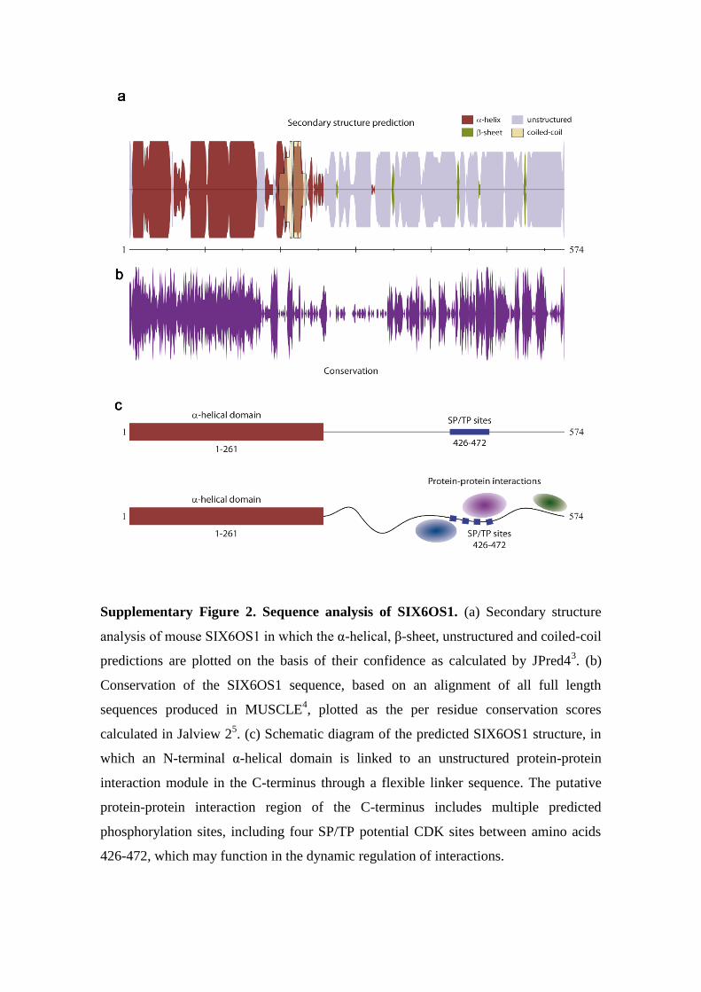
Supplementary Figure 3. Validation of SIX6OS1 antibodies.
(a) HEK 293T cells were transfected with a plasmid encoding GFP-SIX6OS1 or GFP
and the whole extracts were analyzed by western blot using goat α-SIX6OS1 (left
panel), rabbit α-SIX6OS1 (central panel) and α-GFP (right panel). Ponceau S staining
of the blotted membranes was used as loading control. A band around 100 kDa,
corresponding to the expected GFP-SIX6OS1 fusion protein (32,7 kDa + 70 kDa), was
detected with the goat α-SIX6OS1, rabbit α-SIX6OS1 and goat α-GFP (arrowheads). (b)
Immunofluorescence of HEK 293T cells transfected with plasmids encoding GFP-
SIX6OS1 or GFP. SIX6OS1 was detected with either goat α-SIX6OS1 (left panel) or
rabbit α-SIX6OS1 (right panel, red) and GFP by direct fluorescence signal (green).
Green and red signals co-localize in the cytoplasm of the transfected HEK 293T cells.
(c) Double immunofluorescence of spermatocytes at pachytene stage obtained from
Six6os1+/+
and Six6os1-/-
mice using the polyclonal rabbit antibody α-SIX6OS1 (green)
and mouse α-SYCP3 (red). The experiments were reproduced three times. Bars
represent 10 µm.
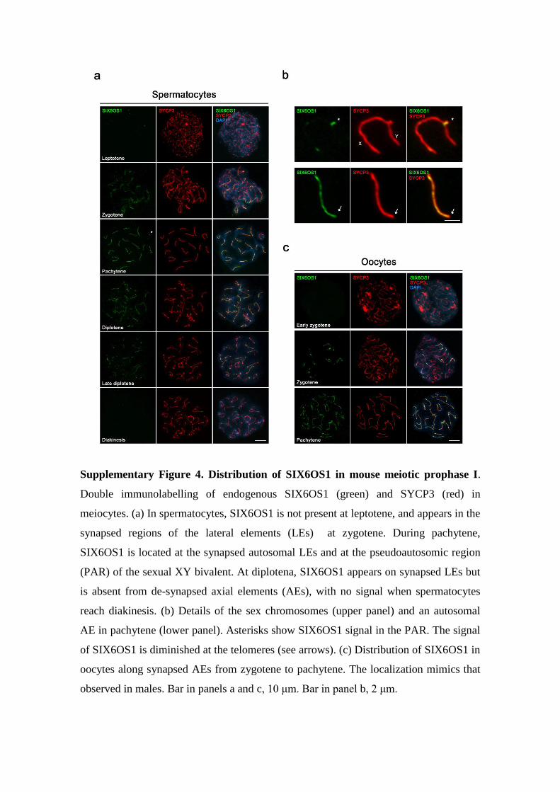
Supplementary Figure 4. Distribution of SIX6OS1 in mouse meiotic prophase I.
Double immunolabelling of endogenous SIX6OS1 (green) and SYCP3 (red) in
meiocytes. (a) In spermatocytes, SIX6OS1 is not present at leptotene, and appears in the
synapsed regions of the lateral elements (LEs) at zygotene. During pachytene,
SIX6OS1 is located at the synapsed autosomal LEs and at the pseudoautosomic region
(PAR) of the sexual XY bivalent. At diplotena, SIX6OS1 appears on synapsed LEs but
is absent from de-synapsed axial elements (AEs), with no signal when spermatocytes
reach diakinesis. (b) Details of the sex chromosomes (upper panel) and an autosomal
AE in pachytene (lower panel). Asterisks show SIX6OS1 signal in the PAR. The signal
of SIX6OS1 is diminished at the telomeres (see arrows). (c) Distribution of SIX6OS1 in
oocytes along synapsed AEs from zygotene to pachytene. The localization mimics that
observed in males. Bar in panels a and c, 10 μm. Bar in panel b, 2 μm.
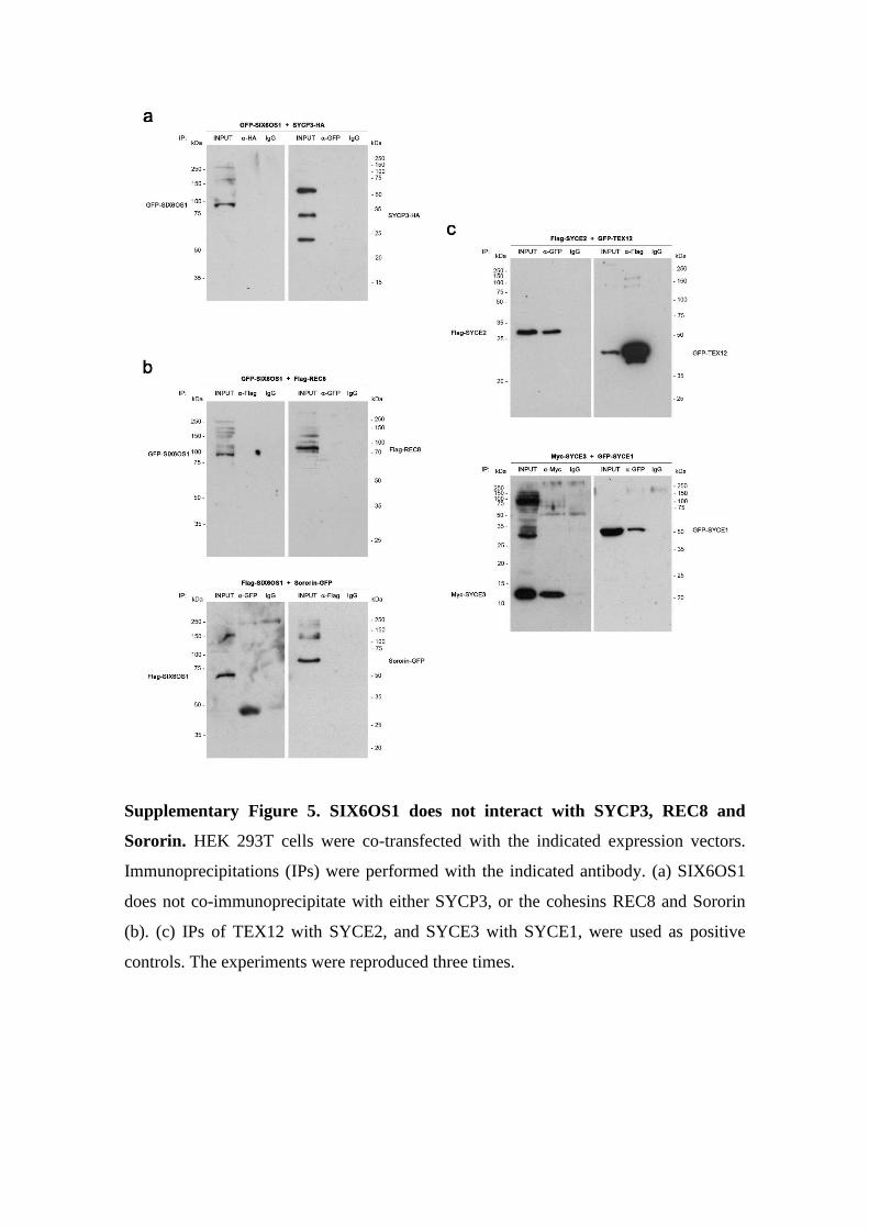
Supplementary Figure 5. SIX6OS1 does not interact with SYCP3, REC8 and
Sororin. HEK 293T cells were co-transfected with the indicated expression vectors.
Immunoprecipitations (IPs) were performed with the indicated antibody. (a) SIX6OS1
does not co-immunoprecipitate with either SYCP3, or the cohesins REC8 and Sororin
(b). (c) IPs of TEX12 with SYCE2, and SYCE3 with SYCE1, were used as positive
controls. The experiments were reproduced three times.
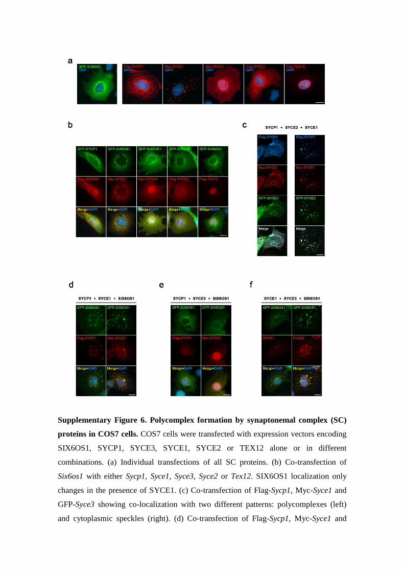
Supplementary Figure 6. Polycomplex formation by synaptonemal complex (SC)
proteins in COS7 cells. COS7 cells were transfected with expression vectors encoding
SIX6OS1, SYCP1, SYCE3, SYCE1, SYCE2 or TEX12 alone or in different
combinations. (a) Individual transfections of all SC proteins. (b) Co-transfection of
Six6os1 with either Sycp1, Syce1, Syce3, Syce2 or Tex12. SIX6OS1 localization only
changes in the presence of SYCE1. (c) Co-transfection of Flag-Sycp1, Myc-Syce1 and
GFP-Syce3 showing co-localization with two different patterns: polycomplexes (left)
and cytoplasmic speckles (right). (d) Co-transfection of Flag-Sycp1, Myc-Syce1 and
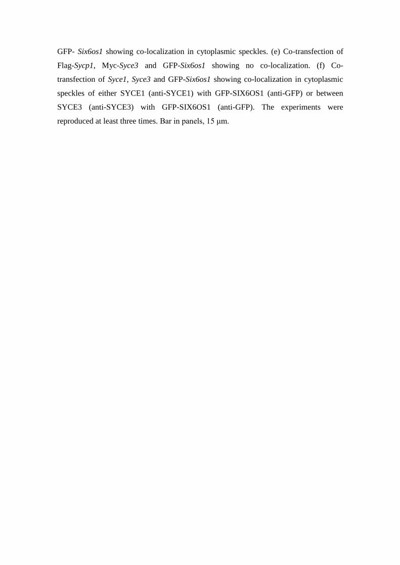
GFP- Six6os1 showing co-localization in cytoplasmic speckles. (e) Co-transfection of
Flag-Sycp1, Myc-Syce3 and GFP-Six6os1 showing no co-localization. (f) Co-
transfection of Syce1, Syce3 and GFP-Six6os1 showing co-localization in cytoplasmic
speckles of either SYCE1 (anti-SYCE1) with GFP-SIX6OS1 (anti-GFP) or between
SYCE3 (anti-SYCE3) with GFP-SIX6OS1 (anti-GFP). The experiments were
reproduced at least three times. Bar in panels, 15 μm.
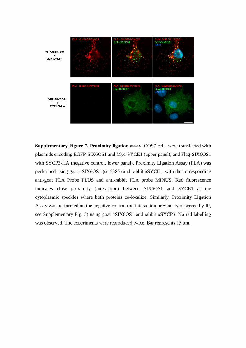
Supplementary Figure 7. Proximity ligation assay. COS7 cells were transfected with
plasmids encoding EGFP-SIX6OS1 and Myc-SYCE1 (upper panel), and Flag-SIX6OS1
with SYCP3-HA (negative control, lower panel). Proximity Ligation Assay (PLA) was
performed using goat αSIX6OS1 (sc-5385) and rabbit αSYCE1, with the corresponding
anti-goat PLA Probe PLUS and anti-rabbit PLA probe MINUS. Red fluorescence
indicates close proximity (interaction) between SIX6OS1 and SYCE1 at the
cytoplasmic speckles where both proteins co-localize. Similarly, Proximity Ligation
Assay was performed on the negative control (no interaction previously observed by IP,
see Supplementary Fig. 5) using goat αSIX6OS1 and rabbit αSYCP3. No red labelling
was observed. The experiments were reproduced twice. Bar represents 15 μm.
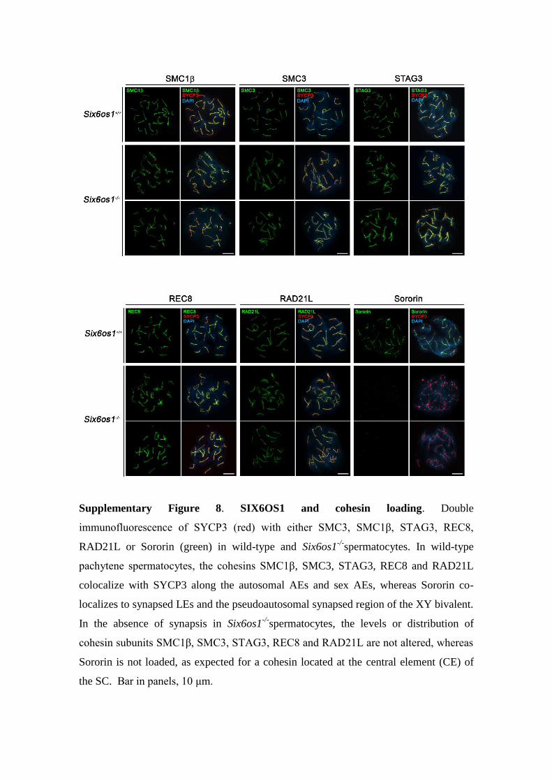
Supplementary Figure 8. SIX6OS1 and cohesin loading. Double
immunofluorescence of SYCP3 (red) with either SMC3, SMC1β, STAG3, REC8,
RAD21L or Sororin (green) in wild-type and Six6os1-/-
spermatocytes. In wild-type
pachytene spermatocytes, the cohesins SMC1β, SMC3, STAG3, REC8 and RAD21L
colocalize with SYCP3 along the autosomal AEs and sex AEs, whereas Sororin co-
localizes to synapsed LEs and the pseudoautosomal synapsed region of the XY bivalent.
In the absence of synapsis in Six6os1-/-
spermatocytes, the levels or distribution of
cohesin subunits SMC1β, SMC3, STAG3, REC8 and RAD21L are not altered, whereas
Sororin is not loaded, as expected for a cohesin located at the central element (CE) of
the SC. Bar in panels, 10 μm.
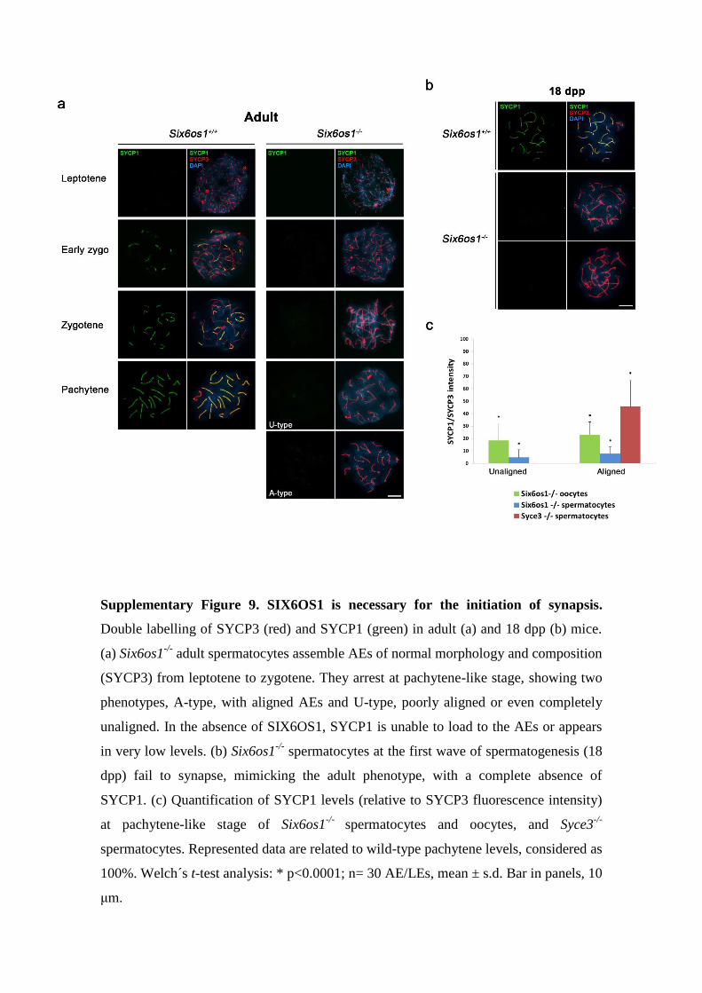
Supplementary Figure 9. SIX6OS1 is necessary for the initiation of synapsis.
Double labelling of SYCP3 (red) and SYCP1 (green) in adult (a) and 18 dpp (b) mice.
(a) Six6os1-/-
adult spermatocytes assemble AEs of normal morphology and composition
(SYCP3) from leptotene to zygotene. They arrest at pachytene-like stage, showing two
phenotypes, A-type, with aligned AEs and U-type, poorly aligned or even completely
unaligned. In the absence of SIX6OS1, SYCP1 is unable to load to the AEs or appears
in very low levels. (b) Six6os1-/-
spermatocytes at the first wave of spermatogenesis (18
dpp) fail to synapse, mimicking the adult phenotype, with a complete absence of
SYCP1. (c) Quantification of SYCP1 levels (relative to SYCP3 fluorescence intensity)
at pachytene-like stage of Six6os1-/-
spermatocytes and oocytes, and Syce3-/-
spermatocytes. Represented data are related to wild-type pachytene levels, considered as
100%. Welch´s t-test analysis: * p<0.0001; n= 30 AE/LEs, mean ± s.d. Bar in panels, 10
μm.
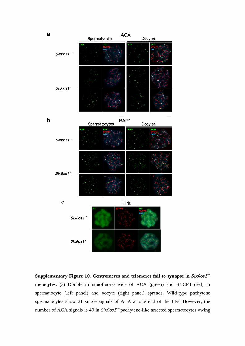
Supplementary Figure 10. Centromeres and telomeres fail to synapse in Six6os1-/-
meiocytes. (a) Double immunofluorescence of ACA (green) and SYCP3 (red) in
spermatocyte (left panel) and oocyte (right panel) spreads. Wild-type pachytene
spermatocytes show 21 single signals of ACA at one end of the LEs. However, the
number of ACA signals is 40 in Six6os1-/-
pachytene-like arrested spermatocytes owing

to the absence of synapsis. Six6os1-/-
oocytes show 40 ACA signals vs 20 in wild-type.
(b) Co-labelling of RAP1 (green) and SYCP3 (red) in spermatocyte (left panel) and
oocyte (right panel) spreads. Six6os1-/-
meiocytes show 80 RAP1 foci, one at each end
(telomere) of the AEs, while wild-type meiocytes show 40 RAP1 signals. (c) Double
immunofluorescence of H1t (green) and SYCP3 (red) in spermatocytes from Six6os1-/-
and Six6os1+/+
showing loading of H1t in the arrested pachytene-like and wild-type
pachytene spermatocytes. Bar in panels, 10 μm.
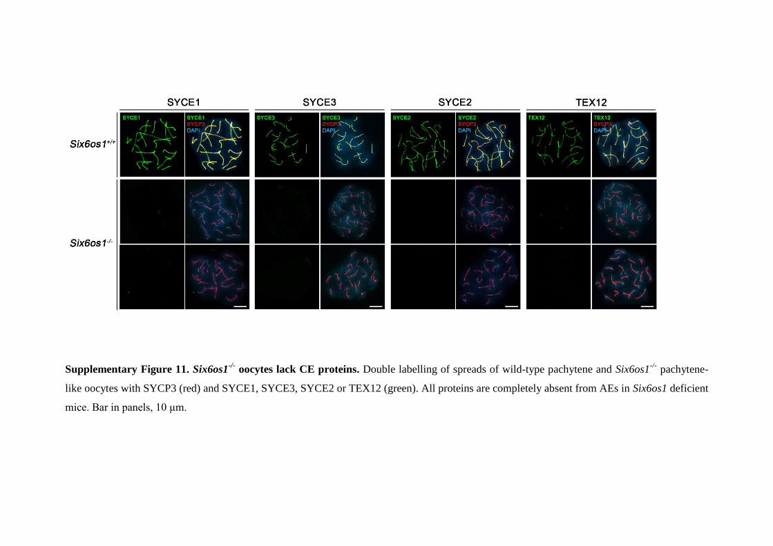
Supplementary Figure 11. Six6os1-/-
oocytes lack CE proteins. Double labelling of spreads of wild-type pachytene and Six6os1-/-
pachytene-
like oocytes with SYCP3 (red) and SYCE1, SYCE3, SYCE2 or TEX12 (green). All proteins are completely absent from AEs in Six6os1 deficient
mice. Bar in panels, 10 μm.
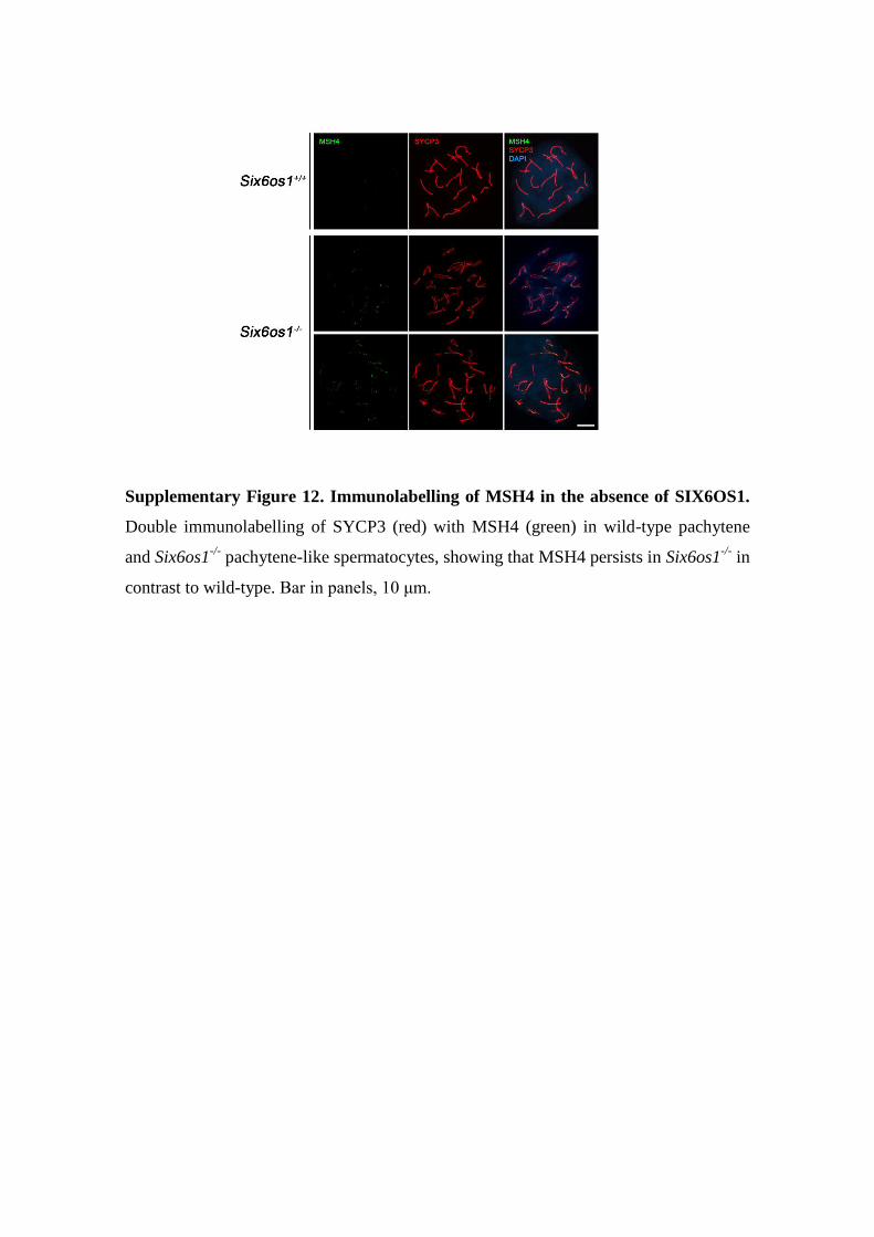
Supplementary Figure 12. Immunolabelling of MSH4 in the absence of SIX6OS1.
Double immunolabelling of SYCP3 (red) with MSH4 (green) in wild-type pachytene
and Six6os1-/-
pachytene-like spermatocytes, showing that MSH4 persists in Six6os1-/-
in
contrast to wild-type. Bar in panels, 10 μm.
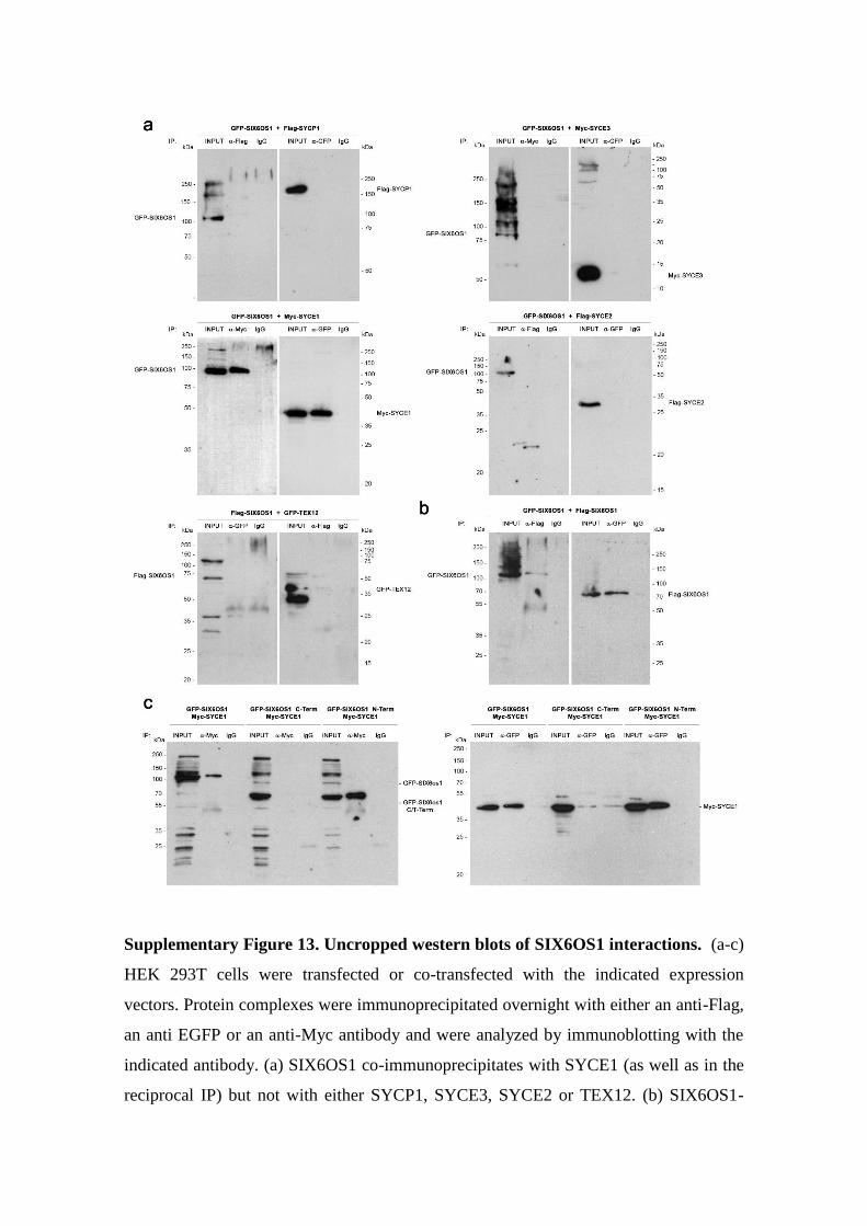
Supplementary Figure 13. Uncropped western blots of SIX6OS1 interactions. (a-c)
HEK 293T cells were transfected or co-transfected with the indicated expression
vectors. Protein complexes were immunoprecipitated overnight with either an anti-Flag,
an anti EGFP or an anti-Myc antibody and were analyzed by immunoblotting with the
indicated antibody. (a) SIX6OS1 co-immunoprecipitates with SYCE1 (as well as in the
reciprocal IP) but not with either SYCP1, SYCE3, SYCE2 or TEX12. (b) SIX6OS1-
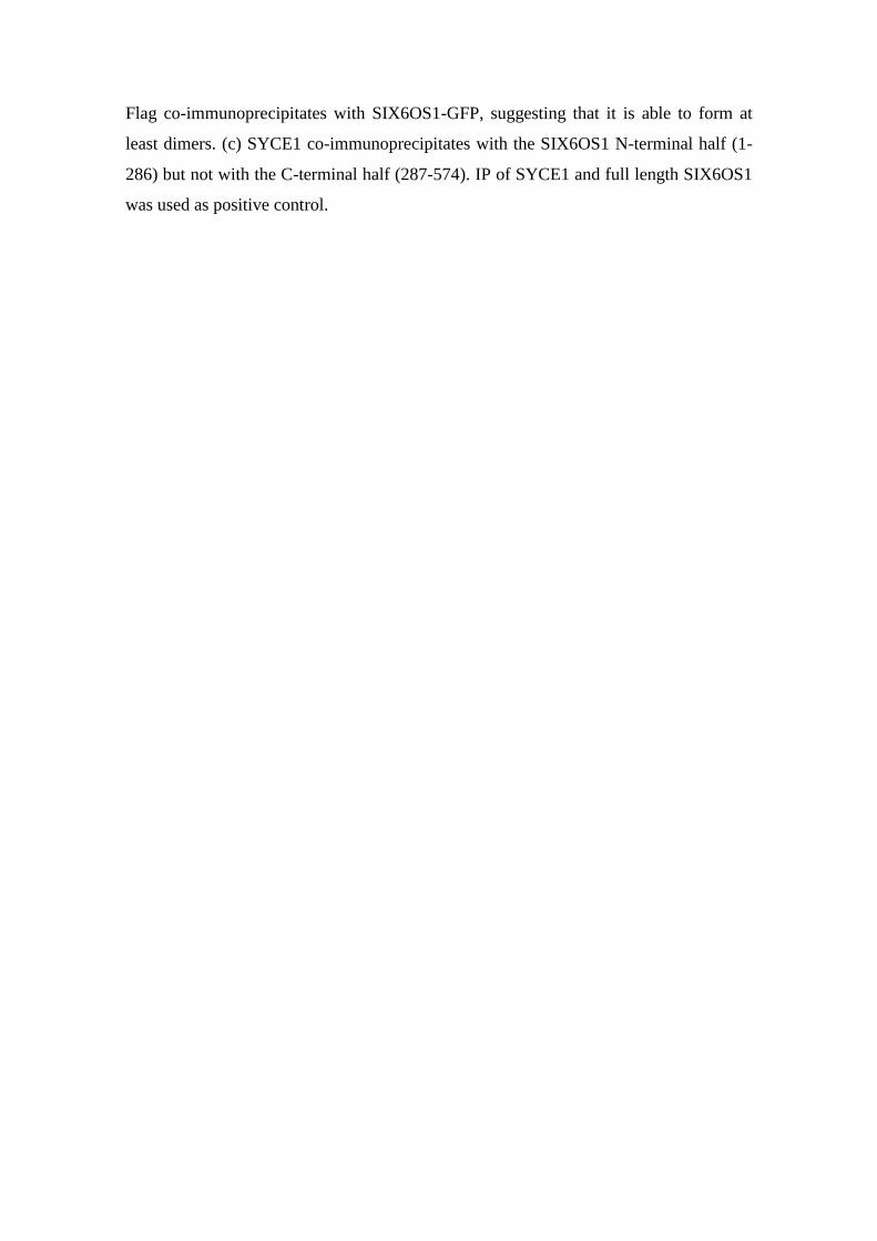
Flag co-immunoprecipitates with SIX6OS1-GFP, suggesting that it is able to form at
least dimers. (c) SYCE1 co-immunoprecipitates with the SIX6OS1 N-terminal half (1-
286) but not with the C-terminal half (287-574). IP of SYCE1 and full length SIX6OS1
was used as positive control.
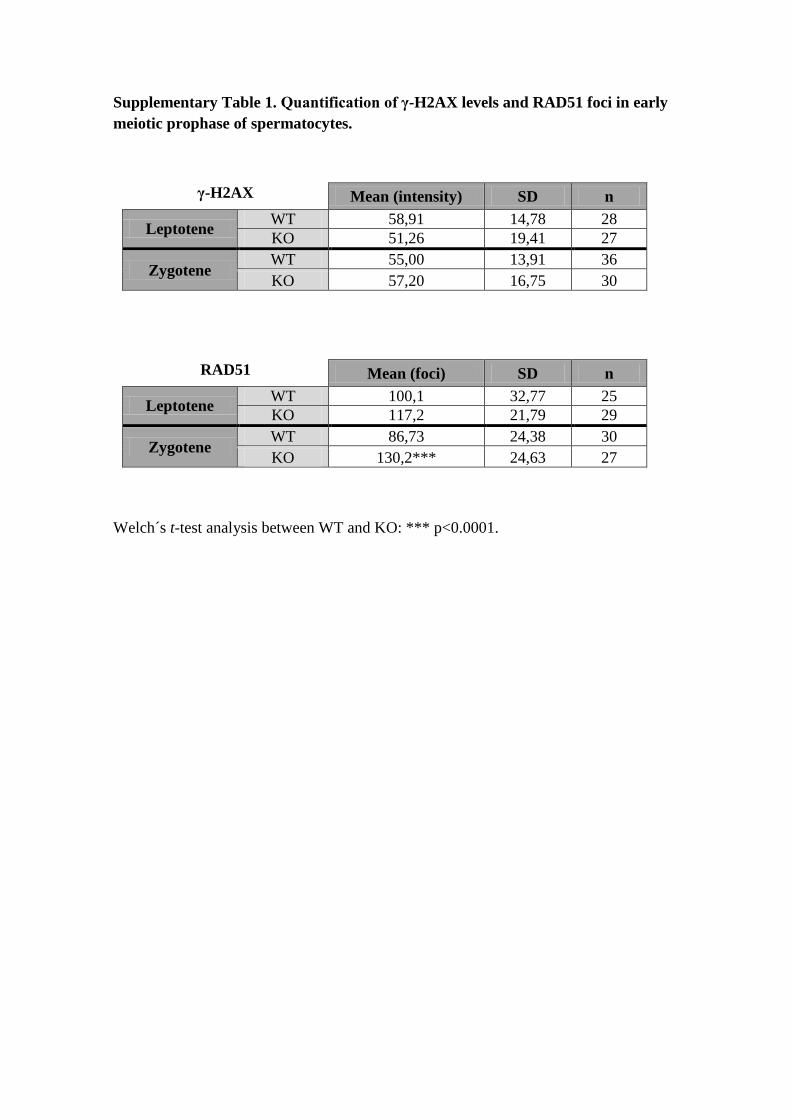
Supplementary Table 1. Quantification of γ-H2AX levels and RAD51 foci in early
meiotic prophase of spermatocytes.
γ-H2AX
Mean (intensity) SD n
Leptotene WT 58,91 14,78 28
KO 51,26 19,41 27
Zygotene WT 55,00 13,91 36
KO 57,20 16,75 30
RAD51
Mean (foci) SD n
Leptotene WT 100,1 32,77 25
KO 117,2 21,79 29
Zygotene WT 86,73 24,38 30
KO 130,2*** 24,63 27
Welch´s t-test analysis between WT and KO: *** p<0.0001.
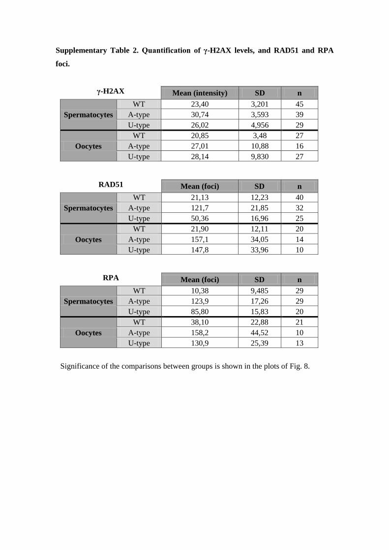
Supplementary Table 2. Quantification of γ-H2AX levels, and RAD51 and RPA
foci.
γ-H2AX
Mean (intensity) SD n
Spermatocytes
WT 23,40 3,201 45
A-type 30,74 3,593 39
U-type 26,02 4,956 29
Oocytes
WT 20,85 3,48 27
A-type 27,01 10,88 16
U-type 28,14 9,830 27
RAD51
Mean (foci) SD n
Spermatocytes
WT 21,13 12,23 40
A-type 121,7 21,85 32
U-type 50,36 16,96 25
Oocytes
WT 21,90 12,11 20
A-type 157,1 34,05 14
U-type 147,8 33,96 10
RPA
Mean (foci) SD n
Spermatocytes
WT 10,38 9,485 29
A-type 123,9 17,26 29
U-type 85,80 15,83 20
Oocytes
WT 38,10 22,88 21
A-type 158,2 44,52 10
U-type 130,9 25,39 13
Significance of the comparisons between groups is shown in the plots of Fig. 8.
Supplementary References
1. Fraune, J., et al. Hydra meiosis reveals unexpected conservation of structural
synaptonemal complex proteins across metazoans. Proceedings of the National
Academy of Sciences of the United States of America 109, 16588-16593 (2012).
2. Fraune, J., Brochier-Armanet, C., Alsheimer, M. & Benavente, R. Phylogenies
of central element proteins reveal the dynamic evolutionary history of the
mammalian synaptonemal complex: ancient and recent components. Genetics
195, 781-793 (2013).
3. Drozdetskiy, A., Cole, C., Procter, J. & Barton, G.J. JPred4: a protein secondary
structure prediction server. Nucleic acids research 43, W389-394 (2015).
4. Edgar, R.C. MUSCLE: multiple sequence alignment with high accuracy and
high throughput. Nucleic acids research 32, 1792-1797 (2004).
5. Waterhouse, A.M., Procter, J.B., Martin, D.M., Clamp, M. & Barton, G.J.
Jalview Version 2--a multiple sequence alignment editor and analysis
workbench. Bioinformatics 25, 1189-1191 (2009).


![Genetic mapping of a lobed-leaf gene associated …...leaf [1,17,18]. Some molecular markers linked to lobed-leaf genes were identified. For example, a SCAR marker linked to the lobed-leaf](https://static.fdocuments.in/doc/165x107/5e771cd79b545f444838ff7a/genetic-mapping-of-a-lobed-leaf-gene-associated-leaf-11718-some-molecular.jpg)