Supplementary Figures, Table, and Movie of...
8
1 Supplementary Figures, Table, and Movie Figure S1 Distribution of ALDH1A1, calbindin, and TH-positive neurons in the SNpc and VTA of 2-month-old nTg mice Representative images show ALDH1A1 (green), calbindin (red), and TH (blue) staining in the SNpc and VTA of 2-month-old control mice. The dash line marks the boundary between SNpc and VTA. DM: dorsomedial, VL: ventrolateral. Scale bar: 100μm Figure S2 Distribution of Aldh family genes in the brain of P56 nTg mice (A) In situ hybridization of Th, Aldh1a1, Aldh1l1, Adh2 in coronal sections of postnatal day 56 (P56)male C56/BL6 mice (Allen Brain Atlas, http://www.brain-map.org/). (B) In situ hybridization of Th, Aldh1a1, Aldh1a2, Aldh1a3, Aldh1b1, Aldh3a2, Aldh3b1, Aldh3b2, Aldh4a1, Aldh5a1, Aldh6a1, Aldh7a1, Aldh8a1, and Aldh18a1 in sagittal sections of P56 male C56/BL6 mice (Allen Brain Atlas, http://www.brain-map.org/). Figure S3 Distribution of ALDH1A1-positive and negative DA neurons in human SNpc (A-B) 3D reconstruction of DA neurons in human SNpc shows the distribution of ALDH1A1 and NM-double positive (ALDH1A1 + /NM + , yellow) as well as ALDH1A1-negative but NM- positive (ALDH1A1 - /NM + , red) neurons remaining in the SNpc of control (Ctrl, A) and PD (B) human brains.
Transcript of Supplementary Figures, Table, and Movie of...

1
Supplementary Figures, Table, and Movie
Figure S1 Distribution of ALDH1A1, calbindin, and TH-positive neurons in the SNpc and
VTA of 2-month-old nTg mice
Representative images show ALDH1A1 (green), calbindin (red), and TH (blue) staining in the
SNpc and VTA of 2-month-old control mice. The dash line marks the boundary between SNpc
and VTA. DM: dorsomedial, VL: ventrolateral. Scale bar: 100µm
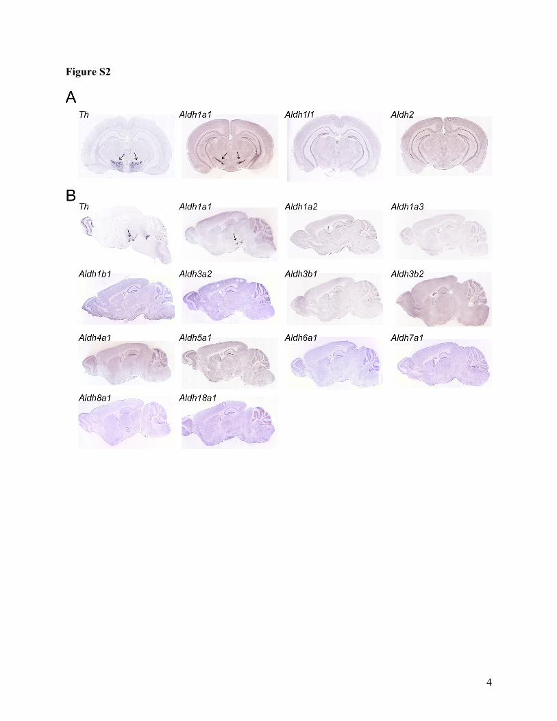
Figure S2 Distribution of Aldh family genes in the brain of P56 nTg mice
(A) In situ hybridization of Th, Aldh1a1, Aldh1l1, Adh2 in coronal sections of postnatal day 56
(P56)male C56/BL6 mice (Allen Brain Atlas, http://www.brain-map.org/).
(B) In situ hybridization of Th, Aldh1a1, Aldh1a2, Aldh1a3, Aldh1b1, Aldh3a2, Aldh3b1,
Aldh3b2, Aldh4a1, Aldh5a1, Aldh6a1, Aldh7a1, Aldh8a1, and Aldh18a1 in sagittal sections of
P56 male C56/BL6 mice (Allen Brain Atlas, http://www.brain-map.org/).
Figure S3 Distribution of ALDH1A1-positive and negative DA neurons in human SNpc
(A-B) 3D reconstruction of DA neurons in human SNpc shows the distribution of ALDH1A1
and NM-double positive (ALDH1A1+/NM+, yellow) as well as ALDH1A1-negative but NM-
positive (ALDH1A1-/NM+, red) neurons remaining in the SNpc of control (Ctrl, A) and PD (B)
human brains.

2
Fig. S4 Reduction of ALDH1A1 staining in DA neurons at the SNpc of patients with
sporadic PD
Representative bright-field images show ALDH1A1 staining (purple) in the VM, PL and VL of
control and severe PD SNpc. DA neurons were marked by the presence of NM (brown) in the
soma. The small panels highlight the soma (left) and neurites (right) in the boxed areas of the
large panels. Scale bar: 100µm (large panel); 10µm (small panel).
Fig. S5 Expression of transgenic α-synuclein in cultured cortical neurons
Western blot analyses show expression of transgenic α-synuclein in cultured cortical neurons
derived from P0 CaMKII-tTA/tetO-A53T pups. The C20 α-synuclein antibody recognizes both
human and mouse α-synuclein, while the syn211 antibody recognizes only human α-synuclein.
The presence of DOX suppresses the expression of transgenic α-synuclein. The expression of
actin was used as the loading control.
Table S1. List of control and PD cases.
Movie. 3D reconstructions of DA neurons in the midbrain of 18-month-old nTg (movie 1) and A53T (movie 2) mice.

3
Figure S1
4
Figure S2

5
Figure S3

6
Figure S4

7
Figure S5

Table S1: Human brain information Case Diagnosis Age Sex Duration Braak Neuropathologic Stage Depigmentation SNpc LB UPDRS Hoehn&Yahr Source
#1 Control 81 M 0 III No Lewy bodies Normal 0 6.5 N/A BSHRI #2 Control 80 M 0 I No Lewy bodies Normal 0 3 N/A BSHRI #3 Control 86 M 0 II No Lewy bodies Normal 0 2 N/A BSHRI #4 Control 89 M 0 II No Lewy bodies Normal 0 7 N/A BSHRI #5 Control 78 M 0 N/A No Lewy bodies Normal 0 N/A N/A JHUMS #6 Control 76 M 0 N/A No Lewy bodies Normal 0 N/A N/A JHUMS #7 Control 86 M 0 N/A No Lewy bodies Normal 0 NA N/A JHUMS #8 Control 87 F 0 III No Lewy bodies Normal 0 1 N/A BSHRI #9 Control 73 F 0 I No Lewy bodies Normal 0 N/A N/A BSHRI
Case Diagnosis Age Sex Duration Braak Neuropathologic Stage Depigmentation SNpc LB UPDRS Hoehn&Yahr Source #10 PD 90 M 15 II Brainstem Predominant Moderate 1 N/A N/A BSHRI #11 PD 89 M 1 III Brainstem Predominant Mild 0 0 N/A BSHRI #12 PD 71 M 10 III-IV Limbic Moderate N/A N/A 5 JHUMS #13 PD 80 F 11 III Brainstem/Limbic Moderate 3 N/A N/A BSHRI #14 PD 78 F 16 III Neocortical Severe 3 N/A N/A BSHRI #15 PD 79 F 20 IV Neocortical Severe 3 24 N/A BSHRI #16 PD 77 F 21 II Brainstem Predominant Moderate 3 N/A N/A BSHRI #17 PD 73 F 7 II Brainstem Predominant Mild N/A N/A 2 JHUMS #18 PD 85 F 13 V-VI Neocortical Severe N/A N/A 4 JHUMS #19 PD 73 F 18 III-IV Limbic Severe N/A N/A 4 JHUMS
Duration(Yrs): Disease duration in years UPDRS: time elapsed (months) from last UPDRS until death Braak: Braak tangle stage score Neuropathologic Stage: Lewy body progression through 10 brain regions SNpc LB: the Lewy pathology density score for SNpc for that case N/A: data not availalble BSHRI: Banner Sun Health Research Institute
JHUMS: the Johns Hopkins University School of Medicine


![5 movie mas mayo [movie-mas.com]](https://static.fdocuments.in/doc/165x107/559b4a451a28ab9c678b4596/5-movie-mas-mayo-movie-mascom.jpg)