Supplemental Material New Genetic variants for Cardiac ...
103
1 Supplemental Material New Genetic variants for Cardiac Structure and Function – The EchoGen Consortium Conflicts of interest The authors of this manuscript declare to have the following potential conflicts of interest according to JCI’s policy: Ownership: GFM: owner of Cardiovascular Engineering, Inc., a company that develops and manufactures devices to measure vascular stiffness; Income: EI: advisor and consultant for Precision Wellness, Inc., and advisor for Cellink for work unrelated to the present project; GFM: consultant to and receives honoraria from Novartis, Merck, Servier and Philips; PSW: honoraria for lectures or consulting from Boehringer Ingelheim, Bayer Health Care, AstraZeneca, Sanofi-Aventis and Public Health, Heinrich-Heine-University Düsseldorf. SB: honoraria for lectures from Abbott, Abbott Diagnostics, Astra Zeneca, Bayer, Boehringer Ingelheim, Medtronic, Pfizer, Roche, SIEMENS Diagnostics, SIEMENS, Thermo Fisher and member of Advisory Boards and consultant for Boehringer Ingelheim, Bayer, Novartis, Roche and Thermo Fisher. SJS: consulting fees from Bayer, Novartis, and Sanofi., Research support: AZ: research support from BiomarCaRE (EU) and DZHK (BMBF); CAM: >50K-NIH, Burroughs Welcome Foundation, Leducq Foundation, AHA, Google, AstraZeneca, Merck, Sanofi; EJB received NI grant support 1R01HL128914, 2R01 HL092577. GFM: research grant from Novartis and by research grants HL094898,
Transcript of Supplemental Material New Genetic variants for Cardiac ...

1
Supplemental Material
New Genetic variants for Cardiac Structure and Function –
The EchoGen Consortium
Conflicts of interest
The authors of this manuscript declare to have the following potential conflicts of
interest according to JCI’s policy:
Ownership: GFM: owner of Cardiovascular Engineering, Inc., a company that develops
and manufactures devices to measure vascular stiffness;
Income: EI: advisor and consultant for Precision Wellness, Inc., and advisor for Cellink
for work unrelated to the present project; GFM: consultant to and receives honoraria
from Novartis, Merck, Servier and Philips; PSW: honoraria for lectures or consulting
from Boehringer Ingelheim, Bayer Health Care, AstraZeneca, Sanofi-Aventis and Public
Health, Heinrich-Heine-University Düsseldorf. SB: honoraria for lectures from Abbott,
Abbott Diagnostics, Astra Zeneca, Bayer, Boehringer Ingelheim, Medtronic, Pfizer,
Roche, SIEMENS Diagnostics, SIEMENS, Thermo Fisher and member of Advisory
Boards and consultant for Boehringer Ingelheim, Bayer, Novartis, Roche and Thermo
Fisher. SJS: consulting fees from Bayer, Novartis, and Sanofi.,
Research support: AZ: research support from BiomarCaRE (EU) and DZHK (BMBF);
CAM: >50K-NIH, Burroughs Welcome Foundation, Leducq Foundation, AHA, Google,
AstraZeneca, Merck, Sanofi; EJB received NI grant support 1R01HL128914, 2R01
HL092577. GFM: research grant from Novartis and by research grants HL094898,

2
DK082447, HL107385, HL104184 and HL126136 from the National Institutes of Health;
PSW, KL, TM: research grant from Boehringer Ingelheim (grant for the conduct of the
Gutenberg Health Study); SJS: research grant support from Actelion, AstraZeneca, and
Novartis; SB: research funding from Abbott, Abbott Diagnostics, Bayer, Boehringer
Ingelheim, SIEMENS and Thermo Fisher; TZ: research grant by the German Federal
Ministry of Education and Research (BMBF) to the German Center for Cardiovascular
Disease (DZHK) (81Z1710101). PSW: research grant by the Federal Ministry of
Education and Research (BMBF 01EO1003 and 01EO1503), German Federal Ministry
of Education and Research (BMBF) to the German Center for Cardiovascular Disease
(DZHK) (81Z1710101), Federal Institute for Occupational Safety and Health (BAuA),
DFG no. WI 3881/2-2; EU Grants No. 278913, 278397, Initiative “Health Economy”,
Ministry of Health and Ministry of Economics, and Federal Ministry of Education and
Research, Rhineland-Palatinate; Mainz Heart Foundation, Boehringer Ingelheim,
Sanofi-Aventis, Bayer Health Care, Daiichi Sankyo Europe.
Intellectual property: Not applicable.

Supplemental Material: New Genetic variants for Cardiac Structure and Function – EchoGen
1
Online Methods and Supplemental Material:
New Genetic variants for Cardiac Structure and Function –
The EchoGen Consortium
1 ONLINE METHODS ......................................................................................................... 2
1.1 Echocardiographic Methods ................................................................................. 2
1.2 Methods used for look-up in other cohorts ........................................................... 6
1.3 Pathway Analysis ............................................................................................... 11
2 SUPPLEMENTAL MATERIAL ........................................................................................... 14
2.1 Abbreviations of participating cohorts ................................................................ 14
2.2 Description of cohorts (Table S1) ....................................................................... 15
2.3 Baseline characteristics of cohorts (Table S2) ................................................... 18
2.4 Echo methods (Table S3) .................................................................................. 21
2.5 Echo values by cohorts (Table S4) .................................................................... 22
2.6 Genotyping Methods and Imputation (Table S5) ............................................... 24
3 Additional tables and figures (Table S6-S19, Figures S1-S20) ................................ 28
4 Acknowledgements ................................................................................................... 72
5 References ................................................................................................................ 88

Supplemental Material: New Genetic variants for Cardiac Structure and Function – EchoGen
2
1 ONLINE METHODS
1.1 Echocardiographic Methods
1.1.1 Harmonizationandqualityissues
An important issue for meta-analysis of data from different cohorts is the compliance
with high quality phenotyping standards. Echocardiographic data used for these
analyzes were recorded for research but not taken from clinical routine. For
echocardiography potential sources of variability have been identified and described
previously1, including inter- and intra-reader and sonographer variability, within subject
variability, temporal drift within laboratories, and biases due to missing data2,3. Methods
that have previously demonstrated to enhance echo reproducibility have been applied
by the participating cohorts of the EchoGen consortium at each study site, standardized
equipment and imaging and reading protocols, trained sonographers and readers who
periodically undergo reading review sessions, averaging multiple measurements,
substituting 2D measurements when M-mode measurements are unreliable,
assessment of intra-reader and inter-reader agreement, and analyses of temporal
drifts1,3-6.
1.1.2 Echoexaminations,parametersanddefinitions
Transthoracic echocardiography was performed in each cohort by trained technicians or
physicians. All measurements were performed according to the current American and
European guidelines for the echocardiographic assessment of the left ventricle7. For the
present investigation we analyzed 5 LV structural, 2 systolic functional, and 7 diastolic
functional parameters, as well as 2 binary diastolic traits.

Supplemental Material: New Genetic variants for Cardiac Structure and Function – EchoGen
3
- LV structural parameters:
Two-dimensional guided M-mode measurements of the parasternal long axis view of
the LV and aortic root were obtained:
o LV diastolic internal dimension (LVDD)
o LV wall thickness (LVWT), calculated as the sum of posterior wall and
interventricular septum measurements
o LV mass (LVM), calculated by using the formula 0.8 [1.04{(LV diastolic internal
dimension + interventricular septum + posterior wall)3 −(LV diastolic internal
dimension)3}] + 0.6 7,8
o Left atrial antero-posterior diameter (LA)
o Aortic root diameter (AoD) , diameter of the aortic root (at the maximal diameter
of the sinuses of Valsalva), obtained from the parasternal long-axis view7
o LV systolic function parameters:
Also from the parasternal long axis view the following variables were obtained:
o Left ventricular fractional shortening (FS), a quantitative measure of LV systolic
function, calculated using the formula ([LV end diastolic dimension – LV end
systolic dimension]/ LV end diastolic dimension x 100)
o LV systolic dysfunction (LVSD), defined as the presence of reduced fractional
shortening (<29%, which corresponds to an ejection fraction of 50%) on M-mode
or a diminished ejection fraction (<50%) on 2-dimensional echocardiography 9. If a
cohort only had categorical data on systolic dysfunction available, for example a
categorical visual estimate of LV function, these were recoded into a binary
variable best representing the cut-offs above.

Supplemental Material: New Genetic variants for Cardiac Structure and Function – EchoGen
4
- LV diastolic functional parameters:
For the analyses of all LV diastolic functional parameters only subjects with a
preserved LV ejection fraction (EF) were considered, defined according one of the
following methods (method 1 preferred): 1. EF (modified Simpson method) ≥ 50%; 2.
EF (Teichholz method) ≥ 50%; 3. Fractional shortening ≥ 29%; 4. Visual estimation of
EF as fair or poor. Further details about the application of the different methods by
cohorts is given in Supplemental Material Online, Table S 3.
From the apical 4-chamber view the following variables were obtained by Doppler
imaging of the mitral inflow:
o Peak velocity of the mitral E-wave (Mv-E)
o Peak velocity of the mitral A-wave (Mv-A)
o Ratio of the peak velocity of the mitral E-Wave divided by the peak
velocity of the mitral A-wave. This ration was determined during breathing
baseline in all cohorts (E/A) and during Valsalva maneuver in some cohorts
(E/A_val).
o Deceleration time of the mitral E-wave (DecTime)
In addition, a pulse-wave Doppler imaging was obtained from the apical 5-chamber
or long-axis view and the sample volume placed within the LVOT, but in proximity to
the anterior mitral valve leaflet to record both inflow and outflow signals and measure
the following parameter:

Supplemental Material: New Genetic variants for Cardiac Structure and Function – EchoGen
5
o Isovolumetric relaxation time (IVRT), an additional index of diastolic
function, defined as the interval from the closure of the aortic valve to the
opening of the mitral valve.
Moreover, tissue Doppler imaging (TDI) was applied in some cohorts to obtain the
following diastolic parameters:
o Peak velocity of the excursion of the lateral mitral annulus in the early
diastolic phase (E´)
o Ratio of the peak velocity of the excursion of the lateral mitral annulus in
the early diastolic phase by TDI and the peak velocity of the mitral E-
wave by Doppler imaging (E/E´)
Alternative to TDI Doppler imaging of the pulmonary venous inflow was used in some
cohorts to achieve these parameters: systolic pulmonary venous forward flow (S),
diastolic pulmonary venous forward flow (D), and ratio of systolic and diastolic
pulmonary venous flow (S/D).
- Binary diastolic LV traits:
For the definition of these variables the classification of diastolic function by Redfield
et al. was applied10. Measurement of diastolic function was based on Doppler
imaging of the mitral inflow and either TDI of the mitral annulus or Doppler imaging of
the pulmonary venous inflow in combination with E/A_val measured by Doppler
imaging of the mitral inflow.
The following binary diastolic traits were calculated:
o Diastolic Dysfunction with preserved ejection fraction (DDpEF): Cases
were defined by an EF ≥ 50%, no symptoms of heart failure, no medicated

Supplemental Material: New Genetic variants for Cardiac Structure and Function – EchoGen
6
heart failure AND evidence for mild, moderate or severe LV diastolic
dysfunction. Controls were defined by an EF ≥ 50%, no symptoms of heart
failure, no medicated heart failure AND a normal LV diastolic function.
o Heart Failure with preserved ejection fraction – definition (HFpEF): Cases
were defined by an EF ≥ 50%, symptoms of heart failure (NYHA class ≥ 2)
AND/OR medicated heart failure AND evidence for mild, moderate or severe
LV diastolic dysfunction. Controls were asymptomatic individuals with
preserved systolic and diastolic LV function defined as an EF ≥ 50%, no
symptoms of heart failure, no medicated heart failure AND normal LV diastolic
function.
Participants with valvular disease were excluded from the analyses of left ventricular
dimensions and systolic function, if this information was known or recorded during the
echocardiographic examination. Detailed echocardiographic methods used and
distributions of traits in each cohort study are reported in the Supplemental material,
Tables S3 and S4.
1.2 Methods used for look-up in other cohorts
AortaGen
To determine whether SNPs that were associated with aortic diameter were also
associated with aortic stiffness, as assessed by carotid-femoral pulse wave velocity, we
evaluated the top SNP from each of the 7 novel aortic diameter loci. Lookups were
derived from a previously published meta-analysis of genome wide association results
for carotid-femoral pulse wave velocity from 9 cohorts that included 20,634

Supplemental Material: New Genetic variants for Cardiac Structure and Function – EchoGen
7
participants11. After accounting for 7 tests, the adjusted threshold for significance was
set at a P values < 0.007.
CHARGE-HF
The association of the two novel SNPs for left ventricular diastolic diameter with incident
heart failure was assessed using the summary statistics of the meta-analysis of four
population-based cohorts of adults of European ancestry (Cardiovascular Health Study,
Framingham Heart Study, Atherosclerosis Risk In Communities Study and Rotterdam
Study) including a total of 20,926 participants free of diagnosed heart failure at baseline,
in whom 2,526 cases of incident heart failure occurred during a mean follow-up of 11.5
years12. There is partial overlap between the samples of the Cardiovascular Health
Study, the Framingham Heart Study and the Rotterdam Study included in CHARGE-HF
and in the present study.
Generation R Study
The Generation R Study is a population-based, prospective cohort study from fetal life
onwards, including pregnant women with an expected delivery date between April 2002
and January 2006, living in the city of Rotterdam, the Netherlands. A detailed
description of the design of study has been published previously12. DNA was extracted
from cord blood, or, if cord blood was unavailable, from blood samples taken at the 6-
year visit, according to a standardized protocol.
Information on SNPs used in the present analysis was extracted from the GWAs
database, details of which are described in the supplemental material of this paper.
Analyses were restricted to singleton, live born children of European ethnic origin
without congenital heart or kidney malformations, in whom echocardiography was

Supplemental Material: New Genetic variants for Cardiac Structure and Function – EchoGen
8
performed during the follow-up at age 6. Analyses were adjusted for age, sex, height,
weight and the first four principal components based on the GWAs data.
LURIC
The Ludwigshafen Risk and Cardiovascular Health (LURIC) study is an ongoing
prospective study of more than 3,300 individuals of German ancestry in whom
cardiovascular and metabolic phenotypes (CAD, MI, dyslipidemia, hypertension,
metabolic syndrome and diabetes mellitus) have been defined or ruled out using
standardized methodologies in all study participants. Inclusion criteria for LURIC were:
German ancestry (limitation of genetic heterogeneity), clinical stability (except for acute
coronary syndromes) and availability of a coronary angiogram. Exclusion criteria were:
any acute illness other than acute coronary syndromes, any chronic disease where non-
cardiac disease predominated and a history of malignancy within the last five years. A
10-year clinical follow-up for total and cause specific mortality has been completed.
Participants were genotyped using the Affymetrix 6.0 array and datasets were imputed
to the HapMap2 and 1000G reference panels. Association of SNPs with all-cause
mortality, cardiovascular mortality and death due to heart failure was analyzed using the
Cox proportional hazards model.
CARDIOGRAMPLUSC4D
The associations of the novel SNPs with myocardial infarction and coronary artery
disease were looked up in the publicly available dataset of the CARDIOGRAMplusC4D
Consortium, including more than 180,000 individuals of which 60,000 are cases of
coronary artery diasease and myocardial infarction [downloaded from
www.CARDIOGRAMPLUSC4D.org] 13. The results of the additive models were used.

Supplemental Material: New Genetic variants for Cardiac Structure and Function – EchoGen
9
1.2.1 Humanleftventricletissue
Samples of cardiac tissue (n=313) were acquired from patients from the Myocardial
Applied Genomics Network (MAGNet; www.med.upenn.edu/magnet). Left ventricular
free-wall tissue was harvested at the time of cardiac surgery from subjects with heart
failure undergoing transplantation and from unused donor hearts. The heart was
perfused with cold cardioplegia prior to cardiectomy to arrest contraction and prevent
ischemic damage. Tissue specimens were then obtained and frozen in liquid nitrogen.
Genomic DNA was extracted using the Gentra Puregene Tissue Kit (Qiagen, CA)
according to manufacturer’s instructions. Total RNA was extracted using the miRNeasy
Kit (Qiagen) including DNAse treatment. RNA concentration and quality was determined
using the NanoVue Plus™ spectrophotometer (GE Healthcare) and the Agilent 2100
RNA Nano Chip (Agilent).
DNA samples were genotyped using Affymetrix Genome Wide SNP Array 6.0. We
applied quality control (QC) filters to exclude unreliable samples, samples with cryptic
relatedness and samples that were not genetically inferred Caucasian. For the analysis
reported here, we eliminated SNPs with genotype call rate < 95%, with minor allele
frequency (MAF) < 15%, or if there was significant departure from Hardy-Weinberg
equilibrium (p < 10-6). A total of 360,046 SNPs passed QC and were available for
analysis. To improve cross study comparisons, genotype imputation was performed
using the Minimac (v 2012.11.16) program14. Imputation results were filtered at an
imputation quality threshold of 0.5 and a MAF threshold of 0.15. For imputed
genotypes, we used dosage value as genotype. To assess gene expression, RNA was
hybridized with Affymetrix Genechip ST1.1 arrays using manufacturer instructions. CEL

Supplemental Material: New Genetic variants for Cardiac Structure and Function – EchoGen
10
files were normalized with robust multiarray analysis (RMA) using Bioconductor15. To
remove potential batch effects, expression values were further adjusted using
ComBat16.
We tested whether an association existed between the genotype of the 41
echocardiographic trait SNPs and gene expression of proximal (or neighboring) genes.
A cis eQTL analysis was performed using transcripts +/- 1Mb from each of the 41 SNPs
using a linear regression model and adjusted for multiple comparisons using a
Bonferroni correction (total of 687 SNP-transcript association tests examined). Our
analysis used a joint-effects model that allowed for different strengths of association in
comparatively healthy hearts from unused donors versus those with end-stage heart
failure. Specifically, we fit linear regression models, Y = age + sex + study site + D +
β1(g) + β2(g x D), where Y is the log2 transformed expression level of a given
expression trait, g is the dosage value of the test SNP, and D is the patient group (D = 1
for heart failure and D = 0 for unused donors). Association between Y and g was
assessed by testing H0: β1 = β2 = 0 using a likelihood ratio test. Significance of the test
statistic was evaluated by comparing with a Chi-squared distribution with two degrees of
freedom. We considered P < 7.50 x 10⁻⁵ (0.05/687 tests) to be statistically significant.
To test whether statistically significant associations were likely to be mediated through
the strongest eSNPs in the region, we fit analogous models that conditioned on
genotypes for the strongest observed cis eSNP for each transcript. Echocardiographic
SNPs that showed substantial attenuation of the strength of association with a specific
transcript upon adjustment for the best eSNP for that transcript suggest that the

Supplemental Material: New Genetic variants for Cardiac Structure and Function – EchoGen
11
echocardiographic trait association is mediated by influence of the causal variant (not
necessarily the SNP identified) on expression of that gene.
1.2.2 Wholeblood
In 5,311 human whole blood samples of the results of Westra et al.17, all transcripts of
which the center of the corresponding array probe is located within 250 kb of the SNP
position were correlated with each replicated SNP.
1.2.3 Monocytes
In an expression dataset of human monocytes from 1,372 participants of the GHS18,
relation of SNPs to the gene expression was calculated by linear regression assuming
an additive model adjusted for sex and age. A cis association was defined as an
association with expression levels of a gene at 250 kb up- or downstream of the SNP
position. A significance threshold of 0.001 (Bonferroni correction of P = 0.01 for 10
tests) was used to adjust for multiple testing.
1.3 Pathway Analysis
The collective effects of multiple genetic variants on biological systems were
investigated by pathway analysis. For each of the ~2.5 million tested SNPs, we
assigned an overall score to indicate its association with echo-related traits, which was
equivalent to the most significant p-value among the seven structural and systolic traits.
These genetic variants were then mapped back to the human reference genome (NCBI
Build 36, 2006) and we examined their locations relative to RefSeq genes (Mar 17,
2013). We took a region of 110kb upstream to 40kb downstream of each gene's
transcript boundaries and determined SNP with the lowest score within that region 19. Of

Supplemental Material: New Genetic variants for Cardiac Structure and Function – EchoGen
12
the 23,696 genes evaluated, 379 reached a score less than 1.0 x 10⁻⁵. These genes
were then imported into Ingenuity IPA for pathway analysis (Ingenuity Systems,
Redwood, CA). Fisher's exact test was used to justify the enrichment of each of the
canonical pathways, and false discovery rate (FDR) was used to adjust for multiple
testing 20. Our analysis reveals that four canonical pathways were significantly enriched
(FDR<0.05) with echo related genes, including the protein kinase A signaling pathway,
death receptor Signaling, the Wnt/Ca+ pathway and the P2Y purigenic receptor
signaling pathway. The results suggest that the disruption of these signaling pathways
might be the potential mechanisms affecting echo traits.
We also combined the association with both systolic and diastolic traits, and
performed pathway analysis. Protein kinase A signaling remained the most significant
pathway in the combined analysis (P value: 5.9 x 10⁻⁷). The other three pathways, death
receptor signaling pathway (P value: 2.2 x 10⁻³), Wnt/Ca+ pathway (P value: 2.8 x 10⁻³),
and P2Y purigenic receptor signaling pathway (P value: 1.0 x 10⁻²), also retained
nominal significance although p-values were attenuated. None of the pathways reached
the FDR cutoff for the diastolic traits alone.
We then examined the interactions between the top echo-related loci by
DAPPLE21. Variants with P value less than 5x10⁻⁷ were used as the input of DAPPLE
software, which then built both direct and indirect interaction networks from seed genes
nearby the top loci. No significant interactions were found between loci (P value: 0.68
for direct interactions, and P value: 0.51 for indirect interactions). The analysis by
SNIPPER (http://csg.sph.umich.edu/boehnke/snipper/) did not find direct interactions
between the top echo-related loci.

Supplemental Material: New Genetic variants for Cardiac Structure and Function – EchoGen
13
The potential regulatory effect of the top loci was also investigated using all
tissue types represented in the ENCODE data22. Seven loci were found be located
within enhancer histone marks (rs1454157, rs10774625, rs6702619, rs1532292,
rs17608766, rs11207426, and rs9470361). In addition, seven loci were located within
DNase hypersensitive sites in multiple cell lines, suggesting that these loci might be
involved into important regulatory processes in different developmental stages.
We also used the DEPICT tool to further explore functionality of the identified
SNPs23. SNPs within the major histocompatibility complex region were excluded
(chromosome 6, base pairs 25,000,000 through 35,000,000). LD r2 > 0.5 distance was
used to define locus boundaries yielding 54 loci for AoD and 27 loci for LVD comprising
83 and 55 genes, respectively. DEPICT was run using default settings, that is using 500
permutations for bias adjustment, 20 replications for false discovery rate estimation,
normalized expression data from 77,840 Affymetrix microarrays for gene set
reconstitution (see ref.24 for details), 14,461 reconstituted gene sets for gene set
enrichment analysis, and testing 209 tissue/cell types assembled from 37,427
Affymetrix U133 Plus 2.0 Array samples for enrichment in tissue/cell type expression.

Supplemental Material: New Genetic variants for Cardiac Structure and Function – EchoGen
14
2 SUPPLEMENTAL MATERIAL
2.1 Abbreviations of participating cohorts
AortaGen AortaGen Consortium AGES Age, Gene/Environment Susceptibility ASCOT Anglo-Scandinavian Cardiac Outcomes Trial ASPS Austrian Stroke Prevention Study CARDIA Coronary Artery Risk Development in Young Adults CARLA Cardiovascular Disease, Living and Ageing in Halle CHARGE-HF Cohorts for Heart and Aging Research in Genomic Epidemiology – Heart
Failure Working Group CHS Cardiovascular Health Study Cilento Genetic Park of Cilento and Vallo di Diano Project FHS1, FHS2,
FHS3
Framingham Heart Study, original cohort, offspring and third generation cohorts
Generation R Generation R Study GHS-I, -II, -III Gutenberg Health Study, waves 1-3 HyperGEN Hypertension Genetic Epidemiology Network JHS
KNHI
Jackson Heart Study Kompetenznetz Herzinsuffizienz (Competence Network Heart Failure)
KORA-F3, -F4 Cooperative Health Research in the Region of Augsburg, waves 3 and 4 LURIC Ludwigshafen Risk and Cardiovascular Health Study MICROS Microisolates in South Tyrol Study MPP Malmö Preventive Project NOMAS Northern Manhattan Study PIVUS Prospective Investigation of the Vasculature in Uppsala Seniors RSI, -II, -III Rotterdam Study, subcohorts 1-3 SHIP
SHIP-Trend
Study of Health in Pomerania Study of Health in Pomerania, independent baseline cohort
ULSAM Uppsala Longitudinal Study of Adult Men YFS The Cardiovascular Risk in Young Finns Study

Supplemental Material: New Genetic variants for Cardiac Structure and Function – EchoGen
15
2.2 Description of cohorts (Table S1)
Table S 1: General cohort descriptives Cohort / study Study Design Total sample
size of cohort Country Inclusion / exclusion criteria for this analysis Reference
AGES Population-based 5,660 Iceland Sample exclusion criteria included sample failure, genotype mismatch with reference panel, and sex mismatch, resulting in clean genotype data on 3,219 individuals.
25
ASCOT Substudy of ASCOT clinical trial (ASCOT-HACVD)
885 Ireland, UK Samples with CNV370 genotype data and part of the ASCOT-HACVD study were included in the study. Samples with genotype call rate <95% were excluded from the genetic association study.
26
ASPS Community-based prospective cohort study
2,007 Austria Non-European ancestry not included, sample failures, genotyped sex different from recorded sex
27
CARDIA Prospective Cohort Study 5,115 US Non-caucasian participants excluded 28 CARLA Prospective, population-
based 1,779 Germany Subjects with successful genotyping and available echocardiography
measurements, exclusion of subjects of the baseline examination (2002-2006) due to missing echocardiographic variables.
29
CHS Prospective, population-based
5,888 US Samples in this analysis were limited to those of European ancestry, those with successful genotyping and available echocardiography measurements.
30
Cilento Cross-sectional Isolated Population Study
2,137 Italy Echocardiography and genotyping data available 31,32
FHS1 Prospective family-based 5,209 US Echo available and person free of MI and CHF at exam cycle 20. 33
FHS2 Prospective family-based 5,124 US Echo available and person free of MI and CHF for at least two exam cycles
(2,4,5,6).
34
FHS3 Prospective family-based 4,095 US Free of MI and CHF with echo data available at exam 1. 35
Generation R Population-based cohort 9,901 The Netherlands Inclusion: Caucasian, live birth, Exclusion: twins 12
GHS-I, -II, -III Population-based GHS-I 3,192 GHS-II 1,179 GHS-III 9,750
Germany Missing echocardiographic traits or genetic data 36
HyperGEN Population-based family study
2,407 US Samples in this analysis were restricted to Caucasian families. 37

Supplemental Material: New Genetic variants for Cardiac Structure and Function – EchoGen
16
Table S 1, continued Cohort / study Study Design Total sample
size of cohort Country Inclusion / exclusion criteria for this analysis Reference
JHS Prospective and Community based study
3,029 US Missing Echocardiographic traits in the first examination visit (2000-2004) 38
KNHI Prospective population-based cohort at high CV risk
1,732 Germany Excluded were: 1) atrial fibrillation at investigation 2) Non-Caucasian, sex mismatch, relatedness to other study participant, individual call rate < 90%
39
KORA-F3 Population-based 3006 Germany participants between 35 and 79 years with echocardiograms, genome-wide association data and free of prevalent MI and CHF (n=589)
40-42
KORA-F4 Population-based 4,261 Germany Participants between 25 and 74 years with echocardiograms, genome-wide association data and free of prevalent MI and CHF (n=373)
40-42
LURIC Case-control 3,316 Germany Availability of genotypes 43 MICROS Population-based 1,340 Italy None 44 MPP Prospective population-
based cohort, with oversampling of subjects with impaired glucose tolerance and diabetes
1,791 Sweden Echocardiography and DNA available. 45
NOMAS Population-based incidence and case-control study
3,298 US Samples with call rate ≤95%, mismatch between genotyped gender and self-reported gender, genetic ancestry outliers (samples beyond 6 SD from the mean of PC1-10 in each racial group), and one from each pair of samples with PI_HAT≥0.25 were removed from analysis.
46
PIVUS Prospective population-based study
1,016 Sweden Sample call rate <95%, genotype heterozygosity > +-3 standard deviations, gender discordance, and duplicates.
47
RS-I Prospective population-based cohort study
7,983 the Netherlands
Inclusion: Availability of GWAs data and phenotype data. Exclusion: Sample call rate < 97.5%, excess autosomal heterozygosity, mismatch between called and phenotypic gender, ethnic outliers identified by the IBS clustering analysis with >4 standard deviations from population mean or IBS probabilities >97%, familiar relationships
48
RS-II Prospective population-based cohort study
3,011 the Netherlands
Inclusion: Availability of GWAs data and phenotype data. Exclusion: Sample call rate < 97.5%, excess autosomal heterozygosity, mismatch between called and phenotypic gender, ethnic outliers identified by the IBS clustering analysis with >4 standard deviations from population mean or IBS probabilities >97%, familiar relationships
48

Supplemental Material: New Genetic variants for Cardiac Structure and Function – EchoGen
17
Table S 1, continued Cohort / study Study Design Total sample
size of cohort Country Inclusion / exclusion criteria for this analysis Reference
RS-III Prospective population-based cohort study
3,932 the Netherlands
Inclusion: Availability of GWAs data and phenotype data. Exclusion: Sample call rate < 97.5%, excess autosomal heterozygosity, mismatch between called and phenotypic gender, ethnic outliers identified by the IBS clustering analysis with >4 standard deviations from population mean or IBS probabilities >97%, familiar relationships
48
SHIP Prospective population-based study
4308 (SHIP-0) 3300 (SHIP-1)
Germany Excluded arrays with CallRate < 92%, duplicate samples (by estimated IBD) and individuals with reported/genotyped gender mismatch. For diastolic analyses data from the first 5-year follow-up were used (3300 total sample / 2400 with echo and genotyping data).
49
SHIP-Trend Prospective population-based study
4,420 Germany Excluded arrays with CallRate < 94%, duplicate samples (by estimated IBD) and individuals with reported/genotyped gender mismatch
49
ULSAM Prospective population-based study
1,221 Sweden Sample call rate <95%, genotype heterozygosity > +-3 standard deviations, gender discordance, and duplicates.
50
YFS Prospective population-based study
2,063 Finland Genotyping sample call rate < 95 %, excess heterozygosity, duplicates, cryptic relatedness, MDS outliers , gender mismatch
51

Supplemental Material: New Genetic variants for Cardiac Structure and Function – EchoGen
18
2.3 Baseline characteristics of cohorts (Table S2)
Table S 2: Selected characteristics of participants Cohort / study AGES ASCOT ASPS CARDIA CHS FHS FHS-3 GHS-I GHS-II GHS-III HyperGEN
Age, y mean (SD) 76 (6) 64 (8) 66 (8) 31 (3) 75 (5) 74.9
(4.9)
51.7
(9.7)
40.16
(8.86)
56 (11) 55 (11) 55 (11) 50 (14)
Age range, y 67-95 40-80 49-90 22-37 65-96 68-93 26-79 19-72 35-74 36-74 35-74 18 - 87
Female sex, No. (%) 315 (57) 336 (22) 462 (57) 849 (53) 1981 (60)
430 (60) 1746
(54)
2050
(53.1)
1558 (49) 590 (50) 4797 (49) 639 (50.4)
Height, cm mean (SD) 167 (10) 170 (9) 166 (9) 171 (9) 164 (9) 162 (10) 168 (9) 171 (9) 171 (9) 169 (10) 170 (10) 170 (9)
Weight, kg mean (SD) 75 (14) 85 (15) 73 (13) 73 (16) 72 (14) 70 (14) 76 (16) 79 (19) 79 (16) 79 (17) 80 (17) 85 (19)
BMI, kg/m2 mean (SD) 26.9 (4.2)
29.4 (4.7)
26.8 (4.1)
24.9 (4.6)
26.4 (4.4)
26.7
(4.4)
26.9
(4.7)
26.9 (5.5) 27.2 (4.8) 27.3 (5.0) 27.5 (5.1) 29.4 (6.1)
Obesity, No (%) 110 (20) 336 (38) 154 (19) 189 (12) 568 (17) 138 (19) 593 (18) 877 (23) 761 (24) 298 (25) 2516 (26) 513 (40.4)
Systolic BP, mmHg mean (SD) 143 (21) 135 (11) 143 (22) 106 (11) 133 (20)
147 (23) 125 (15) 117 (14) 134 (18) 131 (17) 131 (17) 123.5 (19)
Hypertension, No. (%) 430 (78) 885 (100)
281 (34) 63 (4) 1512 (47)
524 (74) 870 (27) 718 (19) 1692 (53) 570 (48) 4786 (49) 659 (52)
Smoking, No. (%) 59 (11) 207 (23) 95 (11) 350 (22) 221 (7) 74 (10) 619 (20) 671 (17) 585 (18) 249 (21) 1910 (20) 116 (9)
Diabetes, No. (%) 72 (13) 165 (19) 74 (9) 30 (2) 272 (8) 42 (6) 130 (4) 116 (3) 237 (7) 90 (8) 735 (8) 165 (13)

Supplemental Material: New Genetic variants for Cardiac Structure and Function – EchoGen
19
Table S 2, continued Cohort / study KNHI KORA-F3 KORA-F4 MICROS PIVUS RS-I RS-II RS-III SHIP SHIP-Trend ULSAM
Age, y mean (SD) 64 (9) 62 (10) 58 (8) 46 (17) 75 (0) 75 (6) 68 (7) 56 (6) 54 (6) 50 (14) 71 (1)
Age range, y 50-85 35-79 25-74 19-83 75-76 65-99 58-98 46-97 25-86 20-81 69.7-73.3
Female sex, No. (%) 315 (56) 290 (56) 201 (54) 110 (64) 282 (55) 1465 (59) 955 (56) 988 (56) 1825 (52) 554 (56) 0 (0)
Height, cm mean (SD) 167 (10) 167 (9) 167 (9) 170 (8) 168 (9) 166 (9) 168.0 (9) 171 (9) 169 (4) 170 (9) 175 (6)
Weight, kg mean (SD) 79 (15) 77 (13) 77 (13) 70 (13) 75 (14) 76 (13) 78.5 (14) 81 (16) 79 (17) 79 (15) 80 (12)
BMI, kg/m2 mean (SD) 28.5 (7.7) 27.0 (4.0) 26.9 (4) 24.2 (3.7) 26.9 (4.4) 27.4 (4.1) 27.8 (4.0) 27.7 (4.7) 27.8 (4.8) 27.3 (4.6) 26.3 (3.4)
Obesity, No (%) 162 (29) 114 (22) 82 (22) 10 (5) 177 (21) 537 (23) 404 (25) 419 (24) 895 (26) 255 (26) 155 (13)
Systolic BP, mmHg mean (SD) 145 (24) 133 (19) 135 (20) N/A 149 (19) 153 (21) 145 (20) 133 (19) 137 (21) 124 (17) 147 (19)
Hypertension, No. (%) 422 (79) 106 (18) 75 (20) N/A 746 (83) 2133 (87) 1208 (74) 824 (47) 901 (26) 391 (40) 909 (75)
Smoking, No. (%) 251 (45) 112 (19) 67 (18) N/A 51 (6) 298 (12) 275 (16) 467 (27) 955 (27) 216 (22) 245 (21)
Diabetes, No. (%) 108 (19) N/A N/A N/A 126 (15) 329 (14) 180 (11) 119 (7) 278 (8) 31 (3) 131 (11)

Supplemental Material: New Genetic variants for Cardiac Structure and Function – EchoGen
20
Table S 2, continued Cohort / study CARLA Cilento MPP YFS Generation R JHS NOMAS
Age, y mean (SD) 67 (10) 53 (19) 68 (6) 42 (5) 6 (0) 55 (13) 70 (9)
Age range, y 50 - 87 12-98 56-79 34-49 5-9 21-93 41-97
Female sex, No. (%) 636 (45) 745 (56) 525 (29) 937 (55) 1025 (49) 1109 (62) 653 (59)
Height, cm mean (SD) 167 (9) 162.0 (10) 171.6 (9) 172 (9) 120 (6) 169 (9) 162 (10)
Weight, kg mean (SD) 79 (15) 70 (14) 83.5 (15) 79 (17) 23 (3) 92 (22) 75 (14)
BMI, kg/m2 mean (SD) 28.3 (4.5) 26.5 (4.8) 28.3 (4.3) 26.5 (5.0) 15.9 (1.4) 32.0 (7.5) 28.7 (5.1)
Obesity, No (%) 998 (71) 271 (20) 172 (10) 347 (20) 32 (1) 931 (52) 383 (35)
Systolic BP, mmHg mean (SD) 138 (20) 124.2 (14) 148 (20) 119 (14) 102.1 (7.9) 127 (18) 138 (19)
Hypertension, No. (%) 1140 (81) 569 (42) 1419 (80) 141 (8) 3 (0) 56 (1003) 726 (66)
Smoking, No. (%) 197 (14) 281 (21) 325 (18.1) 347 (20) N/A 235 (13) 174 (16)
Diabetes, No. (%) 275 (20) 124 (9) 633 (35.3) 40 (2) 0 (0) 286 (17) 219 (19.9)

Supplemental Material: New Genetic variants for Cardiac Structure and Function – EchoGen
21
2.4 Echo methods (Table S3) Table S 3: Echo methods used by cohorts Cohort / study
Echo device LVEF method (as exclusion criteria for
diastolic traits) 1= LVEF (Simpson) 2= LVEF
(Teichholz) 3=Fractional shortening 4= visual
estimation
Definition of binary diastolic traits used
1= tissue imaging 2= pulmonary venous inflow and mitral inflow at peak
Valsalva maneuver
Reference
AGES Siemens Medical Systems, Sequoia C256, Acuson
4 1 52
ASCOT ATL, HDI 5000 1 or 2 1 53
ASPS GE Medical, Vingmed CFM 750 and CFM 800
2 or 4 N/A 1
CARDIA ACUSON 128 N/A N/A 54,55
CARLA GE Medical, Vivid 5 1, 2 or 3 1 56,57
CHS Toshiba, SSH-160A 4 N/A 55,58
Cilento GE HealthCare, Vivid 3 1 N/A N/A
FHS1, FHS2, FHS3
Hewlett Packard, 77020AC / Sonos 1000
3 or 4 1 59
GenerationR ATL-Philips, Model HDI 5000 or GE Medical Systems, Logiq
E9
N/A N/A 60
GHS-I, -II, -III Philips, IE33 1 1 61
HyperGEN Acuson 128 2 N/A 62-64
JHS Hewlett Packard 4500 4 2 65
KNHI Hewlett-Packard Sonos 5500 1 2 66
KORA-F3, -F4
Hewlett Packard, Sonos 1500 4 1 1,67
MICROS Toshiba, Aplio XG 1 2 N/A
MPP Acuson, Sequoia or Philips, Sonos 5500
4 1 45
NOMAS Philips, IE33 1 or 4 1 68
PIVUS Accuso, XP128 2 N/A 69
RS-I Esaote Biomedica AU3, Acuson, Cypress; GE, Vivid I
(for TDI)
3 or 4 N/A 70
RS-II, -III Acuson, Cypress; GE, Vivid I (for TDI)
3 or 4 N/A 70
SHIP GE Medical, Vingmed CFM 800A
2 2 49,71
SHIP-Trend GE Medical, Vivid i 2 1 49
ULSAM Hewlett Packard, Sonos 1500 2 N/A 72
YFS ACUSON, Sequoia 512 1 or 2 1 N/A
Supplemental Material: New Genetic variants for Cardiac Structure and Function – EchoGen
22
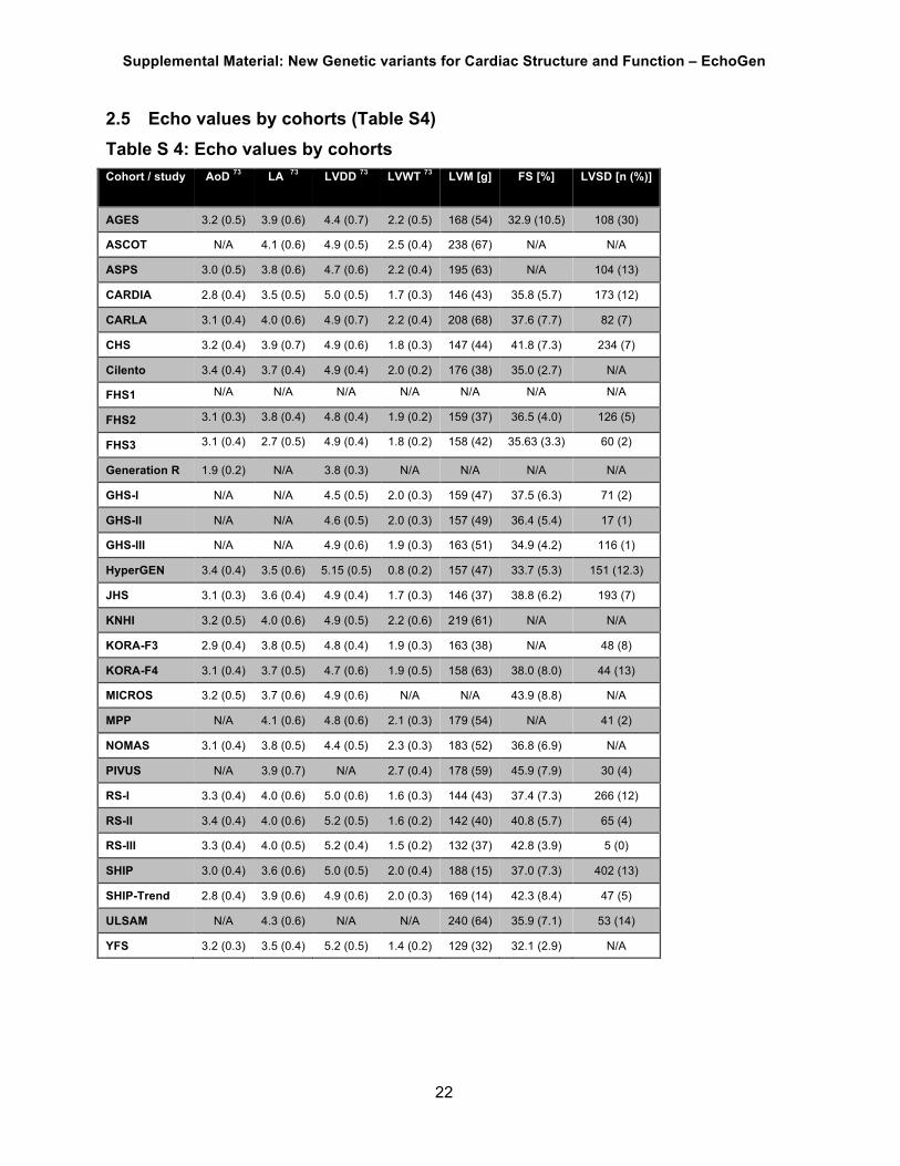
2.5 Echo values by cohorts (Table S4) Table S 4: Echo values by cohorts Cohort / study AoD 73 LA 73 LVDD 73 LVWT 73 LVM [g] FS [%] LVSD [n (%)]
AGES 3.2 (0.5) 3.9 (0.6) 4.4 (0.7) 2.2 (0.5) 168 (54) 32.9 (10.5) 108 (30)
ASCOT N/A 4.1 (0.6) 4.9 (0.5) 2.5 (0.4) 238 (67) N/A N/A
ASPS 3.0 (0.5) 3.8 (0.6) 4.7 (0.6) 2.2 (0.4) 195 (63) N/A 104 (13)
CARDIA 2.8 (0.4) 3.5 (0.5) 5.0 (0.5) 1.7 (0.3) 146 (43) 35.8 (5.7) 173 (12)
CARLA 3.1 (0.4) 4.0 (0.6) 4.9 (0.7) 2.2 (0.4) 208 (68) 37.6 (7.7) 82 (7)
CHS 3.2 (0.4) 3.9 (0.7) 4.9 (0.6) 1.8 (0.3) 147 (44) 41.8 (7.3) 234 (7)
Cilento 3.4 (0.4) 3.7 (0.4) 4.9 (0.4) 2.0 (0.2) 176 (38) 35.0 (2.7) N/A
FHS1 N/A N/A N/A N/A N/A N/A N/A
FHS2 3.1 (0.3) 3.8 (0.4) 4.8 (0.4) 1.9 (0.2) 159 (37) 36.5 (4.0) 126 (5)
FHS3 3.1 (0.4) 2.7 (0.5) 4.9 (0.4) 1.8 (0.2) 158 (42) 35.63 (3.3) 60 (2)
Generation R 1.9 (0.2) N/A 3.8 (0.3) N/A N/A N/A N/A
GHS-I N/A N/A 4.5 (0.5) 2.0 (0.3) 159 (47) 37.5 (6.3) 71 (2)
GHS-II N/A N/A 4.6 (0.5) 2.0 (0.3) 157 (49) 36.4 (5.4) 17 (1)
GHS-III N/A N/A 4.9 (0.6) 1.9 (0.3) 163 (51) 34.9 (4.2) 116 (1)
HyperGEN 3.4 (0.4) 3.5 (0.6) 5.15 (0.5) 0.8 (0.2) 157 (47) 33.7 (5.3) 151 (12.3)
JHS 3.1 (0.3) 3.6 (0.4) 4.9 (0.4) 1.7 (0.3) 146 (37) 38.8 (6.2) 193 (7)
KNHI 3.2 (0.5) 4.0 (0.6) 4.9 (0.5) 2.2 (0.6) 219 (61) N/A N/A
KORA-F3 2.9 (0.4) 3.8 (0.5) 4.8 (0.4) 1.9 (0.3) 163 (38) N/A 48 (8)
KORA-F4 3.1 (0.4) 3.7 (0.5) 4.7 (0.6) 1.9 (0.5) 158 (63) 38.0 (8.0) 44 (13)
MICROS 3.2 (0.5) 3.7 (0.6) 4.9 (0.6) N/A N/A 43.9 (8.8) N/A
MPP N/A 4.1 (0.6) 4.8 (0.6) 2.1 (0.3) 179 (54) N/A 41 (2)
NOMAS 3.1 (0.4) 3.8 (0.5) 4.4 (0.5) 2.3 (0.3) 183 (52) 36.8 (6.9) N/A
PIVUS N/A 3.9 (0.7) N/A 2.7 (0.4) 178 (59) 45.9 (7.9) 30 (4)
RS-I 3.3 (0.4) 4.0 (0.6) 5.0 (0.6) 1.6 (0.3) 144 (43) 37.4 (7.3) 266 (12)
RS-II 3.4 (0.4) 4.0 (0.6) 5.2 (0.5) 1.6 (0.2) 142 (40) 40.8 (5.7) 65 (4)
RS-III 3.3 (0.4) 4.0 (0.5) 5.2 (0.4) 1.5 (0.2) 132 (37) 42.8 (3.9) 5 (0)
SHIP 3.0 (0.4) 3.6 (0.6) 5.0 (0.5) 2.0 (0.4) 188 (15) 37.0 (7.3) 402 (13)
SHIP-Trend 2.8 (0.4) 3.9 (0.6) 4.9 (0.6) 2.0 (0.3) 169 (14) 42.3 (8.4) 47 (5)
ULSAM N/A 4.3 (0.6) N/A N/A 240 (64) 35.9 (7.1) 53 (14)
YFS 3.2 (0.3) 3.5 (0.4) 5.2 (0.5) 1.4 (0.2) 129 (32) 32.1 (2.9) N/A
Supplemental Material: New Genetic variants for Cardiac Structure and Function – EchoGen
23
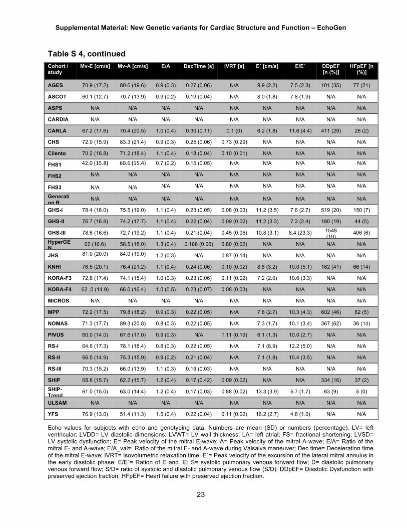
Table S 4, continued Cohort / study
Mv-E [cm/s] Mv-A [cm/s] E/A DecTime [s] IVRT [s] E´ [cm/s] E/E´ DDpEF [n (%)]
HFpEF [n (%)]
AGES 70.9 (17.2) 80.6 (19.6) 0.9 (0.3) 0.27 (0.06) N/A 9.9 (2.2) 7.5 (2.3) 101 (35) 77 (21)
ASCOT 60.1 (12.7) 70.7 (13.9) 0.9 (0.2) 0.19 (0.04) N/A 8.0 (1.8) 7.8 (1.9) N/A N/A
ASPS N/A N/A N/A N/A N/A N/A N/A N/A N/A
CARDIA N/A N/A N/A N/A N/A N/A N/A N/A N/A
CARLA 67.2 (17.6) 70.4 (20.5) 1.0 (0.4) 0.30 (0.11) 0.1 (0) 6.2 (1.8) 11.6 (4.4) 411 (29) 26 (2)
CHS 72.0 (15.9) 83.3 (21.4) 0.9 (0.3) 0.25 (0.06) 0.73 (0.29) N/A N/A N/A N/A
Cilento 70.2 (16.8) 71.2 (18.4) 1.1 (0.4) 0.18 (0.04) 0.10 (0.01) N/A N/A N/A N/A
FHS1 42.0(15.8) 60.6(15.4) 0.7 (0.2) 0.15 (0.05) N/A N/A N/A N/A N/A
FHS2 N/A N/A N/A N/A N/A N/A N/A N/A N/A
FHS3 N/A N/A N/A N/A N/A N/A N/A N/A N/A Generation R
N/A N/A N/A N/A N/A N/A N/A N/A N/A
GHS-I 78.4 (18.0) 75.5 (19.0) 1.1 (0.4) 0.23 (0.05) 0.08 (0.03) 11.2 (3.5) 7.6 (2.7) 519 (20) 150 (7)
GHS-II 76.7 (16.8) 74.2 (17.7) 1.1 (0.4) 0.22 (0.04) 0.09 (0.02) 11.2 (3.3) 7.3 (2.4) 180 (19) 44 (5)
GHS-III 78.6 (16.6) 72.7 (19.2) 1.1 (0.4) 0.21 (0.04) 0.45 (0.05) 10.8 (3.1) 8.4 (23.3) 1548 (19)
406 (6)
HyperGEN
62 (16.6) 58.5 (18.0) 1.3 (0.4) 0.186 (0.06) 0.80 (0.02) N/A N/A N/A N/A
JHS 81.0 (20.0) 84.0 (19.0) 1.2 (0.3) N/A 0.87 (0.14) N/A N/A N/A N/A
KNHI 76.5 (20.1) 76.4 (21.2) 1.1 (0.4) 0.24 (0.06) 0.10 (0.02) 8.8 (3.2) 10.0 (5.1) 162 (41) 66 (14)
KORA-F3 72.8 (17.4) 74.1 (15.4) 1.0 (0.3) 0.23 (0.06) 0.11 (0.02) 7.2 (2.0) 10.6 (3.3) N/A N/A
KORA-F4 62 .0 (14.9) 66.0 (16.4) 1.0 (0.5) 0.23 (0.07) 0.08 (0.03) N/A N/A N/A N/A
MICROS N/A N/A N/A N/A N/A N/A N/A N/A N/A
MPP 72.2 (17.5) 79.8 (18.2) 0.9 (0.3) 0.22 (0.05) N/A 7.8 (2.7) 10.3 (4.3) 602 (46) 62 (5)
NOMAS 71.3 (17.7) 89.3 (20.8) 0.8 (0.3) 0.22 (0.05) N/A 7.3 (1.7) 10.1 (3.4) 367 (62) 36 (14)
PIVUS 60.0 (14.0) 67.6 (17.0) 0.9 (0.3) N/A 1.11 (0.19) 6.1 (1.3) 10.0 (2.7) N/A N/A
RS-I 64.6 (17.3) 78.1 (18.4) 0.8 (0.3) 0.22 (0.05) N/A 7.1 (8.9) 12.2 (5.0) N/A N/A
RS-II 66.5 (14.9) 75.3 (15.9) 0.9 (0.2) 0.21 (0.04) N/A 7.1 (1.8) 10.4 (3.5) N/A N/A
RS-III 70.3 (15.2) 66.0 (13.9) 1.1 (0.3) 0.19 (0.03) N/A N/A N/A N/A N/A
SHIP 68.8 (15.7) 62.2 (15.7) 1.2 (0.4) 0.17 (0.42) 0.09 (0.02) N/A N/A 334 (16) 37 (2)
SHIP-Trend
61.0 (15.0) 63.0 (14.4) 1.2 (0.4) 0.17 (0.03) 0.88 (0.02) 13.3 (3.9) 5.7 (1.7) 63 (9) 5 (0)
ULSAM N/A N/A N/A N/A N/A N/A N/A N/A N/A
YFS 76.9 (13.0) 51.4 (11.3) 1.5 (0.4) 0.22 (0.04) 0.11 (0.02) 16.2 (2.7) 4.8 (1.0) N/A N/A
Echo values for subjects with echo and genotyping data. Numbers are mean (SD) or numbers (percentage). LV= left ventricular; LVDD= LV diastolic dimensions; LVWT= LV wall thickness; LA= left atrial; FS= fractional shortening; LVSD= LV systolic dysfunction; E= Peak velocity of the mitral E-wave; A= Peak velocity of the mitral A-wave; E/A= Ratio of the mitral E- and A-wave; E/A_val= Ratio of the mitral E- and A-wave during Valsalva maneuver; Dec time= Deceleration time of the mitral E-wave; IVRT= Isovolumetric relaxation time; E´= Peak velocity of the excursion of the lateral mitral annulus in the early diastolic phase; E/E´= Ration of E and ´E; S= systolic pulmonary venous forward flow; D= diastolic pulmonary venous forward flow; S/D= ratio of systolic and diastolic pulmonary venous flow (S/D); DDpEF= Diastolic Dysfunction with preserved ejection fraction; HFpEF= Heart failure with preserved ejection fraction.

Supplemental Material: New Genetic variants for Cardiac Structure and Function – EchoGen
24
2.6 Genotyping Methods and Imputation (Table S5) Table S 5: Genotyping information by cohorts Cohort / study
Array type Genotype calling
QC filters for genotyped SNPs used for imputation
No of SNPs
used for imputatio
n
Imputation
software
Imputation Backbone for phased
CEU haplotypes
(NCBI build)
Filtering of
imputed genotypes
Data management and statistical
analysis
Population stratification or Principal Component
s
AGES Illumina 370CNV Illumina BeadStudio
call rate < 97% pHWE< 1 x 10⁻⁶, mishap (PLINK
haplotype-based test for non-random missing genotype data[2])
p < 1 x 10⁻⁹, and mismatched positions between Illumina,
dbSNP and/or HapMap
325,094 MACH version 1.0.16
HapMap, release 22 (build 36)
None ProABEL None
ASCOT Illumina HumanCNV370-
Duo
Illumina BeadStudio
CR<0.97, MAF<0.05, HWE_pvalue<1e-07
283,291 BEAGLE
HapMap II release 24
CEU reference
panel
Information score<0.6, MAF<5%
PLINK, R Adjusted for first 10PCs
ASPS Illumina Human 610-Quad BeadChip
Illuminus call rate <97.5%, MAF <1%, ,
pHWE <1E-6
550,635 MACH v1.0.15
HapMap, release 22 (build 36)
None SPSS, ProbABEL, R
None
CARDIA Affymetrix 6.0 BEAGLE, Birdseed
call rate <95%, MAF<3%, pHWE
<10E-4
579,630 BEAGLE
HapMap, release 22 (build 36)
Rsq<0.3, MAF<1%
ProbABEL, PLINK, R
None
CARLA ABI N/A N/A N/A N/A NCBI built 37
N/A R version 2.14.1 None
CHS Illumina 370CNV BeadChip
system
Illumina BeadStudio
call rate < 97%, HWE P < 10-5, > 2 duplicate errors or Mendelian inconsistencies (for reference
CEPH trios), heterozygote frequency = 0, SNP not found in
HapMap.
306,655 BIMBAM
v0.99
HapMap, release 22 (build 36)
SNPs were excluded
for variance on the allele
dosage ≤0.01
R, robust SE estimates
None

Supplemental Material: New Genetic variants for Cardiac Structure and Function – EchoGen
25
Table S 5, continued Cohort / study
Array type Genotype calling
QC filters for genotyped SNPs used for imputation
No of SNPs
used for imputati
on
Imputation software
Imputation Backbone for phased
CEU haplotypes (NCBI build)
Filtering of
imputed genotype
s
Data management and statistical
analysis
Population stratification or
Principal Components
Cilento Illumina 370K (n=859) Illumina OmniExpress(n=
758)
Illumina BeadStudio
Imputation was performed using the following filters: SNPs in common
between the two arrays, call rate<95%, MAF<1%. For the
directly typed SNPs in common between the two groups, the real
genotype was used in the association analysis, while the
imputation dosage was considered for the other SNPs.
190862 MACH, minimac
1000G Phase I integrated
Release Version 3 (build 37)
None R, linear model, GenABEL and
ProbABEL (mmscore
function was used to account for relatedness)
none
FHS1, FHS2, FHS3
Affymetrix 500K; Affymetrix 50K supplemental
BRLMM pHWE<1e-6, call rate<97%, mishap p<1e-9, MAF<0.01, Mendelian
errors>100, SNPs not in Hapmap or strandedness issues merging with
Hapmap
378,163 MACH version 1.0.15
HapMap, release 22 (build 36)
None R, linear mixed effect models
and GEE models, robust variance option to account for relatedness
Adjused for first PC estimated
from Eigenstrat associated with
calcium levels (P <0.05).(35)
Generation R
Ilumina 610 Quad and 660W
Genomestudio 2009 V.1.1.9
call rate <98%, MAF <1%, pHWE <10E-6
492.871 MACH and mini-mac
HapMap release 22
None MACH2QTL Adjusted for the first 4 PC's
GHS-I, -II, -III
Affymetrix SNP 6.0
Birdseed2 call rate ≤98%, MAF ≤0.01 and pHWE≤0.0001
662,405 (GHS-I) 673,914 (GHS-II)
IMPUTE v2.1.0
HapMap, release 24 (build 36)
None MetABEL, R None
HyperGEN Affymetrix 5.0 Birdsuite monomorphic SNPs, X-chromosomem SNPs, non-
Mendelian inheritance errors, missing rate >5%, MAF<1%,Hardy-
Weinberg p-value <10-6
358327 MACH HapMap, release 22 (build 36)
Rsq<0.3, MAF<1%, MAF<1%, HWP <10-
6
R, LMEKIN package
Adjusted for 10 PCs
JHS Affymetrix SNP 6.0
Birdseed2 pHWE<1e-6, call rate<95%, MAF<0.01, Imputation quality >0.30
868,969 (JHS)
796,384 (ARIC-JHS)
MACH version 1.0.16
HapMap, release 22 (build 36)
None ProbABEL Adjusted for the first 10 PCs

Supplemental Material: New Genetic variants for Cardiac Structure and Function – EchoGen
26
Table S 5, continued Cohort / study
Array type Genotype calling
QC filters for genotyped SNPs used for imputation
No of SNPs used for
imputation
Imputation
software
Imputation Backbone for phased
CEU haplotypes
(NCBI build)
Filtering of imputed
genotypes
Data management and statistical
analysis
Population stratification or Principal
Components
KNHI Affymetrix 500K (n= 127),
Affymetrix 5.0 (n= 17) Affymetrix
6.0 (n = 417)
Affy 500K: BRLMM Affy 5.0:
BRLMM-P Affy 6.0:
Birdseed2
per chip type: SNP call rate <95%, MAF <0.01, pHWE <10-5; in sample: SNP call rate <90%;
exclusion of SNPs with discordance rates >5% between
chip types, exclusion of discordant genotypes for subjects typed on
>1chip
SNPs with call rate >90% in sample: 384,234 (out of
736,307)
MACH "HapMap, release 22 (build 36)
dbSNP 126
None (QC filters on meta-
level: RSQ < 0.6)
PLINK, R, linear models
with ProbABEL, GenABEL
None (subjects
excluded if related, wrong
ethnicity, sex-
mismatch, or individual call rate <90%)
KORA-F3 Affymetrix 500K BRLMM pHWE<1e-6, individual call rate<93%, snp call rate<95%,
MAF<0.01
379,392 Impute v1.0.0
HapMap, release 22 (build 36)
None R, linear models
None
KORA-F4 Affymetrix 6.0 Birdseed2 On chip level only subjects with overall genotyping efficiencies of at least 93% were included resulting in an average genotyping efficiency of 93% per chip. In addition the called sex had to agree with the sex in the
KORA study database.
630,550 MACH v1.0.16
HapMap, release 22 (build 36)
None R, MACH2QTL, ProbABEL
None
LURIC Affymetrix 6.0 Birdseed2 pHWE<1e-4, individual call rate<95%, snp call rate<98%,
MAF<0.01
686,195 MACH v1
HapMap, release 22 (build 36)
None PLINK, R, SPSS
None
MAGNet Affymetrix 6.0 Birdseed2 genotype call rate > 95% minor allele frequency (MAF) > 15%
Hardy-Weinberg equilibrium (p > 10-6)
360,046 Minimac (v2012.1
1.16) program
N/A imputation quality
threshold of 0.5 and a
MAF threshold of
0.15
PLINK,R None
MICROS HumanHap 300v2 Illumina BeadStudi
o
call rate <98%, MAF <1%, pHWE
<10E-6
290,356 MACH v1
HapMap, release 22 (build 36)
None ProbABEL,mmscore
argument
None
MPP Sequenom MassArray iPlex
MassArray Typer 4.0
N/A N/A N/A N/A N/A SAS 9.2 None

Supplemental Material: New Genetic variants for Cardiac Structure and Function – EchoGen
27
Table S 5, continued Cohort / study
Array type Genotype calling
QC filters for genotyped SNPs used for
imputation
No of SNPs used for
imputation
Imputation
software
Imputation Backbone for phased CEU
haplotypes (NCBI build)
Filtering of
imputed
genotypes
Data management
and statistical analysis
Population stratification or
Principal Components
NOMAS Affymetrix SNP 6.0
Affymetric Power Tools
call rate<95%, pHWE<1e-6
804,944 (Black) 815,972
(Hispanic) 795,588 (White)
IMPUTE2
1000 Genomes phase 1, version 3
reference panel
None SAS, PLINK Adjusted for PCs estimated using
Eigenstrat (first 2 PCs for Hispanics, first PC for Blacks and Whites)
PIVUS Human Omni Express and Metabochip
Illumina BeadStudio
For SNPs with MAF >=0.05: pHWE<1e-6, call rate<95%; For SNPs with MAF <0.05: pHWE<1e-6, call rate<99%; MAF<0.01
738,879 IMPUTE version 2.1.2
HapMap, release 22 (build 36)
None SNPTEST Adjusted for the first 2 PCs estimated from MDS using PLINK
RS-I,-II, -III
550K (RS-I and RS-II) and 610K (RS-III)
Illumina arrays
Illumina BeadStudio
Genecall
pHWE<1e-6, SNP call rate<97.5%, MAF<0.01
RS-I: 512,349; RS-II:
466,389; RS-III: 514,073
MACH HapMap, release 22 (build 36)
None R, ProbABEL None
SHIP Affymetrix SNP 6.0
Birdseed2 None 869,224 IMPUTE v0.5.0
HapMap, release 22 (build 36)
duplicate
RSID but
different
positions
QUICKTEST version 0.95 (Params: --
method-score),
InforSense, InterSystems
Caché
We observed no population stratification
using principle components estimated
using Eigenstrat. [Rice et al. PMID: 16862161]
SHIP-Trend
Illumina Human Omni
2.5
GenomeStudio Genotyping Module v1.0
excluded: pHWE <= 0.0001 or CallRate <= 0.9
or monomorphic SNPs
1,782,967 IMPUTE v2.1.2.3
HapMap, release 22 (build 36)
duplicate
RSID but
different
positions
QUICKTEST version 0.95 (Params: --
method-score),
InforSense, InterSystems
Caché
We observed no population stratification
using principle components estimated
using Eigenstrat. [Rice et al. PMID: 16862161]
ULSAM Human Omni Express and Metabochip
Illumina BeadStudio
For SNPs with MAF >=0.05: pHWE<1e-6, call rate<95%; For SNPs with MAF <0.05: pHWE<1e-6, call rate<99%; MAF<0.01
738,879 IMPUTE HapMap, release 22 (build 36)
None SNPTEST Adjusted for the first 2 PCs estimated from MDS using PLINK
YFS Illumina 670k Illuminus call rate <95%, MAF<1%, pHWE
<10E-6
546,677 SHAPEIT v1,
IMPUTE v2.2.2
1000 Genomes Phase I (build 37)
None R, SNPTEST None

Supplemental Material: New Genetic variants for Cardiac Structure and Function – EchoGen
28
3 Additional tables and figures (Table S6-S19, Figures S1-S20)
Table S 6: Individual study lambdas for left ventricular structure and systolic function echocardiographic traits
Study AoD LA LVDD LVWT LVM FS LVSD
AGES 1.01 1.03 0.99 0.98 1.00 1.02 1.00
ASCOT NA 1.01 1.01 0.99 0.94 NA NA
ASPS 1.01 1.02 0.99 1.01 0.99 NA 1.01
CARDIA 1.02 0.99 0.99 1.00 1.01 1.00 1.02
CHS 1.03 1.09 1.02 1.00 1.02 1.01 1.01
FHS-2 1.03 1.04 1.01 1.03 1.04 1.01 0.99
FHS-3 1.03 1.02 1.02 1.03 1.03 1.03 1.03
GHS-I NA NA 1.01 1.02 1.01 1.01 1.01
GHS-II NA NA 1.00 1.01 1.01 1.00 0.99
HyperGen 1.10 1.08 1.12 1.16 1.10 1.10 1.10
KNHI 1.00 1.02 1.01 1.01 0.99 NA NA
KORA-F3 1.00 1.01 1.00 1.00 0.99 0.99 1.01
KORA-F4 1.01 1.01 1.02 1.00 1.00 1.01 0.99
MICROS 1.01 1.02 1.03 1.02 NA 1.01 NA
PIVUS 1.01 1.00 1.02 1.01 1.02 0.98 0.97
RS-I 1.02 1.02 1.00 1.01 1.01 1.01 1.01
RS-II 0.98 1.01 1.00 0.98 1.00 1.00 0.99
RS-III 1.03 1.03 1.03 1.02 1.04 1.01 NA
SHIP 1.02 1.02 1.01 1.02 0.99 1.02 1.01
SHIP-Trend 1.01 1.01 1.02 1.00 1.01 1.00 0.99
ULSAM NA 1.02 NA NA 1.00 1.00 1.03

Supplemental Material: New Genetic variants for Cardiac Structure and Function – EchoGen
29
Table S 6, continued: Individual study lambdas for left ventricular structure and systolic function echocardiographic traits Study Mv-E Mv-A E/A DecTime IVRT E´ E/E´ DDpEF HFpEF
AGES 0.99 0.99 1.00 1.02 NA 0.99 0.97 1.01 1.00
ASCOT 1.01 0.99 1.01 1.01 NA 1.01 1.00 NA NA
CARDIA 1.00 1.00 1.01 NA 1.01 NA NA NA NA
CHS 1.02 1.01 1.01 1.03 1.02 NA NA NA NA
FHS-1 1.02 1.03 1.01 1.03 NA NA NA NA NA
GHS-I 1.01 1.01 1.00 1.01 1.00 1.02 1.02 1.02 1.00
GHS-II 1.01 1.003 1.01 1.01 0.99 1.01 1.01 1.00 0.99
HyperGen 1.16 1.11 1.01 1.09 1.07 NA NA NA NA
KNHI 1.00 0.99 0.99 1.02 0.99 0.99 1.00 1.03 0.94
KORA-F3 1.01 1.01 1.01 1.01 1.04 1.00 1.00 1.02 1.05
KORA-F4 1.00 0.99 1.01 NA 0.99 NA NA NA NA
PIVUS 1.00 1.01 1.01 NA 1.02 1.01 1.02 NA NA
RS-I 1.01 1.02 1.02 1.01 NA 1.01 1.01 NA NA
RS-II 1.01 1.01 0.98 0.98 NA NA NA NA NA
RS-III 1.02 1.02 1.03 1.00 NA NA NA NA NA
SHIP 1.00 1.01 1.01 1.01 1.02 NA NA 0.99 1.02
SHIP-Trend 1.00 1.00 1.01 1.01 1.00 1.00 NA 1.03 NA

Supplemental Material: New Genetic variants for Cardiac Structure and Function – EchoGen
30
Table S 7: Meta-analysis results of SNPs per phenotype with a P value < 10-4
and
MAF < 0.01 (LVSD: MAF < 0.03)
See Supplemental Excel file “Supplemental Table 7.xls”

Supplemental Material: New Genetic variants for Cardiac Structure and Function – EchoGen
31
Table S 8: Lookup of novel findings for left ventricular structure and systolic function echocardiographic traits in Caucasian children and generalizability to different ethnicities
Subpopulation Caucasian Children* Hispanics African Americans Cohort Generation R Northern Manhattan Study Northern Manhattan Study &
Jackson Heart Study
Trait SNP Chr Effect/
non-effect allele
EAF Effect (SE)
P value N EAF Effect
(SE) P
value N EAF1 Effect (SE)
P value N
AoD, cm
rs806322 13 A/G 0.61 -0.017 (0.005) 6.65 x 10-4* 2070 0.49 0.009
(0.020) 0.638 515 0.48 -0.01 (0.01) 0.54 1300
rs6702619 1 G/T 0.48 0.011 (0.005) 0.024 2070 0.28 0.017
(0.020) 0.408 515 0.14 0.03 (0.02) 0.19 1300
rs17696696 16 G/T 0.60 -0.002 (0.005) 0.676 2070 0.49 -0.008
(0.019) 0.682 515 0.34 -0.02 (0.01) 0.07 1300
rs7127129 11 G/A 0.42 -0.006 (0.005) 0.260 2070 0.29 -0.031
(0.021) 0.130 515 0.18 -0.01 (0.02) 0.62 1300
rs17608766 17 C/T 0.15 0.019 (0.007) 5.92 x 10⁻³ 2070 0.09 0.003
(0.039) 0.934 515 0.03 0.07 (0.04) 0.05 1300
rs4765663 12 C/G 0.14 -0.001 (0.008) 0.908 2070 0.11 0.006
(0.029) 0.845 515 0.08 0.02 (0.02) 0.24 1300
rs11207426 1 A/G 0.38 0.004 (0.005) 0.429 2070 0.31 0.024
(0.021) 0.241 515 0.45 -0.01 (0.01) 0.53 1300
LVDD, cm
rs12541595 8 T/G 0.31 -0.002 (0.008) 0.776 2069 0.23 0.032
(0.027) 0.225 785 0.07 0.02 (0.02) 0.44 1576
rs10774625 12 G/A 0.52 0.007 (0.008) 0.362 2069 0.70 0.002
(0.025) 0.930 785 0.93 -0.001 (0.03) 0.96 1576
Mv-A rs12440869 15 T/A Not available 0.34 -0.621 (1.164) 0.594 575 0.34 0.41 (0.94) 0.66 520
Betas in italics represent directional consistency with meta-analysis results; Bold results represent nominally significant findings (P < 0.05), results marked with * are significant after Bonferroni correction for 10 SNPS (P < 0.005) 1 From Jackson Heart Study 2 Northern Manhattan Study N = 97 for AoD and 194 for LVDD; Jackson Heart Study N = 1203 for AoD and 1382 for LVDD AoD= diameter of the aortic root; LVDD= LV diastolic internal dimension; Chr= chromosome; EAF= effect allele frequency; SE= standard error

Supplemental Material: New Genetic variants for Cardiac Structure and Function – EchoGen
32
Table S9. Generalizability of novel findings for echocardiographic traits – Effect of genome-wide significant SNPs associated with aortic root diameter on pulse wave velocity in the AortaGen consortium
Trait SNP Effect / non-effect allele Effect (SE) P
Pulse wave velocity, m/s
rs806322 A/G 0.005 (0.012) 0.689rs6702619 G/T 0.003 (0.011) 0.805
rs17696696 G/T 0.042 (0.011) 1.55 x 10⁻⁴
rs7127129 G/A 0.000 (0.011) 0.979 rs17608766 C/T -0.038 (0.016) 0.018 rs4765663 C/G 0.007 (0.015) 0.630
rs11207426 A/G 0.028 (0.012) 0.021
SE= standard error.

Supplemental Material: New Genetic variants for Cardiac Structure and Function – EchoGen
33
Table S10. Generalizability of novel findings for echocardiographic traits – Effect of genome-wide significant SNPs on incident heart failure HF and mortality in patients with HF the CHARGE – Heart Failure consortium
HR= hazard ratio; CI= confidence interval; HF= heart failure.
SNP Effect/ non-effect allele
HR (95% CI) Incident HF
P Incident HF
HR (95% CI) Mortality in HF
P Mortality in HF
rs806322 A/G 0.97 (0.91-1.03) 0.306 1.00 (0.93-1.08) 0.971
rs6702619 G/T 0.99 (0.93-1.04) 0.636 1.02 (0.95-1.09) 0.635
rs17696696 G/T 1.00 (0.94-1.06) 0.950 1.06 (0.99-1.14) 0.103
rs7127129 G/A 0.99 (0.93-1.05) 0.705 1.00 (0.93-1.08) 0.941
rs17608766 C/T 1.11 (1.01-1.21) 0.033 0.98 (0.87-1.11) 0.799
rs4765663 C/G 1.04 (0.97-1.12) 0.254 1.01 (0.92-1.10) 0.907
rs11207426 A/G 1.07 (1.01-1.14) 0.027 1.04 (0.97-1.12) 0.289
rs12541595 T/G 0.96 (0.90-1.03) 0.232 1.00 (0.92-1.08) 0.999
rs10774625 G/A 1.00 (0.95-1.06) 0.963 1.01 (0.94-1.09) 0.726
rs12440869 T/A 1.04 (0.97-1.11) 0.263 0.93 (0.86-1.00) 0.062

Supplemental Material: New Genetic variants for Cardiac Structure and Function – EchoGen
34
Table S11. Generalizability of novel findings for echocardiographic traits – Effect of the novel genome-wide significant SNPs on all-cause,
cardiovascular and heart failure mortality in the LURIC cohort of patients with suspected coronary artery disease
SNP Effect/
non-effect allele
HR (95% CI) Mortality
P Mortality
HR (95% CI) CV mortality
P CV
mortality HR (95% CI) HF mortality
P HF
mortality rs806322 A/G 1.09 (0.99-1.20) 0.079 1.05 (0.93-1.19) 0.428 0.83 (0.65-1.07) 0.145
rs6702619 G/T 1.01 (0.92-1.11) 0.806 0.99 (0.88-1.11) 0.827 0.87 (0.68-1.12) 0.279 rs17696696 G/T 0.93 (0.85-1.03) 0.146 0.96 (0.85-1.08) 0.451 0.94 (0.74-1.21) 0.642 rs7127129 G/A 0.95 (0.86-1.04) 0.275 0.97 (0.86-1.09) 0.600 0.85 (0.66-1.09) 0.205
rs17608766 C/T 0.98 (0.86-1.12) 0.758 0.97 (0.82-1.15) 0.717 0.92 (0.64-1.31) 0.638 rs4765663 C/G 1.01 (0.89-1.14) 0.875 0.95 (0.81-1.11) 0.517 1.07 (0.77-1.47) 0.701
rs11207426 A/G 1.08 (0.98-1.19) 0.109 1.05 (0.93-1.19) 0.399 1.43 (1.13-1.83) 4.0 x 10⁻³ rs12541595 T/G 0.99 (0.89-1.09) 0.788 0.97 (0.85-1.10) 0.615 0.96 (0.74-1.26) 0.784 rs10774625 G/A 0.95 (0.86-1-04) 0.238 0.94 (0.83-1.05) 0.273 0.89 (0.70-1.14) 0.368 rs12440869 T/A 0.97 (0.87-1.08) 0.546 0.90 (0.78-1.03) 0.131 0.95 (0.71-1.26) 0.710
HR= hazard ratio; CI= confidence interval; CV= cardiovascular; HF= heart failure.

Supplemental Material: New Genetic variants for Cardiac Structure and Function – EchoGen
35
Table S12. Generalizability of novel findings for echocardiographic traits – Effect of the novel genome-wide significant SNPs on myocardial infarction (MI) and coronary artery disease (CAD) in the CARDIOGRAMplusC4D Consortium
SNP Effect/
non-effect allele
HR (95% CI) MI
P MI HR (95% CI)
CAD P
CAD
rs806322 A/G 1.00 (0.98-1.02) 0.696 1.00 (0.98-1.02) 0.912
rs6702619 G/T 0.99 (0.97-1.01) 0.288 1.00 (0.98-1.02) 0.960
rs17696696 G/T 1.03 (1.00-1.05) 0.021 1.04 (1.02-1.06) 8.45 x 10⁻⁶
rs7127129 G/A 1.02 (1.00-1.05) 0.023 1.02 (1.00-1.03) 0.113
rs17608766 C/T 1.05 (1.02-1.09) 0.002 1.05(1.02-1.09) 4.94 x 10⁻⁴
rs4765663 C/G 0.99 (0.96-1.01) 0.323 0.99 (0.97-1.02) 0.551
rs11207426 A/G 0.99 (0.97-1.01) 0.218 0.98 (0.96-0.99) 0.011
rs12541595 T/G 1.00 (0.98-1.03) 0.736 1.00 (0.99-1.03) 0.636
rs10774625 G/A 0.93 (0.91-0.95) 5.09 x 10⁻¹¹ 0.94 (0.92-0.96) 2.69 x 10⁻¹⁰
rs12440869 T/A 1.00 (0.98-1.03) 0.688 1.00 (0.98-1.02) 0.799
HR= hazard ratio; CI= confidence interval; MI= myocardial infarction; CAD = coronary artery
disease.

Supplemental Material: New Genetic variants for Cardiac Structure and Function – EchoGen
36
Table S 13: Top canonical pathways enriched with echo-related genes from Ingenuity Pathway Analysis
Ingenuity Canonical Pathways P value FDR Ratio Genes Protein Kinase A Signaling 5.8 x 10⁻⁶ 0.002 19/386 ADCY8, CAMK2B,
CDC27, CREB5,
DUSP12, DUSP18,
FLNC, GLI3, GNB4,
HIST2H3C, LEF1,
MYL4, NFKBIA,
OPN1SW, PLCE1,
PLCG1, PLN,
PRKCA, TTN
Death Receptor Signaling 6.9 x 10⁻⁵ 0.012 8/92 ACTA2, ART1,
CRADD, FADD,
FAS, LIMK1,
NFKBIA, TNFRSF1A
Wnt/Ca+ pathway 2.2 x 10⁻⁴ 0.019 6/56 AXIN1, CREB5,
DVL1, PLCE1,
PLCG1, PRKCA
P2Y Purigenic Receptor Signaling 4.1 x 10⁻⁴ 0.028 8/119 ADCY8, CREB5,
GNB4, ITGB3,
OPN1SW, PLCE1,
PLCG1, PRKCA
FDR: False discovery rate; Ratio: number of molecules in a given pathway that meet cut criteria, divided by total number of molecules that make up that pathway

Supplemental Material: New Genetic variants for Cardiac Structure and Function – EchoGen
37
Table S 14: Top canonical pathways enriched with echo-related genes from Ingenuity Pathway Analysis: additional informations on gene loci Gene Length Most significant SNP
within gene region Locus
ACTA2 56317 rs2862834 10q23.31
ADCY8 260289 rs263255 8q24.22
ART1 19286 rs7103680 11p15.4
AXIN1 65237 rs2685127 16p13.3
CAMK2B 108482 rs7804804 7p13
CDC27 71355 rs2292864 17q21.32
CRADD 173381 rs10859579 12q22
CREB5 526572 rs917275 7p15.1
DUSP12 7372 rs953301 1q23.3
DUSP18 5834 rs734479 22q12.2
DVL1 13835 rs12142199 1p36.33
FADD 4240 rs7127129 11q13.3
FAS 25255 rs2862834 10q23.31
FLNC 28846 rs7786074 7q32.1
GLI3 276071 rs7782675 7p14.1
GNB4 55496 rs4855074 3q26.33
HIST2H3C 507 rs532941 1q21.2
ITGB3 58870 rs2292864 17q21.32
LEF1 121412 rs7665502 4q25
LIMK1 38749 rs4717863 7q11.23
MYL4 14618 rs3892085 17q21.32
NFKBIA 3245 rs8019505 14q13.2
OPN1SW 3302 rs7786074 7q32.1
PLCE1 239319 rs3758524 10q23.33
PLCG1 38197 rs2866372 20q12
PLN 12146 rs11967375 6q22.31
PRKCA 507937 rs6504434 17q24.2
TNFRSF1A 13361 rs7958488 12p13.31
TTN 281432 rs12052983 2q31.2

Supplemental Material: New Genetic variants for Cardiac Structure and Function – EchoGen
38
Table S 15: DEPICT
Results of the DEPICT analyses are summarized in the Supplemental Excel file
“Supplemental Table 15.xlsx”
Supplemental Material: New Genetic variants for Cardiac Structure and Function – EchoGen
39
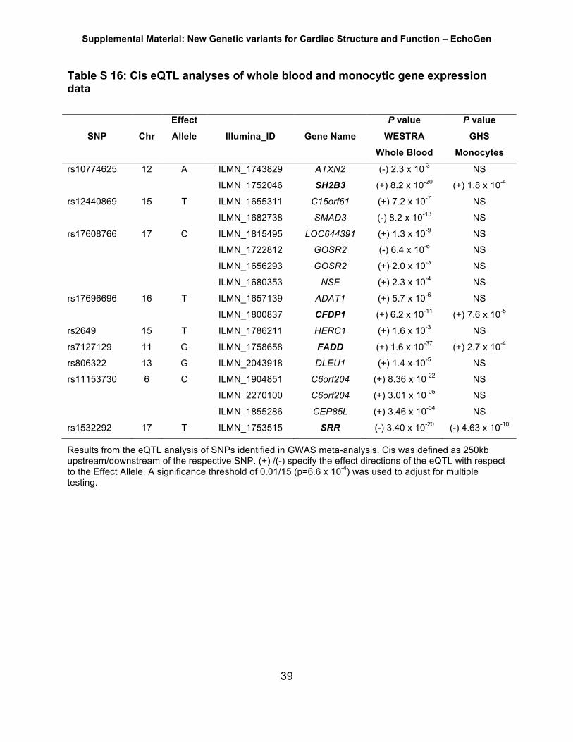
Table S 16: Cis eQTL analyses of whole blood and monocytic gene expression data
Effect P value P value SNP Chr Allele Illumina_ID Gene Name WESTRA
Whole Blood GHS
Monocytes
rs10774625 12 A ILMN_1743829 ATXN2 (-) 2.3 x 10⁻³ NS
ILMN_1752046 SH2B3 (+) 8.2 x 10⁻²⁰ (+) 1.8 x 10⁻⁴
rs12440869 15 T ILMN_1655311 C15orf61 (+) 7.2 x 10⁻⁷ NS
ILMN_1682738 SMAD3 (-) 8.2 x 10⁻¹³ NS
rs17608766 17 C ILMN_1815495 LOC644391 (+) 1.3 x 10⁻⁹ NS
ILMN_1722812 GOSR2 (-) 6.4 x 10⁻⁶ NS
ILMN_1656293 GOSR2 (+) 2.0 x 10⁻³ NS
ILMN_1680353 NSF (+) 2.3 x 10⁻⁴ NS
rs17696696 16 T ILMN_1657139 ADAT1 (+) 5.7 x 10⁻⁶ NS
ILMN_1800837 CFDP1 (+) 6.2 x 10⁻¹¹ (+) 7.6 x 10⁻⁵
rs2649 15 T ILMN_1786211 HERC1 (+) 1.6 x 10⁻³ NS
rs7127129 11 G ILMN_1758658 FADD (+) 1.6 x 10-37 (+) 2.7 x 10⁻⁴
rs806322 13 G ILMN_2043918 DLEU1 (+) 1.4 x 10⁻⁵ NS
rs11153730 6 C ILMN_1904851 C6orf204 (+) 8.36 x 10⁻²² NS
ILMN_2270100 C6orf204 (+) 3.01 x 10-05 NS
ILMN_1855286 CEP85L (+) 3.46 x 10-04 NS
rs1532292 17 T ILMN_1753515 SRR (-) 3.40 x 10⁻²⁰ (-) 4.63 x 10⁻¹⁰
Results from the eQTL analysis of SNPs identified in GWAS meta-analysis. Cis was defined as 250kb upstream/downstream of the respective SNP. (+) /(-) specify the effect directions of the eQTL with respect to the Effect Allele. A significance threshold of 0.01/15 (p=6.6 x 10⁻⁴) was used to adjust for multiple testing.

Supplemental Material: New Genetic variants for Cardiac Structure and Function – EchoGen
40
Table S 17: Significant eQTLs in the Genotype-Tissue Expression (GTEx) database
SNPID GencodeID GeneSymbolEffectallele Pvalue Effectsize Tissue
rs17696696 ENSG00000261783.1 RP11-252K23.2 G 2.00E-23 -0.77 Cells-Transformedfibroblastsrs17696696 ENSG00000261783.1 RP11-252K23.2 G 2.50E-22 -0.69 Thyroidrs17696696 ENSG00000261783.1 RP11-252K23.2 G 1.70E-20 -0.87 Adipose-Visceral(Omentum)rs17696696 ENSG00000153774.4 CFDP1 G 6.20E-17 -0.34 Cells-Transformedfibroblastsrs17696696 ENSG00000050820.12 BCAR1 G 8.40E-17 -0.48 Esophagus-Mucosars17696696 ENSG00000261783.1 RP11-252K23.2 G 3.10E-16 -0.72 Artery-Aortars17696696 ENSG00000261783.1 RP11-252K23.2 G 8.80E-16 -0.63 Esophagus-Mucosars17696696 ENSG00000261783.1 RP11-252K23.2 G 1.60E-15 -0.59 Lungrs17696696 ENSG00000261783.1 RP11-252K23.2 G 5.70E-15 -0.58 Artery-Tibialrs17696696 ENSG00000261783.1 RP11-252K23.2 G 6.80E-15 -0.58 Nerve-Tibialrs17696696 ENSG00000261783.1 RP11-252K23.2 G 2.70E-14 -0.52 Adipose-Subcutaneousrs17696696 ENSG00000261783.1 RP11-252K23.2 G 1.60E-10 -0.58 Esophagus-Muscularisrs17696696 ENSG00000050820.12 BCAR1 G 1.70E-10 0.26 Artery-Aortars17696696 ENSG00000261783.1 RP11-252K23.2 G 6.90E-10 -0.69 AdrenalGlandrs17696696 ENSG00000050820.12 BCAR1 G 7.30E-10 0.19 Artery-Tibialrs17696696 ENSG00000261783.1 RP11-252K23.2 G 1.30E-09 -0.72 Pancreasrs17696696 ENSG00000261783.1 RP11-252K23.2 G 1.30E-09 -0.45 Skin-SunExposed(Lowerleg)rs17696696 ENSG00000261783.1 RP11-252K23.2 G 2.00E-09 -0.56 Breast-MammaryTissuers17696696 ENSG00000261783.1 RP11-252K23.2 G 1.50E-08 -0.43 WholeBloodrs17696696 ENSG00000261783.1 RP11-252K23.2 G 1.60E-08 -0.62 Artery-Coronaryrs17696696 ENSG00000153774.4 CFDP1 G 1.10E-07 -0.19 Adipose-Subcutaneousrs17696696 ENSG00000261783.1 RP11-252K23.2 G 2.50E-07 -0.63 Vaginars17696696 ENSG00000261783.1 RP11-252K23.2 G 3.00E-07 -0.63 Colon-Sigmoidrs17696696 ENSG00000261783.1 RP11-252K23.2 G 5.10E-07 -0.72 Brain-CerebellarHemispherers17696696 ENSG00000261783.1 RP11-252K23.2 G 5.30E-07 -0.64 Prostaters17696696 ENSG00000166822.8 TMEM170A G 6.30E-07 0.23 Skin-SunExposed(Lowerleg)rs17696696 ENSG00000261783.1 RP11-252K23.2 G 1.40E-06 -0.53 Stomachrs17696696 ENSG00000153774.4 CFDP1 G 1.70E-06 -0.17 Skin-SunExposed(Lowerleg)rs17696696 ENSG00000153774.4 CFDP1 G 1.80E-06 -0.41 Brain-Hippocampusrs17696696 ENSG00000166822.8 TMEM170A G 2.00E-06 -0.21 Nerve-Tibialrs17696696 ENSG00000261783.1 RP11-252K23.2 G 4.00E-06 -0.47 Colon-Transversers17696696 ENSG00000153774.4 CFDP1 G 6.10E-06 -0.17 Nerve-Tibialrs7127129 ENSG00000254721.1 RP11-805J14.5 G 5.40E-10 0.23 Cells-Transformedfibroblastsrs7127129 ENSG00000168040.4 FADD G 2.10E-09 0.22 Cells-Transformedfibroblastsrs17608766 ENSG00000179673.3 RPRML C 1.60E-10 -0.56 Muscle-Skeletalrs17608766 ENSG00000179673.3 RPRML C 3.00E-05 -0.44 Adipose-Subcutaneousrs11207426 ENSG00000224609.2 RP11-470E16.1 A 7.20E-21 0.68 Artery-Tibialrs11207426 ENSG00000224609.2 RP11-470E16.1 A 8.00E-16 0.49 Adipose-Subcutaneousrs11207426 ENSG00000224609.2 RP11-470E16.1 A 4.20E-15 0.62 Artery-Aortars11207426 ENSG00000224609.2 RP11-470E16.1 A 7.90E-15 0.56 Nerve-Tibialrs11207426 ENSG00000224609.2 RP11-470E16.1 A 6.70E-12 0.5 Skin-SunExposed(Lowerleg)rs11207426 ENSG00000224609.2 RP11-470E16.1 A 8.90E-08 0.53 Artery-Coronaryrs11207426 ENSG00000224609.2 RP11-470E16.1 A 1.50E-07 -0.23 Muscle-Skeletalrs11207426 ENSG00000224609.2 RP11-470E16.1 A 2.00E-07 0.51 Skin-NotSunExposed(Suprapubic)rs11207426 ENSG00000224609.2 RP11-470E16.1 A 2.20E-07 0.43 Breast-MammaryTissuers12541595 ENSG00000170873.14 MTSS1 T 7.00E-18 -0.61 Heart-LeftVentriclers12541595 ENSG00000249816.2 LINC00964 T 7.00E-11 -0.53 Heart-LeftVentriclers10774625 ENSG00000111275.8 ALDH2 G 1.00E-08 -0.26 Skin-SunExposed(Lowerleg)rs12440869 ENSG00000103591.8 AAGAB T 1.30E-06 -0.19 Esophagus-Muscularisrs1532292 ENSG00000236838.2 AC090617.1 G 3.50E-11 0.78 Testisrs1532292 ENSG00000167720.8 SRR G 2.50E-10 0.33 Cells-Transformedfibroblastsrs1532292 ENSG00000167720.8 SRR G 7.70E-10 0.38 Adipose-Subcutaneousrs1532292 ENSG00000141258.8 SGSM2 G 7.20E-09 -0.21 Esophagus-Mucosars1532292 ENSG00000167720.8 SRR G 8.00E-09 0.42 AdrenalGland

Supplemental Material: New Genetic variants for Cardiac Structure and Function – EchoGen
41
rs1532292 ENSG00000167720.8 SRR G 1.00E-08 0.37 Artery-Tibialrs1532292 ENSG00000167720.8 SRR G 1.10E-08 0.41 Esophagus-Mucosars1532292 ENSG00000167720.8 SRR G 1.70E-08 0.34 Lungrs1532292 ENSG00000167720.8 SRR G 3.30E-07 0.38 Breast-MammaryTissuers1532292 ENSG00000167720.8 SRR G 3.60E-07 0.38 Colon-Transversers1532292 ENSG00000167720.8 SRR G 7.90E-07 0.3 Esophagus-Muscularisrs1532292 ENSG00000167720.8 SRR G 1.40E-06 0.42 Stomachrs1532292 ENSG00000141258.8 SGSM2 G 1.70E-06 -0.22 Skin-SunExposed(Lowerleg)rs1532292 ENSG00000167720.8 SRR G 1.70E-06 0.34 Artery-Aortars1532292 ENSG00000141258.8 SGSM2 G 3.80E-06 -0.23 Skin-NotSunExposed(Suprapubic)rs1532292 ENSG00000167720.8 SRR G 4.00E-06 0.31 Thyroidrs1532292 ENSG00000263345.1 RP1-59D14.5 G 1.10E-05 -0.22 Skin-SunExposed(Lowerleg)rs11153730 ENSG00000217330.1 SSXP10 C 1.10E-10 0.36 Artery-Tibialrs11153730 ENSG00000217330.1 SSXP10 C 9.70E-07 0.39 Artery-Aortars11153730 ENSG00000217330.1 SSXP10 C 6.40E-06 0.48 Heart-AtrialAppendage
This searches our precalculated eQTLs as generated by Matrix eQTL for tissues having more than 70 samples, using a +/- 1 Mb cis window around the transcript start site (TSS). These results have been filtered using a q-value threshold. More details are presented on the documentation page (Analysis Methods). URL: http://gtexportal.org/home/
Supplemental Material: New Genetic variants for Cardiac Structure and Function – EchoGen
42
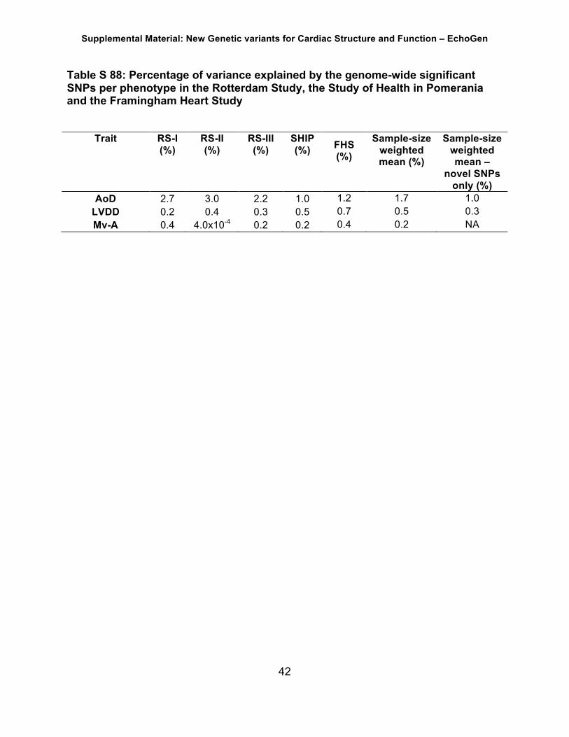
Table S 88: Percentage of variance explained by the genome-wide significant SNPs per phenotype in the Rotterdam Study, the Study of Health in Pomerania and the Framingham Heart Study
Trait RS-I (%)
RS-II (%)
RS-III (%)
SHIP (%)
FHS (%)
Sample-size weighted mean (%)
Sample-size weighted mean –
novel SNPs only (%)
AoD 2.7 3.0 2.2 1.0 1.2 1.7 1.0 LVDD 0.2 0.4 0.3 0.5 0.7 0.5 0.3 Mv-A 0.4 4.0x10⁻⁴ 0.2 0.2 0.4 0.2 NA

Supplemental Material: New Genetic variants for Cardiac Structure and Function – EchoGen
43
Table S 19: Explained variance of all autosomal SNPs (MAF ≥ 1%) in SHIP Trait Explained Variance (%) SE (%) P value N AoD 29 8.2 1.1 x 10⁻⁴ 3,458
LA 16 8.3 2.1 x 10⁻² 3,457
LVDD 18 9.0 1.9 x 10⁻² 3,168
LVWT 18 8.9 1.4 x 10⁻² 3,168
LVM 12 8.5 0.07 3,168
FS 17 9.1 2.3 x 10⁻² 3,116
LVSD 8.2 9.3 0.19 3,120
Mv-A 11 12.2 0.18 2,315
DecTime 16 11.9 0.09 2,364
IVRT 24 12.2 1.9 x 10⁻² 2,303
HFpEF 20 16.6 0.13 1,766
AoD= aortic root diameter; LA= left atrial size; LVDD= left ventricular end-diastolic diameter; LVWT= left ventricular wall thickness; LVM= left ventricular mass; FS= left ventricular fractional shortening; LVSD= left ventricular systolic dysfunction; A= peak velocity of the mitral A-wave; DecTime= deceleration time; IVRT= isovolumetric relaxation time; HFpEF= heart failure with preserved ejection fraction; SE= standard error; * P values are from GCTA analyses.11 The explained variance for the traits E (peak velocity of the mitral E-wave), E/A (ratio of the peak velocity of the mitral E-Wave divided by the peak velocity of the mitral A-wave) and DDpEF (diastolic dysfunction with preserved ejection fraction) was 0 – therefore the numbers are not displayed in this table. For the traits E´ (Peak velocity of the excursion of the lateral mitral annulus in the early diastolic phase) and E/E´ (Ratio of the peak velocity of the excursion of the lateral mitral annulus in the early diastolic phase by TDI and the peak velocity of the mitral E-wave by Doppler imaging) no values were calculated due to the low number of samples available in SHIP.

Supplemental Material: New Genetic variants for Cardiac Structure and Function – EchoGen
44
Figure S 1: QQ plot - AoD. P values were obtained by calculating Wald test statistics (sample size: n=26,741).
Figure S 2: QQ plot – LA. P values were obtained by calculating Wald test statistics (sample size: n=26,189).

Supplemental Material: New Genetic variants for Cardiac Structure and Function – EchoGen
45
Figure S 3: QQ plot – LVDD. P values were obtained by calculating Wald test statistics (sample size: n=30,201).
Figure S 4: QQ plot – LVWT. P values were obtained by calculating Wald test statistics (sample size: n=30,043).

Supplemental Material: New Genetic variants for Cardiac Structure and Function – EchoGen
46
Figure S 5: QQ plot – LVM. P values were obtained by calculating Wald test statistics (sample size: n=30,142).
Figure S 6: QQ plot – FS. P values were obtained by calculating Wald test statistics (sample size: n=28,083).

Supplemental Material: New Genetic variants for Cardiac Structure and Function – EchoGen
47
Figure S 7: LVSD. P values were obtained by calculating Wald test (sample size: n=27,864).
Figure S 8: QQ plot Mv-E. P values were obtained by calculating Wald test (sample size: n=21,852).

Supplemental Material: New Genetic variants for Cardiac Structure and Function – EchoGen
48
Figure S 9: QQ plot – Mv-A. P values were obtained by calculating Wald test statistics (sample size: n=21,643).
Figure S 10: QQ plot – E/A. P values were obtained by calculating Wald test statistics (sample size: n=21,348).

Supplemental Material: New Genetic variants for Cardiac Structure and Function – EchoGen
49
Figure S 11: QQ plot – DecTime. P values were obtained by calculating Wald test statistics (sample size: n=16,681).
Figure S 12: QQ plot – IVRT. P values were obtained by calculating Wald test statistics (sample size: n=12,151).

Supplemental Material: New Genetic variants for Cardiac Structure and Function – EchoGen
50
Figure S 13: QQ plot – E’. P values were obtained by calculating Wald test statistics (sample size: n=8,500).
Figure S 14: QQ plot – E/E’. P values were obtained by calculating Wald test statistics (sample size: n=7,832).

Supplemental Material: New Genetic variants for Cardiac Structure and Function – EchoGen
51
Figure S 15: QQ plot – DDpEF. P values were obtained by calculating Wald test statistics (sample size: n=7,262).
Figure S 16: QQ plot – HFpEF. P values were obtained by calculating Wald test statistics (sample size: n=5,297).

Supplemental Material: New Genetic variants for Cardiac Structure and Function – EchoGen
52
Figure S 17: Forest plots for genome-wide significant hits
n=30,704

Supplemental Material: New Genetic variants for Cardiac Structure and Function – EchoGen
53
n=30,704

Supplemental Material: New Genetic variants for Cardiac Structure and Function – EchoGen
54
n=30,704

Supplemental Material: New Genetic variants for Cardiac Structure and Function – EchoGen
55
n=30,704

Supplemental Material: New Genetic variants for Cardiac Structure and Function – EchoGen
56
n=30,704

Supplemental Material: New Genetic variants for Cardiac Structure and Function – EchoGen
57
n=30,704

Supplemental Material: New Genetic variants for Cardiac Structure and Function – EchoGen
58
n=30,704

Supplemental Material: New Genetic variants for Cardiac Structure and Function – EchoGen
59
n=43,623
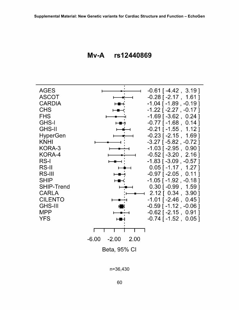
Supplemental Material: New Genetic variants for Cardiac Structure and Function – EchoGen
60
n=36,430

Supplemental Material: New Genetic variants for Cardiac Structure and Function – EchoGen
61
Figure S 18: Regional plots for all genome-wide significant hits. P values were obtained by calculating Wald test statistics
AoD, n=26,741

Supplemental Material: New Genetic variants for Cardiac Structure and Function – EchoGen
62

Supplemental Material: New Genetic variants for Cardiac Structure and Function – EchoGen
63

Supplemental Material: New Genetic variants for Cardiac Structure and Function – EchoGen
64
LVDD, n=30,201

Supplemental Material: New Genetic variants for Cardiac Structure and Function – EchoGen
65
Mv-A, n=21,643

Supplemental Material: New Genetic variants for Cardiac Structure and Function – EchoGen
66
Figure S 19: Funnel plots for the novel genome-wide significant hits

Supplemental Material: New Genetic variants for Cardiac Structure and Function – EchoGen
67

Supplemental Material: New Genetic variants for Cardiac Structure and Function – EchoGen
68

Supplemental Material: New Genetic variants for Cardiac Structure and Function – EchoGen
69

Supplemental Material: New Genetic variants for Cardiac Structure and Function – EchoGen
70

Supplemental Material: New Genetic variants for Cardiac Structure and Function – EchoGen
71
Figure S 20: Protein Kinase A Signaling (pathway analysis)
Pathyway analysis using Ingenuity IPA (Ingenuity Systems, Redwood, CA, USA). Red nodes are genes related to echo traits.

Supplemental Material: New Genetic variants for Cardiac Structure and Function – EchoGen
72
4 Acknowledgements
General funding and acknowledgements
This work was supported by a grant from the National Heart, Lung, and Blood Institute,
USA (contract no: N01-HL-25195; R01HL 093328 to RSV), a MAIFOR grant from the
University Medical Center Mainz, Germany (to PSW), the “Center for Translational
Vascular Biology (CTVB)” of the Johannes Gutenberg-University of Mainz, and the
Federal Ministry of Research and Education, Germany (contract no: BMBF 01EO1003
to PSW). This work was also supported by the research project Greifswald Approach to
Individualized Medicine (GANI_MED). GANI_MED has been funded by the Federal
Ministry of Education and Research and the Ministry of Cultural Affairs of the Federal
State of Mecklenburg, West Pomerania (contract no: 03IS2061A).
We would like to thank all study participants, and the colleagues and coworkers from all
cohorts and sites who were involved in the generation of data or in the analysis. We
especially thank Andrew Johnson (Framingham Heart Study) for generation of the gene
annotation database used for analysis. We thank the German Center for Cardiovascular
Research (DZHK e.V.) for supporting the analysis and publication of this project. RSV is
a member of the Scientific Advisory Board of the DZHK. Data on coronary artery
disease and myocardial infarction have been contributed by CARDIoGRAMplusC4D
investigators and have been downloaded from www.CARDIOGRAMPLUSC4D.ORG
Study-specific acknowledgements, funding and ethics statements
AortaGen Consortium
For full acknowledgements and funding information, please refer to Mitchell et al. (45).

Supplemental Material: New Genetic variants for Cardiac Structure and Function – EchoGen
73
Age, Gene/Environment Susceptibility (AGES) Study
The Age, Gene/Environment Susceptibility Study has been funded by NIH contract N01-
AG-1-2100, the NIA Intramural Research Program, Hjartavernd (the Icelandic Heart
Association), and the Althingi (the Icelandic Parliament). The study is approved by the
Icelandic National Bioethics Committee, VSN: 00-063. The researchers are indebted to
the participants for their willingness to participate in the study. The AGES Reykjavik
Study GWAS was approved by the National Bioethics Committee (VSN: 00-063) and
the Data Protection Authority. Informed consent was obtained from all participants.
Anglo-Scandinavian Cardiac Outcomes Trial - Hypertension Association
Cardiovascular Disease (ASCOT) Study
The Anglo-Scandinavian Cardiac Outcomes Trial and the establishment of the
associated genetic repository were funded by Pfizer, New York, USA with some
additional funding provided by Servier Research Group, Paris, France, and Leo
Laboratories, Copenhagen, Denmark. Genotyping was funded by Barts, the London
School of Medicine and Dentistry, and by the Centre Nationale de Genotypage Paris.
The research was also in part funded by an Irish Research Council GREP award. We
thank the other investigators, the staff, and the participants of the ASCOT study for their
important contributions. The study conformed to good clinical practice guidelines and
was approved by the respective local hospital ethics committees (St. Mary's Hospital,
London, UK and Beaumont Hospital, Dublin, Ireland). Written informed consent for the
study was obtained from all participants.
Austrian Stroke Prevention Study (ASPS)

Supplemental Material: New Genetic variants for Cardiac Structure and Function – EchoGen
74
Current analyses of ASPS are funded by the Austrian Science Fund Project
P20545_P05 Genetics of cerebral small vessel disease (to H Schmidt). We are
indebted to Birgit Reinhart for her continuous administrative support in the setting of the
Austrian Stroke Prevention Study and to Johann Semmler for his high-quality technical
assistance. The authors thank the staff and the participants of the ASPS for their
valuable contributions. The study protocol was approved and accepted by the ethics
committee of the Medical University of Graz, Austria, and informed consent was
obtained from all study participants.
Coronary Artery Risk Development in Young Adults (CARDIA) Study
The CARDIA Study is conducted and supported by the National Heart, Lung, and Blood
Institute in collaboration with the University of Alabama at Birmingham
(HHSN268201300025C & HHSN268201300026C), Northwestern University
(HHSN268201300027C), University of Minnesota (HHSN268201300028C), Kaiser
Foundation Research Institute (HHSN268201300029C), and Johns Hopkins University
School of Medicine (HHSN268200900041C). CARDIA is also partially supported by the
Intramural Research Program of the National Institute on Aging. Genotyping and
imputation were funded as part of the Gene Environment Association Studies
(GENEVA) through grants U01-HG004729, U01-HG04424, and U01-HG004446 from
the National Human Genome Research Institute. This manuscript has been reviewed
and approved by CARDIA for scientific content. SJ Shah is supported by R01
HL1075755. Informed consent was obtained from all participants, and the institutional
review board at each participating CARDIA center approved the study.
Cardiovascular Disease, Living and Ageing in Halle (CARLA) Study

Supplemental Material: New Genetic variants for Cardiac Structure and Function – EchoGen
75
This study was funded by a grant from the Deutsche Forschungsgemeinschaft (DFG,
German Research Foundation) as part of the Collaborative Research Center 598 ‘‘Heart
failure in the elderly–cellular mechanisms and therapy’’ at the Medical Faculty of the
Martin-Luther-University Halle-Wittenberg; by a grant of the Wilhelm-Roux Programme
of the Martin-Luther-University Halle-Wittenberg; by the Federal Employment Office;
and by the Ministry of Education and Cultural Affairs of Saxony-Anhalt. The study was in
accordance with the declaration of Helsinki. All participants gave their written informed
consent. The study was approved by the local ethic commission of the Medical Faculty
of the Martin-Luther-University Halle-Wittenberg.
Cohorts for Heart and Aging Research in Genomic Epidemiology– Heart Failure
Working Group (CHARGE-HF)
For a full list of CHARGE-HF working group members contributing to this work and for
CHARGE-HF acknowledgements, please reference (3).
Cardiovascular Health Study (CHS)
Cardiovascular Health Study: This CHS research was supported by NHLBI contracts
HHSN268201200036C, HHSN268200800007C, N01HC55222, N01HC85079,
N01HC85080, N01HC85081, N01HC85082, N01HC85083, N01HC85086,
N01HC85084, N01HC35129; and NHLBI grants U01HL080295, R01HL087652,
R01HL105756, R01HL103612, and R01HL120393 with additional contribution from the
National Institute of Neurological Disorders and Stroke (NINDS). Additional support was
provided through R01AG023629 from the National Institute on Aging (NIA). A full list of
principal CHS investigators and institutions can be found at CHS-NHLBI.org. The
provision of genotyping data was supported in part by the National Center for Advancing

Supplemental Material: New Genetic variants for Cardiac Structure and Function – EchoGen
76
Translational Sciences, CTSI grant UL1TR000124, and the National Institute of
Diabetes and Digestive and Kidney Disease Diabetes Research Center (DRC) grant
DK063491 to the Southern California Diabetes Endocrinology Research Center. The
content is solely the responsibility of the authors and does not necessarily represent the
official views of the National Institutes of Health. CHS was approved by institutional
review committees at each site, the subjects gave informed consent, and those included
in the present analysis consented to the use of their genetic information for the study of
cardiovascular disease.
Genetic Park of Cilento and Vallo di Diano Project (Cilento)
This work was supported by grants from the Italian Ministry of Universities and CNR
(Interomics Flagship Project, PON03PE_00060_7), the Assessorato Ricerca Regione
Campania, the Fondazione con il SUD (2011-PDR-13), and the Istituto Banco di Napoli
- Fondazione to MC. We thank the populations of Cilento for their participation in the
study.
We thank Dr. Gianluigi Iovino for echocardiographic examinations. The study design
was approved by the ethics committee of Azienda Sanitaria Locale Napoli 1. The study
was conducted according to the criteria set by the declaration of Helsinki and each
subject signed an informed consent before participating to the study.
DEPICT
TH Pers is supported by The Danish Council for Independent Research Medical
Sciences (FSS) and The Alfred Benzon Foundation.
Framingham Heart Study, original cohort, offspring and third generation (FHS1,
FHS2, FHS3)

Supplemental Material: New Genetic variants for Cardiac Structure and Function – EchoGen
77
This work was partially supported by the National Heart, Lung and Blood Institute's
Framingham Heart Study (Contracts No. N01-HC-25195 and HHSN268201500001I),
research grants (5R01HL107385-04 1R01HL126136-01A1), and a contract with
Affymetrix, Inc for genotyping services (Contract No. N02-HL-6-4278). A portion of this
research utilized the Linux Cluster for Genetic Analysis (LinGA-II) funded by the Robert
Dawson Evans Endowment of the Department of Medicine at Boston University School
of Medicine and Boston Medical Center. This research was conducted in part using data
and resources from the Framingham Heart Study of the National Heart Lung and Blood
Institute of the National Institutes of Health and Boston University School of Medicine.
The analyses reflect intellectual input and resource development from the Framingham
Heart Study investigators participating in the SNP Health Association Resource
(SHARe) project. Each study participant provided written informed consent for
participation in the study and for genetic analyses and the study was approved by the
Boston University Medical Center Institutional Review Board.
The Generation R Study (Generation R)
The general design of Generation R Study is made possible by financial support from
the Erasmus Medical Center, Rotterdam, the Erasmus University Rotterdam, the
Netherlands Organization for Health Research and Development (ZonMw), the
Netherlands Organisation for Scientific Research (NWO), the Ministry of Health, Welfare
and Sport and the Ministry of Youth and Families. VW Jaddoe received an additional
grant from the Netherlands Organization for Health Research and Development (VIDI
016.136.361) and a European Research Council Consolidator Grant (ERC-2014-CoG-
648916). The Generation R Study is conducted by the Erasmus Medical Center in close

Supplemental Material: New Genetic variants for Cardiac Structure and Function – EchoGen
78
collaboration with the School of Law and Faculty of Social Sciences of the Erasmus
University Rotterdam, the Municipal Health Service Rotterdam area, Rotterdam, the
Rotterdam Homecare Foundation, Rotterdam and the Stichting Trombosedienst &
Artsenlaboratorium Rijnmond (STAR-MDC), Rotterdam. We gratefully acknowledge the
contribution of children and parents, general practitioners, hospitals, midwives and
pharmacies in Rotterdam. The generation and management of GWAS genotype data
for the Generation R Study were done at the Genetic Laboratory of the Department of
Internal Medicine, Erasmus MC, the Netherlands. We would like to thank Karol Estrada,
Dr. Tobias A. Knoch, Anis Abuseiris, Luc V. de Zeeuw, and Rob de Graaf, for their help
in creating GRIMP, BigGRID, MediGRID, and Services@MediGRID/D-Grid, (funded by
the German Bundesministerium fuer Forschung und Technology; grants 01 AK 803 A-
H, 01 IG 07015 G) for access to their grid computing resources. We thank Mila Jhamai,
Manoushka Ganesh, Pascal Arp, Marijn Verkerk, Lizbeth Herrera and Marjolein Peters
for their help in creating, managing and QC of the GWAS database. Also, we thank
Karol Estrada for his support in creation and analysis of imputed data. The Generation
R Study is made possible by financial support from the Erasmus Medical Center,
Rotterdam, the Erasmus University Rotterdam and the Netherlands Organization for
Health Research and Development. The study protocol was approved by the Medical
Ethics Committee of the Erasmus Medical Center, Rotterdam. Written informed consent
was obtained for all participants.
Gutenberg Health Study (GHS-I, -II, -III)
The Gutenberg Health Study is funded through the government of Rhineland-Palatinate
(„Stiftung Rheinland-Pfalz für Innovation“, contract AZ 961-386261/733), the research

Supplemental Material: New Genetic variants for Cardiac Structure and Function – EchoGen
79
programs “Wissen schafft Zukunft” and “Center for Translational Vascular Biology
(CTVB)” of the Johannes Gutenberg-University of Mainz, and its contract with
Boehringer Ingelheim and PHILIPS Medical Systems, including an unrestricted grant for
the Gutenberg Health Study. PS Wild is funded by the Federal Ministry of Education
and Research (BMBF 01EO1003) and he received honoraria for lectures or consulting
from Boehringer Ingelheim and Bayer HealthCare, Leverkusen. We thank all study
participants for their willingness to provide data for this research project and we are
indebted to all coworkers for their enthusiastic commitment. The study followed the
recommendations of the Declaration of Helsinki and was approved by the ethics
committee of the Chamber of Physicians of Rhineland-Palatinate, Germany (reference
no. 837.020.07). Written informed consent was obtained from all participants.
Hypertension Genetic Epidemiology Network (HyperGEN)
The HyperGEN Echocardiography ancillary study was funded by the National Institutes
of Health (R01 HL 55673). The HyperGEN parent study was funded by cooperative
agreements (U10) with the National Heart, Lung, and Blood Institute: HL54471,
HL54472, HL54473, HL54495, HL54496, HL54497, HL54509, HL54515. The authors
thank the HyperGEN participants and staff for their valuable contributions. HyperGEN
was approved by the institutional review committees at each site. All participants gave
informed consent, and those included in the present analysis consented to the use of
their genetic information for cardiovascular disease or related conditions.
Jackson Heart Study (JHS)
The Jackson Heart Study is supported by contracts HSN268201300046C,
HHSN268201300047C, HHSN268201300048C, HHSN268201300049C,

Supplemental Material: New Genetic variants for Cardiac Structure and Function – EchoGen
80
HHSN268201300050C from the National Heart, Lung, and Blood Institute on Minority
Health and Health Disparities. The authors thank the Jackson Heart Study team
(University of Mississippi Medical Center, Jackson State University and Tougaloo
College) and participants for their long-term commitment that continues to improve our
understanding of the genetic epidemiology of cardiovascular and other chronic
diseases. The study protocol was approved by the University of Mississippi Medical
Center Internal Review Board committee on human subjects. All participants gave
informed consent.
Kompetenznetz Herzinsuffizienz (KNHI)
EB, BP, HB and GH received funding by the national genome research network NGFN
of the BMBF (grant numbers 01GS0837 and 01GS0422, funding MS and DMalzahn);
FE and BP received funding by the competence network heart failure Germany. The
study was approved by the ethics committee of the Georg-August-University in
Göttingen, Germany and all participants gave informed consent.
Cooperative Health Research in the Region of Augsburg, followup studies F3 and
F4 (KORA-F3 and –F4)
The Cooperative Health Research in the Region of Augsburg studies were financed by
the Helmholtz Zentrum München, German Research Center for Environmental Health,
Neuherberg, Germany and supported by grants from the German Federal Ministry of
Education and Research (BMBF). Part of this work was financed by the German
National Genome Research Network (NGFN). Our research was supported within the
Munich Center of Health Sciences (MC Health) as part of LMUinnovativ. The studies

Supplemental Material: New Genetic variants for Cardiac Structure and Function – EchoGen
81
were approved by the local ethics committees and all participants gave informed
consent.
Ludwigshafen Risk and Cardiovascular Health (LURIC) Study
This work was supported by the 7th Framework Program (AtheroRemo, grant
agreement number 201668 and RiskyCAD, grant agreement number 305739) of the EU
and by the INTERREG-IV-Oberrhein-Program (Project A28, Genetic mechanisms of
cardiovascular diseases) with support from the European Regional Development Fund
(ERDF) and the Wissenschaftsoffensive TMO. We extend our appreciation to the
participants of the LURIC study and thank the LURIC study team who were either
temporarily or permanently involved in patient recruitment as well as sample and data
handling, in addition to the laboratory staff at the Ludwigshafen General Hospital and
the Universities of Freiburg and Ulm, Germany. The study was approved by the ethics
committee at the "Landesärztekammer Rheinland-Pfalz" and was conducted in
accordance with the "Declaration of Helsinki”. Informed written consent was obtained
from all participants.
Myocardial Applied Genomics Network (MAGNet)
MAGNet (http://www.med.upenn.edu/magnet/) is funded by NIH R01HL105993.
MAGNet protocols have been approved by institutional review boards at University of
Pennsylvania, Stanford University, and Cleveland Clinic, Gift of Life.
Microisolates in South Tyrol (MICROS) Study
The MICROS study was supported by the Ministry of Health and Department of
Educational Assistance, University and Research of the Autonomous Province of
Bolzano, the South Tyrolean Sparkasse Foundation, and the European Union

Supplemental Material: New Genetic variants for Cardiac Structure and Function – EchoGen
82
framework program 6 EUROSPAN project (contract no. LSHG-CT-2006-018947). For
the MICROS study, we thank the primary care practitioners Raffaela Stocker, Stefan
Waldner, Toni Pizzecco, Josef Plangger and Ugo Marcadent, and Helmuth Weiss and
the personnel of the Hospital of Silandro (Department of Laboratory Medicine) for their
participation and collaboration in the research project. The MICROS study was
approved by the ethical committee of the Autonomous Province of Bolzano. Informed
consent was obtained from all participants.
Malmö Preventive Project (MPP)
This work in the MPP was supported by grants from the the Swedish Heart-Lung
Foundation (JGS, OM, PN), the Swedish Research Council (to JGS and OM), the
European Research Council (OM), the Faculty of Medicine of Lund University (OM),
Skåne University Hospital (JGS, OM) and the Crafoord Foundation (JGS, OM). This
study was approved by the Ethics Committee of Lund University. All participants
provided written informed consent before entering the study.
Northern Manhattan Study (NOMAS)
NOMAS is supported by the National Institute of Neurological Disorders and Stroke
(NINDS) of the National Institutes of Health (NIH) through grants R37 NS2993 (Dr.
Sacco and Elkind) and R01 NS36286 (Dr. Di Tullio). MDT is supported by grant NINDS
R01 NS083784. RLS and NDD are supported by McKnight Brain Foundation. The
authors gratefully acknowledge Ashley Beecham and Shengru Guo for their analytical
support. The authors also gratefully acknowledge the McKnight Institute for providing
funding to genotype NOMAS participants. All patients provided informed consent to

Supplemental Material: New Genetic variants for Cardiac Structure and Function – EchoGen
83
participate in the study. The study was approved by the Institutional Review Boards of
Columbia University and the University of Miami.
Prospective Investigation of the Vasculature in Uppsala Seniors (PIVUS) Study
This study was supported by grants from the Swedish Foundation for Strategic
Research (project grant no. ICA08-0047), the Swedish Research Council (project grant
no. 2012-1397), the Swedish Heart-Lung Foundation (project grant no. 20120197), the
Swedish Society of Medicine, and Uppsala University. Genotyping was performed by
the SNP&SEQ Technology Platform in Uppsala (www.genotyping.se), which is
supported by Uppsala University, Uppsala University Hospital, Science for Life
Laboratory - Uppsala and the Swedish Research Council (Contracts 80576801 and
70374401). Genotyping was performed by the SNP&SEQ Technology Platform in
Uppsala (www.genotyping.se). We thank Tomas Axelsson, Ann-Christine Wiman and
Caisa Pöntinen for their excellent assistance with genotyping. APM is a Wellcome Trust
Senior Research Fellow in Basic Biomedical Science (grant number WT098017). The
study was approved by the Ethics Committee of Uppsala University and all participants
provided informed consent.
Rotterdam Study (RS)-I, -II, -III
The generation and management of GWAS genotype data for the Rotterdam Study is
supported by the Netherlands Organisation of Scientific Research NWO Investments
(nr. 175.010.2005.011, 911-03-012). This study is funded by the Research Institute for
Diseases in the Elderly (014-93-015; RIDE2), the Netherlands Genomics Initiative
(NGI)/Netherlands Organisation for Scientific Research (NWO) project nr. 050-060-810.
The Rotterdam Study is funded by Erasmus Medical Center and Erasmus University,

Supplemental Material: New Genetic variants for Cardiac Structure and Function – EchoGen
84
Rotterdam, Netherlands Organization for the Health Research and Development
(ZonMw), the Research Institute for Diseases in the Elderly (RIDE), the Ministry of
Education, Culture and Science, the Ministry for Health, Welfare and Sports, the
European Commission (DG XII), and the Municipality of Rotterdam. OH Franco works in
ErasmusAGE, a center for aging research across the life course funded by Nestlé
Nutrition (Nestec Ltd.), Metagenics Inc. and AXA. Nestlé Nutrition (Nestec Ltd.),
Metagenics Inc. and AXA had no role in design and conduct of the study; collection,
management, analysis, and interpretation of the data; and preparation, review or
approval of the manuscript. The generation and management of GWAS genotype data
for the Rotterdam Study is supported by the Netherlands Organisation of Scientific
Research NWO Investments (nr. 175.010.2005.011, 911-03-012). This study is funded
by the Research Institute for Diseases in the Elderly (014-93-015; RIDE2), the
Netherlands Genomics Initiative (NGI)/Netherlands Organisation for Scientific Research
(NWO) project nr. 050-060-810. We thank Pascal Arp, Mila Jhamai, Marijn Verkerk,
Lizbeth Herrera and Marjolein Peters for their help in creating the GWAS database, and
Karol Estrada and Maksim V. Struchalin for their support in creation and analysis of
imputed data. The Rotterdam Study is funded by Erasmus Medical Center and Erasmus
University, Rotterdam, Netherlands Organization for the Health Research and
Development (ZonMw), the Research Institute for Diseases in the Elderly (RIDE), the
Ministry of Education, Culture and Science, the Ministry for Health, Welfare and Sports,
the European Commission (DG XII), and the Municipality of Rotterdam. The authors are
grateful to the study participants, the staff of the Rotterdam Study and the participating
general practitioners and pharmacists. The Rotterdam Study has been approved by the

Supplemental Material: New Genetic variants for Cardiac Structure and Function – EchoGen
85
Medical Ethics Committee of the Erasmus MC and by the Dutch Ministry of Health,
Welfare and Sport, implementing the “Wet Bevolkings Onderzoek: ERGO (Population
Screening Act: Rotterdam Study). All participants provided written informed consent to
participate in the study and to obtain information from their treating physicians.
Study of Health in Pomerania (SHIP and SHIP-Trend)
SHIP is part of the Community Medicine Research net of the University of Greifswald,
Germany, which is funded by the Federal Ministry of Education and Research (grants
no. 01ZZ9603, 01ZZ0103, and 01ZZ0403), the Ministry of Cultural Affairs as well as the
Social Ministry of the Federal State of Mecklenburg-West Pomerania. Echocardiography
in the 5-year follow-up (SHIP-1) was funded by the Competence Network Heart Failure
of the Federal Ministry of Education and Research. Genome-wide data have been
supported by the Federal Ministry of Education and Research (grant no. 03ZIK012) and
a joint grant from Siemens Healthcare, Erlangen, Germany and the Federal State of
Mecklenburg- West Pomerania. The University of Greifswald is a member of the Caché
Campus program of the InterSystems GmbH. All participants gave informed written
consent. The study followed the recommendations of the Declaration of Helsinki and
was approved by the Ethics Committee of the University of Greifswald.
Uppsala Longitudinal Study of Adult Men (ULSAM)
This study was supported by grants from the Swedish Foundation for Strategic
Research (project grant no. ICA08-0047), the Swedish Research Council (project grant
no. 2012-1397), the Swedish Heart-Lung Foundation (project grant no. 20120197), the
Swedish Society of Medicine, and Uppsala University. Genotyping was performed by
the SNP&SEQ Technology Platform in Uppsala (www.genotyping.se), which is

Supplemental Material: New Genetic variants for Cardiac Structure and Function – EchoGen
86
supported by Uppsala University, Uppsala University Hospital, Science for Life
Laboratory - Uppsala and the Swedish Research Council (Contracts 80576801 and
70374401). Genotyping was performed by the SNP&SEQ Technology Platform in
Uppsala (www.genotyping.se). APM is a Senior Fellow in Basic Biomedical Science
under award WT098017. We thank Tomas Axelsson, Ann-Christine Wiman and Caisa
Pöntinen for their excellent assistance with genotyping. The study was approved by the
Ethics Committee of Uppsala University and all participants provided informed consent.
The Cardiovascular Risk in Young Finns Study (YFS)
The Young Finns Study has been financially supported by the Academy of Finland:
grants 286284, 134309 (Eye), 126925, 121584, 124282, 129378 (Salve), 117787
(Gendi), and 41071 (Skidi); the Social Insurance Institution of Finland; Competitive
State Research Financing of the Expert Responsibility area of Kuopio, Tampere and
Turku University Hospitals (grant X51001); Juho Vainio Foundation; Paavo Nurmi
Foundation; Finnish Foundation for Cardiovascular Research ; Finnish Cultural
Foundation; Tampere Tuberculosis Foundation; Emil Aaltonen Foundation; Yrjö
Jahnsson Foundation; Signe and Ane Gyllenberg Foundation; and Diabetes Research
Foundation of Finnish Diabetes Association. The expert technical assistance in the
statistical analyses by Irina Lisinen is gratefully acknowledged. The study was approved
by the local ethics committees (University Hospitals of Helsinki, Turku, Tampere, Kuopio
and Oulu) and was conducted following the guidelines of the Declaration of Helsinki. All
participants gave their written informed consent.

Supplemental Material: New Genetic variants for Cardiac Structure and Function – EchoGen
87
Role of the sponsors
The sponsors had no role in the study design, analyses, drafting of the manuscript, or
the decision to publish.
Drs.Wild, Felix, Schillert, Teumer, Zeller, Vasan and Dörr had full access to all of the
data in the study and take responsibility for the integrity of the data and the accuracy of
the data analysis.

Supplemental Material: New Genetic variants for Cardiac Structure and Function – EchoGen
88
5 References
1. Vasan, R.S. et al. Genetic variants associated with cardiac structure and function:
a meta-analysis and replication of genome-wide association data. JAMA 302, 168-
78 (2009).
2. Anand, I.S., Florea, V.G., Solomon, S.D., Konstam, M.A. & Udelson, J.E.
Noninvasive assessment of left ventricular remodeling: concepts, techniques, and
implications for clinical trials. J Card Fail 8, S452-64 (2002).
3. Gardin, J.M. How reliable are serial echocardiographic measurements in detecting
regression in left ventricular hypertrophy and changes in function? J Am Coll
Cardiol 34, 1633-6 (1999).
4. Devereux, R.B. et al. An appraisal of echocardiography as an epidemiological tool.
The Strong Heart Study. Ann Epidemiol 13, 238-44 (2003).
5. Gosse, P. et al. Centralized echocardiogram quality control in a multicenter study
of regression of left ventricular hypertrophy in hypertension. J Hypertens 16, 531-5
(1998).
6. Palmieri, V. et al. Reliability of echocardiographic assessment of left ventricular
structure and function: the PRESERVE study. Prospective Randomized Study
Evaluating Regression of Ventricular Enlargement. J Am Coll Cardiol 34, 1625-32
(1999).
7. Lang, R.M. et al. Recommendations for chamber quantification. Eur J
Echocardiogr 7, 79-108 (2006).
8. Devereux, R.B. et al. Echocardiographic assessment of left ventricular
hypertrophy: comparison to necropsy findings. Am J Cardiol 57, 450-8 (1986).
9. Vasan, R.S. et al. Plasma natriuretic peptides for community screening for left
ventricular hypertrophy and systolic dysfunction: the Framingham heart study.
JAMA 288, 1252-9 (2002).
10. Redfield, M.M. et al. Burden of systolic and diastolic ventricular dysfunction in the
community: appreciating the scope of the heart failure epidemic. JAMA 289, 194-
202 (2003).
11. Yang, J., Lee, S.H., Goddard, M.E. & Visscher, P.M. GCTA: a tool for genome-
wide complex trait analysis. Am J Hum Genet 88, 76-82 (2011).

Supplemental Material: New Genetic variants for Cardiac Structure and Function – EchoGen
89
12. Jaddoe, V.W. et al. The Generation R Study: design and cohort update 2012. Eur
J Epidemiol 27, 739-56 (2012).
13. Nikpay, M. et al. A comprehensive 1,000 Genomes-based genome-wide
association meta-analysis of coronary artery disease. Nat Genet 47, 1121-30
(2015).
14. Howie, B., Fuchsberger, C., Stephens, M., Marchini, J. & Abecasis, G.R. Fast and
accurate genotype imputation in genome-wide association studies through pre-
phasing. Nat Genet 44, 955-9 (2012).
15. Irizarry, R.A. et al. Exploration, normalization, and summaries of high density
oligonucleotide array probe level data. Biostatistics 4, 249-64 (2003).
16. Johnson, W.E., Li, C. & Rabinovic, A. Adjusting batch effects in microarray
expression data using empirical Bayes methods. Biostatistics 8, 118-27 (2007).
17. Westra, H.J. et al. Systematic identification of trans eQTLs as putative drivers of
known disease associations. Nat Genet 45, 1238-43 (2013).
18. Zeller, T. et al. Genetics and beyond--the transcriptome of human monocytes and
disease susceptibility. PLoS One 5, e10693 (2010).
19. Segre, A.V. et al. Common inherited variation in mitochondrial genes is not
enriched for associations with type 2 diabetes or related glycemic traits. PLoS
Genet 6(2010).
20. Benjamini, Y. & Hochberg, Y. Controlling the False Discovery Rate - a Practical
and Powerful Approach to Multiple Testing. Journal of the Royal Statistical Society
Series B-Methodological 57, 289-300 (1995).
21. Rossin, E.J. et al. Proteins encoded in genomic regions associated with immune-
mediated disease physically interact and suggest underlying biology. PLoS Genet
7, e1001273 (2011).
22. Consortium, E.P. An integrated encyclopedia of DNA elements in the human
genome. Nature 489, 57-74 (2012).
23. Pers, T.H. et al. Biological interpretation of genome-wide association studies using
predicted gene functions. . Nature Communications 5, 5890 (2014).
24. Fehrmann, R.S.N. et al. Gene expression analysis identifies global gene dosage
sensitivity in cancer. Nature Genetics (2015).

Supplemental Material: New Genetic variants for Cardiac Structure and Function – EchoGen
90
25. Harris, T.B. et al. Age, Gene/Environment Susceptibility-Reykjavik Study:
multidisciplinary applied phenomics. Am J Epidemiol 165, 1076-87 (2007).
26. Dahlöf, B. et al. Prevention of cardiovascular events with an antihypertensive
regimen of amlodipine adding perindopril as required versus atenolol adding
bendroflumethiazide as required, in the Anglo-Scandinavian Cardiac Outcomes
Trial-Blood Pressure Lowering Arm (ASCOT-BPLA): a multicentre randomised
controlled trial. Lancet 366, 895-906 (2005).
27. Schmidt, R. et al. Assessment of cerebrovascular risk profiles in healthy persons:
definition of research goals and the Austrian Stroke Prevention Study (ASPS).
Neuroepidemiology 13, 308-13 (1994).
28. Friedman, G.D. et al. CARDIA: study design, recruitment, and some
characteristics of the examined subjects. J Clin Epidemiol 41, 1105-16 (1988).
29. Greiser, K.H. et al. Cardiovascular disease, risk factors and heart rate variability in
the elderly general population: design and objectives of the CARdiovascular
disease, Living and Ageing in Halle (CARLA) Study. BMC Cardiovasc Disord 5, 33
(2005).
30. Fried, L.P. et al. The Cardiovascular Health Study: design and rationale. Ann
Epidemiol 1, 263-76 (1991).
31. Colonna, V. et al. Campora: a young genetic isolate in South Italy. Hum Hered 64,
123-35 (2007).
32. Colonna, V. et al. Comparing population structure as inferred from genealogical
versus genetic information. Eur J Hum Genet 17, 1635-41 (2009).
33. Dawber, T.R., Meadors, G.F. & Moore, F.E., Jr. Epidemiological approaches to
heart disease: the Framingham Study. Am J Public Health Nations Health 41, 279-
81 (1951).
34. Kannel, W.B., Feinleib, M., McNamara, P.M., Garrison, R.J. & Castelli, W.P. An
investigation of coronary heart disease in families. The Framingham offspring
study. Am J Epidemiol 110, 281-90 (1979).
35. Splansky, G.L. et al. The Third Generation Cohort of the National Heart, Lung, and
Blood Institute's Framingham Heart Study: design, recruitment, and initial
examination. Am J Epidemiol 165, 1328-35 (2007).

Supplemental Material: New Genetic variants for Cardiac Structure and Function – EchoGen
91
36. Wild, P.S. et al. [The Gutenberg Health Study]. Bundesgesundheitsblatt
Gesundheitsforschung Gesundheitsschutz 55, 824-9 (2012).
37. Williams, R.R. et al. NHLBI family blood pressure program: methodology and
recruitment in the HyperGEN network. Hypertension genetic epidemiology
network. Ann Epidemiol 10, 389-400 (2000).
38. Taylor, H.A., Jr. et al. Toward resolution of cardiovascular health disparities in
African Americans: design and methods of the Jackson Heart Study. Ethn Dis 15,
S6-4-17 (2005).
39. Pilz, S. et al. Associations of methylarginines and homoarginine with diastolic
dysfunction and cardiovascular risk factors in patients with preserved left
ventricular ejection fraction. J Card Fail 20, 923-30 (2014).
40. Schunkert, H. et al. Cardiovascular phenotypes and functional parameters in the
general population--results of the MONICA/KORA studies. Gesundheitswesen 67 Suppl 1, S74-8 (2005).
41. Wichmann, H.E., Gieger, C., Illig, T. & Group, M.K.S. KORA-gen--resource for
population genetics, controls and a broad spectrum of disease phenotypes.
Gesundheitswesen 67 Suppl 1, S26-30 (2005).
42. Holle, R., Happich, M., Lowel, H., Wichmann, H.E. & Group, M.K.S. KORA--a
research platform for population based health research. Gesundheitswesen 67 Suppl 1, S19-25 (2005).
43. Winkelmann, B.R. et al. Rationale and design of the LURIC study--a resource for
functional genomics, pharmacogenomics and long-term prognosis of
cardiovascular disease. Pharmacogenomics 2, S1-73 (2001).
44. Pattaro, C. et al. The genetic study of three population microisolates in South Tyrol
(MICROS): study design and epidemiological perspectives. BMC Med Genet 8, 29
(2007).
45. Leosdottir, M. et al. Myocardial structure and function by echocardiography in
relation to glucometabolic status in elderly subjects from 2 population-based
cohorts: a cross-sectional study. Am Heart J 159, 414-420 e4 (2010).
46. Sacco, R.L. et al. Design of a family study among high-risk Caribbean Hispanics:
the Northern Manhattan Family Study. Ethn Dis 17, 351-7 (2007).

Supplemental Material: New Genetic variants for Cardiac Structure and Function – EchoGen
92
47. Lind, L., Fors, N., Hall, J., Marttala, K. & Stenborg, A. A comparison of three
different methods to evaluate endothelium-dependent vasodilation in the elderly:
the Prospective Investigation of the Vasculature in Uppsala Seniors (PIVUS)
study. Arterioscler Thromb Vasc Biol 25, 2368-75 (2005).
48. Hofman, A. et al. The Rotterdam Study: objectives and design update. Eur J
Epidemiol 22, 819-29 (2007).
49. Völzke, H. et al. Cohort profile: the study of health in Pomerania. Int J Epidemiol
40, 294-307 (2011).
50. Hedstrand, H. A study of middle-aged men with particular reference to risk factors
for cardiovascular disease. Ups J Med Sci Suppl 19, 1-61 (1975).
51. Raitakari, O.T. et al. Cohort profile: the cardiovascular risk in Young Finns Study.
Int J Epidemiol 37, 1220-6 (2008).
52. McAreavey, D. et al. Correlation of echocardiographic findings with cerebral
infarction in elderly adults: the AGES-Reykjavik study. Stroke 41, 2223-8 (2010).
53. Stanton, A. et al. An intensive phenotyping study to enable the future examination
of genetic influences on hypertension-associated cardiovascular disease. J Hum
Hypertens 15 Suppl 1, S13-8 (2001).
54. Paczek, A. et al. Comparison of M-mode and two-dimensional echocardiographic
algorithms used to estimate left ventricular mass: the Coronary Artery Risk
Development in Young Adults Study. J Am Soc Echocardiogr 8, 780-92 (1995).
55. Gardin, J.M. et al. Sex, age, and disease affect echocardiographic left ventricular
mass and systolic function in the free-living elderly. The Cardiovascular Health
Study. Circulation 91, 1739-48 (1995).
56. Medenwald, D. et al. Inflammation and echocardiographic parameters of
ventricular hypertrophy in a cohort with preserved cardiac function. Open Heart 1,
e000004 (2014).
57. Tiller, D. et al. Prevalence of symptomatic heart failure with reduced and with
normal ejection fraction in an elderly general population-the CARLA study. PLoS
One 8, e59225 (2013).

Supplemental Material: New Genetic variants for Cardiac Structure and Function – EchoGen
93
58. Gardin, J.M. et al. Echocardiographic design of a multicenter investigation of free-
living elderly subjects: the Cardiovascular Health Study. J Am Soc Echocardiogr 5,
63-72 (1992).
59. Sundstrom, J. et al. Relations of plasma total TIMP-1 levels to cardiovascular risk
factors and echocardiographic measures: the Framingham heart study. Eur Heart
J 25, 1509-16 (2004).
60. Taal, H.R. et al. Parental smoking during pregnancy and cardiovascular structures
and function in childhood: the Generation R Study. Int J Epidemiol 42, 1371-80
(2013).
61. Wild, P.S. et al. Distribution and categorization of left ventricular measurements in
the general population: results from the population-based Gutenberg Heart Study.
Circ Cardiovasc Imaging 3, 604-13 (2010).
62. Devereux, R.B., Dahlof, B., Levy, D. & Pfeffer, M.A. Comparison of enalapril
versus nifedipine to decrease left ventricular hypertrophy in systemic hypertension
(the PRESERVE trial). Am J Cardiol 78, 61-5 (1996).
63. Devereux, R.B. et al. Relations of Doppler stroke volume and its components to
left ventricular stroke volume in normotensive and hypertensive American Indians:
the Strong Heart Study. Am J Hypertens 10, 619-28 (1997).
64. Devereux, R.B. et al. Relations of left ventricular mass to demographic and
hemodynamic variables in American Indians: the Strong Heart Study. Circulation
96, 1416-23 (1997).
65. Samdarshi, T.E. et al. Distribution and determinants of Doppler-derived diastolic
flow indices in African Americans: the Jackson Heart Study (JHS). Am Heart J
158, 209-16 (2009).
66. Wachter, R. et al. Impact of obstructive sleep apnoea on diastolic function. Eur
Respir J 41, 376-83 (2013).
67. Baessler, A. et al. Association of the Ghrelin receptor gene region with left
ventricular hypertrophy in the general population: results of the MONICA/KORA
Augsburg Echocardiographic Substudy. Hypertension 47, 920-7 (2006).
68. Russo, C. et al. Race/ethnic disparities in left ventricular diastolic function in a
triethnic community cohort. Am Heart J 160, 152-8 (2010).

Supplemental Material: New Genetic variants for Cardiac Structure and Function – EchoGen
94
69. Sundstrom, J., Arnlov, J., Stolare, K. & Lind, L. Blood pressure-independent
relations of left ventricular geometry to the metabolic syndrome and insulin
resistance: a population-based study. Heart 94, 874-8 (2008).
70. Kardys, I. et al. Echocardiographic parameters and all-cause mortality: the
Rotterdam Study. Int J Cardiol 133, 198-204 (2009).
71. Dörr, M. et al. The association of thyroid function with cardiac mass and left
ventricular hypertrophy. J Clin Endocrinol Metab 90, 673-7 (2005).
72. Andren, B., Lind, L., Hedenstierna, G. & Lithell, H. Left ventricular hypertrophy and
geometry in a population sample of elderly males. Eur Heart J 17, 1800-7 (1996).
73. McMahon, D.M., Liu, J., Zhang, H., Torres, M.E. & Best, R.G. Maternal obesity,
folate intake, and neural tube defects in offspring. Birth Defects Res Part A Clin
Mol Teratol 97, 115-122 (2013).
Consortium Details
AortaGen Consortium Members: Gary F. Mitchell, Germaine C. Verwoert, Kirill V. Tarasov,
Aaron Isaacs, Albert V. Smith, Yasmin, Ernst R. Rietzschel, Toshiko Tanaka, Yongmei Liu,
Afshin Parsa, Samer S. Najjar, Kevin M. O'Shaughnessy, Sigurdur Sigurdsson, Marc L. De
Buyzere, Martin G. Larson, Mark P.S. Sie, Jeanette S. Andrews, Wendy S. Post, Francesco
U.S. Mattace-Raso, Carmel M. McEniery, Gudny Eiriksdottir, Patrick Segers, Ramachandran S.
Vasan, Marie Josee E. van Rijn, Timothy D. Howard, Patrick F. McArdle, Abbas Dehghan,
Elizabeth S. Jewell, Stephen J. Newhouse, Sofie Bekaert, Naomi M. Hamburg, Anne B.
Newman, Albert Hofman, Angelo Scuteri, Dirk De Bacquer, Mohammad Arfan Ikram, Bruce M.
Psaty, Christian Fuchsberger, Matthias Olden, Louise V. Wain, Paul Elliott, Nicholas L. Smith,
Janine F. Felix, Jeanette Erdmann, Joseph A. Vita, Kim Sutton-Tyrrell, Eric J.G. Sijbrands,
Serena Sanna, Lenore J. Launer, Tim De Meyer, Andrew D. Johnson, Anna F.C. Schut, David
M. Herrington, Fernando Rivadeneira, Manuela Uda, Ian B. Wilkinson, Thor Aspelund, Thierry
C. Gillebert, Luc Van Bortel, Emelia J. Benjamin, Ben A. Oostra, Jingzhong Ding, Quince
Gibson, André G. Uitterlinden, Gonçalo R. Abecasis, John R. Cockcroft, Vilmundur Gudnason,
Guy G. De Backer, Luigi Ferrucci, Tamara B. Harris, Alan R. Shuldiner, Cornelia M. van Duijn,
Daniel Levy, Edward G. Lakatta, Jacqueline C.M. Witteman. Affiliations at the time of the
original publication: Cardiovascular Engineering, Inc, Norwood, MA (G.F.M.); the Department of
Epidemiology (G.C.V., A.D., A.H., M.A.I., J.F.F., F.R., A.G.U., J.C.M.W.), Department of Internal
Medicine (G.C.V., F.U.S.M., E.J.G.S., F.R., A.G.U.), Genetic Epidemiology Unit (A.I., M.P.S.S.,
M.J.E.V.R., A.F.C.S., B.A.O., C.M.V.D.), Erasmus Medical Center, Rotterdam, The Netherlands;
the Netherlands Consortium on Healthy Aging, The Netherlands (G.C.V., F.U.S.M., A.D., M.A.I.,
J.F.F., F.R., A.G.U., J.C.M.W.); Laboratory of Cardiovascular Science (K.V.T., S.S.N., A.S.,
E.G.L.), Laboratory of Genetics (K.V.T.), Clinical Research Branch (T.T., L.F.), Laboratory of
Epidemiology, Demography, and Biometry (L.J.L., T.B.H.), Intramural Research Program,

National Institute on Aging, NIH, Baltimore, MD; Centre for Medical Systems Biology, Leiden,
The Netherlands (A.I., B.A.O., C.M.V.D.); Icelandic Heart Association, Kopavogur, Iceland
(A.V.S., S.S., G.E., T.A., V.G.); University of Iceland, Reykjavik, Iceland (A.V.S., T.A., V.G.);
Clinical Pharmacology Unit, University of Cambridge, Addenbrooke's Hospital, Cambridge,
United Kingdom (Y., K.M.O., C.M.M., S.J.N., I.B.W.); Department of Cardiology, Ghent
University Hospital, Belgium (E.R.R., T.C.G., M.L.D.B.); the Department of Internal Medicine
(E.R.R.), Department of Public Health (D.D.B., G.G.D.B.), bioMMeda (Biofluid, Tissue, and Solid
Mechanics for Medical Applications), Institute Biomedical Technology (P.S.), the Department of
Molecular Biotechnology (Faculty of Bioscience Engineering) (T.D.M.), the Clinical Research
Center (S.B.), the Heymans Institute of Pharmacology (L.V.B.), Ghent University, Belgium; the
Department of Epidemiology and Prevention (Y.L.), the Department of Biostatistical Sciences,
Division of Public Health Sciences (J.S.A.), the Department of Cardiology (D.M.H.), Sticht
Center on Aging (J.D.), Wake Forest University School of Medicine, Winston-Salem, NC;
University of Maryland School of Medicine, Baltimore, MD (A.P., P.F.M., Q.G., A.R.S.); the
Division of Cardiology, Washington Hospital Center, Washington, DC (S.S.N.); the Department
of Mathematics and Statistics, Boston University, Boston, MA (M.G.L.); NHLBI's Framingham
Study, Framingham, MA (M.G.L., R.S.V., M.O., A.D.J., E.J.B., D.L.); Division of Cardiology, The
Johns Hopkins University School of Medicine, Baltimore, MD (W.S.P.); Evans Department of
Medicine, Whitaker Cardiovascular Institute, and Preventive Medicine and Cardiology Sections,
Boston University School of Medicine, Boston, MA (R.S.V., N.M.H., J.A.V., E.J.B.); Center for
Genomics and Personalized Medicine Research, Wake Forest School of Medicine, Winston-
Salem, NC (T.D.H.); Center for Statistical Genetics, Department of Biostatistics, University of
Michigan, Ann Arbor, MI (E.S.J., C.F., G.R.A.); the Department of Epidemiology, Graduate
School of Public Health, University of Pittsburgh, Pittsburgh, PA (A.B.N., K.S.T.); Cardiovascular
Health Study, Departments of Epidemiology (B.M.P., N.L.S.), Medicine and Health Services
(B.M.P.), University of Washington, Seattle, WA; Group Health Research Institute, Group

Health, Seattle, WA (B.M.P., N.L.S.); Seattle Epidemiologic Research and Information Center of
the Department of VA Office of Research and Development, Seattle, WA (N.L.S.); the
Department of Internal Medicine II, Department of Epidemiology and Preventive Medicine,
University Medical Center Regensburg, Regensburg, Germany (M.O.); the Department of
Health Sciences, University of Leicester, Leicester, United Kingdom (L.V.W.); MRC-HPA Center
for Environment and Health, School of Public Health, Imperial College London, St Mary's
Campus, London, United Kingdom (P.E.); German Cancer Research Center (DKFZ), Division of
Clinical Epidemiology and Aging Research, Heidelberg, Germany (J.F.F.); and Universität zu
Lübeck, Medizinische Klinik II, Lübeck, Germany (J.E.); Istituto di Neurogenetica e
Neurofarmacologia, Consiglio Nazionale delle Ricerche, Cittadella Universitaria di Monseratto,
Monserrato, Cagliari, Italy (S.S., M.U.); Department of Cardiology, Wales Heart Research
Institute, Cardiff University, Cardiff, United Kingdom (J.R.C.); Geriatric Research and Education
Clinical Center, VA Medical Center, Baltimore, MD (A.R.S.); Center for Population Studies, and
the Division of Intramural Research of the National Heart, Lung, and Blood Institute, National
Institutes of Health, Bethesda, MD (A.D.J., D.L.)
CHARGE-HF Consortium Members: Nicholas L. Smith, Janine F. Felix, Alanna C. Morrison,
Serkalem Demissie, Nicole L. Glazer, Laura R. Loehr, L. Adrienne Cupples, Abbas Dehghan,
Thomas Lumley, Wayne D. Rosamond, Wolfgang Lieb, Fernando Rivadeneira, Joshua C. Bis,
Aaron R. Folsom, Emelia Benjamin, Yurii S. Aulchenko, Talin Haritunians, David Couper,
Joanne Murabito, Ying A. Wang, Bruno H. Stricker, John S. Gottdiener, Patricia P. Chang,
Thomas J. Wang, Kenneth M. Rice, Albert Hofman, Susan R. Heckbert, Ervin R. Fox,
Christopher J. O'Donnell, Andre G. Uitterlinden, Jerome I. Rotter, James T. Willerson, Daniel
Levy, Cornelia M. van Duijn, Bruce M. Psaty, Jacqueline C.M. Witteman, Eric Boerwinkle,
Ramachandran S. Vasan. Affiliations at the time of the original publication: Cardiovascular
Health Study: Departments of Epidemiology (N.L.S., S.R.H., B.M.P.), Medicine (N.L.G., J.C.B.,

B.M.P.), Biostatistics (T.L., K.M.R.), and Health Services (B.M.P.) University of Washington,
Seattle, Wash.; Seattle Epidemiologic Research and Information Center of the Department of
Veterans Affairs Office of Research and Development (N.L.S.), Seattle, Wash.; Group Health
Research Institute (S.R.H., B.M.P., N.L.S.) Group Health, Seattle, Wash.; and Department of
Cardiology (J.S.G.), University of Maryland, Baltimore, Md; Medical Genetics Institute (T.H.,
J.I.R.), Cedars-Sinai Medical Center, Los Angeles, Calif; Artherosclerosis Risk in Communities
Study: University of Texas at Houston Health Science Center (A.C.M., E.B., J.T.W.), Houston,
Tex.; Departments of Medicine (L.R.L., P.P.C.), Biostatistics (D.C.), and Epidemiology (W.D.R.,
L.R.L.), University of North Carolina at Chapel Hill; National Institute of Environmental Health
Sciences (L.R.L.), Research Triangle Park, N.C., University of Minnesota School of Public
Health, Division of Epidemiology and Community Health, School of Public Health (A.R.F.),
Minneapolis, Minn.; Department of Medicine (E.R.F.), University of Mississippi Medical Center,
Jackson, Miss; Texas Heart Institute (J.T.W.), Houston, Tex; Framingham Heart Study,
Department of Biostatistics (S.D., L.A.C., Y.A.W.), Boston University School of Public Health,
Boston, Mass.; Sections of Preventive Medicine and Cardiology (R.S.V.), Boston University
School of Medicine, Boston, Mass.; National Heart Lung and Blood Institute (C.J.O., D.L.),
Bethesda, MD; Massachusetts General Hospital (T.J.W., C.J.O.), Harvard Medical School,
Boston, Mass.; and Framingham Heart Study (L.A.C., W.L., E.B., J.M., T.J.W., C.J.O., D.L.,
R.S.V.), Framingham, Mass; Rotterdam Study: Departments of Epidemiology (J.F.F., A.D.,
Y.S.A., B.H.S., A.H., C.M.v.D., J.C.M.W.) and Internal Medicine (F.R., A.G.U.), Erasmus MC,
Rotterdam, the Netherlands; and Member of the Netherlands Consortium on Healthy Aging,
Leiden, The Netherlands (J.F.F., J.C.M.W.)
KNHI Investigators: Heike Bickeböller, Gerd Hasenfuss, Anke Kockskämper, Albrecht Schmidt,
Melanie Sohns. Affiliations: Department of Genetic Epidemiology, University Medical Center,
Georg-August University, Göttingen, Germany (H.B., M.S.), Department of Cardiology and
Pneumology, University Medical Center of Göttingen, Georg-August University, Göttingen,

Germany (G.H., A.K.), DZHK (German Center for Cardiovascular Research), partner site
Goettingen, Germany (G.H.), Medizinische Klinik II - Kardiologie und Angiologie, Agaplesion
EKH Mittelhessen, Giessen, Germany (A.K.), Department of Cardiology, Medical University
Graz, Graz, Austria (A.S.)

1
STROBE Statement—Checklist of items that should be included in reports of cohort studies Item
No Recommendation (a) Indicate the study’s design with a commonly used term in the title or the abstract Title and abstract 1 (b) Provide in the abstract an informative and balanced summary of what was done and what was found
Introduction Background/rationale 2 Explain the scientific background and rationale for the investigation being reported Objectives 3 State specific objectives, including any prespecified hypotheses
Methods Study design 4 Present key elements of study design early in the paper Setting 5 Describe the setting, locations, and relevant dates, including periods of recruitment,
exposure, follow-up, and data collection (a) Give the eligibility criteria, and the sources and methods of selection of participants. Describe methods of follow-up
Participants 6
(b) For matched studies, give matching criteria and number of exposed and unexposed
Variables 7 Clearly define all outcomes, exposures, predictors, potential confounders, and effect modifiers. Give diagnostic criteria, if applicable
Data sources/ measurement
8* For each variable of interest, give sources of data and details of methods of assessment (measurement). Describe comparability of assessment methods if there is more than one group
Bias 9 Describe any efforts to address potential sources of bias Study size 10 Explain how the study size was arrived at Quantitative variables 11 Explain how quantitative variables were handled in the analyses. If applicable,
describe which groupings were chosen and why (a) Describe all statistical methods, including those used to control for confounding (b) Describe any methods used to examine subgroups and interactions (c) Explain how missing data were addressed (d) If applicable, explain how loss to follow-up was addressed
Statistical methods 12
(e) Describe any sensitivity analyses
Results (a) Report numbers of individuals at each stage of study—eg numbers potentially eligible, examined for eligibility, confirmed eligible, included in the study, completing follow-up, and analysed (b) Give reasons for non-participation at each stage
Participants 13*
(c) Consider use of a flow diagram (a) Give characteristics of study participants (eg demographic, clinical, social) and information on exposures and potential confounders (b) Indicate number of participants with missing data for each variable of interest
Descriptive data 14*
(c) Summarise follow-up time (eg, average and total amount) Outcome data 15* Report numbers of outcome events or summary measures over time
(a) Give unadjusted estimates and, if applicable, confounder-adjusted estimates and their precision (eg, 95% confidence interval). Make clear which confounders were adjusted for and why they were included (b) Report category boundaries when continuous variables were categorized
Main results 16
(c) If relevant, consider translating estimates of relative risk into absolute risk for a meaningful time period

2
Other analyses 17 Report other analyses done—eg analyses of subgroups and interactions, and sensitivity analyses
Discussion Key results 18 Summarise key results with reference to study objectives Limitations 19 Discuss limitations of the study, taking into account sources of potential bias or
imprecision. Discuss both direction and magnitude of any potential bias Interpretation 20 Give a cautious overall interpretation of results considering objectives, limitations,
multiplicity of analyses, results from similar studies, and other relevant evidence Generalisability 21 Discuss the generalisability (external validity) of the study results
Other information Funding 22 Give the source of funding and the role of the funders for the present study and, if
applicable, for the original study on which the present article is based *Give information separately for exposed and unexposed groups. Note: An Explanation and Elaboration article discusses each checklist item and gives methodological background and published examples of transparent reporting. The STROBE checklist is best used in conjunction with this article (freely available on the Web sites of PLoS Medicine at http://www.plosmedicine.org/, Annals of Internal Medicine at http://www.annals.org/, and Epidemiology at http://www.epidem.com/). Information on the STROBE Initiative is available at http://www.strobe-statement.org.