Supplemental Information The Deubiquitinating Enzyme … · Supplemental Information The...
28
Neuron, Volume 83 Supplemental Information The Deubiquitinating Enzyme USP5 Modulates Neuropathic and Inflammatory Pain by Enhancing Ca v 3.2 Channel Activity Agustin García-Caballero, Vinicius M. Gadotti, Patrick Stemkowski, Norbert Weiss, Ivana A. Souza, Victoria Hodgkinson, Chris Bladen, Lina Chen, Jawed Hamid, Anne Pizzoccaro, Mickael Deage, Amaury François, Emmanuel Bourinet, and Gerald W. Zamponi
Transcript of Supplemental Information The Deubiquitinating Enzyme … · Supplemental Information The...

Neuron, Volume 83
Supplemental Information
The Deubiquitinating Enzyme USP5
Modulates Neuropathic and Inflammatory
Pain by Enhancing Cav3.2 Channel Activity Agustin García-Caballero, Vinicius M. Gadotti, Patrick Stemkowski, Norbert Weiss, Ivana A. Souza, Victoria Hodgkinson, Chris Bladen, Lina Chen, Jawed Hamid, Anne Pizzoccaro, Mickael Deage, Amaury François, Emmanuel Bourinet, and Gerald W. Zamponi
Supplemental information, Garcia-Caballero et al., 1

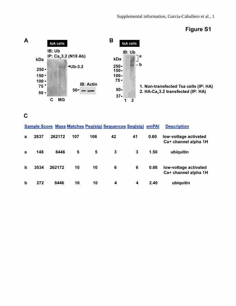
Supplemental information, Garcia-Caballero et al., 2 Figure S1. Ubiquitination of Cav3.2 channels in tsA201 cells (related to Figure 1)
(A) Same experiment as in Fig. 1A, but with a different precipitating Cav3.2 antibody (N-18, Santa
Cruz). (B) Immunoprecipitation of HA-Cav3.2 from tsA-201 cells, followed by probing with a
ubiquitin antibody revealing a ubiquitin band just above 250 kDa and a high MW smear. Two bands
(labeled a and b) were excised from the corresponding Coomassie gel and subjected to mass
spectrometry analysis. (C) Mass spectrometry results from bands “a” and “b” from the Coomassie gel.

Supplemental information, Garcia-Caballero et al., 3

Supplemental information, Garcia-Caballero et al., 4 Figure S2. Ubiquitination of Cav3.2 channels by WWP1 and WWP2 (related to Figure 2)
(A) WWP1 immunoprecipitates analyzed by Western blot with a specific anti-WWP1 antibody from
tsa-201 cells transfected with WWP1-shRNA or vector alone (control). This experiment was repeated
three times. (B) Western blot of ubiquitinated WT Cav3.2 and mutant Y1594N channels expressed in
tsA-201 cells. An α-tubulin blot is shown as sample loading control. (C) Western blot of WT Cav3.2
and mutant Y1594N channel immunoprecipitates from tsA-201 cells. The membrane from panel B,
was stripped and re-probed with anti-Cav3.2 antibody. (D) Quantification of ubiquitinated Cav3.2
channels expressed as the ratio of ubiquitinated Cav3.2 vs. total Cav3.2 channels by densitometry
(measured via relative integrated density values from western blots). (E) WWP2 does not
immunoprecipitate with Cav3.2 channels from DRG tissue, but does so from mouse brain (F). (G)
Western blot analysis comparing the expression of WWP2 in brain lysate and DRG cell lysate. Note
that WWP2 is absent from DRG tissue.

Supplemental information, Garcia-Caballero et al., 5

Supplemental information, Garcia-Caballero et al., 6 Figure S3. Expression of USP5 in DRG neurons, and USP15 controls (related to Figure 3)
(A) Primer pairs used for PCR analysis to detect mRNA expression of the long (L) and short (S) form
of USP5. Primer pair #12/#80 detects both the long and alternately spliced short form of USP5 and
allows for the comparison of relative abundance of the two isoforms. Primer pair #55/#80 selectively
amplifies the long form. (B) Epifluorescence image of a mouse DRG slice immunostained with a USP5
antibody (red) and with nuclei labeled via DAPI (blue). Note that USP5 is present in nuclei, but
excluded from the nucleolus. (C) Representative example of a mouse DRG slice stained with a USP5
antibody without (top) or with (bottom) preabsorption of the antibody with recombinant USP5. (D)
Co-immunoprecipitation assay using rat brain lysates. IgG, USP5, Cav3.2, USP15 immunoprecipitates
were probed for Cav3.2. Inputs were probed for actin as control (bottom panel). A representative
experiment is shown, n=3. (E) Western blot of USP15 immunoprecipitates from rat brain, as positive
control (n=3). Inputs were probed for actin as control (bottom panel). (F) Co-staining of DRG neurons
for USP5 (red), CGRP (green) and substance P (blue). The dashed area in the merged image is
magnified below.

Supplemental information, Garcia-Caballero et al., 7

Supplemental information, Garcia-Caballero et al., 8 Figure S4. USP5 regulates Cav3.2 ubiquitin modification and channel activity (related to Figure 5)
(A) USP5 Western blot of whole cells lysates from CAD cells transfected with or without USP5-
shRNA. A representative experiment is shown (n=3). (B) Cav3.2 immunoprecipitates probed for
ubiquitin by western blot from CAD cells pretreated with 5 µM MG132 and transfected with USP5-
shRNA or USP15-shRNA (which served as the control condition), as detected with an anti-ubiquitin
antibody. A representative experiment is shown (n=4). (C) Cav3.2 immunoprecipitates probed for
Cav3.2 are shown from a stripped membrane used in panel B. A blot for actin is shown as loading
control (right panel). A representative experiment is shown (n=4). (D) Quantification of the USP5-
shRNA effect on the ubiquitination state of Cav3.2 channels expressed as a ratio of ubiquitinated vs.
non-ubiquitinated channels (Ub-Cav3.2 channels/Cav3.2 channels), obtained from relative integrated
density values from western blots. Data from 4 experiments are included in the bar chart (*P<0.05, t-
test). (E) Representative whole cell T-type current traces evoked by a series of increasing membrane
depolarizations of CAD cells under control conditions and after treatment with USP-shRNA (F)
Cav3.2 channel peak currents of CAD cells transfected with shRNA for USP5 (n=18) or eGFP (n=20).
(G) Calcium current sensitivity to 50 μM nickel (n=5) or 3 μM (n=4) cadmium in CAD cells at a test
potential of -20 mV. The data show that low voltage activated channels expressed in these cells are
consistent with Cav3.2.

Supplemental information, Garcia-Caballero et al., 9

Supplemental information, Garcia-Caballero et al., 10 Figure S5. Effects of USP5 on pain responses in mice (related to Figure 5)
(A, B) Blind analysis of the nocifensive behavior of mice not-treated (N.t) or treated i.t. with USP5-
shRNA or USP15-shRNA (control) in the first (A) and second (B) phases of formalin-induced
nociception. Each column represents the mean ± SEM (n=5-6) and is representative of 2 independent
experimental runs. A one-way ANOVA revealed a significant effect of USP5-shRNA treatment
[F(1.163) = 4.4, p = 0.0021) for the first, and [F(14.08) = 4.4, p = 0.0135) for the second phase.

Supplemental information, Garcia-Caballero et al., 11

Supplemental information, Garcia-Caballero et al., 12 Figure S6. Characterization of the Tat-3.2-III-IV peptide (related to Figures 6 - 8)
(A) In vitro competition assay by affinity precipitation using biotinylated or non-biotinylated peptides
and mouse brain lysates, analyzed by western blot using an anti-USP5 antibody. A biotinylated form
of the Tat-Cav3.21569-1589 III-IV linker peptide (25 μg) interacted with USP5 from mouse brain lysate
(lane 1), and this interaction was reduced by 48% (when normalized to the tubulin control shown in the
right panel) a two-fold excess of non biotinylated Tat-Cav3.21569-1589 III-IV linker peptide (50 μg) (lane
2). In contrast to non-biotinylated Tat-Cav3.21569-1589 C-terminus peptide (50 μg) did not alter USP5
binding (lane 3). (B, C) Cumulative probability plots of mEPSC inter-event interval in a Substantia
gelatinosa neuron under control conditions and in the presence of nickel (B), and after washout of
nickel and in the presence of the Tat-3.2-III-IV peptide (C). These data correspond to the raw current
traces shown in Figures 6 D. All plots were prepared from 5 minutes of data acquisition in the
continuous presence of tetrodotoxin and picrotoxin (P < 0.001, Kolmogorov–Smirnov 2-sample test).
(D) Effect of nickel on EPSCs evoked from primary afferent fibres in lamina II of the spinal cord.
Scatter plots summarize the effect nickel on PPR (left) and eEPSC amplitude (right) in multiple cells.
Similar to the Tat-3.2-III-IV peptide, nickel significantly increased PPR (from 0.649 ± 0.093 to 1.056 ±
0.171; p=0.022, n=7, paired t test) with no concurrent change in eEPSC amplitude (from -238.5 ± 77.5
pA to -184.9 ± 50.8; p = 0.146, n=7, paired t test). Black bars are Means ± SEM, and black lines
between points indicate paired experiments. All recordings were performed at – 70 mV in the
continuous presence of 10 µM Cd2+ and bicuculline, and 1 µM strychnine.

Supplemental information, Garcia-Caballero et al., 13

Supplemental information, Garcia-Caballero et al., 14 Figure S7. In vivo effect of the Tat-Cav3.2-III-IV peptide (related to Figures 6 - 8).
(A) Time course of mechanical hyperalgesia of CFA-injected mice treated intraperitoneally (i.p.) with
the Tat-3.2-III-IV linker peptide (15 mg/kg). Each column represents the mean ± SEM (n=6-8) and is
representative of 2 independent experimental runs. Statistical analyses were performed by two-way
ANOVA followed by Tukey's test. (B) Time course of basal mechanical threshold of non-injured mice
treated with the Tat-3.2-III-IV linker or control peptide (10.0 μg/i.t). Statistical analyses were
performed by two-way ANOVA followed by Tukey's test. Each data set represents the mean ± SEM
(n=5) and is representative of 2 independent experimental runs. The dashed line indicates the range of
data points where injured animals differed from the sham group.

Supplemental information, Garcia-Caballero et al., 15

Supplemental information, Garcia-Caballero et al., 16 Figure S8. Regulation of Cav3.2 channels by ubiquitination and deubiquitination (related to
Discussion)
(A) Schematic representation of the Cav3.2 channel. The domain III-IV linker appears to be a hot spot
for channel regulation by ubiquitination. It contains key ubiquitination sites (i.e., residue 1560) as well
as binding sites for the ubiquitin ligase WWP1 (PY motif) and USP5. (B) Cav3.2 channels are
ubiquitinated by WWP1 and de-ubiquitinated by USP5. Ubiquitinated channels are less stable in the
plasma membrane, and are thus internalized and subject to degradation. Enhanced association of the
channel with USP5 results in an increased population of deubiquitinated (and thus more stable)
channels. It is possible that deubiquitination may also occur on endosomes, leading to recycling of
channels.

Supplemental information, Garcia-Caballero et al., 17 Supplemental experimental procedures
Cell culture and transfection
Human embryonic kidney tsA-201 cells were cultured as described previously (Altier et al., 2006).
Cells were transfected with calcium phosphate and used for biochemical analysis 72hrs post-
transfection. Neuronal derived CAD cells were cultured as described previously (Altier et al., 2011).
These cells were transfected with Lipofectamine 2000 (Invitrogen) and used for biochemical and
electrophysiological experiments 48-72 hrs post-transfection. Both tsA-201 and CAD cells were
preincubated with 5 μM MG132 (MG) or 100 μM Chloroquine (Chl) overnight.
Animals
Male C57BL/6J (wild-type) or Cacna1h (Cav3.2 null) mice (mus musculus, 22-28g, 8 weeks old,
background C57BL/6J) were used. Animals were housed at a maximum of five per cage (30 × 20 × 15
cm) with ad libitum access to food and water. They were kept in controlled temperature of 23 ± 1°C on
a 12 h light/dark cycles (lights on at 7:00 a.m.). Experiments were performed between 10 am and 3
pm, with the exception of the experiments involving USP5 shRNA in CFA treated animals, and
experiments involving delivery of the Tat peptides in CFA treated animals (as the duration of these
experiments exceeded 5 hrs). All testing procedures were performed following the protocol approved
by the Institutional Animal Care and Use Committee and all efforts were made to minimize animal
suffering according to the policies and recommendations of the International Association for the Study
of Pain. Different cohorts of mice were used for each test, except when stated. The observer was blind
to the experimental conditions in the experiment examining the effect of USP5-shRNA on acute pain
(formalin test), the effect of Tat-peptides on chronic inflammatory pain (CFA model) in either wild-

Supplemental information, Garcia-Caballero et al., 18 type or Cav3.2 null mice, the effects of Tat-peptides in long term CCI mice and in mibefradil-treated
animals. Cav3.2 null mice (Chen et al., 2003) were purchased from Jackson Labs.
Formalin test
The Formalin test is a widely used model that allows the evaluation of two different types of pain:
neurogenic (first) and inflammatory (second) pain (Dubuisson and Dennis, 1977; Hunskaar and Hole,
1987). Animals received 20 μl of formalin solution (1.25%, made up in PBS) injected intraplantarly
(i.pl.) in the ventral surface of the right hindpaw. Following intraplantar injection of formalin, animals
were immediately placed individually in observation chambers and the time spent licking or biting the
injected paw was recorded with a chronometer and considered as a nocifensive response. A blinded
experimenter observed animals individually from 0-5 min (neurogenic phase) and 15-30 min
(inflammatory phase). Mice received USP5-shRNA (12.5 µg/i.t.) or USP15-shRNA (control, 12.5
µg/i.t.) 1 day before the formalin test.
Chronic constriction injury (CCI)-induced neuropathy
Neuropathic pain was induced by loose ligatures of sciatic nerve according to the method described by
Bennett and Xie (Bennett and Xie, 1988) with minor modifications, and as performed previously in our
laboratory (Gadotti and Zamponi, 2011). Mice were anaesthetized (isoflurane, 5% induction and 2.5%
maintenance) and the right sciatic nerve was exposed at the level of the thigh by blunt dissection
through the biceps femoris. Proximal to the sciatic nerve trifurcation, about ten mm of nerve was freed
of adhering tissue and 4 loose ligatures (silk suture 6-0) were loosely tied around it with about 1-2 mm
spacing so that the epineural circulation was preserved. In sham-operated mice, the nerve was exposed
but not injured. For the studies using USP5-shRNA treatment, the same cohorts of mice were analysed
for either mechanical or thermal allodynia. Animals received USP5-shRNA (12.5 µg/i.t.) or USP15-

Supplemental information, Garcia-Caballero et al., 19 shRNA (control, 12.5 µg/i.t.) 1 day before nerve injury in order to allow us to verify its prophylactic
(pre-administered USP5-shRNA) effect. A second injection was given on the 12th day after nerve
injury to determine the therapeutic action of the USP5-shRNA treatment. For experiments involving
the Tat-peptides, mice received either Tat-3.2-III-IV (10.0 µg/i.t.) or Tat-3.2-CT (10.0 µg/i.t.) 5 days or
20 days after CCI.
CFA-induced persistent inflammatory pain
To induce inflammatory chronic pain, mice received 20 μl of Complete Freund's Adjuvant (CFA)
injected subcutaneously in the plantar surface of the right hindpaw (i.pl.) (Ferreira et al., 2001). Control
groups received 20 μl of PBS in the ipsilateral paw. This dose of CFA produces significant hindpaw
swelling and hypernociception that can be detected after 24 hours. Animals received USP5-shRNA
(12.5 µg/i.t.) or USP15-shRNA (control, 12.5 µg/i.t.), or the Tat-peptides (10.0 µg/i.t. or 15.0 mg/kg,
i.p.) 3 days following CFA injection.
Behavioral assays
Mechanical hyperalgesia was measured using the digital plantar aesthesiometer (DPA, UgoBasile,
Varese, Italy). Animals were placed individually in a small enclosed testing arena (20 cm × 18.5 cm ×
13 cm, length × width × height) on top of a wire mesh floor. Mice were allowed to acclimate for a
period of at least 90 minutes. The DPA device was positioned beneath the animal, so that the filament
was directly under the plantar surface of the ipsilateral hind paw. Each paw was tested three times per
session. To evaluate the time-dependence of the USP5-shRNA treatment, mechanical withdrawal
thresholds were determined at 1 day prior to CFA injection (Baseline), and at 0, 3, 6, 9, 12, 24, 36, 48,
72 and 96 hours after CFA injection; or at 1 day prior to CCI (Baseline), and at -1, 4, 7, 11, 14 and 18

Supplemental information, Garcia-Caballero et al., 20 days after CCI. For Tat-peptide treatment, mechanical withdrawal was analyzed at 0, 15, 40, 90, 180
and 360 minutes after treatment, 5 days after CCI.
Thermal hyperalgesia was examined by measuring the latency to withdrawal of ipsilateral hind
paws on a focused beam of radiant heat (IR=30) of a Plantar Test apparatus (UgoBasile, Varese, Italy).
Animals were placed individually in a small enclosed testing arena (20 cm × 18.5 cm × 13 cm, length ×
width × height) on top of a wire mesh floor. Mice were allowed to acclimate for a period of at least 90
minutes. The device was positioned beneath the animal, so that the radiant heat was directly under the
plantar surface of the ipsilateral hind paw. Three trials for each mouse were performed. The apparatus
was set at a cut-off time of 20 s to avoid tissue damage. For USP5-shRNA treatment, thermal
hyperalgesia was evaluated at -1, 3, 6, 10, 13 and 17 days after CCI.
Intrathecal drug treatment
Injections were given to fully conscious mice using the method described by Hylden and Wilcox
(Hylden and Wilcox, 1980). Animals were manually restrained, the dorsal fur of each mouse was
shaved, the spinal column was arched, and a 30-gauge needle attached in a PE20 Polyethylene tube to a
25-μl Hamilton microsyringe (Hamilton, Birmingham, UK) was inserted into the subdural space
between the L4 and L5vertebrae. Correct i.t. positioning of the needle tip was confirmed by a
characteristic tail-flick response of animal. Intrathecal injections of volumes between 2-10 μl were
given over a period between 5-10 seconds. For intrathecal delivery of shRNA, no transfection reagents
were added as these constructs are taken up into DRG neurons spontaneously. When mice were
injected with the Tat III-IV linker peptide (10.0 µg/i.t.) at a concentration of 5µg/µl over a period of 5
seconds, we observed that 3 out of 10 mice developed shaking behavior which terminated no later than
15 minutes after injection. When mice received the same dose of the Tat III-IV linker peptide (10.0
µg/i.t.), but at a lower concentration (2.5 µg/µl) over a period of 10 seconds, no abnormal behavior was
Supplemental information, Garcia-Caballero et al., 21 observed. In contrast, there were no noticeable behavioral consequences associated with
intraperitoneal delivery of the Tat peptides.
Heterologous expression and patch-clamp recordings in CAD cells
CAD cells were grown in a Dulbecco’s modified Eagle’s culture medium containing 10% fetal bovine
serum and 1% penicillin/streptomycin and maintained under standard conditions at 37°C in a
humidified atmosphere containing 5% CO2 and transiently transfected using phosphate-calcium buffer.
CAD cells were transfected with shUSP5 along with EGFP in a ratio 1:0.1. In control experiments,
shUSP5 was replaced by an equal amount of EGFP. Whole-cell Ba2+ currents were recorded 48-72 hrs
after transfection using pipettes (4-5 MΩ) filled with a solution containing (in mM): 110 CsCl, 3 Mg-
ATP, 0.5 Na-GTP, 2.5 MgCl2, 5 D-glucose, 10 EGTA, 10 HEPES (pH 7.3 with CsOH). The external
solution contained (in mM): 10 BaCl2, 1 MgCl2, 140 TEACl, 10 D-glucose, 10 HEPES (pH 7.2 with
TEAOH). Whole-cell recordings were performed using an Axopatch 200B amplifier (Axon
Instruments, Union City, CA). Acquisition and analysis were performed using pClamp9 and Clampfit 9
software, respectively (Axon Instruments). All traces were corrected on-line for leak currents, digitized
at 10 KHz and filtered at 2 KHz.
Harvesting of dorsal root ganglia and dorsal horn
L4-L6 dorsal root ganglia (DRGs) from 8 week old mice were dissected and collected in ice-cold
dissection Hank's Buffered Salt Solution-HBSS (Invitrogen 14185-052) containing 0.25% 4-(2-
hydroxyethyl)-1-piperazineethanesulfonic acid -HEPES (sigma H7523), osmolarity 310 mOsm and pH
7.2), then DRGs were washed once with fresh ice-cold dissection solution and cut into 2-3 pieces. DRG
tissues were incubated at 37°C in DMEM (Invitrogen 11995-073) containing 0.15% collagenase
(Invitrogen 17018-029) for 90 min, followed by 0.25% trypsin-ethylenediaminetetraacetic acid

Supplemental information, Garcia-Caballero et al., 22 (EDTA) (Invitrogen 25200-056) for another 10 min. DRG tissues were washed three times with warm
complete culture medium (DMEM supplemented with 10% heat-inactivated fetal bovine serum (FBS)
(Invitrogen 26140) and 1% penicillin/streptomycin (Invitrogen 15140)) to remove collagenase and
trypsin enzymes. DRG neurons were dissociated by three to five passages through a fire-polished
Pasteur pipette. Cells were plated on dishes pretreated with poly-D-Lysine (Sigma P7280) and laminin
(Sigma L2020) and cells were kept at 37°C in 5% CO2 incubator. On the next morning the medium
was replaced by fresh complete culture medium. Experiments were performed 12 hours after cells
were plated. Cells were preincubated with 5 μM MG132 (MG) or 100 μM Chloroquine (Chl)
overnight. A set of experiments was done using ipsilateral lumbar 5 (L5) mouse DRG harvested 90
minutes after treatment with Tat peptides, and 3 days after CFA injection. For CCI experiments, L4-L6
DRG or dorsal horn was harvested 10 or 20 days after surgery.
RNA Extraction, Reverse Transcription, and RT-PCR.
Total RNAs were prepared from pooled mouse lumbar sensory ganglia, lumbar spinal cord, brain, or
from DRG primary culture using the TRIzol method according to the manufacturer’s instructions
(Invitrogen, Carlsbad, CA). RNAs were treated with DNase-I RNase-Free (Ambion, Austin, TX).
Reverse transcriptions were performed using total RNA with random primers and the Moloney murine
leukemia virus reverse transcriptase (Invitrogen). The expression levels of the long and short USP5
isoforms were estimated with a mix of common forward and reverse primers and a specific forward
primer for the long form (Forward com 5’ GTCAAGACCACGCGCTTT 3’, Forward long specific 5’
GCAACGAAGACGAAGACTCC 3’, Reverse com 5’ CAGCCCGCTCTAAGCTATTG 3’). This
generated 2 bands for the long isoform (597 and 362 bp) and a single band for the short isoform (528
bp) (see Fig. S3A).

Supplemental information, Garcia-Caballero et al., 23 Immunohistofluorescence
Mice (P-60 to P80) were anesthetized with pentobarbital injection and transcardially perfused with
HBSS (pH 7.4, 4° C). Lumbar spinal cords and lumbar DRGs were dissected from the perfused mice.
Tissues were fixed in TBS containing 4% PFA (pH7.4) at room temperature for 30 and 60 minutes,
respectively. Tissues were washed three times 10 minutes in TBS at 4°C and then embedded in 4%
agarose and sectioned at 20-30 µm using a vibratome. For spared nerve injury (SNI) spinal cords,
animals were transcardially perfused with HBSS and then with PB containing 4% PFA (pH7.4). After
dissection, spinal cords were post fixed 60 minutes at 4°C, and cryoprotected in 30% sucrose
containing PB. Free floating section, or sections on slides were blocked with TBS containing 0.05%
tween 20 (TBST), 0.2% Triton X 100 and 10% normal goat serum at room temperature for 1 hr. Tissue
sections were incubated with primary antibodies diluted in blocking solution at 4°C overnight (rabbit
anti-USP5 (Protein Tech 15158-1-AP, 1/500), mouse anti-NF200 (Sigma N0142, 1:200)), chicken anti
CGRP (Neuromics CH14100, 1/500), guinea pig anti Substance P (Neuromics GP14103, 1/200), goat
anti IBA-1 (Abcam AB5076, 1/2000). Then, sections were washed four times with TBST, and
incubated with secondary antibodies (Alexa 568 conjugated goat anti rabbit, and Alexa 647 conjugated
goat anti mouse antibodies (Invitrogen, 1/1000), CF488A conjugated donkey anti chicken antibody
(Sigma 1/500), Cy3 conjugated goat anti mouse antibody (Jackson laboratories 1/500) Alexa 647
conjugated goat anti guinea pig antibody (Jackson laboratories 1/500), Alexa 647 conjugated donkey
anti rabbit antibody (Invitrogen 1/500), Alexa 568 conjugated donkey anti goat antibody (Invitrogen
1/500)), diluted in blocking solution at room temperature for 1 hr, washed again four times with TBST,
and mounted on superfrost slides with Vectashield (Vectors laboratories). Alexa 488 conjugated IB4
(Invitrogen) was diluted at 1/200 and incubated together with secondary antibodies. For preabsorption
experiments, recombinant USP5 protein (Sino Biological Inc, 12772-H08B) was incubated overnight at
4°C with the anti-USP5 antibody at a 10/1 molar ratio. Following this step, classical immuno-

Supplemental information, Garcia-Caballero et al., 24 histofluorescence experiments were performed as described with a direct comparison of labeling
obtained with non preabsorbed and preabsorbed anti-USP5 antibodies. Confocal images were taken
with a Zeiss LSM780 or a Leica SP8 confocal microscope (at the Montpellier RIO imaging facility)
and processed with Image J and photoshop CS4 software.
Spinal cord slice preparation
Electrophysiological analysis of the effects Tat-3.2-III-IV peptide in the spinal cord was determined in
6 – 10 week old male C57BL/6 mice (Charles River Laboratories). Acute slices from naïve and sham
animals were prepared in a manner similar to that previously described (Moran and Smith, 2002).
Briefly, mice were deeply anesthetized with isoflurane and, immediately following decapitation, a
laminectomy was performed. The lumbar enlargement of the spinal cord with attached dorsal and
ventral rootlets was transferred into ice-cold oxygenated (95% O2 – 5% CO2) dissection solution,
containing (in mM) 220 sucrose, 125 NaCl, 25 NaHCO3, 3 KCl, 1.25 NaH2PO4, 1 CaCl2 and 6 MgCl2.
The dura mater was removed and the spinal cord was then glued to a 4% agar block with cyanoacrylate
glue (Vetbond, Western Drug Distribution Center Ltd.). Transverse slices (300 µm) were cut using a
vibrating microtome (Vibratome, TPI) in ice-cold oxygenated dissection solution and were then
incubated for 1 h at 31 – 34 in oxygenated artificial cerebrospinal fluid (ACSF), containing (in mM)
125 NaCl, 25 NaHCO3, 20 D-glucose, 3 KCl, 1.25 NaH2PO4, 2 CaCl2 and 1 MgCl2 (pH 7.4).
Spinal cord slice recordings
For electrophysiological recording, slices were superfused at room temperature ( 23 ) with
oxygenated ACSF. The substantia gelatinosa appeared as translucent bands under infrared red
differential-interference contrast optics (Axioskop II, Zeiss) and neurons were patched under visual
control (Moran and Smith, 2002). Recording electrodes had resistances of 5 – 10 MΩ when filled with

Supplemental information, Garcia-Caballero et al., 25 an internal solution, containing (in mM) 130 potassium gluconate, 1 MgCl2, 2 CaCl2, 10 HEPES, 10
EGTA, 4 Mg-ATP and 0.3 Na-GTP (pH 7.2, 290 – 300 mOsm). In some experiments, QX314 (Tocris;
1 or 5 mM) was included in the internal solution to prevent action potential discharge. All recordings
were at -70 mV, using an Axopatch 200B amplifier (Axon Instruments / Molecular Devices) in
voltage-clamp mode where series resistance and capacitance were compensated (≥ 75 %) with
amplifier circuitry. Data were acquired at a sampling rate of 10 kHz and filtered at 1-5 kHz with
pClamp 10 software (Molecular Devices).
Miniature excitatory postsynaptic currents (mEPSCs) were recorded in the presence of 1 µM
TTX (Alomone Labs) and 20 µM picrotoxin (Tocris). Typically, experiments involved a five minute
recording of baseline activity, followed by a three minute application of nickel (100 µM) and a five
minute recording of activity in the continued presence of this ion. Following a five minute wash, and
another 5 minute recording period, the Tat-3.2-III-IV peptide (10 µg/ml) was applied for three minutes,
followed by a 5 minute recording period. Mini Analysis Program (Synaptosoft, Inc.) was used to
distinguish mEPSCs from baseline noise, using appropriate amplitude and area threshold settings for
each neuron. All detected events were then visually re-examined where acceptable events had a sharp
onset, an exponential offset, a total duration of < 50 ms and amplitude of at least double the baseline
noise (Chen et al, 2009). The Mini Analysis Program was further used to determine inter-event interval
of mEPSCs from all events recorded, during each 5 min session. Neurons with fewer than 100 events at
the outset of experiment were rejected from analysis.
Evoked excitatory postsynaptic currents (eEPSCs) were recorded in the presence 1 µM
strychnine and 10 µM bicuculline (both from Abcam ). Cadmium (10 µM) was included in the bath to
limit the contribution of HVA calcium channel activity to evoked responses. EPSCs were evoked
(stimulus duration: 10 or 100 µs) by a concentric bipolar stimulating electrode (FHC) placed near the
dorsal root entry zone (Moran and Smith, 2002). EPSCs were identified to be monosynaptic by their

Supplemental information, Garcia-Caballero et al., 26 ability to follow stimulation (1 Hz) with a constant latency and without failures. Paired-pulses (inter-
stimulus interval: 50 or 100 ms) were delivered at 0.2 Hz where 5-10 sweeps were used to calculate
paired-pulse ratio (pulse 2 / pulse 1) and eEPSC amplitude (pulse 1) before and 5-10 min after nickel
(100 µM) or Tat-3.2-III-IV peptide (10 µg/ml).
Calcium Imaging
For functional analysis of the effects Tat-3.2-III-IV peptide in DRG neuron culture, DRG cells were
loaded with 5 μM Fluo-4 (Molecular Probes) for 25 minutes prior to imaging. Low KCl bath solution
containing in mM- KCl 1, CaCl2 10, NaCl 125, MgCl2 2, Glucose 10, HEPES 10- and including 3 µM
cadmium to eliminate the contribution of HVA channels, and calcium signals were evoked with high
KCl (KCl 50, CaCl2 10, NaCl 75, MgCl2 2, Glucose 10, HEPES 10) stimuli. Neurons were imaged on
an inverted microscope with CCD camera, and digitized. ROI’s were fitted around perimeters of all
cells, and intensity of signal recorded using ImageJ software. Intensities were expressed in relation to
baseline fluorescence preceding stimulus, resulting in ΔF/F values. After stimulation, and a recovery
period, cells were treated with the Tat-3.2-III-IV peptide, Tat-3.2-CT peptide or nickel (100 µM) for 5
minutes, before a second high KCl stimulation. Data was reported as a ratio of the ΔF/F values after
treatment: initial. DRG cells were rejected from analysis if they did not demonstrate a robust initial
response to high KCl, or if the fluorescence intensity values after treatment were 0.

Supplemental information, Garcia-Caballero et al., 27 Supplemental references:
Altier, C., Garcia-Caballero, A., Simms, B., You, H., Chen, L., Walcher, J., Tedford, H.W., Hermosilla, T., and Zamponi GW (2011). The Cavβ subunit prevents RFP2-mediated ubiquitination and proteasomal degradation of L-type channels. Nat Neurosci 14, 173-180. Bennett, G.J., and Xie, Y.K. (1988). A peripheral mononeuropathy in rat that produces disorders of pain sensation like those seen in man. Pain 33, 87-107. Chen, C.C., Lamping, K.G., Nuno, D.W., Barresi, R., Prouty, S.J., Lavoie, J.L., Cribbs, L.L., England, S.K., Sigmund, C.D., Weiss, R.M., et al. (2003). Abnormal coronary function in mice deficient in alpha1H T-type Ca2+ channels. Science 302, 1416-1418. Chen, Y., Balasubramanyan, S., Lai, A.Y., Todd, K.G. and Smith, P.A. (2009). Effects of sciatic nerve axotomy on excitatory synaptic transmission in rat substantia gelatinosa. J Neurophysiol 102, 3203-3215. Dubuisson, D., and Dennis, S.G. (1977). The formalin test: a quantitative study of the analgesic effects of morphine, meperidine, and brain stem stimulation in rats and cats. Pain 4, 161-174. Gadotti, V.M., and Zamponi, G.W. (2011). Cellular prion protein protects from inflammatory and neuropathic pain. Mol Pain 7, 59. Hunskaar, S., and Hole, K. (1987). The formalin test in mice: dissociation between inflammatory and non-inflammatory pain. Pain 30, 103-114. Hylden, J.L., and Wilcox, G.L. (1980). Intrathecal morphine in mice: a new technique. Eur J Pharmacol 67, 313-316. Moran, T.D. and Smith, P.A. (2002) Morphine-3beta-D-glucuronide suppresses inhibitory synaptic transmission in rat substantia gelatinosa. J Pharmacol Exp Ther 302, 568-576.