Supplemental Information Cholesterol-Dependent … Information Cholesterol-Dependent Degradation ......
25
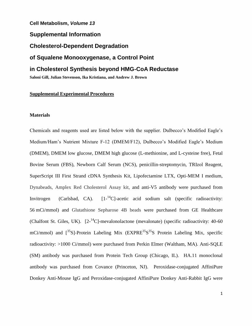
1 Cell Metabolism, Volume 13 Supplemental Information Cholesterol-Dependent Degradation of Squalene Monooxygenase, a Control Point in Cholesterol Synthesis beyond HMG-CoA Reductase Saloni Gill, Julian Stevenson, Ika Kristiana, and Andrew J. Brown Supplemental Experimental Procedures Materials Chemicals and reagents used are listed below with the supplier. Dulbecco’s Modified Eagle’s Medium/Ham’s Nutrient Mixture F-12 (DMEM/F12), Dulbecco’s Modified Eagle’s Medium (DMEM), DMEM low glucose, DMEM high glucose (L-methionine, and L-cysteine free), Fetal Bovine Serum (FBS), Newborn Calf Serum (NCS), penicillin-streptomycin, TRIzol Reagent, SuperScript III First Strand cDNA Synthesis Kit, Lipofectamine LTX, Opti-MEM I medium, Dynabeads, Amplex Red Cholesterol Assay kit, and anti-V5 antibody were purchased from Invitrogen (Carlsbad, CA). [1- 14 C]-acetic acid sodium salt (specific radioactivity: 56 mCi/mmol) and Glutathione Sepharose 4B beads were purchased from GE Healthcare (Chalfont St. Giles, UK). [2- 14 C]-mevalonolactone (mevalonate) (specific radioactivity: 40-60 mCi/mmol) and [ 35 S]-Protein Labeling Mix (EXPRE 35 S 35 S Protein Labeling Mix, specific radioactivity: >1000 Ci/mmol) were purchased from Perkin Elmer (Waltham, MA). Anti-SQLE (SM) antibody was purchased from Protein Tech Group (Chicago, IL). HA.11 monoclonal antibody was purchased from Covance (Princeton, NJ). Peroxidase-conjugated AffiniPure Donkey Anti-Mouse IgG and Peroxidase-conjugated AffiniPure Donkey Anti-Rabbit IgG were
Transcript of Supplemental Information Cholesterol-Dependent … Information Cholesterol-Dependent Degradation ......
1
Cell Metabolism, Volume 13
Supplemental Information
Cholesterol-Dependent Degradation
of Squalene Monooxygenase, a Control Point
in Cholesterol Synthesis beyond HMG-CoA Reductase
Saloni Gill, Julian Stevenson, Ika Kristiana, and Andrew J. Brown
Supplemental Experimental Procedures
Materials
Chemicals and reagents used are listed below with the supplier. Dulbecco’s Modified Eagle’s
Medium/Ham’s Nutrient Mixture F-12 (DMEM/F12), Dulbecco’s Modified Eagle’s Medium
(DMEM), DMEM low glucose, DMEM high glucose (L-methionine, and L-cysteine free), Fetal
Bovine Serum (FBS), Newborn Calf Serum (NCS), penicillin-streptomycin, TRIzol Reagent,
SuperScript III First Strand cDNA Synthesis Kit, Lipofectamine LTX, Opti-MEM I medium,
Dynabeads, Amplex Red Cholesterol Assay kit, and anti-V5 antibody were purchased from
Invitrogen (Carlsbad, CA). [1-14
C]-acetic acid sodium salt (specific radioactivity:
56 mCi/mmol) and Glutathione Sepharose 4B beads were purchased from GE Healthcare
(Chalfont St. Giles, UK). [2-14
C]-mevalonolactone (mevalonate) (specific radioactivity: 40-60
mCi/mmol) and [35
S]-Protein Labeling Mix (EXPRE35
S35
S Protein Labeling Mix, specific
radioactivity: >1000 Ci/mmol) were purchased from Perkin Elmer (Waltham, MA). Anti-SQLE
(SM) antibody was purchased from Protein Tech Group (Chicago, IL). HA.11 monoclonal
antibody was purchased from Covance (Princeton, NJ). Peroxidase-conjugated AffiniPure
Donkey Anti-Mouse IgG and Peroxidase-conjugated AffiniPure Donkey Anti-Rabbit IgG were

2
obtained from Jackson ImmunoResearch Laboratories (West Grove, PA). Lipoprotein-Deficient
Serum (LPDS) was prepared from NCS as described previously (Goldstein et al., 1983). LDL
(d=1.019-1.063 g/ml) was isolated by standard ultracentrifugation techniques from the plasma of
healthy male volunteers (Brown et al., 1996). N-Acetyl-Leu-Leu-Norleu-al (ALLN), anti-α-
tubulin antibody, butylated hydroxytoluene, chloroquine, compactin (also called mevastatin),
cycloheximide, desmosterol, Dulbecco’s phosphate buffered saline (PBS), lactacystin, methyl-β-
cyclodextrin, mevalonate, Z-Leu-Leu-Leu-al (MG132), primers, protease inhibitor cocktail, N-
ethylmaleimide, sodium oleate, sodium dodecyl sulfate (SDS), IGEPAL CA-630, sodium
deoxycholate, Zaragozic acid A trisodium salt (squalene synthase inhibitor, SSi), methionine,
and cysteine were obtained from Sigma (St. Louis, MO). The squalene epoxidase inhibitor,
GR144000X (squalene monooxygenase inhibitor, SMi) was kindly donated by GlaxoSmithKline
(Middlesex, UK). iProof High-Fidelity DNA Polymerase and Precision Plus Kaleidoscope
protein marker were from Bio-Rad Laboratories (Hercules, CA). SYBR Green SensiMix dT was
from Quantace (Norwood, MA). 24(S),24-Epoxycholesterol (24,25EC) was obtained from Enzo
Life Sciences (Farmingdale, NY). Cholesterol, lanosterol, lathosterol, 7-dehydrocholesterol
(7DHC), 24,25-dihydrolanosterol (24,25DHL), 7α-hydroxycholesterol (7αHC), 7β-
hydroxycholesterol (7βHC), 7-ketocholesterol (7KC), 19-hydroxycholesterol (19HC), 25-
hydroxycholesterol (25HC), and 27-hydroxycholesterol (27HC) were obtained from Steraloids
(Newport, RI). If not otherwise mentioned, oxysterols were delivered in ethanol. Sterols and
oxysterols complexed with methyl-β-cyclodextrin were prepared as described (Brown et al.,
2002). Sterol/CD complexes were diluted without addition of further cyclodextrin, so a constant
molar ratio of ~0.1 sterol to methyl-β-cyclodextrin was used. All solvents used for thin layer
chromatography (TLC) were analytical reagent grade from Ajax Finechem (Taren Point, NSW,
Australia). Chinese Hamster Ovary-7 (CHO-7), SRD-1, SRD-13A, and HEK293 cells were

3
generous gifts of Drs. Michael S. Brown and Joseph L. Goldstein (UT Southwestern Medical
Center, Dallas, TX). SRD-15 cells were generously donated by Dr Russell DeBose-Boyd (UT
Southwestern Medical Center, Dallas, TX). HepG2 cells and primary human fibroblasts were
kind gifts from the Centre for Vascular Research (UNSW, Sydney, NSW, Australia). BE(2)C
cells were generously donated by Dr Louise Lutze-Mann (UNSW, Sydney, NSW, Australia).
The HA-tagged ubiquitin plasmid, pMT123, encoding 8 tandem HA-ubiquitins (Treier et al.,
1994), was a gift from Dr Dirk Bohmann (University of Rochester Medical Center, Rochester,
NY).
Media Recipes
Media formulations are described below.
Medium*
A DMEM/F12 supplemented with 5% LPDS
B Medium A containing 5 µM compactin, and 50 µM mevalonate
C DMEM/F12 supplemented with 5% NCS
D DMEM low glucose supplemented with 10% FBS
E DMEM low glucose supplemented with 5% LPDS
F Medium E containing 5 µM compactin, and 50 µM mevalonate
G DMEM high glucose supplemented with 10% FBS
H DMEM high glucose supplemented with 5% LPDS
I Medium H containing 5 µM compactin, and 50 µM mevalonate
*All media containing penicillin (100 U/ml), streptomycin (100 μg/ml), and L-glutamine (2 mM)

4
Cell Culture
All cells were maintained in monolayer at 37°C in 5% CO2. Cells were seeded at the following
densities: 1×10⁵
cells/well in triplicate in 12-well plates for quantitative real-time PCR; 2×10⁶
cells/6 cm dish for immunoprecipitation; 4×10⁶
cells/10 cm dish for cell fractionation and
glutathione sepharose pulldown; 2×10⁵
cells/well in 6-well plates for all other experiments.
CHO-7, SRD-1, and SRD-15 cells were maintained in medium A, HepG2 cells in medium D,
and BE(2)C, HEK293, and fibroblasts in medium G. SRD-13A cells were maintained in
medium C supplemented with 5 μg/ml cholesterol, 1 mM mevalonate, and 20 μM sodium oleate.
Unless otherwise stated, cells were statin pretreated in media containing 5 µM statin (compactin)
and 50 µM mevalonate overnight (CHO-7, SRD-1, SRD-13A, and SRD-15 cells in medium B,
HepG2 cells in medium F, and BE(2)C, HEK293, and fibroblasts in medium I). Cells were then
treated with test agents (added in ethanol or dimethylsulfoxide to refreshed media) as indicated
in the figure legends. Within an experiment, the final concentration of solvents was kept constant
between conditions and did not exceed 0.28 % (v/v).

5
Primer Sequences
Primers are described in the following two tables.
Primer sequences for quantitative real-time PCR analysis
Gene Direction Primer Sequence (5’-3’) Reference
SQLE
(hamster)
HMGCR
(hamster)
Pbgd
(mouse)
Forward
Reverse
Forward
Reverse
Forward
Reverse
TCTGATACACGGCTACATAG
ACTTGCCATGGTGGAAAGCAAC
CTGGTGATGGGAGCTTGCTGTG
AATCACAAGCACGAGGAAGAC
G
AGATTCTTGATACTGCACTC
TGAAAGACAACAGCATCACA
Present study
(Du et al., 2006)
(Wong et al., 2006)

6
Primer sequences used for cloning and site-directed mutagenesis
Name Primer Sequence (5'-3')
topoSQEF ATGTGGACTTTTCTGGGCATTGC
topoSQER ATGAACCATATACTTCATTTCTGAG
pcTOPO TK F GCTTAGGGTTAGGCGTTTTGCGCTGCTTCGGCAGCTGCTTCA
TCCCCGTGAC
pcTOPO TK R GCTTGGGTCTCCCTATAGTGAGTCGTATTATACAATTCCGCA
GCTTTTAGAGC
SM Ntrm KO F CCAGTGTGGTGGAATTGCCCTTATGGGAACCAATATTTCAGA
AACAAGC
pcV155 R TCTTCATGCAATTGTCGGTC
pcDNA3 MCS C F AAGGGCAATTCTGCAGATATCCAGCACAGTGG
pcTOPO EV R CTGCAGAATTGCCCTTAAGGGCAATTCCACCACACTGGAC
GAr oligo F GCTGGAGCAGGCGGTGGAGCAGGTGCTGGAGGTGCAGGTGG
AGCAGGCGGTGCAGGAGCA
GAr oligo R ACCTGCTCCACCTCCAGCACCTGCACCACCTGCTCCTGCACC
GCCTGCTCCACCTGCACC
SM N GAr F CCAGTGTGGTGGAATTGCCCTTATGGCTGGAGCAGGCGGTG
GAGC
SM N GAr R GAAAGTGGCAATGCCCAGAAAAGTCCAACCTGCTCCACCTC
CAGCAC
pcC GAr F CGTACCGGTCATCATCACCATCACCATGCTGGAGCAGGCGGT

7
GGAGC
pcC GAr R GAGGCTGATCAGCGGGTTTAAACTCAACCTGCTCCACCTCCA
GCAC
pcGFP F GCCGCTCGAGTCTAGAGGGCCCGCGGTTCGAAATGGTGAGC
AAGGGCGAGGAG
pcGFP R CGAGACCGAGGAGAGGGTTAGGGATAGGCTTACCCTTGTAC
AGCTCGTCCATGCC
Cd GFP F GCCGGCAGCGGCGCCGTGAGCAAGGGCGAGGAGC
SM d476 GFP R GGCGCCGCTGCCGGCTTTTCTGCGCCTCCTGGCCTC
C GST F GCCGGCAGCGGCGCCTCCCCTATACTAGGTTATTGGAAAATT
C GST R GTTAGGGATAGGCTTACCGTCACGATGCGGCCGCTCG
C V5 F GGTAAGCCTATCCCTAACCCTCTCCTCGGTCTCG
SM N Ub F GTGGTGGAATTGCCCTTATGGGCTACCCCTATGATGTG
SM N Ub R CCCAGAAAAGTCCACATACCACCTCTGAGACGGAGGACC
SM M1 F ATGTGGACTTTTCTGGGCATTGC
pcDNA3 MCS n R AAGGGCAATTCCACCACACTGG
SM 2K15R F CTATTTTTATAGGAGGTTCGGGGACTTC
SM K82R R GGGGATCTGGCCCAGAAGAAG
SM K90R F CAGAAAATAGGGAGCAGCTC
SM K100R R TATTGGTTCCTCTTCTGCGCC
SM d476 K0 GFP R GGCGCCGCTGCCGGCCCGTCTGCGCCTCCTGGCCTC
SM K157R R CTGTCAGGCTCCCTTAAGTCTCTC
SM K268R F GGGAGTTCAGTACAAGGATAGGGAGACTGGAGATATCAAGG

8
SM K293R R GGAGACCAGGCTCCTCCTGAACTTGG
SM K318R F CTTTCTTATGAAGAATGCACCACAGTTTAGGGCAAATCATGC
TGAAC
SM 2K400R R GAAGAACACCTCGCCTCCTCACTGATGAAGG
SM K429R R CAGTTTTCTCCATAGCCTTATATCTTTAAAAGC
SM K436R F GAAAACTGCTAAGGGGTATCCCTGACC
SM K496R R CATTCGCCACCAAGCCTGAAATAAAGAAAAC
SM K536R F GTATTTTTGCTTTAGGTCAGAACCTTGG
SM K570R R GAACCATATACCTCATTTCTGAG
SM L5A F CCTTATGTGGACTTTTGCCGGCATTGCC
SM SFS F GCATTGCCACTTTCACCAGCTTTAGCAAGAAGTTCGGGGACT
TCAT
SM Y44S F CTCGCTGGGCCTGGTGCTCTCCAGCCGCTGTCGCCACC
Construction of Expression Plasmids
PCRs were performed using iProof polymerase, with verification by sequencing.
pCMV-SM-V5 contains the protein coding sequence of human squalene monooxygenase
(identical to NM_003129.3 gi.62865634, 927-2651, NP_003120) with a C-terminal V5 epitope
and His tag. It was produced by TA cloning into pcDNA3.1-V5-His TOPO vector (Invitrogen)
using the primers topoSQEF and topoSQER for PCR. pTK-SM-V5 is identical to pCMV-SM-
V5, but with expression driven by the thymidine kinase promoter. It was prepared through
9
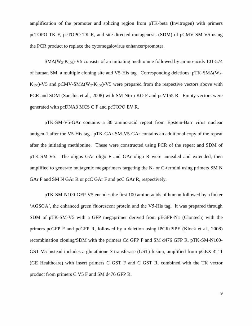
amplification of the promoter and splicing region from pTK-beta (Invitrogen) with primers
pcTOPO TK F, pcTOPO TK R, and site-directed mutagenesis (SDM) of pCMV-SM-V5 using
the PCR product to replace the cytomegalovirus enhancer/promoter.
SMΔ(W2-K100)-V5 consists of an initiating methionine followed by amino-acids 101-574
of human SM, a multiple cloning site and V5-His tag. Corresponding deletions, pTK-SMΔ(W2-
K100)-V5 and pCMV-SMΔ(W2-K100)-V5 were prepared from the respective vectors above with
PCR and SDM (Sanchis et al., 2008) with SM Ntrm KO F and pcV155 R. Empty vectors were
generated with pcDNA3 MCS C F and pcTOPO EV R.
pTK-SM-V5-GAr contains a 30 amino-acid repeat from Epstein-Barr virus nuclear
antigen-1 after the V5-His tag. pTK-GAr-SM-V5-GAr contains an additional copy of the repeat
after the initiating methionine. These were constructed using PCR of the repeat and SDM of
pTK-SM-V5. The oligos GAr oligo F and GAr oligo R were annealed and extended, then
amplified to generate mutagenic megaprimers targeting the N- or C-termini using primers SM N
GAr F and SM N GAr R or pcC GAr F and pcC GAr R, respectively.
pTK-SM-N100-GFP-V5 encodes the first 100 amino-acids of human followed by a linker
‘AGSGA’, the enhanced green fluorescent protein and the V5-His tag. It was prepared through
SDM of pTK-SM-V5 with a GFP megaprimer derived from pEGFP-N1 (Clontech) with the
primers pcGFP F and pcGFP R, followed by a deletion using iPCR/PIPE (Klock et al., 2008)
recombination cloning/SDM with the primers Cd GFP F and SM d476 GFP R. pTK-SM-N100-
GST-V5 instead includes a glutathione S-transferase (GST) fusion, amplified from pGEX-4T-1
(GE Healthcare) with insert primers C GST F and C GST R, combined with the TK vector
product from primers C V5 F and SM d476 GFP R.
10
Wild-type or mutant ubiquitin was fused to the N-terminus of SM-N100-GFP-V5 with
amplification of the insert with SM N Ub F and SM N Ub R from pRK5-HA-Ubiquitin-WT or -
K48R (Lim et al., 2005) (Ted Dawson, Johns Hopkins University School of Medicine, Addgene
Plasmids 17608 and 17604 respectively), and the vector with SM M1 F and pcDNA3 MCS n R,
yielding pTK-Ub-WT-SM-N100-GFP-V5 or pTK-Ub-K48R-SM-N100-GFP-V5, respectively.
Substitution point mutations were prepared using SDM with combinations of the primers
SM 2K15R F, SM K82R R, K90R F, SM K100R R, SM d476 K0 GFP R, SM K157R R, SM
K268R F, SM K293R R, SM K318R F, SM 2K400R R, SM K429R R, SM K436R F, SM
K496R R, SM K536R F, SM K570R R, and alternatively SM L5A F, SM SFS F (Y12,14S) or
SM Y44S F.
Metabolic Labeling of Squalene and Cholesterol: Lipid Extraction and Thin Layer
Chromatography
Cells were washed once with PBS, lyzed in 500 µl 0.1 M NaOH, and rinsed with 1.25 ml H2O.
Protein concentrations were measured by the Bicinchoninic Acid method (Pierce, Rockford, IL).
Lysates were saponified with 500 µl 20% KOH (w/v) in methanol, butylated hydroxytoluene (1
µl, 20 mM), and EDTA (20 µl, 20 mM) at 70ºC for 1 hr. After cooling, the lipids were extracted
with 2 ml hexane and evaporated to dryness. Extracts were re-dissolved in 60 µl hexane and
aliquots corresponding to equivalent amounts of protein separated on Silica Gel 60 F254 plates
(Merck, Whitehouse Station, NJ) with a mobile phase of hexane: diethyl ether: glacial acetic acid
(60:40:1, v/v/v). Bands corresponding to cholesterol and squalene (with relative Rf values of
~0.4 and ~0.9, respectively) were visualized using the FLA-5100 phosphorimager (Fujifilm,
11
Tokyo, Japan). The relative intensities of bands were quantified using Sciencelab ImageGauge
4.0 Software (Fujifilm).
Immunoprecipitation or GST Pull-Down of Ectopic SM
Following transfection, statin pretreatment and treatment as indicated in the respective figure
legends, CHO-7 cells were washed once and lyzed in modified RIPA buffer (1.0% IGEPAL CA-
630, 0.1% SDS, 0.5% sodium deoxycholate, 1 mM sodium orthovanadate, 150 mM NaCl, 1 mM
Na EDTA, 20 mM Tris-HCl, pH 7.4) supplemented with protease inhibitor cocktail. For the
ubiquitination assays, N-ethylmaleimide (10 mM) and ALLN (25 μg/ml) were also added to the
RIPA buffer during lysis. Protein concentrations were measured by the Bicinchoninic Acid
method (Pierce, Rockford, IL). After protein standardization, lysates were immunoprecipitated
with monoclonal anti-V5-conjugated Dynabeads (Invitrogen), according to the manufacturer’s
instructions. Pull-down of the N100-GST fusion protein was achieved with an overnight
incubation with glutathione sepharose beads at 4oC. Following 4 washes with RIPA buffer, beads
were resuspended in 50 μl of ‘loading buffer’ (2 vol RIPA: 2 vol 10% SDS: 1 vol 5x Laemmli
buffer). Samples were subjected to 7.5% or 10% SDS-PAGE followed by immunoblot analysis
with anti-V5 (for SM) and anti-HA (for ubiquitin) antibodies. For the [35
S] metabolic labeling
experiments, the gels were visualized using the FLA-5100 phosphorimager (Fujifilm, Tokyo,
Japan). The relative intensities of bands were quantified using Sciencelab ImageGauge 4.0
Software (Fujifilm).

12
Cell Fractionation
Fractions were prepared according to (Feramisco et al., 2004), with minor modifications. CHO-7
cells in 10 cm dishes were grown in medium A (without antibiotic) and transfected with 5 µg of
DNA. Cells were harvested after 24 hr by scraping into ice-cold PBS, washed, resuspended in
Buffer A (10 mM HEPES-KOH pH 7.4, 10 mM KCl, 1.5 mM MgCl2, 100 mM NaCl, 5 mM Na
EDTA, 5 mM Na EGTA, and 250 mM sucrose), and passed through an 18 G needle 50 times.
The lysate of equalized protein content was centrifuged at 1,000 x g for 5 min, 4°C, and the post-
nuclear supernatant centrifuged at 100,000 x g for 30 min, 4°C, with resuspension of the
resulting membrane pellet in an equal volume of the same buffer. The 1,000 x g pellet was
resuspended in Buffer B (20 mM Hepes-KOH (pH 7.6), 25% (v/v) glycerol, 0.42 M NaCl, 1.5
mM MgCl2, 5 mM sodium EDTA, and 5 mM sodium EGTA), rotated at 4°C for 1 hr, and
similarly centrifuged at 100,000 x g for 30 min at 4°C, with the supernatant yielding the nuclear
fraction. Fractions of equal volume were analyzed with SDS-PAGE and immunoblotting.
13
Figure S1
Acetyl-CoA HMG-CoA Mevalonate
Squalene
DOS
24(S),25-Epoxylanosterol
Cholesterol
HMGR
MOS
Isoprenoids
Ubiquinol
Dolichol
Shunt Pathway
24(S),25-Epoxycholesterol
Lanosterol
Lanosterol
Synthase
[14C]-Squal
[14C]-Chol
- Pretreatment + Pretreatment
Lane 1 2 3 4 5 6 7 8
A
B
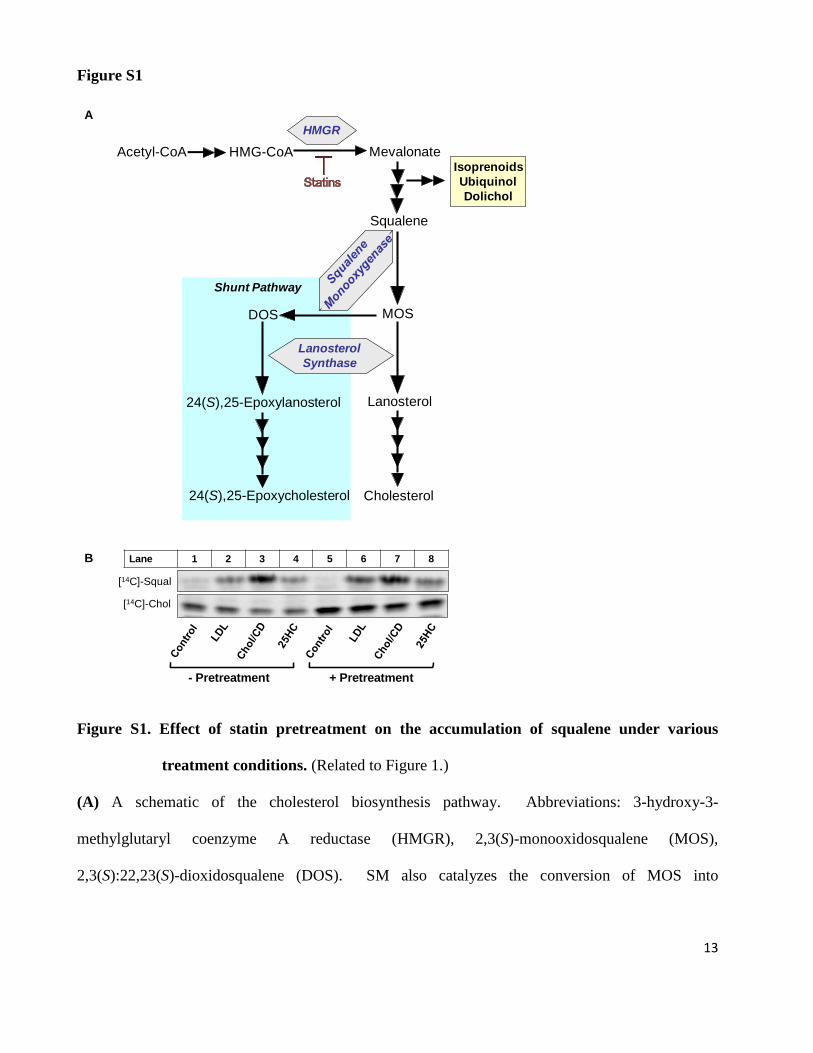
Figure S1. Effect of statin pretreatment on the accumulation of squalene under various
treatment conditions. (Related to Figure 1.)
(A) A schematic of the cholesterol biosynthesis pathway. Abbreviations: 3-hydroxy-3-
methylglutaryl coenzyme A reductase (HMGR), 2,3(S)-monooxidosqualene (MOS),
2,3(S):22,23(S)-dioxidosqualene (DOS). SM also catalyzes the conversion of MOS into
14
2,3(S):22,23(S)-dioxidosqualene, the precursor for the potent oxysterol 24(S),25-epoxycholesterol,
which fine-tunes acute cholesterol synthesis (Wong et al., 2008). Adapted from (Gill et al., 2008).
(B) CHO-7 cells were statin pretreated as indicated and then treated in medium A with 50 μg/ml
LDL, 20 μg/ml Chol/CD, or 1 μg/ml 25HC and labeled with [14
C]-acetate for 4 hr. Bands
corresponding to cholesterol and squalene were visualized by phosphorimaging and the image
shown is representative of at least 2 separate experiments.

15
Figure S2
Serum LPDS NCS
V5
α-tubulin
Lane 1 2
Figure S2. Serum derived cholesterol promotes degradation of transfected human SM.
(Related to Figure 3.)
CHO-7 cells were transfected with 1 μg of pTK-SM-V5, statin pretreated overnight, and treated in
medium B or C (supplemented with 5 μM compactin and 50 μM mevalonate) as indicated for 8 hr,
both containing 10 μg/ml cycloheximide. The immunoblot shown is representative of at least 2
separate experiments.

16
Figure S3
B
Sterol
SREBP Inhibition
In vitro Binding Scap Insig
Ub/Deg. HMGR SM
7α-Hydroxycholesterol 7β-Hydroxycholesterol 7-Ketocholesterol
+/- +/- +/-
- - -
+/- +/- +/-
- - -
+ + +
19-Hydroxycholesterol - - - + +
24(S),25-Epoxycholesterol 25-Hydroxycholesterol 27-Hydroxycholesterol
+ + +
- - -
+ + +
+ + +
- - -
Cholesterol Desmosterol 7-Dehydrocholesterol Lathosterol
+ + - -
+ +
nd nd
- -
nd nd
- -
+/- nd
+ + - -
24,25-Dihydrolanosterol Lanosterol
- -
nd -
nd -
+ -
- -
Figure S3. Effect of sterol depletion on cellular cholesterol levels and comparison of sterol
specificity for selected processes. (Related to Figure 4.)
(A) CHO-7 cells were pretreated overnight (16 hr) in either medium A, medium B, medium C, or
medium C with 5 µM compactin (statin) and 50 µM mevalonate. The media was then refreshed
with the same formulations between conditions as for the pretreatments, but with the addition of 10
0
2
4
6
8
10
12
14
16
LPDS LPDS +
CPN
NCS NCS +
CPN
μg
ch
ol/
mg
pro
tein
A
Serum LPDS NCS
Statin - + - +

17
µg/mL cycloheximide, and harvested after 8 hr for cholesterol mass determination similarly to
Figure 4H. The data is presented as mean + SEM from 3 separate experiments.
(B) Comparison of the ability (high, + ; moderate, +/- ; negligible, - ; not determined, nd ) of the
listed sterols to inhibit SREBP processing, bind to SCAP or Insig, and stimulate ubiquitination (or
degradation) of HMGR or SM. Adapted from (Radhakrishnan et al., 2007), and partly derived from
data from (Adams et al., 2004; Song et al., 2004; Song et al., 2005) and (Fitzky et al., 2001). The
last column summarizes the results from Figures 4F and G of the current work.

18
Figure S4
0
0.2
0.4
0.6
0.8
1
1.2
1.4
1 2 3 4 5 6 7 8
Re
lati
ve
SM
pro
tein
le
ve
ls
Chol/CD - +
SM
α-tubulin
Lane 1 2 3 4 5 6 7 8A B Lane 1 2 3 4 5 6 7 8 9
Chol/CD - +
SM
α-tubulin
Time (hr) 0 4
Figure S4. Effect of proteasomal and lysosomal inhibitors on cholesterol regulated turnover
of SM. (Related to Figure 5.)
(A and B) SRD-1 cells were statin pretreated overnight and treated in medium B containing
cycloheximide (10 μg/ml) with or without Chol/CD (20 μg/ml) and with MG132 (10 μM),
ALLN (25 μg/ml), lactacystin (10 μM) and chloroquine (200 μM) as indicated for 4 hr.
(A) Densitometric values for SM protein were normalized to the vehicle-treated control
condition, which was set to 1. The data is presented as mean + SEM from 4 separate
experiments. In cholesterol-treated cells, all three proteasomal inhibitors significantly increased
SM levels above control values (p < 0.05 by paired t-test).
(B) This immunoblot is representative of 3 separate experiments.

19
Figure S5
Lane 1 2 3 4 5 6
Fraction N M C N M C
pSM-V5 WT Δ(W2-K100)
B
V5
A
1 2 3
N M C
N100-GFP

20
Lane 1 2 3 4
α-tubulin
Chol/CD - + - +
WT KO
V5
Lane 1 2 3 4 5 6
Chol/CD - + - + - +
WT Y12,14S Y44S
V5
C
D
α-tubulin
Lane 1 2 3 4 5 6 7 8
V5
α-tubulin
Chol/CD - + - + - + - +
WT N K R MK R CK R
K to R mutants
N K R MK R CK R
V5
E
1 2 3 4
- + - +
WT L5ApTK-N100-
GFP-V5
pTK-SM-V5
pTK -N100-
GST-V5
Lane 1 2 3 4
α-tubulin
Chol/CD - + - +
Ubiquitin
fusionWT K48R
GF
Chol/CD - +
MG132 +
+
pUb-HA +
V5
HA-
Ubiquitin
Lane 1 2
GST
Pulldown
pTK-N100-
GST-V5
N100-GFP-V5

21
Figure S5. SM sequence conservation, membrane localization, and mutation experiments.
(Related to Figure 7.)
(A) Multiple sequence alignment of SM protein for selected species, constructed using
ClustalW2. Alignment of human (Homo sapiens, NP_003120), rat (Rattus norvegicus, P52020),
finch (Taeniopygia guttata, XP_002187271), zebra fish (Danio rerio, NP_001103509), lancelet
(Branchiostoma floridae, XP_002594656), slime mould (Dictyostelium discoideum, XP_629022)
and yeast (Saccharomyces cerevisiae, P32476).
(B) Membrane localization of truncation mutants. CHO-7 cells were transfected as indicated
and harvested for cell fractionation as described in the Supplemental Experimental Procedures.
SM protein was analyzed by immunoblotting. N, nuclear; M, membrane (100,000 x g pellet); C,
cytosol (100,000 x g supernatant).
(C-E) Substitution mutants of SM show cholesterol-dependent regulation. CHO-7 cells were
transfected with 0.5 μg (C, E) or 1 μg (D) of plasmid as indicated – SM-N100-KO-GST-V5
(KO) used in (E) contains no lysines in the first 100 hundred amino-acids, but others remain
within GST and the V5 tag. Following statin pretreatment, cells were treated in medium B
containing cycloheximide (10 μg/ml) with or without Chol/CD (20 μg/ml) for 8 hr. SM protein
was analyzed by immunoblotting. Immunoblots shown are each representative of at least 2
separate experiments.
(F) CHO-7 cells were transfected with 0.5 μg pTK-Ub-WT-SM-N100-GFP-V5 (UbWT) or pTK-
Ub-K48R-SM-N100-GFP-V5 (UbK48R), statin pretreated and treated as in (D-E) (representative
of 2 separate experiments). Polyubiquitin chains recognized by the proteasome are made up of
G76-K48 inter-ubiquitin linkages, so the K48R mutant causes premature chain termination,
inhibiting degradation (Ward et al., 1995). Expression of TK-driven N100-GFP was abrogated
when co-transfected with CMV-driven mutant ubiquitin (data not shown), possibly due to

22
transcriptional squelching by the CMV promoter. Hence to avoid this, ubiquitin was delivered
under the control of the TK promoter by fusing it to the N-terminus of SM. This approach is
possible because ubiquitin fusions are efficiently processed to liberate free ubiquitin by
deubiquitinating enzymes (Treier et al., 1994). Thus, the size of the immunoblotted N100-GFP
did not shift.
(G) CHO-7 cells were transfected with 1.5 μg of pTK-SM-N100-GST-V5 (N100-GST) and 0.5
μg of pMT123 (pUb-HA, HA-tagged ubiquitin). Following statin pretreatment, cells were
treated in medium B with or without Chol/CD (20 μg/ml) and MG132 (10 μM) for 1 hr. N100-
GST protein was pulled down with glutathione sepharose beads and immunoblotted for V5
(N100-GST) and HA-ubiquitin. Representative of 3 separate experiments.

23
Supplemental References
Brown, A.J., Dean, R.T., and Jessup, W. (1996). Free and esterified oxysterol: formation during
copper-oxidation of low density lipoprotein and uptake by macrophages. J Lipid Res 37, 320-
335.
Brown, A.J., Sun, L., Feramisco, J.D., Brown, M.S., and Goldstein, J.L. (2002). Cholesterol
addition to ER membranes alters conformation of SCAP, the SREBP escort protein that regulates
cholesterol metabolism. Mol Cell 10, 237-245.
Du, X., Kristiana, I., Wong, J., and Brown, A.J. (2006). Involvement of Akt in ER-to-Golgi
transport of SCAP/SREBP: a link between a key cell proliferative pathway and membrane
synthesis. Mol Biol Cell 17, 2735-2745.
Feramisco, J.D., Goldstein, J.L., and Brown, M.S. (2004). Membrane topology of human insig-1,
a protein regulator of lipid synthesis. J Biol Chem 279, 8487-8496.
Fitzky, B.U., Moebius, F.F., Asaoka, H., Waage-Baudet, H., Xu, L., Xu, G., Maeda, N.,
Kluckman, K., Hiller, S., Yu, H., Batta, A.K., Shefer, S., Chen, T., Salen, G., Sulik, K., Simoni,
R.D., Ness, G.C., Glossmann, H., Patel, S.B., and Tint, G.S. (2001). 7-Dehydrocholesterol-
dependent proteolysis of HMG-CoA reductase suppresses sterol biosynthesis in a mouse model
of Smith-Lemli-Opitz/RSH syndrome. J Clin Invest 108, 905-915.
Gill, S., Chow, R., and Brown, A.J. (2008). Sterol regulators of cholesterol homeostasis and
beyond: the oxysterol hypothesis revisited and revised. Prog Lipid Res 47, 391-404.
Goldstein, J.L., Basu, S.K., and Brown, M.S. (1983). Receptor-mediated endocytosis of low-
density lipoprotein in cultured cells. Methods Enzymol 98, 241-260.

24
Klock, H.E., Koesema, E.J., Knuth, M.W., and Lesley, S.A. (2008). Combining the polymerase
incomplete primer extension method for cloning and mutagenesis with microscreening to
accelerate structural genomics efforts. Proteins 71, 982-994.
Lim, K.L., Chew, K.C., Tan, J.M., Wang, C., Chung, K.K., Zhang, Y., Tanaka, Y., Smith, W.,
Engelender, S., Ross, C.A., Dawson, V.L., and Dawson, T.M. (2005). Parkin mediates
nonclassical, proteasomal-independent ubiquitination of synphilin-1: implications for Lewy body
formation. J Neurosci 25, 2002-2009.
Radhakrishnan, A., Ikeda, Y., Kwon, H.J., Brown, M.S., and Goldstein, J.L. (2007). Sterol-
regulated transport of SREBPs from endoplasmic reticulum to Golgi: oxysterols block transport
by binding to Insig. Proc Natl Acad Sci U S A 104, 6511-6518.
Sanchis, J., Fernandez, L., Carballeira, J.D., Drone, J., Gumulya, Y., Hobenreich, H., Kahakeaw,
D., Kille, S., Lohmer, R., Peyralans, J.J., Podtetenieff, J., Prasad, S., Soni, P., Taglieber, A., Wu,
S., Zilly, F.E., and Reetz, M.T. (2008). Improved PCR method for the creation of saturation
mutagenesis libraries in directed evolution: application to difficult-to-amplify templates. Appl
Microbiol Biotechnol 81, 387-397.
Treier, M., Staszewski, L.M., and Bohmann, D. (1994). Ubiquitin-dependent c-Jun degradation
in vivo is mediated by the delta domain. Cell 78, 787-798.
Ward, C.L., Omura, S., and Kopito, R.R. (1995). Degradation of CFTR by the ubiquitin-
proteasome pathway. Cell 83, 121-127.

25
Wong, J., Quinn, C.M., and Brown, A.J. (2006). SREBP-2 positively regulates transcription of
the cholesterol efflux gene, ABCA1, by generating oxysterol ligands for LXR. Biochem J 400,
485-491.