Supplemental Data CD44 Mediates Successful … Volume 29 1 Supplemental Data CD44 Mediates...
14
Immunity, Volume 29 1 Supplemental Data CD44 Mediates Successful Interstitial Navigation by Killer T Cells and Enables Efficient Antitumor Immunity Paulus Mrass, Ichiko Kinjyo, Lai Guan Ng, Steven L. Reiner, Ellen Puré, and Wolfgang Weninger Supplemental Experimental Procedures Generation and transduction of CTL Single cell suspensions from spleens of TCR transgenic OT-I animals were stimulated with SIINFEKL peptide (1μg ml -1 ) for 2 hours, washed and placed into culture. After 2 days, rmIL-2 (20 ng ml -1 ) was added, and cells were cultured for an additional 6 days. In studies involving retroviral transduction, a similar protocol was used with the exception that that the cells were exposed to SIINFEKL peptide together with IL-2 for 24h. Production of retrovirus and retroviral transduction was carried out as described previously (Pearce et al., 2003). Transduction efficiency of T cells in all experiments was ~50%. Generation of retroviral plasmids In experiments where the migration of OT-I and OT-IxCd44 -/- CTL in identical tumor regions was compared, T cells were transduced with MigR1-based retroviral plasmids containing ECFP or YFP coding sequences (Mrass et al., 2006). In rescue experiments, Cd44 -/- -T cells were transduced with retroviral plasmids containing full-length CD44 or CD44 mutants (a schematic overview of the CD44 constructs is provided in Figure S9). The constructs were generated as follows: CD44 coding sequences were subcloned into the BglII/EcoRI site of retroviral MigR1 vector (Pear et al., 1998) or fused in-frame at their C-terminus to the GFP coding sequence of retroviral vector FGM, a MigR1 variant,
Transcript of Supplemental Data CD44 Mediates Successful … Volume 29 1 Supplemental Data CD44 Mediates...
Immunity, Volume 29
1
Supplemental Data
CD44 Mediates Successful Interstitial
Navigation by Killer T Cells and Enables
Efficient Antitumor Immunity
Paulus Mrass, Ichiko Kinjyo, Lai Guan Ng, Steven L. Reiner, Ellen Puré, and Wolfgang Weninger
Supplemental Experimental Procedures
Generation and transduction of CTL
Single cell suspensions from spleens of TCR transgenic OT-I animals were stimulated
with SIINFEKL peptide (1µg ml-1) for 2 hours, washed and placed into culture. After 2
days, rmIL-2 (20 ng ml-1) was added, and cells were cultured for an additional 6 days.
In studies involving retroviral transduction, a similar protocol was used with the
exception that that the cells were exposed to SIINFEKL peptide together with IL-2 for
24h. Production of retrovirus and retroviral transduction was carried out as described
previously (Pearce et al., 2003). Transduction efficiency of T cells in all experiments was
~50%.
Generation of retroviral plasmids
In experiments where the migration of OT-I and OT-IxCd44-/- CTL in identical tumor
regions was compared, T cells were transduced with MigR1-based retroviral plasmids
containing ECFP or YFP coding sequences (Mrass et al., 2006). In rescue experiments,
Cd44-/--T cells were transduced with retroviral plasmids containing full-length CD44 or
CD44 mutants (a schematic overview of the CD44 constructs is provided in Figure S9).
The constructs were generated as follows: CD44 coding sequences were subcloned into
the BglII/EcoRI site of retroviral MigR1 vector (Pear et al., 1998) or fused in-frame at
their C-terminus to the GFP coding sequence of retroviral vector FGM, a MigR1 variant,
Immunity, Volume 29
2
lacking the internal ribosome entry site sequences. The coding sequence of full length
murine CD44 was amplified by PCR from mouse splenocyte cDNA with high fidelity Pfx
polymerase (Invitrogen) using the primers, FLfwd: 5’-
ATGAAGATCTCCACCATGGACA AGTTTTGGTGGCACACAGCTTG with FLrev:
5’-ATGCTGAATTCCTACACCC CAATCTTCATGTCCACACTC for MigR1 or
FLfusionrev: 5’-ATGCTGAATTC TCACCCCAATCTTCATGTCCACACTCTG for
FGM. The PCR product corresponds to murine CD44 cDNA (BC005676). For the
generation of CD44 mutants the MigR1 plasmid containing full length CD44 was used as
a template. To generate CD44∆ECD the primer ∆ECDfwd: 5’-
ATGAAGATCTCCACCATGGACAAGTTTTGGTGGCACACAGCTTGGGGACTTTGCC
TCTTGCAGTTGAGCCTGGCACAGGACAGTGGAGTGACCACAAC-3’ (signal
peptide is shown in Italic) was used with FLrev or FLfusionrev primers. To generate
CD44∆ICD, ∆ICDrev: 5’-ATGCTGAAT TCCTACTGCCCACACCTTCTCCTACTA
TTGAC-3’ or ∆ICDfusionrev: 5’-ATGC TGAATTCCCTGCCCACACCTTCTCCTACT
ATTGAC-3’ for C-terminal fusion were used with FLfwd primer. All constructs were
verified by sequencing.
Immunity, Volume 29
3
Supplemental Figures
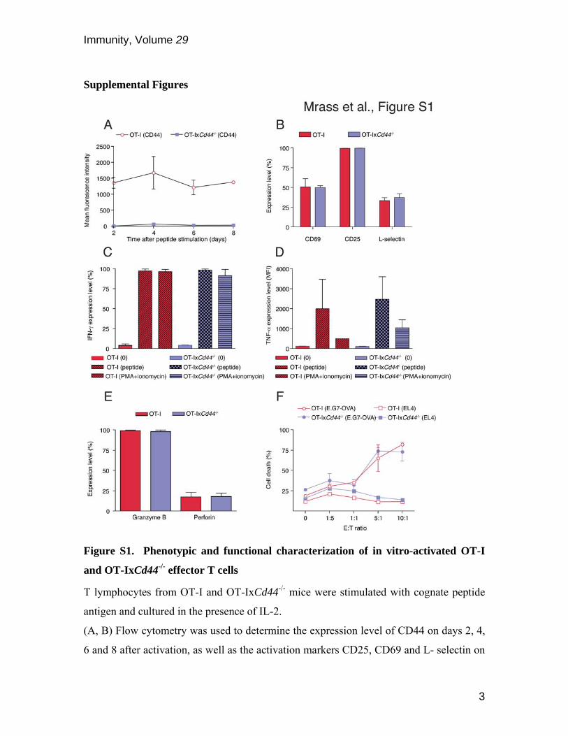
Figure S1. Phenotypic and functional characterization of in vitro-activated OT-I
and OT-IxCd44-/- effector T cells
T lymphocytes from OT-I and OT-IxCd44-/- mice were stimulated with cognate peptide
antigen and cultured in the presence of IL-2.
(A, B) Flow cytometry was used to determine the expression level of CD44 on days 2, 4,
6 and 8 after activation, as well as the activation markers CD25, CD69 and L- selectin on
Immunity, Volume 29
4
day 8 of culture (n=5 experiments).
(C, D) On day 8 after peptide stimulation, T lymphocytes were restimulated with cognate
peptide antigen or PMA and ionomycin. Production of IFN-γ and TNF-α was determined
by intracellular staining using flow cytometry (n=3 experiments).
(E) The expression level of granzyme B and perforin of T lymphocytes was determined
by flow cytometry on day 8 of culture.
(F) Graded numbers of T lymphocytes on day 8 of culture were incubated with EL4 or
E.G7-OVA tumor cells to determine antigen specific cytotoxicity. Death of the tumor
cells was determined using a cytolysis assay.
Immunity, Volume 29
5
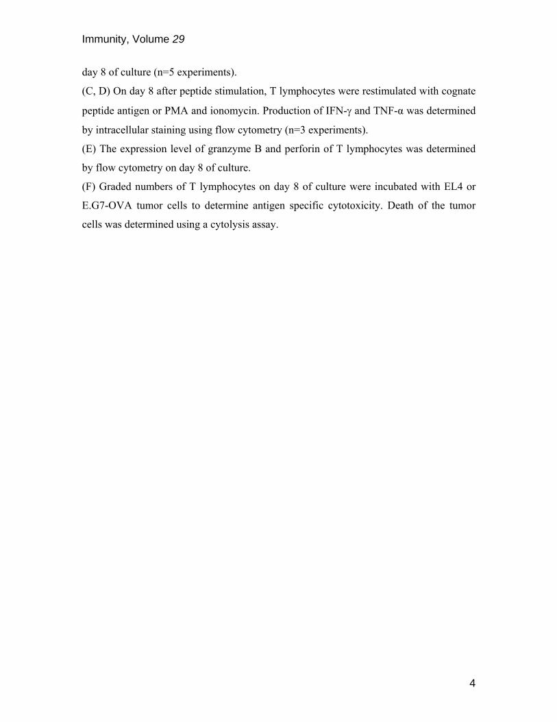
Figure S2. Migration of naïve T cells.
(A) Single cell suspensions from spleens of OT-IxDPEGFP or OT-IxCd44-/-xDPEGFP mice
were plated on collagen gel, and time-lapse sequences generated with fluorescence
microscopy. GFP+ T cells were tracked and the velocity was determined.
(B-F) Lymph nodes from OT-IxCd44-/-xDPEGFP mice were explanted and imaged with 2-
photon microscopy. GFP+ T cells were tracked and motility parameters determined. The
obtained values were compared to previously published data from CD44-wildtype T cells
(Mrass et al, 2006).
Immunity, Volume 29
6
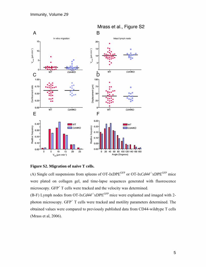
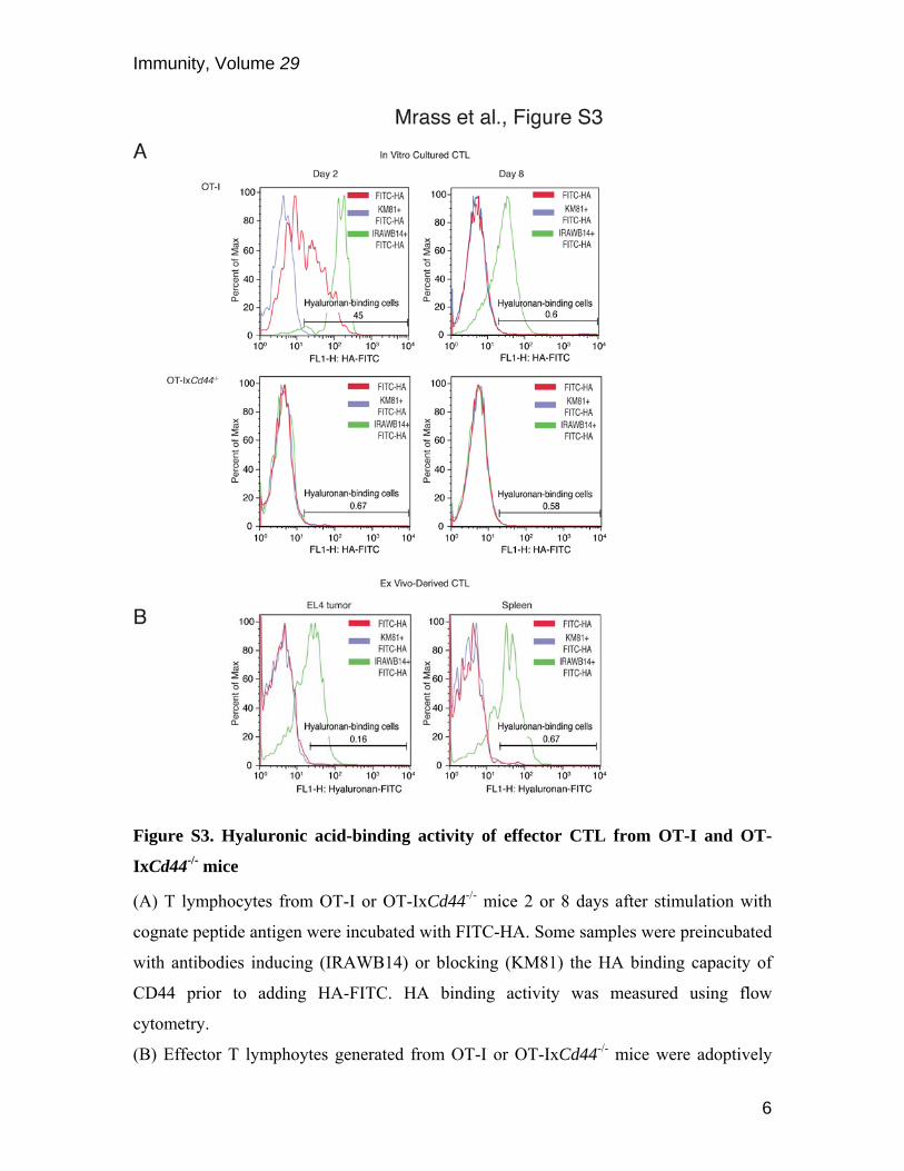
Figure S3. Hyaluronic acid-binding activity of effector CTL from OT-I and OT-
IxCd44-/- mice
(A) T lymphocytes from OT-I or OT-IxCd44-/- mice 2 or 8 days after stimulation with
cognate peptide antigen were incubated with FITC-HA. Some samples were preincubated
with antibodies inducing (IRAWB14) or blocking (KM81) the HA binding capacity of
CD44 prior to adding HA-FITC. HA binding activity was measured using flow
cytometry.
(B) Effector T lymphoytes generated from OT-I or OT-IxCd44-/- mice were adoptively

Immunity, Volume 29
7
transferred into EL4 tumor-bearing mice. After 3 days, single cell suspensions from
spleen and tumor tissues were generated followed by incubation of the samples with
FITC-HA, IRAWB14 and KM81, as in (A). Antigen-specific T cells were identified
using anti-CD8 antibody and an MHC class I-SIINFEKL tretramer. HA binding activity
of CD8+tetramer+ T cells was quantified by flow cytometry.
Immunity, Volume 29
8
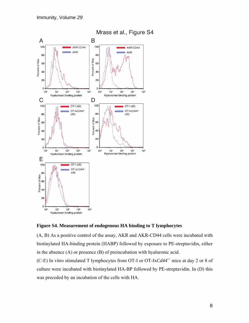
Figure S4. Measurement of endogenous HA binding to T lymphocytes
(A, B) As a positive control of the assay, AKR and AKR-CD44 cells were incubated with
biotinylated HA-binding protein (HABP) followed by exposure to PE-streptavidin, either
in the absence (A) or presence (B) of preincubation with hyaluronic acid.
(C-E) In vitro stimulated T lymphocytes from OT-I or OT-IxCd44-/- mice at day 2 or 8 of
culture were incubated with biotinylated HA-BP followed by PE-streptavidin. In (D) this
was preceded by an incubation of the cells with HA.
Immunity, Volume 29
9
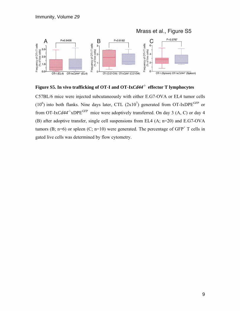
Figure S5. In vivo trafficking of OT-I and OT-IxCd44-/- effector T lymphocytes
C57BL/6 mice were injected subcutaneously with either E.G7-OVA or EL4 tumor cells
(106) into both flanks. Nine days later, CTL (2x10⁷) generated from OT-IxDPEGFP or
from OT-IxCd44-/-xDPEGFP mice were adoptively transferred. On day 3 (A, C) or day 4
(B) after adoptive transfer, single cell suspensions from EL4 (A; n=20) and E.G7-OVA
tumors (B; n=6) or spleen (C; n=10) were generated. The percentage of GFP+ T cells in
gated live cells was determined by flow cytometry.

Immunity, Volume 29
10
Figure S6. CD44-expression levels on T lymphocytes after retroviral introduction
Peptide-activated T cells derived from OT-I or OT-IxCd44-/- mice were transduced with
MigR1 or MigR1-CD44 retroviruses. Seven days after transduction, CD44 cell surface
expression levels were determined with flow cytometry.
Immunity, Volume 29
11
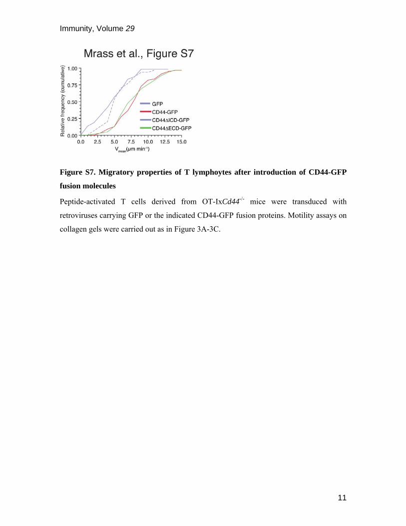
Figure S7. Migratory properties of T lymphoytes after introduction of CD44-GFP
fusion molecules
Peptide-activated T cells derived from OT-IxCd44-/- mice were transduced with
retroviruses carrying GFP or the indicated CD44-GFP fusion proteins. Motility assays on
collagen gels were carried out as in Figure 3A-3C.
Immunity, Volume 29
12
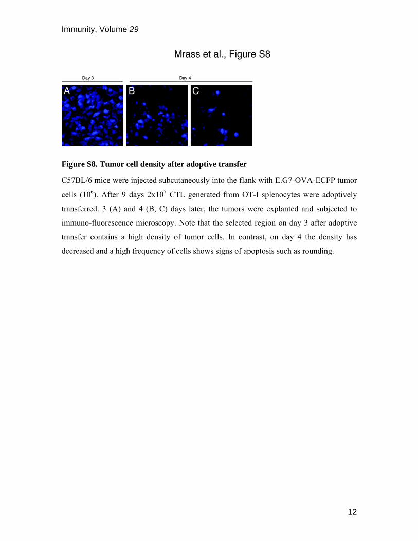
Figure S8. Tumor cell density after adoptive transfer
C57BL/6 mice were injected subcutaneously into the flank with E.G7-OVA-ECFP tumor
cells (106). After 9 days 2x10⁷ CTL generated from OT-I splenocytes were adoptively
transferred. 3 (A) and 4 (B, C) days later, the tumors were explanted and subjected to
immuno-fluorescence microscopy. Note that the selected region on day 3 after adoptive
transfer contains a high density of tumor cells. In contrast, on day 4 the density has
decreased and a high frequency of cells shows signs of apoptosis such as rounding.
Immunity, Volume 29
13
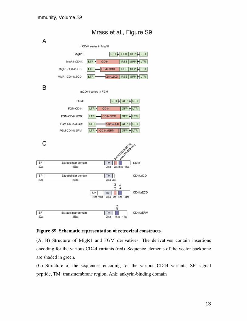
Figure S9. Schematic representation of retroviral constructs
(A, B) Structure of MigR1 and FGM derivatives. The derivatives contain insertions
encoding for the various CD44 variants (red). Sequence elements of the vector backbone
are shaded in green.
(C) Structure of the sequences encoding for the various CD44 variants. SP: signal
peptide, TM: transmembrane region, Ank: ankyrin-binding domain
Immunity, Volume 29
14
Supplemental References
Mrass, P., Takano, H., Ng, L. G., Daxini, S., Lasaro, M. O., Iparraguirre, A., Cavanagh, L. L., von Andrian, U. H., Ertl, H. C., Haydon, P. G., and Weninger, W. (2006). Random migration precedes stable target cell interactions of tumor-infiltrating T cells. J Exp Med 203, 2749-2761. Pear, W. S., Miller, J. P., Xu, L., Pui, J. C., Soffer, B., Quackenbush, R. C., Pendergast, A. M., Bronson, R., Aster, J. C., Scott, M. L., and Baltimore, D. (1998). Efficient and rapid induction of a chronic myelogenous leukemia-like myeloproliferative disease in mice receiving P210 bcr/abl-transduced bone marrow. Blood 92, 3780-3792. Pearce, E. L., Mullen, A. C., Martins, G. A., Krawczyk, C. M., Hutchins, A. S., Zediak, V. P., Banica, M., DiCioccio, C. B., Gross, D. A., Mao, C. A., et al. (2003). Control of effector CD8+ T cell function by the transcription factor Eomesodermin. Science 302, 1041-1043.