Sunil Kumar Deshmukh and Shilpa Amit Verekar
5
INTRODUCTION The Gir Forest National Park and Wildlife Sanctuary is a forest and wildlife sanctuary in Gujarat, India. Established in 1965, with a total area of 1412 km² (about 258 km² for the fully protected area (the national park) and 1153 km² for the Sanctuary), the park is located 43 km in the north-east from Somnath, 65 km to the south-east of Junagadh and 60 km to south west of Amreli. It is the sole home of the Asiatic Lions (Panthera leo persica) and is considered to be one of the most important protected areas in Asia due to its supported species. Gir National Park and Sanctuary does not have a designated area for tourists, however, to reduce the tourism hazard to the wildlife and to promote nature education, an Interpretation Zone has been created at Devalia within the sanctuary. Within its chained fences, it covers all habitat types and wildlife of Gir with its feeding-cum-living cages for the carnivores and a double-gate entry system. Its unique location, tropical climate and geographic diversity make it a potentially interesting area to study the distribution of keratinophilic fungi. We therefore undertook this study and report the results obtained. METHODOLOGY Collection and processing of soil samples Eighty samples were collected from 6 sites in the Gir Forest National Park and Wildlife Sanctuary in the month of November 2013 in sterile tightly closed polythene bags. These sites were Forest soil, Grass land soil, barren land, the raw road (pagdandi), near water reservoirs and resting areas of animals. The samples were collected from the superficial layer, depth not exceeding 35 cm, with a plastic spoon and transferred to sterile polythene bags, brought to o the laboratory, stored at 15 C and processed immediately. The hair bait technique of Vanbreuseghem, (1952) was used to isolate keratinophilic fungi. For this purpose, sterile Petri dishes were half filled with the soil samples and moistened with water and baited by burying sterile human hairs in the soil. These dishes were incubated at room temperature (25 ±1 °C) and examined for fungal growth over a period of four weeks. Isolation and identification of keratinophilic fungi After observing the growth under a stereoscopic binocular microscope it was cultured on Sabouraud's dextrose agar supplemented with chloramphenicol (50 mg/l) and cycloheximide (500 mg/l).The slopes /plates were incubated at room temperature for five to ten days following which the cultures were microscopically checked for purity and subcultured to get pure cultures. These fungi were identified based on the various available monographs (Sigler and Carmichael, 1976; Oorschot, 1980; Currah, 1985; Arx, 1986; Cano and Guarro, 1990). Molecular identification of keratinophilic fungi Molecular characteristics of the cultures were studied by determination of their DNA sequences of the ITS1-5.8S- ITS2 region. Genomic DNA was extracted by the mini prep protocol of Lee and Taylor (1990). The ITS1-5.8S- ITS2 rDNA was amplified using ITS1 and ITS4 as the forward and reverse primers, respectively, as described by White et al. (1990). Amplification was performed in 100μL reaction volumes containing 10X buffer 10μl, MgCl (25mM) 2μl, dNTP (10mM) 2μl, ITS1 primer 2 (20pm) 2μl, ITS4 primer (20pm) 2μl, Taq Polymerase (2.5U) 1μl, DNA Sample (5μg/ml) 3μl, and Milli Q Water 78μl. The PCR reaction was carried out using a Thermal Cycler (M.J. research, PTC 200) with conditions as o follows: denaturation for five minutes at 94 C, 34 cycles o o o of (30 sec at 94 C, 30 sec at 55 C, 1 min at 72 C) extension o o for four minutes at 72 C and storage at 4 C. Negative controls were used in each set of reactions. The final products were analyzed by electrophoresis on 1.2% agarose (Sigma). The PCR products were purified using Qiagen Gel extraction kit (CAT No. 28704) and the PCR products of expected size were sequenced using ITS1 and ITS4 primers in an Applied Biosystem 3730 DNA analyzer at GenOmbiotech, Pune, India. KAVAKA 43: 6-10 (2014) Isolation of keratinophilic fungi from selected soils of The Gir Forest National Park and Wildlife Sanctuary, Gujarat, (India) Sunil Kumar Deshmukh and Shilpa Amit Verekar Department of Natural Products, Piramal Enterprises Limited, 1, Nirlon Complex, Off Western Express Highway, Near NSE Complex, Goregaon (East), Mumbai 400 063, India Corresponding author email: [email protected] (Submitted in November, 2014; Accepted on December 20, 2014) ABSTRACT Eighty samples were collected from six different sites in the vicinity of Gir Forest National Park and screened for the presence of keratinophilic fungi using hair baiting technique for isolation. Seventy three isolates were recovered and identified. The cultures were identified using macro- and micro morphological features. Their identification was also confirmed by the BLAST search of sequences of the ITS1-5.8S-ITS2 rDNA region against the NCBI/Genbank data and compared with deposited sequences for identification purpose. Ten species of seven genera were isolated, viz. Aphanoascus durus (2.50 %), Aphanoascus fulvuscence (5.00 %), Arthrographis kalrae (2.50 %), Auxarthron conjugatum (1.25 %), Chrysosporium indicum (16.25 %), Chrysosporium tropicum (6.25 %), Chrysosporium zonatum (3.75 %), Chrysosporium state of Ctenomyces serratus (7.50 %), Microsporum gypseum (12.50 %), and Trichophyton mentagrophytes (3.75 %). This study indicates that the soils of Gir Forest National Park may be significant reservoirs of certain keratinophilic fungi. Key words: India, keratinophilic fungi, Gir Forest National Park, soil fungi, 6
Transcript of Sunil Kumar Deshmukh and Shilpa Amit Verekar

INTRODUCTION
The Gir Forest National Park and Wildlife Sanctuary is a forest and wildlife sanctuary in Gujarat, India. Established in 1965, with a total area of 1412 km² (about 258 km² for the fully protected area (the national park) and 1153 km² for the Sanctuary), the park is located 43 km in the north-east from Somnath, 65 km to the south-east of Junagadh and 60 km to south west of Amreli. It is the sole home of the Asiatic Lions (Panthera leo persica) and is considered to be one of the most important protected areas in Asia due to its supported species. Gir National Park and Sanctuary does not have a designated area for tourists, however, to reduce the tourism hazard to the wildlife and to promote nature education, an Interpretation Zone has been created at Devalia within the sanctuary. Within its chained fences, it covers all habitat types and wildlife of Gir with its feeding-cum-living cages for the carnivores and a double-gate entry system. Its unique location, tropical climate and geographic diversity make it a potentially interesting area to study the distribution of keratinophilic fungi. We therefore undertook this study and report the results obtained.
METHODOLOGY
Collection and processing of soil samples
Eighty samples were collected from 6 sites in the Gir Forest National Park and Wildlife Sanctuary in the month of November 2013 in sterile tightly closed polythene bags. These sites were Forest soil, Grass land soil, barren land, the raw road (pagdandi), near water reservoirs and resting areas of animals. The samples were collected from the superficial layer, depth not exceeding 35 cm, with a plastic spoon and transferred to sterile polythene bags, brought to
othe laboratory, stored at 15 C and processed immediately. The hair bait technique of Vanbreuseghem, (1952) was used to isolate keratinophilic fungi. For this purpose, sterile Petri dishes were half filled with the soil samples and moistened with water and baited by burying sterile human hairs in the soil. These dishes were incubated at
room temperature (25 ±1 °C) and examined for fungal growth over a period of four weeks.
Isolation and identification of keratinophilic fungi
After observing the growth under a stereoscopic binocular microscope it was cultured on Sabouraud's dextrose agar supplemented with chloramphenicol (50 mg/l) and cycloheximide (500 mg/l).The slopes /plates were incubated at room temperature for five to ten days following which the cultures were microscopically checked for purity and subcultured to get pure cultures. These fungi were identified based on the various available monographs (Sigler and Carmichael, 1976; Oorschot, 1980; Currah, 1985; Arx, 1986; Cano and Guarro, 1990).
Molecular identification of keratinophilic fungi
Molecular characteristics of the cultures were studied by determination of their DNA sequences of the ITS1-5.8S-ITS2 region. Genomic DNA was extracted by the mini prep protocol of Lee and Taylor (1990). The ITS1-5.8S-ITS2 rDNA was amplified using ITS1 and ITS4 as the forward and reverse primers, respectively, as described by White et al. (1990). Amplification was performed in 100µL reaction volumes containing 10X buffer 10µl, MgCl (25mM) 2µl, dNTP (10mM) 2µl, ITS1 primer 2
(20pm) 2µl, ITS4 primer (20pm) 2µl, Taq Polymerase (2.5U) 1µl, DNA Sample (5µg/ml) 3µl, and Milli Q Water 78µl. The PCR reaction was carried out using a Thermal Cycler (M.J. research, PTC 200) with conditions as
ofollows: denaturation for five minutes at 94 C, 34 cycles o o oof (30 sec at 94 C, 30 sec at 55 C, 1 min at 72 C) extension
o ofor four minutes at 72 C and storage at 4 C. Negative controls were used in each set of reactions. The final products were analyzed by electrophoresis on 1.2% agarose (Sigma). The PCR products were purified using Qiagen Gel extraction kit (CAT No. 28704) and the PCR products of expected size were sequenced using ITS1 and ITS4 primers in an Applied Biosystem 3730 DNA analyzer at GenOmbiotech, Pune, India.
KAVAKA 43: 6-10 (2014)
Isolation of keratinophilic fungi from selected soils of The Gir Forest National Park and Wildlife Sanctuary, Gujarat, (India)
Sunil Kumar Deshmukh and Shilpa Amit Verekar
Department of Natural Products, Piramal Enterprises Limited, 1, Nirlon Complex, Off Western Express Highway, Near NSE Complex, Goregaon (East), Mumbai 400 063, India Corresponding author email: [email protected] (Submitted in November, 2014; Accepted on December 20, 2014)
ABSTRACT
Eighty samples were collected from six different sites in the vicinity of Gir Forest National Park and screened for the presence of keratinophilic fungi using hair baiting technique for isolation. Seventy three isolates were recovered and identified. The cultures were identified using macro- and micro morphological features. Their identification was also confirmed by the BLAST search of sequences of the ITS1-5.8S-ITS2 rDNA region against the NCBI/Genbank data and compared with deposited sequences for identification purpose. Ten species of seven genera were isolated, viz. Aphanoascus durus (2.50 %), Aphanoascus fulvuscence (5.00 %), Arthrographis kalrae (2.50 %), Auxarthron conjugatum (1.25 %), Chrysosporium indicum (16.25 %), Chrysosporium tropicum (6.25 %), Chrysosporium zonatum (3.75 %), Chrysosporium state of Ctenomyces serratus (7.50 %), Microsporum gypseum (12.50 %), and Trichophyton mentagrophytes (3.75 %). This study indicates that the soils of Gir Forest National Park may be significant reservoirs of certain keratinophilic fungi.
Key words: India, keratinophilic fungi, Gir Forest National Park, soil fungi,
6

Phylogenetic analysis
Similarity analysis of the nucleotides was performed by BLAST searches against sequences available in GenBank (Altschul et al., 1990). For phylogenetic tree construction, multiple sequences were obtained from GenBank and the alignments were performed using MEGA6 (Tamura et al., 2013).
RESULTS
The results of the isolations are presented in Table 1. They show that out of 80 samples only 48 yielded keratinophilic fungi which were categorized into 10 species of 7 genera, namely Aphanoascus durus (2.50 %), Aphanoascus
fulvuscence (5.00 %), Arthrographis kalrae (2.50 %), Auxarthron conjugatum (1.25 %), Chrysosporium indicum (16.25 %), Chrysosporium tropicum (6.25 %), Chrysosporium zonatum (3.75 %), Chrysosporium state of Ctenomyces serratus (7.50 %), Microsporum gypseum (12.50 %), and Trichophyton mentagrophytes (3.75 %) (Fig. 1). All the ten strains yielded unique PCR amplification. The sequences of the ITS1- 5.8S- ITS2 rDNA region for the ten strains were from 527 bp to 677 bp. Chrysosporium state of Ctenomyces serratus and Trichophyton mentagrophytes were the smallest and the largest, respectively. The other species showed a product size of approximately 600 bp. There was considerable
Fig. 1. Microscopic structures of some of the kerationophilic fungi from The Gir Forest National Park and Wildlife Sanctuary, (India). A. Chrysosporium indicum, B. Chrysosporium tropicum, C. Chrysosporium state of Ctenomyces serratus, D. Chrysosporium zonatum, E. Conidial stage of Aphanoascus fulvuscence, F. Conidial stage of Aphanoascus durus, G. Microsporum gypseum, H. Trichophyton mentagrophytes, I. Arthrographis kalrae, J. Auxarthron conjugatum (Ascocarp) and K. Auxarthron conjugatum (Mature peridial hyphae).
Source of soil samples
Fo
res
tso
il
Gr
ass
lan
dso
il
ba
rela
nd
Th
era
wro
ad
(pa
gd
an
di)
Nea
rw
ate
rr
ese
rv
oir
s
Res
tin
ga
rea
so
fa
nim
als
To
tal
% Dis
trib
uti
on
Number of samples examined 15 15 10 10 15 15 80 Number of samples positive 8 7 7 6 12 8 48 Distribution (%) 53.33 46.66 70.00 60.00 80.00 53.33 60.00 1 Aphanoascus durus 1 1 - - - - 2 2.50
2 Aphanoascus fulvuscence 1 1 - - 1 1 4 5.00 3 Arthrographis kalrae - - - - - 1 1 1.25 4 Auxarthron conjugatum - - 1 - - - 1 1.25 5 Chrysosporium indicum 2 2 2 2 4 1 13 16.25
6 Chrysosporium tropicum 1 1 1 1 1 - 5 6.25 7 Chrysosporium zonatum 1 1 1 - - 3 3.75 8 Chrysosporium state of
Ctenomyces serratus 1 - 1 - 3 1 6 7.50
9 Microsporum gypseum 2 1 1 2 2 2 10 12.5 10 Trichophyton mentagrophytes - - - - 1 2 3 3.75 Total 8 7 7 6 12 8 48 60.00
Table 1. Distribution of keratinophilic fungi from Gir Forest National Park soil samples
7Sunil Kumar Deshmukh and Shilpa Amit Verekar
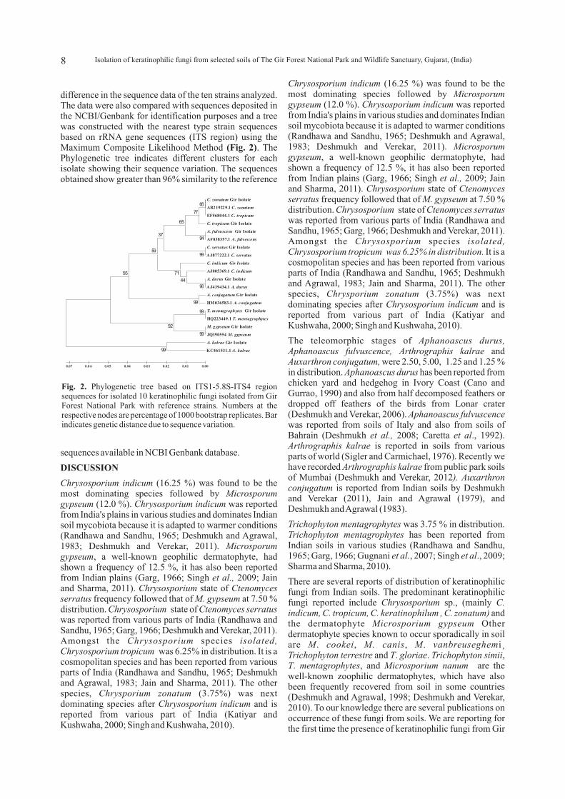
difference in the sequence data of the ten strains analyzed. The data were also compared with sequences deposited in the NCBI/Genbank for identification purposes and a tree was constructed with the nearest type strain sequences based on rRNA gene sequences (ITS region) using the Maximum Composite Likelihood Method (Fig. 2). The Phylogenetic tree indicates different clusters for each isolate showing their sequence variation. The sequences obtained show greater than 96% similarity to the reference
sequences available in NCBI Genbank database.
DISCUSSION
Chrysosporium indicum (16.25 %) was found to be the most dominating species followed by Microsporum gypseum (12.0 %). Chrysosporium indicum was reported from India's plains in various studies and dominates Indian soil mycobiota because it is adapted to warmer conditions (Randhawa and Sandhu, 1965; Deshmukh and Agrawal, 1983; Deshmukh and Verekar, 2011). Microsporum gypseum, a well-known geophilic dermatophyte, had shown a frequency of 12.5 %, it has also been reported from Indian plains (Garg, 1966; Singh et al., 2009; Jain and Sharma, 2011). Chrysosporium state of Ctenomyces serratus frequency followed that of M. gypseum at 7.50 % distribution. Chrysosporium state of Ctenomyces serratus was reported from various parts of India (Randhawa and Sandhu, 1965; Garg, 1966; Deshmukh and Verekar, 2011). Amongst the Chrysosporium species isolated, Chrysosporium tropicum was 6.25% in distribution. It is a cosmopolitan species and has been reported from various parts of India (Randhawa and Sandhu, 1965; Deshmukh and Agrawal, 1983; Jain and Sharma, 2011). The other species, Chrysporium zonatum (3.75%) was next dominating species after Chrysosporium indicum and is reported from various part of India (Katiyar and Kushwaha, 2000; Singh and Kushwaha, 2010).
Chrysosporium indicum (16.25 %) was found to be the most dominating species followed by Microsporum gypseum (12.0 %). Chrysosporium indicum was reported from India's plains in various studies and dominates Indian soil mycobiota because it is adapted to warmer conditions (Randhawa and Sandhu, 1965; Deshmukh and Agrawal, 1983; Deshmukh and Verekar, 2011). Microsporum gypseum, a well-known geophilic dermatophyte, had shown a frequency of 12.5 %, it has also been reported from Indian plains (Garg, 1966; Singh et al., 2009; Jain and Sharma, 2011). Chrysosporium state of Ctenomyces serratus frequency followed that of M. gypseum at 7.50 % distribution. Chrysosporium state of Ctenomyces serratus was reported from various parts of India (Randhawa and Sandhu, 1965; Garg, 1966; Deshmukh and Verekar, 2011). Amongst the Chrysosporium species isolated, Chrysosporium tropicum was 6.25% in distribution. It is a cosmopolitan species and has been reported from various parts of India (Randhawa and Sandhu, 1965; Deshmukh and Agrawal, 1983; Jain and Sharma, 2011). The other species, Chrysporium zonatum (3.75%) was next dominating species after Chrysosporium indicum and is reported from various part of India (Katiyar and Kushwaha, 2000; Singh and Kushwaha, 2010).
The teleomorphic stages of Aphanoascus durus, Aphanoascus fulvuscence, Arthrographis kalrae and Auxarthron conjugatum, were 2.50, 5.00, 1.25 and 1.25 % in distribution. Aphanoascus durus has been reported from chicken yard and hedgehog in Ivory Coast (Cano and Gurrao, 1990) and also from half decomposed feathers or dropped off feathers of the birds from Lonar crater (Deshmukh and Verekar, 2006). Aphanoascus fulvuscence was reported from soils of Italy and also from soils of Bahrain (Deshmukh et al., 2008; Caretta et al., 1992). Arthrographis kalrae is reported in soils from various parts of world (Sigler and Carmichael, 1976). Recently we have recorded Arthrographis kalrae from public park soils of Mumbai (Deshmukh and Verekar, 2012). Auxarthron conjugatum is reported from Indian soils by Deshmukh and Verekar (2011), Jain and Agrawal (1979), and Deshmukh and Agrawal (1983).
Trichophyton mentagrophytes was 3.75 % in distribution. Trichophyton mentagrophytes has been reported from Indian soils in various studies (Randhawa and Sandhu, 1965; Garg, 1966; Gugnani et al., 2007; Singh et al., 2009; Sharma and Sharma, 2010).
There are several reports of distribution of keratinophilic fungi from Indian soils. The predominant keratinophilic fungi reported include Chrysosporium sp., (mainly C. indicum, C. tropicum, C. keratinophilum , C. zonatum) and the dermatophyte Microsporium gypseum Other dermatophyte species known to occur sporadically in soil are M. cookei, M. canis, M. vanbreuseghemi¸ Trichophyton terrestre and T. gloriae. Trichophyton simii, T. mentagrophytes, and Microsporium nanum are the well-known zoophilic dermatophytes, which have also been frequently recovered from soil in some countries (Deshmukh and Agrawal, 1998; Deshmukh and Verekar, 2010). To our knowledge there are several publications on occurrence of these fungi from soils. We are reporting for the first time the presence of keratinophilic fungi from Gir
C. zonatum Gir Isolate
AB21922 9.1 C. zonatum
EF568044.1 C. tropicum
C. tropicum Gir Isolate
A. fulvuscens Gir Isolate
AF038357.1 A. fulvescens
C. serratus Gir Isolate
AJ877222.1 C. serratus
C. ind icum Gir Isolate
AJ005369.1 C. indicum
A. durus Gir Isolate
AJ439434.1 A. durus
A. conjugatum Gir Isolate
HM036583.1 A. conjugatum
T. mentagrophytes Gir Isolate
HQ223449.1 T. mentagrophytes
M. g ypseum Gir Isolate
JQ390554 M. gypseum
A. kalrae Gir Isolate
KC461531.1 A. kalrae
99
99
99
94
96
66
99
77
44
65
71
92
99
37
59
55
0.00 0.01 0.02 0.03 0.04 0.05 0 .0 6 0.07
Fig. 2. Phylogenetic tree based on ITS1-5.8S-ITS4 region sequences for isolated 10 keratinophilic fungi isolated from Gir Forest National Park with reference strains. Numbers at the respective nodes are percentage of 1000 bootstrap replicates. Bar indicates genetic distance due to sequence variation.
8 Isolation of keratinophilic fungi from selected soils of The Gir Forest National Park and Wildlife Sanctuary, Gujarat, (India)
Forest National park.
There are reports on various types of infections caused by species of Chrysosporium in man and animals particularly in immuno-compromised patients (Anstead et al., 2012). For example Chrysosporium zonatum was reported to cause disseminated infection in a patient with chronic granulomatous disease (Roilides et al., 1999). In Japan, C. zonatum strains were isolated from bronchial lavage from a female in Chiba and from a male in Kyushu. Both patients presented pulmonary cavity sites (Sigler et al., 1998). Chrysosporium tropicum was reported from comb lesion in two different breeds of chicken in India (Saidi et al., 1994).
CONCLUSION
Our study indicates the presence of these fungi in Gir National Park. The occurrence of these fungi in these areas is an indicative that they may be a source of superficial infections to man and animals on one hand. Whereas on the other hand these fungi may be playing an important role in decomposing the keratinic matter added to the soil by human and animal activity
REFERENCES
Altschul, S.F., Gish, W., Miller, W., Myers, E.W. and Lipman, D.J. 1990. Basic local alignment search tool. J. Mol. Biol. 215: 403-410.
Arx, von J. A. 1986. The ascomycetes genus Gymnoascus. Persoonia 13:173-183.
Anstead, G.M., Sutton, D.A. and Graybill, J.R. 2012. Adiaspiromycosis causing respiratory failure and a review of human infections due to Emmonsia and Chrysosporium spp. J. Clin. Microbiol. 50(4):1346-1354.
Cano, J. and Gurrao, J. 1990. The genus Aphanoascus. Mycological Research 94: 355-377.
Caretta, G., Mangiarotti, A M and Piontelli, E. 1992. Keratinophilic fungi isolated from soil of Italian parks in the province of Pavia. Eur. J. Epidemiol. 8(3):330-9
Currah, R.S. 1985. Taxonomy of Onygenales: A r t h r o d e r m a c e a e , G y m n o a s c a c e a e , Myxotrichaceae and Onygenaceae. Mycotaxon. 24: 1-216.
Deshmukh, S.K. 2002. Isolation of Dermatophytes and other keratinophilic fungi from Karnala Bird Sanctuary, Maharashtra (India). Journal of Basic and Applied Mycology 1(2): 194-196.
Deshmukh, S.K. and Agrawal, S.C. 1983. Prevalence of dermatophytes and other keratinophilic fungi in soils of Madhya Pradesh (India). Mykosen. 26 (11): 574-577.
Deshmukh, S.K. and Agrawal, S.C. 1998. Biology of keratinophilic fungi and related dermatophytes. In: Microbes: For health, wealth and sustainable environment (Ed.: Varma, Ajit). Malhotra Publishing House, New Delhi, India pages 253-272.
Deshmukh, S.K. and Verekar, S.A. 2006. Keratinophilic
fungi from the vicinity of meteorite crater soils of Lonar (India). Mycopathologia 162(4): 303-306.
Deshmukh, S.K., Mandeel, Q.A. and Verekar, S.A. 2008. Isolation of Keratinophilic fungi from selected soils of Bahrain. Mycopathologia 165(3): 143-147.
Deshmukh, S.K. and Verekar S.A. 2011. Incidence of Keratinophilic fungi from the soils of Vedanthangal Water Bird Sanctuary (India). Mycoses 54: 487-490.
Deshmukh, S.K. and Verekar, S.A. 2010. Keratinophilic Fungi - Distribution and prospects. In: Taxonomy and ecology of Indian Fungi (Eds.: Mukerji, K.G. and Manoharachary, C.). I.K. International Publishing House, New Delhi. India. pp. 207- 222.
Deshmukh, S.K. and Verekar, S.A. 2012, Prevalence of keratinophilic fungi in public parks of Mumbai, India. Microbiology research 3: e24-e27.
Garg, A.K. 1966. Isolation of dermatophytes and other keratinophilic fungi from soils in India. Sabouraudia 4:259-264.
Gugnani, H. C., Paliwal-Joshi, A., Rahman, H., Padhye, A. A., Singh, T. S. K., Das, T. K., Khanal, B., Bajaj, R., Rao, S. and Chukhani, R. 2007. Occurrence of pathogenic fungi in soil of burrows of rats and of other sites in bamboo plantations in India and Nepal. Mycoses 50(6): 507-511.
Jain, N, and Sharma, M. 2011. Distribution of dermatophytes and other related fungi in Jaipur city with particular reference to soil pH. Mycoses 54(1): 5258.
Jain, P.C. and Agrawal, S.C. 1979. Some additions to Indian Malbranchea. Kavaka 7: 69-72.
Katiyar, S. and Kushwaha, R.K.S. 2000. Human hair colonizing fungi in water sediments of India Mycopathologia 152: 81-84.
Lee, S.B., and Taylor, J.W. 1990. Isolation of DNA from fungal mycelium, single cells In: PCR protocols: A guide to methods and applications (Eds.: Innis, M. A., Gelfand, D.H., Sninsky, J.J. and White, T.J.). Academic Press San Diego, California, USA, p 282-287.
Oorschot, CAN van. 1980. A revision of Chrysosporium and allied genera. Stud. Mycol. 20: 1-89.
Randhawa, H.S. and Sandhu, R.S. 1965. A survey of soil i n h a b i t i n g d e r m a t o p h y t e s a n d r e l a t e d kerationophilic fungi of India. Sabouraudia 4: 7179.
Roilides, E., Sigler, L., Bibashi, E., Katsifa, H., Flaris, N. and Panteliadis, C. 1999. Disseminated infection due to Chrysosporium zonatum in a patient with Chronic granulomatous disease and review of non- Aspergillus fungal infection in patients with this disease. J Clin Microbiol. 37:18-25.
Sharma, M. and Sharma M. 2010. Incidence of dermatophytes and other keratinophilic fungi in the schools and college playground soils of Jaipur, India. Afr. J. Microbiol. Res. 4(24): 2647-2654.
9Sunil Kumar Deshmukh and Shilpa Amit Verekar
Singh, I. and Kushwaha, R.K.S. 2010. Dermatophytes and related keratinophilic fungi in soil of parks and agricultural fields of Uttar Pradesh, India. Indian J Dermatol. 55(3): 306-308
Singh, I., Mishra, A. and Kushwaha, R.K.S. 2009. Dermatophytes, related keratinophilic and opportunistic fungi in indoor dust of houses and hospitals. Indian J Med Microbiol. 27(3):242-246.
Sigler, L., and Carmichael, J.W. 1976. Taxonomy of Malbranchea and some other hyphomycetes with arthroconidia. Mycotaxon 4: 349-488.
Saidi, S.A., Bhatt, S., Richard, J.L., Sikdar, A. and Ghosh, G. R. 1994. Chrysosporium tropicum as a probable cause of mycoses of poultry in India. Mycopathologia 125:143-7.
Sigler, L., Flis, A.L. and Carmichael, J.W. 1998. The genus Uncinocarpus (Onygenaceae) and its synonym Brunneospora: new concepts, combinations and connections to anamorphs in Chrysosporium, and further evidence of relationship with Coccidioides immitis. Canad. J. Bot. 76:1624-36.
Tamura, K., Stecher, G., Peterson, D., Filipski, A. and Kumar, S. 2013. “MEGA6 : Molecular evolutionary genetics analysis version 6.0”. Mol. Biol. Evol. 30: 2725-2729.
Vanbreuseghem, R. 1952. Technique biologique pour 1' isolement des dermatophytes du sol. Ann. Soc. Belge. Med. Trop. 32:173-8.
White, T.J., Bruns, T., Lee, S. and Taylor, J. 1990. Amplification and direct sequencing of fungal ribosomal RNA genes for phylogenetics. In: PCR protocols: A guide to methods and applications. (Eds.: Innis, A., Gelfand, D.H., Sninsky, J.J. and White, T.J.) Academic Press, San Diego, California, USA, p 315-22.
10 Isolation of keratinophilic fungi from selected soils of The Gir Forest National Park and Wildlife Sanctuary, Gujarat, (India)