Summary of Safety and Effectivness (SSED)Template · • Hyphema obscuring the surgeon’s view •...
37
PMA P170034: FDA Summary of Safety and Effectiveness Data Page 1 SUMMARY OF SAFETY AND EFFECTIVENESS DATA (SSED) I. GENERAL INFORMATION Device Generic Name: Intraocular Pressure Lowering Implant Device Trade Name: Hydrus ® Microstent Device Procode: OGO Applicant’s Name and Address: Ivantis, Inc. 38 Discovery, Suite 150 Irvine, CA 92618 Date(s) of Panel Recommendation: None Premarket Approval Application (PMA) Number: P170034 Date of FDA Notice of Approval: August 10, 2018 II. INDICATIONS FOR USE The Hydrus ® Microstent is indicated for use in conjunction with cataract surgery for the reduction of intraocular pressure (IOP) in adult patients with mild to moderate primary open-angle glaucoma (POAG). III. CONTRAINDICATIONS The Hydrus ® Microstent is contraindicated under the following circumstances or conditions: • In eyes with angle closure glaucoma • In eyes with traumatic, malignant, uveitic, or neovascular glaucoma or discernible congenital anomalies of the anterior chamber (AC) angle. IV. WARNINGS AND PRECAUTIONS The warnings and precautions can be found in the Hydrus ® Microstent labeling. V. DEVICE DESCRIPTION The Hydrus ® Microstent is a crescent-shaped, implantable microstent pre-loaded onto a hand-held delivery system. The microstent (Figure 1) is composed of nitinol, a metal alloy of nickel (Ni) and titanium (Ti) with super-elastic properties. The implant is laser cut from nitinol tubing to a design with alternating “spines” for structural support and “windows” to provide
Transcript of Summary of Safety and Effectivness (SSED)Template · • Hyphema obscuring the surgeon’s view •...

PMA P170034: FDA Summary of Safety and Effectiveness Data Page 1
SUMMARY OF SAFETY AND EFFECTIVENESS DATA (SSED) I. GENERAL INFORMATION
Device Generic Name: Intraocular Pressure Lowering Implant
Device Trade Name: Hydrus® Microstent
Device Procode: OGO
Applicant’s Name and Address: Ivantis, Inc. 38 Discovery, Suite 150 Irvine, CA 92618
Date(s) of Panel Recommendation: None
Premarket Approval Application (PMA) Number: P170034
Date of FDA Notice of Approval: August 10, 2018
II. INDICATIONS FOR USE
The Hydrus® Microstent is indicated for use in conjunction with cataract surgery for the reduction of intraocular pressure (IOP) in adult patients with mild to moderate primary open-angle glaucoma (POAG).
III. CONTRAINDICATIONS
The Hydrus® Microstent is contraindicated under the following circumstances or conditions:
• In eyes with angle closure glaucoma • In eyes with traumatic, malignant, uveitic, or neovascular glaucoma or discernible
congenital anomalies of the anterior chamber (AC) angle.
IV. WARNINGS AND PRECAUTIONS
The warnings and precautions can be found in the Hydrus® Microstent labeling. V. DEVICE DESCRIPTION
The Hydrus® Microstent is a crescent-shaped, implantable microstent pre-loaded onto a hand-held delivery system. The microstent (Figure 1) is composed of nitinol, a metal alloy of nickel (Ni) and titanium (Ti) with super-elastic properties. The implant is laser cut from nitinol tubing to a design with alternating “spines” for structural support and “windows” to provide

PMA P170034: FDA Summary of Safety and Effectiveness Data Page 2
outflow pathways for aqueous humor. After laser cutting, the shape of the implant is heat-set to a curvature intended to match the curvature of Schlemm’s canal and is electro-polished to create a smooth surface. The microstent is approximately 8 mm in overall length with major and minor axes of 292 µm and 185 µm, respectively. The length and curvature of the implant are designed to occupy approximately 90° or 3 clock-hours of Schlemm’s canal. The implant is designed to have adequate structural thickness to support the tissue of the canal while providing maximum open flow areas through the canal, with the proximal portion of the implant exiting the canal through the trabecular meshwork (TM) to allow inflow of aqueous humor from the anterior chamber. The proximal end is also notched to allow for it to be interlocked to the notched cannula tip of the hand-held delivery system.
Figure 1: Hydrus® Implant
The microstent is manually implanted into the eye using a hand-held delivery system (Figure 2) through insertion of a stainless-steel cannula into the anterior chamber (AC) of the eye. The delivery system is designed for use in either the right or left hand, allowing for surgeon individual preference and hand position. To accommodate a range of hand positions, a rotatable sleeve at the distal end allows positioning and alignment of the cannula by the surgeon to direct the implant into Schlemm's canal. The tracking wheel on the delivery system serves as the control mechanism to advance the implant into the canal or retract the implant into the cannula.
Figure 2: Hydrus®Delivery System
Spines
Windows
Distal Tip
Inlet
PMA P170034: FDA Summary of Safety and Effectiveness Data Page 3
To deliver the microstent into Schlemm’s canal, the cannula of the delivery system is inserted through a clear corneal incision approximately 1.5 mm in length. The cannula tip is then advanced through the TM until it enters Schlemm’s canal and the entry point into the meshwork is coincident with the end of the cannula bevel. The target tissue is visualized using a gonioscopic prism. After observing that the distal cannula tip is properly positioned through the TM into Schlemm's canal, the tracking wheel on the delivery system is used to advance and release the microstent. It should be noted that the design of the device that will be marketed differs from the version of the device, described above, that was used during the pivotal trial. The modified version of the device included changes to the design of the interlocking mechanism “Side Interlock” that allows the implant to be released from the delivery device. See Section XI for additional details regarding the modified version of the device. The Hydrus® Microstent is packaged in sterile-barrier packaging and provided “STERILE” by gamma irradiation. Note: the Hydrus® Microstent will also be referred to as the Hydrus® implant or Hydrus® in this document.
VI. ALTERNATIVE PRACTICES AND PROCEDURES
There are several alternatives for the correction of mild to moderate POAG. These alternatives include:
• Non-surgical treatment, such as IOP-lowering medications (topical eye drops or systemic IOP lowering drugs)
• Laser treatment • Other incisional glaucoma surgery
Each alternative has its own advantages and disadvantages. A patient should fully discuss these alternatives with his/her physician to select the method that best meets expectations and lifestyle.
VII. MARKETING HISTORY
The Hydrus® Microstent is currently approved for commercial distribution in the European Union, Canada, Australia, New Zealand, Costa Rica and Columbia.
The Hydrus® Microstent has not been withdrawn from marketing for any reason relating to the safety and effectiveness of the device.

PMA P170034: FDA Summary of Safety and Effectiveness Data Page 4
VIII. PROBABLE ADVERSE EFFECTS OF THE DEVICE ON HEALTH
Below is a list of the probable adverse effects (e.g., complications) associated with the use of the device. Potential intraoperative complications and AEs may include, but are not limited to, the following: • Anterior capsule tear • Choroidal detachment • Choroidal hemorrhage or effusion • Corneal abrasion • Corneal edema • Cyclodialysis • Descemet’s membrane detachment • Device malposition • Difficulty with microstent implantation, or inability to implant the microstent • Hyphema obscuring the surgeon’s view • Inadvertent perforation of the sclera • Inadvertent loss of vitreous not associated with the cataract removal • Iridodialysis • Iris prolapse/wound incarceration • Posterior capsular rupture • Significant iris injury or trauma • Significant iris injury or trauma • Vitreous in AC • Vitreous loss not associated with the cataract procedure • Zonular dialysis Potential postoperative complications and AEs may include, but are not limited to, the following: • Angle recession • Anterior uveitis/iritis • Atrophy/phthisis • Choroidal detachment, hemorrhage, or effusion • Chronic pain in the implanted eye • Circulating blood in the AC • Corneal edema • Corneal opacification or decompensation • Elevated IOP requiring treatment with oral or intravenous medications or with
surgical intervention • Endophthalmitis • Flat or shallow AC with lens/corneal touch • Inadvertent bleb • Increase in vertical cup-to-disc ratio (C/D) and/or worsening of visual field • Layered hyphema

PMA P170034: FDA Summary of Safety and Effectiveness Data Page 5
• Loss of best corrected visual acuity (BCVA) • Maculopathy, including hypotony maculopathy • Microstent-cornea or microstent-iris touch • Microstent explantation • Microstent malposition, dislodgement, or movement • Microstent migration • Microstent obstruction (partial or complete with blood or inflammatory material) • Peripheral anterior synechiae (PAS) formation with or without microstent
obstruction • Persistent hypotony • Presence of a shallow AC with peripheral iridocorneal apposition • Ptosis • Retinal complications (dialysis, flap tears, retinal detachment, or proliferative
vitreoretinopathy) • Scleral ectasia • Significant foreign body sensation • Unplanned secondary ocular surgical re-intervention • Vitreous in AC • Vitreous hemorrhage associated with hyphema
The occurrence of some of these events may involve the necessity of secondary (additional) surgical intervention (SSI). For the specific adverse events that occurred in the clinical study of the Hydrus® Microstent, please see Section X below.
IX. SUMMARY OF NONCLINICAL STUDIES
A. Laboratory Studies As stated in the Device Description section, the applicant made a minor design change. Therefore, some testing performed on the original design was leveraged to support device approval.
i. Biocompatibility
Biocompatibility testing was performed on the Hydrus® Microstent implant or representative samples of the finished device (Table 1A) and on the patient-contacting components of the Hydrus® delivery system (Table 1B). The biocompatibility testing was performed in accordance with International Standard Organization (ISO) 10993-1 - Biological evaluation of medical devices - Part 1: Evaluation and testing within a risk management process, - Part 3: Tests for genotoxicity, carcinogenicity and reproductive toxicity, - Part 5: Tests for in vitro cytotoxicity, - Part 6: Tests for local effects after implantation, - Part 10: Tests for irritation and skin sensitization, and - Part 11: Tests for systemic toxicity.
All biocompatibility testing was conducted in accordance with the provisions of 21 CFR 58, Good Laboratory Practice for Nonclinical Laboratory Studies.

PMA P170034: FDA Summary of Safety and Effectiveness Data Page 6
TABLE 1A: BIOCOMPATIBILITY TESTING OF THE HYDRUS®IMPLANT
Test Purpose Acceptance Criteria
Results
MEM Elution w/ L-929 Mouse Fibroblast Cells (ISO 10993-5)
Evaluate the potential for cellular toxicity of the implant
Non-cytotoxic Pass
Agarose Overlay (Direct Contact) w/L-929 Mouse Fibroblast (ISO 10993-5)
Evaluate the potential for cellular toxicity of the implant
Non-cytotoxic Pass
Cell Growth Inhibition Assay w/L-929 Mouse Fibroblast Cells (ISO 10993-5)
Evaluate the potential for cellular toxicity of the implant Non-cytotoxic Pass
Guinea Pig Maximization (ISO 10993-10)
Evaluate the sensitization capacity of the implant Non-sensitizer Pass
Rabbit Intracutaneous Reactivity (ISO 10993-10)
Evaluate the potential of the implant to induce local irritation
Non-irritant Pass
Mouse Acute Systemic Toxicity (ISO 10993-11)
Evaluate the potential for systemic toxicity of the implant
Non-toxic Pass
Rabbit Pyrogen (ISO 10993-11)
Evaluate the potential of the implant to cause a febrile response
Non-pyrogenic Pass
Repeat Exposure Systemic Toxicity (Mouse 14-Day Intraperitoneal Injection; ISO 10993-11)
Evaluate the subacute systemic toxicity potential of the implant Non-toxic Pass
Repeat Exposure Systemic Toxicity (Mouse 14-Day Intravenous Injection; ISO 10993-11)
Evaluate the subchronic systemic toxicity potential of the implant Non-toxic Pass
Bacterial Reverse Mutation Mutagenicity Test (Ames Test; ISO 10993-3)
Evaluate the mutagenic potential of the implant Non-mutagenic Pass
Mammalian Erythrocyte Micronucleus test
Evaluate the potential of the implant to induce micronuclei formation
Non-genotoxic Pass
In vitro Mouse Lymphoma Assay (ISO 10993-3)
Evaluate the potential of the implant to induce forward mutations in mouse lymphoma cells
Non-genotoxic Pass
Rabbit Muscle Implantation (13 weeks; ISO 10993-6)
Evaluate the local effects of the implant in living skeletal muscle tissue
Non-irritant Pass
6-Month Ocular Implantation Study in Rabbits (ISO 10993-6)
Evaluate the ocular local tissue response to the implant
No significant biological local
response Pass

PMA P170034: FDA Summary of Safety and Effectiveness Data Page 7
In addition to the biocompatibility tests summarized in Table 1A, the applicant conducted a risk assessment to evaluate the carcinogenicity potential of the Nickel. The risk assessment concluded that the amount of Nickel available in the implant does not pose a carcinogenic risk.
TABLE 1B: BIOCOMPATIBILITY TESTING OF THE HYDRUS®DELIVERY SYSTEM
Test Purpose Acceptance Criteria Results
MEM Elution w/L-929 Mouse Fibroblast Cells (ISO 10993-5)
Evaluate the potential for cellular toxicity of the delivery system
Non-cytotoxic Pass
Guinea Pig Maximization (ISO 10993-10)
Evaluate the allergic potential or sensitization capacity of the delivery system
Non-sensitizer Pass
Intraocular Irritation in Rabbits (ISO 10993-10)
Evaluate the ocular irritation of the delivery system Non-irritant Pass
Rabbit Intracutaneous Reactivity (ISO 10993-10)
Evaluate the potential of the delivery system to induce local irritation
Non-irritant Pass
Mouse Acute Systemic Toxicity (ISO 10993-11)
Evaluate the potential for systemic toxicity of the delivery system
Non-toxic Pass
Rabbit Pyrogen (ISO 10993-11)
Evaluate the potential of the delivery system to cause a febrile response
Non-pyrogenic Pass
Note: Biocompatibility testing was performed on the orginal design of the Hydrus® Microstent.
ii. Physicochemcal testing
Physicochemical testing was conducted to physically characterize and verify the stability of the microstent throughout the potential implant life span. Physicochemical testing (Table 2) was conducted on the Hydrus® implant (or representative samples of the finished device) in accordance with ANSI Z80.27 - Ophthalmics - Implantable Glaucoma Devices and FDA Guidance Document - Premarket Studies of Implantable Minimally Invasive Glaucoma Surgical (MIGS) Devices (December 15, 2015).

PMA P170034: FDA Summary of Safety and Effectiveness Data Page 8
TABLE 2: PHYSICOCHEMICAL TESTING OF THE HYDRUS®IMPLANT
Test Purpose Acceptance Criteria Results
Corrosion Resistance
Determine susceptibility of microstent to galvanic corrosion per ASTM F2129-08 (dynamic polarization technique)
Withstand a breakdown potential >600mV without damage of surface or physical properties
Resistant to corrosion
Exhaustive Extraction
Determine identity and amount of extractable substances
No extractable substances at levels that would affect the human eye
Safe levels of extractable substances
Leachables Determine identity and amount of saline leachables
No leachable substances at levels that would affect the human eye
Safe levels of leachable substances
Nickel Elution Quantify amount of nickel ion released from microstent
Risk of adverse effects resulting from nickel leaching is negligible
Negligible nickel levels
Hydrolytic Stability
Demonstrate 5 year accelerated hydrolytic stability of the microstent in situ
Microstent is hydrolytically stable for 5 years; no detectable adverse effect on surface characteristics of finished microstents
Hydrolytically stable
Insoluble Organics
Evaluate microstent for evidence of inorganic compounds on the finished device at the end of the manufacturing process
No significant levels of insoluble organics detected
Negligible organic substances
Note: Physicochemical testing was performed on the orginal design of the Hydrus® Microstent.
B. Animal Studies
No animal studies other than those described in Section (i) Biocompatibility, above were conducted for the Hydrus® Microstent. Refer to the last row of Table 1A for description of long term (6 month) ocular implant study in rabbits.
C. Additional Studies
i. Physical and Mechanical Testing
The Hydrus® implant and delivery system were subjected to physical and mechanical testing in accordance with ANSI Z80.27 (Table 3).

PMA P170034: FDA Summary of Safety and Effectiveness Data Page 9
TABLE 3: PHYSICAL AND MECHANICAL TESTING OF THE HYDRUS®MICROSTENT
Test Purpose Acceptance Criteria Results
Material Properties Nitinol (NiTi) Alloy
Verify nitinol base material satisfies composition and mechanical characteristics for NiTi surgical implants (ASTM F2063-12)
Meets material specifications as shown in ASTM
F2063-12
Passed
Austenitic Finish Transition Temperature (Af)
Verify Af transition temperature satisfies specification using differential scanning calorimetry testing (ASTM F2004-05)
16±3º C Passed
Dimensional Properties
Verify overall dimensions are within specifications
Meets length, wall thickness and radius
of curvature specifications
Passed
Edge and Surface Quality
Verify edges and surface of microstent are smooth, free of cracks, protrusion, pits, dings, inclusions and stringers
Criteria for light microscope and SEM Passed
Structural Integrity Verify microstent satisfies mechanical strength requirements per ANSI Z80.27-14
Tensile force at breakage >0.5N Passed
Outflow Facility
Verify microstent is able to increase outflow of aqueous humor sufficient to lower IOP in bench test model
Significant increase in outflow over baseline
in cadaver eyes Passed
Delivery System Performance
Verify delivery system consistently satisfies performance requirements for microstent delivery under simulated use conditions
Must satisfy all performance criteria
for successful delivery of the microstent
Passed
Note: Physical and Mechanical testing was performed on the orginal and modified design of the Hydrus® Microstent.
ii. Magnetic Response Compatibility
A series of tests was conducted to evaluate the compatibility of the microstent with standard magnetic resonance imaging (MRI) techniques. The results are summarized in Table 4.

PMA P170034: FDA Summary of Safety and Effectiveness Data Page 10
TABLE 4: MAGNETIC RESONANCE COMPATIBILITY
Test Purpose Acceptance Criteria Results
Magnetically induced displacement ASTM F2052-15
Evaluate MRI field effects on movement of the microstent <45o deflection angle Passed
Magnetically induced torque ASTM F2213-11
Evaluate MRI field effects on movement of the microstent <45o deflection angle Passed
Magnetically induced heating ASTM F2182-11
Evaluate the amount of MRI related heating of the microstent
Temperature effect is physiologically
compatible
Temperature value scaled to whole body averaged SAR of 4-
W/kg for the First Level Controlled Operating
Mode is 2.2°C.
Image artifacts ASTM F2119-13
Evaluate the potential for the microstent to produce image artifacts under MRI
For characterization only
The maximum artifact size extends
approximately 2 mm when imaged using a
gradient echo sequence and a 3-Tesla MR
System.” Classification for magnetic resonance imaging compatibility ASTM F2503-13
Evaluate the compatibility of the microstent with current standard magnetic resonance imaging techniques
Meets requirements too be labeled “MR
conditional”
Passed “MR conditional”
Note: Magnetic response compatability testing was performed on the orginal design of the Hydrus® Microstent.
iii. Sterilization, Package Integrity, Shelf Life, and Transport Stability
The Hydrus® Microstent is supplied with the implant pre-loaded in the handheld delivery system. The device is sealed in sterile-barrier (Tyvek®) packaging and packaged in a chipboard shelf box. The packaged device is sterilized by exposure to gamma radiation. Microbiological studies have been conducted to demonstrate that the packaged device satisfies domestic and international requirements to be labeled ‘STERILE’ with a Sterility Assurance Level (SAL) of 10-6. The sterilization cycle was validated in accordance with the provisions of ISO 11137-1:2006 - Sterilization of health care products – Radiation – Part 1: Requirements for development, validation and routine control of a sterilization process for medical devices and ISO 11137-2:2012 - Sterilization of health care products – Radiation - Part 2: Establishing the sterilization dose. The methodology used was that applicable to substantiating 25 kGy as a minimum sterilization dose (Method VDmax25). Successful completion of the sterilization validation qualifies dose monitoring as a means for routine lot release of the device. Bacterial endotoxin testing was conducted in accordance with FDA guidance Endotoxin Testing Recommendations for Single-Use Intraocular Ophthalmic Devices, Guidance for

PMA P170034: FDA Summary of Safety and Effectiveness Data Page 11
Industry and FDA Staff, August 2015. Limulus amoebocyte lysate testing per USP on sterile finished devices demonstrated that the concentration of bacterial endotoxin were below 0.2 EU per device which is the current limit established for intraocular implants. Sterilization, packaging, shipping, and shelf life studies were conducted to verify that the packaging for the device maintains a sterile barrier and that the device performance meets product specifications through the labeled shelf life. The results of the sterilization, packaging, shelf life and transport stability studies are summarized in Table 5.
TABLE 5: STERILIZATION, PACKAGING, TRANSPORTATION AND SHELF LIFE TESTING
Test Purpose Acceptance Criteria Results
Gamma Irradiation Sterilization Validation
Demonstrate device is ‘STERILE’ to an SAL of 10-6 per ISO 11137-1:2006 and ISO 11137-2:2012
SAL 10-6 (minimum) Passed
Bioburden Establish upper limit for product bioburden to establish sterility (< 200 CFU/Device)
< 200 CFU/Device Passed
Bacterial Endotoxin
Verify bacterial endotoxin levels satisfy FDA Guidance for single-use ophthalmic devices (< 0.2 EU/Device)
< 0.2 EU/Device Passed
Package Evaluation – Bubble Leak
Verify package seal integrity (ASTM F2096-11) Pass/Fail Passed
Package Evaluation – Seal Peel Test
Verify package seal integrity > 1.0 Lbf Passed
Transport Stability
Verify package and device stability after transportation challenges per ASTM D4169-14, Cycle 13
Meets product specifications after
challenges Passed
Shelf Life Stability
Verify packaging and device satisfy specifications through labeled expiration date
Meets product specifications after
aging
Passed at 2 Years
Note: Sterilization, package iuntegrity, shelf life, and transport stability testing was performed on the orginal and modified design of the Hydrus® Microstent.
X. SUMMARY OF PRIMARY CLINICAL STUDY
The applicant performed a clinical study to establish a reasonable assurance of safety and effectiveness of cataract surgery with the Hydrus® Microstent for the reduction of intraocular pressure (IOP) in adults with mild to moderate primary open-angle glaucoma (POAG) in the US, Canada, Mexico , the United Kingdom, Italy, Spain, Poland and the Philippines under IDE# G110048. Data from this clinical study were the primary basis for the PMA approval decision. A summary of the clinical study is presented below.

PMA P170034: FDA Summary of Safety and Effectiveness Data Page 12
A. Study Design
Patients were treated between February 2012 and May 2015. The database for this PMA reflected data collected through July 2017 and included 556 patients. There were 38 investigational sites.
The study was a prospective, randomized, comparative, multicenter investigation conducted in the United States and other countries. A total of 556 participants from 38 study sites were randomized in a 2:1 fashion to undergo either implantation of the Hydrus® Microstent after uncomplicated cataract removal surgery (Hydrus® group) or cataract removal surgery alone without implantation of the Hydrus® Microstent (Control group). A total of 369 and 187 participants were randomized to the Hydrus® group and control group, respectively. In each participant, only one eye was considered to be the study eye. Enrollment began in February 2012 and the last participant was randomized in May 2015. Randomized participants were followed for two years postoperatively. The database for this PMA reflected data collected through July, 2017. The participants and medical monitor was masked to treatment assignments. Additionally, the observers reading the dial of the Goldmann applanation tonometers were masked. In the initial phase of the trial, 79 participants (51 in the Hydrus® group and 28 in the control group) were randomized and followed for a minimum of three months postoperatively. Safety data, which included three- to six-month follow-up for these 79 participants, were submitted for FDA review. Approval for expansion to enroll the full cohort was granted on November 15, 2013. During the expansion phase, 477 additional participants (318 in the Hydrus® group and 159 in the control group) were randomized. The primary effectiveness endpoint was the proportion of eyes in which diurnal IOP (DIOP) was reduced by ≥20% at 24 months postoperative compared to baseline after washout of topical glaucoma medications. The study hypothesis was that a larger proportion of eyes who received the Hydrus®device would meet the primary effectiveness endpoint than the proportion who received cataract surgery alone. As an additional performance goal, at least 50% of the Hydrus®group must achieve the primary effectiveness endpoint. The sample size calculation was based on assumptions and criteria related to the primary effectiveness endpoint and two proposed hypotheses, the first specifying the Hydrus® group achieving superiority over the control group, the other related to pre-specified performance goal. The selected sample size is the larger of the two (based on the latter hypothesis). For safety, a sample size of at least 300 Hydrus® and 150 control participants was determined to have a 95% confidence level in observing at least one safety event estimated to occur at a rate of 1% with a statistical power of 80%. This sample size was sufficient for detection of a mean difference of 1.36 mm

PMA P170034: FDA Summary of Safety and Effectiveness Data Page 13
Hg for the secondary effectiveness endpoint at 90% power. After accounting for the 2:1 randomization ratio and an assumed 10% annual attrition rate, the sample size was calculated to be 558 participants for a 24-month follow-up period. The trial included a data safety monitoring board (DSMB), medical monitor, and specular microscopy reading center. 1. Clinical Inclusion and Exclusion Criteria
Enrollment in the Horizon study was limited to patients who met the following inclusion criteria: • At least 45 years of age. • An age-related cataract associated with best-corrected visual acuity (BCVA)
of 20/40 or worse, eligible for phacoemulsification; if BCVA is better than 20/40, testing with a brightness acuity testing (BAT) meter on a medium setting must result in BCVA 20/40 or worse.
• A diagnosis of POAG treated with 1 to 4 hypotensive medications. • Optic nerve appearance characteristic of glaucoma. • Medicated IOP ≤ 31 mmHg. • Diurnal IOP ≥ 22 mmHg and ≤ 34 mmHg after wash out of ocular
hypotensive medications. • IOP increase > 3 mmHg after wash out of ocular hypotensive medications. • Visual field (VF) examination using the Humphrey® Field Analyzer (24-2
SITA standard testing strategy), meeting protocol-specified minimum criteria for glaucoma defined as: o Mild: visual field loss on Humphrey visual field testing, with mean
deviation (MD) between 0 and -6dB; fewer than 25% of points depressed below the 5% level and fewer than 15% of points depressed below the 1% level on pattern deviation plot; and no point within central 5° with sensitivity <15dB.
o Moderate: visual field loss on Humphrey visual field testing, with mean deviation worse than -6dB but no worse than -12dB; fewer than 50% of points depressed below the 5% level, and fewer than 25% of points depressed below the 1% level on pattern deviation plot; no points within central 5° with sensitivity of ≤0 dB; and only one hemifield containing a point with sensitivity <15dB within 5° of fixation.
• In participants where the VF exam is not confirmatory for glaucomatous defect, retinal nerve fiber layer optical scanning laser imaging supporting ophthalmoscopy findings shall be performed.
• Open AC angle defined as Shaffer grade ≥ III in all four quadrants. • Cup:Disc (C:D) ratio ≤ 0.8. • Absence of peripheral anterior synechiae (PAS), rubeosis or other angle
abnormalities that could impair placement of the implant.

PMA P170034: FDA Summary of Safety and Effectiveness Data Page 14
• Participants is able and willing to attend scheduled follow-up exams for 2 years post-operatively (and up to 5 years postoperatively as part of a post-approval study).
• Participant understands and signs the informed consent.
Patients were not permitted to enroll in the Horizon study if they met any of the following exclusion criteria (refers to study eye unless specified otherwise): • Closed angle forms of glaucoma • Congenital or developmental glaucoma • Secondary glaucomas (such as neovascular, uveitic, pseudoexfoliative,
pigmentary, lens-induced, steroid-induced, trauma induced, or glaucoma associated with increased episcleral venous pressure).
• Use of more than four ocular hypotensive medications (combination medications count as two medications).
• Previous argon laser trabeculoplasty, trabeculectomy, tube shunts, or any other prior filtration or cilioablative surgery.
• Prior surgery for an ab-interno or ab-externo device implanted in or through the Schlemm’s canal.
• Inability to complete a reliable 24-2 SITA Standard Humphrey visual field on the study eye at screening (fixation losses, false positive errors and false negative errors should not be greater than 33%).
• Use of oral hypotensive medication for glaucoma for treatment of the fellow eye.
• Participants with advanced glaucoma or any person who presents with an unacceptable risk from a washout of ocular hypotensive medications.
• BCVA worse than 20/80 in the fellow eye. • A 24-2 SITA Standard Humphrey visual field mean deviation (MD) of worse
than -12dB in the fellow eye. • Central corneal thickness > 620 microns and < 480 microns. • Proliferative diabetic retinopathy. • Previous surgery for retinal detachment. • Previous corneal surgery or clinically significant corneal dystrophy, e.g.,
Fuch’s dystrophy (>12 confluent guttae) • Unclear ocular media preventing visualization of the fundus or anterior
chamber angle. • Degenerative visual disorders such as neovascular age-related macular
degeneration. • Clinically significant ocular pathology, other than cataract and glaucoma. • Clinically significant ocular inflammation or infection within 6 months prior
to screening.
PMA P170034: FDA Summary of Safety and Effectiveness Data Page 15
• Presence of extensive iris processes that obscure visualization of the trabecular meshwork.
• Unable to discontinue use of blood thinners in accordance with surgeon’s standard postoperative instructions.
• Known or suspected elevated episcleral venous pressure due to Sturge- Weber syndrome, nanophthalmos, or orbital congestive disease.
• Uncontrolled systemic disease that in the opinion of the Investigator would put participants with health at risk and/or prevent the participant from completing all study visits.
• Current participation or participation in another investigational drug or device clinical trial (which includes the fellow eye) within the past 30 calendar days.
• Pregnant or nursing women; or women of child bearing age not using medically acceptable contraceptives.
Individuals who met the following intraoperative eligibility criteria were randomized into the treatment or control arms of this study: • An intact and centered capsulorrhexis • An intact posterior capsular bag • A well-centered monofocal Intraocular lens (IOL) placed in the capsular bag • A clear view of an open angle and visualization of the angle with direct
gonioscopy following intracameral instillation of a miotic agent, and • No evidence of zonular dehiscence/rupture • Did not have intraoperative floppy iris syndrome
2. Follow-up Schedule
All participants were scheduled to return for follow-up examinations at defined intervals through 24 months postoperatively. Table 6 shows the schedule of events and procedures at each protocol-required visit. Adverse events and complications were recorded at all visits.

PMA P170034: FDA Summary of Safety and Effectiveness Data Page 16
TABLE 6: SCHEDULE OF EVENTS AND PROCEDURES
Procedure
Screening (Both Eyes)
(w/in 30d of washout start)
Baseline (w/in 13d of
minimum washout)
Surgery (w/in 14d of
minimum washout)
1D
Postop
7D Postop
(±2d)
1M Postop
(±7d)
3M Postop (±14d)
6M Postop (±21d)
12M Postop
(-28d/ +42d)
18M Postop (±28d)
24M Postop
(-28d/ +42d)
3Y, 4Y & 5Y Postop
(±84d)
Ophthalmic and medical history X
Ocular Medication Status X X X X X X X X X X X X5
Glaucoma Medication Status X X X X X X X X X X X X5
Manifest Refraction X X X X X X X X X
BCVA X (Snellen)
X (ETDRS)
X
(pinhole) (Snellen)
X (ETDRS)
X (ETDRS)
X (ETDRS)
X (ETDRS)
X (ETDRS)
X (ETDRS)
X (ETDRS)
X (Snellen)
IOP (Goldmann Applanation Tonometry) X X X X X X X2 X X2 X6
Washed Out Diurnal IOP (Goldmann Applanation Tonometry) X1 X1 X1
Pachymetry CCT X X X X X
Gonioscopy X X X X X X X X X
Slit Lamp Exam X X X X X X X X X X X
Visual Field (Humphrey 24-2 SITA-Std)
X X X X X
Fundus Exam/Disc evaluation X X X X X X
Specular Microscopy X X X7 X7 X7 X7 X7
Nerve Fiber Layer Imaging X3
Ocular Symptom Questionnaire X X X
Adverse Event Assessment X4 X X X X X X X X X X8 1 Washed out diurnal IOP measurements will be performed after subject has washed out of any ocular hypotensive medications. All diurnal IOP measurement must be done PRIOR to the Gonioscopy,
Fundus Exam and contact Pachymetry. An additional visit may be necessary to perform the diurnal IOP measurements. 2 Only required for those participants taking ocular hypotensive medications at the time of this visit. 3 Only required for those participants whose VF testing was not confirmatory for glaucomatous defect per the protocol defined criteria. 4 Only adverse events associated with the ocular medication wash out. 5 Study eye only for ocular medications other than hypotensive medications.
6 Single operator tonometry may be conducted at 3, 4 and 5 year visits. 7 Includes specular microscopy of the fellow eye for the central region only 8 Report all SAEs and only study eye ocular AEs.

PMA P170034: FDA Summary of Safety and Effectiveness Data Page 17
The key timepoints are shown below in the tables summarizing safety and effectiveness.
3. Clinical Endpoints
With regard to safety, anticipated and unanticipated AEs were reported for all participants randomized in the study. BCVA, central corneal pachymetry, slit lamp and fundus exams, gonioscopy and central corneal endothelial cell density (ECD) were also used to assess safety With regards to effectiveness, the primary effectiveness endpoint for this trial was: • Proportion of participants with a reduction of at least 20% (i.e., ≥20%) in mean
diurnal IOP from baseline in the study eye at 24 months following medication washout. These participants were defined as “IOP responders.”
Participants were defined as non-responders if they did not achieve the primary effectiveness endpoint; if they were missing 24-month IOP assessment outcomes; if ocular hypotensive medications were not washed out at the 24-month visit; if they underwent an IOP-affecting secondary surgical procedure (i.e., iridotomy, iridectomy, trabeculectomy, glaucoma shunt implantation, argon laser trabeculoplasty, selective laser trabeculoplasty); or other surgery that would affect IOP. The secondary effectiveness was: • The mean diurnal unmedicated IOP change from baseline at 24 months was
compared between the Hydrus®group and control group. Each endpoint required a comparison between the Hydrus®and control groups. The primary effectiveness analysis was performed using the Intent-to-Treat (ITT) population, consisting of all randomized participants grouped according to their randomization assignment.
B. Accountability of PMA Cohort
At the time of database lock, of 556 patients randomized in the PMA study, 95% (527) patients are available for analysis at the completion of the study, the 24 month post-operative visit. Patients completing the 24 month visit included 357 of 369 (97%) in the Hydrus®group and 170 of 187 (91%) in the control. Table 7 provides subject accountability for the ITT population. A total of 1,143 participants were enrolled (i.e., signed informed consent) and underwent screening prior to completion of randomization in May 2015. Of the 1,143 enrolled, 572 were screen failures. In addition, there were 15 participants who were excluded

PMA P170034: FDA Summary of Safety and Effectiveness Data Page 18
intraoperatively due to failure to meet all intraoperative eligibility criteria (most frequently unmet criterion being inadequate visualization of the AC angle) and were therefore not randomized. Hence, a total of 556 participants underwent cataract surgery and subsequently were randomized to either the Hydrus®group (n=369) or the control group (n=187). At 24 months postoperatively, 357 subjects in the Hydrus®group and 170 control group subjects completed the study, including the 24-month washout postoperative visit.
TABLE 7
SUBJECT ACCOUNTABILITY (ITT/SAFETY POPULATION)
Eye Status 1D 1W 1M 3M 6M 12M 18M 24M 36M
Cataract Surgery & Hydrus®Implant N = (369) Available for analysis
369 (100.0%)
368 (99.7%)
369 (100.0%)
368 (99.7%)
367 (99.5%)
365 (98.9%)
362 (98.1%)
357 (96.7%)
108 (29.3%)
Missing 0 (0.0%)
1 (<1%)
0 (0.0%)
1 (<1%)
2 (<1%)
4 (1.1%)
7 (1.9%)
12 (3.3%)
30 (8.1%)
Discontinued 0 (0.0%)
0 (0.0%)
0 (0.0%)
0 (0.0%)
1 (<1%)
3 (<1%)
4 (1.1%)
5 (1.4%)
8 (2.2%)
Missed visit 0 (0.0%)
1 (<1%)
0 (0.0%)
1 (<1%)
1 (<1%)
0 (0.0%)
0 (0.0%)
0 (0.0%)
1 (<1%)
Lost to follow-up
0 (0.0%)
0 (0.0%)
0 (0.0%)
0 (0.0%)
0 (0.0%)
1 (<1%)
3 (<1%)
7 (1.9%)
21 (5.7%)
Active 0 (0.0%)
0 (0.0%)
0 (0.0%)
0 (0.0%)
0 (0.0%)
0 (0.0%)
0 (0.0%)
0 (0.0%)
231 (62.6%)
Accountability 369/369 (100.0%
368/369 (99.7%)
369/369 (100.0%
368/369 (99.7%)
367/368 (99.7%)
365/366 (99.7%)
362/365 (99.2%)
357/364 (98.1%)
108/130 (83.1%)
Cataract Surgery Only N = (187) Available for analysis
187 (100.0%)
187 (100.0%)
187 (100.0%)
186 (99.5%)
183 (97.9%)
180 (96.3%)
176 (94.1%)
170 (90.9%)
50 (26.7%)
Missing 0 (0.0%)
0 (0.0%)
0 (0.0%)
1 (<1%)
4 (2.1%)
7 (3.7%)
11 (5.9%)
17 (9.1%)
25 (13.4%)
Discontinued 0 (0.0%)
0 (0.0%)
0 (0.0%)
0 (0.0%)
1 (<1%)
1 (<1%)
3 (1.6%)
5 (2.7%)
5 (2.7%)
Missed visit 0 (0.0%)
0 (0.0%)
0 (0.0%)
1 (<1%)
0 (0.0%)
0 (0.0%)
0 (0.0%)
0 (0.0%)
1 (<1%)
Lost to follow-up
0 (0.0%)
0 (0.0%)
0 (0.0%)
0 (0.0%)
3 (1.6%)
6 (3.2%)
8 (4.3%)
12 (6.4%)
19 (10.2%)
Active 0 (0.0%)
0 (0.0%)
0 (0.0%)
0 (0.0%)
0 (0.0%)
0 (0.0%)
0 (0.0%)
0 (0.0%)
112 (59.9%)
Accountability 187/187 (100.0%
187/187 (100.0%
187/187 (100.0%
186/187 (99.5%)
183/186 (98.4%)
180/186 (96.8%)
176/184 (95.7%)
170/182 (93.4%)
50/70 (71.4%)
Available for Analysis: available records prior to the glaucoma related secondary surgical intervention if it existed. Per ISO 11979-7:2006(E) related definitions are as follows: Discontinued: subject discontinued treatment prior to the visit for any reasons (e.g., death or device replacement). This does not include participants that are lost to follow up. Lost to follow-up: participants that have missed the visit and there is no information available about them. % = n ÷ N × 100%. Accountability = available ÷ (randomized - discontinued - active) × 100%.
Participants were analyzed in 3 population cohorts:
Intent-to Treat (ITT) - All participants randomized and grouped according to their randomization assignment (as randomized). The ITT is used for the analyses of the primary and secondary effectiveness endpoints.
PMA P170034: FDA Summary of Safety and Effectiveness Data Page 19
Per Protocol (PP) - ITT participants who meet the following conditions: • Met all protocol eligibility criteria • Had treatment consistent with randomization schedule • Completed the 24-month medication washout without secondary surgical
intervention to control IOP or additional procedures that could affect IOP (such as cyclodialysis cleft)
• Had preoperative visual field mean deviation (MD) of <0 • Without any major protocol deviation established before the data review and
database closure Safety - All participants who were randomized and treated. Participants in the
Hydrus®group were grouped according to whether the implantation procedure was attempted (as treated) and includes those participants for whom implantation was not successful.
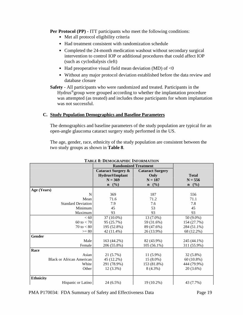
C. Study Population Demographics and Baseline Parameters
The demographics and baseline parameters of the study population are typical for an open-angle glaucoma cataract surgery study performed in the US. The age, gender, race, ethnicity of the study population are consistent between the two study groups as shown in Table 8.
TABLE 8: DEMOGRAPHIC INFORMATION Randomized Treatment Cataract Surgery &
Hydrus®Implant Cataract Surgery
Only
Total N = 369
n (%) N = 187 n (%)
N = 556 n (%)
Age (Years) N 369 187 556
Mean 71.6 71.2 71.1 Standard Deviation 7.9 7.6 7.8
Minimum 45 53 45 Maximum 93 93 93
< 60 37 (10.0%) 13 (7.0%) 50 (9.0%) 60 to < 70 95 (25.7%) 59 (31.6%) 154 (27.7%) 70 to < 80 195 (52.8%) 89 (47.6%) 284 (51.1%)
>= 80 42 (11.4%) 26 (13.9%) 68 (12.2%) Gender
Male 163 (44.2%) 82 (43.9%) 245 (44.1%) Female 206 (55.8%) 105 (56.1%) 311 (55.9%)
Race Asian 21 (5.7%) 11 (5.9%) 32 (5.8%)
Black or African American 45 (12.2%) 15 (8.0%) 60 (10.8%) White 291 (78.9%) 153 (81.8%) 444 (79.9%) Other 12 (3.3%) 8 (4.3%) 20 (3.6%)
Ethnicity
Hispanic or Latino 24 (6.5%) 19 (10.2%) 43 (7.7%)

PMA P170034: FDA Summary of Safety and Effectiveness Data Page 20
Randomized Treatment Cataract Surgery &
Hydrus®Implant Cataract Surgery
Only
Total N = 369
n (%) N = 187 n (%)
N = 556 n (%)
Not Hispanic or Latino 345 (93.5%) 168 (89.8%) 513 (92.3%) Study Eye
OD 178 (48.2%) 92 (49.2%) 270 (48.6%) OS 191 (51.8%) 95 (50.8%) 286 (51.4%)
Glaucoma related baseline parameters including use of ocular hypotensive medications, medicated IOP at screening, unmedicated IOP at baseline, visual acuity, visual field parameters (mean deviation and pattern standard deviation), and central corneal thickness are consistent between the two study groups as shown in Table 9.
TABLE 9: SCREENING AND BASELINE PARAMETERS FOR RANDOMIZED SUBJECTS Randomized Treatment
Parameter Cataract Surgery &
Hydrus®Implant Cataract Surgery
Only
Total N = 369
n (%) N = 187 n (%)
N = 556 n (%)
Number of ocular hypotensive medications at Screening1 1 194 (52.6%) 101 (54.0%) 295 (53.1%) 2 100 (27.1%) 48 (25.7%) 148 (26.6%) 3 65 (17.6%) 28 (15.0%) 93 (16.7%) 4 10 (2.7%) 10 (5.3%) 20 (3.6%) Screening IOP (mmHg) N 369 187 556 Mean (SD) 17.9 (3.1) 18.1 (3.1) 18.0 (3.1) Median 18.0 18.0 18.0 Minimum, Maximum 10.0, 30.0 8.5, 30.0 8.5, 30.0 Unmedicated IOP (mmHg) at Baseline N 369 187 556 Mean (SD) 25.5 (3.0) 25.4 (2.9) 25.5 (3.0) Median 24.7 24.7 24.7 Minimum, Maximum 22.0, 34.0 22.0, 34.0 22.0, 34.0 Baseline BCVA (ETDRS) N 369 186 555 Mean (SD) — LogMAR
Mean Snellen 0.304 (0.203)
20/40 0.296 (0.186)
20/40 0.301 (0.198)
20/40 Mean (SD) — LogMAR
Mean Snellen 0.300 20/40
0.300 20/40
0.300 20/40
Mean (SD) — LogMAR Mean Snellen
-0.160, 1.080 20/240, 20/14
-0.100, 0.840 20/138, 20/16
-0.160, 1.080 20/240, 20/14
Visual Field, Mean Deviation (MD, dB) N 369 187 556 Mean (SD) -3.61 (2.49) -3.61 (2.60) -3.61 (2.53) Median -3.20 -2.99 -3.13 Minimum, Maximum -10.39, 2.41 -11.85, 0.34 -11.85, 2.41 Visual Field, Pattern Standard Deviation (PSD, dB) N 369 187 556

PMA P170034: FDA Summary of Safety and Effectiveness Data Page 21
Randomized Treatment
Parameter Cataract Surgery &
Hydrus®Implant Cataract Surgery
Only
Total N = 369
n (%) N = 187 n (%)
N = 556 n (%)
Mean (SD) 3.18 (2.18) 3.13 (1.85) 3.17 (2.07) Median 2.41 2.36 2.37 Minimum, Maximum 1.06, 15.26 1.00, 10.68 1.00, 15.26 Corneal Thickness (µ) at Screening N 369 187 556 Mean (SD) 548.03 (31.59) 549.29 (34.54) 548.45 (32.59) Median 548.67 550.00 549.00 Minimum, Maximum 481.00, 619.67 480.33, 616.00 480.33, 619.67 Smaller sample sizes (n) for some clinical parameters were due to missing values. 1 Combo medication was counted as two medications. Oral medication was counted as one medication.
D. Safety and Effectiveness Results
1. Safety Results The safety analysis was based on the Safety cohort (556: 369 Cataract Surgery and Hydrus®Implant, and 187: cataract sugery only randomized participants and 527 participants available for analysis at the 24-month evaluation). The key safety outcomes for this study are presented below in Tables 10-12. Implantation of the Hydrus® Microstent was successful in most cases, with non-implantation reported in 11 participants (3.0%; 11/369). The reasons for unsuccessful implantation were anatomic restrictions, poor visualization due to hyphema, excessive movement of the participant, suboptimal placement, difficulty viewing the target area intended for implantation. The microstent was successfully implanted with one delivery attempt in 316 participants (85.6%: 316/369). Surgeons experienced difficulty with implantation with 58 participants (15.7%; 58/369). The most frequent reasons for implantation difficulty were encountering anatomic restriction and poor visualization. In 28 participants (7.5%; 28/369), more than one Hydrus® system was required. The most frequent reason for requiring the use of more than one system is the need for repositioning. Participants who underwent attempted Hydrus®implantation but did not receive the implant are included in the Safety cohort.
Adverse events (AE) that occurred in the PMA clinical study pivotal trail:
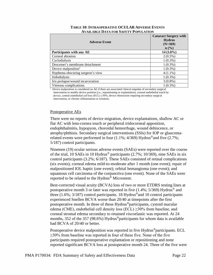
Intraoperative AEs
A total of 19 intraoperative AEs were reported in 19 out of 369 Hydrus®
participants (5.1%). Intraoperative AEs in the Hydrus® group are shown in Table 10.
PMA P170034: FDA Summary of Safety and Effectiveness Data Page 22
TABLE 10: INTRAOPERATIVE OCULAR ADVERSE EVENTS AVAILABLE DATA FOR SAFETY POPULATION
Adverse Event
Cataract Surgery with Hydrus (N=369) n (%)
Participants with any AE 14 (3.8%) Corneal abrasion 2 (0.5%) Cyclodialysis 1 (0.3%) Descemet’s membrane detachment 1 (0.3%) Device malposition1 1 (0.3%) Hyphema obscuring surgeon’s view 4 (1.1%) Iridodialysis 1 (0.3%) Iris prolapse/wound incarceration 3 (0.8%) Vitreous complications 1 (0.3%)
1 Device malposition is considered an AE if there are associated clinical sequelae of secondary surgical intervention to modify device position (i.e., repositioning or explantation), corneal endothelial touch by device, central endothelial cell loss (ECL) ≥30%, device obstruction requiring secondary surgical intervention, or chronic inflammation or irritation.
Postoperative AEs
There were no reports of device migration, device explantations, shallow AC or flat AC with lens-cornea touch or peripheral iridocorneal apposition, endophthalmitis, hypopyon, choroidal hemorrhage, wound dehiscence, or atrophy/phthisis. Secondary surgical interventions (SSIs) for IOP or glaucoma-related events were performed in four (1.1%; 4/369) Hydrus®and five (2.7%; 5/187) control participants.
Nineteen (19) ocular serious adverse events (SAEs) were reported over the course of the trial, 10 SAEs in 10 Hydrus® participants (2.7%; 10/369), nine SAEs in six control participants (3.2%; 6/187). These SAEs consisted of retinal complications (six events); corneal edema mild-to-moderate after 1 month (one event); repair of malpositioned IOL haptic (one event); orbital hemangioma (one event); and squamous cell carcinoma of the conjunctiva (one event). None of the SAEs were reported to be related to the Hydrus® Microstent.
Best-corrected visual acuity (BCVA) loss of two or more ETDRS testing lines at postoperative month 3 or later was reported in five (1.4%; 5/369) Hydrus® and three (1.6%; 3/187) control participants. 18 Hydrus®and 10 control participants experienced Snellen BCVA worse than 20/40 at timepoints after the first postoperative month. In three of these Hydrus®participants, cystoid macular edema (CME), endothelial cell density loss (ECL) ≥30% from baseline, and corneal stromal edema secondary to retained viscoelastic was reported. At 24 months, 352 of the 357 (98.6%) Hydrus®participants for whom data is available had BCVA of 20/40 or better.
Postoperative device malposition was reported in five Hydrus®participants. ECL ≥30% from baseline was reported in four of these five. None of the five participants required postoperative explantation or repositioning and none reported significant BCVA loss at postoperative month 24. Three of the five were
PMA P170034: FDA Summary of Safety and Effectiveness Data Page 23
considered non-responders at postoperative month 24 per protocol definition, two did not have 24-month washout diurnal IOP data available, and one was considered a responder at postoperative month 24.
Device obstruction of any kind was reported in 40 (10.8%; 40/369) Hydrus®participants. Seven cases were reported as complete obstruction and seven were reported as complete obstruction with peripheral anterior synechiae (PAS) formation. Twenty (20) cases were reported as partial device obstruction (defined as tissue in the inlet of the Hydrus®with the aqueous humor outflow path appearing only partially obstructed), and six were reported as partial obstruction with PAS formation. In two, blood caused a temporary obstruction of the Hydrus®
inlet that resolved by the first postoperative week. In 25 cases, the obstructing material was fibrinous material, fibrous tissue, or iris tissue. Laser membranectomy was performed three times on two participants and laser goniosynechialysis was performed on one participant to treat the obstruction, without success. All obstructions remained stable through postoperative month 24 without requiring explantation. Last available BCVA was 20/40 or better at the last visit for all eyes.
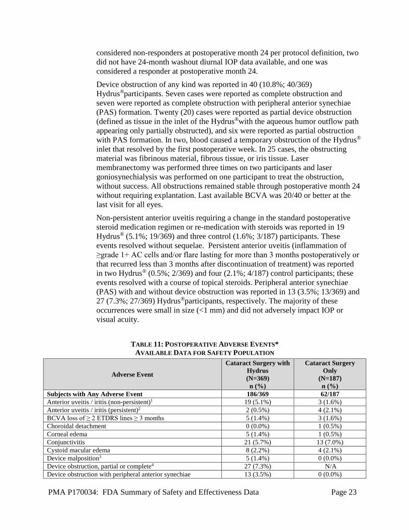
Non-persistent anterior uveitis requiring a change in the standard postoperative steroid medication regimen or re-medication with steroids was reported in 19 Hydrus® (5.1%; 19/369) and three control (1.6%; 3/187) participants. These events resolved without sequelae. Persistent anterior uveitis (inflammation of ≥grade 1+ AC cells and/or flare lasting for more than 3 months postoperatively or that recurred less than 3 months after discontinuation of treatment) was reported in two Hydrus® (0.5%; 2/369) and four (2.1%; 4/187) control participants; these events resolved with a course of topical steroids. Peripheral anterior synechiae (PAS) with and without device obstruction was reported in 13 (3.5%; 13/369) and 27 (7.3%; 27/369) Hydrus®participants, respectively. The majority of these occurrences were small in size (<1 mm) and did not adversely impact IOP or visual acuity.
TABLE 11: POSTOPERATIVE ADVERSE EVENTS* AVAILABLE DATA FOR SAFETY POPULATION
Adverse Event
Cataract Surgery with Hydrus (N=369) n (%)
Cataract Surgery Only
(N=187) n (%)
Subjects with Any Adverse Event 186/369 62/187 Anterior uveitis / iritis (non-persistent)1 19 (5.1%) 3 (1.6%) Anterior uveitis / iritis (persistent)2 2 (0.5%) 4 (2.1%) BCVA loss of ≥ 2 ETDRS lines ≥ 3 months 5 (1.4%) 3 (1.6%) Choroidal detachment 0 (0.0%) 1 (0.5%) Corneal edema 5 (1.4%) 1 (0.5%) Conjunctivitis 21 (5.7%) 13 (7.0%) Cystoid macular edema 8 (2.2%) 4 (2.1%) Device malposition3 5 (1.4%) 0 (0.0%) Device obstruction, partial or complete4 27 (7.3%) N/A Device obstruction with peripheral anterior synechiae 13 (3.5%) 0 (0.0%)

PMA P170034: FDA Summary of Safety and Effectiveness Data Page 24
Dry eye 14 (3.8%) 6 (3.2%) Hyphema (>2mm at >1 day) 4 (1.1%) 1 (0.5%) Hypotony (IOP <6 mmHg ≥ 1 month) 0 (0.0%) 1 (0.5%) IOP elevated >10 mmHg from baseline > 1 month 2 (0.5%) 5 (2.7%) Peripheral anterior synechiae without device obstruction 27 (7.3%) N/A Peripheral anterior synechiae - no device implanted 2 0.5%) 4 (2.1%) Subconjunctival hemorrhage 9 (2.4%) 0 (0.0%) Surgical re-intervention in study eye (not paracentesis prior to 1 week postop) 9 (2.4%) 9 (4.8%)
Vitreous hemorrhage associated with hyphema 2 (0.5%) 0 (0.0%) Worsening in visual field MD by > 2.5 dB compared with preoperative 16 (4.3%) 10 (5.3%)
Worsening ocular symptoms: a 2-point worsening to severe or more > 3 months postop 16 (4.3%) 9 (4.8%)
*Occurring at 2% or greater in either group, or other adverse events known to be associated with glaucoma procedures or potential risks with stent implantations
1 Anterior chamber cell and flare requiring change in steroid treatment 2 ≥Grade 1+ anterior chamber cells and/or flare lasting for more than 3 months postoperatively or recurring less than 3 months after discontinuation
of treatment (requiring change in steroid regimen) 3 Device malposition is considered an AE if there are associated clinical sequelae of secondary surgical intervention to modify device position (i.e.,
repositioning or explantation), corneal endothelial touch by device, central endothelial cell loss (ECL) ≥30%, device obstruction requiring secondary surgical intervention, or chronic inflammation or irritation. None of the eyes reported with device malposition required surgical intervention to remove or reposition the microstent.
4 In two eyes with device obstruction, three YAG laser procedures were performed on two eyes. These procedures were not successful in removing the obstruction.
In addition to the AEs reported in Table 11, AEs that occurred at <2% in both groups included blepharitis, blurry vision, chalazion, corneal edema, diplopia (monocular), ocular pain, diabetic retinopathy (non-proliferative), epiretinal membrane, retinal break, retinal detachment/repair, retinal tear with vitreous hemorrhage, retinopathy (central serous), superficial punctate keratitis and vitreous floaters.
Adverse events that occurred at <2% in the Hydrus®group included age related macular degeneration (dry and wet form), anterior capsule fibrosis, asthenopia, blood reflux, central serous retinopathy, chronic pain in study eye >3 months, circulating blood in the anterior chamber (i.e., not yet settled inferiorly), conjunctival cyst, conjunctival injection, corneal abrasion, corneal haze, cyst, decentered IOL, dermatitis (eyelid), diabetic retinopathy (proliferative), diplopia, disc hemorrhage, dysphotopsia (including glare and intermittent flashes), eyelid edema, foreign body in eye, hordeolum, IOP <6 mmHg due to thin cornea, lacrimal obstruction, lesion on eyelid, malpositioned IOL haptic, orbital hemangioma, persistent mydriasis, photosensitivity, ptosis, punctal stenosis, pupil irregularity, retained lens fragment, retinal macroaneurysm, retinal pigment epithelial detachment, significant foreign body sensation >3 months, squamous cell carcinoma, vitreous in anterior chamber, and vitreous opacities.
Ten (10) ocular serious adverse events (SAE) were reported over the course of the study in the Hydrus®group in which 8 of the 10 SAEs required either medical or surgical intervention. These SAEs consisted of retinal complications (6 events); corneal edema mild-to-moderate after 1 month (1 event); repair of malposition IOL haptic (1 event); orbital hemangioma (1 event); and squamous cell carcinoma

PMA P170034: FDA Summary of Safety and Effectiveness Data Page 25
of the conjunctiva (1 event). None of the events were reported to be related to the Hydrus® Microstent.
Secondary Ocular Surgical Interventions
Secondary ocular surgical intervention (SSI) were reported through 24 months in both groups as shown in Table 12. Secondary ocular surgeries for IOP or glaucoma-related events occurred in four (1.1%; 4/369) Hydrus®and five (2.7%; 5/187) control participants. SSIs for any ocular adverse event through 24 months occurred in 16 Hydrus® (4.3%; 16/369) and 10 control (5.3%; 10/187) participants.
TABLE 12: SECONDARY SURGICAL INTERVENTIONS (SSI) FOR ANY OCULAR ADVERSE EVENT
THROUGH 24 MONTHS AVAILABLE DATA FOR SAFETY POPULATION
Adverse Event
Cataract Surgery with Hydrus (N=369) n (%)
Cataract Surgery Only
(N=187) n (%)
Subjects with SSIs for any ocular adverse event* 16 (4.3%) 10 (5.3%) Secondary surgeries for IOP or glaucoma-related Events 4 (1.1%) 5 (2.7%) Anterior chamber paracentesis 1 (0.3%) 2 (1.1%) Goniosynchialysis - with laser 1 (0.3%) 0 (0.0%) Glaucoma shunt implantation 0 (0.0%) 2 (1.1%) Express shunt removal 0 (0.0%) 1 (0.5%) Selective laser trabeculoplasty (SLT) 0 (0.0%) 1 (0.5%) Trabeculectomy with Express shunt implantation 0 (0.0%) 2 (1.1%) Tube with pars plana vitrectomy and scleral reinforcement 0 (0.0%) 1 (0.5%) YAG membranectomy or membranotomy 3 (0.8%) 0 (0.0%) Other secondary surgical interventions 12 (3.3%) 5 (2.7%) Anterior chamber irrigation and aspiration 1 (0.3%) 0 (0.0%) Canthoplasty 1 (0.3%) 0 (0.0%) Descemet membrane endothelial keratoplasty 1 (0.3%) 0 (0.0%) Haptic reposition 1 (0.3%) 0 (0.0%) Orbital tumor biopsy 1 (0.3%) 0 (0.0%) Pars plana vitrectomy with laserpexy 0 (0.0%) 1 (0.5%) Pars plana vitrectomy with membrane peel 1 (0.3%) 1 (0.5%) Punctoplasty 1 (0.3%) 0 (0.0%) Retinal detachment repair - vitrectomy and/or scleral buckle 1 (0.3%) 2 (1.1%) Retained lens material removal 1 (0.3%) 0 (0.0%) Retinal laser 3 (0.8%) 3 (1.6%) Vitrectomy with retinal detachment repair 1 (0.3%) 0 (0.0%) *A subject may have had multiple SSIs.
Other Postoperative Observations
Reporting of other ocular observations was at the study investigator’s discretion. Similar data may not be reported for every subject, or consistently within the course of a given subject’s study participation. Consequently, no conclusions

PMA P170034: FDA Summary of Safety and Effectiveness Data Page 26
regarding the overall frequency of these findings can be drawn from the incidence rates noted. The other ocular observations that were reported postoperatively and which could impact safety in Hydrus® participants included, but were not limited to: transient layered hyphema (<2mm) or microhyphema (13%; 48/369); transient anterior chamber shallowing (0.1%; 3/369); iris erosion (0.1%; 3/369); pupil peaking (4.3%; 16/369); and early hypotony (IOP <6 mmHg with onset ≤ 2 weeks and accompanied by corneal folds) (0.1%; 4/369).
Additional Safety Data Gathered after 24-Months
After the 24-month visit, the following IOP or glaucoma-related secondary ocular surgeries (not included in Table 12) have been reported for five Hydrus® participants:
• Three participants underwent selective laser trabeculoplasty (SLT) three years after Hydrus® implantation
• One participant underwent three IOP-lowering procedures: laser cyclodestructive procedure, gonio puncture with laser, and Baerveldt shunt with graft patch
• One participant underwent trabeculectomy. None of these procedures required Hydrus®explant or repositioning.
Corneal Endothelial Cell Density
Mean endothelial cell density (ECD) was evaluated by specular microscopy preoperatively and postoperatively. Mean percent change in central ECD was slightly higher for the Hydrus®group (-14%, SD: 14%, 95% CI: -16%, -13%) compared to the control group (-10%, SD: 11%, 95% CI: -12%, -8%) through 24 months. Most of the central ECD loss occurred in the early postoperative period (from preoperative to 3 months) in both treatment groups as a result of surgery. Minimal to no central cell loss is noted between sequential visits after the 3-month visit. A higher proportion of Hydrus® participants (13.6%; 47/346) had ≥30% central ECD loss (ECL) from baseline at 24 months compared to the proportion of control participants (7.2%; 12/167). In these cases, unless accompanied by other precipitating adverse events, ECL was not associated with corneal edema after 1 month, or any other clinical sequelae, including persistent BCVA loss. Nine participants with ECL, 8 Hydrus® eye and one control ye, there was ECL that had not yet stabilized by the last reported visit. ECD monitoring for available participants with ECL will continue until the conclusion of the study (up to five years).
2. Effectiveness Results
The analysis of effectiveness was based on the 556 evaluable patients at the 24-month time point. Key effectiveness outcomes are presented in Tables 13 and 14.

PMA P170034: FDA Summary of Safety and Effectiveness Data Page 27
Results from the primary and secondary endpoints are shown in Table 13. The primary effectiveness endpoint was met, with 77.2%:285/369) in the Hydrus®group and 57.8%: 108/187 in the Control group achieving a clinically significant (≥ 20%) decrease in unmedicated mean DIOP from baseline to the hypotensive medication-free 24-month postoperative examination. This difference between groups was statistically significant (p < 0.001).
The secondary endpoint, a clinically significant mean change in IOP between baseline and hypotensive medication-free 24-month postoperative examination, was met. The mean reduction in unmedicated mean DIOP from baseline to 24 months was 7.5 mmHg (SD=4.1) in the Hydrus®group compared to 5.3 mmHg (SD=4.2) in the Control group (p < 0.001).
TABLE 13: PRIMARY AND SECONDARY EFFECTIVENESS RESULTS
Effectiveness Endpoint (Evaluated at 24 Months Postoperatively)
Hydrus (N=369)
Control (N=187)
Difference (Hydrus-Control)
p-value
Primary Effectiveness Endpoint
Proportion of subject eyes with unmedicated mean IOP reduction > 20% from baseline 77.2% 57.8% 19.5% <0.001
Secondary Effectiveness Endpoint
Difference in unmedicated mean DIOP (mmHg) reduction from baseline -7.5 -5.3 -2.3 <0.001
Additional detail regarding the reasons participants did not achieve the primary endpoint (IOP non-responders) is shown in Table 14.
TABLE 14: SUMMARY OF 24-MONTH IOP NON-RESPONDER CATEGORIES Cataract Surgery with
Hydrus (N=369) n (%)
Cataract Surgery Only
(N=187) n (%)
Total Non-Responders 84 (22.8%) 79 (42.2%) Non-Responders: 24-month unmedicated mean DIOP reductions < 20% vs. baseline 61 (16.5%) 54 (28.9%)
Non-Responders for reasons other than IOP reduction 23 (6.2%) 25 (13.4%) Had glaucoma-related events or secondary surgical procedures1
4 (1.1%) 5 (2.7%)
Unable to washout glaucoma medications 5 (1.4%) 3 (1.6%) Missing 24-month data 14 (3.8%) 17 (9.1%)
n = number of eyes meeting corresponding criteria 1 Secondary glaucoma surgeries included laser goniosynchialysis, YAG laser membranectomy, and selective laser trabeculoplasty (SLT)

PMA P170034: FDA Summary of Safety and Effectiveness Data Page 28
3. Pediatric Extrapolation In this premarket application, existing clinical data was not leveraged to support approval of a pediatric patient population.
E. Financial Disclosure
The Financial Disclosure by Clinical Investigators regulation (21 CFR 54) requires applicants who submit a marketing application to include certain information concerning the compensation to, and financial interests and arrangement of, any clinical investigator conducting clinical studies covered by the regulation. The pivotal clinical study included 38 principal investigators of which none were full-time or part-time employees of the applicant and 1 of investigators had disclosable financial interests/arrangements as defined in 21 CFR 54.2(a), (b), (c) and (f) and described below:
• Compensation to the investigator for conducting the study where the value could be influenced by the outcome of the study: none
• Significant payment of other sorts: none • Proprietary interest in the product tested held by the investigator: none • Significant equity interest held by investigator in sponsor of covered study: 1
The applicant has adequately disclosed the financial interest/arrangements with clinical investigators. Statistical analyses were conducted by FDA to determine whether the financial interests/arrangements had any impact on the clinical study outcome. The information provided does not raise any questions about the reliability of the data.
XI. SUMMARY OF SUPPLEMENTAL CLINICAL INFORMATION
A. Side Interlock Delivery System
During the Horizon Study, the Hydrus® Microstent was implanted with a delivery system different than the commercially available delivery system. The delivery system in the Horizon study had a different interlocking mechanism for attaching and releasing the microstent. Releasing the microstent for implantation in the Horizon study required the microstent to be advanced into Schlemm’s canal and released by reversing the wheel on the delivery system. The commercially available delivery system requires that the microstent be advanced into the canal, and is released passively from the side-interlocking mechanism by advancing the wheel to its full forward position (i.e., reversing the wheel is not required). Additionally, using the commercially available delivery system, the microstent can be reattached onto the delivery system to facilitate repositioning the microstent, if required.
The commercially available (modified-design) Hydrus® Microstent was introduced for investigation into two multicenter randomized controlled trials outside of the United States (OUS) that had been ongoing prior to the availability of the modified-design

PMA P170034: FDA Summary of Safety and Effectiveness Data Page 29
Hydrus® microstent. The patient populations and study questions under investigation in these trials are slightly different from those of the HORIZON study. The interim results (reflecting follow-up of 12 months or greater) from a small sub-group of participants who received the modified-design microstent in each of these two trials suggest that the intraoperative performance and safety results for the modified-design system are comparable to the product used in the HORIZON study. AEs pertaining to device placement and stability were similar in nature to AEs observed in the Hydrus®group of the HORIZON study.
B. Patient Questionnaire Data Patient-reported information on ocular symptoms was collected using a shortened (7-item) version of an 18-item questionnaire1 adapted from the 43-item glaucoma-related symptom questionnaire2 used in the Collaborative Initial Glaucoma Treatment Study (CIGTS). The questionnaire asked participants to report on the presence of eye irritation or burning, foreign body sensation, droopy eyelids, excessive tearing, skin sensitivity around the eyes, eye pain, and red eyes. Participants were also asked to rate how bothersome the symptom was and the degree to which they attributed the symptom to glaucoma or glaucoma treatment. The majority of participants in each group did not report the presence of symptoms at baseline, postoperative month 12, and postoperative month 24 and these proportions were similar between groups. The results of the patient reported information on ocular symptoms are provided in Tables 15-17. It has not been determined how well this questionnaire applies to glaucoma patients undergoing implantation of a MIGS device with or without cataract surgery. The original CIGTS questionnaire was developed with a cohort of patients who were newly diagnosed with open-angle glaucoma (primary open-angle, exfoliation, and pigmentary) and who were required to be new to IOP-lowering medication use (2 weeks of lifetime use or less) or any other glaucoma treatment as part of eligibility. These participants were randomized to treatment with topical IOP-lowering medication or trabeculectomy; cataract surgery and implantation of a MIGS device were not part of the planned interventions in the CIGTS trial. Also, the impact of symptoms on visual function or quality of life was not assessed, and the questionnaire was not administered during unscheduled visits.
1 Musch DC et al, JAMA Ophthalmol 2017;135(12):1345-1351 2 Janz NK et al, Ophthalmology 2001;108:887-898

PMA P170034: FDA Summary of Safety and Effectiveness Data Page 30
TABLE 15 CIGTS SYMPTOMS IMPACT GLAUCOMA SUBSCALE: LOCAL EYE
Cataract Surgery & Hydrus®Implant (369 Subjects)
Cataract Surgery Only (187 Subjects)
Symptom Baseline n (%)
12M n (%)
24M n (%)
Baseline n (%)
12M n (%)
24M n (%)
Total 369 365 357 187 180 170 Missed Assessment 5 8 4 1 2 1
Eye Irritation Burning, N 364 357 353 185 178 169 No 296 (81.3%) 306 (85.7%) 311 (88.1%) 163 (88.1%) 141 (79.2%) 139 (82.2%) Yes 68 (18.7%) 51 (14.3%) 42 (11.9%) 22 (11.9%) 37 (20.8%) 30 (17.8%) Bothersome1 A lot 6 (8.8%) 4 (7.7%) 1 (2.3%) 5 (20.8%) 0 (0.0%) 6 (20.0%) A moderate amount 11 (16.2%) 7 (13.5%) 17 (39.5%) 3 (12.5%) 8 (22.2%) 6 (20.0%) Some 18 (26.5%) 18 (34.6%) 8 (18.6%) 4 (16.7%) 10 (27.8%) 2 (6.7%) A little 20 (29.4%) 18 (34.6%) 10 (23.3%) 9 (37.5%) 14 (38.9%) 13 (43.3%) Not at all 13 (19.1%) 5 (9.6%) 7 (16.3%) 3 (12.5%) 4 (11.1%) 3 (10.0%) Not Reported 296 305 310 161 142 139 Eye Pain, N 364 357 353 186 178 168 No 344 (94.5%) 336 (94.1%) 327 (92.6%) 171 (91.9%) 163 (91.6%) 157 (93.5%) Yes 20 (5.5%) 21 (5.9%) 26 (7.4%) 15 (8.1%) 15 (8.4%) 11 (6.5%) Bothersome1 A lot 2 (9.5%) 4 (19.0%) 1 (3.7%) 1 (5.9%) 1 (7.1%) 1 (8.3%) A moderate amount 4 (19.0%) 1 (4.8%) 4 (14.8%) 5 (29.4%) 3 (21.4%) 3 (25.0%) Some 7 (33.3%) 3 (14.3%) 6 (22.2%) 3 (17.6%) 3 (21.4%) 3 (25.0%) A little 6 (28.6%) 8 (38.1%) 11 (40.7%) 5 (29.4%) 6 (42.9%) 3 (25.0%) Not at all 2 (9.5%) 5 (23.8%) 5 (18.5%) 3 (17.6%) 1 (7.1%) 2 (16.7%) Not Reported 343 336 326 169 164 156 Excessive Tearing, N 364 357 353 185 178 169 No 331 (90.9%) 320 (89.6%) 309 (87.5%) 172 (93.0%) 156 (87.6%) 151 (89.3%) Yes 33 (9.1%) 37 (10.4%) 44 (12.5%) 13 (7.0%) 22 (12.4%) 18 (10.7%) Bothersome1 A lot 5 (15.2%) 6 (16.2%) 6 (13.3%) 2 (14.3%) 1 (4.3%) 3 (16.7%) A moderate amount 3 (9.1%) 5 (13.5%) 8 (17.8%) 1 (7.1%) 6 (26.1%) 4 (22.2%) Some 7 (21.2%) 6 (16.2%) 11 (24.4%) 2 (14.3%) 3 (13.0%) 4 (22.2%) A little 13 (39.4%) 9 (24.3%) 12 (26.7%) 8 (57.1%) 9 (39.1%) 5 (27.8%) Not at all 5 (15.2%) 11 (29.7%) 8 (17.8%) 1 (7.1%) 4 (17.4%) 2 (11.1%) Not Reported 331 320 308 171 155 151 Droopy Eyelids, N 364 357 353 185 178 168 No 341 (93.7%) 337 (94.4%) 341 (96.6%) 175 (94.6%) 170 (95.5%) 162 (96.4%) Yes 23 (6.3%) 20 (5.6%) 12 (3.4%) 10 (5.4%) 8 (4.5%) 6 (3.6%) Bothersome1 A lot 0 (0.0%) 2 (9.5%) 1 (7.7%) 3 (25.0%) 1 (14.3%) 1 (14.3%) A moderate amount 2 (8.7%) 2 (9.5%) 0 (0.0%) 1 (8.3%) 2 (28.6%) 0 (0.0%) Some 5 (21.7%) 2 (9.5%) 2 (15.4%) 0 (0.0%) 0 (0.0%) 1 (14.3%) A little 7 (30.4%) 8 (38.1%) 5 (38.5%) 4 (33.3%) 1 (14.3%) 3 (42.9%) Not at all 9 (39.1%) 7 (33.3%) 5 (38.5%) 4 (33.3%) 3 (42.9%) 2 (28.6%) Not Reported 341 336 340 173 171 161

PMA P170034: FDA Summary of Safety and Effectiveness Data Page 31
Cataract Surgery & Hydrus®Implant
(369 Subjects) Cataract Surgery Only
(187 Subjects) Symptom Baseline
n (%) 12M
n (%) 24M
n (%) Baseline n (%)
12M n (%)
24M n (%)
Total 369 365 357 187 180 170 Missed Assessment 5 8 4 1 2 1
Red Eyes, N 364 357 353 185 178 169 No 316 (86.8%) 310 (86.8%) 306 (86.7%) 158 (85.4%) 152 (85.4%) 150 (88.8%) Yes 48 (13.2%) 47 (13.2%) 47 (13.3%) 27 (14.6%) 26 (14.6%) 19 (11.2%) Bothersome1 A lot 5 (10.2%) 2 (4.3%) 1 (2.2%) 5 (17.2%) 0 (0.0%) 1 (5.3%) A moderate amount 7 (14.3%) 6 (12.8%) 8 (17.4%) 2 (6.9%) 4 (16.0%) 4 (21.1%) Some 9 (18.4%) 9 (19.1%) 7 (15.2%) 4 (13.8%) 8 (32.0%) 3 (15.8%) A little 15 (30.6%) 22 (46.8%) 11 (23.9%) 9 (31.0%) 8 (32.0%) 7 (36.8%) Not at all 13 (26.5%) 8 (17.0%) 19 (41.3%) 9 (31.0%) 5 (20.0%) 4 (21.1%) Not Reported 315 310 307 156 153 150 Feeling Like Something is in Your Eye, N
364 356 353 186 178 169
No 296 (81.3%) 280 (78.7%) 297 (84.1%) 143 (76.9%) 152 (85.4%) 131 (77.5%) Yes 68 (18.7%) 76 (21.3%) 56 (15.9%) 43 (23.1%) 26 (14.6%) 38 (22.5%) Bothersome1 A lot 6 (8.7%) 2 (2.6%) 5 (8.9%) 5 (11.1%) 2 (8.0%) 7 (18.9%) A moderate amount 7 (10.1%) 12 (15.8%) 4 (7.1%) 7 (15.6%) 4 (16.0%) 4 (10.8%) Some 19 (27.5%) 19 (25.0%) 18 (32.1%) 7 (15.6%) 7 (28.0%) 4 (10.8%) A little 25 (36.2%) 31 (40.8%) 17 (30.4%) 19 (42.2%) 10 (40.0%) 18 (48.6%) Not at all 12 (17.4%) 12 (15.8%) 12 (21.4%) 7 (15.6%) 2 (8.0%) 4 (10.8%) Not Reported 295 280 297 141 153 132 Skin Sensitivity or Irritation around the Eye, N
364 357 353 186 178 169
No 339 (93.1%) 328 (91.9%) 329 (93.2%) 170 (91.4%) 160 (89.9%) 157 (92.9%) Yes 25 (6.9%) 29 (8.1%) 24 (6.8%) 16 (8.6%) 18 (10.1%) 12 (7.1%) Bothersome1 A lot 2 (7.7%) 4 (13.3%) 0 (0.0%) 2 (11.1%) 3 (17.6%) 3 (25.0%) A moderate amount 3 (11.5%) 1 (3.3%) 6 (23.1%) 3 (16.7%) 6 (35.3%) 2 (16.7%) Some 6 (23.1%) 10 (33.3%) 2 (7.7%) 3 (16.7%) 1 (5.9%) 1 (8.3%) A little 12 (46.2%) 10 (33.3%) 11 (42.3%) 6 (33.3%) 6 (35.3%) 5 (41.7%) Not at all 3 (11.5%) 5 (16.7%) 7 (26.9%) 4 (22.2%) 1 (5.9%) 1 (8.3%) Not Reported 338 327 327 168 161 157 N = number of available subjects with non-missing Yes/No response. N < number of subjects with assessment = missing response for the corresponding CIGTS symptom questionnaire. % for No or Yes = n/N x 100%. Not reported = number of subjects who responded to the symptom questionnaire but with missing data for 'bothersome'. 1 The denominator for the % is the number of subjects reported with the response of the corresponding sub-question (i.e., 'bothersome').
Subjects might report 'No' symptom but with a response to 'bothersome'. Also, subjects might report 'Yes' to the symptom but fail to respond to 'bothersome'. As such, the total number of subjects with the responses could be different from the total number of subjects reported with 'Yes' for the corresponding symptom.
At 24 months, no change in bothersome grade was reported 73.4% (254/346) to 91.6% (316/345) of the Hydrus®group and 67.1% (112/187) to 91% (151/167) of the control group. Increase from baseline in the bothersome score of two or more grades to a “moderate amount” or “a lot” at any postoperative visit were reported as adverse events (AEs). Such AEs were reported in 4.3% (16/369) of the Hydrus®group and 4.8% (9/187) of the control group. Results for worsening symptom scores by group are provided in Table 16.

PMA P170034: FDA Summary of Safety and Effectiveness Data Page 32
TABLE 16: OCULAR SYMPTOMS ADVERSE EVENTS
Worsened ≥2 grades to Cataract Surgery &
Hydrus®Implant (369 Subjects)
Cataract Surgery Only (187 Subjects)
Bothersome Grade "A Lot" or to "A Moderate Amount"
12M n/N (%)
24M n/N (%)
12M n/N (%)
24M n/N (%)
Eye Irritation Burning 1/365 (<1%) 3/357 (<1%) 1/180 (<1%) 2/170 (1.2%) Eye Pain 0/365 (0.0%) 1/357 (<1%) 1/180 (<1%) 0/170 (0.0%) Excessive Tearing 2/365 (<1%) 3/357 (<1%) 4/180 (2.2%) 2/170 (1.2%) Droopy Eyelids 0/365 (0.0%) 0/357 (0.0%) 1/180 (<1%) 0/170 (0.0%) Red Eyes 1/365 (<1%) 0/357 (0.0%) 1/180 (<1%) 2/170 (1.2%) Feeling Like Something is in Your Eye
5/364 (1.4%) 2/357 (<1%) 2/180 (1.1%) 1/170 (<1%)
Skin Sensitivity or Irritation around the Eye
1/365 (<1%) 1/357 (<1%) 3/180 (1.7%) 0/170 (0.0%)
Overall 9/365 (2.5%) 7/357 (2.0%) 5/180 (2.8%) 4/170 (2.4%) For each symptom, grades of 1, 2, 3, 4, and 5 were assigned to non-missing bothersome response of "not at all," "a little," "some," "a moderate amount," and "a lot," respectively. Otherwise a bothersome grade of 0 was assigned to a response of "no symptom" or a response of "symptom not due to glaucoma/treatment." Change = postop bothersome grade - baseline bothersome grade. A positive value means a worsening in the symptom. N = number of subjects with a non-missing bothersome grade change from baseline for the corresponding symptom. % = n/N x 100%.
TABLE 17: OCULAR SYMPTOMS REPORTED AS BOTHERING PATIENTS “A MODERATE AMOUNT” OR “A LOT”
AVAILABLE DATA FROM SAFETY POPULATION
Ocular Symptom
Cataract Surgery & Hydrus®Implant
(369 Subjects)
Cataract Surgery Only (187 Subjects)
Baseline n (%)
12M n (%)
24M n (%)
Baseline n (%)
12M n (%)
24M n (%)
Total 369 365 357 187 180 170 Missed Assessment 5 8 4 1 2 1
Eye Irritation Burning, N 364 357 353 185 178 169 A lot 6 (8.8%) 4 (7.7%) 1 (2.3%) 5 (20.8%) 0 (0.0%) 6 (20.0%) A moderate amount 11 (16.2%) 7 (13.5%) 17 (39.5%) 3 (12.5%) 8 (22.2%) 6 (20.0%) Eye Pain, N 364 357 353 186 178 168 A lot 2 (9.5%) 4 (19.0%) 1 (3.7%) 1 (5.9%) 1 (7.1%) 1 (8.3%) A moderate amount 4 (19.0%) 1 (4.8%) 4 (14.8%) 5 (29.4%) 3 (21.4%) 3 (25.0%) Excessive Tearing, N 364 357 353 185 178 169 A lot 5 (15.2%) 6 (16.2%) 6 (13.3%) 2 (14.3%) 1 (4.3%) 3 (16.7%) A moderate amount 3 (9.1%) 5 (13.5%) 8 (17.8%) 1 (7.1%) 6 (26.1%) 4 (22.2%) Droopy Eyelids, N 364 357 353 185 178 168 A lot 0 (0.0%) 2 (9.5%) 1 (7.7%) 3 (25.0%) 1 (14.3%) 1 (14.3%) A moderate amount 2 (8.7%) 2 (9.5%) 0 (0.0%) 1 (8.3%) 2 (28.6%) 0 (0.0%) Red Eyes, N 364 357 353 185 178 169 A lot 5 (10.2%) 2 (4.3%) 1 (2.2%) 5 (17.2%) 0 (0.0%) 1 (5.3%) A moderate amount 7 (14.3%) 6 (12.8%) 8 (17.4%) 2 (6.9%) 4 (16.0%) 4 (21.1%) Feeling Like Something is in Your Eye, N 364 356 353 186 178 169
A lot 6 (8.7%) 2 (2.6%) 5 (8.9%) 5 (11.1%) 2 (8.0%) 7 (18.9%) A moderate amount 7 (10.1%) 12 (15.8%) 4 (7.1%) 7 (15.6%) 4 (16.0%) 4 (10.8%) Skin Sensitivity or Irritation 364 357 353 186 178 169

PMA P170034: FDA Summary of Safety and Effectiveness Data Page 33
Ocular Symptom
Cataract Surgery & Hydrus®Implant
(369 Subjects)
Cataract Surgery Only (187 Subjects)
Baseline n (%)
12M n (%)
24M n (%)
Baseline n (%)
12M n (%)
24M n (%)
around the Eye, N A lot 2 (7.7%) 4 (13.3%) 0 (0.0%) 2 (11.1%) 3 (17.6%) 3 (25.0%) A moderate amount 3 (11.5%) 1 (3.3%) 6 (23.1%) 3 (16.7%) 6 (35.3%) 2 (16.7%)
% = n/N (100)
C. Patient Use of Hypotensive Medications
Of the participants who were responders, 82.8% in the Hydrus® group (236/285) and 56.5% in the control group (61/108) were not using ocular hypotensive medications at 24 months.
XII. PANEL MEETING RECOMMENDATION AND FDA’S POST-PANEL ACTION
In accordance with the provisions of section 515(c)(3) of the act as amended by the Safe Medical Devices Act of 1990, this PMA was not referred to the Ophthalmic Devices Panel, an FDA advisory committee, for review and recommendation because the information in the PMA substantially duplicates information previously reviewed by this panel.
XIII. CONCLUSIONS DRAWN FROM PRECLINICAL AND CLINICAL STUDIES
A. Effectiveness Conclusions
The Hydrus® Microstent pivotal trial (Horizon Study) met both the primary and secondary effectiveness endpoints.
B. Safety Conclusions
The risks of the device are based on nonclinical laboratory as well as data collected in a clinical study conducted to support PMA approval as described above. Device-related serious AEs were not reported. Device-related non-serious AEs included: • The most common AEs in the Hydrus®arm included:
o Non-persistent anterior uveitis / iritis consisting of AC cell and flare requiring a change in post-operative steroid treatment (5.1% or 19/369)
o Microstent malposition (1.4% or 5/369)
o Microstent obstruction, partial or complete, with or without peripheral anterior synechiae (10.8% or 40/369)
o Microstent obstruction with peripheral anterior synechiae (3.5% or 13/369)
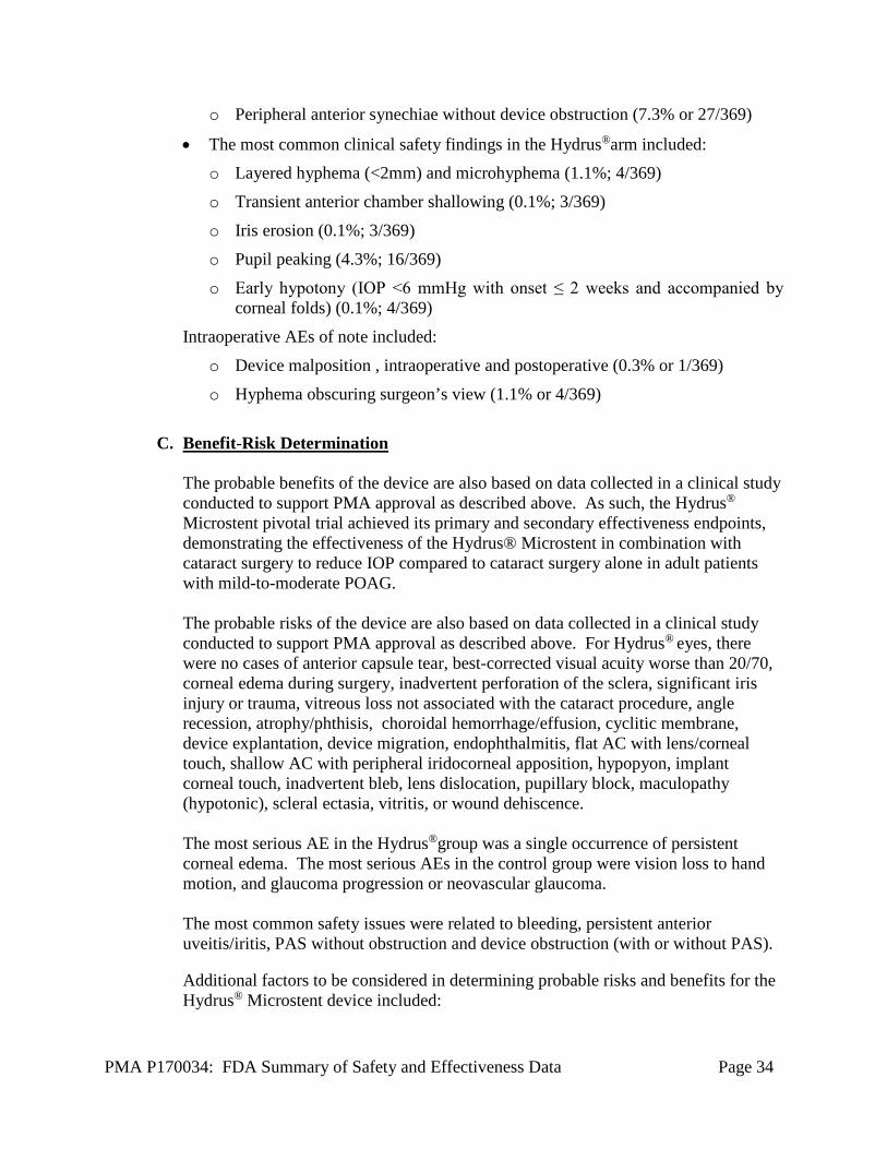
PMA P170034: FDA Summary of Safety and Effectiveness Data Page 34
o Peripheral anterior synechiae without device obstruction (7.3% or 27/369)
• The most common clinical safety findings in the Hydrus®arm included: o Layered hyphema (<2mm) and microhyphema (1.1%; 4/369)
o Transient anterior chamber shallowing (0.1%; 3/369)
o Iris erosion (0.1%; 3/369)
o Pupil peaking (4.3%; 16/369)
o Early hypotony (IOP <6 mmHg with onset ≤ 2 weeks and accompanied by corneal folds) (0.1%; 4/369)
Intraoperative AEs of note included:
o Device malposition , intraoperative and postoperative (0.3% or 1/369)
o Hyphema obscuring surgeon’s view (1.1% or 4/369)
C. Benefit-Risk Determination
The probable benefits of the device are also based on data collected in a clinical study conducted to support PMA approval as described above. As such, the Hydrus®
Microstent pivotal trial achieved its primary and secondary effectiveness endpoints, demonstrating the effectiveness of the Hydrus® Microstent in combination with cataract surgery to reduce IOP compared to cataract surgery alone in adult patients with mild-to-moderate POAG. The probable risks of the device are also based on data collected in a clinical study conducted to support PMA approval as described above. For Hydrus® eyes, there were no cases of anterior capsule tear, best-corrected visual acuity worse than 20/70, corneal edema during surgery, inadvertent perforation of the sclera, significant iris injury or trauma, vitreous loss not associated with the cataract procedure, angle recession, atrophy/phthisis, choroidal hemorrhage/effusion, cyclitic membrane, device explantation, device migration, endophthalmitis, flat AC with lens/corneal touch, shallow AC with peripheral iridocorneal apposition, hypopyon, implant corneal touch, inadvertent bleb, lens dislocation, pupillary block, maculopathy (hypotonic), scleral ectasia, vitritis, or wound dehiscence. The most serious AE in the Hydrus®group was a single occurrence of persistent corneal edema. The most serious AEs in the control group were vision loss to hand motion, and glaucoma progression or neovascular glaucoma. The most common safety issues were related to bleeding, persistent anterior uveitis/iritis, PAS without obstruction and device obstruction (with or without PAS). Additional factors to be considered in determining probable risks and benefits for the Hydrus® Microstent device included:
PMA P170034: FDA Summary of Safety and Effectiveness Data Page 35
• The Horizon Study was a prospective, multicenter, randomized, controlled study in which 556 participants were randomized and followed for 24 months postoperatively. The study incorporated glaucoma medication washout, safety and effectiveness determination with 2-year follow-up, specular microscopy, and sample size which exceeded the sufficient number of participants needed to achieve 95% probability of detecting AEs occurring at a rate of 1%.
• There was a high degree of participant accountability. Ninety-seven percent (97%) of participants randomized (n = 357) completed the 24-month study follow-up period, which is significant given the age and co-morbidity associated with the study subjects.
• The applicant proposes a surgeon training program. • Mild to moderate primary open angle glaucoma can also be managed with
medicine, lasers, and other incisional glaucoma surgeries. Conventional incisional glaucoma surgeries (i.e., tube or trabeculectomy) are typically reserved for more severe disease because it is marked with a turbulent postoperative course.
1. Patient Perspectives
This submission did not include specific information on patient perspectives for this device. In conclusion, given the available information above, the data support that for the reduction of intraocular pressure in adult patients with mild to moderate POAG the probable benefits of the Hydrus® Microstent outweigh the probable risks when used in conjunction with cataract surgery.
D. Overall Conclusions
The data in this application support the reasonable assurance of safety and effectiveness of this device when used in accordance with the indications for use. The Hydrus® Microstent in conjunction with cataract surgery is a novel addition to an ophthalmologist’s toolkit to address mild to moderate POAG which is not anticipated to preclude other options.
Since it is implanted in conjunction with cataract surgery, the Hydrus® Microstent offers a safer surgical option with the aim of a modest reduction in intraocular pressure.
XIV. CDRH DECISION
CDRH issued an approval order on August 10, 2018. The final conditions of approval cited in the approval order are described below. 1. ODE Lead PMA Post-Approval Study – Continuation Follow-up of the Premarket
Cohort for the Hydrus® Microstent. The Office of Device Evaluation (ODE) will have

PMA P170034: FDA Summary of Safety and Effectiveness Data Page 36
the lead for this clinical study, which was initiated prior to device approval. This study will be conducted as per the protocol outlined in our June 29, 2018 email. On June 20, 2018, you agreed to conduct a study as follows: The study is prospective, randomized, multicenter pivotal study (HORIZON Study, CP 11-001) conducted under IDE G110048 to collect additional long-term safety information. All available subjects in both the Hydrus® Microstent and control groups who consented to continuation in the study will be followed 5 years postoperatively. The study is designed to evaluate the long-term rate of clinically relevant complications associated with Hydrus® Microstent placement and stability. The sample size will include 556 subjects (369 Hydrus, 187 controls). This is based on the age of the study population and other factors, it is estimated that a minimum of 70% of subjects (233 Hydrus subjects and 110 control subjects) will complete 5-year postoperative evaluations in the continuation study. The primary safety endpoint is the rate of occurrence of sight-threatening adverse events associated with Hydrus® Microstent at 60 months. The secondary safety outcomes included Best Corrected Visual Acuity (BCVA), rate of occurrence of ocular adverse events, slit lamp, gonioscopy and fundus findings, visual field mean deviation (MD), central corneal thickness, central corneal endothelial cell density (ECD), rate of occurrence of device malposition, rate of occurrence of device obstruction, and rate of occurrence of peripheral anterior synechiae (PAS). The secondary safety endpoints include the mean change in intraocular pressure (IOP) and the proportion of patients who are not using ocular hypotensive medications with a 20% or greater reduction in IOP from baseline in the HORIZON Study.
2. OSB Lead PMA Post-Approval Study – Modified Hydrus® Microstent New Enrollment PAS: The Office of Surveillance and Biometrics (OSB) will have the lead for studies initiated after device approval. This study will be conducted as per the protocol outlined in our July 16, 2018 email. On July 20, 2018, you agreed to conduct a study as follows: The study is a is a prospective, non-randomized, multicenter, single arm, post approval study of the Hydrus® Microstent. The study is designed to evaluate the rate of Hydrus® Microstent malposition and its associated clinical sequelae within 12 months post-operation. The study will include 20 to 30 sites across in the US. A total of 545 adult patients with mild to moderate primary open angle glaucoma (POAG) undergoing cataract surgery, who are treated with the modified Hydrus® Microstent and Delivery System, will be enrolled. Assuming no more than 35% screen failures and 10% of attrition rate, approximately 330 subjects are to be treated to ensure that 300 eyes of 300 subjects are evaluable at 12 months of follow up. Eligible subjects will be followed for twelve-months post implantation with the following frequency of assessments: Preoperative, Operative Day, and Postoperative Day 1, Week 1, and 1, 3, 6, and 12 months.

PMA P170034: FDA Summary of Safety and Effectiveness Data Page 37
The primary safety endpoint is the rate of occurrence of clinically significant device malposition associated with clinical sequelae, for example secondary surgical intervention to modify device position or to remove the device (explantation), corneal endothelial touch by device, iris touch by the device associated with intraocular inflammation, pigment dispersion or other sequelae, central endothelial cell loss (ECL) ≥30%, compromised corneal function, e.g., corneal edema, opacification, etc., best-corrected visual acuity loss of 2 lines (10 letters) or more on the Early Treatment Diabetic Retinopathy Study (ETDRS) chart, device obstruction requiring secondary surgical intervention, persistent anterior chamber inflammation with peripheral anterior synechiae, and chronic pain. The secondary safety endpoints include the occurrence of intraoperative ocular adverse events and post-operative ocular adverse events. The study will also evaluate the rate of device malposition that is not clinically significant. The occurrence of eyes reported with clinically significant device malposition and other adverse events will be reported descriptively.
The applicant’s manufacturing facilities have been inspected and found to be in compliance with the device Quality System (QS) regulation (21 CFR 820).
XV. APPROVAL SPECIFICATIONS
Directions for use: See device labeling. Hazards to Health from Use of the Device: See Indications, Contraindications, Warnings, Precautions, and Adverse Events in the device labeling. Post-approval Requirements and Restrictions: See approval order.
XVI. REFERENCES
1. Janz NK, Wren PA, et al, Quality of life in newly diagnosed glaucoma patients - The Collaborative Initial Glaucoma Treatment Study (CIGTS). Ophthalmology 2001; 5:887-895.
2. Musch DC, Taver ME, et al, Development of an 18-Item Measure of Symptom
Burden in Patients With Glaucoma From the Collaborative Initial Glaucoma Treatment Study’s Symptom and Health Problem Checklist. JAMA Ophthalmology 2017;135(12):1345-1351.