
SUMMARY OF SAFETY AND EFFECTIVENESS DATA (SSED) I. …SUMMARY OF SAFETY AND EFFECTIVENESS DATA...
38
Page 1 of 26 SUMMARY OF SAFETY AND EFFECTIVENESS DATA (SSED) I. GENERAL INFORMATION Device Generic Names: Ablation Catheter and Accessories Device Trade Names: HeartLight ® Catheter HeartLight ® Endoscope HeartLight ® Balloon Fill Media Device procode: OAE Applicant’s Name and Address: CardioFocus, Inc. 500 Nickerson Road Marlborough, MA 01752 Date(s) of Panel Recommendation: none Premarket Approval Application (PMA) Number: P150026 Date of FDA Notice of Approval: April 1, 2016 Priority Review: none II. INDICATIONS FOR USE The HeartLight ® Endoscopic Ablation System is indicated for the treatment of drug refractory recurrent symptomatic paroxysmal atrial fibrillation. III. CONTRAINDICATIONS The HeartLight ® Catheter should not be used: In patients who have had a ventriculotomy or atriotomy within the preceding four weeks as the recent surgery may increase the risk of perforation; In patients with prosthetic valves as the catheter may damage the prosthesis; In patients with an active systemic infection as this may increase the risk for cardiac infection; In patients with unstable angina; In patients with an interatrial baffle or patch because the opening could persist and produce an iatrogenic atrial shunt following transseptal puncture; In the ventricle because of the danger of catheter entrapment in the chordae tendineae; In patients with conditions where the manipulation of the catheter within the heart would be unsafe (for example, presence of intracardiac thrombus and myxoma);
Transcript of SUMMARY OF SAFETY AND EFFECTIVENESS DATA (SSED) I. …SUMMARY OF SAFETY AND EFFECTIVENESS DATA...

Page 1 of 26
SUMMARY OF SAFETY AND EFFECTIVENESS DATA (SSED) I. GENERAL INFORMATION Device Generic Names: Ablation Catheter and Accessories Device Trade Names: HeartLight® Catheter HeartLight® Endoscope HeartLight® Balloon Fill Media Device procode: OAE Applicant’s Name and Address: CardioFocus, Inc. 500 Nickerson Road Marlborough, MA 01752 Date(s) of Panel Recommendation: none Premarket Approval Application (PMA) Number: P150026 Date of FDA Notice of Approval: April 1, 2016 Priority Review: none II. INDICATIONS FOR USE The HeartLight® Endoscopic Ablation System is indicated for the treatment of drug refractory recurrent symptomatic paroxysmal atrial fibrillation. III. CONTRAINDICATIONS
The HeartLight® Catheter should not be used: In patients who have had a ventriculotomy or atriotomy within the preceding four
weeks as the recent surgery may increase the risk of perforation; In patients with prosthetic valves as the catheter may damage the prosthesis; In patients with an active systemic infection as this may increase the risk for cardiac
infection; In patients with unstable angina; In patients with an interatrial baffle or patch because the opening could persist and
produce an iatrogenic atrial shunt following transseptal puncture; In the ventricle because of the danger of catheter entrapment in the chordae
tendineae; In patients with conditions where the manipulation of the catheter within the heart
would be unsafe (for example, presence of intracardiac thrombus and myxoma);
Page 2 of 26
In patients with one or more pulmonary vein stents. IV. WARNINGS AND PRECAUTIONS
The warnings and precautions can be found in the HeartLight® Catheter Instructions for Use (IFU), and the HeartLight® Console User Manual. V. DEVICE DESCRIPTION The HeartLight® System consists of the HeartLight Catheter, Endoscope and Balloon Fill Media, and a console. The HeartLight® Catheter is a sterile, single-use, disposable device that delivers infrared laser energy to create a rise in tissue temperature resulting in thermal ablation of the target tissue. The HeartLight® Catheter is comprised of the following basic elements and features: a multi-lumen Catheter an inflatable, compliant Balloon at the distal end. The Balloon is inflated with a sterile
Balloon Fill Media (BFM), Deuterium Oxide (D2O) admixture (packaged separately) the Lesion Generator that delivers light energy two optical fibers for illuminating the tissue with white light to permit visualization by
the Endoscope The endoscope (packaged separately) is a 2F, 145cm usable length, reusable device compatible with the HeartLight® Catheter. BFM is a single use sterile disposable liquid composed of heavy water (deuterium oxide) and sodium diatrizoate (contrast) supplied in 95ml glass bottles. BFM is used to inflate the balloon of the HeartLight® Catheter. The following devices are required in addition to the HeartLight® Catheter, Endoscope and BFM to perform catheter ablation procedures: HeartLight Console Electrophysiology (EP) laboratory with standard equipment such as an EP recording
system An appropriate 12F ID deflectable sheath

Page 3 of 26
VI. ALTERNATIVE PRACTICES AND PROCEDURES Alternative therapy for symptomatic paroxysmal atrial fibrillation includes the following: • Catheter ablation using commercially available PMA-approved devices • Pharmacological therapy for rate and/or rhythm control • Electrical or pharmacologic cardioversion • Surgical intervention to create atrial lesions • AVN ablation and pacemaker implantation Each alternative has its own advantages and disadvantages. VII. MARKETING HISTORY The CardioFocus HeartLight Endoscopic Ablation System has been marketed in Germany, The Czech Republic, The United Kingdom, The Netherlands, Belgium, Switzerland, Spain, Italy, Sweden and Australia. VIII. POTENTIAL ADVERSE EFFECTS OF THE DEVICE ON HEALTH
The following adverse events have been documented for catheter ablation procedures: Adverse reaction to anesthesia Air Embolism Anemia Anxiety Aspiration Pneumonia Atrio-esophageal fistula,
esophageal ulceration, or esophageal tear
Arteriovenous (AV) fistula Back pain Bleeding from puncture site Blood Clot / Thrombotic /
thromboembolic event / Deep Vein Thrombosis
Blurred vision or vision changes Bradycardia Bronchitis Bruise Cardiac perforation / tamponade /
tear Cardiopulmonary arrest Chest pain / discomfort / pressure Complete heart block;
Hemoptysis Hypertension / hypotension Incision site pain or tenderness Infection Major Bleeding Myocardial Infarction Nausea/vomiting Nerve injury Neurological deficits Pain or severe coughing during
energy delivery Pericardial effusion Pericarditis Phrenic nerve damage leading to
diaphragmatic paralysis Phrenic nerve palsy Pneumothorax Pleural effusion Pseudo-aneurysms Pulmonary edema Pulmonary Vein Stenosis/
Occlusion Pyrogenic reaction

Page 4 of 26
Coronary artery spasm, dissection or thrombosis
Cough Death Diarrhea Dizziness/vertigo Dysphagia Esophago-mediastinal fistula Fatigue Fever Headache Hematoma / ecchymosis Hemothorax Hemorrhage
Scarring Sepsis Shortness of breath Stroke / Transient Ischemic Attack
(TIA) / cerebrovascular accident Tachyarrhythmia Ulceration Urinary infection Wound healing difficulties Valvular damage Vascular complication requiring
surgery Vascular damage / tear Vasovagal reactions
For the specific adverse events that occurred in the clinical studies, please see Section X below. IX. SUMMARY OF PRECLINICAL STUDIES Pre-clinical testing of the HeartLight® System included verification and validation testing (catheter, endoscope, BFM, and console hardware/software testing), biocompatibility, shelf life testing, and animal studies. Performance testing was conducted to demonstrate design integrity. All tests performed which were identified in standards or guidance documents were based on the product specification requirements. In the tests described below, the devices were manufactured by trained operators. “Pass” as used below denotes that the devices and system met established product specifications and/or performance criteria, or were in conformance with the requirements of the standards identified. Testing results confirmed that the HeartLight® System met product specifications. A. Laboratory Studies In Vitro Bench Studies – HeartLight® Catheter, Endoscope, and BFM In-vitro bench testing was conducted to ensure the HeartLight® Catheter, Endoscope, and BFM met product requirements and applicable standards. Table 1 summarizes the bench testing for the disposable HeartLight® System devices including mechanical integrity and performance test results.

Page 5 of 26
Table 1: HeartLight® Catheter, Endoscope, and BFM Bench Testing Results Summary
Test Description Acceptance Criteria ResultC
athe
ter
Tensile strength of all joints All catheter joints exceed requirements in ISO 10555-1 standard
Pass
Standard operation and compatibility with the deflectable sheath
Successfully operates per IFU and compatible with 12F ID sheath
Pass
Kink resistance No kinking when looped in tight radius per EN 83868
Pass
Atramautic tip Force needed to deflect tip less than product specification
Pass
Balloon cooling Balloon temperature less than product specification
Pass
Freedom from leakage Leak rate less than ISO 10555-1 PassEnergy Accuracy Energy accuracy of system to exceed
requirement of IEC 60601-22 Pass
Emergency balloon deflation Endoscope connector to be compatible with standard male slip luer
Pass
Balloon pullback force Balloon pullback force to be less than balloon bond strength
Pass
Balloon burst strength Balloon burst pressure to be less than product specification
Pass
Corrosion resistance No corrosion after testing per ISO 10555-1 Pass
End
osco
pe
Tensile strength of all joints All endoscope joints exceed requirements in ISO 10555-1 standard
Pass
Standard operation and compatibility with the catheter
Successfully operates per IFU with the HeartLight® Catheter
Pass
Kink resistance No kinking when looped in tight radius per EN 83868
Pass
Optical performance Optical resolution, field of view, boresight error, depth of field, image clarity, and resistance to fluid ingress must meet product specifications
Pass
Reuse May be cleaned and resterilized for up to 10 usesper IFU
Pass
BF
M
Standard operation and compatibility with catheter and endoscope
Successfully operates per IFU with the HeartLight® Catheter and Endoscope
Pass
Optical clarity Water clear to allow endoscopic visualization through balloon
Pass
Transparency Transparent to visible light and 980 nm laser light PassSodium Diatrizoate concentration
BFM to be 10% Sodium Diatrizoate for radiopacity
Pass

Page 6 of 26
In Vitro Bench Testing – HeartLight® Console (Hardware & Software) The HeartLight® Console was evaluated in accordance with internationally recognized standards for electrical safety and electromagnetic compatibility. External third party agency testing was completed to ensure all applicable IEC 60601-1 and applicable collateral and particular standards requirements were met. Passing results were concluded for testing conducted to IEC 60601-1, IEC 60601-1-2, IEC 60601-2-18, IEC 60601-2-22, IEC 60825-1, IEC 60234, and IEC 62366. Biocompatibility Testing Biocompatibility testing was conducted in accordance with ISO 10993-1 for external communicating devices in contact with circulating blood for less than 24 hours. Biocompatibility testing was performed in accordance with applicable parts of ISO 10993-1 and establishes biocompatibility and material safety of the proposed device. The biocompatibility results are summarized in Tables 2 and 3. Table 2: Biocompatibility Testing Summary – Catheter and Endoscope
Biological Test/Method ResultCytotoxicity – L929 MEM Elution (ISO 10993-5) Pass Sensitization – Mouse Local Lymph Node (ISO 10993-10) Pass Intracutaneous Reactivity – Rabbit (ISO 10993-10) Pass Acute Systemic Toxicity – Mouse Systemic Injection (ISO 10993-11) Pass Pyrogenicity – Material Mediated Rabbit Pyrogen (ISO 10993-11) Pass Hemocompatibility – Blood Compatibility Test for Hemolysis (ISO 10993-4)
Pass
Hemocompatibility – Complement Activation Direct & Indirect Contact (ISO 10993-4)
Pass
Thrombogenicity – Canine (ISO 10993-4) Pass Table 3: Biocompatibility Testing Summary – BFM
Biological Test/Method ResultCytotoxicity – L929 Neutral Red Uptake (ISO 10993-5) Pass Sensitization – Mouse Local Lymph Node (ISO 10993-10) Pass Intracutaneous Injection – Direct Injection Rabbit (ISO 10993-10) Pass Pyrogenicity – Direct Exposure Rabbit Pyrogen Test (ISO 10993-11) Pass Hemocompatibility – Blood Compatibility Test for Hemolysis (ISO 10993-4)
Pass
Hemocompatibility – Direct Contact Complement Activation (ISO 10993-4)
Pass

Page 7 of 26
Patient contacting materials of the HeartLight® Catheter are provided in Table 4. There are no patient contacting materials in the HeartLight® Endoscope or BFM during normal use. Table 4. List of Patient Contact Materials and Components
Description Material Patient Contact Catheter shaft Polyether block amide Direct contact Outer shaft Polyether block amide Direct contact Balloon Urethane Direct contact Adhesives Acrylated Urethane Direct contact Balloon introducer
Perfluoroalkoxy Indirect contact
B. Animal Studies Appropriate acute and chronic animal studies were conducted to support the safe and effective use of the HeartLight System. The pivotal animal studies were of four types: safety of intravascular administration of balloon fill media, acute and chronic PV isolation and minimization of the risk of thrombus formation. Acute in vivo animal testing was conducted to demonstrate safety and acute effectiveness in a setting that simulated a clinical ablation procedure. The animal study was conducted on a total of 17 pigs. One-hundred percent of attempted PV’s were blocked acutely. There were no adverse effects or other indications of a safety concern. The overall device performance from a gross pathological perspective was characteristic of cardiac ablation. No safety issues related to the device were recorded. All animals survived the procedures without any significant adverse events. Chronic in vivo animal testing was conducted to demonstrate safety and acute effectiveness in a setting that simulated a clinical ablation procedure. The animal study was conducted on a total of 5 pigs. All attempted PV’s were blocked acutely. There were two device malfunctions reported. There were no adverse effects or other indications of a safety concern. Sixty percent of PV’s remained blocked approximately 4-weeks post-ablation. Histopathological analysis demonstrated an excellent ability to achieve transmural circumferential lesions. All animals survived the procedures without any significant adverse events. A study to assess for the potential risk of coagulum formation was conducted in 20 pigs. This study provided adequate evidence in a realistic in vivo situation that use of the low dose of 5.5W for 30 seconds does not cause coagulum when delivered into a combination of tissue and moving blood. There were no adverse events reported in any of the animals and no other anomalous observations on gross examination. The effects of a direct administration of the Balloon Fill Media (BFM) into the left atria near the pulmonary vein in vivo were studied in 5 dogs. This study demonstrated no deleterious effects of worst-case volume of BFM released into the left atrium. The results of the acute and chronic in vivo studies of the HeartLight System indicate

Page 8 of 26
that the HeartLight System can create ablative lesions in pulmonary vein ostia that lead to chronic isolation (block) as confirmed by electrical mapping and histopathology review without a significant concern related to safety of the device. A summary of the relevant animals studies performed on the Hearthlight system is provided in Table 5. Table 5. Summary of Animals Studies Performed on the HeartLight® System
Study Type Number of Animals
Follow-up Duration
Relevant Findings
Non-GLP Proof-of-Principle Study
17 Porcine Acute Device could electrically isolate RSPV acutely
Lesion formation easily visualized
Gross evaluation of lesions appeared circumferential, and 100% transmural
Microscopic lesion evaluation: Circumferential lesions >70% but <100% transmurality
No adverse events
Non-GLP Ablation of Two Pulmonary Veins (LSPV and RSPV)
5 Porcine Chronic -- 28 + 2 days
At end of study, 3/5 targeted PVs remained electrically blocked
5/5 targeted PVs had 100% complete circumferential lesions with 98.55% transmurality
No adverse events
Two device malfunctions: o A Thermal Safety Sensor error
that stopped ablation
o A pinhole in the balloon observed after withdrawal
Non-GLP Dosing Study with energy delivery into tissue and blood
20 Porcine Acute Clot observed in 11% lesions with a dose of 7.0W/30sec
Clot observed in 15% lesions with a dose of 6.3W/30sec
No clot observed with a dose of 5.5W/30sec for targeted tissue + moving blood
Positive control: dose of 8.5W/30sec, clot adhered to the balloon, 100% clot in animals

Page 9 of 26
Study Type Number of Animals
Follow-up Duration
Relevant Findings
Non-GLP Study to determine safety of Deuterium oxide (D2O)
5 Canine Acute Accidental release of D2O will have no deleterious effects.
Non-GLP Chronic RSPV/LSPV Isolation Comparison Study to a Predicate Device
13 Porcine*:8 in test arm
2 in control arm
*3 early deaths -- all 3 animals developed arrhythmia and could not be resuscitated
Study endpoint varied between 1-2 months
Test arm: energy delivery ranged from 5.5W/30sec to 16W/30sec
o One balloon pinhole observed at 16W/30sec dose
o 100% Transmural lesions with 5.5W/30sec dose
o 83% electrical block at end of study
Control arm o 0% electrical block at end of study
Adverse Events
o 3 animal deaths in test arm; was not believe to be device related
Non-GLP Unbalanced 3 Arm Study to demonstrate acute and chronic PV isolation at low and normal dose compared to RF PVI.
14 Porcine:
5 in low dose test
5 in high dose test
4 in RF control
Chronic -- 28 + 2 days
Acute PVI in 100% of all study animals
Chronic PVI:
o 100% (5/5) Normal Dose*
o 60% (3/5) Low Dose^
o 75% (3/4) RF Ablation
Microscopic Evaluation: o 100% (5/5) transmural and
circumferential lesions, Normal Dose
o 80% (4/5) transmural and circumferential lesions, Low Dose
o 100% (3/3) transmural and circumferential lesions, RF -- one PV not available for evaluation.
No Adverse Events
*Normal Dose not specified; ^ Low Dose = 5.5W/30sec

Page 10 of 26
Study Type Number of Animals
Follow-up Duration
Relevant Findings
Non-GLP Unbalanced 3 Arm Alternative Dose Study to determine if higher power/shorter laser delivery was effective
16 Porcine:
4 in 0% lesion overlap group
6 in 25% lesion overlap group
6 in 50% lesion overlap group
Chronic -- 28 + 2 days
Dose of 12W/10sec was used with varying degrees of lesion overlap
Acute PVI in 100% of all study animal
Chronic PVI lesion-overlap and dose dependent with no group achieving 100% chronic PVI
Non-GLP Usability Study of modified test system
6 Porcine Acute The following attributes were considered acceptable by the physician:
o Fluoroscopic visualization of catheter
o Ease of positioning/rotating the balloon to obtain tissue contact
o Ease of positioning the lesion generator
o Use of EASAC Console controls
o Ability to enter data into console
o Ability to retrieve data from console
o Ability to visualize tissue contact
NOTE: The risk of thromboemboli was not studied in an animal setting. C. Other Studies Sterilization The HeartLight® Catheter, Endoscope, and BFM are supplied sterile. The catheter and endoscope are sterilized using ethylene oxide (EO) gas to a sterility assurance level (SAL) of 10-6. The sterilization process was validated according to ISO 11135-1. Catheters and endoscope meet the ISO allowable limits for EO/ECH gas residuals as set forth in ISO 10993-7. Catheters are routinely tested for pyrogens of non-material mediated origin and meet the USP criteria for devices in contact with blood. The catheters are single use only. The endoscope may be cleaned and resterilized following the initial use. The cleaning and resterilization have been validated in accordance with FDA Guidance for Reprocessing Medical Devices in Health Care Settings. The BFM is aseptically processed and has been validated per ISO 13408-1 and ISO 13408-2. Every

Page 11 of 26
lot of BFM is tested for pyrogens and sterility. Packaging and Shelf Life The HeartLight® Catheter is packaged on a plastic tray inside a heat sealed pouch. The sealed catheter pouch is packaged inside a rectangular cardboard box. The HeartLight® Endoscope is packaged in a coiled configuration inside heat sealed pouches. The sealed endoscope pouches are placed in a paperboard box. The HeartLight® BFM is packaged in a 100 ml vial with crimp sealed septum. Five vials are contained in a polystyrene container. Each device is labeled with the appropriate shelf life. The catheter and endoscope have been validated with a one year shelf life, and the BFM has been validated with a four year shelf life. Table 6 provides a summary of testing conducted to support the shelf life and packaging. Table 6. HeartLight® System Shelf Life and Packaging Testing
Test Method Acceptance Criteria Result
Cat
hete
r an
d E
ndos
cope
Stability and Ship Testing
ASTM D4169ASTM F1980ISO 11607-1 ISO 11607-2
Catheter and endoscope function after sterilization, aging, and ship testing: Devices meet product specifications No interaction with packaging which could adversely affect the device No shipping damage Packaging integrity after aging and distribution: Seal strength test Bubble leak testing Labels remain legible, adhere to packaging Manual peel-off force (pouch): Packaging easy to open Peel-off – Adhesive No material torn
Pass
Bal
loon
Fill
Mat
eria
l Stability Testing
Real time testing per product specification
Function after aging: BFM meets product specifications Closure passes integrity test No interaction with packaging which could adversely affect the device Labels remain legible, adhere to packaging
Pass
Ship Testing
ASTM D4169 Packaging integrity after ship testing Closure passes integrity test
Pass

Page 12 of 26
Con
sole
Ship Testing
IEC 60601-1 IEC 60068-2-64 ASTM D4169
Console function after ship testing Pass
X. SUMMARY OF PRIMARY CLINICAL STUDY A clinical study named HeartLight was performed to establish a reasonable assurance of safety and effectiveness of the HeartLight System to treat drug refractory recurrent symptomatic paroxysmal atrial fibrillation (AF) in the US under IDE G090080. Data from this clinical study were the basis for the PMA approval decision. A summary of the clinical study is provided below. A. Study Design The HeartLight (HL) study was a prospective, randomized, controlled, open-label, multi-center U.S. investigation. In this study, the investigational device was the HeartLight System and the control device was the ThermoCool ablation catheter (P030031/S011) that received FDA approval for the treatment of paroxysmal AF. This study was sponsored by CardioFocus, Inc. and conducted at nineteen (19) clinical study sites throughout the United States. The study was conducted from early 2012 to last participant follow-up in November 2014. The first participant was enrolled March 12, 2012 and the last participant was enrolled October 11, 2013. A core lab was utilized during the study to evaluate and assess the transtelephonic event monitor (TTM) tracings and 24-hour Holter recordings. A safety monitoring committee (Clinical Oversight Committee) comprised of an independent Medical Monitor and two other independent members (one physician and one statistician), was utilized to ensure ongoing monitoring of participant safety throughout the enrollment and ablation phase of the study. The Clinical Oversight Committee reviewed all serious adverse events (SAEs) throughout the conduct of the study. 1. Inclusion and Exclusion Criteria
Enrollment in the HL study was limited to patients who met the following inclusion criteria: Be 18 - 75 years of age. Diagnosed with symptomatic paroxysmal atrial fibrillation (AF) where paroxysmal
atrial fibrillation is defined as AF with self-terminating episodes lasting no longer than 7 days.
Failure (resistance or intolerance) of at least one (1) specified Class I, II or III antiarrhythmic drugs (AAD) as evidenced by recurrent symptomatic atrial fibrillation or intolerable side-effects due to AAD.

Page 13 of 26
Have at least two (2) symptomatic episodes of AF, (attacks lasting ≥ 1 minute) in the six months prior to enrollment.
Have at least one documented episode of AF in the past twelve months prior to enrollment where documentation of atrial fibrillation episode includes electrocardiogram (ECG), transtelephonic monitor (TTM), Holter monitor (HM), other event recorder, or telemetry strip.
Understands the nature of the study procedure and provides written informed
consent approved by the Local Ethics Committee (EC) or Institutional Review Board (IRB) of the respective clinical site.
Willing to comply with specified pre-, post- and follow-up testing, evaluations and requirements.
Expected to remain available (geographically stable) for at least 12 months after enrollment.
Patients were not permitted to enroll in the HL study if they met any of the following exclusion criteria: Any pulmonary vein with an average diameter > 35 mm. Atrial fibrillation secondary to a reversible cause or of non-cardiac origin. More than four (4) electrical cardioversions in the year prior to enrollment
but not including cardioversions performed within 24 hours of arrhythmia onset. Documented left atrial thrombus on imaging (e.g., transesophageal
echocardiogram, angiogram). Cannot be removed from anti-arrhythmic drugs for reasons other than AF
which includes participants with Wolff-Parkinson-White (WPW) Syndrome and participants with a history of ventricular tachycardia (VT).
NYHA functional Class III or Class IV heart failure. Unstable angina. Left ventricular ejection fraction < 30%. History of any valvular cardiac surgical procedure. Coronary artery bypass graft (CABG) procedure within 6 months prior to
enrollment. Any other cardiac surgery within three months prior to enrollment. Awaiting cardiac transplantation or other cardiac surgery within the next year. Left atrial size > 50 mm as measured in the parasternal antero-posterior view. Previous left heart ablation procedure, either by surgery or percutaneous
catheter, for atrial fibrillation or atrial flutter.
Myocardial infarction (MI) within 60 days prior to enrollment. Uncontrolled bleeding, diathesis, coagulopathy or pro-coagulant state.
Active systemic infection or sepsis. Diagnosed with atrial myxoma (soft tumor).
Presence of an implantable cardioverter/defibrillator (ICD). History of a documented thromboembolic event such as stroke or transient
ischemic neurological attack (TIA) in the three months prior to enrollment.

Page 14 of 26
Significant gastrointestinal (GI) or genitourinary bleed within three months prior to enrollment.
Significant pulmonary disease, malfunction of lungs or respiratory systems or history of primary pulmonary hypertension.
Currently enrolled in another investigational device or drug trial that has not completed the required follow-up period and would conflict with this study.
Previously enrolled in this Study. Woman of childbearing potential who is pregnant, lactating or not using
adequate birth control. Other co-morbid condition(s) that could limit the participant’s ability to
participate in the study or to comply with follow-up requirements, or impact the scientific integrity of the study.
Any condition in the opinion of the Investigator that would compromise the participant’s safety in the Study or whose condition poses an inordinately high procedural risk such as known contraindication to contrast media.
2. Follow-up Schedule
All patients were scheduled to return for follow-up examinations at 1, 3, 6 and 12 month post procedure. Adverse events were recorded at all visits. Table 7 lists the protocol-required baseline, procedural, and follow-up assessments for all participants.
Table 7. Study Schedule
Study Activities
Pri
or
to
Ran
do
miz
atio
n1
Aft
er R
and
om
izat
ion
an
d P
rio
r to
Tre
atm
ent
Tre
atm
ent
Pre
-Dis
char
ge2
1 M
on
th
± 2
wee
k
Wee
kly
Un
til
Stu
dy
Fai
lure
3 M
on
ths
± 1
mo
nth
6 M
on
ths
± 1
mo
nth
12-m
on
ths
± 1
mo
nth
Informed Consent, Eligibility Assessments, Medical History
X
Physical Exam X X X X X X
Pregnancy test for women, if applicable
X or X
12-lead ECG X X X X X X
TTE within 6 months of enrollment X X
CT or MRI for PV assessment X or X X X3
NIH Stroke Scale (NIHSS) X or X X X

Page 15 of 26
Assessment4
24-Hour Holter (3 lead) until Failure or End of Study
X X
Issue event recorder before 3 month visit
X X
Weekly & Symptomatic Event Monitoring (1 minute) - begin end Blanking Period
X
Anticoagulation Therapy5 X X X X X
Adverse Events X X X X X X 1 No study-related assessment shall be conducted until study participant has completed an informed consent form. 2 All study participants will be contacted within one week after discharge to assess for any possible adverse effects. 3 12 month CT only required if one or more PV’s had a greater than 50% narrowing at 3-months. 4 Performed by a neurologist or individual certified in NIHSS testing. 5 Oral anticoagulation therapy should be continued for 12-months unless participant develops a contraindication or is at a minimal risk for stroke. If the participant has no conventional risk factors for stroke, anticoagulation therapy may be discontinued during the Study at the discretion of the Investigator. Participants should receive anti-coagulation therapy in accordance with the ACC/AHA/ESC 2006 Guidelines for the management of Patients with Atrial Fibrillation, as periodically updated or follow the 2012 HRS Expert Consensus Statement, in particular Section 6.6. Use of any FDA approved anti-coagulant including Coumadin, Pradaxa (dabigatran) or Xarelto (rivaroxaban) is permitted.
3. Study Endpoints
Primary Effectiveness Endpoint
The primary effectiveness endpoint was freedom from protocol-defined treatment failure, which included documented symptomatic AF occurrence of at least 60 seconds occurring after the 90-day blanking period. Ablation-induced left atrial flutter or atrial tachycardia (atypical AFL or AT) occurring after the 90-day blanking period was considered a treatment failure. When AFL or AT was identified in follow-up but could not be classified as definitively right-sided flutter, it was considered left sided atrial flutter and therefore the participant was considered a study failure. Treatment failure was also defined as any participant that did not have all clinically relevant (a PV less than 10mm in greatest diameter may not be clinically relevant) PVs isolated. Any Class I, II or III antiarrhythmic drug (AAD) prescribed for AF during the 9-12 months post-procedure was also considered a treatment failure. Any participant that had cardiac surgery, left heart ablation or an implantable ICD for AF during follow-up before the 12-month visit was considered a treatment failure.

Page 16 of 26
Secondary Effectiveness Endpoints Pre-specified additional comparisons between the two groups included the following secondary endpoints in a hierarchical order:
Ranking Secondary Endpoint 1 PVs reconnected during procedure 2 Rate of Chronic Durable PV Isolation 3 Success on Previously Ineffective AAD 4 Success in Isolating all Attempted PVs Acutely 5 Recurrence of Asymptomatic Atrial Fibrillation 6 Technical (Acute) Success
Primary Safety Endpoint The primary safety endpoint was a composite of the following Primary Adverse Events (PAEs) through 12 months. Transient ischemic attack (within 1 month of treatment) Cerebrovascular accident including stroke caused by air embolism Major bleeding that requires transfusion (within 1 week of treatment) Cardiac perforation, tamponade or clinically significant pericardial effusion (within
1 month of treatment) Pulmonary vein stenosis (> 50% diameter decrease) (during the 12-month
evaluation period) Myocardial infarction (Q-wave only – within 1 week of treatment) Diaphragmatic paralysis (that persists after blanking period) Atrio-esophageal fistula (within 6 months of treatment) Death (during the 12-month evaluation period and cause possibly related to
device or procedure or if unknown) Atrial fibrillation or flutter that requires cardioversion
All adverse events that met the PAE definition were included in the PAE rate for the study because no distinction was made between procedure or device-relatedness in the definition of PAE, with the exception of death.
4. Study success criteria
Study success is achieved when the following criteria are met: The protocol-defined primary effectiveness endpoint and primary safety endpoint
are met by demonstrating non-inferiority of the HeartLight System to the control device. A non-inferiority margin of 15% for effectiveness and 8% for safety were utilized in the HeartLight study analyses; and
The lower bound of the one-sided 95% confidence interval for the HL group primary effectiveness success rate exceeds 32%.

Page 17 of 26
B. Accountability of PMA Cohort At the time of database lock, of 366 patients enrolled in the PMA study, 89.3% (327) patients were available for analysis at the completion of the study, the 12-month post-procedure visit. Participant disposition is given in Table 8 below for the 366 enrolled participants. There were a total of 353 participants randomized to the two treatment groups (178 HL, 175 controls). There were a total of 342 participants (170 HL, 172 controls) in the safety population. All 342 participants in the safety population were eligible for the primary effectiveness endpoint analysis (i.e., the MITT population), and 334 participants (167 HL, 167 Control) in whom the primary effectiveness endpoint was evaluable. Table 8. Participant Disposition
Pivotal Cohort1 366 Enrollment Failure Analysis Population 13/366 (3.6%) Screen failures 11/366 (3.0%) Withdrawal prior to Randomization 2/366 (0.5%)
HeartLight Control
ITT Population2 178 175 Treatment not initiated 8/178 (4.5%) 3/175 (1.7%) Safety Population3 170/178 (95.5%) 172/175
(98.3%) Treatment not received 0/170 (0.0%) 0/172 (0.0%) MITT Population4 170/178 (95.5%) 172/175
(98.3%) Evaluable for Primary Endpoint Analysis 167/170 (98.2%) 167/172
(97.1%) Completed study5 165/170 (97.1%) 162/172
(94.2%) Early Withdrawal; Primary Reason: 5/170 (2.9%) 10/172 (5.8%) Investigator Decision 0/5 (0.0%) 0/10 (0.0%) Participant withdrew consent 1/5 (20.0%) 5/10 (50.0%) Participant Lost-to-follow-up 1/5 (20.0%) 4/10 (40.0%) Adverse Event 1/5 (20.0%) 1/10 (10.0%) Death 1/5 (20.0%) 0/10 (0.0%) Other 1/5 (20.0%) 0/10 (0.0%)
1 All participants who signed the Informed Consent (i.e. enrolled). 2 Consists of participants who are randomized into the study. 3 Consists of participants who are randomized and for whom treatment was initiated (i.e., treatment catheter is inserted in the vasculature). 4 Consists of participants who are randomized and received treatment (i.e. energy is delivered by the treatment catheter) in at least one vein. 5 Completed study is defined as those participants who did not withdraw from the study early.

Page 18 of 26
The following definitions were used to classify analysis populations. Enrollment Failure Analysis Population Participants who enrolled in the study (executed informed consent) and were not randomized. This population was not included in the safety or effectiveness analyses, but reported on separately. Intent to Treat Population (ITT) The ITT population was defined as all participants randomized into the study. All available data, regardless of specific time windows, was included in any ITT analysis. Participants were analyzed according to the investigational treatment assigned regardless of the subsequent sequence of events. Primary Safety Analysis Population (Safety Population) The primary analysis population for the safety endpoint included enrolled participants in whom treatment was initiated. Treatment was considered to be initiated when the treatment catheter was inserted into the vasculature. Primary Effectiveness Analysis Population (MITT) The primary analysis population for the effectiveness endpoint included enrolled participants who received treatment. Participants were considered to have received treatment when energy was delivered with the treatment catheter. This analysis population was labeled the Modified Intent-to-Treat (MITT) population. C. Study Population Demographics and Baseline Parameters The HL study population consisted of mostly non-Hispanic white ethnic background (96.2%), had a mean age of 59.9 years with 66.4% being male. The baseline characteristics were comparable between the randomized groups, as summarized in tables 9 through 11 below.

Page 19 of 26
Table 9. Baseline Demographics
HeartLight (n=170)
Control (n=172)
Difference [95% CI]
Gender Male 118 (69.4%) 109 (63.4%) 6.04 [-3.95, 16.03]
Female 52 (30.6%) 63 (36.6%)
Age (years)
Mean ± SD (n) 59.7±10.4 (170)
60.1±8.9 (172) -0.42 [-2.49, 1.64]
Median (Min, Max)
62 (26,75) 62 (29,75)
Race
White 164 (96.5%) 168 (97.7%) -1.20 [-4.78, 2.37]
Black/African American
5 (2.9%) 0 (0.0%) 2.94 [0.40, 5.48]
Asian 1 (0.6%) 2 (1.2%) -0.57 [-2.55, 1.40]
American Indian/ Alaska Native
0 (0.0%) 0 (0.0%) N/A
Native Hawaiian/ Pacific Islander
0 (0.0%) 2 (1.2%) -1.16 [-2.76, 0.44]
Ethnicity
Hispanic or Latino
0 (0.0%) 3 (1.7%) -1.74 [-3.70, 0.21]
Not Hispanic or Latino
170 (100.0%) 169 (98.3%)
Table10. AF Related Medical History
AF Related Medical History HeartLight
(n=170) Control (n=172)
Difference [95% CI]
Duration of AF Symptoms (Years)
Mean ± SD 4.8±5.9 (170)
5.3±6.6 (172)
-0.49 [-1.82, 0.85]
Median (Min, Max)
2 (0, 40)
3 (0, 40)
Duration of AF Symptoms (Years)
≤ 1 year 62/170 (36.5%)
51/172 (29.7%)
6.82 [-3.13, 16.77]
> 1 year 108/170 (63.5%)
121/172 (70.3%)
Previous Catheter Ablation for Atrial Fibrillation
0/170 (0.0%)
0/172 (0.0%)
N/A
History of Atrial Flutter 42/170 (24.7%)
41/172 (23.8%)
0.87 [-8.22, 9.96]
Page 20 of 26
AF Related Medical History HeartLight
(n=170) Control (n=172)
Difference [95% CI]
Previous Catheter Ablation for Atrial Flutter
15/170 (8.8%)
15/172 (8.7%)
0.10 [-5.89, 6.10]
Implantable cardioverter/ defibrillator (ICD)
0/170 (0.0%)
0/172 (0.0%)
N/A
Previous Cardioversions 55/170 (32.4%)
45/172 (26.2%)
6.19 [-3.43, 15.81]
Number of failed AADs 1.8 +/- 0.78 1.9 +/- 0.89 -0.13 [-0.31, 0.05]
Failed AADs Class I Class II Class III
84 (49.4%) 86 (50.6%) 98 (57.6%)
101 (58.7%) 81 (47.1%) 99 (57.6%)
Failure of Class II AAD only 20 (11.8%) 16 (9.3%) 2.5 [-4.0, 9.1]
Table 11. Other Medical History
Other Medical History HeartLight (n=170)
Control (n=172)
Difference [95% CI]
Myocardial Infarction 7/170 (4.1%)
7/172 (4.1%)
0.05 [-4.15, 4.25]
Hypertension 101/170 (59.4%)
100/172 (58.1%)
1.27 [-9.16, 11.71]
Coronary Artery Disease 36/170 (21.2%)
35/172 (20.3%)
0.83 [-7.77, 9.43]
Coronary Artery Bypass Grafting 5/170 (2.9%)
7/172 (4.1%)
-1.13 [-5.02, 2.77]
Previous Cardiac Surgery 0/170 (0.0%)
1/172 (0.6%)
-0.58 [-1.72, 0.55]
Prior Cardiac Valvular Surgery 0/170 (0.0%)
0/172 (0.0%)
N/A
Diabetes 26/170 (15.3%)
17/172 (9.9%)
5.41 [-1.60, 12.42]
Heart Failure 9/170 (5.3%)
4/172 (2.3%)
2.97 [-1.08, 7.02]
Stroke or TIA 11/170 (6.5%)
13/172 (7.6%)
-1.09 [-6.50, 4.32]
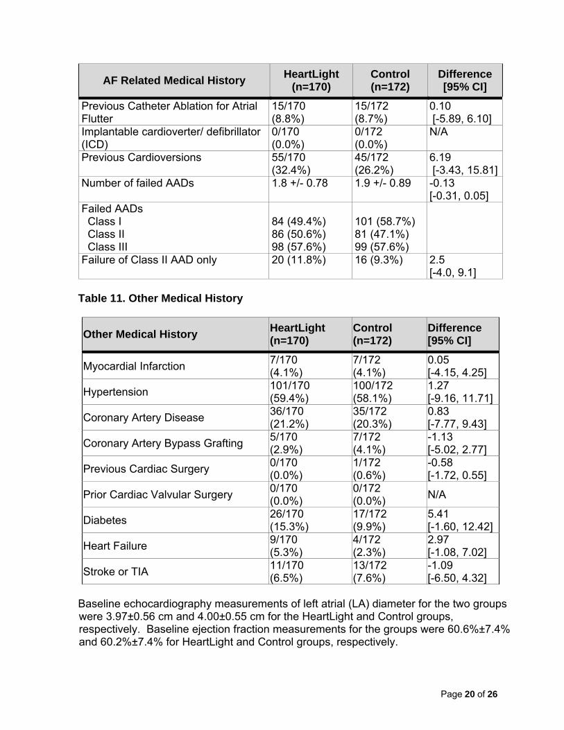
Baseline echocardiography measurements of left atrial (LA) diameter for the two groups were 3.97±0.56 cm and 4.00±0.55 cm for the HeartLight and Control groups, respectively. Baseline ejection fraction measurements for the groups were 60.6%±7.4% and 60.2%±7.4% for HeartLight and Control groups, respectively.
Page 21 of 26
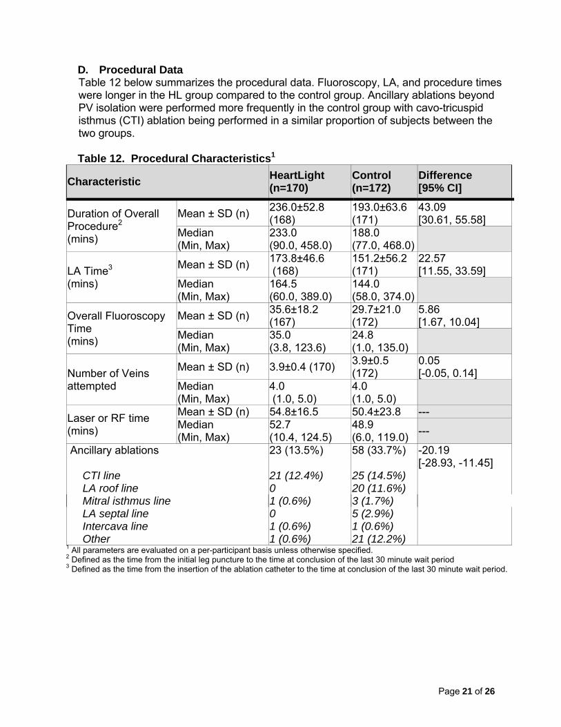
D. Procedural Data Table 12 below summarizes the procedural data. Fluoroscopy, LA, and procedure times were longer in the HL group compared to the control group. Ancillary ablations beyond PV isolation were performed more frequently in the control group with cavo-tricuspid isthmus (CTI) ablation being performed in a similar proportion of subjects between the two groups. Table 12. Procedural Characteristics1
Characteristic HeartLight (n=170)
Control (n=172)
Difference [95% CI]
Duration of Overall Procedure2 (mins)
Mean ± SD (n) 236.0±52.8 (168)
193.0±63.6 (171)
43.09 [30.61, 55.58]
Median (Min, Max)
233.0 (90.0, 458.0)
188.0 (77.0, 468.0)
LA Time3 (mins)
Mean ± SD (n) 173.8±46.6 (168)
151.2±56.2 (171)
22.57 [11.55, 33.59]
Median (Min, Max)
164.5 (60.0, 389.0)
144.0 (58.0, 374.0)
Overall Fluoroscopy Time (mins)
Mean ± SD (n) 35.6±18.2 (167)
29.7±21.0 (172)
5.86 [1.67, 10.04]
Median (Min, Max)
35.0 (3.8, 123.6)
24.8 (1.0, 135.0)
Number of Veins attempted
Mean ± SD (n) 3.9±0.4 (170) 3.9±0.5 (172)
0.05 [-0.05, 0.14]
Median (Min, Max)
4.0 (1.0, 5.0)
4.0 (1.0, 5.0)
Laser or RF time (mins)
Mean ± SD (n) 54.8±16.5 50.4±23.8 --- Median (Min, Max)
52.7 (10.4, 124.5)
48.9 (6.0, 119.0)
---
Ancillary ablations 23 (13.5%) 58 (33.7%) -20.19 [-28.93, -11.45]
CTI line 21 (12.4%) 25 (14.5%) LA roof line 0 20 (11.6%) Mitral isthmus line 1 (0.6%) 3 (1.7%) LA septal line 0 5 (2.9%) Intercava line 1 (0.6%) 1 (0.6%) Other 1 (0.6%) 21 (12.2%)
1 All parameters are evaluated on a per-participant basis unless otherwise specified. 2 Defined as the time from the initial leg puncture to the time at conclusion of the last 30 minute wait period 3 Defined as the time from the insertion of the ablation catheter to the time at conclusion of the last 30 minute wait period.
Page 22 of 26
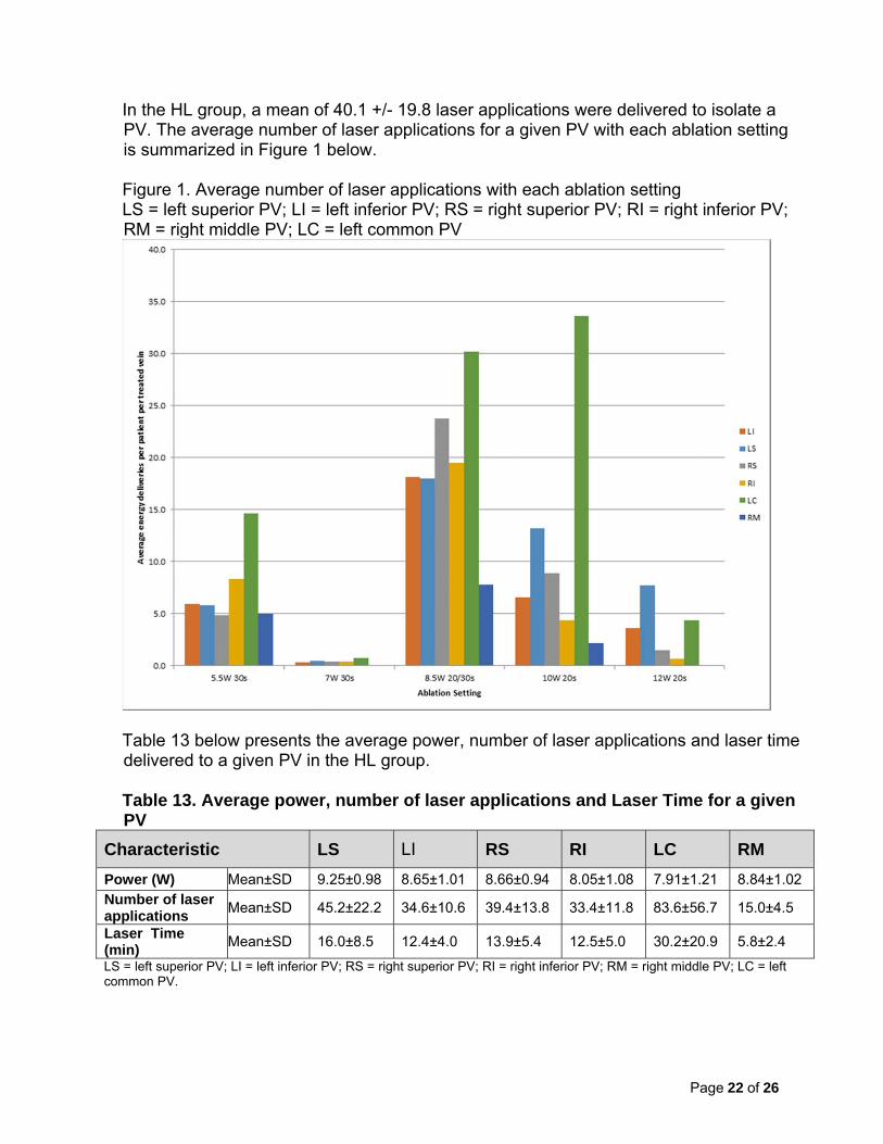
In the HL group, a mean of 40.1 +/- 19.8 laser applications were delivered to isolate a PV. The average number of laser applications for a given PV with each ablation setting is summarized in Figure 1 below. Figure 1. Average number of laser applications with each ablation setting LS = left superior PV; LI = left inferior PV; RS = right superior PV; RI = right inferior PV; RM = right middle PV; LC = left common PV
Table 13 below presents the average power, number of laser applications and laser time delivered to a given PV in the HL group. Table 13. Average power, number of laser applications and Laser Time for a given PV
Characteristic LS LI RS RI LC RM
Power (W) Mean±SD 9.25±0.98 8.65±1.01 8.66±0.94 8.05±1.08 7.91±1.21 8.84±1.02 Number of laser applications
Mean±SD 45.2±22.2 34.6±10.6 39.4±13.8 33.4±11.8 83.6±56.7 15.0±4.5
Laser Time (min)
Mean±SD 16.0±8.5 12.4±4.0 13.9±5.4 12.5±5.0 30.2±20.9 5.8±2.4
LS = left superior PV; LI = left inferior PV; RS = right superior PV; RI = right inferior PV; RM = right middle PV; LC = left common PV.

Page 23 of 26
E. Safety and Effectiveness Results Primary Effectiveness Endpoint As shown in the table 14 below, the HL group and the control group had similar primary effectiveness success rate – 61.1% vs. 61.7%, and the results met the pre-specified noninferiority margin of 15%. In addition, the lower bound of the one-sided confidence interval for the HL group (54.5%) exceeded the pre-specified threshold of 32%, meeting the second study success criterion. Table 14. Primary Effectiveness Endpoint-Non-Inferiority Analysis
HeartLight (n=170)
Control (n=172)
Difference (HeartLight-Control)
p-value
Number of Participants Evaluated1 167 167 Primary Effectiveness Endpoint Successes
61.08% (102)
61.68% (103)
-0.60 0.0032
Lower Bound of 95% Confidence Interval3
54.5%4 55.1% -9.285
1 Consists of participants who completed the 12-month follow-up or were identified as a failure prior to early withdrawal. 2 Calculated using the Farrington-Manning method for non-inferiority. 3Lower bound of the 1-sided 95% confidence interval is presented. 4 Study success is declared if the lower bound is greater than the pre-specified threshold rate of 32%. 5 Study success is declared if the lower bound is greater than the pre-specified non-inferiority margin of -15%.
As shown in table 15 below, the reasons for primary effectiveness failure were well balanced between the study groups, with most subjects failing the primary effectiveness endpoint due to symptomatic AF lasting >= 1 minute after the 90-day blanking period. Table 15. Reasons for Primary Effectiveness Failure
HeartLight (n=170)
Control (n=172)
Number of Participants Evaluated 167 167 Number of Participants who are primary effectiveness endpoint failures
65 (38.2%) 64 (37.2%)
Reason for Failure1: Did not have all clinically relevant2 PVs isolated acutely using randomized treatment device
10 (15.4%) 7 (10.9%)
Symptomatic AF lasting >=1 minute after the 90-day blanking period, documented by3:
34 (52.3%) 40 (62.5%)
TTM (core lab) 28 (82.4%) 29 (72.5%) 24-hour Holter (core lab) 6 (17.6%) 8 (20.0%) 12-lead ECG (not core lab) 12 (35.3%) 11 (27.5%) Other: Holter, mobile, pacemaker, telemetry (not core lab)
5 (14.7%) 7 (17.5%)

Page 24 of 26
HeartLight (n=170)
Control (n=172)
Any Class I, II or III AAD prescribed for AF at any time during the 9-12 months post-ablation index procedure
7 (10.8%) 3 (4.7%)
Ablation-induced left atrial flutter after the 90-day blanking period4
8 (12.3%) 7 (10.9%)
Additional Intervention for AF5 5 (7.7%) 7 (10.9%) Other6 1 (1.5%) 0 (0.0%)
1 Participants may have more than one reason for failure but the first occurrence of failure is classified as the primary reason. If multiple reasons for failure occurred on the same day then the reason for failure will be determined hierarchically in the order presented. 2 Defined as excluding PVs with no potentials and small PVs (<10 mm) unless ablation was attempted 3 Symptomatic A Fib can be documented by more than one method; therefore the percentages may not add up to 100% 4 If atrial flutter or atrial tachycardia cannot be classified as definitively right-sided flutter, it must be classified as left-atrial flutter. 5 Cardiac surgery, left-sided heart ablation and Implantable ICD for AF. Because the protocol allows for the Control participants to have a repeat left-sided heart ablation performed within 80 days of the index procedure, these procedures are not included here as reasons for failure unless there was no documented symptomatic episode of AF. 6 Right-sided flutter ablation during index procedure without history of flutter or flutter seen during index procedure and/or left-sided procedure using non-randomized treatment device during index procedure.
Figure 2 below presents the Kaplan-Meier curve for each of the treatment groups for freedom from primary effectiveness failure and shows similar pattern in time to primary effectiveness failure between the treatment groups. The curve made an initial drop for both groups at day 0, representing acute procedure failures. A second drop occurred at day 90 representing recurrence of atrial tachyarrhythmia soon after the 90-day blanking period. A third drop occurred at day 270, representing the time point at which being on an AAD for AF counts toward a treatment failure.

Page 25 of 26
Figure 2. KM Curves of Primary Effectiveness Endpoint
Secondary Effectiveness Endpoints Table 16 below summaries the secondary effectiveness results. The two groups showed similar results in all but one secondary effectiveness endpoint. While both groups had a very high acute success rate, the HL group had a lower incidence of PV reconnection during the procedure.

Page 26 of 26
Table 16. Secondary Effectiveness Endpoints
HeartLight (n=170)
Control (n=172)
P Value7
Secondary Effectiveness Endpoints Evaluated on a per-participant basis Success in isolating all attempted PVs acutely1 94.1%
(160/170) 97.1% (167/172)
N/A
Chronic Durable PV Isolation (per participant)2 13.6% (3/22)
16.7% (3/18)
N/A
Recurrence of Asymptomatic AF3 12.6% (21/167)
14.4% (24/167)
N/A
Evaluated on a per-vein basis PVs reconnected during procedure4 2.71%
(18/664) 5.72% (38/664)
0.006
Technical (Acute) Success5 97.3% (649/667)
97.9% (658/672)
N/A
Chronic Durable PV Isolation (per vein)6 52.7% (49/93)
46.4% (32/69)
0.511 1 Success in isolating all attempted PVs acutely is defined as the percentage of participants that have all attempted pulmonary veins isolated during the index procedure. 2 Chronic durable PV isolation on a per-participant basis is calculated as the number of participants requiring a 2nd procedure with all PVs isolated acutely during index procedure that remain isolated at start of 2nd procedure / number of participants requiring a 2nd procedure with all PVs isolated acutely during index procedure (not tested for statistical significance). 3 Percentage of participants with asymptomatic AF that lasts one minute or more outside the 90-day blanking period, independent of any reports of symptomatic AF. 4 Percentage of attempted PVs that reconnect during the index procedure. 5 Technical Success is defined as the number of clinically relevant pulmonary veins successfully isolated /number of clinically relevant veins *100. 6 Chronic durable PV isolation on a per-vein basis is calculated as # of PVs isolated acutely during index procedure that remain isolated at start of 2nd procedure / # of PVs isolated acutely during index procedure*100. 7 A hierarchical closed test procedure was used to account for multiple testing and Control the maximum overall Type I error rate. Endpoints were tested in the order described in the study design section, each tested at a significance level of p <0.05. The test procedure was stopped the first time statistical significance was not achieved. Secondary effectiveness endpoints were calculated using a t-test.
Primary Safety Endpoint – Primary Adverse Events Table 17 summaries the primary safety endpoint results. The PAE rate was 11.8% in the HL group vs. 14.5% in the control group. The difference in the PAE rate between the two groups was 2.8%. The upper 95% confidence interval of 3.5% was less than the pre-specified non-inferiority margin of 8%, demonstrating success in the primary safety endpoint.

Page 27 of 26
Table 17. Primary Safety Endpoint1 Non-Inferiority Analysis
HeartLight (n=170)
Control (n=172)
Difference (HeartLight-Control) [95% Confidence Interval2] p-value
Percent (Number) of Participants with a PAE3
11.76% (20) 14.53% (25) -2.77 [3.45]
0.0024
1 Primary safety endpoint is the composite of Primary Adverse Events (PAEs) through 12 months. 2 Upper bound of the 1-sided 95% confidence interval is presented. Study success is declared if the upper bound does not exceed the pre-specified non-inferiority margin of 8%. 3 Each participant is only counted once in the overall percentage of participants with a PAE. 4 Calculated using the Farrington-Manning method for non-inferiority.
Table 18 below summaries the PAEs. The two groups were comparable in all PAEs but two, namely phrenic nerve injury resulting in diaphragmatic paralysis and PV stenosis. As has been reported with other balloon-based PV isolation technologies, phrenic nerve injury resulting in diaphragmatic paralysis was more frequent with HL ablation compared to conventional open irrigated RF ablation (3.5% vs. 0.6%). However, PV stenosis was more frequent in the control group treated with conventional open irrigated RF ablation compared to the HL group (2.9% vs 0). Table 18. Primary Adverse Events (PAEs)
HeartLight (n=170)
Control (n=172)
Number of events
x (%) of participants1
Number of events
x (%) of participants1
Transient ischemic attack (within 1 month of treatment)
0 0 (0.0%) 0 0 (0.0%)
Cerebrovascular accident including stroke caused by air embolism
2 2 (1.2%) 1 1 (0.6%)
Major bleeding requiring transfusion (within 1 week of treatment)
0 0 (0.0%) 1 1 (0.6%)
Cardiac perforation, tamponade or clinically significant pericardial effusion (within 1 month of treatment)
2 2 (1.2%) 3 3 (1.7%)
Pulmonary Vein Stenosis > 50%2 (during the 12-month evaluation period)
0 0 (0.0%) 5 5 (2.9%)
Myocardial infarction (Q-wave only within 1 week of treatment)
0 0 (0.0%) 0 0 (0.0%)
Diaphragmatic paralysis (that persists after the blanking period)
6 6 (3.5%) 1 1 (0.6%)
Atrio-esophageal fistula (within 6 months of treatment)
0 0 (0.0%) 0 0 (0.0%)
Death (during the 12-month evaluation period and cause possibly related to device or procedure or if unknown)
0 0 (0.0%) 0 0 (0.0%)

Page 28 of 26
HeartLight (n=170)
Control (n=172)
Number of events
x (%) of participants1
Number of events
x (%) of participants1
Atrial Fibrillation or flutter requiring cardioversion
14 14 (8.2%) 16 16 (9.3%)
Number of participants experienced at least one Primary Adverse Event
-- 20 -- 25
1 A participant is counted only once within each Primary Adverse Event Name category however, could be counted multiple times across different Primary Adverse Event Name categories. 2 Based on change in PV size.
Additional Safety Information from HL Study Serious Adverse Events Serious adverse events (SAEs) that occurred in the HL group and control group are presented in table 19 below. A total of 26 SAEs in 23 study subjects were reported by investigators during the 12-month follow-up period. The overall proportion of subjects with one or more SAE appeared to be slighter higher in the HL group than that in the control group (8.2% vs. 5.2%). Of note, not all primary AEs were SAEs. For example, two events of phrenic nerve injury leading to diaphragmatic paralysis (one in each study group) were considered non-serious by investigational sites, but were classified as PAEs. Table 19. All Serious Adverse Events (SAEs)1
Adverse Event Name HeartLight (n=170)
Control (n=172)
Cardiac Perforation 0 (0.0%) 1 (0.6%)Cardiac Tamponade 2 (1.2%) 2 (1.2%)Cerebrovascular Event -- Other 0 (0.0%) 1 (0.6%)Cerebrovascular Event -- Stroke 1 (0.6%) 0 (0.0%)Chest Pain/Discomfort 2 (1.2%) 2 (1.2%)Diaphragmatic paralysis 1 (0.6%) 0 (0.0%)Hemorrhage 1 (0.6%) 0 (0.0%)Other: Fall 1 (0.6%) 0 (0.0%)Other: Methicillin Susp. SA infected PPM and RA & RV leads 0 (0.0%) 1 (0.6%)Other: Moderate drop in Hemoglobin 1 (0.6%) 0 (0.0%)Other: Pulmonary Emboli - Multiple 1 (0.6%) 0 (0.0%)Pericardial effusion 1 (0.6%) 1 (0.6%)Pericarditis 0 (0.0%) 2 (1.2%)Phrenic nerve damage leading to diaphragmatic paralysis 4 (2.4%) 0 (0.0%)Pseudoaneurysm 1 (0.6%) 0 (0.0%)Total 14 (8.2%) 9 (5.2%)
1 Defined as events that place the participant’s health in jeopardy and that occur despite following all labeling precautions and instructions for use and where all attempts at correction by medical intervention do not resolve the event. A participant is counted only once within each Adverse Event Name category however, could be counted multiple times across different Adverse Event Name categories.

Page 29 of 26
Death Summary One subject in the HL group expired during the 12 month follow-up period. The subject was a 66 year old female with a past medical history significant for hypertension, cerebrovascular accident, NYHA Class II heart failure, pulmonary hypertension, and AF/atrial flutter. She underwent an acutely successful PV isolation procedure using the HL system without immediate complications. One month post procedure, atrial tachyarrhythmia recurred and required multiple DC cardioversion. This was followed by acute on chronic diastolic heart failure with right heart catheterization revealing increased right-sided pressure and severe pulmonary hypertension that required maximal medical therapy. Four months post the index procedure, a repeat ablation procedure using an approved RF ablation catheter was performed for AF and atrial flutter without immediate complications. Post procedure, the subject was evaluated and treated for NYHA Class III/IV heart failure. Approximately 7 months after the index procedure and approximately 3 months after the RF ablation procedure, the subject fell at home and was found dead by her family. No clear reason for death was documented. Family declined autopsy. The event was adjudicated by the independent Clinical Oversight Committee as not related to the study device or the index ablation procedure but related to pre-existing pulmonary hypertension. Phrenic Nerve Injury Phrenic nerve injury resulting in diaphragmatic paralysis occurred in 6 subjects (3.5%) in the HL group and one subject (0.6%) in the control group. All 7 occurrences of diaphragmatic paralysis were classified as primary AEs because they had persisted for at least 3 months. Of the 6 cases of diaphragmatic paralysis in the HL group, 3 resolved by 12 months post the index procedure, 3 persisted at 12 months, with 2 resolving at 17 months and 23 months, respectively. The single diaphragmatic paralysis in the control group was persistent at 12 months. Among the 6 HL subjects who had phrenic nerve injury, 5 were women. The power setting in these 6 subjects was 8.5 W or 10 W. All 6 subjects had one or more associated symptoms/signs including shortness of breath (n = 5), decreased lung sounds (n = 2), respiratory failure requiring re-intubation (n = 1), hypoxia with confusion (n = 1), inability to extubate (n = 1) and wheezing (n = 1) during the period in which hemi-diaphragmatic abnormalities were noted. PV stenosis CT or MR imaging was performed at baseline and 3 month post ablation to screen for PV stenosis. Subjects with PV narrowing of > 50% on 3 month CT/MRI underwent repeat PV imaging at 12 month. Table 20 below summarizes PV narrowing and stenosis detected in the HL study. On a per-vein basis, the rate of PV narrowing of 20%-50% was similar between the two groups. No PV stenosis was detected in the HL group. However, 5 subjects in the control group had a single PV with > 50% narrowing. None of these 5 subjects had PV stenosis related symptoms or required therapeutic intervention.

Page 30 of 26
Table 20. Pulmonary Vein Narrowing and Stenosis on a Per-Vein Basis
3-month FU1 12-month FU2
HL (n=622)
Control (n=632)
Difference (HL-Control) [95% Confidence Interval]
HL (n=3)
Control (n=12)
Difference (HL-Control) [95% Confidence Interval]
PV Narrowing (>20 to <= 50% decrease from baseline)
136 (21.9%)
156 (24.7%)
-2.82 [-7.49, 1.86]
2 (66.7%)
3 (25.0%)
41.67 [-17.03, 100.00]
PV Stenosis (>50% decrease from baseline)
0 (0.0%)
5 (0.8%)
-0.79 [-1.48, -0.10]
0 (0.0%)
3 (25.0%)
-25.00 -49.50, -0.50]
1 Includes veins attempted during the Index Procedure and imaged at 3 months. 2 Only those veins that showed >50% decrease at 3 months are required to be imaged at 12 months.
Stroke There were two (2) strokes (1.2%) in the HL group (one occurred prior to discharge and the other a week after discharge) and one stroke (0.6%) in the control group that occurred prior to discharge. Both strokes in the HL group were considered embolic in nature with one stroke being acute cerebellar infarct resulting in left hemiparesis and ataxia , and the other being sub-acute infarcts resulting visual changes. The stroke in the control group was a small infarct at the right caudal nucleus, resulting in left sided weakness with tremors. Both strokes from the HL group recovered completely, and the control subject with stroke had minor residual effects by the conclusion of study follow-up. Additional Analyses Effect of Operator Experience
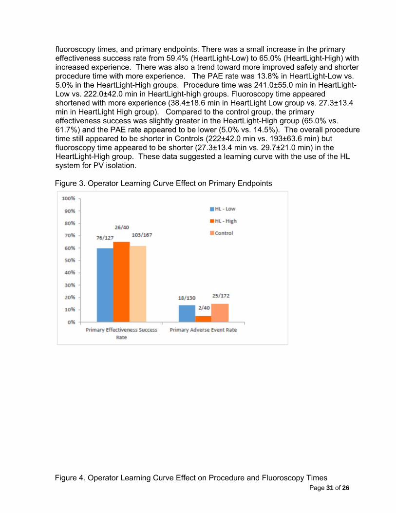
An analysis was conducted to examine learning curve by individual operator’s life-time HL procedure experience. This post-hoc analysis was conducted to examine the effects of learning curve on HeartLight ablation procedure metrics. There were 30 primary operators that performed at least one randomized HL procedure that were included in this analysis. All 30 operators had extensive experience with standard radiofrequency ablation and limited experience with the HeartLight System. Each procedure where an operator was a Primary Operator (conducted more than half of a HL procedure) counted towards the total number of lifetime HL procedures, regardless of study protocol or where (study center) that case was performed. By the end of the pivotal clinical study, only half (15/30) of the operators had performed more than 3 randomized HeartLight procedures and only 17% (5/30) had performed 15 or more lifetime HL procedures. The five operators with ≥15 lifetime procedures of experience with HeartLight (HeartLight-High; 40 procedures) were compared to the 25 operators with <15 lifetime procedures of experience (HeartLight-Low; 130 procedures). Fifteen procedures was selected as an experience threshold for experience because it has been previously used to determine the HeartLight learning curve [Ref 1]. Figures 3 and 4 below summarize the operator learning curve effect on procedure and
Page 31 of 26
fluoroscopy times, and primary endpoints. There was a small increase in the primary effectiveness success rate from 59.4% (HeartLight-Low) to 65.0% (HeartLight-High) with increased experience. There was also a trend toward more improved safety and shorter procedure time with more experience. The PAE rate was 13.8% in HeartLight-Low vs. 5.0% in the HeartLight-High groups. Procedure time was 241.0±55.0 min in HeartLight-Low vs. 222.0±42.0 min in HeartLight-high groups. Fluoroscopy time appeared shortened with more experience (38.4±18.6 min in HeartLight Low group vs. 27.3±13.4 min in HeartLight High group). Compared to the control group, the primary effectiveness success was slightly greater in the HeartLight-High group (65.0% vs. 61.7%) and the PAE rate appeared to be lower (5.0% vs. 14.5%). The overall procedure time still appeared to be shorter in Controls (222±42.0 min vs. 193±63.6 min) but fluoroscopy time appeared to be shorter (27.3±13.4 min vs. 29.7±21.0 min) in the HeartLight-High group. These data suggested a learning curve with the use of the HL system for PV isolation. Figure 3. Operator Learning Curve Effect on Primary Endpoints
Figure 4. Operator Learning Curve Effect on Procedure and Fluoroscopy Times

Page 32 of 26
Gender Analysis
A gender analysis was performed to assess the differences in primary effectiveness and safety endpoints between female and male subjects. As shown in the table 21 below, there was no gender discrepancy in primary effectiveness success. However, there was a greater PAE rate for female HL subjects (25.0%) compared to both male HL subjects (5.9%) and female controls (11.1%).
Table 21. Primary Endpoints by Gender
Male Female
HeartLight (n=118)
Control (n=109)
p-value
HeartLight (n=52)
Control (n=63)
p- value
Primary Effectiveness Endpoint
62.1% (72/116)
65.4% (70/107)
0.603 58.8% (30/51)
55.0% (33/60)
0.685
Primary Safety Endpoint
5.9% (7/118)
16.5% (18/109)
0.011 25.0% (13/52)
11.1% (7/63)
0.051
An additional analysis was performed to assess differences between female and male subjects in PAE rate excluding cardioversion for AF and atrial flutter, a more clinically meaningful measure of major complication rate. As shown in table 22 below, the PAE rate excluding cardioversion (or major complication rate) appeared to be greater in female HL subjects (11.5% or 6/52) than that in female controls (4.8% or 3/63) and male HL subjects (2.5%, 3/118). The greater major complication rate in female HL subjects was primarily driven by a high incidence of phrenic nerve injury resulting in diaphragmatic paralysis (9.6% or 5/52).

Page 33 of 26
Table 22. Primary Safety Endpoint without Cardioversion by Gender
Male Female
HeartLight Control p-value• HeartLight Control p-value•
Primary AE rate without cardioversion
2.5% (3/118)
7.3% (8/109)
0.084 11.5% (6/52)
4.8% (3/63)
0.159
Logistical regression analyses were conducted to investigate whether the major complication rate was consistent between female and male subjects. Since the treatment-by-gender interaction term reached statistical significance (p = 0.04), there was evidence suggesting that males and females may have different safety profiles for the HL system as compared to the control catheter.
It should be noted that this study was not powered to determine gender-specific safety profile of the HL system. Although women were well represented in the study (33.7% female), the number of female subjects enrolled was small (52 subjects in the HL group). Therefore, no firm conclusion could be made regarding the safety profile of the HL system in women. Moreover, there is no known anatomical or clinical reason for gender disparity in the risk of phrenic nerve injury associated with ablation using the HL system. A post approval study that enrolls a large number of female subjects is warranted to further evaluate the safety profile of the HL system including the risk of phrenic nerve injury in women.
F. Financial Disclosure
The Financial Disclosure by Clinical Investigators regulation (21 CFR Part 54), requires CardioFocus to include certain information concerning the compensation to, and financial interests and arrangement of, any clinical investigator conducting clinical studies covered by the regulation. The pivotal clinical study included 93 investigators of which 0 were full-time or part-time employees of the sponsor and one (1) had disclosable financial interests/arrangements as defined in 21 CFR 54.2(a), (b), (c) and (f) and described below:
Compensation to the investigator for conducting the study where the value could be influenced by the outcome of the study: 0
Significant payment of other sorts: 1 Proprietary interest in the product tested held by the investigator: 0 Significant equity interest held by investigator in sponsor of covered study:
0
The applicant has adequately disclosed the financial interest/arrangements with clinical investigators. Statistical analyses were conducted by FDA to determine whether the financial interests/arrangements had any impact on the clinical study outcome. The information provided does not raise any questions about the reliability of the data.

Page 34 of 26

Page 35 of 26
XI. PANEL MEETING RECOMMENDATION AND FDA’S POST-PANEL ACTION In accordance with the provisions of section 515(c)(3) of the act as amended by the Safe Medical Devices Act of 1990, this PMA was not referred to the Circulatory System Devices Advisory Panel, an FDA advisory committee, for review and recommendation because the information in the PMA substantially duplicates information previously reviewed by this panel.
XII. CONCLUSIONS DRAWN FROM PRECLINICAL AND CLINICAL STUDIES
A. Effectiveness Conclusions
The HL study met its primary effectiveness endpoint by demonstrating non-inferiority of the HL system to the FDA approved ThermoCool ablation catheter with respect to freedom from symptomatic atrial tachyarrhythmia recurrence off AAD at 12 months post ablation. The observed primary effectiveness success rate in the HL group, which was almost identical to that in the control group, was in line with other AF ablation studies for catheter-based technologies for the same indication for use. Moreover, upon completion of the ablation procedure, electrical PV isolation was achieved in the vast majority of the clinically relevant PVs (97.3%) by using the HL system alone. There was no gender discrepancy in primary effectiveness success. These data provide a reasonable assurance that the HL system is effective for the treatment of symptomatic drug refractory paroxysmal AF.
B. Safety Conclusions
The risks of the device are based on nonclinical laboratory and animal studies as well as data collected in a clinical study conducted to support PMA approval as described above. The HL study met its primary safety endpoint by demonstrating non-inferiority of the HL system to the FDA approved ThermoCool ablation catheter with respect to PAE rate. The observed PAE rate was slightly lower in the HL group compared to the control group but the difference was not statistically significant (11.8% vs. 14.5%). The major complication rates in both groups were in line with other AF ablation studies for similar catheter-based technologies for the same indication for use. Consistent with reports of other balloon-based PV isolation technologies, phrenic nerve injury resulting in diaphragmatic paralysis was more frequent with HL ablation compared to the approved open irrigated RF ablation (3.5% vs. 0.6%). The rate of persistent diaphragmatic paralysis at 1 year was 1.8% in the HL group and 0.6% in the control group, respectively. All but one case of diaphragmatic paralysis in the HL group resolved by the end of study follow-up. On the other hand, PV stenosis (defined as > 50% narrowing) was less frequent with ablation using the HL catheter compared to ablation using the approved open irrigated RF catheter (0 vs. 2.9%).

Page 36 of 26
The major complication rate appeared to be greater in female subjects treated with the HL catheter (11.5% or 6/52) compared to female controls (4.8% or 3/63) and male subjects treated with the HL catheter (2.5%, 3/118). The greater major complication rate in female HL subjects was primarily driven by a high incidence of phrenic nerve injury resulting in diaphragmatic paralysis (9.6% or 5/52). It should be noted that the pivotal study was not powered to determine gender-specific safety profile of the HL system. Although women were well represented in the study (33.7% female), the number of female subjects enrolled was small (52 subjects in the HL group). Therefore, no firm conclusion could be made regarding the safety profile of the HL system in women. Moreover, there is no known anatomical or clinical reason for gender disparity in the risk of phrenic nerve injury associated ablation using the HL system. A post approval study that enrolls a large number of female subjects is warranted to further evaluate the safety profile of the HL system including the risk of phrenic nerve injury in women. Taken together, these data provide a reasonable assurance that the HL system is safe for the treatment of symptomatic drug refractory paroxysmal AF.
C. Benefit-Risk Conclusions
The preclinical and clinical data presented support the notion that the probable benefits outweigh the probable risks when the HL system is used for the treatment of symptomatic drug refractory paroxysmal AF.
D. Overall Conclusions
The preclinical and clinical data in this application support the reasonable assurance of safety and effectiveness of the HL system when used in accordance with the Indications for Use. A post-approval study that enrolls a large number of female subjects is warranted to further evaluate the safety profile of the HL system including the risk of phrenic nerve injury in women.
XIII. CDRH DECISION CDRH issued an approval order on April 1, 2016. The final conditions of approval cited in the approval order are described below. In addition to the Annual Report requirements, the applicant must provide the following data in post-approval study (PAS) reports for the PAS listed below: OSB Lead PMA Post-Approval Study- of the HeartLight Endoscopic Ablation System for the Treatment of Atrial Fibrillation; Short Title: CF HeartLight PAS: The Office of Surveillance and Biometrics (OSB) will have the lead for studies initiated after device approval. This study will be conducted as per protocol dated March 30, 2016, Version 5.0. On March 30, 2016 (email) the applicant agreed to conduct a study as follows: The purpose of the study is to evaluate clinical outcomes in subjects treated with the

Page 37 of 26
HeartLight System during commercial use, and to specifically investigate safety outcomes for females in addition to the safety and effectiveness outcomes for the entire study group; This will be a prospective single-armed study design, study subjects will be clinically eligible for the HeartLight device; Two Hundred and fifty subjects will be enrolled in the study, with a minimum of 135 of those subjects being female; Study subjects will be followed for three years; The primary effectiveness endpoint will be freedom from symptomatic Atrial Fibrillation at one-year, and will be compared to a performance goal of 55% effectiveness at one-year post procedure; The primary safety outcome for the entire cohort will be the percentage of subjects experiencing a Primary Adverse Event by one-year of follow-up, and will be compared to a performance goal of 14% PAE rate at one-year post-procedure; The same safety outcome will be assessed for females with the same performance goal; Longer term (three year) safety and effectiveness will be assessed as secondary outcomes; The long-term effect of operator experience will also be assessed as a secondary outcome; PAS reporting will occur every six-months for the first two years of the PAS and then yearly thereafter. The applicant’s manufacturing facilities have been inspected and found to be in compliance with the device Quality System (QS) regulation (21 CFR 820).
Page 38 of 26
XIV. APPROVAL SPECIFICATIONS Directions for use: See device labeling Hazards to Health from Use of the Device: See Indications, Contraindications, Warnings, Precautions, and Adverse Events in the device labeling. Post-approval Requirements and Restrictions: See approval order. XV. REFERENCES [REF1] Dukkipati SR, Kuck KH, Neuzil P et al. Pulmonary vein isolation using a visually guided laser balloon catheter: the first 200-patient multicenter clinical experience. Circ Arrhythm Electrophysiol 2013;6:467-72.