Sumit K. Soni & Rakshapal Singh & Ashutosh Awasthi - CIMAP Staff
16
1 23 Environmental Science and Pollution Research ISSN 0944-1344 Environ Sci Pollut Res DOI 10.1007/s11356-012-1178-4 In vitro Cr(VI) reduction by cell-free extracts of chromate-reducing bacteria isolated from tannery effluent irrigated soil Sumit K. Soni, Rakshapal Singh, Ashutosh Awasthi, Mangal Singh & Alok Kalra
Transcript of Sumit K. Soni & Rakshapal Singh & Ashutosh Awasthi - CIMAP Staff

1 23
Environmental Science and PollutionResearch ISSN 0944-1344 Environ Sci Pollut ResDOI 10.1007/s11356-012-1178-4
In vitro Cr(VI) reduction by cell-freeextracts of chromate-reducing bacteriaisolated from tannery effluent irrigated soil
Sumit K. Soni, Rakshapal Singh,Ashutosh Awasthi, Mangal Singh & AlokKalra

1 23
Your article is protected by copyright and
all rights are held exclusively by Springer-
Verlag. This e-offprint is for personal use only
and shall not be self-archived in electronic
repositories. If you wish to self-archive your
work, please use the accepted author’s
version for posting to your own website or
your institution’s repository. You may further
deposit the accepted author’s version on a
funder’s repository at a funder’s request,
provided it is not made publicly available until
12 months after publication.
RESEARCH ARTICLE
In vitro Cr(VI) reduction by cell-free extractsof chromate-reducing bacteria isolated from tannery effluentirrigated soil
Sumit K. Soni & Rakshapal Singh & Ashutosh Awasthi &Mangal Singh & Alok Kalra
Received: 27 April 2012 /Accepted: 30 August 2012# Springer-Verlag 2012
Abstract Four efficient Cr(VI)-reducing bacterial strainswere isolated from rhizospheric soil of plants irrigated withtannery effluent and investigated for in vitro Cr(VI) reduc-tion. Based on 16S rRNA gene sequencing, the isolatedstrains SUCR44, SUCR140, SUCR186, and SUCR188 wereidentified as Bacillus sp. (JN674188), Microbacterium sp.(JN674183), Bacillus thuringiensis (JN674184), andBacillus subtilis (JN674195), respectively. All four isolatescould completely reduce Cr(VI) in culture media at 0.2 mMconcentration within a period of 24–120 h; SUCR140 com-pletely reduced Cr(VI) within 24 h. Assay with the permea-bilized cells (treated with Triton X-100 and Tween 80) andcell-free assay demonstrated that the Cr(VI) reduction activitywas mainly associated with the soluble fraction of cells.Considering the major amount of chromium being reducedwithin 24–48 h, these fractions could have been releasedextracellularly also during their growth. At the temperatureoptima of 28 °C and pH7.0, the specific activity of Cr(VI)reduction was determined to be 0.32, 0.42, 0.34, and0.28 μmol Cr(VI)min−1mg−1 protein for isolates SUCR44,SUCR140, SUCR186, and SUCR188, respectively. Additionof 0.1 mM NADH enhanced the Cr(VI) reduction in the cell-free extracts of all four strains. The Cr(VI) reduction activityin cell-free extracts of all the isolates was stable in presence ofdifferent metal ions tested except Hg2+. Beside this, urea and
thiourea also reduced the activity of chromate reduction tosignificant levels.
Keywords Chromate reduction . Specific activity .
Cell-free extracts . Cr(VI) . Chromate-reducing bacteria
Introduction
Hexavalent chromium is released as a by-product byseveral industrial activities like tanning, wood preserva-tion, production of steel, paper, pigment, dye, welding,chrome plating, thermonuclear weapons, etc.( Patra et al.2010). Tannery industries are one of the most pollutingindustries causing chromium pollution in the environ-ment. In India, there are more than 2,500 tanneries, andmost of them (nearly 80 %) are engaged in chrome tanningprocess (Chandra et al. 2010). Besides, several agronomicpractices, including the use of organic biomass like sewagesludge or fertilizers that contain varying degree of chromium,contribute to environment contamination (Viti et al. 2003). Cr(VI) exists in solution as CrO4
2−, and due to structural simi-larity with SO4
2−, can overcome the cellular permeabilitybarrier, entering via sulfate transport pathways (Patra et al.2010), rapidly reducing to Cr(V) and generating free radicals(Mabbett and Macaskie 2001). Due to generation of freeradicals, it is toxic (Wise et al. 2004) to all forms of livingsystems including microorganisms by causing oxidative stress(Ackerley et al. 2006), DNA damage (Mabbett and Macaskie2001), and altered gene expression (Bagchi et al. 2002).Moreover, Cr(VI) is also mutagenic (Puzon et al. 2002), carci-nogenic (Codd et al. 2003), and teratogenic (Asmatullah et al.1998), and has been recognized as a priority pollutant (Cheungand Gu 2007). In view of the seriousness of Cr(VI) pollutionand its alarming effects on human health, the USEnvironmental Protection Agency has listed it in class A human
Responsible editor: Robert Duran
S. K. Soni :R. Singh :A. Awasthi :M. Singh :A. KalraDepartment of Microbial Technology and Entomology,CSIR-Central Institute of Medicinal and Aromatic Plants(CSIR-CIMAP),Lucknow 226015, India
A. Kalra (*)CSIR-Central Institute ofMedicinal andAromatic Plants, P.O. CIMAP,Lucknow 226015 Uttar Pradesh, Indiae-mail: [email protected]
Environ Sci Pollut ResDOI 10.1007/s11356-012-1178-4
Author's personal copy
carcinogen categories (Costa and Klein 2006). The toxicity ofCr(VI) in plants is also observed at multiple levels, reducedoverall growth, and inhibition of enzyme functions leading tolower yields (Shanker et al. 2005).
Although hexavalent chromium is highly toxic, its triva-lent form is an essential micronutrient for animal and humanbeing involved in glucose metabolism (Vincent 2000), stim-ulation of enzyme system (Karuppanapandian et al. 2009),stabilization of nucleic acids by increasing the processivityof DNA polymerase (Snow and Xu 1991), and is relativelyinert and much less toxic than the hexavalent form (Krishnaand Philip 2005).
Metal pollutants are nondegradable and can only be trans-formed to less toxic oxidation states or removed either byadsorption/accumulation or by physicochemical treatments.However, it has been observed that these processes are costlyand unreliable (Malik 2004). On the other hand, microbialreclamation is safe, ecofriendly, is a cost-effective technology,and an alternative to the traditional physicochemical methods.Several microorganisms have the exceptional ability to sur-vive noxious metal-polluted environments by developingmechanisms to avoid metal toxicity like metal resistanceplasmids, metal efflux channels, adsorption uptake, DNAmethylation, and metal biotransformation either directly byenzymatic reduction to less mobile and toxic forms orindirectly through making complexes with metabolites(such as H2S) (Pei et al. 2009). A variety of Cr-resistantbacteria with high potential of Cr(VI)-reducing abilityhave been reported including Bacillus, Pseudomonas,Deinococcus, Enterobacter, Agrobacterium, Escherichia,Shewanella, Thermus, and other species (Opperman2008a). However, Cr(VI) resistance and Cr(VI) reductionhave been considered to be unrelated (Ohtake et al. 1987).The availability of selected strains able to resist and reducechromate elevate the possibility of employingmicroorganismsfor bioremediation of Cr(VI) contaminated site.
Several types of enzymatic Cr(VI) reduction have beenreported in bacteria, which include Cr(VI) reductase, alde-hyde oxidase, cytochrome P450, DT-diaphorase, etc. (Patraet al. 2010). Similarly, several oxidoreductases with differ-ent metabolic functions have also been reported to catalyseCr(VI) reduction in bacteria, which include nitroreductase(Kwak et al. 2003), iron reductase, quinone reductases(Gonzalez et al. 2005), hydrogenases (Chardin et al.2003), flavin reductases (Ackerley et al. 2004), as well asNADH/NADPH-dependent reductases (Puzon et al. 2002).The enzymatic chromate reduction occurs both in anaer-obic and aerobic bacteria (Cervantes et al. 2001). Inanaerobic bacteria, chromate reduction occurs in pres-ence of membrane bound enzymes. In contrast, enzymesfor chromate reduction have been localized as solublecytosolic proteins in most of the aerobic bacteria (Puzonet al. 2002).
The present study was carried out to explore the locali-zation and mechanisms associated with Cr(VI) reduction infour Cr(VI) tolerant bacteria, isolated from the soil irrigatedwith tannery effluents, with higher activities to reduce chro-mate. The optimal conditions (temperature and pH) for Cr(VI) reduction as well as the effects on Cr(VI) reductionrates due to the presence of metal ions, protein denaturants,and electron donors were also elucidated in this study.
Material and methods
Soil sampling
Soil samples were collected from a long-term tannery efflu-ent irrigated site near Kanpur (26°28′ N and 80°24′ E),India. Well-growing plants were uprooted, and soil adheringloosely to the roots was removed by shaking the plants. Thesoil firmly adhering to the roots, designated as rhizosphericsoil, was collected. The samples were passed through 2-mmsieve and well dispersed. The properties of the soils areshown in Table 1. A portion of the soil samples was airdried for chemical analyses. Soil pH was measured in dis-tilled water with a ratio of soil/solution of 1:2.5. DTPA-extractable heavy metals (Lindsay and Norvell 1978) weredetected by optical emission spectrophotometer (PerkinElmer) (Table 2).
Isolation of bacterial strains
A large number of bacterial isolates (more than 200) wereisolated from rhizospheric soil, using Luria agar (casein enzy-mic hydolysate, 10 gL−1; yeast extract, 5.0 gL−1; sodiumchloride, 5.0 gL−1; agar powder, 15 gL−1; Himedia, India)medium supplemented with filter-sterilized 1,000 mg Cr(VI)L−1 as K2CrO4. The Cr(VI) stock solutions were filter steril-ized using a 0.22-μm membrane filter. Plates were incubatedat 28 °C, and actively growing strains were isolated after1 week. These Cr-tolerant strains were further evaluated forchromate reduction at 0.2 mM concentration of Cr(VI).
On the basis of chromium reduction (data not provided),four strains (SUCR44, SUCR140, SUCR186, and SUCR188)were selected for further studies.
Table 1 The basic properties of rhizosphere soils
Soil samplenumber
Total N(kgha−1)
Available P(kgha−1)
Available K(kgha−1)
pH EC (μscm−1)
1 580 87 732 6.34 14,550
2 586 174 573 6.74 5,930
3 596 175 500 6.76 5,950
4 486 162 556 6.81 6,070
Environ Sci Pollut Res
Author's personal copy

Quantification of bacterial growth and Cr(VI) reductionof selected strains at different concentration of Cr(VI)
The culture flasks (100 ml) containing 20 ml nutrient broth(sodium chloride, 5.0 gL−1; beef extract, 1.5 gL−1; yeastextract, 1.5 gL−1; peptic digest of animal tissue, 5.0 gL−1,pH7.0±0.2; Himedia, India) supplemented with different con-centrations of Cr(VI), i.e., 0.2, 0.4, 0.6, 0.8, and 1.0 mM, wereinoculated with 0.5 mL of logarithmic phase bacterial culture(OD 1.2±0.1 at 600 nm), grown for 18 h in nutrient broth. Allthe cultures including biotic [nutrient broth without Cr(VI)]and abiotic [nutrient broth with Cr(VI) but not inoculated withbacteria] controls in triplicate were incubated for 120 h at 28 °C temperature with shaking at 150 rpm. The density of thebacteria was monitored at definite time intervals by measuringoptical density of the culture at 600 nm. Tomeasure the Cr(VI)reduction, 1 mL culture from each of the above flasks wascentrifuged (6,000×g for 10 min), and the Cr(VI) in thesupernatant was analyzed spectrophotometrically at 540 nm,according to the 1,5-diphenylcarbazide method described byAPHA (1995).
Effect of temperature and pH on Cr(VI) reduction
Chromium reduction was studied at different temperatures(20, 28, 35, and 42 °C) and pH (6.0, 7.0, and 8.0). The initialpH was adjusted using with 1 N HCl and 1 N NaOH.Appropriate buffers (0.05 M), phosphate buffer (pH6.0), andTris–HCl buffer (pH7.0 and 8.0) were added for avoiding theshifting of pH (Olajuyigbe and Ajele 2005). Flasks containing20 mL nutrient broth amended with K2CrO4 to final concen-tration of 0.2 mM Cr(VI) were inoculated with 0.5 mL oflogarithmic phase bacterial culture (OD 1.2±0.1 at 600 nm),grown for 18 h in nutrient broth. All the cultures includingbiotic and abiotic controls, in triplicate, were incubated for120 h with shaking at 150 rpm. Aliquots (2 mL) were with-drawn at regular time interval (every 24 h) from each repli-cated tube and centrifuged at 6,000×g for 10 min. Theconcentration of Cr(VI) in the supernatant was analyzed forCr(VI) reduction. Experiments for all isolates were done intriplicates and were repeated twice.
Resting cell assay
Bacterial cultures in 100 mL of nutrient broth were grownovernight (18 h) at 28 °C with shaking at 150 rpm. Cellswere harvested from aforesaid cultures (OD at 600 nm were1.2±0.1) by centrifugation at 6,000×g at 4 °C for 10 min.Pellets of bacterial cultures were washed twice with 5 mL of0.1 M potassium phosphate buffer, pH7.0 and resuspendedin same buffer. These cell suspensions were spiked with0.2 mM concentration of Cr(VI) as K2CrO4 and adjustedthe final system volume to 10 mL. The tubes were vortexedT
able
2Con
centratio
n(m
gkg
−1)of
heavymetals(D
TPA
-extractable)in
rhizosph
eresoils
contam
inated
with
tann
eryeffluents
Sam
plenu
mber
Corresponding
plants
Cd
Cr
Cu
Fe
Mg
Mn
Pb
Zn
1Triticum
aestivam
0.10
±0.03
107.95
±0.24
0.76
±0.08
87.56±0.68
275.45
±33
.87
12.93±0.61
1.83
±0.15
90.50
±0.070
2Brassicaoleracea
0.11
±0.01
516
6.15
±46
.42
0.45
±0.03
567
.62±28
.88
225.50
±36
.76
5.78
±1.53
0.77
±0.10
60.46
±0.088
3Brassicacampestris
0.06
±0.01
461
.12±4.06
50.32
±0.03
652
.37±8.83
255.58
±42
.30
8.47
±1.62
0.43
±0.12
30.15
±0.035
4Trifo
lium
alexandrinum
0.08
±0.01
210
8.12
±12
.55
0.57
±0.07
060
.83±3.12
304.75
±43
.48
10.25±0.53
0.30
±0.03
50.26
±0.053
Environ Sci Pollut Res
Author's personal copy

briefly for 2 min and incubated at 28 °C for 6 h. Aliquots(1 mL) were withdrawn at regular time interval and ana-lyzed for Cr(VI) reduction. Cr(VI) spiked in heat-treated(100 °C for 30 min) resuspended cells served as controls.Experiments for all isolates were done in triplicates andwere repeated twice.
Permeabilized cell assay
Overnight (18 h) grown cells of bacterial isolates wereharvested and washed twice with potassium phosphate buff-er pH7.0, as described above and resuspended in the samebuffer. Suspended cells were treated with 0.2 % (v/v) Tween80 and 0.2 % (v/v) Triton X-100 by vortexing for 20 min toachieve cell permeabilization. Cr(VI) as K2CrO4 (0.2 mMconcentration) was added to suspended cells, and the finalvolume was adjusted to 10 mL, and the samples wereincubated for 6 h at 28 °C. Aliquots (1 mL) were withdrawnat regular time intervals and analyzed for Cr(VI) reductionas described above. Permeabilized cells heated at 100 °C for30 min served as controls. Experiments for all isolates weredone in triplicates and were repeated twice.
Cell-free assay and localization of chromate reductionactivity
Cell-free extracts of bacterial isolates prepared by followingpreviously published protocol (Desai et al. 2008a). Cellsgrown for 18 h in 250 mL nutrient broth were harvested(OD at 600 nm were 1.2±0.1) by centrifugation at 6,000×gfor 10 min at 4 °C, washed and resuspended in 20 mL of0.1 M potassium phosphate buffer pH7.0. These cell sus-pensions were placed in ice bath and disrupted using anUltrasonic Probe (Rivotek, frequency 30 KHz±3 KHz) at120 W with 15-s pulses at 15-s interval for 30 min.Sonicates thus obtained were then ultracentrifuged at175,000×g (Beckman Coulter) for 90 min at 4 °C. Thecytosolic fractions or supernatants thus obtained were fil-tered through 0.22 μm filters to yield the cell-free extractsdevoid of membrane fractions and were immediately usedfor Cr(VI) reduction assay. The sonicated cell pellets wereaccordingly resuspended in same volume of phosphate buff-er. Aliquots of 300 μL of cell-free extracts or cytosolicfractions and sonicated pellet or membrane fractions wereused for chromate reduction assay in order to localize thechromate reduction activity in the cells of each isolate.Experiments for all isolates were done in triplicates withfreshly prepared cell-free extracts.
Enzyme assays
Chromate reduction was estimated using a standard calibra-tion curve of Cr(VI) as in the form of K2CrO4. The reaction
system (of 1 mL) used contained Cr(VI) final concentrations(0.2 mM) in 0.7 mL of 0.1 M potassium phosphate bufferwith 0.3 mL aliquots of cell-free extracts for chromatereduction. The system volume of 1 mL was kept constantfor all experiments. Assay conditions were kept constantwith a reaction time of 30 min at 28 °C. Abiotic controlcontained 0.2 mM Cr(VI) in 0.7 mL of phosphate buffer(0.1 M) with 0.3 mL of heat (100 °C for 30 min) treated cell-free extract. Experiments for all isolates were done in trip-licates. Specific activity was defined as unit chromate re-ductase activity per milligram protein concentration in thecell-free extract. Protein concentrations of cell-free extractwere estimated using Folin-phenol reagent by reading ab-sorbance at 750 nm, following the principle of Lowry et al.(1951). Known concentrations of bovine serum albumin(BSA) prepared in phosphate buffer (pH7.0) were used fordrawing the standard calibration curve.
Effect of metal ions, electron donors, and proteindenaturants on Cr(VI) reduction by cell-free extracts
Hexavalent chromate reductase activity in the cell-free ex-tract of bacterial isolates was also determined in the pres-ence of (0.1 mM each) metal ions (Cd2+, Pb2+, Hg2+, Ni2+,Cu2+, Co2+, and Zn+2) supplemented as CdCl2, Pb(NO3)2,HgCl2, NiCl2, CuCl2, CoCl2, ZnCl2, electron donors(NADH, succinate, and citrate) protein denaturants (ureaand thiourea) by incubating for 30 min at 28 °C.
Extraction of DNA from bacterial culture
Bacterial genomic DNAwas isolated from overnight growncells using standard procedures (Chachaty and Saulnier2000).The extracted DNA was electrophoresed on 0.8 %agarose gel in TAE buffer and visualized under UV inUvitec (Bangalore Genei, India) to check for integrity. Thequantity of the extracted DNA was checked spectrophoto-metrically (Nanodrop ND1000)
Amplification of 16S rRNA
The universal primers (forward 5′-AGAGTTTGATCCTGGCTCAG-3′ and reverse 5′- ACGGCTACCTTGTTACGACTT-3′) described earlier (Awasthi et al. 2011) were usedfor amplification of the 16S rRNA gene from the bacterialstrain. Approximately 25 ng of bacterial genomic DNA and10 pmol of forward and reverse primer, 0.6 U of Taqpolymerase, and 2.5 μL of 10× buffer (Bangalore Genei,India) were used for amplification in a Mastercyler gradient(Eppendorf) programmed as 94 °C for 5 min; 34 cycles of94 °C for 1 min, 57.4 °C for 1 min, 72 °C for 2 min; 72 °Cfor 10 min; and 4 °C for an infinite period. The amplifica-tion of PCR products were checked in 1.26 % agarose gels
Environ Sci Pollut Res
Author's personal copy

in TAE buffer stained with ethidium bromide (0.5 mgmL−1)and visualized under UV in Uvitec. The PCR product waspurified using PCR Cleanup Kit (Genexy) according to themanufacturer’s instructions and directly sequenced using theforward universal primer and Big Dye® Terminator v3.1cycle sequencing kit (Applied Biosystems, USA) on a3130xl Genetic Analyzer (Applied Biosystems, USA) usingthe manufacturer’s protocol.
Molecular characterization and phylogenetic analysisof isolates
16S sequence analysis was carried out using the nucleotideBLAST (http://www.ncbi.nlm.nih.gov/BLAST/) to identifyand download the nearest neighbor sequences from theNCBI database. All the sequences were aligned usingClustalW alignment tool. ClustalW was accessed throughthe MEGA, version 5 software (Tamura et al. 2011). Thephylogenetic tree was constructed using bootstrappedneighbor-joining tree method from MEGA5.
Statistical analysis
Data were subjected to two-way ANOVA to determine themain effects and interactions among factors. Differences amongtreatment means were compared using Tukey post hoc test withthe help of ASSISTAT Version 7.6 beta software (2011).
Results
Screening and identification of bacterial isolates
Homology searching (Table 3) and Blast analysis using 16SrRNA gene sequencing revealed that the newly isolated strainsSUCR44, SUCR186, and SUCR188 belong to Bacillus andexhibited 99 %, 98 and 100 % similarities with Bacillus cereus(EU162012), Bacillus thuringiensis (FJ236808), and Bacillussubtilis (JN641293), respectively, whereas SUCR140was iden-tified as Microbacterium and showed 99 % similarity withMicrobacterium paraoxydans (HM235673). Phylogeneticpositions in relation to other related organisms have beenshown in Fig. 1a and b. 16S rRNA gene sequence ofSUCR44, SUCR140, SUCR186, and SUCR188 have been
submitted to the NCBI GenBank under the accession numbersJN674188, JN674183, JN674184, and JN674195, respectively.
Reduction of Cr(VI) and growth of bacterial strains
The growth of bacterial strains was affected with increase inCr(VI) concentration (Fig. 2). The growth of bacterialstrains and corresponding Cr(VI) reduction at differenttimes intervals with graded concentration of Cr(VI) (0.2–1.0 mM), as potassium chromate, has been depicted inFig. 2. All the four isolates completely (100 %) reducedthe Cr(VI) at 0.2 mM concentration within a period of 24–120 h. However, strain SUCR140 could completely reducethe Cr(VI) within 24 h. There were negligible levels ofchromate reductions in abiotic controls.
Effect of temperature and pH on Cr(VI) reductionby bacterial strains
Over the strains, the microbial growth was higher, both inpresence and absence of Cr(VI), at pH7.0 at 28 °C (Fig. 3).However, the Cr(VI) reducing strength of bacterial strainswas found to be affected by strain identity, temperature, pH,and time (Table 4). Significant interactions were noticedamong these parameters. Chromate reduction by all fourbacterial strains was investigated at regular time intervalsat different temperatures (20–42 °C) and pH (6.0–8.0)(Fig. 4). Maximum Cr(VI) reduction activity of strainsSUCR44 and SUCR140 at 0.2 mM Cr(VI) was establishedat 28 °C at pH7.0, while SUCR186 and SUCR188 showedmaximum Cr(VI) reduction activity at pH 6.0 at 28 °C. Overthe pH, maximum reduction of chromate was observed at28 °C, this activity of all the strains decreased at both lower(20 °C) and higher temperatures (35 and 42 °C). Reductionof chromate was negligible in case of abiotic control at allthe temperature and pH (data not presented) after 120 h.
Localization of chromium reduction activity
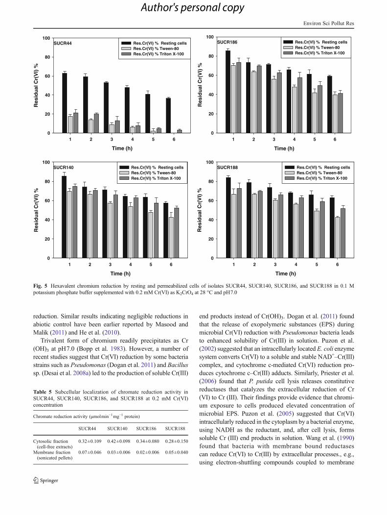
For detecting the localization, chromate reduction assayswere carried out using resting and permeabilized cells ofall the four strains by exposing the cells to 0.2 mM Cr(VI)for 6 h at 28 °C. Figure 5 shows the concentration ofresidual Cr(VI) upon exposure of resting and permeabilized
Table 3 Homology search ofbacterial isolates Isolate Identification GenBank
accession no.Similar organism Accession
numberSequencesimilarity (%)
SUCR44 Bacillus sp. JN674188 Bacillus cereus EU162012 99
SUCR140 Microbacterium sp. JN674183 Microbacterium paraoxydans HM235673 99
SUCR186 Bacillus thuringiensis JN674184 Bacillus thuringiensis FJ236808 98
SUCR188 Bacillus subtilis JN674195 Bacillus subtilis JN641293 100
Environ Sci Pollut Res
Author's personal copy

SUCR186(JN674184)
Bacillus thuringiensis str.B4(1)(FJ236808)
Bacillus pseudomycoides str.IARI-AN-13(JN411285)
Bacillus cereus str.PGOa4(EU162012)
SUCR44(JN674188)
Bacillus mycoides str.IARI-S-5(JN411479)
Bacillus shandongensis str.NSIII-20(JN993732)
Bacillus marisflavi str.IARI-S-14(JN411488)
Bacillus simplex str.IARI-MB-5(JN411330)
Bacillus koreensis str.TSI-2(JN993703)
Bacillus flexus str.IARI-AB-27(JN411314)
Bacillus megaterium str.IARI-A-10(JN411430)
Bacillus aryabhattai str.IARI-AN-21(JN411292)
Bacillus firmus str.IARI-J-28(JN411422)
Bacillus oceanisediminis str.M-FJ9(JF731240)
Bacillus selenatarsenatis str.NBSL41(JN624922)
Bacillus endophyticus str.IARI-J-22(JN411416)
Bacillus humi str.NBSL47(JN624924)
Bacillus licheniformis str.IARI-AB-16(JN411308)
Bacillus oleronius str.LZ034(JQ023623)
Bacillus pumilus str.IARI-A-5(JN411426)
Bacillus altitudinis str.IARI-MB-9(JN411334)
Bacillus methylotrophicus str.LZ039(JQ023625)
Bacillus mojavensis str.IARI-AB-10(JN411304)
SUCR188(JN674195)
Bacillus subtilis str.DDKRC5(JN641293)
Bacillus niacini str.TSII-13(JN993716)
Bacillus beijingensis(HQ424467)
Bacillus ginsengi(HQ424468)
Bacillus baekryungensis str.IARI-AB-24(JN411313)
Bacillus badius str.NBSL50(JN624927)
E.coli str.ATCC 11775T(X80725)
0.02
Microbacterium oxydans str.58(JN853773)
Microbacterium hydrocarbonoxydans str.SAI2(HQ220178)
Microbacterium foliorum str.NSIII-5(JN993727)
Microbacterium keratanolyticum str.JS424(JQ014176)
Microbacterium aurum str.B8A(GU441767)
Microbacterium aerolatum str.KNUC9057(JF505991)
Microbacterium oleivorans str.ANA51(HQ219882)
Microbacterium natoriense str.NW41(JF915347)
SUCR140(JN674183)
Microbacterium paraoxydans str.PAN 1974(HM235673)
Microbacterium terricola str.gilbert4(JN084156)
Microbacterium aoyamense str.I-C-6(GU593650)
Microbacterium trichotecenolyticum str.B26(EU169182)
Microbacterium nematophilum str.S1-11(FJ218360)
Microbacterium arabinogalactanolyticum str.21A(JF792087)
Microbacterium hominis str.LFR11(JF682041)
Microbacterium xylanilyticum str.HR87(JF700460)
Microbacterium schleiferi str.B-G-PYD9(HM629348)
Microbacterium flavescens str.LLR84(JF682100)
Microbacterium laevaniformans str.C-D-PYD2(HM755674)
Microbacterium dextranolyticum str.Na27(HQ831382)
Microbacterium barkeri str.IISR WP 25(JF907694)
Microbacterium arborescens str.ATY37(HQ219956)
Microbacterium imperiale str.RA38(JN585692)
E.coli str.ATCC 11775T(X80725)
0.02
a
b
Fig. 1 a Phylogenetic treeconstructed from the 16S rRNAgene of strains SUCR44,SUCR186, and SUCR188 andrelated organisms constructedusing neighbor-joining algo-rithm from an alignmentof 710 nucleotides. Accessionnumbers of correspondingsequences are given inparentheses, and scale bar rep-resents 1 base substitution per20 nucleotide positions.The bootstrap probabilities cal-culated from 1,000 replications.E. coli str. ATCC 11775T wastaken as an out-group.b Phylogenetic tree constructedfrom the 16S rRNA gene ofstrain SUCR140 andrelated organisms constructedusing neighbor-joining algo-rithm from an alignmentof 716 nucleotides. Accessionnumbers of correspondingsequences are given in paren-theses, and scale bar represents1 base substitution per20 nucleotide positions. Thebootstrap probabilities calculat-ed from 1,000 replications. E.coli str. ATCC 11775T wastaken as an out-group
Environ Sci Pollut Res
Author's personal copy

cells of all the four strains. As observed from the figure, cellpermeabilization significantly increased the Cr(VI) reduc-tion in all four strains. Among the two reagents used forpermeabilization namely Tween 80 and Triton X-100,Tween 80 brought about higher permeabilization of bacterialcells, which resulted in increased Cr(VI) reduction. Ontreatment with Tween 80, complete reduction of Cr(VI)was observed at 0.2 mM Cr(VI) concentration by permea-bilized cells of SUCR44, whereas 58, 62, and 57 % of Cr(VI) could be reduced by SUCR140, SUCR186, andSUCR188 respectively. On the other hand, permeabilizationthrough Triton X100 resulted in reduction of 97, 48, 59, and51 % by the strains SUCR44, SUCR140, SUCR186, andSUCR188, respectively. Considering Cr(VI) reduction byresting cell as 100 %, the Cr(VI) reduction by SUCR44,SUCR140, SUCR186, and SUCR188 was increased by 35,56, 49, and 57 %, respectively, on treatment with Tween 80.Similarly, 12, 51, 41, and 30 % enhancements in Cr(VI)reduction were observed on treatment with Triton X100 of
respective aforesaid strains. Chromate reduction assays werefollowed using initial concentration of 0.2 mM Cr(VI) withultrasonicated cytosolic fraction or cell-free extract andmembrane fraction (ultrasonicated pellet). As observed fromTable 5, reduction of Cr(VI) is mainly associated with solu-ble fraction (cell-free extracts) in all the four strains, indicat-ing the presence of hexavalent chromate-reducing principlein the cytoplasm (cytosolic fraction). No significant activityof chromate reduction was noticed in membrane fractionderived from ultrasonicated cells of all the isolates. Heated(100 °C for 30 min) cell-free extracts acting as control failedto reduce Cr(VI). These results confirm the presence ofsoluble enzymatic mechanism in the cytoplasmic fraction(crude cell-free extracts) of all the four strains. At the tem-perature optima of 28 °C and pH7.0, the specific activity ofCr(VI) reduction was determined to be 0.32 (0.16 %), 0.42(0.21 %), 0.34 (0.17 %), and 0.28 (0.14 %)μmol Cr(VI)min−1mg−1 for isolates SUCR44, SUCR140, SUCR186, andSUCR188, respectively (Table 5).
Fig. 2 Kinetics of growth and Cr(VI) reduction. Bacterial isolates were cultured with Cr(VI) 0.2, 0.4, 0.6, 0.8, and 1.0 mMCr(VI) as K2CrO4 and % Cr(VI) reduction in nutrient broth medium at 28 °C and pH7.0
Environ Sci Pollut Res
Author's personal copy

Effect of metal ions, protein denaturants, and electrondonors on chromium reduction activity of cell-free extract
Chromium reduction activity of the cell-free extract of isolatedstrains was estimated in presence of (0.1 mM) metal ions,protein denaturants, and electron donors at initial Cr(VI) con-centrations of 0.2 mMupon incubation at 28 °C and pH7.0 for30 min in 0.1 M potassium phosphate buffer. Among themetal ions tested, 0.1 mM of Cd2+ inhibited the reduction ofCr(VI) by the cell-free extracts of SUCR140, SUCR186, and
SUCR188 by 17, 21, and 32 %, respectively, whereas nosignificant inhibition in the reduction of Cr(VI) was noticedin SUCR44. Cr(VI) reduction activity of cell-free extract of allthe strains was not affected by Pb2+, whereas other divalentcations such as Ni2+ and Zn2+ could influence the Cr (VI)reduction activity of some strains. Hg2+ strongly inhibited theCr(VI) reduction in all the four isolates by 86–93 %. On theother hand, reduction of Cr(VI) by the cell-free extract ofSUCR44, SUCR140, SUCR186, and SUCR188 was en-hanced by Cu2+, increase of 37, 33, 44, and 28 %, respective-ly, as observed from Table 6. Co2+ also stimulated the Cr(VI)reduction in crude cell-free extract of SUCR44, SUCR140,SUCR186, and SUCR188, an increase of 15, 16, 35, and18 %, respectively. Urea, a protein denaturant, inhibited theCr(VI) reduction by 78, 88, 94, and 89 % in SUCR44,SUCR140 SUCR186, and SUCR188, respectively, while an-other protein denaturant thiourea inhibited the Cr(VI) by 88,86, 91, and 86 %, respectively, indicating the denaturation ofprotein(s) responsible for inhibition of Cr(VI) reduction in allfour strains. The specific activity of Cr(VI) reduction in thecell-free extracts of all the strains showed an increase with theaddition of 0.1 mMNADH; addition of 0.1 mMNADH in thereaction mixture containing cell-free extract increased thereduction of Cr(VI) by 141, 148, 159, and 150 % in the strainsSUCR44, SUCR140, SUCR186, and SUCR188, respectively.Citrate, a possible electron donor, during the reduction of Cr(VI) increased Cr(VI) reduction by 30, 21, 32, and 32 % inSUCR44, SUCR140, SUCR186, and SUCR188, respectively,as observed from data presented in Table 6. Succinate had nosignificant effect on the reduction of Cr(VI) by the cell-freeextract of all the four isolates.
Discussion
The irrigationwater contaminated with heavy metals is knownto cause disturbance in microbial communities with emer-gence of bacterial species having elevated metal tolerance(Stepanauskas et al. 2005). Chromium irrigation exerts astrong selective pressure on microbial flora of tannery soils(Viti et al. 2003). In the present investigation, bacterial strainstolerating and reducing Cr(VI) were isolated from rhizo-spheric soil receiving long-term augmentations of chromatefrom tanneries. The isolates belonged to the genera BacillusandMicrobacterium. Strains of the genus Bacillus are knownto tolerate and reduce Cr(VI) (Campos et al. 1995; Liu et al.2006). Unlike the genus Bacillus, the bacteria belonging togeneraMicrobacterium are rarely known for Cr(VI) reductionunder aerobic condition. Pattanapipitpaisal et al. (2001) earlierreported thatMicrobacterium sp. MP30 can reduce the Cr(VI)under anaerobic condition. In our experiments, higher Cr(VI)concentrations caused decrease in growth rate in all fourstrains tested when compared to growth at lower Cr(VI)
Table 4 Summary of statistical analysis: the main effect and interac-tion of bacterial strains, pH, time, and temperature on Cr(VI) reductionwere analyzed by factorial ANOVA
Treatmentsa df SS F
Strains 3 7,005.77162 850.5864*
Temperature 3 143,058.06921 17,369.0009*
pH 2 5,936.39659 1,081.1268*
Time 4 63,171.67061 5,752.3606*
Strains×temperature 9 42,549.09603 1,721.9937*
Strains×pH 6 11,458.98900 695.6308*
Strains×time 12 1,144.97787 34.7536*
Temperature×pH 6 36,251.26776 2,200.6738*
Temperature×time 12 8,463.93102 256.9062*
pH×time 8 248.30293 11.3051*
Strains×temperature×pH 18 43,614.25402 882.5507*
Strains×temperature×time 36 4,505.75202 45.5878*
Strains×pH×time 24 453.06145 6.8759*
Temperature×pH×time 24 1,407.86789 21.3665*
Strains×temperature×pH×time 72 1,921.25134 565.6957*
Error 240 658.91214
*p<0.01)a Strains (SUCR44, SUCR140, SUCR186, and SUCR188); tempera-ture (20, 28, 35, and 42 °C); pH (6.0, 7.0, 8.0); times [(24, 48, 72, 96,and 120) in hours]
Temperature oC
15 20 25 30 35 40 45
OD
(60
0 n
m)
1.5
2.0
2.5
3.0
3.5
4.0
4.5
Temperature vs pH 6.0 with Cr(VI) Temperature vs pH 6.0 without Cr(VI) Temperature vs pH 7.0 with Cr(VI) Temperature vs pH 7.0 without Cr(VI) Temperature vs pH 8.0 with Cr(VI) Temperature vs pH 8.0 without Cr(VI)
Fig. 3 Bacterial growth (over the strains) after 48 h in the presence andabsence of Cr(VI) at different temperature and pH
Environ Sci Pollut Res
Author's personal copy

concentrations. It is also clear from our results (Fig. 2) thatincrease in chromate reduction was growth dependent; higherreduction were noticed during the first 48 h corresponding tolog phase of the microbial growth. Most likely, bacterialgrowth and Cr(VI)-induced damage are competing processes,and bacteria can cope with Cr(VI) exposure only as long asmetabolizable C-sources are available. Liu et al. (2006) no-ticed that this phenomenonmight be explained as an increasedtime period for adaptation or repair during the exposure ofhigh level of Cr(VI) in the medium. The high level of Cr(VI)in the medium induces frameshift errors and, to a greaterextent, base pair substitution both in G–C and A–T base pairs(DeFlora et al. 1990). It has also been proposed that bacterialSOS function can repair the DNA damage caused by Cr(VI)(Oh and Choi 1997). Hexavalent chromate reduction by all thefour strains was investigated at different temperatures (20–42 °C), an important factor affecting microbial Cr(VI) reduc-tion. Maximum Cr(VI) reduction of all the four strains wasestablished at 28 °C, which also corresponds to maximumgrowth of the bacterial strains (Fig. 3) again, indicating thatCr(VI) reduction was growth dependent. Such growth-
dependent chromate reduction has also been earlier reportedby Desai et al. (2008a). It has been reported that the optimaltemperature of Cr(VI) reduction could be in the range of 25–37 °C (Cheung and Gu 2007; Ibrahim et al. 2012). However,optimum temperature of Cr(VI) reduction of thermophilicThermus scotoductus SA-01 (Opperman et al. 2008b) andBacillus firmus KUCr1 (Sau et al. 2010) have been reportedat 65 and 70 °C, respectively. Cr(VI) reduction was found tobe influenced by pH. Maximum Cr(VI) reduction activity ofstrains SUCR44 and SUCR140 at 0.2 mMCr(VI) was noticedat pH7.0, while SUCR186 and SUCR188 showed maximumCr(VI) reduction activity at pH6.0. Wang et al. (1990)reported that reduction of Cr(VI) in bacterial strain occurredat pH6.0–8.0 and was strongly inhibited at pH5.0 and 9.0.(Bopp et al. 1983). Our results clearly indicated that chromatereduction was dependent on pH, temperature, strain identity(Table 4), and the significant interaction observed amongthem, suggesting that different strains perform differentlyunder different temperature and pH. Negligible reductionwas noticed in abiotic control at all temperature and pH,indicating the direct interaction of microbes in Cr(VI)
pH
6 7 8
Res
idu
al C
r(V
I) %
0
20
40
60
80
100
120Res.Cr(VI) % 24 hrs Res.Cr(VI) % 48 hrs Res.Cr(VI) % 72 hrs Res.Cr(VI) % 96 hrs Res.Cr(VI) % 120 hrs
0244RCUS oC
pH
6 7 8
Res
idu
al C
r(V
I) %
0
20
40
60
80
100
120Res.Cr(VI) % 24 hrs Res.Cr(VI) % 48 hrs Res.Cr(VI) % 72 hrs Res.Cr(VI) % 96 hrs Res.Cr(VI) % 120 hrs
8244RCUS oC
pH
6.0 7.0 8.0
Res
idu
al C
r(V
I) %
0
20
40
60
80
100
120Res.Cr(VI) % 24 hrs Res.Cr(VI) % 48 hrs Res.Cr(VI) % 72 hrs Res.Cr(VI) % 96 hrs Res.Cr(VI) % 120 hrs
5344RCUS oC
pH
6.0 7.0 8.0
Res
idu
al C
r(V
I) %
0
20
40
60
80
100
120Res.Cr(VI) % 24 hrs Res.Cr(VI) % 48 hrs Res.Cr(VI) % 72 hrs Res.Cr(VI) % 96 hrs Res.Cr(VI) % 120 hrs
2444RCUS oC
pH
6.0 7.0 8.0
Res
idu
al C
r(V
I) %
0
20
40
60
80
100
120Res.Cr(VI) % 24 hrs Res.Cr(VI) % 48 hrs Res.Cr(VI) % 72 hrs Res.Cr(VI) % 96 hrs Res.Cr(VI) % 120 hrs
02041RCUS oC
pH
6.0 7.0 8.0
Res
idu
al C
r(V
I) %
0
20
40
60
80
100
120Res.Cr(VI) % 24 hrs Res.Cr(VI) % 48 hrs Res.Cr(VI) % 72 hrs Res.Cr(VI) % 96 hrs Res.Cr(VI) % 120 hrs
82041RCUS oC
pH
6.0 7.0 8.0
Res
idu
al C
r(V
I) %
0
20
40
60
80
100
120Res.Cr(VI) % 24 hrs Res.Cr(VI) % 48 hrs Res.Cr(VI) % 72 hrs Res.Cr(VI) % 96 hrs Res.Cr(VI) % 120 hrs
53041RCUS oC
pH
6.0 7.0 8.0
Res
idu
al C
r(V
I) %
0
20
40
60
80
100
120Res.Cr(VI) % 24 hrs Res.Cr(VI) % 48 hrs Res.Cr(VI) % 72 hrs Res.Cr(VI) % 96 hrs Res.Cr(VI) % 120 hrs
24041RCUS oC
pH
6.0 7.0 8.0
Res
idu
al C
r(V
I) %
0
20
40
60
80
100
120Res.Cr(VI) % 24 hrs Res.Cr(VI) % 48 hrs Res.Cr(VI) % 72 hrs Res.Cr(VI) % 96 hrs Res.Cr(VI) % 120 hrs
02681RCUS oC
pH
6.0 7.0 8.0
Res
idu
al C
r(V
I) %
0
20
40
60
80
100
120Res.Cr(VI) % 24 hrs Res.Cr(VI) % 48 hrs Res.Cr(VI) % 72 hrs Res.Cr(VI) % 96 hrs Res.Cr(VI) % 120 hrs
82681RCUS oC
pH
6.0 7.0 8.0
Res
idu
al C
r(V
I) %
0
20
40
60
80
100
120Res.Cr(VI) % 24 hrs Res.Cr(VI) % 48 hrs Res.Cr(VI) % 72 hrs Res.Cr(VI) % 96 hrs Res.Cr(VI) % 120 hrs
53681RCUS oC
pH
6.0 7.0 8.0
Res
idu
al C
r(V
I) %
0
20
40
60
80
100
120Res.Cr(VI) % 24 hrs Res.Cr(VI) % 48 hrs Res.Cr(VI) % 72 hrs Res.Cr(VI) % 96 hrs Res.Cr(VI) % 120 hrs
24681RCUS oC
pH
6.0 7.0 8.0
Res
idu
al C
r(V
I) %
0
20
40
60
80
100
120Res.Cr(VI) % 24 hrs Res.Cr(VI) % 48 hrs Res.Cr(VI) % 72 hrs Res.Cr(VI) % 96 hrs Res.Cr(VI) % 120 hrs
02881RCUS oC
pH
6.0 7.0 8.0
Res
idu
al C
r(V
I) %
0
20
40
60
80
100
120Res.Cr(VI) % 24 hrs Res.Cr(VI) % 48 hrs Res.Cr(VI) % 72 hrs Res.Cr(VI) % 96 hrs Res.Cr(VI) % 120 hrs
82881RCUS oC
pH
6.0 7.0 8.0
Res
idu
al C
r(V
I) %
0
20
40
60
80
100
120Res.Cr(VI) % 24 hrs Res.Cr(VI) % 48 hrs Res.Cr(VI) % 72 hrs Res.Cr(VI) % 96 hrs Res.Cr(VI) % 120 hrs
53881RCUS oC
pH
6.0 7.0 8.0
Res
idu
al C
r(V
I) %
0
20
40
60
80
100
120Res.Cr(VI) % 24 hrs Res.Cr(VI) % 48 hrs Res.Cr(VI) % 72 hrs Res.Cr(VI) % 96 hrs Res.Cr(VI) % 120 hrs
24881RCUS oC
Fig. 4 Effect of pH and temperature on Cr(VI) reduction by bacterial strains supplemented with 0.2 mM Cr(VI) as K2CrO4
Environ Sci Pollut Res
Author's personal copy
reduction. Similar results indicating negligible reductions inabiotic control have been earlier reported by Masood andMalik (2011) and He et al. (2010).
Trivalent form of chromium readily precipitates as Cr(OH)3 at pH7.0 (Bopp et al. 1983). However, a number ofrecent studies suggest that Cr(VI) reduction by some bacteriastrains such as Pseudomonas (Dogan et al. 2011) and Bacillussp. (Desai et al. 2008a) led to the production of soluble Cr(III)
end products instead of Cr(OH)3. Dogan et al. (2011) foundthat the release of exopolymeric substances (EPS) duringmicrobial Cr(VI) reduction with Pseudomonas bacteria leadsto enhanced solubility of Cr(III) in solution. Puzon et al.(2002) suggested that an intracellularly located E. coli enzymesystem converts Cr(VI) to a soluble and stable NAD+–Cr(III)complex, and cytochrome c-mediated Cr(VI) reduction pro-duces cytochrome c–Cr(III) adducts. Similarly, Priester et al.(2006) found that P. putida cell lysis releases constitutivereductases that catalyzes the extracellular reduction of Cr(VI) to Cr (III). Their findings provide evidence that chromi-um exposure to cells produced elevated concentration ofmicrobial EPS. Puzon et al. (2005) suggested that Cr(VI)intracellularly reduced in the cytoplasm by a bacterial enzyme,using NADH as the reductant, and, after cell lysis, formssoluble Cr (III) end products in solution. Wang et al. (1990)found that bacteria with membrane bound reductasescan reduce Cr(VI) to Cr(III) by extracellular processes., e.g.,using electron-shuttling compounds coupled to membrane
Time (h)
1 2 3 4 5 6
Res
idu
al C
r(V
I) %
0
20
40
60
80
100Res.Cr(VI) % Resting cells Res.Cr(VI) % Tween-80 Res.Cr(VI) % Triton X-100
SUCR44
Time (h)
1 2 3 4 5 6
Res
idu
al C
r(V
I) %
0
20
40
60
80
100Res.Cr(VI) % Resting cells Res.Cr(VI) % Tween-80 Res.Cr(VI) % Triton X-100
SUCR140
Time (h)
1 2 3 4 5 6
Res
idu
al C
r(V
I) %
0
20
40
60
80
100Res.Cr(VI) % Resting cells Res.Cr(VI) % Tween-80 Res.Cr(VI) % Triton X-100
SUCR186
Time (h)
1 2 3 4 5 6
Res
idu
al C
r(V
I) %
0
20
40
60
80
100Res.Cr(VI) % Resting cells Res.Cr(VI) % Tween-80 Res.Cr(VI) % Triton X-100
SUCR188
Fig. 5 Hexavalent chromium reduction by resting and permeabilized cells of isolates SUCR44, SUCR140, SUCR186, and SUCR188 in 0.1 Mpotassium phosphate buffer supplemented with 0.2 mM Cr(VI) as K2CrO4 at 28 °C and pH7.0
Table 5 Subcellular localization of chromate reduction activity inSUCR44, SUCR140, SUCR186, and SUCR188 at 0.2 mM Cr(VI)concentration
Chromate reduction activity (μmolmin−1mg−1 protein)
SUCR44 SUCR140 SUCR186 SUCR188
Cytosolic fraction(cell-free extracts)
0.32±0.109 0.42±0.098 0.34±0.080 0.28±0.150
Membrane fraction(sonicated pellets)
0.07±0.046 0.03±0.006 0.02±0.006 0.05±0.040
Environ Sci Pollut Res
Author's personal copy

reductases. The metal reduction can also be mediated by thesurfaces of bacterial spores (Junier et al. 2009) and suchmechanism may be relevant for spore forming bacteria likeBacillus.
Resting and permeabilized cell assays provided the betterevidence of the presence of an Cr(VI) reduction mechanismin cytosol fraction as observed in previous findings ofMegharaj et al. (2003). Permeabilization with Tween 80and Triton X-100 resulted in increased Cr(VI) reduction,indicating that cytoplasmic proteins were released and allthe four strains reduced Cr(VI) through soluble cytosolicreductases and not through membrane associated reductases.The inability to reduce hexavalent chromium by boiled cell-free extract, which served as control, showed that reductionprocess is enzymatic and not due to absorption or chemicalreaction. The cell lysis played an important role in Cr(VI)reduction. Ishibashi et al. (1990), Pal et al. (2005), andElangovan et al. (2006) observed that chromate reductaseactivity was associated with soluble protein and not with themembrane fraction. Desai et al. (2008b) suggested that asoluble chromate reductase associated with the cytoplasmicmembrane catalyzed Cr(VI) reduction by Pseudomonas sp.G1DM21 by transferring initial one electron to Cr(VI) toform an intermediate Cr(V), followed by two electron trans-fer for Cr(III) formation. Priester et al. (2006) reported thatthe chromate reductases originated in the cytoplasm leftcells by cell lysis and reduces Cr(VI) extracellularly. Ourresults indicate that, although the chromate-reducing frac-tion is located in cytosol, these fractions might be releasedextracellularly considering major amount of chromium be-ing reduced by all strains during initial 48 h of their growth.
McLean and Beveridge (2001) reported that the extracellu-lar reductase activity in cell filtrates from 48-h-old cultureswas due to either secretion or cell lysis.
Metal ions have been known to affect chromate reductaseactivity. Reduction of Cr(VI) by the cell-free extract wasenhanced by Cu2+. Camargo et al. (2003) reported the stimu-lation of Cr(VI) reduction in Bacillus sp. ES 29 on addition of1 mM of Cu2+. Elangovan et al. (2006) also found that onaddition of 1 mM Cu2+ reduction of Cr(VI) was stimulated inBacillus sp. The stimulation of enzyme activity by Cu2+ mightbe due to its nature as a prosthetic group of many reductaseenzymes (Sau et al. 2010). Camargo et al. (2003) reported thatthe increase in the reduction of Cr(VI) in presence of Cu2+ hasbeen attributed to its action as an electron-transport protectoror its action as a single electron redox center. On the contrary,Cu2+ has also been reported to inhibit the membrane associ-ated chromate reductase activity of Enterobacter cloacae(Ohtake et al. 1990) and soluble chromate reductase activityin Pseudomonas putida (Park et al. 2000) and B. sphaericusAND 303 (Pal et al. 2005). Addition of Pb2+ showed adiminutive inhibitory effect on Cr(VI) reduction, while Hg2+
strongly inhibited the Cr(VI) reduction activity, whereas otherdivalent cation such as Ni2+, Cd2+, and Zn2+ inhibited the Cr(VI) reduction to a variable degree. These variations seem tobe due to the different functional nature of the reductaseenzymes, which warrants further investigation. Metal ionsmay affect microbial Cr(VI) reduction in two ways: destruc-tion of cells (decrease in cell growth) and inhibition ofenzymes responsible for Cr(VI) reduction. Metal ions mayabsorb on to cell walls or complex with enzymes responsiblefor Cr(VI) reduction. The absorption of metal ions onto cell
Table 6 Effect of 0.1 mM metalions
Protein denaturants and electrondonors on hexavalent chromatereductase activity in the crudecell-free extracts of SUCR44,SUCR140, SUCR186, andSUCR188 in 0.1 M potassiumphosphate buffer of pH7, on in-cubation of 30 min at 28 °C
Metals ions SUCR44 SUCR140 SUCR186 SUCR188Specific activity(μmolmin−1mg−1
protein)
Specific activity(μmolmin−1mg−1
protein)
Specific activity(μmolmin−1mg−1
protein)
Specific activity(μmolmin−1mg−1
protein)
CFE (control) 0.32±0.109 0.42±0.098 0.34±0.080 0.28±0.150
Cd2+ 0.29±0.057 0.35±0.075 0.27±0.092 0.19 ±0.063
Pb2+ 0.35±0.086 0.40±0.103 0.32±0.069 0.30±0.051
Hg2+ 0.03±0.005 0.06±0.023 0.04±0.017 0.02±0.005
Ni2+ 0.25±0.121 0.44±0.080 0.32±0.063 0.22±0.080
Cu2+ 0.44±0.098 0.56±0.127 0.49±0.075 0.36±0.127
Co2+ 0.37±0.109 0.49±0.167 0.42±0.063 0.33±0.092
Zn+2 0.33±0.121 0.36±0.040 0.30±0.086 0.23±0.069
Protein denaturants
Urea 0.07±0.034 0.05±0.023 0.02±0.011 0.03±0.011
Thiourea 0.04±0.011 0.06±0.017 0.03±0.011 0.04±0.017
Electron donors
NADH 0.77±0.032 1.04±0.248 0.88±0.178 0.70±0.138
Succinate 0.31±0.080 0.45±0.069 0.33±0.103 0.30±0.075
Citrate 0.40±0.196 0.51±0.127 0.45±0.161 0.37±0.086
Environ Sci Pollut Res
Author's personal copy
walls or the formation of metal–enzyme complexes may leadto inactivation of chromate reductase enzymes or sites respon-sible for Cr reduction (Mabbett et al. 2002; Dogan et al. 2011).The reduction of Cr(VI) by cell-free extract was stimulated byCo2+. Similar results have been observed in Bacillus firmusstrain KUCr1 (Sau et al. 2010) at 0.2 mM, though Pal et al.(2005) reported the inhibition of Cr(VI) reduction activity ofBacillus sphaericus AND 303 by Co2+ at 100 μM concentra-tion. On the contrary, Desai et al. (2008a) reported the en-hancement of Cr(VI) reduction activity even at 1,000 μMconcentration. Mercury is the most commonly reported inhib-itor of reductase, which suggests the role of thiol group incatalysis (Park et al. 2000); Cr(VI) reduction by all the fourstrains was highly inhibited by mercuric ion. The electrondonor such as citrate had significant stimulatory effect on Cr(VI) reduction. Studies by Mabbett et al. (2002) and Desai etal. (2008a) show that the presence of low molecular organicmolecules such as citrate protected the chromate reductaseenzymes from inactivation by removing toxic products ofmicrobial reduction. Mabbett et al. (2002) also found a closeconnection between the amount of Cr(VI) reduced and theequilibrium constants of Cr-ligand complexes with more Cr(VI) being reduced with much stronger complexes. Succinate,on the other hand, did not show any noteworthy effect on Cr(VI) reduction. Desai et al. (2008b) observed improved chro-mate reductase activity by Pseudomonas sp. G1DM21 in thepresence of electron donors such as citrate, acetate, and succi-nate. The Cr(VI) reduction of Bacillus sp. andMicrobacteriumsp. belongs to the NADH-dependent type. An addition ofNADH improved Cr(VI) reduction both in Bacillus andMicrobacterium. There have been reports supporting NADH-dependent Cr(VI) reduction from Pseudomonas sp.,Escherichia coli, and Bacillus sp.; probably, these can useNADH as the electron donor (Bae et al. 2005). Previous reportshave demonstrated that intracellular Cr(VI) accepts a singleelectron from an NADH molecule forming a Cr(V) intermedi-ate, which in turn accepts two electrons from two molecules ofNADH to form stable Cr(III) (Suzuki et al. 1992). Urea andthiourea are well known protein denaturizing agents. In ourstudy, both inhibited the Cr(VI) reduction.
To conclude, all the four strains were tolerant to Cr(VI) andcompletely reduced Cr(VI) through soluble reductases withina period of 24–120 h. SUCR140 could completely reduce theCr(VI) within 24 h at 28 °C at pH7.0 but failed to reduce itcompletely at pH6.0 even after 120 h where SUCR 188performed the best. Interestingly, SUCR 140 at higher temper-atures worked well even at pH6.0 whereas SUCR 188 couldperform well only at low temperatures (≤28 °C). At 35 °C,SUCR44 and SUCR186 showed relatively higher chromatereductase activity, which improved at pH8.0. Therefore,SUCR140 and SUCR188 could bemore useful for subtropicalareas, while strain SUCR140 might also be useful for themoderate acidic regions experiencing high temperature. On
the other hand, strains SUCR44 and SUCR186 might beuseful under neutral and moderate alkaline soil environmentfor tropical regions. Our studies clearly suggest a stronginteraction among strains, pH, and temperature. Therefore,this study provides useful information for identifying thestrains of Cr(VI) reducing bacteria, which would performbetter under different soil environments and may be employedinto sustainable microphytoremediation.
Acknowledgments The authors wish to thank the Director, CSIR-Central Institute of Medicinal and Aromatic Plants, Lucknow, India, forproviding necessary facilities and encouragement during the course ofinvestigation, Dr HP Singh for his help in statistical analysis, SukhmalChand and Kundan Wasnik for soil analysis, and the Indian Council ofMedical Research (ICMR), New Delhi, India, for providing financialsupport to SKS.
References
Ackerley DF, Gonzalez CF, Park CH, Blake R II, Keyhan M, Matin A(2004) Chromate-reducing properties of soluble flavoproteinsfrom Pseudomonas putida and Escherichia coli. Appl EnvironMicrobiol 70(2):873–882
Ackerley DF, Barak Y, Lynch SV, Curtin J, Matin A (2006) Effect ofchromate stress on Escherichia coli K-12. J Bacteriol 188:3371–3381
Asmatullah Qureshi SN, Shakoori AR (1998) Hexavalent chromiuminduced congenital abnormalities in chick embryos. J Appl Tox-icol 18(3):167–171
Awasthi A, Bharti N, Nair P, Singh R, Shukla AK, Gupta MM, DarokarMP, Kalra A (2011) Synergistic effect of Glomus mosseae andnitrogen fixing Bacillus subtilis strain Daz26 on artemisinin con-tent in Artemisia annua L. Appl Soil Ecol 49:125–130
Bae WC, Lee HK, Choe YC, Jahng DK, Lee SH, Kim SJ, Lee JH,Jeong BC (2005) Purification and characterization of NADPHdependent Cr(VI) reductase from Escherichia coli ATCC 33456.J Microbiol 43:21–27
Bagchi D, Stohs SJ, Downs BW, Bagchi M, Preuss HG (2002) Cyto-toxicity and oxidative mechanism of different forms of chromium.Toxicology 180:5–22
Bopp LH, Chakrabarty AM, Ehrlich HL (1983) Chromate resis-tance plasmid in Pseudomonas fluorescens. J Bacteriol 155:1105–1109
Camargo FAO, Bento FM, Okeke BC, Frankenberger WT (2003) Chro-mate reduction by chromium-resistant bacteria isolated from soilscontaminated with dichromate. J Environ Qual 32:1228–1233
Campos J, Martinez-Pacheco M, Cervantes C (1995) Hexavalent-chromium reduction by a chromate-resistant Bacillus sp. strain.Ant van Leeuwen 68:203–208
Cervantes C, Campos-Garcia J, Gutierrez-Corona F, Loza-Tavera H,Torres-Guzman JC, Moreno-Sanchez R (2001) Interactions ofchromium with microorganisms and plants. FEMS MicrobiolRev 25:335–347
Chachaty E, Saulnier P (2000) Isolating chromosomal DNA frombacteria. In: Rapley R (ed) The nucleic acid protocols handbook,vol 1. Humana, Totowa, pp 29–32
Chandra R, Bharagava RN, Kapley A, Purohit HJ (2010) Bacterialdiversity, organic pollutants and their metabolites in two aerationlagoons of common effluent treatment plant (CFTP) during thedegradation and detoxification of tannery wastewater. BioresTechnol 102(3):2333–2341
Environ Sci Pollut Res
Author's personal copy

Chardin B, Giudici-Orticoni MT, DeLuca G, Guigliarelli B, Bru-schi M (2003) Hydrogenases in sulfate-reducing bacteriafunction as chromium reductase. Appl Microbiol Biotechnol63(3):315–321
Cheung KH, Gu JD (2007) Mechanism of hexavalent chromium de-toxification by microorganisms and bioremediation applicationpotential: a review. Int Biodeterior Biodegrad 59:8–15
Codd R, Irwin JA, Lay PA (2003) Sialoglycoprotein and carbohydratecomplexes in chromium toxicity. Curr Opi Chem Biol 17(2):213–219
Costa M, Klein CB (2006) Toxicity and carcinogenicity of chromiumin humans. Crit Rev Toxicol 36:155–163
DeFlora S, Bagnasco M, Serra D, Zanacchi P (1990) Genotoxicity ofchromium compounds: a review. Mut Res 238:99–172
Desai C, Jain K, Madamwar D (2008a) Evaluation of In vitro Cr(VI)reduction potential in cytosolic extracts of three indigenous Ba-cillus sp. Isolated from Cr(VI) polluted industrial landfill. BioresTechnol 99:6059–6069
Desai C, Jain K, Madamwar D (2008b) Hexavalent chromate reductaseactivity in cytosolic of Pseudomonas sp.G1DM21 isolated fromCr(VI) contaminated industrial landfill. Proc Biochem 43:713–721
Dogan NM, Kantar C, Gulcan S, Dodge CJ, Yilmaz BC, Mazmanci MA(2011) Chromium (VI) bioremoval by Pseudomonas bacteria: Roleof microbial exudates for natural attenuation and biotreatment of Cr(VI) contamination. Environ Sci Technol 45:2278–2285
Elangovan R, Abhipsa S, Rohit B, Ligy P, Chandraraj K (2006)Reduction of Cr(VI) by a Bacillus sp. Biotechnol Lett 28:247–252
Gonzalez CF, Ackerley DF, Lynch SV, Matin A (2005) ChrR, a solublequinone reductase of Pseudomonas putida that defends againstH2O2. J Biol Chem 280:2590–2595
He M, Li X, Guo L, Miller S, Rensing C, Wang G (2010) Character-ization and genomic analysis of chromate resistant and reducingBacillus cereus strain SJ1. BMC Microbiol 10:221
Ibrahim ASS, Elbadawi BY, El-Tayeb AM, Al-Salamah AA (2012)Hexavalent chromium reduction by novel chromate resistant alka-liphilic Bacillus sp. strain KSUCr9a. African. J Biotechnol 11(16):3832–3841
Ishibashi Y, Cervantes C, Silver S (1990) Chromium reduction inPseudomonas putida. Appl Environ Microbiol 56(7):2268–2270
Junier P, Frutschi, Wigginton NS, Schofield EJ, Bargar JR, LatmaniBR (2009) Metal reduction by spores of Desulfotomaculumreducen. Environ Microbiol 11(12):3007–3017
Karuppanapandian T, Sinha PB, Kamarul HA, Manoharan K (2009)Chromium induced accumulation of peroxide content, stimulationof antioxidative enzyme and lipid peroxidation in green gram(vigna radiata L cv wilczek) leaves. Afr J Biotechnol 8(3):475–479
Krishna RK, Philip L (2005) Bioremediation of Cr(VI) in contaminat-ed soils. J Hazard Mater 121:109–117
Kwak YH, Lee DS, Kim HB (2003) Vibrio harveyi nitroreductase is alsoa chromate reductase. Appl Environ Microbiol 69:4390–4395
LindsayWL,NorvellWA (1978) Development of a DTPA soil test for zinc,iron, manganese, and copper. Soil Sci Soc Am J 42(3):421–428
Liu YG, Xu WH, Zeng GM, Li X, Gao H (2006) Cr(VI) reduction byBacillus sp. isolated from chromium landfill. Pro Biochem 41(9):1981–1986
Lowry OH, Roseberough NJ, Lewis AF, Randall JR (1951) Proteinmeasurement with the folin phenol reagent. J Biol Chem193:265–275
Mabbett AN, Macaskie LE (2001) A novel isolate of Desulfovibrio sp.with enhanced ability to reduce Cr (VI). Biotechnol Lett 23:683–687
Mabbett AN, Lloyd JR, Macaskie LE (2002) Effect of complexingagents on reduction of Cr(VI) by Desulfovibrio vulgaris ATCC29579. Biotechnol Bioeng 79(4):389–397
Malik A (2004) Metal bioremediation by growing cells. Environ Int30:261–278
Masood F, Malik A (2011) Hexavalent chromium reduction by Bacil-lus sp. strain FM1 isolated from heavy-metal contaminated soil.Bull Environ Contam Toxicol 86:114–119
McLean J, Beveridge TJ (2001) Chromate reduction by a pseudomo-nas isolated from a site contaminated with chromated copperarsenate. Appl Environ Microbiol 67:1076–1084
Megharaj M, Avudainayagam S, Naidu R (2003) Toxicity of hexava-lent chromium and its reduction by bacteria isolated from soilcontaminated with tannery waste. Curr Microbiol 47:51–54
Oh YS, Choi SC (1997) Reduction of hexavalent chromium by Pseu-domonas aeruginosa HP014. J Microbiol 35:25–29
Ohtake H, Cervantes C, Silver S (1987) Decreased chromate uptake inPseudomonas fluorescens carrying a chromate resistance plasmid.J Bacteriol 169:3853–3856
Ohtake H, Fujii E, Toda K (1990) Reduction of toxic chromate in anindustrial effluent by use of chromate reducing strain of Enter-obactor cloacae. Environ Technol 11:663–668
Olajuyigbe FM, Ajele JO (2005) Production dynamics of extracellularprotease from Bacillus species. Afr J Biotechnol 4(8):776–779
Opperman DJ (2008a) The mechanism of chromate reduction by Ther-mus scotoductus SA-01. PhD thesis, University of the Free State
Opperman DJ, Piater LA, Heerden E (2008) A novel chromate reduc-tase from Thermus scotoductus SA-01 related to old yellowenzyme. J Bacteriol 190(8):3076–3082
Pal A, Dutta S, Paul AK (2005) Reduction of hexavalent chromium bycell free extract of Bacillus sphaericus AND 303 isolated fromserpentine soil. Curr Microbiol 51:327–330
Park CH, KeyhanM,Wielinga B, Fendorf S,Matin A (2000) Purificationto homogeneity and characterization of a novelPseudomonas putidachromate reductase. Appl Environ Microbiol 66:1788–1795
Patra RC, Malik B, Beer M, Megharaj M, Naidu R (2010) Molecularcharacterization of chromium (VI) reducing potential in Grampositive bacteria isolated from contaminated sites. Soil Biol Bio-chem 42:1857–1863
Pattanapipitpaisal P, Brown NL, Macaskie LF (2001) Chromate reduc-tion and 16S rRNA identification of bacteria isolated from a Cr(VI)-contaminated site. Appl Microbiol Biotechnol 57:257–261
Pei QH, Shahir S, Santhana AS (2009) Chromium (VI) resistance andremoval by Acinetobacter haemolyticus. World J Microbiol Bio-technol 25:1085–1093
Priester JH, Olson SG, Webb SM, Neu MP, Hersman LE, Ve HoldenPA (2006) Enhanced exopolymer production and chromium sta-bilization in Pseudomonas putida unsaturated biofilms. ApplEnviron Microbiol 72(3):1988–1996
APHA (American Public Health Association); American Water WorksAssociation (AWWA); Water Environment Federation (WEF)(1995) Standard Methods for the Examination of Water andWastewater, 19th ed. Washington, DC
Puzon GJ, Petersen JN, Roberts AG, Kramer DM, Xun L (2002) Abacterial flavin reductase system reduces chromates (III)–NAD+
complex. Biochem Biophy Res 294(1):76–81Puzon GJ, Roberts AG, Kramer DM, Xun L (2005) Formation of
soluble organo-chromium(III) complexes after chromate reduc-tion in the presence of cellular organics. Environ Sci Technol39:2811–2817
Sau GB, Chatterjee S, Mukherjee SK (2010) Chromate reduction by cellfree extract of Bacillus firmusKUCr1. Pol JMicrobiol 59(3):185–190
Shanker AK, Cervantes C, Loza-Tavera H, Avudainayagam S (2005)Chromium toxicity in plants. Environ Int 31:735–753
Snow ET, Xu LS (1991) Chromium (III) bound to DNA templatespromotes increased polymerase processivity and decreased fidel-ity during replication in vitro. Biochemistry 30(47):11238–11245
Stepanauskas R, Glenn TC, Jagoe CH, Tuckfield RC, Lindell AH,McArthur JV (2005) Elevated microbial tolerance to metals andantibiotics in metal-contaminated industrial environments. Envi-ron Sci Technol 39(10):3671–3678
Environ Sci Pollut Res
Author's personal copy

Suzuki T, Miyata N, Horitsu H, Kawai K, Takamizawa K, Tai Y,Okazaki M (1992) NAD(P)H-dependent chromium(VI) reductaseof Pseudomonas ambigua G-1: a Cr(V) intermediate is formedduring the reduction of Cr(VI) to Cr(III). J Bacteriol 174:5340–5345
Tamura K, Peterson D, Peterson N, Stecher G, Nei M, Kumar S (2011)MEGA5: Molecular Evolutionary Genetics Analysis using max-imum likelihood, evolutionary distance, and maximum parsimonymethods. Mol Biol Evol 28:2731–2739
Vincent JB (2000) Elucidating a biological role for chromium at amolecular level. Acc Chem Res 33(7):503–510
Viti C, Pace A, Giovannetti L (2003) Characterization of Cr(VI)resistant bacteria isolated from chromium contaminated soil bytannery activity. Curr Microbiol 46:1–5
Wang P, Mori T, Toda K, Ohtake H (1990) Membrane-associatedchromate reductase activity from Enterobacter cloacae. J Bacter-iol 172:1670–1672
Wise SS, Elmore LW, Holt SE, Little JE, Anto nucci PG, Bryant BH,Pierce WSJ (2004) Telomerase mediated lifespan extension ofhuman bronchial cells does not affect hexavalent chromium in-duced cytotoxicity or genotoxicity. Mol Cell Biochem 255(1–2):103–112
Environ Sci Pollut Res
Author's personal copy