Sulfonamido-2-arylbenzoxazole GroEL/ES inhibitors are ...
47
Page 1 of 47 Journal of Medicinal Chemistry Sulfonamido 2 arylbenzoxazole GroEL/ES inhibitors are potent antibacterials against methicillin resistant Staphylococcus aureus (MRSA) AUTHOR NAMES Sanofar Abdeen, a Trent Kunkle, a Nilshad Salim, a Anne-Marie Ray, a Najiba Mammadova, a,1 Corey Summers, a,2 Mckayla Stevens, a Andrew J. Ambrose, b Yangshin Park, a,e,f Peter G. Schultz, c Arthur L. Horwich, d Quyen Hoang, a,e,f Eli Chapman, b and Steven M. Johnson a, * AUTHOR ADDRESSES a Indiana University School of Medicine, Department of Biochemistry and Molecular Biology, 635 Barnhill Dr., Indianapolis, IN 46202 b The University of Arizona, College of Pharmacy, Department of Pharmacology and Toxicology, 1703 E. Mabel St., PO Box 210207, Tucson, AZ 85721 c The Scripps Research Institute, Department of Chemistry, 10550 North Torrey Pines Rd., La Jolla, CA 92037 d HHMI, Department of Genetics, Yale School of Medicine, Boyer Center for Molecular Medicine, 295 Congress Ave., New Haven, CT 06510 e Stark Neurosciences Research Institute, Indiana University School of Medicine. 320 W. 15th Street, Suite 414, Indianapolis, IN 46202 f Department of Neurology, Indiana University School of Medicine. 635 Barnhill Drive, Indianapolis, IN 46202 PRESENT ADDRESSES 1 Department of Genetics, Development and Cell Biology, Iowa State University, 1210 Molecular Biology Building, Pannel Dr, Ames, IA 50011 2 Department of Kinesiology, Iowa State University, 235 Barbara E. Forker Building, Beach Rd, Ames, IA 50011 KEYWORDS. GroEL, GroES, HSP60, HSP10, molecular chaperone, chaperonin, proteostasis, small molecule inhibitors, ESKAPE pathogens MRSA, antibiotics. 1 2 3 4 5 6 7 8 9 10 11 12 13 14 15 16 17 18 19 20 21 22 23 24 25 26 27 28 29 30 31 32 33 34 35 36 37 38 39 40 41 42 43 44 45 46 47 48 49 50 51 52 53 54 55 ___________________________________________________________________ This is the author's manuscript of the article published in final edited form as: Abdeen, S., Kunkle, T., Salim, N., Ray, A.-M., Mammadova, N., Summers, C., … Johnson, S. M. (2018). Sulfonamido-2- arylbenzoxazole GroEL/ES inhibitors are potent antibacterials against methicillin-resistant Staphylococcus aureus (MRSA). Journal of Medicinal Chemistry. https://doi.org/10.1021/acs.jmedchem.8b00989
Transcript of Sulfonamido-2-arylbenzoxazole GroEL/ES inhibitors are ...

Page 1 of 47 Journal of Medicinal Chemistry
Sulfonamido 2 arylbenzoxazole GroEL/ES inhibitors are potent antibacterials against
methicillin resistant Staphylococcus aureus (MRSA)
AUTHOR NAMES
Sanofar Abdeen,a Trent Kunkle,
a Nilshad Salim,
a Anne-Marie Ray,
a Najiba Mammadova,
a,1
Corey Summers,a,2 Mckayla Stevens,
a Andrew J. Ambrose,
b Yangshin Park,
a,e,f Peter G. Schultz,
c
Arthur L. Horwich,d Quyen Hoang,
a,e,f Eli Chapman,
b and Steven M. Johnson
a,*
AUTHOR ADDRESSES
a Indiana University School of Medicine, Department of Biochemistry and Molecular Biology, 635 Barnhill Dr., Indianapolis, IN 46202 b The University of Arizona, College of Pharmacy, Department of Pharmacology and Toxicology, 1703 E. Mabel St., PO Box 210207, Tucson, AZ 85721 c The Scripps Research Institute, Department of Chemistry, 10550 North Torrey Pines Rd., La Jolla, CA 92037 d HHMI, Department of Genetics, Yale School of Medicine, Boyer Center for Molecular Medicine, 295 Congress Ave., New Haven, CT 06510 e Stark Neurosciences Research Institute, Indiana University School of Medicine. 320 W. 15th Street, Suite 414, Indianapolis, IN 46202 f Department of Neurology, Indiana University School of Medicine. 635 Barnhill Drive, Indianapolis, IN 46202
PRESENT ADDRESSES
1 Department of Genetics, Development and Cell Biology, Iowa State University, 1210 Molecular Biology Building, Pannel Dr, Ames, IA 50011 2 Department of Kinesiology, Iowa State University, 235 Barbara E. Forker Building, Beach Rd, Ames, IA 50011
KEYWORDS.
GroEL, GroES, HSP60, HSP10, molecular chaperone, chaperonin, proteostasis, small molecule inhibitors, ESKAPE pathogens MRSA, antibiotics.
12345678910111213141516171819202122232425262728293031323334353637383940414243444546474849505152535455 ___________________________________________________________________
This is the author's manuscript of the article published in final edited form as:
Abdeen, S., Kunkle, T., Salim, N., Ray, A.-M., Mammadova, N., Summers, C., … Johnson, S. M. (2018). Sulfonamido-2-arylbenzoxazole GroEL/ES inhibitors are potent antibacterials against methicillin-resistant Staphylococcus aureus (MRSA). Journal of Medicinal Chemistry. https://doi.org/10.1021/acs.jmedchem.8b00989

2
ABSTRACT
Extending from a study we recently published examining the anti-trypanosomal effects of
a series of GroEL/ES inhibitors based on a pseudo-symmetrical bis-sulfonamido-2-
phenylbenzoxazole scaffold, here, we report the antibiotic effects of asymmetric analogs of this
scaffold against a panel of bacteria known as the ESKAPE pathogens (Enterococcus faecium,
Staphylococcus aureus, Klebsiella pneumoniae, Acinetobacter baumannii, Pseudomonas
aeruginosa, and Enterobacter species). While GroEL/ES inhibitors were largely ineffective
against K. pneumoniae, A. baumannii, P. aeruginosa, and E. cloacae (Gram-negative bacteria),
many analogs were potent inhibitors of E. faecium and S. aureus proliferation (Gram-positive
bacteria – EC50 values of the most potent analogs were in the 1-2 µM range). Furthermore, even
though some compounds inhibit human HSP60/10 biochemical functions in vitro (IC50 values in
the 1-10 µM range), many of these exhibited moderate to low cytotoxicity to human liver and
kidney cells (CC50 values >20 µM).
Page 2 of 47
ACS Paragon Plus Environment
Journal of Medicinal Chemistry
123456789101112131415161718192021222324252627282930313233343536373839404142434445464748495051525354555657585960

3
INTRODUCTION:
The persistence of antibiotic resistant pathogens is a significant health and economic
burden worldwide. Six of the most problematic drug resistant Gram-positive (Gr+) and Gram-
negative (Gr-) bacteria are commonly referred to as the ESKAPE pathogens. This panel of drug
resistant bacteria includes Enterococcus faecium (Gr+), Staphylococcus aureus (Gr+), Klebsiella
pneumoniae (Gr-), Acinetobacter baumannii (Gr-), Pseudomonas aeruginosa (Gr-), and
Enterobacter species (Gr-).1-5 Of particular significance, the CDC recently estimated that
~80,000 individuals are infected with MRSA in the US annually, with ~11,000 deaths – the
highest mortality rate amongst all the antibiotic resistant bacteria. S. aureus-associated
infections range from mild skin infections to life-threatening endocarditis, osteomyelitis, or
bacteremia, which are usually treated with penicillinase-resistant beta-lactams or vancomycin.
For more resistant strains, newer classes of antibiotics can be prescribed, such as linezolid,
daptomycin, dalfopristin, telavancin, tigecycline, ceftaroline, oritavancin, dalbavancin, or
tedizolid.6-8 Unfortunately, despite the availability of several antimicrobials, treating MRSA
infections remains a significant challenge owing to off-target toxicities of some antibiotics, lack
of efficacy in life-threatening clinical infections such as bacteraemia, endocarditis, etc., and the
emergence of pan-drug resistant strains.7 Thus, there is an urgent need for new antibacterial
drugs that function against previously unexploited targets and pathways to circumvent
predisposed resistance mechanisms.
Towards developing mechanistically unique antibacterial candidates, we have focused on
exploiting bacterial protein homeostasis pathways. A network of molecular chaperones and
proteases collectively functions to maintain protein homeostasis by assisting proteins to fold to
their native, functional states, or ensuring their proper degradation and recycling.9, 10 Since such
Page 3 of 47
ACS Paragon Plus Environment
Journal of Medicinal Chemistry
123456789101112131415161718192021222324252627282930313233343536373839404142434445464748495051525354555657585960

4
quality control mechanisms are vital to cell survival, targeting them with small molecule
inhibitors should be an effective antibacterial strategy. While recent studies have investigated
targeting DnaK (a molecular chaperone belonging to the HSP70 family) and Clp proteases,
targeting of the bacterial GroEL/GroES chaperonin system has gone largely unexplored.11-14 E.
coli GroEL, which is the prototypical member of the HSP60 chaperonin family of molecular
chaperones, is a homo-tetradecameric protein that forms two, seven-subunit rings that stack
back-to-back with one another.15-17 Through a series of events driven by ATP binding and
hydrolysis, unfolded substrate polypeptides are bound within the central cavity of a GroEL ring
and encapsulated by the GroES co-chaperonin “lid”, allowing protein folding within the
sequestered chamber.17-21 Since the GroEL/ES chaperonin system is essential for E. coli survival
under all growth conditions, as it likely is for other bacteria, it represents an excellent target for
antibacterial development.22, 23 Furthermore, GroEL is highly conserved in prokaryotes
(generally >50% sequence identity between bacterial species, whether Gram-positive or Gram-
negative), thus, targeting this molecular machinery has the potential for broad spectrum
applicability. However, since human HSP60 is also highly conserved (48% identity with E. coli
GroEL), we also need to consider whether GroEL inhibitors have off-target effects against
HSP60 in human cells.
Towards our goal of exploiting the GroEL/ES chaperonin system as an antibacterial
strategy, we previously reported a high-throughput screen that identified 235 small molecule
inhibitors of the E. coli GroEL/ES chaperonin system.24 In a subsequent study, we evaluated 22
of these GroEL/ES inhibitor hits for their antibacterial properties against the ESKAPE
pathogens.25 In another study, we evaluated a series of compound 1-based GroEL/ES inhibitors
for their antibiotic effects against Trypanosoma brucei parasites – the causative agents of African
Page 4 of 47
ACS Paragon Plus Environment
Journal of Medicinal Chemistry
123456789101112131415161718192021222324252627282930313233343536373839404142434445464748495051525354555657585960

5
sleeping sickness (Figure 1A).26 As an extension of those two studies, herein, we have explored
additional compound 1 analogs for their ability to selectively inhibit the prototypical E. coli
GroEL/ES chaperonin system and growth of the ESKAPE pathogens over human HSP60/10 and
liver and kidney cells. Overall, this study enabled us to better understand the bioactivity profiles
of this molecular scaffold, which provided invaluable SAR to guide future studies to more
rationally optimize the pharmacological properties of these antibacterial GroEL/ES chaperonin
system inhibitors.
RESULTS AND DISCUSSION
Identifying preliminary SAR of previously developed pseudo-symmetrical compound 1
analogs for antibacterial effects against the ESKAPE pathogens.
From our previous study exploring the antibacterial effects of our different GroEL/ES
inhibitor scaffolds, we found that the parent hit, compound 1 (based on the benzimidazole core,
Figure 1A), exhibited no antibacterial effects against any of the ESKAPE pathogens.25
Therefore, before embarking on synthesis of new compound 1 analogs, we tested our previously
developed benzoxazole-based GroEL/ES inhibitors (compounds 2-14), which have anti-
trypanosomal activities, for their antibacterial properties.26 We expected that this initial
evaluation would tell us whether or not this scaffold would be worthwhile to pursue for
antibacterial development and, if so, allow us to identify which substituents, substitution
patterns, and aryl substructures would provide the most potent antibacterial effects. We
employed a standard antibacterial efficacy assay in liquid culture as previously reported, with
one modification: the media was cation-adjusted by addition of 25 mg/L Ca2+ and 12.5 mg/L
Mg2+ to better mimic the concentrations of these divalent cations in vivo, which can alter the
Page 5 of 47
ACS Paragon Plus Environment
Journal of Medicinal Chemistry
123456789101112131415161718192021222324252627282930313233343536373839404142434445464748495051525354555657585960

6
antibacterial effects of some compounds (e.g. daptomycin is a more potent inhibitor when media
is supplemented with additional Ca2+ and Mg2+ ions).27 Briefly, bacteria were grown at 37°C
without shaking (stagnant assay) in media stamped with test compounds. Compounds were first
tested at single concentrations of 100 µM, then in dose-response for those that exhibited >50%
inhibition of bacterial proliferation (refer to Tables S1A and S2A in the Supporting Information
for a tabulation of all EC50 results). After 6-8 h (S. aureus, MRSA, K. pneumonia, E. cloacae,
and P. aeruginosa) or 24 h (E. faecium and A. baumannii), the absorbance of each well was read
at 600 nm to monitor turbidity from bacterial growth, from which EC50 results were obtained by
plotting the dose-response measurements and fitting data with non-linear regression. A detailed
protocol for this assay is listed in the Supporting Information.
Preliminary results from these proliferation assays indicated that all but one compound
(2h-p) were inactive against the four Gram-negative KAPE bacterial species, and only three were
able to inhibit E. faecium (the ortho-, meta-, and para-substituted analogs of hydroxylated
compound 2h); however, several compounds were able to inhibit S. aureus and MRSA
proliferation (Figure 1B, and Tables S1A and S2A in the Supporting Information). Furthermore,
these compounds were reasonably selective at killing MRSA in relation to their cytotoxicity
CC50 values obtained from liver (THLE 3) and kidney (HEK 293) cell viability assays, which
were determined in the previous study targeting T. brucei parasites.26 Preliminary SAR revealed
that the sulfonamide end-capping thiophene and para-chlorophenyl groups gave the most potent
and selective aryl and substituent/substitution patterns, respectively (Figure 1B). Based on these
initial results, we decided to combine these two moieties, reasoning that inhibition properties of
each could be additive, and developed two parallel series of 2-chlorothiophene-based
Page 6 of 47
ACS Paragon Plus Environment
Journal of Medicinal Chemistry
123456789101112131415161718192021222324252627282930313233343536373839404142434445464748495051525354555657585960

7
asymmetrical analogs (Figure 1C, analogs 15-34) containing variable substructures at the R2
positions.
Synthesis of the two parallel series of 2-chlorothiophene-based analogs.
We rationalized that the previously developed pseudo-symmetrical compound 1 analogs
(i.e. with the same sulfonamide end-capping substructures on either side of the molecules) may
not be truly optimized for binding to and inhibiting the chaperonin system. Thus, to better
complement the envisioned asymmetric binding sites, we designed two parallel series of analogs
with a variety of alkyl and aryl substructures at the R2 positions on the Right (R-series) and Left
(L-series) sides of the 2-phenylbenzoxazole scaffold, while keeping the 2-
chlorothiophenesulfonamide moiety on the opposite sides (Figure 1C). The same R2-groups
were used to create the panel of 19 matched-pairs for the different R- and L-series analogs, with
an additional pseudo-symmetrical analog (15) that contained the 2-chlorothiophenesulfonamide
on both sides (thus, a total of 39 new compounds were synthesized for evaluation). We expected
that these matched pairs of analogs would help us to identify whether or not the directionality of
the 2-phenylbenzoxazole core would significantly impact inhibitor effects in the various
biochemical and cell-based assays they would be tested in. The general syntheses of these
analogs are shown in Scheme 1, with detailed procedures and compound characterizations
presented in the Experimental section and Supporting Information. Each of the R- and L-series
of analogs were synthesized through 5-step linear protocols employing facile reactions. All final
test compounds were characterized by 1H-NMR and LC-MS analyses for structural confirmation,
and by two independent sets of HPLC conditions for purity identification (all were >95% pure
under both conditions).
Page 7 of 47
ACS Paragon Plus Environment
Journal of Medicinal Chemistry
123456789101112131415161718192021222324252627282930313233343536373839404142434445464748495051525354555657585960
8
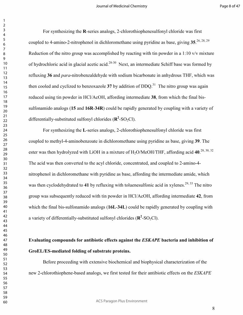
For synthesizing the R-series analogs, 2-chlorothiophenesulfonyl chloride was first
coupled to 4-amino-2-nitrophenol in dichloromethane using pyridine as base, giving 35.26, 28, 29
Reduction of the nitro group was accomplished by reacting with tin powder in a 1:10 v/v mixture
of hydrochloric acid in glacial acetic acid.28-30 Next, an intermediate Schiff base was formed by
refluxing 36 and para-nitrobenzaldehyde with sodium bicarbonate in anhydrous THF, which was
then cooled and cyclized to benzoxazole 37 by addition of DDQ.31 The nitro group was again
reduced using tin powder in HCl/AcOH, affording intermediate 38, from which the final bis-
sulfonamido analogs (15 and 16R-34R) could be rapidly generated by coupling with a variety of
differentially-substituted sulfonyl chlorides (R2-SO2Cl).
For synthesizing the L-series analogs, 2-chlorothiophenesulfonyl chloride was first
coupled to methyl-4-aminobenzoate in dichloromethane using pyridine as base, giving 39. The
ester was then hydrolyzed with LiOH in a mixture of H2O/MeOH/THF, affording acid 40.28, 30, 32
The acid was then converted to the acyl chloride, concentrated, and coupled to 2-amino-4-
nitrophenol in dichloromethane with pyridine as base, affording the intermediate amide, which
was then cyclodehydrated to 41 by refluxing with toluenesulfonic acid in xylenes.29, 33 The nitro
group was subsequently reduced with tin powder in HCl/AcOH, affording intermediate 42, from
which the final bis-sulfonamido analogs (16L-34L) could be rapidly generated by coupling with
a variety of differentially-substituted sulfonyl chlorides (R2-SO2Cl).
Evaluating compounds for antibiotic effects against the ESKAPE bacteria and inhibition of
GroEL/ES-mediated folding of substrate proteins.
Before proceeding with extensive biochemical and biophysical characterization of the
new 2-chlorothiophene-based analogs, we first tested for their antibiotic effects on the ESKAPE
Page 8 of 47
ACS Paragon Plus Environment
Journal of Medicinal Chemistry
123456789101112131415161718192021222324252627282930313233343536373839404142434445464748495051525354555657585960

9
bacteria using the proliferation assay described above (refer to Tables S3A and S4A in the
Supporting Information for tabulations of all EC50 results). Much like the initial testing with the
pseudo-symmetrical analogs, all but one of the 2-chlorothiophene analogs were inactive against
the four Gram-negative KAPE bacterial species (27R had an EC50 of 37 µM against A.
baumannii). However, 14 of the 39 new analogs (36%) exhibited antibacterial effects against E.
faecium, compared to 3 out of 50 (6%) of the pseudo-symmetrical compounds. What was
particularly impressive was that 27 out of the 39 new analogs (69%) inhibited S. aureus and
MRSA, with greater overall potency, compared to the 20 out of 50 (40%) of the pseudo-
symmetrical compounds (Figure 2A presents a correlation plot of EC50 values for each
compound determined in these two assays). To compare assay results between the R- and L-
series analogs, we analyzed the log(EC50) values of each data set using two-tailed, paired t-tests
(95% confidence level) and looked at differences between paired values (results are plotted in
Figure S1 in the Supporting Information). For the E. faecium EC50 results, we did not see a
statistical difference between results from the R- and L-series analogs (Figure S1A). However,
there was a statistically significant difference between results of the two series for inhibiting S.
aureus and MRSA proliferation, with the L-series generally providing more potent inhibitors of
bacterial proliferation for each strain (Figures S1B and S1C). Although exceptions are evident
for individual compounds, there was no statistical difference between inhibiting the drug
susceptible S. aureus and MRSA strains when the data sets were analyzed for all 89 compounds
as a whole (Figure S5A).
Having ascertained that the majority of the R- and L-series analogs were potent inhibitors
of S. aureus and MRSA proliferation, we next evaluated their abilities to inhibit the biochemical
function of the GroEL/ES chaperonin system. For this, we employed our previously reported
Page 9 of 47
ACS Paragon Plus Environment
Journal of Medicinal Chemistry
123456789101112131415161718192021222324252627282930313233343536373839404142434445464748495051525354555657585960

10
assays that monitor GroEL/ES-mediated refolding of two denatured substrate enzymes, malate
dehydrogenase (dMDH) and rhodanese (dRho).25 Since these were coupled assays that monitor
chaperonin-mediated refolding of substrates by virtue of the enzymatic activities of the refolded
substrates, we further counter-screened against the native MDH and Rho enzymes to ensure that
compounds were not simply false-positives of the reporter reactions. Detailed protocols for these
assays and tabulation of all IC50 results are presented in the Supporting Information.
Because the previously developed pseudo-symmetrical analogs (1-14) were not tested in
the GroEL/ES-dRho refolding assay, nor the native rhodanese enzymatic reporter reaction
counter screen, we also evaluated those compounds in these two assays. When looking at all 89
compounds as a whole, although a correlation is evident, we noticed a statistically significant
difference between the GroEL/ES-dMDH and GroEL/ES-dRho refolding IC50 results (Figures
2B and S5B), where compounds were slightly more potent at inhibiting GroEL/ES-mediated
refolding of denatured MDH. This difference does not appear to be a result of compounds
preferentially inhibiting the native MDH reporter enzyme over rhodanese – while eleven analogs
inhibited the native MDH enzymatic reporter reaction, five inhibited native rhodanese, and only
to a minor extent. Since only one compound inhibits both native malate dehydrogenase and
native rhodanese (28R), and only to a minor extent, this supports that compounds are on-target
for inhibiting the GroEL/ES-mediated folding cycle.
For the purposes of categorizing inhibitor potencies in the GroEL/ES-mediated refolding
assays, we consider compounds with IC50 values >100 µM to be inactive, 30-100 µM to be weak
inhibitors, 10-30 µM moderate inhibitors, 1-10 µM potent inhibitors, and <1 µM very potent and
acting near stoichiometrically since the concentration of GroEL tetradecamer is 50 nM during
the refolding cycle (i.e. 700 nM of GroEL subunits). Upon further dissection of the GroEL/ES-
Page 10 of 47
ACS Paragon Plus Environment
Journal of Medicinal Chemistry
123456789101112131415161718192021222324252627282930313233343536373839404142434445464748495051525354555657585960

11
dMDH refolding assay IC50 results, we observed that the asymmetric R- and L-series analogs are
generally more potent than the previously developed pseudo-symmetric analogs: 28 out of 38
(74%) of the asymmetric R- and L-series analogs (16-34) have IC50 values less than 10 µM,
whereas only 16 out of 51 (31%) of the pseudo-symmetric analogs (1-15) have IC50 values less
than 10 µM. These results are not entirely surprising since the R- and L-series analogs were
hypothesized to better complement the envisioned asymmetric binding sites than the pseudo-
symmetric compounds. However, we caution on over-interpreting these results since the analog
groupings did contain different alkyl and aryl substructures on the sulfonamide end-caps. When
comparing the R- and L-series analogs with each other, though, we do not see any statistically
significant differences in either the GroEL/ES-dMDH or GroEL/ES-dRho refolding assay results
(Figure S2A and S2B), suggesting that the orientation of the 2-phenylbenzoxazole core scaffold
does not play a significant factor in these compounds binding to and inhibiting the GroEL/ES
chaperonin system.
When comparing IC50 results for compounds tested in the biochemical GroEL/ES-dMDH
refolding assay with EC50 results for testing in the S. aureus proliferation assay (Figure 2C and
Tables S3A-B and S4A-B), we note that no compounds are active against bacteria unless they
are able to inhibit GroEL. In general, the more potent compounds are at inhibiting the GroEL/ES
chaperonin system, the more potent their antibacterial effects, in particular for the L-series
inhibitors. However, several exceptions are evident where potent GroEL/ES inhibitors are
poorly effective, or ineffective, against bacteria, which may owe to poor cell wall permeability
and/or efflux, as we had previously observed for other GroEL/ES inhibitor scaffolds.25 Also,
while correlations between biochemical IC50 and cell-based EC50 are only suggestive of
mechanisms of action in cells, they are not confirmatory. Thus, whether compounds are “on-
Page 11 of 47
ACS Paragon Plus Environment
Journal of Medicinal Chemistry
123456789101112131415161718192021222324252627282930313233343536373839404142434445464748495051525354555657585960

12
target” for GroEL within bacteria is still unknown and warrants further investigation. These
studies are ongoing and will be reported in the future.
Characterizing GroEL-inhibitor binding interactions using Isothermal Titration
Calorimetry.
To further interrogate inhibitor mechanisms of action against GroEL, we used isothermal
titration calorimetry (ITC) to identify the thermodynamic parameters, binding affinities, and
binding stoichiometries of compounds 20R and 20L. While these two compounds were only
moderately active at inhibiting S. aureus and MRSA proliferation, they were strong inhibitors of
GroEL/ES-mediated folding functions, and thus likely had high binding affinities. Furthermore,
because they bore primary amines that would be charged under physiological conditions, they
were much more soluble than other inhibitors and thus more amenable to ITC analysis, which
requires high concentrations of both protein and ligands in matched aqueous buffers. We
performed ITC analyses by titrating 400 µM GroEL (monomer concentration) into solutions
containing either 50 µM 20R or 20L (detailed protocols are presented in the Experimental
section). Two representative isotherms for the binding of 20R and 20L to GroEL are presented
in Figure 3. After subtraction of background heats of mixing and dilution, plots of the integrated
heats from compound binding fit well to a single-site binding model.34 Averaged results for the
various binding parameters (Kd, n, ∆H, ∆S, and ∆G) obtained from replicate analyses (six
replicates for 20R, and five replicates for 20L) are presented in Table 1.
From the averaged results, we found that 20R had a Kd of 36 nM for binding to E. coli
GroEL, while the Kd of 20L was about four-fold higher (150 nM), which corresponds reasonably
well with the relative IC50 values for inhibiting GroEL/ES-mediated refolding of dMDH and
Page 12 of 47
ACS Paragon Plus Environment
Journal of Medicinal Chemistry
123456789101112131415161718192021222324252627282930313233343536373839404142434445464748495051525354555657585960

13
dRho – the IC50 values for 20L were typically two- to four-fold higher than for 20R. For both
compounds, binding was predominantly enthalpically driven, with minor entropic contributions
to affinity. What was particularly interesting from these analyses was the stoichiometry of
binding of each compound to GroEL. Since GroEL consists of 14 identical subunits, we
anticipated that compounds could bind with a stoichiometry of 14, or potentially 7 if there is
negative cooperativity between the two GroEL rings (also assuming that for inhibitor binding
there is no negative cooperativity between subunits within a ring). Since IC50 values were
previously found to correlate between GroEL/ES-mediated refolding and ATPase assays for this
inhibitor scaffold, the most likely binding sites may be the ATP pockets, of which there are 1 per
GroEL subunit (14 per oligomer).26 However, 20R bound with a stoichiometry of roughly 18
molecules per GroEL tetradecamer, and 20L with 23 molecules, which could indicate more than
one potential binding site per GroEL subunit, and potentially an unknown site outside of the ATP
pockets. We are currently pursuing X-ray crystallographic studies to identify specific inhibitor
binding sites, which we will report on in future studies.
Counter-screening compounds for inhibition of HSP60/10-mediated refolding of denatured
MDH and for cytotoxicity in human cell viability assays.
Having established that the R- and L-series analogs were potent GroEL/ES chaperonin
system inhibitors with anti-staphylococcal properties, we next investigated their selectivity
profiles compared to the human HSP60/10 chaperonin system. For this, we evaluated
compounds in our previously reported HSP60/10-dMDH refolding assay, which is analogous to
that used for determining inhibition of bacterial GroEL/ES, but alternatively employs the human
chaperonin system. A detailed protocol and tabulation of all IC50 results for this assay are
Page 13 of 47
ACS Paragon Plus Environment
Journal of Medicinal Chemistry
123456789101112131415161718192021222324252627282930313233343536373839404142434445464748495051525354555657585960
14
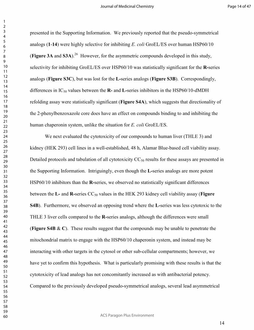
presented in the Supporting Information. We previously reported that the pseudo-symmetrical
analogs (1-14) were highly selective for inhibiting E. coli GroEL/ES over human HSP60/10
(Figure 3A and S3A).26 However, for the asymmetric compounds developed in this study,
selectivity for inhibiting GroEL/ES over HSP60/10 was statistically significant for the R-series
analogs (Figure S3C), but was lost for the L-series analogs (Figure S3B). Correspondingly,
differences in IC50 values between the R- and L-series inhibitors in the HSP60/10-dMDH
refolding assay were statistically significant (Figure S4A), which suggests that directionality of
the 2-phenylbenzoxazole core does have an effect on compounds binding to and inhibiting the
human chaperonin system, unlike the situation for E. coli GroEL/ES.
We next evaluated the cytotoxicity of our compounds to human liver (THLE 3) and
kidney (HEK 293) cell lines in a well-established, 48 h, Alamar Blue-based cell viability assay.
Detailed protocols and tabulation of all cytotoxicity CC50 results for these assays are presented in
the Supporting Information. Intriguingly, even though the L-series analogs are more potent
HSP60/10 inhibitors than the R-series, we observed no statistically significant differences
between the L- and R-series CC50 values in the HEK 293 kidney cell viability assay (Figure
S4B). Furthermore, we observed an opposing trend where the L-series was less cytotoxic to the
THLE 3 liver cells compared to the R-series analogs, although the differences were small
(Figure S4B & C). These results suggest that the compounds may be unable to penetrate the
mitochondrial matrix to engage with the HSP60/10 chaperonin system, and instead may be
interacting with other targets in the cytosol or other sub-cellular compartments; however, we
have yet to confirm this hypothesis. What is particularly promising with these results is that the
cytotoxicity of lead analogs has not concomitantly increased as with antibacterial potency.
Compared to the previously developed pseudo-symmetrical analogs, several lead asymmetrical
Page 14 of 47
ACS Paragon Plus Environment
Journal of Medicinal Chemistry
123456789101112131415161718192021222324252627282930313233343536373839404142434445464748495051525354555657585960

15
analogs have appreciably higher Selectivity Indices (SI) for inhibiting MRSA proliferation over
cytotoxicity in the liver cell viability assay (Figure 4C). The biochemical and cell-based IC50,
EC50, and CC50 results for the top four lead inhibitors of the pseudo-symmetrical, R-series, and
L-series analogs are presented in Table 2, ranked based on their selectivity indices. Notably,
compounds 24L and 25L have selectivity indices of 31 and 46, respectively.
Investigating the potential for MRSA to gain resistance to lead inhibitors.
While selectivity indices (and therapeutic windows with regards to in vivo evaluation) are
important criteria to consider for antibacterial development, it is also important to determine
whether or not bacteria will be able to rapidly develop resistance to lead candidates, especially if
they function through new mechanisms of action. To gauge the efficacy of our GroEL/ES
inhibitors for eluding acute antibacterial resistance mechanisms, we performed a step-wise,
liquid culture resistance assay by consecutively passaging the MRSA strain (ATCC BAA-44) for
12 cycles over 12 consecutive days in the presence of serially-diluted inhibitors 20R, 28R, and
vancomycin for comparison (a detailed protocol is presented in the Experimental section).35, 36
At the end of each cycle (i.e. each 24 h passage), EC50 values were determined for the test
compounds, with the assumption that EC50 values would increase over time if MRSA was able to
generate acute resistance. A plot of test compound EC50 values over time is presented in Figure
5A. Despite the fact that 28R was a potent MRSA growth inhibitor (initial EC50 value of 1.7
µM), the bacteria rapidly developed resistance to compound concentrations in excess of 100 µM.
However, after culturing in the absence of inhibitors for two days (24 h on agar, and another 24 h
in liquid media), bacteria regained sensitivity to 28R (Figure 5B), suggesting resistance is
reversible, potentially from up-regulation of efflux pumps. Most importantly, we found that the
Page 15 of 47
ACS Paragon Plus Environment
Journal of Medicinal Chemistry
123456789101112131415161718192021222324252627282930313233343536373839404142434445464748495051525354555657585960

16
parent MRSA strain was unable to generate resistance against 20R over the 12 day passage, even
though this analog was only a moderate inhibitor of MRSA proliferation (EC50 = 19 µΜ). These
results suggest that pan-resistance to these GroEL inhibitor analogs may be difficult to develop,
which supports the continued optimization of lead antibacterial candidates based on this
molecular scaffold.
CONCLUSIONS.
From exploratory screening of the antibacterial effects of our previously reported pseudo-
symmetrical compound 1 analogs,26 we were able to develop two parallel R- and L-series of 2-
chlorothiophene-based asymmetrical analogs with significantly improved antibacterial efficacy
profiles against MRSA. While there were no statistical differences between either the R- or L-
series analogs for inhibiting in the GroEL/ES-mediated refolding assays, the L-series analogs
were found to be more potent at inhibiting S. aureus and MRSA proliferation – i.e. preferred
from an antibiotic development perspective (Table 3). While a general trend is observed
between inhibitor potencies in the biochemical GroEL/ES-dMDH refolding assay and the MRSA
proliferation assay (in particular for the L-series analogs), further studies are needed to
conclusively determine whether or not inhibitors are on-target in bacteria. Using ITC to
characterize the binding interactions between GroEL and inhibitors 20R and 20L, we found that
inhibitor binding was predominantly enthalpically driven. Furthermore, we observed that >14
molecules were bound per GroEL tetradecamer (i.e. more than one molecule bound per GroEL
subunit), suggesting that inhibitors could be binding to unknown sites outside of the ATP
pockets. Current studies are underway to identify and characterize these putative binding sites so
that more rigorous structure-based optimization of lead candidates can be conducted. Even
Page 16 of 47
ACS Paragon Plus Environment
Journal of Medicinal Chemistry
123456789101112131415161718192021222324252627282930313233343536373839404142434445464748495051525354555657585960

17
though the R-series analogs were overall less potent at inhibiting HSP60/10, the L-series was
less cytotoxic against the human liver cells tested, and is likely preferential for further
antibacterial optimization going forward. However, before pursuing in vivo development of this
series, further medicinal chemistry optimization is warranted to increase the selectivity indices
for killing MRSA over cytotoxicity to human cells.
EXPERIMENTAL.
General Synthetic Methods.
Unless otherwise stated, all chemicals were purchased from commercial suppliers and
used without further purification. Reaction progress was monitored by thin-layer
chromatography on silica gel 60 F254 coated glass plates (EM Sciences). Flash chromatography
was performed using a Biotage Isolera One flash chromatography system and eluting through
Biotage KP-Sil Zip or Snap silica gel columns for normal-phase separations (hexanes:EtOAc
gradients), or Snap KP-C18-HS columns for reverse-phase separations (H2O:MeOH gradients).
Reverse-phase high-performance liquid chromatography (RP-HPLC) was performed using a
Waters 1525 binary pump, 2489 tunable UV/Vis detector (254 and 280 nm detection), and 2707
autosampler. For preparatory HPLC purification, samples were chromatographically separated
using a Waters XSelect CSH C18 OBD prep column (part number 186005422, 130 Å pore size,
5 µm particle size, 19x150 mm), eluting with a H2O:CH3CN gradient solvent system. Linear
gradients were run from either 100:0, 80:20, or 60:40 A:B to 0:100 A:B (A = 95:5 H2O:CH3CN,
0.05% TFA; B = 5:95 H2O:CH3CN, 0.05% TFA. Products from normal-phase separations were
concentrated directly, and reverse-phase separations were concentrated, diluted with H2O,
frozen, and lyophilized. For primary compound purity analyses (HPLC-1), samples were
Page 17 of 47
ACS Paragon Plus Environment
Journal of Medicinal Chemistry
123456789101112131415161718192021222324252627282930313233343536373839404142434445464748495051525354555657585960

18
chromatographically separated using a Waters XSelect CSH C18 column (part number
186005282, 130 Å pore size, 5 µm particle size, 3.0x150 mm), eluting with the above
H2O:CH3CN gradient solvent systems. For secondary purity analyses (HPLC-2) of final test
compounds, samples were chromatographically separated using a Waters XBridge C18 column
(either part number 186003027, 130 Å pore size, 3.5 µm particle size, 3.0x100 mm, or part
number 186003132, 130 Å pore size, 5.0 µm particle size, 3.0x100 mm), eluting with a
H2O:MeOH gradient solvent system. Linear gradients were run from either 100:0, 80:20, 60:40,
or 20:80 A:B to 0:100 A:B (A = 95:5 H2O:MeOH, 0.05% TFA; B = 5:95 H2O:MeOH, 0.05%
TFA). Test compounds were found to be >95% in purity from both RP-HPLC analyses. Mass
spectrometry data were collected using either an Agilent analytical LC-MS at the IU Chemical
Genomics Core Facility (CGCF), or a Thermo-Finnigan LTQ LC-MS in-lab. 1H-NMR spectra
were recorded on either Bruker 300 MHz or 500 MHz spectrometers. Chemical shifts are
reported in parts per million and calibrated to the d6-DMSO solvent peaks at 2.50 ppm. We
previously synthesized compounds 1-14 (including 2a-m-o/m/p)26 and re-synthesized where
necessary due to stock depletion. Synthesis and characterization of intermediates 35-42 are
presented below. General sulfonamide coupling steps are presented for analogs 15 and 16R-34R
and 16L-34L below, with compound characterizations for each analog presented in the
Supporting Information.
35: 5-Chloro-N-(4-hydroxy-3-nitrophenyl) thiophene-2-sulfonamide.
To a stirring mixture of 4-amino-2-nitrophenol (5.34 g, 24.6 mmol) in anhydrous CH2Cl2
(50 mL) was added 5-chlorothiophene-2-sulfonyl chloride (4.21 g, 27.3 mmol) and pyridine
(2.40 mL, 29.4 mmol). The reaction was allowed to stir at room temperature for 18 h and was
Page 18 of 47
ACS Paragon Plus Environment
Journal of Medicinal Chemistry
123456789101112131415161718192021222324252627282930313233343536373839404142434445464748495051525354555657585960
19
then diluted with hexanes and the precipitate was filtered, rinsed with 1 M HCl and water,
collected, and dried to afford 35 as a reddish-brown solid (7.70 g, 94% yield). 1H-NMR (300
MHz, d6-DMSO) δ 10.98 (br s, 1H), 10.62 (br s, 1H), 7.60 (d, J = 2.7 Hz, 1H), 7.39 (d, J = 4.1
Hz, 1H), 7.30 (dd, J = 8.9, 2.7 Hz, 1H), 7.22 (d, J = 4.1 Hz, 1H), 7.10 (d, J = 8.9 Hz, 1H); MS
(ESI) C10H6ClN2O5S2 [M-H]- m/z expected = 332.9, observed = 332.8; HPLC-1 = >99% (RT =
8.1 min).
36: N-(3-Amino-4-hydroxyphenyl)-5-chlorothiophene-2-sulfonamide.
Tin powder (8.73 g, 73.6 mmol) was added slowly to a stirring mixture of 35 (8.11 g,
24.2 mmol) in a 1:10 mixture of HCl:AcOH (24 mL). The reaction was allowed to stir at R.T.
for 18 h, then diluted with EtOAc and H2O, neutralized with NaHCO3, and filtered. The filtrate
was extracted with EtOAc and the organics dried over Na2SO4, filtered, and concentrated. The
residue was diluted in a 50% mixture of DCM in hexanes and the precipitate was filtered, rinsed
with hexanes, collected, and dried to afford 36 as a brown powder (6.08 g, 82% yield). 1H-NMR
(300 MHz, d6-DMSO) δ 9.81 (br s, 1H), 8.99 (br s, 1H) 7.28 (d, J = 4.1 Hz, 1H), 7.18 (d, J = 4.1
Hz, 1H), 6.49 (d, J = 8.3 Hz, 1H), 6.41 (d, J = 2.5 Hz, 1H), 6.13 (dd, J = 8.3, 2.6 Hz, 1H), 4.64
(br s, 2H); MS (ESI) C10H10ClN2O3S2 [M+H]+ m/z expected = 305.0, observed = 304.9; HPLC-1
= 97% (RT = 6.0 min).
37: 5-Chloro-N-(2-(4-nitrophenyl)benzo[d]oxazol-5-yl)thiophene-2-sulfonamide.
Compound 36 (2.94 g, 9.65 mmol), 4-nitrobenzaldehyde (2.03 g, 13.4 mmol), NaHCO3
(2.08 g, 24.8 mmol), and Na2SO4 (3.35 g) were stirred in THF (40 mL) for 4 h at reflux (under
Ar), then cooled to R.T. DDQ (2.85 g, 12.6 mmol) was then add portion-wise and the reaction
Page 19 of 47
ACS Paragon Plus Environment
Journal of Medicinal Chemistry
123456789101112131415161718192021222324252627282930313233343536373839404142434445464748495051525354555657585960

20
was left to stir for 2 h, then filtered. The filtrate was extracted into EtOAc, rinsed with saturated
NaHCO3, 1 M HCl, and brine. The organics layer was dried over Na2SO4, filtered, and
concentrated. The residue was diluted in a 25% mixture of DCM in hexanes and the precipitate
was filtered, rinsed with hexanes, collected, and dried to afford 37 as a brown powder (4.01 g,
95% yield). 1H-NMR (300 MHz, d6-DMSO) δ 10.74 (s, 1H), 8.37-8.48 (m, 4H), 7.82 (d, J = 8.8
Hz, 1H), 7.60 (d, J = 1.9 Hz, 1H), 7.43 (d, J = 4.0 Hz, 1H), 7.25 (dd, J = 8.8, 2.1 Hz, 1H), 7.19
(d, J = 4.1 Hz, 1H); MS (ESI) C17H9ClN3O5S2 [M-H]- m/z expected = 434.0, observed = 433.8;
HPLC-1 = 91% (RT = 7.7 min).
38: N-(2-(4-Aminophenyl)benzo[d]oxazol-5-yl)-5-chlorothiophene-2-sulfonamide.
Tin powder (2.74 g, mmol) was added slowly to a stirring mixture of 37 (2.43 g, 5.58
mmol) in a 1:10 mixture of HCl:AcOH (15 mL). The reaction was allowed to stir at R.T. for 18
h, then diluted with EtOAc and H2O, neutralized with NaHCO3, and filtered. The filtrate was
extracted with EtOAc and the organics dried over Na2SO4, filtered, and concentrated. The
residue was chromatographed over silica (hexanes:EtOAc gradient) and concentrated to afford
38 as an orange solid (936 mg, 41% yield). 1H-NMR (300 MHz, d6-DMSO) δ 10.54 (s, 1H),
7.78-7.86 (m, 2H), 7.60 (d, J = 8.7 Hz, 1H), 7.35-7.40 (m, 2H), 7.18 (d, J = 4.1 Hz, 1H), 7.04
(dd, J = 8.6, 2.2 Hz, 1H), 6.64-6.71 (m, 2H), 6.04 (br s, 2H); MS (ESI) C17H13ClN3O3S2 [M+H]+
m/z expected = 406.0, observed = 405.9; HPLC-1 = 97% (RT = 7.6 min).
General sulfonamidation procedure for the synthesis of 16R-34R.
To a stirring mixture of compound 38 (1 eq.) in anhydrous CH2Cl2 (5 mL) was added the
respective R2 sulfonyl chloride (1.2 eq.) followed by anhydrous pyridine (1.2 eq.). The reaction
Page 20 of 47
ACS Paragon Plus Environment
Journal of Medicinal Chemistry
123456789101112131415161718192021222324252627282930313233343536373839404142434445464748495051525354555657585960
21
was allowed to stir at room temperature for 18 h and was then chromatographed over silica
(hexanes:EtOAc gradient) and concentrated. If necessary, the product was further purified by
preparatory RP-HPLC (H2O:CH3CN gradient), concentrated, and lyophilized. Refer to the
Supporting Information for individual compound characterization data.
39: Methyl 4-((5-chlorothiophene)-2-sulfonamido)benzoate.
To a stirring mixture of methyl-4-aminobenzoate (2.76 g, 18.3 mmol) in anhydrous
CH2Cl2 (50 mL) was added 5-chlorothiophene-2-sulfonyl chloride (4.78 g, 22.0 mmol) and
pyridine (1.80 mL, 22.1 mmol). The reaction was allowed to stir at room temperature for 18 h
and was then diluted with hexanes and acidified with 1 M HCl. The precipitate was then filtered,
rinsed with 1 M HCl and water, collected, and dried to afford 39 as a pinkish-orange solid (5.94
g, 98% yield). 1H-NMR (300 MHz, d6-DMSO) δ 11.16 (br s, 1H), 7.86-7.93 (m, 2H), 7.56 (d, J
= 4.1 Hz, 1H), 7.25-7.31 (m, 2H), 7.22 (d, J = 4.1 Hz, 1H), 3.80 (s, 3H); HPLC-1 = 99% (RT =
6.2 min).
40: 4-((5-Chlorothiophene)-2-sulfonamido)benzoic acid.
LiOH•H2O (7.02 g, 167 mmol) was added to a stirring mixture of 39 (5.67 g, 17.1 mmol)
in THF (20 mL), MeOH (20 mL), and H2O (20mL). The reaction was allowed to stir at room
temperature for 2 days and was then diluted with 1M HCl. The precipitate was filtered, washed
with H2O, collected, and dried to afford 40 as an off-white solid (5.20 g, 96% yield). 1H-NMR
(300 MHz, d6-DMSO) δ 12.85 (br s, 1H), 11.09 (br s, 1H), 7.83-7.90 (m, 2H), 7.54 (d, J = 4.1
Hz, 1H), 7.19-7.28 (m, 3H); HPLC-1 = >99% (RT = 6.7 min).
Page 21 of 47
ACS Paragon Plus Environment
Journal of Medicinal Chemistry
123456789101112131415161718192021222324252627282930313233343536373839404142434445464748495051525354555657585960

22
41: 5-Chloro-N-(4-(5-nitrobenzo[d]oxazol-2-yl)phenyl)thiophene-2-sulfonamide.
Compound 40 (2.96 g, 9.31 mmol) was stirred in SOCl2 (10 mL) for 6 h at 60°C, then
was concentrated to a solid. This was refluxed with 2-amino-4-nitrophenol (2.96 g, 19.2 mmol)
and pyridine (1.50 mL, 18.4 mmol) in anhydrous CH2Cl2 (50 mL) for 6 h, then stirred at R.T. for
3 days. The reaction was then extracted into EtOAc and rinsed with 1 M HCl and brine, dried
over Na2SO4, filtered, and concentrated. This amide intermediate was then refluxed with
TsOH•H2O (3.61 g, 19.0 mmol) in xylenes (50 mL) using a Dean-Stark apparatus to remove the
residual H2O. After 18 h, the reaction was cooled, the xylenes decanted off, the sludge extracted
with EtOAc, and the combined organics dried over Na2SO4, filtered, and concentrated. Flash
chromatographic purification over silica (hexanes:EtOAc gradient) afforded 41 as a peach solid
(1.81 g, 45% yield). 1H-NMR (300 MHz, d6-DMSO) δ 11.25 (br s, 1H), 8.64 (br s, 1H), 8.32 (d,
J = 8.8 Hz, 1H), 8.18 (d, J = 8.1 Hz, 2H), 8.01 (d, J = 8.7 Hz, 1H), 7.60 (br s, 1H), 7.41 (d, J =
8.2 Hz, 2H), 7.24 (br s, 1H); MS (ESI) C17H11ClN3O5S2 [M+H]+ m/z expected = 436.0, observed
= 436.1; HPLC-1 = 88% (RT = 5.5 min).
42: N-(4-(5-Aminobenzo[d]oxazol-2-yl)phenyl)-5-chlorothiophene-2-sulfonamide.
Tin powder (1.53 g, 12.9 mmol) was added slowly to a stirring mixture of 41 (1.78 g,
4.08 mmol) in a 1:7 mixture of HCl:AcOH (8 mL). The reaction was stirred and heated at 60°C
for 3 h, then cooled to R.T., diluted with EtOAc and H2O, neutralized with NaHCO3, and
filtered. The filtrate was extracted with EtOAc and the organics dried over Na2SO4, filtered, and
concentrated. The residue was diluted in a 10% mixture of DCM in hexanes and the precipitate
was filtered, rinsed with hexanes, collected, and dried to afford 42 as a pale-yellow powder (1.59
g, 96% yield). 1H-NMR (300 MHz, d6-DMSO) δ 8.01-8.09 (m, 2H), 7.55 (d, J = 4.1 Hz, 1H),
Page 22 of 47
ACS Paragon Plus Environment
Journal of Medicinal Chemistry
123456789101112131415161718192021222324252627282930313233343536373839404142434445464748495051525354555657585960

23
7.30-7.41 (m, 3H), 7.22 (d, J = 4.1 Hz, 1H), 6.83 (d, J = 2.0 Hz, 1H), 6.64 (dd, J = 8.7, 2.2 Hz,
1H); MS (ESI) C17H13ClN3O3S2 [M+H]+ m/z expected = 406.0, observed = 406.1; HPLC-1 =
86% (RT = 6.8 min).
General sulfonamidation procedure for the synthesis of 16L-34L.
To a stirring mixture of compound 42 (1 eq.) in anhydrous CH2Cl2 (5 mL) was added the
respective R2 sulfonyl chloride (1.2 eq.) followed by anhydrous pyridine (1.2 eq.). The reaction
was allowed to stir at room temperature for 18 h and was then chromatographed over silica
(hexanes:EtOAc gradient) and concentrated. If necessary, the product was further purified by
preparatory RP-HPLC (H2O:CH3CN gradient), concentrated, and lyophilized. Refer to the
Supporting Information for individual compound synthesis and characterization data.
Cell information for compound evaluation.
The ESKAPE bacteria were purchased from the American Type Culture Collection
(ATCC): E. faecium (Orla-Jensen) Schleifer and Kilpper-Balz strain NCTC 7171 (ATCC
19434); S. aureus subsp. aureus Rosenbach strain Seattle 1945 (ATCC 25923); Multi-drug
resistant S. aureus (MRSA) subsp. aureus Rosenbach strain HPV107 (ATCC BAA-44); K.
pneumonia, subsp. pneumoniae (Schroeter) Trevisan strain NCTC 9633 (ATCC 13883); A.
baumannii Bouvet and Grimont strain 2208 (ATCC 19606); P. aeruginosa (Schroeter) Migula
strain NCTC 10332 (ATCC 10145); E. cloacae, subsp. cloacae (Jordan) Hormaeche and
Edwards strain CDC 442-68 (ATCC 13047). HEK 293 kidney cells (ATCC CRL-1573) and
THLE-3 liver cells (ATCC CRL-11233) were used for compound toxicity assays.
Page 23 of 47
ACS Paragon Plus Environment
Journal of Medicinal Chemistry
123456789101112131415161718192021222324252627282930313233343536373839404142434445464748495051525354555657585960
24
Evaluation of compounds for inhibition of bacterial cell proliferation.
All compounds were evaluated for inhibiting the proliferation of each of the ESKAPE
bacteria as per previously reported procedures with one minor modification: Liquid growth
media was cation-adjusted by addition of 25 mg/L Ca2+ and 12.5 mg/L Mg2+ to reflect the free
concentrations of these divalent cations in vivo.27 Detailed protocols for bacterial growth assays
are presented in the Supporting Information.
Protein Expression and purification.
E. coli GroEL and GroES, and human HSP60 and HSP10, were expressed and purified as
previously reported.25 Detailed protocols for these protein purifications are presented in the
Supporting Information.
Evaluation of compounds in GroEL/ES and HSP60/10-mediated dMDH and dRho
refolding assays.
All compounds were evaluated for inhibiting E. coli GroEL/ES and human HSP60/10-
mediated refolding of the denatured MDH and denatured Rho reporter enzymes as per previously
reported procedures.26 Detailed protocols for these assays are presented in the Supporting
Information.
Counter-screening compounds for inhibition of native MDH and Rho enzymatic activity.
All compounds were counter-screened for inhibiting the enzymatic activity of the native
MDH and native Rho reporter enzymes as per previously reported procedures.25, 26 Detailed
protocols for the assays are presented in the Supporting Information.
Page 24 of 47
ACS Paragon Plus Environment
Journal of Medicinal Chemistry
123456789101112131415161718192021222324252627282930313233343536373839404142434445464748495051525354555657585960

25
Evaluation of inhibitors 20R and 20L binding to GroEL using Isothermal Titration
Calorimetry (ITC).
All ITC experiments were performed on a MicroCal VP-ITC system. To minimize
background heats of dilution, matched solutions of GroEL and compounds 20R or 20L were
prepared in buffer containing 20 mM HEPES, pH 7.4, 50 mM KCl, 10 mM MgCl2, and 1%
DMSO. All buffers were freshly prepared from stock solutions on the day of use and degassed
under vacuum. For each analytical run, a solution of 400 µM GroEL (monomer concentration)
in the syringe was titrated into 50 µM solutions of either compound 20R or 20L in the sample
cell (equilibrated at 20°C for 300 seconds): the first injection was 0.4 µL (discarded during
analysis) and subsequent injections (2-20) were each 2.0 µL. Each injection was made over 4 s
durations with 3-min intervals between subsequent injections. The reference power of the
experiment was set to 6 µcal/s and a 5 s filter period. Sample stirring was set at 600 rpm for all
measurements. Six independent replicates were conducted to analyze compound 20R binding,
and five replicates for 20L. Raw thermograms from each replicate were analyzed independently
using the ORIGIN instrument software (MicroCal Inc., version 7.0). In each experiment, the
upper baseline was collected after the binding reaction was saturated, and the terminal 7-8 points
in the linear region were fit to a straight line and subtracted from the entire data set to remove
contributions from background heats of mixing and dilution. The ∆H was obtained by non-linear
least-squares fitting of the plot of ∆H (mol of injectant)-1 versus molar ratio using a single site
binding model.34 The ∆H, ∆S, Kd (KA= 1/ Kd), and binding stoichiometries (n) in each titration
were obtained, and ∆G was calculated using the Gibbs equation at a temperature of 20°C (293.15
K). Standard deviations were determined based on results from each of the replicate analyses.
Page 25 of 47
ACS Paragon Plus Environment
Journal of Medicinal Chemistry
123456789101112131415161718192021222324252627282930313233343536373839404142434445464748495051525354555657585960
26
Evaluation of compound effects on HEK 293 and THLE-3 cell viability.
Evaluation of compound cytotoxicities to HEK 293 kidney and THLE-3 liver cells were
performed using Alamar Blue-based viability assays. HEK 293 cells were maintained in MEM
medium (Corning Cellgro, 10-009 CV) supplemented with 10% FBS (Sigma, F2242). THLE-3
cells were maintained in Clonetics BEBM medium (Lonza, CC-3171) supplemented with the
BEGM bullet kit (Lonza, CC-3170) and 10% FBS. All assays were carried out in 384-well
plates (BRAND cell culture grade plates, 781980). Cells at 80% confluence were harvested and
diluted in growth medium, then 45 µL of the HEK 293 cells (1,500 cells/well) or THLE-3 cells
(1,500 cells/well) were dispensed per well, and plates were sealed with "Breathe Easy" oxygen
permeable membranes (Diversified Biotech) and incubated at 37°C, 5% CO2, for 24 h. The
following day, 1 µL of the compound stocks (10 mM to 4.6 µM, 3-fold dilutions in DMSO) were
pre-diluted by pin-transfer into 25 µL of the relevant growth mediums. Then, 15 µL aliquots of
the diluted compounds were added to the cell assay plates to give inhibitor concentration ranges
of 100 µM to 46 nM during the assay (final DMSO concentration of 0.1% was maintained during
the assay). Plates were sealed with "Breathe Easy" oxygen permeable membranes and incubated
for an additional 48 h at 37°C and 5% CO2. The Alamar Blue reporter reagents were then added
to a final concentration of 10%, the plates incubated at 37°C and 5% CO2, and sample
fluorescence (535 nm excitation, 590 nm emission) was read using a Molecular Devices
FlexStation II 384-well plate reader (readings taken between 4-24 h of incubation so as to
achieve signals in the 30-60% range for conversion of resazurin to resorufin). Cell viability was
calculated as per vendor instructions (Thermo Fisher - Alamar Blue cell viability assay manual).
Cytotoxicity CC50 values for the test compounds were obtained by plotting the % resazurin
Page 26 of 47
ACS Paragon Plus Environment
Journal of Medicinal Chemistry
123456789101112131415161718192021222324252627282930313233343536373839404142434445464748495051525354555657585960
27
reduction results in GraphPad Prism 6 and analyzing by non-linear regression using the
log(inhibitor) vs. response (variable slope) equation. Results presented represent the averages of
CC50 values obtained from at least triplicate experiments.
Evaluation of MRSA resistance generation against lead inhibitors.
To identify potential resistance toward inhibitors, a liquid culture, 12-day serial passage
assay was employed as per procedures reported by Kim. S et al., using the MRSA strain (ATCC
BAA-44).35, 36 Briefly, MRSA bacteria were streaked onto a Tryptic Soy agar plate and grown
overnight at 37°C. A fresh aliquot of Tryptic Soy Broth (TSB) was inoculated with a single
bacterial colony and the cultures were grown overnight at 37°C with shaking (250 rpm). The
overnight culture was then sub-cultured (1:5 dilution) into a fresh aliquot of media and grown at
37°C for 1 h with shaking, then diluted into fresh media to achieve a final OD600 reading of 0.01.
Aliquots of the diluted culture (200 µL) were dispensed to 96 well plates along with addition of 2
µL of test compounds in DMSO (20R, 28R, and vancomycin as a control). The inhibitor
concentration range during the resistance assay was 100 µM to 48.8 nM (2-fold dilution series).
Plates were sealed with "Breathe Easy" oxygen permeable membranes (Diversified Biotech) and
left to incubate at 37°C without shaking (stagnant assay). OD600 readings were taken at the 24 h
time point to monitor for bacterial growth. A second set of baseline control plates were prepared
analogously, without any bacteria added, to correct for possible compound absorbance and/or
precipitation, as well as plate and media baseline effects. For inoculations on subsequent days,
bacteria from the wells with the highest drug concentration where the OD600 was >0.2 were
diluted with fresh media to OD600 of 0.01 and dispensed into a new 96-well plate. Test
compounds were added, and the bacteria propagated again as described above. This procedure
Page 27 of 47
ACS Paragon Plus Environment
Journal of Medicinal Chemistry
123456789101112131415161718192021222324252627282930313233343536373839404142434445464748495051525354555657585960

28
was repeated each day for a total of 12 days to observe changes in EC50 values over each
passage. EC50 values for the test compounds were obtained by plotting the OD600 results from
each passage in GraphPad Prism 6 and analyzing by non-linear regression using the
log(inhibitor) vs. response (variable slope) equation. Results presented represent the averages of
EC50 values obtained from at triplicate experiments.
Calculation of IC50 / EC50 / CC50 values and statistical considerations.
All IC50 / EC50 / CC50 results reported are averages of values determined from individual
dose-response curves in replicate assays as follows: 1) Individual I/E/CC50 values from replicate
assays were first log-transformed and the average log(I/E/CC50) values and standard deviations
(SD) calculated; 2) Replicate log(I/E/CC50) values were evaluated for outliers using the ROUT
method in GraphPad Prism 6 (Q of 10%); and 3) Average I/E/CC50 values were then back-
calculated from the average log(IC50) values. To compare statistical differences between
log(I/E/CC50) values between the matched R- and L-series of analogs, two-tailed, paired t-tests
were performed using GraphPad Prism 6 (95% confidence level) and looking at differences
between paired values (results are plotted in Figures S1-S5 in the Supporting Information). For
compounds where log(I/E/CC50) values were greater than the maximum compound
concentrations tested (i.e. >1.8 and >2.0, or >63 and >100 µM, respectively), results were
represented as 0.1 log units higher than the maximum concentrations tested (i.e. 1.9 and 2.1, or
79 and 126 µM, respectively), so as not to overly bias comparisons because of the unavailability
of definitive values for these inactive compounds.
Corresponding Author
Page 28 of 47
ACS Paragon Plus Environment
Journal of Medicinal Chemistry
123456789101112131415161718192021222324252627282930313233343536373839404142434445464748495051525354555657585960

29
* E-mail: [email protected], Phone: 317-274-2458, Fax: 317-274-4686.
ACKNOWLEDGMENTS.
Research reported in this publication was supported by the National Institute of General Medical
Sciences (NIGMS) of the National Institutes of Health (NIH) under Award Number
R01GM120350. QQH and YP additionally acknowledge support by NIH grants
5R01GM111639 and 5R01GM115844. The content is solely the responsibility of the authors
and does not necessarily represent the official views of the NIH. This work was also supported,
in part, by the Howard Hughes Medical Institute (AH), an IU Biomedical Research Grant (SMJ),
and startup funds from the IU School of Medicine (SMJ) and the University of Arizona (EC).
The human HSP60 expression plasmid (lacking the 26 amino acid N-terminal mitochondrial
signal peptide) was generously donated by Dr. Abdussalam Azem from Tel Aviv University,
Faculty of Life Sciences, Department of Biochemistry, Israel.
ABBREVIATIONS.
MDH, malate dehydrogenase; Rho, rhodanese; HPLC, high-performance liquid chromatography;
1H-NMR, Proton nuclear magnetic resonance; MS, Mass spectrometry; EtOH, ethanol; AcOH,
acetic acid; EtOAc, ethyl acetate; MeOH, methanol; DMSO, dimethyl sulfoxide; IC50 -
Inhibitory concentration for half-maximal signal in biochemical assay; EC50, effective
concentration for half-maximal signal in bacterial proliferation assays; CC50, cytotoxicity
concentration for half-maximal signal in human cell viability assays; ITC, isothermal titration
calorimetry.
Page 29 of 47
ACS Paragon Plus Environment
Journal of Medicinal Chemistry
123456789101112131415161718192021222324252627282930313233343536373839404142434445464748495051525354555657585960

30
Supporting Information.
Supporting information associated with this article can be found in the online version,
which includes tabulation of all biochemical IC50, bacterial proliferation EC50, and human cell
viability CC50 results; log(IC50), log(EC50), and log(CC50) results with standard deviations; plots
of statistical differences between the R- and L-series analogs in the various assays; synthetic
protocols and characterization data for test compounds (HPLC purity, MS, and 1H-NMR);
experimental protocols for protein synthesis and purification, and biochemical, bacterial
proliferation, and human cell viability assays; and SMILES strings of compound structures.
Page 30 of 47
ACS Paragon Plus Environment
Journal of Medicinal Chemistry
123456789101112131415161718192021222324252627282930313233343536373839404142434445464748495051525354555657585960
31
REFERENCES
1. Tacconelli, E.; Magrini, N. Global priority list of antibiotic-resistant bacteria to guide
research, discovery, and development of new antibiotics. World Health Organization:
Geneva, Switzerland, 2017; p 7.
2. Centers for Disease Control and Prevention. Antibiotic Resistance Threats in the United
States, 2013. Centers for Disease Control and Prevention: Atlanta, Georgia, USA, 2013; p
114.
3. Karlowsky, J. A.; Hoban, D. J.; Hackel, M. A.; Lob, S. H.; Sahm, D. F. Antimicrobial
susceptibility of Gram-negative ESKAPE pathogens isolated from hospitalized patients with
intra-abdominal and urinary tract infections in Asia-Pacific countries: SMART 2013-2015. J.
Med. Microbiol. 2017, 66, 61-69.
4. Santajit, S.; Indrawattana, N. Mechanisms of antimicrobial resistance in ESKAPE pathogens.
Biomed. Res. Int. 2016, 2016, 2475067.
5. Rice, L. B. Federal funding for the study of antimicrobial resistance in nosocomial
pathogens: No ESKAPE. J. Infect. Dis. 2008, 197, 1079-1081.
6. Choo, E. J.; Chambers, H. F. Treatment of methicillin-resistant Staphylococcus aureus
bacteremia. Infect. Chemother. 2016, 48, 267-273.
7. Hassoun, A.; Linden, P. K.; Friedman, B. Incidence, prevalence, and management of MRSA
bacteremia across patient populations-a review of recent developments in MRSA
management and treatment. Crit. Care 2017, 21, 211.
8. Boucher, H.; Miller, L. G.; Razonable, R. R. Serious infections caused by methicillin-
resistant Staphylococcus aureus. Clin. Infect. Dis. 2010, 51 Suppl 2, S183-197.
Page 31 of 47
ACS Paragon Plus Environment
Journal of Medicinal Chemistry
123456789101112131415161718192021222324252627282930313233343536373839404142434445464748495051525354555657585960

32
9. Wong, P.; Houry, W. A. Chaperone networks in bacteria: analysis of protein homeostasis in
minimal cells. J. Struct. Biol. 2004, 146, 79-89.
10. Mogk, A.; Huber, D.; Bukau, B. Integrating protein homeostasis strategies in prokaryotes.
Cold Spring Harb. Perspect. Biol. 2011, 3.
11. Chiappori, F.; Fumian, M.; Milanesi, L.; Merelli, I. DnaK as antibiotic target: Hot spot
residues analysis for differential inhibition of the bacterial protein in comparison with the
human HSP70. PLoS One 2015, 10, e0124563.
12. Arita-Morioka, K.; Yamanaka, K.; Mizunoe, Y.; Ogura, T.; Sugimoto, S. Novel strategy for
biofilm inhibition by using small molecules targeting molecular chaperone DnaK.
Antimicrob. Agents Chemother. 2015, 59, 633-641.
13. Sass, P.; Josten, M.; Famulla, K.; Schiffer, G.; Sahl, H. G.; Hamoen, L.; Brotz-Oesterhelt, H.
Antibiotic acyldepsipeptides activate ClpP peptidase to degrade the cell division protein
FtsZ. Proc. Natl. Acad. Sci. U.S.A. 2011, 108, 17474-17479.
14. Evans, C. G.; Chang, L.; Gestwicki, J. E. Heat shock protein 70 (hsp70) as an emerging drug
target. J. Med. Chem. 2010, 53, 4585-4602.
15. Braig, K.; Otwinowski, Z.; Hegde, R.; Boisvert, D. C.; Joachimiak, A.; Horwich, A. L.;
Sigler, P. B. The crystal structure of the bacterial chaperonin GroEL at 2.8 A. Nature 1994,
371, 578-586.
16. Sigler, P. B.; Xu, Z.; Rye, H. S.; Burston, S. G.; Fenton, W. A.; Horwich, A. L. Structure and
function in GroEL-mediated protein folding. Annu. Rev. Biochem. 1998, 67, 581-608.
17. Horwich, A. L.; Farr, G. W.; Fenton, W. A. GroEL-GroES-mediated protein folding. Chem.
Rev. 2006, 106, 1917-1930.
Page 32 of 47
ACS Paragon Plus Environment
Journal of Medicinal Chemistry
123456789101112131415161718192021222324252627282930313233343536373839404142434445464748495051525354555657585960
33
18. Fenton, W. A.; Kashi, Y.; Furtak, K.; Horwich, A. L. Residues in chaperonin GroEL required
for polypeptide binding and release. Nature 1994, 371, 614-619.
19. Fenton, W. A.; Horwich, A. L. GroEL-mediated protein folding. Protein Sci. 1997, 6, 743-
760.
20. Horwich, A. L.; Fenton, W. A.; Chapman, E.; Farr, G. W. Two families of chaperonin:
physiology and mechanism. Annu. Rev. Cell. Dev. Biol. 2007, 23, 115-145.
21. Saibil, H. R.; Fenton, W. A.; Clare, D. K.; Horwich, A. L. Structure and allostery of the
chaperonin GroEL. J. Mol. Biol. 2013, 425, 1476-1487.
22. Chapman, E.; Farr, G. W.; Usaite, R.; Furtak, K.; Fenton, W. A.; Chaudhuri, T. K.; Hondorp,
E. R.; Matthews, R. G.; Wolf, S. G.; Yates, J. R.; Pypaert, M.; Horwich, A. L. Global
aggregation of newly translated proteins in an Escherichia coli strain deficient of the
chaperonin GroEL. Proc. Natl. Acad. Sci. U.S.A. 2006, 103, 15800-15805.
23. Fayet, O.; Ziegelhoffer, T.; Georgopoulos, C. The groES and groEL heat shock gene
products of Escherichia coli are essential for bacterial growth at all temperatures. J.
Bacteriol. 1989, 171, 1379-1385.
24. Johnson, S. M.; Sharif, O.; Mak, P. A.; Wang, H. T.; Engels, I. H.; Brinker, A.; Schultz, P.
G.; Horwich, A. L.; Chapman, E. A biochemical screen for GroEL/GroES inhibitors. Bioorg.
Med. Chem. Lett. 2014, 24, 786-789.
25. Abdeen, S.; Salim, N.; Mammadova, N.; Summers, C. M.; Frankson, R.; Ambrose, A. J.;
Anderson, G. G.; Schultz, P. G.; Horwich, A. L.; Chapman, E.; Johnson, S. M. GroEL/ES
inhibitors as potential antibiotics. Bioorg. Med. Chem. Lett. 2016, 26, 3127-3134.
26. Abdeen, S.; Salim, N.; Mammadova, N.; Summers, C. M.; Goldsmith-Pestana, K.;
McMahon-Pratt, D.; Schultz, P. G.; Horwich, A. L.; Chapman, E.; Johnson, S. M. Targeting
Page 33 of 47
ACS Paragon Plus Environment
Journal of Medicinal Chemistry
123456789101112131415161718192021222324252627282930313233343536373839404142434445464748495051525354555657585960

34
the HSP60/10 chaperonin systems of Trypanosoma brucei as a strategy for treating African
sleeping sickness. Bioorg. Med. Chem. Lett. 2016, 26, 5247-5253.
27. Wiegand, I.; Hilpert, K.; Hancock, R. E. Agar and broth dilution methods to determine the
minimal inhibitory concentration (MIC) of antimicrobial substances. Nat. Protoc. 2008, 3,
163-175.
28. Johnson, S. M.; Connelly, S.; Wilson, I. A.; Kelly, J. W. Toward optimization of the linker
substructure common to transthyretin amyloidogenesis inhibitors using biochemical and
structural studies. J. Med. Chem. 2008, 51, 6348-6358.
29. Connelly, S.; Mortenson, D. E.; Choi, S.; Wilson, I. A.; Powers, E. T.; Kelly, J. W.; Johnson,
S. M. Semi-quantitative models for identifying potent and selective transthyretin
amyloidogenesis inhibitors. Bioorg. Med. Chem. Lett. 2017, 27, 3441-3449.
30. Johnson, S. M.; Connelly, S.; Wilson, I. A.; Kelly, J. W. Toward optimization of the second
aryl substructure common to transthyretin amyloidogenesis inhibitors using biochemical and
structural studies. J. Med. Chem. 2009, 52, 1115-1125.
31. Tipparaju, S. K.; Joyasawal, S.; Pieroni, M.; Kaiser, M.; Brun, R.; Kozikowski, A. P. In
pursuit of natural product leads: Synthesis and biological evaluation of 2-[3-hydroxy-2-[(3-
hydroxypyridine-2-carbonyl)amino]phenyl]benzoxazole-4-carboxylic acid (A-33853) and its
analogues: Discovery of N-(2-benzoxazol-2-ylphenyl)benzamides as novel antileishmanial
chemotypes. J. Med. Chem. 2008, 51, 7344-7347.
32. Wiseman, R. L.; Johnson, S. M.; Kelker, M. S.; Foss, T.; Wilson, I. A.; Kelly, J. W. Kinetic
stabilization of an oligomeric protein by a single ligand binding event. J. Am. Chem. Soc.
2005, 127, 5540-5551.
Page 34 of 47
ACS Paragon Plus Environment
Journal of Medicinal Chemistry
123456789101112131415161718192021222324252627282930313233343536373839404142434445464748495051525354555657585960

35
33. Johnson, S. M.; Connelly, S.; Wilson, I. A.; Kelly, J. W. Biochemical and structural
evaluation of highly selective 2-arylbenzoxazole-based transthyretin amyloidogenesis
inhibitors. J. Med. Chem. 2008, 51, 260-270.
34. Wiseman, T.; Williston, S.; Brandts, J. F.; Lin, L. N. Rapid measurement of binding
constants and heats of binding using a new titration calorimeter. Anal. Biochem. 1989, 179,
131-137.
35. Kim, S.; Lieberman, T. D.; Kishony, R. Alternating antibiotic treatments constrain
evolutionary paths to multidrug resistance. Proc. Natl. Acad. Sci. U.S.A. 2014, 111, 14494-
14499.
36. Fleeman, R.; LaVoi, T. M.; Santos, R. G.; Morales, A.; Nefzi, A.; Welmaker, G. S.; Medina-
Franco, J. L.; Giulianotti, M. A.; Houghten, R. A.; Shaw, L. N. Combinatorial libraries as a
tool for the discovery of novel, broad-spectrum antibacterial agents targeting the ESKAPE
pathogens. J. Med. Chem. 2015, 58, 3340-3355.
Page 35 of 47
ACS Paragon Plus Environment
Journal of Medicinal Chemistry
123456789101112131415161718192021222324252627282930313233343536373839404142434445464748495051525354555657585960

36
Schemes, Figures, and Tables.
Scheme 1a
+
N
O
NH
S
HN
SR2O O
O O
H2NClS
O O
NH
S
O O
N
O
NH
S
O O
OH
NO2
OH
NO2
NH
S
O OOH
NH2
NH2
A.
SCl
SCl
SCl
SSCl Cl
N
O
NH
S
O ONO2
SCl
35
3637
38 15 & 16R-34R
(a)
(b)
(c)
(d)
(e)
N
O
NH
S
HN
SR2
O O
O O
B.
39
4041
42 16L-34L
NH2
O
O
ClS
OO
SCl
HN
S
O O
O
OS
Cl
HN
S
O O
O
HOS
Cl
N
O
O2N
HN
S
O O
SCl
N
O
H2N
HN
S
O O
SCl
SCl
+(f)
(g)
(h)
(i)
(j)
Page 36 of 47
ACS Paragon Plus Environment
Journal of Medicinal Chemistry
123456789101112131415161718192021222324252627282930313233343536373839404142434445464748495051525354555657585960

37
a Reagents and conditions: A. (a) CH2Cl2, rt (94%); (b) tin powder, 10% HCl in AcOH, rt (82%);
(c) 4-nitrobenzaldehyde, NaHCO3, Na2SO4, THF, reflux to rt, then DDQ added (95%); (d) tin
powder, 10% HCl in AcOH, rt (41%); (e) R2-SO2Cl, pyridine, CH2Cl2, rt. B. (f) CH2Cl2, rt
(98%); (g) LiOH•H2O, 1:1:1 THF:MeOH:H20, rt (96%); (h) SOCl2, 60°C for 1 h, concentrated,
then 2-amino-4-nitrophenol and pyridine in CH2Cl2, reflux to rt, then TsOH•H2O, xylenes, reflux
(45%); (i) tin powder, 10% HCl in AcOH, rt (96%); (j) R2-SO2Cl, pyridine, CH2Cl2, rt.
Page 37 of 47
ACS Paragon Plus Environment
Journal of Medicinal Chemistry
123456789101112131415161718192021222324252627282930313233343536373839404142434445464748495051525354555657585960

38
Figure 1. Progression of hit-to-lead compound 1 analogs. A. Compound 1 was an initial hit that
emerged from our high-throughput screening for GroEL/ES inhibitors.24 We recently reported
Page 38 of 47
ACS Paragon Plus Environment
Journal of Medicinal Chemistry
123456789101112131415161718192021222324252627282930313233343536373839404142434445464748495051525354555657585960

39
on a series of pseudo-symmetrical compound 1 analogs (2-14 – R1 represents a variety of alkyl,
aryl, and substituted phenyl substructures) that have antibiotic properties against Trypanosoma
brucei parasites.26 B. In the present study, we initially screened the 1-14 analogs for their ability
to inhibit a panel of Gram-positive and Gram-negative bacteria, and found that the thiophene (6)
and para-chloro-substituted (2c-p) analogs generated the most potent and selective inhibitors of
MRSA proliferation. Data plotted in the gray zones represent EC50 and CC50 results beyond the
assay detection limits (i.e., >100 µM). C. Based on initial SAR, compound 15 was developed
(bearing the 2-chlorothiophene group on both sides) along with two parallel series of
asymmetrical 2-chlorothiophene-based analogs (16-34, Right and Left) containing variable alkyl
and aryl substructures at the R2 positions were developed in an attempt to improve their
selectivity indices between efficacy against bacterial proliferation and cytotoxicity in human cell
viability assays (structures of the different R2 groups are presented in Tables S3A and S4A in
the Supporting Information).
Page 39 of 47
ACS Paragon Plus Environment
Journal of Medicinal Chemistry
123456789101112131415161718192021222324252627282930313233343536373839404142434445464748495051525354555657585960

40
Figure 2. Correlation plots of IC50 and EC50 values for compounds tested in the respective
GroEL/ES-dMDH or dRho refolding assays and bacterial proliferation assays. Each data point
represents results for individual compounds tested in the respective assays (plotted from results
presented in Tables S1-S4 in the Supporting Information). Data plotted in the gray zones
represent results beyond the assay detection limits (i.e., >100 µM). A. Compounds inhibit the
proliferation of S. aureus (drug susceptible) and MRSA (drug resistant) bacteria nearly
equipotently. The L- and R-series asymmetric inhibitors are generally more potent than the
previously developed pseudo-symmetrical inhibitors (1-14). B. Compounds are on-target for
inhibiting chaperonin-mediated substrate refolding as a correlation is evident between IC50
values obtained from the GroEL/ES-dMDH and -dRho refolding assays, and compounds do not
inhibit in both the native MDH and Rho enzymatic activity counter-screens. C. A general trend
is observed between inhibiting the GroEL/ES chaperonin system and MRSA proliferation,
supporting on-target effects in bacteria.
Page 40 of 47
ACS Paragon Plus Environment
Journal of Medicinal Chemistry
123456789101112131415161718192021222324252627282930313233343536373839404142434445464748495051525354555657585960

41
Figure 3. Representative analyses of the binding of 20R (Panel A) and 20L (Panel B) to GroEL
measured by Isothermal Titration Calorimetry (ITC). Top panels show binding isotherms
obtained by titrating GroEL (400 µM monomer concentration) into solutions of either 20R or
20L (50 µM) in the ITC cell. Lower panels show the integrated data (solid squares) fit to a
single-site binding model (solid lines). The molar ratio refers to the binding stoichiometry of
monomeric GroEL to molecules 20R or 20L. Average results for the various binding parameters
(Kd, n, ∆H, ∆S, and ∆G) obtained from replicate analyses are presented in Table 1.
Page 41 of 47
ACS Paragon Plus Environment
Journal of Medicinal Chemistry
123456789101112131415161718192021222324252627282930313233343536373839404142434445464748495051525354555657585960

42
Figure 4. Correlation plots of IC50, EC50, and CC50 values for compounds tested in the
respective GroEL/ES-dMDH and HSP60/10-dMDH refolding assays, liver cell cytotoxicity
assay, and MRSA proliferation assay. Each data point represents results for individual
compounds tested in the respective assays (plotted from results presented in Tables S1-S4 in the
Supporting Information). Data plotted in the gray zones represent results beyond the assay
detection limits (i.e., >100 µM). A. The R-series and previously developed analogs selectively
inhibit the E. coli GroEL/ES over the human HSP60/10 chaperonin system, but the L-series
analogs are not selective. B. No correlation is evident between cytotoxicity of compounds in the
liver cell viability assay and the HSP60/10-dMDH refolding assay, suggesting cytotoxicity may
be from off-target effects in cells. C. Many compounds are able to selectively inhibit MRSA
proliferation with moderate to low cytotoxicity to human liver cells. Results are similar for
compounds tested in the kidney cell viability assay.
Page 42 of 47
ACS Paragon Plus Environment
Journal of Medicinal Chemistry
123456789101112131415161718192021222324252627282930313233343536373839404142434445464748495051525354555657585960

43
Figure 5. Exploring adaptive tolerance by MRSA bacteria to analogs 20R, 28R, and
vancomycin. A. MRSA was serially passaged for 12 days in fresh liquid media supplemented
with the selected compounds. Average EC50 values of compounds tested after each 24 h passage
are plotted from triplicate analyses. MRSA rapidly evolved resistance to 28R, but retained
sensitivity to 20R and vancomycin throughout 12 day experiment. Data plotted in the gray zones
represent EC50 results beyond the assay detection limits (i.e., >100 µM). B. After re-culturing
the 28R-resistant MRSA strain for two days in the absence of inhibitors (24 h on agar, then
another 24 h in liquid medium), the “resistant” MRSA strain regained sensitivity to 28R (average
% growth inhibition results (±SD) are plotted from 4 replicate analyses).
Page 43 of 47
ACS Paragon Plus Environment
Journal of Medicinal Chemistry
123456789101112131415161718192021222324252627282930313233343536373839404142434445464748495051525354555657585960
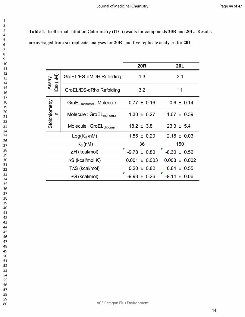
44
Table 1. Isothermal Titration Calorimetry (ITC) results for compounds 20R and 20L. Results
are averaged from six replicate analyses for 20R, and five replicate analyses for 20L.
GroEL/ES-dMDH Refolding
GroEL/ES-dRho Refolding
GroELmonomer : Molecule 0.77 ± 0.16 0.6 ± 0.14
Molecule : GroELmonomer 1.30 ± 0.27 1.67 ± 0.39
Molecule : GroELoligomer 18.2 ± 3.8 23.3 ± 5.4
1.56 ± 0.20 2.18 ± 0.03
-9.78 ± 0.80 -8.30 ± 0.52
0.001 ± 0.003 0.003 ± 0.002
0.20 ± 0.82 0.84 ± 0.55
-9.98 ± 0.26 -9.14 ± 0.06
∆S (kcal/mol�K)
T∆S (kcal/mol)
∆G (kcal/mol)
Assa
y
IC50 (µ
M)
Sto
ich
iom
etr
y
n
Log(Kd /nM)
Kd (nM)
∆H (kcal/mol)
20R 20L
1.3
3.2
36 150
3.1
11
Page 44 of 47
ACS Paragon Plus Environment
Journal of Medicinal Chemistry
123456789101112131415161718192021222324252627282930313233343536373839404142434445464748495051525354555657585960

45
Table 2. Biochemical and cell-based IC50, EC50, and CC50 results for the top four lead inhibitors
based on Selectivity Index (SI) for inhibiting MRSA proliferation over cytotoxicity in the liver
cell viability assay.
dRho dMDH
25L 0.53 24 46 >100 >63 6.4 5.1 4.7
24L 1.1 36 31 >100 43 11 6.7 5.0
27L 2.2 27 12 >100 40 11 5.2 2.4
28L 0.97 11 12 >100 37 1.0 1.1 1.0
33R 4.5 69 15 72 >63 19 10.0 11
24R 2.3 27 12 >100 >63 4.5 2.2 4.5
27R 2.0 22 11 >100 >63 7.3 2.6 6.1
28R 1.7 17 10 53 34 2.0 1.5 2.1
2f-m 2.0 24 12 >100 >63 33 11 58
15 4.6 49 11 >100 52 5.6 2.4 5.9
2i-p 7.3 47 6.4 >100 >63 22 24 51
2c-p 3.5 18 5.2 >100 >63 29 15 96Pseudo-s
ym
metr
ical A
nalo
gs
MRSA
Proliferation
EC50 (µµµµM)
Biochemical Assay IC50 (µµµµM)
L-S
eries A
nalo
gs
Compound Structure & Number
R-S
eries A
nalo
gs
HSP60/10-
dMDH
Refolding
GroEL/ES refolding of:
Cell-Based Assays
Native
Rho
Reporter
Native
MDH
Reporter
Liver Cell
Viability
CC50 (µµµµM)
Selectivity
Index (SI)
(CC50 / EC50)
Page 45 of 47
ACS Paragon Plus Environment
Journal of Medicinal Chemistry
123456789101112131415161718192021222324252627282930313233343536373839404142434445464748495051525354555657585960

46
Table 3. Comparison of the L- and R-series analogs for which overall scaffolds exhibited
preferred inhibition profiles in the various biochemical and cell-based assays. Lead antibacterial
candidates are preferred to potently inhibit GroEL/ES and bacteria, and not inhibit human
HSP60/10 and liver/kidney cell viability. The log(IC50), log(EC50), or log(CC50) values for the
20 matched L- and R-series analogs were analyzed by two-tailed, paired t-tests with 95%
confidence intervals. Comparative plots are presented in Figures S1-S5 in the Supporting
Information.
Assay Inhibition L-Series R-Series
S. aureus growth Preferred
MRSA growth Preferred
GroEL/ES-dMDH refolding
GroEL/ES-dRho refolding
HSP60/10-dMDH refolding Preferred
Liver cell viability Preferred
Kidney cell viability No statistical difference
No statistical difference
No statistical difference
Page 46 of 47
ACS Paragon Plus Environment
Journal of Medicinal Chemistry
123456789101112131415161718192021222324252627282930313233343536373839404142434445464748495051525354555657585960
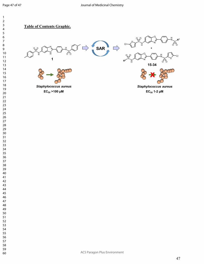
47
Table of Contents Graphic.
Page 47 of 47
ACS Paragon Plus Environment
Journal of Medicinal Chemistry
123456789101112131415161718192021222324252627282930313233343536373839404142434445464748495051525354555657585960


![Oral direct thrombin inhibitors or oral factor Xa inhibitors for the … · [Intervention Review] Oral direct thrombin inhibitors or oral factor Xa inhibitors for the treatment of](https://static.fdocuments.in/doc/165x107/610812dbd3c8b3601c7c48d5/oral-direct-thrombin-inhibitors-or-oral-factor-xa-inhibitors-for-the-intervention.jpg)