Sudden peroneal nerve palsy in an osteoarthritic knee: a ... · PDF fileSudden peroneal nerve...
5
Sudden peroneal nerve palsy in an osteoarthritic knee: a case report Vijay Kumar, Mayur Nayak * , Tahir Ansari, and Rajesh Malhotra Department of Orthopaedics, All India Institute of Medical Sciences, New Delhi 110029, India Received 25 December 2016, Accepted 8 January 2017, Published online 10 March 2017 Abstract – Peroneal nerve injuries have been reported in association with various causes around the knee such as traumatic varus injury, traumatic dislocation, upper tibial osteotomy, knee arthroscopy and total knee arthroplasty. Two instances of varus arthritic knee associated with a peroneal nerve palsy have been reported so far. One presented with gradual onset peroneal nerve palsy that recovered with time and the other with sudden onset peroneal nerve palsy that did not recover. We describe the case of a 63-year-old man who presented with a symptomatic varus arthritic knee and sudden onset peroneal nerve palsy with synovial cysts over the lateral aspect of the knee. We performed a total knee arthroplasty with decompression of the synovial cyst in the same patient. Three months following the surgery the patient was walking pain free with a completely recovered nerve palsy. Key words: Peroneal nerve palsy, Varus arthritic knee, Total knee replacement, Synovial cyst, Decompression. Introduction Peroneal nerve entrapment is the most common entrapment neuropathy found in the lower limb. It is most commonly seen at the fibular neck where the nerve becomes superficial [1, 2]. The causes for neuropathy can be traumatic such as varus injury to the knee [3], dislocation of the knee, [4] procedures such as high tibial osteotomy, [5] knee arthroscopy [6] and total knee arthroplasty [7, 8]. We describe a case of 63-year-old male presenting to us with a symptomatic progressive varus arthritic knee and sudden onset peroneal nerve palsy. A review of literature showed just one similar case in the past; however, the aetiology and the outcome of the surgery were different. Case report A 63-year-old man presented to Orthopaedics Outpatient Department (OPD), All India Institute of Medical Sciences, New Delhi with a history of pain in bilateral knees for 10 years. He had more pain on the left knee as compared to the right knee. He had developed a sudden onset foot drop in the left lower limb for the last four months. His pain in the left knee was severe to an extent that he was not able to walk even up to one block. He had to use a walker for ambulation. His activities of daily living such as using a transport, climbing stairs, squatting and sitting cross legged were limited. He also gave a history of recurrent episodes of giving way in the left knee. He developed a localized swelling on the lateral aspect of left knee, which was insidious in onset and progressed gradually over a period of the last one year. He had no history of any trauma to the left knee. He did not have any back pain or any radiating pain in his left lower limb. He had a history of coronary artery disease in the past for which he received treatment in the form of an angioplasty with a cardiac stent. On physical examination, the patient was 178 cm tall and weighed 74 kg. He walked with high steppage gait and had a varus thrust. The tibiofemoral angle was 10° varus (Figure 1), on weight bearing. On palpation, the patient had medial joint line tenderness and there was patellofemoral crepitus. There were two swellings located on the lateral aspect of the knee. The first swelling was located 1 cm above the lateral joint line and 4 cm lateral to the lateral patellar border and measured 4 cm · 6 cm. The other swelling was located 1 cm below the lateral joint line and 2.5 cm lateral to the lateral patellar tendon measuring 2 cm · 4 cm (Figure 2). The skin around both the swellings was normal. The patient had 10° of flexion contracture and the range of motion was 10–110° flexion. Examination of the ligaments in maximal extension revealed 10 mm of opening of the lateral joint line on varus stress test with a soft end point and with valgus stress the align- ment of the knee improved to normal with a bony end point (Figures 3a and 3b). Neurological examination revealed a 0/5 motor power in the tibialis anterior and the extensor hallucis longus of the left *Corresponding author: [email protected] SICOT J 2017, 3, 22 Ó The Authors, published by EDP Sciences, 2017 DOI: 10.1051/sicotj/2017005 Available online at: www.sicot-j.org This is an Open Access article distributed under the terms of the Creative Commons Attribution License (http://creativecommons.org/licenses/by/4.0), which permits unrestricted use, distribution, and reproduction in any medium, provided the original work is properly cited. OPEN ACCESS CASE REPORT
Transcript of Sudden peroneal nerve palsy in an osteoarthritic knee: a ... · PDF fileSudden peroneal nerve...
Sudden peroneal nerve palsy in an osteoarthritic knee: a casereport
Vijay Kumar, Mayur Nayak*, Tahir Ansari, and Rajesh Malhotra
Department of Orthopaedics, All India Institute of Medical Sciences, New Delhi 110029, India
Received 25 December 2016, Accepted 8 January 2017, Published online 10 March 2017
Abstract – Peroneal nerve injuries have been reported in association with various causes around the knee such astraumatic varus injury, traumatic dislocation, upper tibial osteotomy, knee arthroscopy and total knee arthroplasty.Two instances of varus arthritic knee associated with a peroneal nerve palsy have been reported so far. One presentedwith gradual onset peroneal nerve palsy that recovered with time and the other with sudden onset peroneal nerve palsythat did not recover. We describe the case of a 63-year-old man who presented with a symptomatic varus arthritic kneeand sudden onset peroneal nerve palsy with synovial cysts over the lateral aspect of the knee. We performed a totalknee arthroplasty with decompression of the synovial cyst in the same patient. Three months following the surgery thepatient was walking pain free with a completely recovered nerve palsy.
Key words: Peroneal nerve palsy, Varus arthritic knee, Total knee replacement, Synovial cyst, Decompression.
Introduction
Peroneal nerve entrapment is the most common entrapmentneuropathy found in the lower limb. It is most commonly seenat the fibular neck where the nerve becomes superficial [1, 2].The causes for neuropathy can be traumatic such as varusinjury to the knee [3], dislocation of the knee, [4] proceduressuch as high tibial osteotomy, [5] knee arthroscopy [6] andtotal knee arthroplasty [7, 8].
We describe a case of 63-year-old male presenting to uswith a symptomatic progressive varus arthritic knee andsudden onset peroneal nerve palsy. A review of literatureshowed just one similar case in the past; however, the aetiologyand the outcome of the surgery were different.
Case report
A 63-year-old man presented to Orthopaedics OutpatientDepartment (OPD), All India Institute of Medical Sciences,New Delhi with a history of pain in bilateral knees for10 years. He had more pain on the left knee as compared tothe right knee. He had developed a sudden onset foot dropin the left lower limb for the last four months.
His pain in the left knee was severe to an extent that he wasnot able to walk even up to one block. He had to use a walkerfor ambulation. His activities of daily living such as using atransport, climbing stairs, squatting and sitting cross legged
were limited. He also gave a history of recurrent episodes ofgiving way in the left knee. He developed a localized swellingon the lateral aspect of left knee, which was insidious in onsetand progressed gradually over a period of the last one year.He had no history of any trauma to the left knee. He did nothave any back pain or any radiating pain in his left lower limb.He had a history of coronary artery disease in the past forwhich he received treatment in the form of an angioplasty witha cardiac stent.
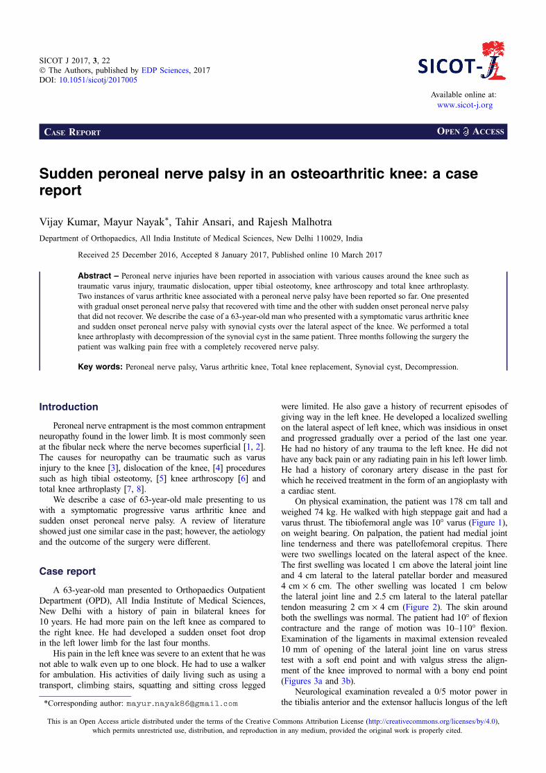
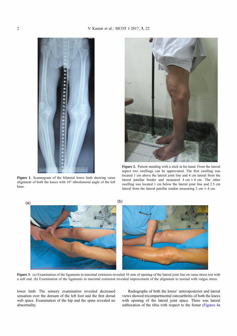
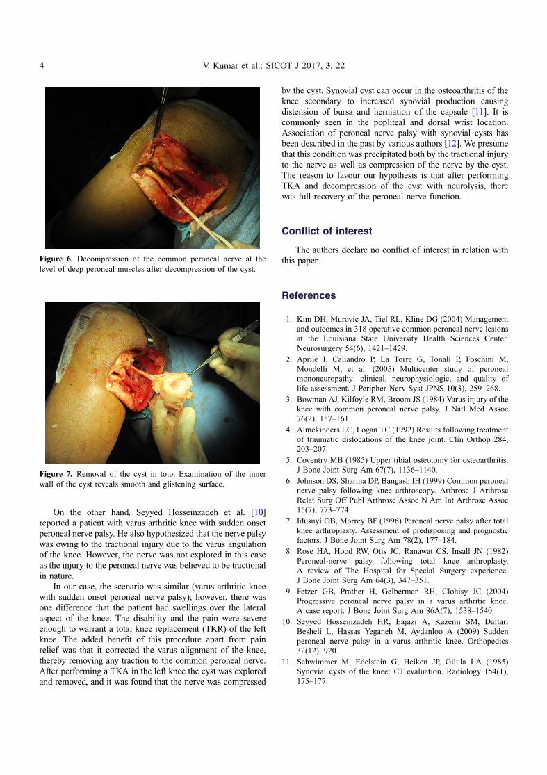
On physical examination, the patient was 178 cm tall andweighed 74 kg. He walked with high steppage gait and had avarus thrust. The tibiofemoral angle was 10� varus (Figure 1),on weight bearing. On palpation, the patient had medial jointline tenderness and there was patellofemoral crepitus. Therewere two swellings located on the lateral aspect of the knee.The first swelling was located 1 cm above the lateral joint lineand 4 cm lateral to the lateral patellar border and measured4 cm · 6 cm. The other swelling was located 1 cm belowthe lateral joint line and 2.5 cm lateral to the lateral patellartendon measuring 2 cm · 4 cm (Figure 2). The skin aroundboth the swellings was normal. The patient had 10� of flexioncontracture and the range of motion was 10–110� flexion.Examination of the ligaments in maximal extension revealed10 mm of opening of the lateral joint line on varus stresstest with a soft end point and with valgus stress the align-ment of the knee improved to normal with a bony end point(Figures 3a and 3b).
Neurological examination revealed a 0/5 motor power inthe tibialis anterior and the extensor hallucis longus of the left*Corresponding author: [email protected]
SICOT J 2017, 3, 22� The Authors, published by EDP Sciences, 2017DOI: 10.1051/sicotj/2017005
Available online at:www.sicot-j.org
This is an Open Access article distributed under the terms of the Creative Commons Attribution License (http://creativecommons.org/licenses/by/4.0),which permits unrestricted use, distribution, and reproduction in any medium, provided the original work is properly cited.
OPEN ACCESSCASE REPORT
lower limb. The sensory examination revealed decreasedsensation over the dorsum of the left foot and the first dorsalweb space. Examination of the hip and the spine revealed noabnormality.
Radiographs of both the knees’ anteroposterior and lateralviews showed tricompartmental osteoarthritis of both the kneeswith opening of the lateral joint space. There was lateralsubluxation of the tibia with respect to the femur (Figures 4a
(a) (b)
Figure 3. (a) Examination of the ligaments in maximal extension revealed 10 mm of opening of the lateral joint line on varus stress test witha soft end. (b) Examination of the ligaments in maximal extension revealed improvement of the alignment to normal with valgus stress.
Figure 1. Scannogram of the bilateral lower limb showing varusalignment of both the knees with 10� tibiofemoral angle of the leftknee.
Figure 2. Patient standing with a stick in his hand. From the lateralaspect two swellings can be appreciated. The first swelling waslocated 1 cm above the lateral joint line and 4 cm lateral from thelateral patellar border and measured 4 cm · 6 cm. The otherswelling was located 1 cm below the lateral joint line and 2.5 cmlateral from the lateral patellar tendon measuring 2 cm · 4 cm.
2 V. Kumar et al.: SICOT J 2017, 3, 22

and 4b). Electromyography and nerve conduction studyrevealed a peroneal nerve neuropathy at the level of the knee.
A total knee arthroplasty (TKA) along with explorationand decompression of peroneal nerve for the left knee wasdone. A primary TKA (NEXGEN; Zimmer Biomet, Warsaw,Indiana) was performed (Figure 5). Ligament balancing wasdone by performing medial release. We used posteriorstabilized insert with cemented femoral and tibial componentswith a tibial stem.
The peroneal nerve was explored and decompressed bymaking a 3–5 cm oblique incision parallel to the course ofnerve at the neck of the fibula. Skin and the subcutaneousfascia were cut in the same plane. With the help ofMetzenbaum scissors and blunt dissection, the retinaculumwas identified. The cysts were found under the fascia. Bluntdissection showed cystic wall. An effort was made to removethe cyst without violating the cystic wall, however decompres-sion of the cyst was necessary. Incision of the cyst extrudedviscous yellow tinted liquid. The decompressed cyst was thenclamped with the Alice forceps and the dissection was started
from proximal to the distal directions taking care not to injurethe common peroneal nerve around it. The cyst was thenremoved in toto and was sent for histopathology (Figure 6).The peroneal nerve was found to be embedded between thetwo cysts and had a flattened surface. The nerve was freedof all the adhesions around it (Figure 7). Furthermore, thenerve was decompressed at the level of deep peroneal musclefascia after exposing the underlying peroneal muscles.
The patient was made to walk 24 h later with protectedweight bearing with the assistance of a walker. The patientwas evaluated at two weeks and three months postoperatively.At three months postoperatively, the knee pain has resolvedcompletely and the patient is able to walk and climb stairsunassisted. The power of the left ankle and toe dorsiflexorshas improved to 5/5.
Discussion
The patient presented to us with varus arthritic knee with afour-month history of sudden onset peroneal nerve palsy.Two instances of varus arthritic knee associated with aperoneal nerve palsy have been reported so far. The first reportwas by Fetzer et al. [9] who reported a patient withvarus arthritic knee with gradual onset peroneal nerve palsy.On performing a TKA with peroneal nerve exploration anddecompression, the function of the nerve improved.The absence of any compressive lesion at the time of theoperation in itself was evident of the fact that the personalnerve dysfunction was due to a tractional injury.
Figure 5. A standing anteroposterior radiograph of the left kneeshows a well-fixed, well-aligned, total knee prosthesis withcemented tibial stem and a malleolar screw in the medial aspect.
Figure 4. (a) Anteroposterior (AP) X-ray of the bilateral kneeshowing decrease in the joint space (left > right) with lateral tibialsubluxation of the left knee. (b) Lateral radiograph of both the kneeshowing multiple osteophytes and degenerative changes in thepatellofemoral joint.
V. Kumar et al.: SICOT J 2017, 3, 22 3
On the other hand, Seyyed Hosseinzadeh et al. [10]reported a patient with varus arthritic knee with sudden onsetperoneal nerve palsy. He also hypothesized that the nerve palsywas owing to the tractional injury due to the varus angulationof the knee. However, the nerve was not explored in this caseas the injury to the peroneal nerve was believed to be tractionalin nature.
In our case, the scenario was similar (varus arthritic kneewith sudden onset peroneal nerve palsy); however, there wasone difference that the patient had swellings over the lateralaspect of the knee. The disability and the pain were severeenough to warrant a total knee replacement (TKR) of the leftknee. The added benefit of this procedure apart from painrelief was that it corrected the varus alignment of the knee,thereby removing any traction to the common peroneal nerve.After performing a TKA in the left knee the cyst was exploredand removed, and it was found that the nerve was compressed
by the cyst. Synovial cyst can occur in the osteoarthritis of theknee secondary to increased synovial production causingdistension of bursa and herniation of the capsule [11]. It iscommonly seen in the popliteal and dorsal wrist location.Association of peroneal nerve palsy with synovial cysts hasbeen described in the past by various authors [12]. We presumethat this condition was precipitated both by the tractional injuryto the nerve as well as compression of the nerve by the cyst.The reason to favour our hypothesis is that after performingTKA and decompression of the cyst with neurolysis, therewas full recovery of the peroneal nerve function.
Conflict of interest
The authors declare no conflict of interest in relation withthis paper.
References
1. Kim DH, Murovic JA, Tiel RL, Kline DG (2004) Managementand outcomes in 318 operative common peroneal nerve lesionsat the Louisiana State University Health Sciences Center.Neurosurgery 54(6), 1421–1429.
2. Aprile I, Caliandro P, La Torre G, Tonali P, Foschini M,Mondelli M, et al. (2005) Multicenter study of peronealmononeuropathy: clinical, neurophysiologic, and quality oflife assessment. J Peripher Nerv Syst JPNS 10(3), 259–268.
3. Bowman AJ, Kilfoyle RM, Broom JS (1984) Varus injury of theknee with common peroneal nerve palsy. J Natl Med Assoc76(2), 157–161.
4. Almekinders LC, Logan TC (1992) Results following treatmentof traumatic dislocations of the knee joint. Clin Orthop 284,203–207.
5. Coventry MB (1985) Upper tibial osteotomy for osteoarthritis.J Bone Joint Surg Am 67(7), 1136–1140.
6. Johnson DS, Sharma DP, Bangash IH (1999) Common peronealnerve palsy following knee arthroscopy. Arthrosc J ArthroscRelat Surg Off Publ Arthrosc Assoc N Am Int Arthrosc Assoc15(7), 773–774.
7. Idusuyi OB, Morrey BF (1996) Peroneal nerve palsy after totalknee arthroplasty. Assessment of predisposing and prognosticfactors. J Bone Joint Surg Am 78(2), 177–184.
8. Rose HA, Hood RW, Otis JC, Ranawat CS, Insall JN (1982)Peroneal-nerve palsy following total knee arthroplasty.A review of The Hospital for Special Surgery experience.J Bone Joint Surg Am 64(3), 347–351.
9. Fetzer GB, Prather H, Gelberman RH, Clohisy JC (2004)Progressive peroneal nerve palsy in a varus arthritic knee.A case report. J Bone Joint Surg Am 86A(7), 1538–1540.
10. Seyyed Hosseinzadeh HR, Eajazi A, Kazemi SM, DaftariBesheli L, Hassas Yeganeh M, Aydanloo A (2009) Suddenperoneal nerve palsy in a varus arthritic knee. Orthopedics32(12), 920.
11. Schwimmer M, Edelstein G, Heiken JP, Gilula LA (1985)Synovial cysts of the knee: CT evaluation. Radiology 154(1),175–177.
Figure 6. Decompression of the common peroneal nerve at thelevel of deep peroneal muscles after decompression of the cyst.
Figure 7. Removal of the cyst in toto. Examination of the innerwall of the cyst reveals smooth and glistening surface.
4 V. Kumar et al.: SICOT J 2017, 3, 22

12. Hersekli MA, Akpinar S, Demirors H, Ozkoc G, Ozalay M,Cesur N, et al. (2004) Synovial cysts of proximal tibiofibularjoint causing peroneal nerve palsy: report of three cases and
review of the literature. Arch Orthop Trauma Surg 124(10),711–714.
Cite this article as: Kumar V, Nayak M, Ansari T & Malhotra R (2017) Sudden peroneal nerve palsy in an osteoarthritic knee: a case report.SICOT J, 3, 22
V. Kumar et al.: SICOT J 2017, 3, 22 5