
Sudan University of Science &Technology College of ......بسم الله الرحمن الرحيم...
56
Sudan University of Science &Technology College of Graduate Studies Assessment of the Placenta Location in Pregnant Women with Previous Cesarean Section ت قييم الحواملء اللنساوقع المشيمة ل مصرية سابقةهن عملية قي لدي تيA thesis submitted for Partial Fulfillment of the Requirement of M.Sc. Degree in Medical Diagnostic Ultrasound. By: Nfhat Yousuf Ahmed Yousuf Supervisor: Dr. Ahmed Mustafa Abukonna 2017
Transcript of Sudan University of Science &Technology College of ......بسم الله الرحمن الرحيم...
ميحرلا نمحرلا هللا مسب
Sudan University of Science &Technology
College of Graduate Studies
Assessment of the Placenta Location in Pregnant Women with
Previous Cesarean Section
تي لديهن عملية قيصرية سابقةموقع المشيمة للنساء الحوامل الال قييمت
A thesis submitted for Partial Fulfillment of the Requirement of M.Sc.
Degree in Medical Diagnostic Ultrasound.
By:
Nfhat Yousuf Ahmed Yousuf
Supervisor:
Dr. Ahmed Mustafa Abukonna
2017

I
االية
قال تعايل:
))وما أوتيتم من العلم إال قليال((
صدق اهلل العظيم
(58االرساء)

II
Dedication
I would like to dedicate my thesis to my mother and my father who have loved and
taken care of me for all my life.
I also dedicate this thesis to my husband who support and encourage me
To my sisters and my brother

III
Acknowledgement
I thank almighty God for giving me the strength, courage and
determination in conducting this study, despite all difficulties.
I would like to thank gratefully my supervisor
Dr. Ahmed Mostafa Abukonna
IV
Abstract
This was a descriptive cross-sectional study conducted at the department of
obstetrics and gynecology in Bahry Educational Hospital and Maternal Specialist
Hospital and Elshaikh Ali Fadoul Hospital; it was conduct during the period from
April to august 2017. The main aim was to assess the association of placenta
location with previous caesarean section (CS) in pregnant women. In antenatal
clinic as per protocol 56 pregnant women were scanned in their second and third
trimester for fetal wellbeing and placental localization after taking a detailed
obstetrical history and clinical examination. All women without previous caesarean
section were excluded. Methodology the data collection from pt by data collection
sheet and analyzed by SPSS.
The result of the study revealed that the fundal placenta was reported in 8.9%,
posterior placenta was 21.4%, anterior placenta was 57.1%, placenta previa was
7.1% and low lying placenta was 5.4%. There was a significant difference in
placental location when there is a CS scar present on the uterus. Women with a
previous history of CS are significantly less likely to have a fundal placenta and
more likely to have the placenta located on the anterior and posterior wall of the
uterus in a subsequent pregnancy. Me was found no statistically significant
difference in the incidence of low-lying placentae between the groups.
Previous CS may have a significant impact on placental location in subsequent
pregnancies. The mechanism behind this effect is not clear; however this
observation may provide indirect evidence that the presence of a CS scar in the
uterus has a more ‘global’ effect on the endometrium than the physical presence of
a scar in isolation.

V
المستخلص
راسة وصفية مستعرضة أجريت في قسم التوليد وأمراض النساء في مستشفى بحري التعليمي هذه د
التخصصي ومستشفى الشيخ علي فضل. اجريت خالل الفترة من أبريل إلى أغسطس الوالدةومستشفى
في . وكان الهدف الرئيسي لتقييم موقع المشيمة مع العملية القيصرية السابقة في النساء الحوامل. 7102
تم فحص النساء الحوامل في الثلث الثاني والثالث من عمر الجنين و موقع 65عيادة الوالدة وفقا للبروتوكول
المشيمة بعد أخذ التاريخ التوليد مفصل والفحص السريري. واستبعدت جميع النساء الالتي لم يسبق لهن والدة
.قيصرية
٪،و كانت 70.2٪، .وكان المشيمة الخلفية 9.8لرحم فيوكشفت نتائج الدراسة أن المشيمة توجد في اعلى ا
٪. كان 6.2٪ وكان انخفاض المشيمة 2.0٪، كانت المشيمة المتقدمة على الجنين 62.0المشيمة األمامية
هناك اختالف كبير في موقع المشيمة عندما يكون هناك ندبة بسبب الجراحة القيصرية موجودة على الرحم.
يخ جراحة قيصرية سابق هم أقل احتماال بكثير أن يكون المشيمة في اعلى الرحم النساء الذين لديهم تار
وأكثر احتماال أن يكون المشيمة تقع على الجدار األمامي والخلفي للرحم في الحمل الالحق. لم نجد أي فرق
.ذو داللة إحصائية في حدوث المشيمة المنخفضة بين المجموعات
السابقة لها تأثير كبير على موقع المشيمة في حاالت الحمل الالحقة. واآللية قد تكون الجراحة القيصرية
الكامنة وراء هذا التأثير ليست واضحة؛ ولكن هذه المالحظة قد توفر أدلة غير مباشرة على أن وجود ندبة
.جراحة في الرحم له تأثير "عالمي" أكثر على بطانة الرحم من الوجود المادي للندبة في العزلة

VI
List of Contents
Topic Page No.
I االيه
Dedication II
Acknowledgement III
Abstract IV
Abstract – Arabic V
List of Contents VI
List of figures VIII
List of tables IX
List of abbreviations X
Chapter One
Introduction
1.1 Introduction 0
1.2 Problem of study 2
1.3 Objectives 2
1.4 Over view of study 2
Chapter Two
Theoretical Background and Previous Studies
2.1 Cesarean section 3
2.2 Prevelance 4
2.3 placenta development 5
2.4 Further information: Placentation 6
2.5 Placental circulation 7
2.6 Physiology of the placenta 9
2.7 Placenta pathology 10
2.8 Ultrasound of placenta 16
2.9 Previous studies 19
Chapter Three
Material and method
3.1 Materials 21
3.2 Methods 21

VII
Chapter Four
Results
4.1 Results 23
Chapter Five
Discussion, Conclusion and Recommendations
5.1 Discussion 26
5.2 Conclusion 28
5.3 Recommendation 29
References 30
Appendices
VIII
List of Figures
Figure Title Page No.
2.1 Placenta 5
2.2 The initial stages of human embryogenesis. 5
2.3 Maternal blood fills the intervillous space, nutrients, water,
and gases are actively and passively exchanged, then
deoxygenated blood is displaced by the next maternal pulse.
7
2.4 Localizing the placenta from a longitudinal, midline section of
the uterus.
17
2.5 Color doppler image show posterior placenta and umblical
cord.
18
4.1 shows the frequency distribution of Placenta site 24

IX
List of Tables
Table No Table title Page No
4-1 Descriptive Statistics 23
4-2 The frequency distribution of Placenta site 23
4-3 Cross tabulation between placenta site and number of
cesarean section.
24
4-4 Cross tabulation between number of parity and number of
cesarean section.
25
X
List of abbreviations
Abbreviations Meaning
APH Ante-Partum Hemorrhage
CS Cesarean Section
EVS Endo Vaginal Study
IUGR Intra-Uterine Growth Retardation
SCH Sub-Chorionic Hematoma
TAS Trans-Abdominal Study
TPS Trans Perineal Study
1
Chapter one
1.1 Introduction:
Placental development is a complex process and the mechanism of placental
localization is not well under-stood. Between 2 and 10 weeks after fertilization,
contact between maternal and fetal tissue is dependent on the cytotrophoblast shell
of the developing placenta and the decidualizing endometrium. This is a dynamic
phase of the pregnancy as the placenta increases in size and complexity. Therefore,
a favorable endometrial environment is essential in order to ensure the
development of an adequate fetal–maternal interface. To date, it is not known if a
scar on the uterus has any effect on the global endometrial environment and on
placental location in particular (Betran et al., 2007).
The frequency of placenta previa decreases with increasing gestational age because
of ‘placental migration’. This is because the placenta-free uterine wall grows faster
than placenta-covered areas. The placenta does not actually move, but the tissue
upon which it is embedded expands, leading to the placenta’s appearing to move
up and away from the cervix. This process applies in the normal intact uterus, but it
is not known if the same mechanism occurs as effectively in a scarred uterus
(Belachew et al., 2017).
As rates of cesarean section continue to increase worldwide, methods for
prediction, surveillance, and management of complications during pregnancy and
delivery associated with previous cesarean section become increasingly important.
Uterine rupture, placenta previa, and placenta accreta are well-known and
potentially life-threatening complications, but are fortunately still rare conditions.
2
It has been reported that, among women with placenta previa, all with abnormal
invasive placentae had a previous cesarean section and anterior placenta previa.
Pictorial ultrasound, including measurements of myometrial thickness and 3D
power Doppler at the placental site, has been used to diagnose invasive
placentation (Wong et al., 2008).
1.2 Problem of study:
Caesarian section increase rapidly worldwide and pregnant women with history of
caesarian delivery considered as risk for placenta previa so ultrasound for placenta
location is very important in follow up for these women.
1.3 objectives of study:
1.3.1 General objective:
To assess the placenta location in pregnant women with previous cesarean section.
1.3.2 Specific objectives:
To evaluate prevalence of placenta previa in pregnant women with
history of cesarean section.
To assess the common placental location site in association with
cesarean section.
To correlate placenta location to the number of cesarean section.
1.4 Thesis over view:
Chapter one is introduction will discuss problem, objective of study. Chapter two
is literature review will discuss (anatomy, physiology, pathology and previous
study). Chapter three will discuss material and methods, Chapter four will discuss
results. Chapter five is discussion, conclusion and recommendation.

3
Chapter two
Theoretical background and Previous Studies
2.1 Cesarean section:
A caesarean section is surgical procedure in which incisions are made through
mother’s abdomen and uterus to deliver one or more babies.
2.1.1 Indications of C-section:
Abnormal presentation (breech or transverse positions)
Prolonged labor or a failure to progress (dystocia)
Fetal distress
Cord prolapse
Uterine rupture or an elevated risk thereof
Hypertension in the mother or baby after amniotic rupture (the waters
breaking)
Tachycardia in the mother or baby after amniotic rupture (the waters
breaking)
Placenta problems (placenta previa, placental abruption or placenta accreta)
Failed labour induction
Failed instrumental delivery (by forceps or ventouse (Sometimes a trial of
forceps/ventouse delivery is attempted, and if unsuccessful, thebaby will
need to be born by caesarean section.)
Large baby weighing >4,000 g (macrosomia) and umbilical cord
abnormalities (vasa previa, multilobate including bilobate and succenturiate-
lobed placentas, velamentous insertion) (Betran et al., 2007).
4
Caesarean delivery is the use of surgery to deliver one or more babies. A
caesarean section Complications of labour and factors increasing the risk
associated with vaginal delivery, such as abnormal presentation (breech or
transverse positions). Babies are usually born head first. If the baby is in another
position the birth may be complicated. In a ‘breech presentation’ the unborn baby
is bottom-down instead of head-down. Babies born bottom-first are more likely to
be harmed during a normal (vaginal) birth than those born head-first. For instance,
the baby might not get enough oxygen during the birth. Having a planned
caesarean may reduce these problems. A review looking at planned caesarean
section for singleton breech presentation with planned vaginal birth concludes that
in the short term, births with a planned caesarean were safer for babies than vaginal
births. Fewer babies died or were seriously hurt when they were born by caesarean.
However, there was tentative evidence that children who were born by caesarean
had more health problems at age two. Caesareans caused some short-term
problems for mothers such as more abdominal pain. They also had some benefits,
such as less urinary incontinence and less perineal pain (Betran et al., 2007).
2.2 Prevalence:
It is generally agreed that the prevalence of caesarean section is higher than needed
in many countries and physicians are encouraged to actively lower the rate, as a
caesarean rate higher than 10-15% is not associated with reductions in maternal or
infant mortality rates. Some evidence supports a higher rate of 19% may result in
better outcomes (Solheim et al., 2011).
Some of these efforts are: emphasizing a long latent phase of labor is not abnormal
and not a justification for C-section; a new definition of the start of active labor
from a cervical dilatation of 4 cm to a dilatation of 6 cm; and allowing at least 2
hours of pushing for women who have previously given birth and 3 hours of
pushing for women who have not previously given birth (Solheim et al., 2011).
5
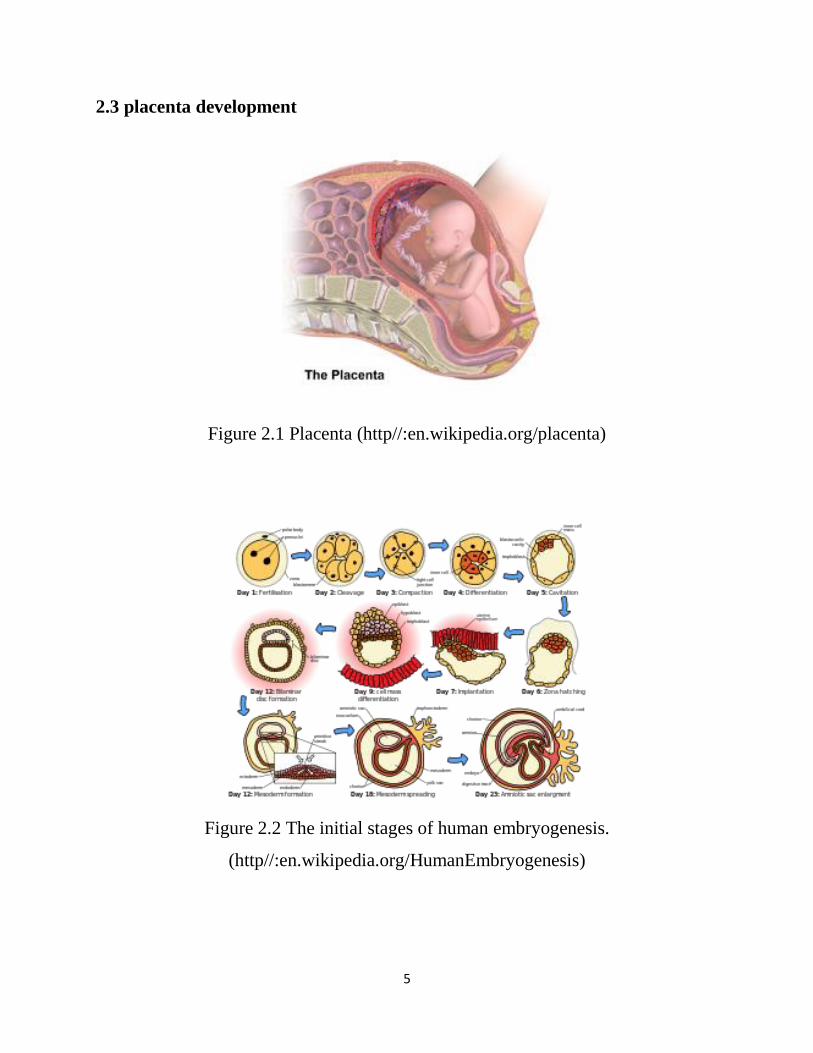
2.3 placenta development
Figure 2.1 Placenta (http//:en.wikipedia.org/placenta)
Figure 2.2 The initial stages of human embryogenesis.
(http//:en.wikipedia.org/HumanEmbryogenesis)
6
2.4 Further information: Placentation
The placenta begins to develop upon implantation of the blastocyst into the
maternal endometrium. The outer layer of the blastocyst becomes the trophoblast,
which forms the outer layer of the placenta. This outer layer is divided into two
further layers: the underlying cytotrophoblast layer and the overlying
syncytiotrophoblast layer. The syncytiotrophoblast is a multinucleated continuous
cell layer that covers the surface of the placenta. It forms as a result of
differentiation and fusion of the underlying cytotrophoblast cells, a process that
continues throughout placental development. The syncytiotrophoblast (otherwise
known as syncytium), thereby contributes to the barrier function 0of the placenta
(Cho et al., 2008).The placenta grows throughout pregnancy. Development of the
maternal blood supply to the placenta is complete by the end of the first trimester
of pregnancy (approximately 12–13 weeks) (Matsubara et al., 2017).
In humans, the placenta averages 22 cm (9 inch) in length and 2–2.5 cm (0.8–
1 inch) in thickness, with the center being the thickest, and the edges being the
thinnest. It typically weighs approximately 500 grams (just over 1 lb). It has a dark
reddish-blue or crimson color. It connects to the fetus by an umbilical cord of
approximately 55–60 cm (22–24 inch) in length, which contains two umbilical
arteries and one umbilical vein. The umbilical cord inserts into the chorionic plate
(has an eccentric attachment). Vessels branch out over the surface of the placenta
and further divide to form a network covered by a thin layer of cells. This results in
the formation of villous tree structures. On the maternal side, these villous tree
structures are grouped into lobules called cotyledons. In humans, the placenta
usually has a disc shape, but size varies vastly between different mammalian
species (Matsubara et al., 2017).
7
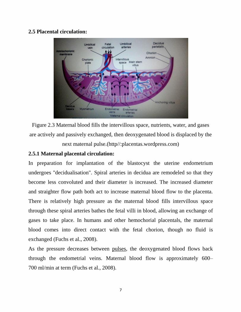
2.5 Placental circulation:
Figure 2.3 Maternal blood fills the intervillous space, nutrients, water, and gases
are actively and passively exchanged, then deoxygenated blood is displaced by the
next maternal pulse.(http//:placentas.wordpress.com)
2.5.1 Maternal placental circulation:
In preparation for implantation of the blastocyst the uterine endometrium
undergoes "decidualisation". Spiral arteries in decidua are remodeled so that they
become less convoluted and their diameter is increased. The increased diameter
and straighter flow path both act to increase maternal blood flow to the placenta.
There is relatively high pressure as the maternal blood fills intervillous space
through these spiral arteries bathes the fetal villi in blood, allowing an exchange of
gases to take place. In humans and other hemochorial placentals, the maternal
blood comes into direct contact with the fetal chorion, though no fluid is
exchanged (Fuchs et al., 2008).
As the pressure decreases between pulses, the deoxygenated blood flows back
through the endometrial veins. Maternal blood flow is approximately 600–
700 ml/min at term (Fuchs et al., 2008).

8
2.5.2 Fetoplacental circulation:
Deoxygenated fetal blood passes through umbilical arteries to the placenta. At the
junction of umbilical cord and placenta, the umbilical arteries branch radially to
form chorionic arteries. Chorionic arteries, in turn, branch into cotyledon arteries.
In the villi, these vessels eventually branch to form an extensive arterio-capillary-
venous system, bringing the fetal blood extremely close to the maternal blood; but
no intermingling of fetal and maternal blood occurs ("placental barrier").
Endothelin and prostanoids cause vasoconstriction in placental arteries, while nitric
oxide causes vasodilation. On the other hand, there is no neural vascular
regulation, and catecholamines have only little effect (Becker et al., 2001).
The fetoplacental circulation is vulnerable to persistent hypoxia or intermittent
hypoxia and reoxygenation, which can lead to generation of excessive free
radicals. This may contribute to pre-eclampsia and other pregnancy complications.
It is proposed that melatonin plays a role as functions nutrition. The placenta
intermediates the transfer of nutrients between mother and fetus. The perfusion of
the intervillous spaces of the placenta with maternal blood allows the transfer of
nutrients and oxygen from the mother to the fetus and the transfer of waste
products and carbon dioxide back from the fetus to the maternal blood. Nutrient
transfer to the fetus can occur via both active and passive transport. Placental
nutrient metabolism was found to play a key role in limiting the transfer of some
nutrients. Adverse pregnancy situations, such as those involving maternal diabetes
or obesity, can increase or decrease levels of nutrient transporters in the placenta
potentially resulting in overgrowth or restricted growth of the fetus (Belachew et
al., 2017).
9
2.5.3 Placental Size and Growth
There is less emphasis nowadays in measurements of the placenta largely because
the information is of limited diagnostic value. Thus, the placenta is not routinely
measured. The most popular measurement is placental thickness (data on placental
area, volume, and weight estimates have all been studied and reported in the
literature). As a guideline, placental thickness should be measured if the placenta
appears to be either thick or thin. Placental thickness measurements should be
made near the mid portion or center of the placenta with one caliper placed at the
amniochorionic surface (chorionic plate) and the second caliper placed at the basal
surface perpendicular to the amniochorionic surface (Proctor et al., 2009).
The measurement should exclude retroplacental veins, myometrium, fibroids, and
contractions of the uterus that might incorrectly increase the measurement. In a
normal pregnancy, placental thickness increases with gestational age. As a rule of
thumb, the mean thickness of the placenta in millimeters is roughly equal to the
gestational age in weeks (e.g. at 20 weeks, mean placental thickness is 20 mm; at
28 weeks, mean placental thickness is 28 mm; and at 36 weeks, mean placental
thickness is 36 mm) (Hunter et al., 2015).
2.6 Physiology of the placenta:
Waste products excreted from the fetus such as urea, uric acid, and creatinine are
transferred to the maternal blood by diffusion across the placenta. Immunity IgG
antibodies can pass through the human placenta, thereby providing protection to
the fetus in utero. This transfer of antibodies begins as early as the 20th week of
gestational age, and certainly by the 24th week. This passive immunity lingers for
several months after birth, thus providing the newborn with a carbon copy of the
mother's long-term humoral immunity to see the infant through the crucial first
months of extrauterine life. IgM, however, cannot cross the placenta, which is why
some infections acquired during pregnancy can be hazardous for the fetus.

10
Furthermore, the placenta functions as a selective maternal-fetal barrier against
transmission of microbes. However, insufficiency in this function may still cause
mother-to-child transmission of infectious diseases (Korkmaz et al., 2013).
The first hormone released by the placenta is called the human chorionic
gonadotropin hormone. This is responsible for stopping the process at the end of
menses when the Corpus luteum ceases activity and atrophies. If hCG did not
interrupt this process, it would lead to spontaneous (Sekiguchi et al., 2013)
2.7 Placenta pathology:
2.7.1 Placenta previa:
The term “placenta previa” refers to a placenta that is “previous” to the fetus in the
birth canal. The incidence at delivery is approximately 0.5% of all pregnancies.
Bleeding in the second and third trimesters is the hallmark of placenta previa. This
bleeding can be life threatening to the mother and fetus. With expectant
management and cesarean delivery, both maternal and perinatal mortality have
decreased over the past 40 years (Sekiguchi et al., 2013)..
The term ‘placenta praevia’ should only be used after 28 weeks. The differentiation
of placental positions has historically been performed by digital assessment of the
lower uterine segment and placenta through the cervix. Using this potentially
hazardous method of evaluation placental position was classified as complete
placenta previa, partial placenta previa, incomplete placenta previa, marginal
placenta previa, low-lying placenta, and placenta distant from the internal cervical
os (Young et al., 2014).
The use of ultrasound to evaluate the position of the placenta in the uterus has both
improved knowledge of the placenta within the uterus and simplified terminology
with respect to placental position. Complete placenta previa describes the situation
in which the internal cervical os is totally covered by the placenta. Marginal
placenta previa denotes placental tissue at the edge of or encroaching on the

11
internal cervical os. A low placenta is one in which the placental edge is within 2
cm, but not covering any portion, of the internal cervical os. The terms
“incomplete placenta previa” and “partial placenta previa” have no place in the
current sonographic assessment of placental position and should be used only by a
clinician performing a digital examination when a “double setup” is necessary to
determine where the leading edge of the placenta lies (Fuchs et al., 2008).
2.7.2 Placenta accreta:
A placenta that is abnormally adherent to the uterine wall after delivery is termed
placenta accrete. Placenta accreta occurs if the placenta invades the myometrium
more deeply, and placenta percreta refers to a placenta that at least in part
protrudes through the uterine serosa. Placenta accreta, increta, and percreta are
serious complications of pregnancy associated with maternal blood loss, need for
hysterectomy, and retained products of conception Although placenta accreta (or
increta or percreta) can occur in any pregnancy, important risk factors include prior
uterine surgery (with risk increasing with increasing number of prior cesarean
deliveries), placenta previa, unexplained elevated maternal serum alpha-fetoprotein
(MS-AFP), increased maternal cell-free placental lactogen, and advancing
maternal age (Fuchs et al., 2008).
Several sonographic signs are associated with placenta accreta. The presence of a
coexisting placenta previa in the majority of cases makes it particularly likely that
the adherent portion of the placenta will be low in the uterus, in the region of a
prior cesarean section scar. This simple fact makes the evaluation of these
placentas much more straight forward with the transvaginal ultrasound probe (Cho
et al., 2008).
Sonographic findings of placenta accreta include loss of the normal hypoechoic
retroplacental-myometrial interface, thin-ning or disruption of the hyperechoic
12
subvesicular uterine serosa, presence of focal exophytic masses, and numerous
placental lakes (Fuchs et al., 2008).
The color Doppler ultrasound findings suggestive of placenta previa accreta
include diffuse lacunar blood flow throughout the placenta, dilated vascular
channels between the placenta and bladder or cervix, absence of the normal
subplacental venous flow, and the demonstration of vessels crossing the placental-
myometrial disruption site . Three-dimensional sonography may also be helpful for
evaluation of vascular anatomy in the setting of a placenta accrete (Fuchs et al.,
2008).
2.7.3 Placenta abruption:
Placental abruption is defined as separation of the placenta prior to the delivery of
the fetus. Placental abruption is one of the worrisome causes of vaginal bleeding in
the latter part of pregnancy because it contributes to perinatal mortality.
Patients typically present with third-trimester vaginal bleeding associated with
abdominal or uterine pain and labor (Fuchs et al., 2008).
Risk factors: History of prior abruption, hypertension, prolonged rupture of
membranes, IUGR, chorioamnionitis, polyhydramnios, maternal thrombophilias,
maternal substance use (tobacco, alcohol, cocaine), maternal trauma, and
advanced maternal age are all risk factors for placental abruption.
A subplacental hematoma between the placenta and uterine wall is a placental
abruption. This should be differentiated from a subchorionic hematoma, in which
the hematoma is underneath the chorion, not the placenta. Although a subchorionic
hematoma can occur anytime during pregnancy, it is more common in the first half
of pregnancy. Preplacental hematoma is a rare condition likely caused by bleeding
from fetal vessels and located on the fetal surface of the placenta under the
chorion. History of placental abruption or previous Caesarian section increases the
risk by a factor of 2.3 (Fuchs et al., 2008).
13
2.7.4 False thickening of the placenta
False placental thickening may be seen with placental abruption if the
retroplacental hematoma has the same echogenicity (isoechoic) as the normal
placental tissue. Color Doppler may be helpful in distinguishing true placental
thickening from pseudo thickening. With true placental thickening, the normal
intraplacental vascular network should be seen from the chorionic to basal surface;
with abruption and a retroplacental hematoma, color will be seen in the placental
tissue and be lacking in the hematoma (Ishikawa et al., 2006).
The graph shows there is significant variance in normal placental thickness at
different gestational ages. This graph indicates the placenta appears to grow until
term but at a slower rate in the third trimester. A placental thickness greater than 4
cm is considered abnormal at any gestational age. Less than 2.5 cm at or greater
than 35 weeks is considered too thin. The four conditions most commonly
associated with placental thickening are:
a) Diabetes mellitus, especially gestational diabetes and class A, B, and C
b) Immune and nonimmune fetal hydrops
c) Fetal infections (e.g. cytomegalovirus)
d) Chromosomal abnormalities, especially triploidy Small or thin placentas are
most commonly associated with maternal hypertensive disease, severe IUGR, and
severe diabetes mellitus (class D, E, R). Rarely, a thin placenta may be due to a
membranous placenta (placenta membranacea or diffusa) which is a thin, poorly
functional placenta that covers the entire surface of the chorionic sac.
The placenta may also appear unusually thin with severe polyhydramnios as it is
stretched over a large surface area of the uterine wall (Allen et al., 2002).
2.7.5 Placental Tumours
All primary and secondary tumours of the placenta are rare. The most common
tumour of the placenta by far is chorioangioma. Other primary tumours of the
14
placenta include teratoma and choriocarcinoma. Choriocarcinoma is most likely to
develop secondary to hydatidiform mole. Melanoma is reported to be the most
common tumour to metastasize to the placenta. Various angiomatous tumours of
the placenta ranging widely in size have been described and because of the
resemblance of their components to the blood vessels and stroma of the chorionic
villus, the term chorioangioma is the most appropriate designation (Alvarez-Goris
et al., 2016).
Macroscopic placental chorioangiomas are reported to occur with an incidence of1
in 16,000 to 1 in 1,500 deliveries. The incidence of clinically insignificant,
microscopic chorioangiomas (not detectable with ultrasound) is reported to be as
high as 1 in 100 placent as. Small lesions (3 cm in diameter) are usually not
associated with fetal or maternal complications. The most common fetal and
maternal complications are IUGR, fetal hydrops (due to fetal congestive heart
failure), polyhydramnios, and premature labour. Chorioangiomas may also
associate with elevated maternal serum alpha-fetoprotein in the absence of other
placental or fetal anomalies (Alvarez-Goris et al., 2016).
Sonographically, chorioangiomas typically appear as solid placental masses
bulging towards the fetal surface of the placenta. In contrast, fibroids arising from
the retroplacental uterine wall cause a bulging effect on the maternal surface of the
placenta and the serosal surface of the uterus. Chorioangiomas have a variable
echo appearance from solid, homogeneous masses resembling placental tissue to
complex masses with septae. The vascularity of chorioangiomas is variable and
may affect outcome. Retroplacental hematoma (clot) appears as a mass of variable
echogenicity between the uterine wall and the uterine surface of the placenta. A
fresh hematoma may be more echogenic than the placenta and with aging
gradually becomes less echogenic. An isoechoic hematoma may mimic a thick
15
placenta although modern systems with good contrast resolution will generally
(Niknejadi et al., 2016).
2.7.6 Subchorionic Hematoma (SCH)
SCH is also known as extramembranous or extrachorionic hematoma. With SCH,
the chorioamniotic membrane appears to bulge towards the amniotic cavity due to
the hematoma between the uterine wall and membrane. This finding is seen more
frequently than a retroplacental hematoma. SCH seen in the first trimester and in
the early second trimester is associated with threatened abortion. An early
pregnancy SCH appears as a fluid collection in the uterine cavity. Before 10-11
weeks gestation, the amnion is still separate from the chorion and one will see the
amniotic and chorionic cavities (Miyake et al., 2015).
2.7.7 Placenta Membranacea
Classically, this term describes a thin membranous placenta covering the entire or
greater part of the chorioamniotic membrane. The expression membranacea is
somewhat misleading, for this form of placenta is not necessarily either thin or
membranous. The essential feature of the anomaly is that all or most of the
chorioamniotic membranes are covered on their outer (endometrial) aspect by
functioning chorionic villi. Exceptionally, there may be a focal thickening to form
a placental disc, but more commonly the gestational sac is diffusely covered by
villous tissue, albeit of varying thickness (Ravangard et al., 2013).
In nearly all instances there is recurrent vaginal bleeding in the late first and
second trimesters the consequence of which is either spontaneous abortion or
premature labor. The bleeding is due to the fact that the placenta membranacea
must also, of necessity, be placenta previa. Fetal survival is usually hampered by
prematurity and IUGR (Ahmed and Gilbert-Barness, 2003).
16
2.7.8 Placenta Annularis
Define as a ring-shaped placenta which surrounds the gestational sac. This type of
placenta is considered by some investigators to be a variant of placenta
membranacea. It is associated with an increased risk of ante- and postpartum
bleeding and IUGR (Allen et al., 2002).
2.7.9 Placenta Extrachorialis
Placenta extrachorialis or extrachorial placenta is a placenta in which the
membranes and decidua have an abnormal relationship to the amniochorionic
surface of the placenta (resulting in a chorionic surface that is smaller than the
basal surface). Placenta circummarginate represents a minor degree of this
abnormality and is not of clinical significance (asymptomatic and very unlikely to
be recognized with prenatal ultrasound). Placenta circumvallate results in
significant raising and folding of the membranes at the edge of the placenta
forming a raised ring of tissue. Placenta circumvallate is usually asymptomatic
however it may be associated with antepartum hemorrhage (APH) and premature
labour (Proctor et al., 2009).
2.8 Ultrasound of placenta:
2.8.1 Normal placenta appearance:
The normal placenta appears as a sonographically uniform structure with mid
amplitude echoes. In the third trimester, the placenta generally appears less
homogeneous and may have small anechoic or hypoechoic areas of different
pathological etiologies. Calcium deposits are seen in the majority of placentas in
the third trimester and appear as high amplitude (white) linear echoes. The fetal or
amniochorionic surface of the placenta (generally referred to by authors as the
chorionic plate) forms a strong interface with the amniotic fluid. This surface is
very angle dependent (specular reflector) and appears as a bright (white) echo
17
when the sound beam strikes at normal incidence (perpendicular to the interface)
(Fuchs et al., 2008).
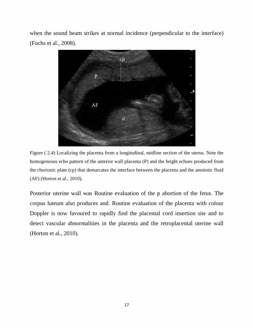
Figure ( 2.4) Localizing the placenta from a longitudinal, midline section of the uterus. Note the
homogeneous echo pattern of the anterior wall placenta (P) and the bright echoes produced from
the chorionic plate (cp) that demarcates the interface between the placenta and the amniotic fluid
(AF) (Horton et al., 2010).
Posterior uterine wall was Routine evaluation of the p abortion of the fetus. The
corpus luteum also produces and. Routine evaluation of the placenta with colour
Doppler is now favoured to rapidly find the placental cord insertion site and to
detect vascular abnormalities in the placenta and the retroplacental uterine wall
(Horton et al., 2010).

18
Figure (2.5) Color doppler image show posterior placenta and umblical cord.
(Horton et al., 2010).
The retroplacental uterine wall consists of the richly vascular myometrium and
decidua basalis. These tissues are distinctly hypoechoic in comparison to the
placenta. After 18 weeks gestation, the normal anterior retroplacental uterine wall
(sometimes referred to as the subplacental complex or the retroplacental space) has
an average thickness of 9.5 mm. The sonographic diagnosis of placental creta
depends on this normal hypoechoic zone being invaded by more echogenic villi
and appearing thinner or not seen (Oppenheimer et al., 2001).
2.8.2 Sonographic technique:
The equipment and transducer deemed most appropriate for the obstetrical
ultrasound study may be used). If the system has electronic beam focussing, the
focal zone should be adjusted to optimally visualize the placenta. The placenta is
best identified by scanning the uterus longitudinally and is easily recognized by its
more echogenic pattern compared with that of the underlying myometrium
(Oppenheimer et al., 2001).
A posterior placenta is more difficult to visualize in its entirety due to attenuation
and shadowing from the overlying fetus. If indicated, positioning the patient in a
19
left or right posterior oblique position may be helpful in better visualizing a
posterior placenta. For the standard trans-abdominal study (TAS), the bladder
should be adequately distended to optimize visualization of the cervix and lower
uterine segment and to show the relationship of the placenta to the internal os.
Over distention of the bladder distorts the appearance of the cervix and lower
uterine segment and may lead to the false positive diagnosis of placenta previa.
Endovaginal (EVS) or transperineal (TPS) techniques should be performed
whenever TAS does not adequately show the relationship of the placenta to the
internal os (e.g. due to attenuation by fetal parts or the patient presents with an
empty bladder) and there is a high index of suspicion of placenta previa (e.g.
patient presents with third trimester bleeding) (Oppenheimer et al., 2001).
2.9 Previous studies:
The relationship between previous cesarean section and subsequent development
of placenta previa and placenta previa with accrete has been assessed by (To and
Leung, 1995), The records of all patients delivered with the diagnosis of placenta
previa during the 10-year period from 1984 to 1993 were reviewed, the result that
From a total of 50,485 deliveries, 421 (0.83%) had placenta previa, 43 (10.2%) of
whom had a history of previous cesarean section. The incidence of placenta previa
was significantly increased in those with a previous cesarean section (1.31%)
compared with those with an unscarred uterus (0.75%) (R.R. 1.64). This risk
increased as the number of previous cesarean sections increased (R.R. 1.53 for one
previous section, 2.63 for two or more). The incidence of an anterior placenta
previa and placenta accreta was significantly increased in those with previous
cesarean scars. The incidence of placenta accreta was 1.18% among patients with
placenta previa, 80% being in patients with previous cesarean section. The relative
risk for placenta accreta in patients with placenta previa was 35 times higher in
those with a previous cesarean section than in those with an unscarred uterus.
20
Study of (Ahmed et al., 2015) aimed to identify the association of placenta previa
with multiparity and previous caesarean section in pregnant women. In antenatal
clinic as per protocol 200 pregnant women were scanned in their second and third
trimester for fetal wellbeing and placental localization after taking a detailed
obstetrical history and clinical examination. All women with or without symptoms
of placenta previa showing placental implantation in lower uterine segment on
ultra sound scan were documented. After completion of the two years data
regarding the detailed obstetrical and surgical history were recorded in a
questionnaire and analyzed using SPSS Software. Sixty five women were
diagnosed as cases of placenta previa. The overall incidence of placenta previa was
found to be 32.5% (65 women). Out of these 7 were primigrvidas, 12 were
multiparous, 34 were grand multiparous .It was clearly evident from the study that
placenta previa is associated with multiparity and previous caesarean section.
Placenta previa was highly significantly associated with previous caesarean section
(P =0000 <0.05). As well as, with multiparity and the association was found to be
as high as previous caesarean section (P =0000<0.05).
21
Chapter three
Material and method
3.1 Materials:
3.1.1 Subjects:
Total sample of 56 pregnant women with history of caesarian section were include
in the study. Pregnant women with no history of caesarian section were excluded.
3.1.2 Machine used:
Ultra sound machine with transducer frequency 3.5MHz, Mindary machine model
DP2200 (2008), made in Germany.
3.2 Method:
3.2.1 Technique used:
Verbal informed consent for the examination was obtained from each patient. The
women were scanned in supine position; coupling agent was applied to ensure
good contrast, curve linear transducer putted longitudinally below women
umbilicus.
The placenta was located transabdominally with a normally filled bladder; for the
purposes of this study and to obtain consistent findings between all operators, it
was decided to have only five placental-location subgroups. If the placenta was
thought to be ‘right anterior’, it was classified as ‘anterior’, and similarly ‘left
posterior’ was considered to be ‘posterior’. Therefore, placental locations were
recorded using the following five subgroups: anterior, posterior, fundal, low-lying
and previa.
3.2.2 Data collection:
This is descriptive cross section study conduct at the department of obstetrics and
gynecology in Bahry educational hospital and maternal specialist hospital and
Elshaikh Ali Fadoul hospital; it was conduct during the period from April to august

22
2017. The data obtained from the patients include (7) variables (age, gravidity,
parity, number of caesarian section, placenta site, placenta pathology and
gestational age).
3.2.3 Data analysis:
The data were analyzed using (SPSS Software) statistical social package for social
sciences (Version 20 SPSS, Chicago, Illinois USA). Descriptive statistics were
calculated for every measured variable, in order to evaluate the studied
sample. All analyses were performed using descriptive frequency and crosstabs
probabilities and a P value of p <0.05 was considered statistically significant.
3.2.4 Ethical approval:
The ethical approval was granted from the hospital and the radiology department;
which include commitment of no disclose of any information concerning the
patient identification.

23
Chapter four
Results
Table (4.1) shows descriptive Statistics.
N Minimum Maximum Mean Std.
Deviation
Age (years) 56 18 39 29.34 5.118
Gravidity 56 2 9 4.11 1.846
Parity 56 1 8 2.93 1.746
Number of Cesarean
Section 56 1 6 2.34 1.352
Gestational age(weeks) 56 17 39 30.96 5.507
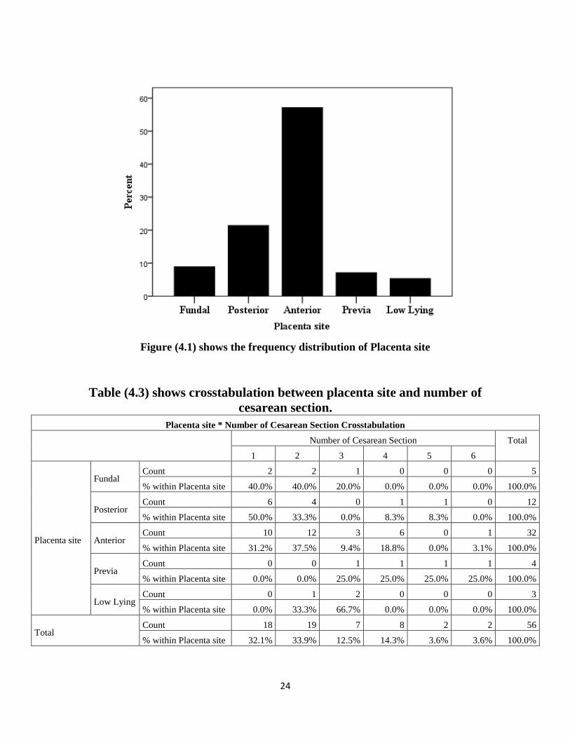
Table (4.2) shows the frequency distribution of Placenta site
Frequency Percent Valid
Percent
Cumulative
Percent
Fundal 5 8.9 8.9 8.9
Posterior 12 21.4 21.4 30.4
Anterior 32 57.1 57.1 87.5
Previa 4 7.1 7.1 94.6
Low Lying 3 5.4 5.4 100.0
Total 56 100.0 100.0
24
Figure (4.1) shows the frequency distribution of Placenta site
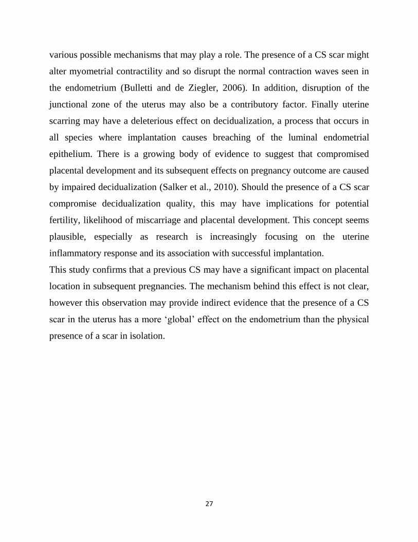
Table (4.3) shows crosstabulation between placenta site and number of
cesarean section.
Placenta site * Number of Cesarean Section Crosstabulation
Number of Cesarean Section Total
1 2 3 4 5 6
Placenta site
Fundal Count 2 2 1 0 0 0 5
% within Placenta site 40.0% 40.0% 20.0% 0.0% 0.0% 0.0% 100.0%
Posterior Count 6 4 0 1 1 0 12
% within Placenta site 50.0% 33.3% 0.0% 8.3% 8.3% 0.0% 100.0%
Anterior Count 10 12 3 6 0 1 32
% within Placenta site 31.2% 37.5% 9.4% 18.8% 0.0% 3.1% 100.0%
Previa Count 0 0 1 1 1 1 4
% within Placenta site 0.0% 0.0% 25.0% 25.0% 25.0% 25.0% 100.0%
Low Lying Count 0 1 2 0 0 0 3
% within Placenta site 0.0% 33.3% 66.7% 0.0% 0.0% 0.0% 100.0%
Total Count 18 19 7 8 2 2 56
% within Placenta site 32.1% 33.9% 12.5% 14.3% 3.6% 3.6% 100.0%

25
Table (4.4) shows crosstabulation between number of parity and placenta
location.
Parity * Placenta site Cross tabulation
Placenta site Total
Fundal Posterior Anterior Previa Low Lying
Parity
1 Count 2 5 5 0 0 12
% within Parity 16.7% 41.7% 41.7% 0.0% 0.0% 100.0%
2 Count 1 4 11 0 1 17
% within Parity 5.9% 23.5% 64.7% 0.0% 5.9% 100.0%
3 Count 2 1 5 1 2 11
% within Parity 18.2% 9.1% 45.5% 9.1% 18.2% 100.0%
4 Count 0 1 2 1 0 4
% within Parity 0.0% 25.0% 50.0% 25.0% 0.0% 100.0%
5 Count 0 0 4 1 0 5
% within Parity 0.0% 0.0% 80.0% 20.0% 0.0% 100.0%
6 Count 0 1 4 1 0 6
% within Parity 0.0% 16.7% 66.7% 16.7% 0.0% 100.0%
8 Count 0 0 1 0 0 1
% within Parity 0.0% 0.0% 100.0% 0.0% 0.0% 100.0%
Total Count 5 12 32 4 3 56
% within Parity 8.9% 21.4% 57.1% 7.1% 5.4% 100.0%

26
Chapter five
Discussion, Conclusion and Recommendations
5.1 Discussion:
This was descriptive study which included 56 pregnant women aged between 18-
39, with mean age 29.3 ±5.118 and gravidity between 2-9 with mean 4.11±1.846
and parity between 1- 8 with mean 2±1.7. The mean number of caesarian section
was 2 ±1.3.
The result of the study showed that the fundal placenta was 8.9%, posterior
placenta was 21.4%, anterior placenta was 57.1%, placenta previa was 7.1% and
low lying placenta was 5.4% (Figure 4.1). The incidence of placenta previa varies
between different reports. Me was found, 2.0%, for women with at least one prior
cesarean section is similar to that previously reported by (1.31%) et al. (To and
Leung, 1995). This results disconfirm the results of Naji et al. (Naji et al., 2013)
stating that more placentae are posterior in women with a previous cesarean
section. On the contrary, I found a majority of the placentae to be anterior, which
also holds true for placenta previa.
I have shown that there is a significant difference in placental location when there
is a CS scar present on the uterus. Women with a previous history of CS are
significantly less likely to have a fundal placenta and more likely to have the
placenta located on the anterior and posterior wall of the uterus in a subsequent
pregnancy. I found no statistically significant difference in the incidence of low-
lying placentae between the groups, though this may have been due to the low
numbers of low-lying placentae encountered overall. My data also suggest that the
presence of a CS scar does not influence placental migration in the event of a low-
lying placenta. My observations suggest that the presence of a CS scar influences
placentation. However, the significance of this observation is not clear. There are
27
various possible mechanisms that may play a role. The presence of a CS scar might
alter myometrial contractility and so disrupt the normal contraction waves seen in
the endometrium (Bulletti and de Ziegler, 2006). In addition, disruption of the
junctional zone of the uterus may also be a contributory factor. Finally uterine
scarring may have a deleterious effect on decidualization, a process that occurs in
all species where implantation causes breaching of the luminal endometrial
epithelium. There is a growing body of evidence to suggest that compromised
placental development and its subsequent effects on pregnancy outcome are caused
by impaired decidualization (Salker et al., 2010). Should the presence of a CS scar
compromise decidualization quality, this may have implications for potential
fertility, likelihood of miscarriage and placental development. This concept seems
plausible, especially as research is increasingly focusing on the uterine
inflammatory response and its association with successful implantation.
This study confirms that a previous CS may have a significant impact on placental
location in subsequent pregnancies. The mechanism behind this effect is not clear,
however this observation may provide indirect evidence that the presence of a CS
scar in the uterus has a more ‘global’ effect on the endometrium than the physical
presence of a scar in isolation.
28
5.2 Conclusion
U/s is most accurate in detection of placenta location from its first appearance until
labor. The most common location was anterior placenta, the least common location
was low laying placenta and previa. The prevalence of placenta previa was 7.1.
Number of CS had significant effect in placenta location. the parity was not
effected on placenta location.
29
5.3 Recommendations:
The sonologist should be more accurate in determine of entire length of
placeta and document both upper and lower margin.
Further studies should be done large sample volume and control group to
compare the prevalence of placenta privia on both groups.
Further studies should be done to assess other pregnancy associated
complication with previous CS.
30
References: AHMED, A. & GILBERT-BARNESS, E. 2003. Placenta membranacea: a developmental
anomaly with diverse clinical presentation. Pediatr Dev Pathol, 6, 201-2.
AHMED, I., ALI, Q., A, M. & M, S. 2015. Association of Caesarean Section and Multiparty
With Placenta Previa in Sudan. IOSR Journal of Dental and Medical Sciences (IOSR-
JDMS), 14, 29-32.
ALLEN, W. R., WILSHER, S., STEWART, F., OUSEY, J. & FOWDEN, A. 2002. The
influence of maternal size on placental, fetal and postnatal growth in the horse. II.
Endocrinology of pregnancy. J Endocrinol, 172, 237-46.
ALVAREZ-GORIS, M. P., DE LA TORRE Y FERNANDEZ, P., HUERTA-HENTSCHEL, J.
M. & SONCHEZ-ZAMORA, R. 2016. [Placental site trophoblastic tumor with atypical
choriocarcinoma. Case Report]. Ginecol Obstet Mex, 84, 324-9.
BECKER, R. H., VONK, R., MENDE, B. C., RAGOSCH, V. & ENTEZAMI, M. 2001. The
relevance of placental location at 20-23 gestational weeks for prediction of placenta
previa at delivery: evaluation of 8650 cases. Ultrasound Obstet Gynecol, 17, 496-501.
BELACHEW, J., EURENIUS, K., MULIC-LUTVICA, A. & AXELSSON, O. 2017. Placental
location, postpartum hemorrhage and retained placenta in women with a previous
cesarean section delivery: a prospective cohort study. Ups J Med Sci, 122, 185-189.
BETRAN, A. P., MERIALDI, M., LAUER, J. A., BING-SHUN, W., THOMAS, J., VAN
LOOK, P. & WAGNER, M. 2007. Rates of caesarean section: analysis of global,
regional and national estimates. Paediatr Perinat Epidemiol, 21, 98-113.
BULLETTI, C. & DE ZIEGLER, D. 2006. Uterine contractility and embryo implantation. Curr
Opin Obstet Gynecol, 18, 473-84.
CHO, J. Y., LEE, Y. H., MOON, M. H. & LEE, J. H. 2008. Difference in migration of placenta
according to the location and type of placenta previa. J Clin Ultrasound, 36, 79-84.
FUCHS, I., DUDENHAUSEN, J. W., SEHOULI, J. & HENRICH, W. 2008. Placenta pathology:
disorders of placental location, placental implantation and cord insertion. Ultraschall
Med, 29, 4-17; quiz 18-23.
HORTON, A. W., PATEL, U. & BELLI, A. M. 2010. An unusual arterial supply to the uterus. A
case report and review of anatomy-implications for uterine artery embolization. Clin
Radiol, 65, 1038-42.
31
HUNTER, D. S., HAZEL, S. J., KIND, K. L., LIU, H., MARINI, D., GILES, L. C., DE
BLASIO, M. J., OWENS, J. A., PITCHER, J. B. & GATFORD, K. L. 2015. Placental
and fetal growth restriction, size at birth and neonatal growth alter cognitive function and
behaviour in sheep in an age- and sex-specific manner. Physiol Behav, 152, 1-10.
ISHIKAWA, H., SEKI, R., YOKONISHI, S., YAMAUCHI, T. & YOKOYAMA, K. 2006.
Relationship between fetal weight, placental growth and litter size in mice from mid- to
late-gestation. Reprod Toxicol, 21, 267-70.
KORKMAZ, C., SAKINCI, M., AKYOL, S. N., KORGUN, E. T. & OZOGUL, C. 2013.
Location of proliferating cell nuclear antigen and p53 protein in human first trimester and
term placenta. Anal Quant Cytopathol Histpathol, 35, 335-43.
MATSUBARA, S., BABA, Y. & TAKAHASHI, H. 2017. Placenta previa and hemorrhage: the
placental location may be an important determinant of the bleeding amount. J Matern
Fetal Neonatal Med, 1-2.
MIYAKE, H., MIYAZAKI-IGARASHI, M. & SUZUKI, S. 2015. Placenta with Old, Diffuse
Infarction that Was Difficult to Differentiate from a Placental Tumor. J Nippon Med Sch,
82, 156-8.
NAJI, O., WYNANTS, L., SMITH, A., ABDALLAH, Y., SASO, S., STALDER, C., VAN
HUFFEL, S., GHAEM-MAGHAMI, S., VAN CALSTER, B., TIMMERMAN, D. &
BOURNE, T. 2013. Does the presence of a Caesarean section scar affect implantation
site and early pregnancy outcome in women attending an early pregnancy assessment
unit? Hum Reprod, 28, 1489-96.
NIKNEJADI, M., AHMADI, F. & AKHBARI, F. 2016. Imaging and Clinical Data of Placental
Site Trophoblastic Tumor: A Case Report. Iran J Radiol, 13, e18480.
OPPENHEIMER, L., HOLMES, P., SIMPSON, N. & DABROWSKI, A. 2001. Diagnosis of
low-lying placenta: can migration in the third trimester predict outcome? Ultrasound
Obstet Gynecol, 18, 100-2.
PROCTOR, L. K., TOAL, M., KEATING, S., CHITAYAT, D., OKUN, N., WINDRIM, R. C.,
SMITH, G. C. & KINGDOM, J. C. 2009. Placental size and the prediction of severe
early-onset intrauterine growth restriction in women with low pregnancy-associated
plasma protein-A. Ultrasound Obstet Gynecol, 34, 274-82.
32
RAVANGARD, S. F., HENDERSON, K. & FULLER, K. 2013. Placenta membranacea. Arch
Gynecol Obstet, 288, 709-12.
SALKER, M., TEKLENBURG, G., MOLOKHIA, M., LAVERY, S., TREW, G.,
AOJANEPONG, T., MARDON, H. J., LOKUGAMAGE, A. U., RAI, R., LANDLES, C.,
ROELEN, B. A., QUENBY, S., KUIJK, E. W., KAVELAARS, A., HEIJNEN, C. J.,
REGAN, L., MACKLON, N. S. & BROSENS, J. J. 2010. Natural selection of human
embryos: impaired decidualization of endometrium disables embryo-maternal
interactions and causes recurrent pregnancy loss. PLoS One, 5, e10287.
SEKIGUCHI, A., NAKAI, A., KAWABATA, I., HAYASHI, M. & TAKESHITA, T. 2013.
Type and location of placenta previa affect preterm delivery risk related to antepartum
hemorrhage. Int J Med Sci, 10, 1683-8.
SOLHEIM, K. N., ESAKOFF, T. F., LITTLE, S. E., CHENG, Y. W., SPARKS, T. N. &
CAUGHEY, A. B. 2011. The effect of cesarean delivery rates on the future incidence of
placenta previa, placenta accreta, and maternal mortality. J Matern Fetal Neonatal Med,
24, 1341-6.
TO, W. W. & LEUNG, W. C. 1995. Placenta previa and previous cesarean section. Int J
Gynaecol Obstet, 51, 25-31.
WONG, H. S., CHEUNG, Y. K., ZUCCOLLO, J., TAIT, J. & PRINGLE, K. C. 2008.
Evaluation of sonographic diagnostic criteria for placenta accreta. J Clin Ultrasound, 36,
551-9.
YOUNG, B. C., NADEL, A. & KAIMAL, A. 2014. Does previa location matter? Surgical
morbidity associated with location of a placenta previa. J Perinatol, 34, 264-7.
34
Appendices
Appendix (1)
Ultrasound image (1) shows posterior placenta at 38 weeeks in PG women with
one Cs
Ultrasound image (2) shows anterior placenta at 37 weeks +6 day in PG women
withone cesarean section
35
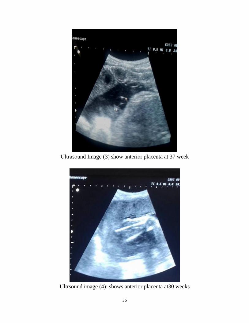
Ultrasound Image (3) show anterior placenta at 37 week
Ultrsound image (4): shows anterior placenta at30 weeks
36
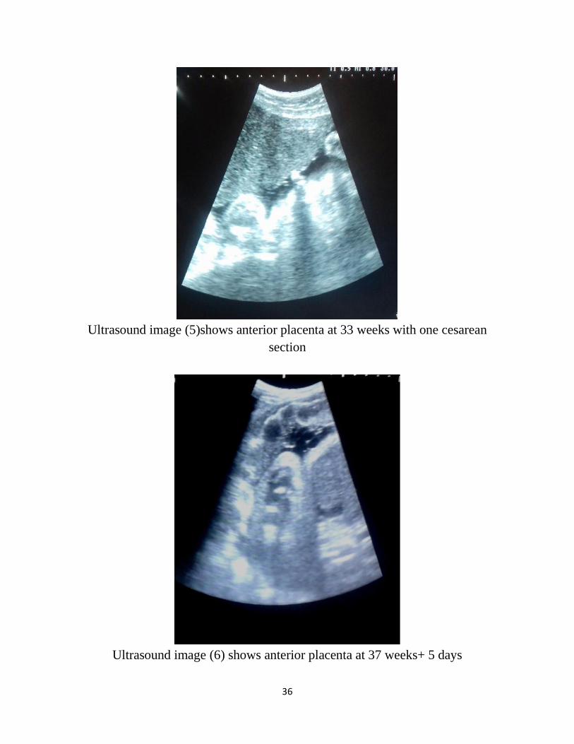
Ultrasound image (5)shows anterior placenta at 33 weeks with one cesarean
section
Ultrasound image (6) shows anterior placenta at 37 weeks+ 5 days

37
Ultrasound image(7) shows anterior placenta at 36 weeks GA
Ultrasound image (8) shows fundal placenta at 39 weeks with one cesarean section

38
Ultrasound image (9) shows posterior placenta at 34 weeks
Ultrasound image(10) shows posterior placenta at 28 weeks

39
Ultrasound image (11) shows anterior placenta at 32 weeks +5 day in PG women
with 4 cesarean section
Ultrasound image (12) shows anterior placenta at 37 weeks
40
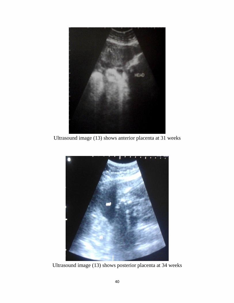
Ultrasound image (13) shows anterior placenta at 31 weeks
Ultrasound image (13) shows posterior placenta at 34 weeks

41
Ultrasound image (14) shows anterior placenta at 39 week +3 d
Ultrasound image number(15) shows anterior placenta at 34 weeks
42
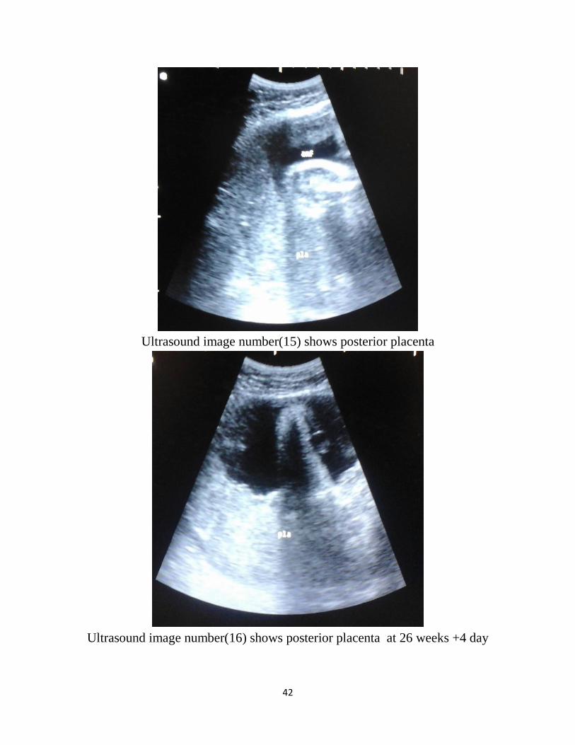
Ultrasound image number(15) shows posterior placenta
Ultrasound image number(16) shows posterior placenta at 26 weeks +4 day
43
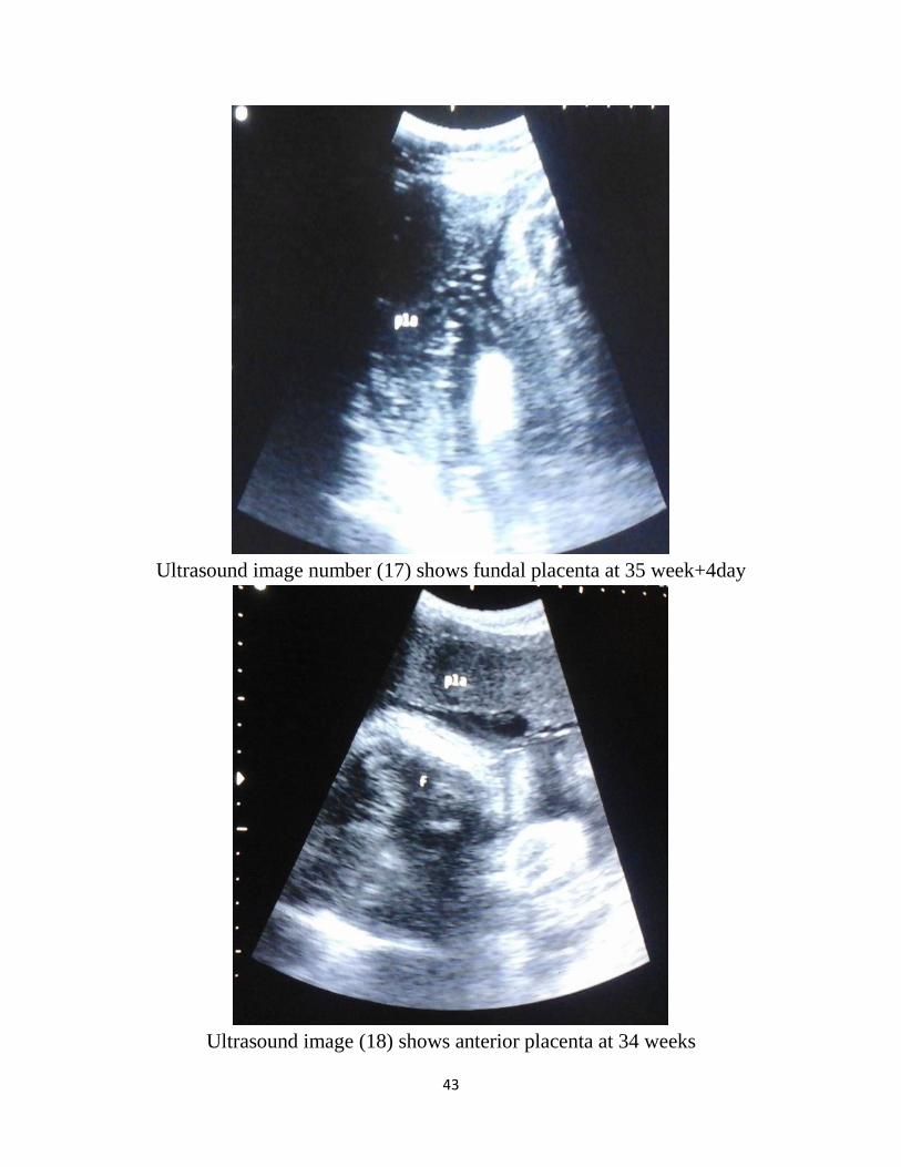
Ultrasound image number (17) shows fundal placenta at 35 week+4day
Ultrasound image (18) shows anterior placenta at 34 weeks
44
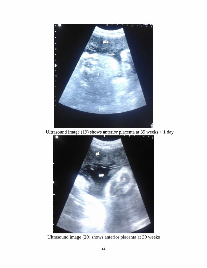
Ultrasound image (19) shows anterior placenta at 35 weeks + 1 day
Ultrasound image (20) shows anterior placenta at 30 weeks
45
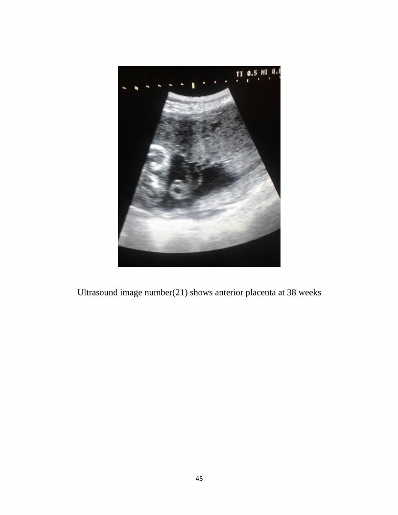
Ultrasound image number(21) shows anterior placenta at 38 weeks
46
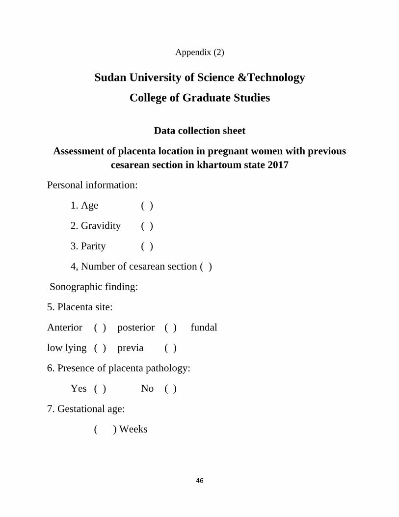
Appendix (2)
Sudan University of Science &Technology
College of Graduate Studies
Data collection sheet
Assessment of placenta location in pregnant women with previous
cesarean section in khartoum state 2017
Personal information:
1. Age ( )
2. Gravidity ( )
3. Parity ( )
4, Number of cesarean section ( )
Sonographic finding:
5. Placenta site:
Anterior ( ) posterior ( ) fundal
low lying ( ) previa ( )
6. Presence of placenta pathology:
Yes ( ) No ( )
7. Gestational age:
( ) Weeks
