
Substrate and Cofactor Range Differences of Two Cysteine ... · and CdoB from R. eutropha H16 (top)...
12
Substrate and Cofactor Range Differences of Two Cysteine Dioxygenases from Ralstonia eutropha H16 Leonie Wenning, a * Nadine Stöveken, a Jan Hendrik Wübbeler, a Alexander Steinbüchel a,b Institut für Molekulare Mikrobiologie und Biotechnologie, Westfälische Wilhelms-Universität Münster, Münster, Germany a ; Faculty of Environmental Sciences, King Abdulaziz University, Jeddah, Saudi Arabia b Cysteine dioxygenases (Cdos), which catalyze the sulfoxidation of cysteine to cysteine sulfinic acid (CSA), have been extensively studied in eukaryotes because of their roles in several diseases. In contrast, only a few prokaryotic enzymes of this type have been investigated. In Ralstonia eutropha H16, two Cdo homologues (CdoA and CdoB) have been identified previously. In vivo studies showed that Escherichia coli cells expressing CdoA could convert 3-mercaptopropionate (3MP) to 3-sulfinopropionate (3SP), whereas no 3SP could be detected in cells expressing CdoB. The objective of this study was to confirm these findings and to study both enzymes in detail by performing an in vitro characterization. The proteins were heterologously expressed and purified to apparent homogeneity by immobilized metal chelate affinity chromatography (IMAC). Subsequent analysis of the enzyme activ- ities revealed striking differences with regard to their substrate ranges and their specificities for the transition metal cofactor, e.g., CdoA catalyzed the sulfoxidation of 3MP to a 3-fold-greater extent than the sulfoxidation of cysteine, whereas CdoB con- verted only cysteine. Moreover, the dependency of the activities of the Cdos from R. eutropha H16 on the metal cofactor in the active center could be demonstrated. The importance of CdoA for the metabolism of the sulfur compounds 3,3=-thiodipropionic acid (TDP) and 3,3=-dithiodipropionic acid (DTDP) by further converting their degradation product, 3MP, was confirmed. Since 3MP can also function as a precursor for polythioester (PTE) synthesis in R. eutropha H16, deletion of cdoA might enable in- creased synthesis of PTEs. C ysteine dioxygenases (Cdos) are thiol-oxygenating enzymes that are well characterized in eukaryotes (1, 2). They catalyze the oxidative conversion of cysteine into cysteine sulfinic acid (CSA) and perform the first step in the catabolism of the highly reactive amino acid cysteine (Fig. 1). Because several neurological disorders, like Alzheimer’s and Parkinson’s diseases (3) and Hallervorden-Spatz disease (4), have been linked to excess levels of cysteine in plasma or the lack of cerebral cysteine dioxygenase activity, the enzyme is exceedingly interesting for medical re- search. Several analyses of the crystal structure were performed, using recombinant Cdos from different mammalian sources (5–7), and revealed an alternative structural motif for coordination of the iron cofactor by Cdos. Whereas most of the nonheme iron pro- teins coordinate the metal via two histidine residues and a carbox- ylic acid group (the 2-His–1-carboxylate facial triad), the ferrous iron in Cdos is arranged in a mutually cis geometry consisting of three histidine residues (3-His facial triad) (1, 8, 9). The loss of Cdo activity after immobilized metal chelate affinity chromatog- raphy (IMAC) purification was reported in several studies (10– 12), and the activity could be reconstituted only by addition of exogenous ferrous iron, whereas other transition metals failed to restore the activity. In addition, the inhibition of Cdo activity by chelating agents, like 1,10-phenanthroline or EDTA (13), empha- sized the strict dependency of the previously characterized Cdos on ferrous iron. Another unique feature of mammalian Cdo is the formation of a cross-linked Cys-Tyr cofactor that is regulated by cysteine and represents an unusual form of substrate-mediated feed-forward activation of enzyme activity (14). The formation of the Cys-Tyr cofactor requires a transition metal [Fe(II)], as well as oxygen, and it is also strictly dependent on the specific Cdo sub- strate cysteine (15). In eukaryotes, the mature Cys-Tyr cofactor- containing Cdo and the cofactor-free enzyme exist. Both forms show catalytic activity, but the cofactor formation leads to a 10- fold increase of Cdo activity and also a prolonged catalytic half-life (14, 16). Besides eukaryotes, the enzyme was also identified in several eubacteria (11, 15, 17–19). Although the translational products of these homologous genes showed only low overall sequence iden- tity to eukaryotic Cdos, structural and catalytic studies verified that the presence of the enzyme is not restricted to higher organ- isms (11). In 2009, we identified a Cdo homologue in the Gram- negative bacterium Variovorax paradoxus TBEA6 (17). Enzymatic studies showed that the enzyme catalyzed the unusual oxidation of 3-mercaptopropionate (3MP) to yield 3-sulfinopropionate (3SP). Because no oxygenation of other thiols, like cysteine or cysteam- ine, was observed, the enzyme was referred to as 3MP dioxygenase (Mdo) (17). The conversion of 3MP into 3SP was also shown for the orthologous Mdo in the 3,3=-dithiodipropionic acid (DTDP)- utilizing bacterium Advenella mimigardefordensis DPN7 T and for a Cdo homologue from Pseudomonas aeruginosa, which is also able to convert cysteine, but to a much lesser extent (18–20). Received 7 August 2015 Accepted 17 November 2015 Accepted manuscript posted online 20 November 2015 Citation Wenning L, Stöveken N, Wübbeler JH, Steinbüchel A. 2016. Substrate and cofactor range differences of two cysteine dioxygenases from Ralstonia eutropha H16. Appl Environ Microbiol 82:910 –921. doi:10.1128/AEM.02568-15. Editor: M. J. Pettinari Address correspondence to Alexander Steinbüchel, [email protected]. * Present address: Leonie Wenning, Chalmers University of Technology, Gothenburg, Sweden. Supplemental material for this article may be found at http://dx.doi.org/10.1128 /AEM.02568-15. Copyright © 2016, American Society for Microbiology. All Rights Reserved. crossmark 910 aem.asm.org February 2016 Volume 82 Number 3 Applied and Environmental Microbiology on June 10, 2019 by guest http://aem.asm.org/ Downloaded from
-
Upload
truonghanh -
Category
Documents
-
view
214 -
download
0
Transcript of Substrate and Cofactor Range Differences of Two Cysteine ... · and CdoB from R. eutropha H16 (top)...

Substrate and Cofactor Range Differences of Two CysteineDioxygenases from Ralstonia eutropha H16
Leonie Wenning,a* Nadine Stöveken,a Jan Hendrik Wübbeler,a Alexander Steinbüchela,b
Institut für Molekulare Mikrobiologie und Biotechnologie, Westfälische Wilhelms-Universität Münster, Münster, Germanya; Faculty of Environmental Sciences, KingAbdulaziz University, Jeddah, Saudi Arabiab
Cysteine dioxygenases (Cdos), which catalyze the sulfoxidation of cysteine to cysteine sulfinic acid (CSA), have been extensivelystudied in eukaryotes because of their roles in several diseases. In contrast, only a few prokaryotic enzymes of this type have beeninvestigated. In Ralstonia eutropha H16, two Cdo homologues (CdoA and CdoB) have been identified previously. In vivo studiesshowed that Escherichia coli cells expressing CdoA could convert 3-mercaptopropionate (3MP) to 3-sulfinopropionate (3SP),whereas no 3SP could be detected in cells expressing CdoB. The objective of this study was to confirm these findings and to studyboth enzymes in detail by performing an in vitro characterization. The proteins were heterologously expressed and purified toapparent homogeneity by immobilized metal chelate affinity chromatography (IMAC). Subsequent analysis of the enzyme activ-ities revealed striking differences with regard to their substrate ranges and their specificities for the transition metal cofactor,e.g., CdoA catalyzed the sulfoxidation of 3MP to a 3-fold-greater extent than the sulfoxidation of cysteine, whereas CdoB con-verted only cysteine. Moreover, the dependency of the activities of the Cdos from R. eutropha H16 on the metal cofactor in theactive center could be demonstrated. The importance of CdoA for the metabolism of the sulfur compounds 3,3=-thiodipropionicacid (TDP) and 3,3=-dithiodipropionic acid (DTDP) by further converting their degradation product, 3MP, was confirmed. Since3MP can also function as a precursor for polythioester (PTE) synthesis in R. eutropha H16, deletion of cdoA might enable in-creased synthesis of PTEs.
Cysteine dioxygenases (Cdos) are thiol-oxygenating enzymesthat are well characterized in eukaryotes (1, 2). They catalyze
the oxidative conversion of cysteine into cysteine sulfinic acid(CSA) and perform the first step in the catabolism of the highlyreactive amino acid cysteine (Fig. 1). Because several neurologicaldisorders, like Alzheimer’s and Parkinson’s diseases (3) andHallervorden-Spatz disease (4), have been linked to excess levelsof cysteine in plasma or the lack of cerebral cysteine dioxygenaseactivity, the enzyme is exceedingly interesting for medical re-search.
Several analyses of the crystal structure were performed, usingrecombinant Cdos from different mammalian sources (5–7), andrevealed an alternative structural motif for coordination of theiron cofactor by Cdos. Whereas most of the nonheme iron pro-teins coordinate the metal via two histidine residues and a carbox-ylic acid group (the 2-His–1-carboxylate facial triad), the ferrousiron in Cdos is arranged in a mutually cis geometry consisting ofthree histidine residues (3-His facial triad) (1, 8, 9). The loss ofCdo activity after immobilized metal chelate affinity chromatog-raphy (IMAC) purification was reported in several studies (10–12), and the activity could be reconstituted only by addition ofexogenous ferrous iron, whereas other transition metals failed torestore the activity. In addition, the inhibition of Cdo activity bychelating agents, like 1,10-phenanthroline or EDTA (13), empha-sized the strict dependency of the previously characterized Cdoson ferrous iron. Another unique feature of mammalian Cdo is theformation of a cross-linked Cys-Tyr cofactor that is regulated bycysteine and represents an unusual form of substrate-mediatedfeed-forward activation of enzyme activity (14). The formation ofthe Cys-Tyr cofactor requires a transition metal [Fe(II)], as well asoxygen, and it is also strictly dependent on the specific Cdo sub-strate cysteine (15). In eukaryotes, the mature Cys-Tyr cofactor-containing Cdo and the cofactor-free enzyme exist. Both forms
show catalytic activity, but the cofactor formation leads to a 10-fold increase of Cdo activity and also a prolonged catalytic half-life(14, 16).
Besides eukaryotes, the enzyme was also identified in severaleubacteria (11, 15, 17–19). Although the translational products ofthese homologous genes showed only low overall sequence iden-tity to eukaryotic Cdos, structural and catalytic studies verifiedthat the presence of the enzyme is not restricted to higher organ-isms (11). In 2009, we identified a Cdo homologue in the Gram-negative bacterium Variovorax paradoxus TBEA6 (17). Enzymaticstudies showed that the enzyme catalyzed the unusual oxidation of3-mercaptopropionate (3MP) to yield 3-sulfinopropionate (3SP).Because no oxygenation of other thiols, like cysteine or cysteam-ine, was observed, the enzyme was referred to as 3MP dioxygenase(Mdo) (17). The conversion of 3MP into 3SP was also shown forthe orthologous Mdo in the 3,3=-dithiodipropionic acid (DTDP)-utilizing bacterium Advenella mimigardefordensis DPN7T and fora Cdo homologue from Pseudomonas aeruginosa, which is alsoable to convert cysteine, but to a much lesser extent (18–20).
Received 7 August 2015 Accepted 17 November 2015
Accepted manuscript posted online 20 November 2015
Citation Wenning L, Stöveken N, Wübbeler JH, Steinbüchel A. 2016. Substrate andcofactor range differences of two cysteine dioxygenases from Ralstonia eutrophaH16. Appl Environ Microbiol 82:910 –921. doi:10.1128/AEM.02568-15.
Editor: M. J. Pettinari
Address correspondence to Alexander Steinbüchel, [email protected].
* Present address: Leonie Wenning, Chalmers University of Technology,Gothenburg, Sweden.
Supplemental material for this article may be found at http://dx.doi.org/10.1128/AEM.02568-15.
Copyright © 2016, American Society for Microbiology. All Rights Reserved.
crossmark
910 aem.asm.org February 2016 Volume 82 Number 3Applied and Environmental Microbiology
on June 10, 2019 by guesthttp://aem
.asm.org/
Dow
nloaded from

The genome sequence of Ralstonia eutropha H16 revealed twoparalogous cdo genes (21), and the amino acid sequences of thetranslational products (CdoA and CdoB) exhibited significant dif-ferences (36% identical amino acids), particularly in the conser-vation of the typical cupin motifs (22). It was demonstrated thatonly one of the two Cdos (CdoA) in R. eutropha H16 is able toconvert 3MP (17) (Fig. 1). In addition, 3MP, 3,3=-thiodipropionicacid (TDP), and DTDP can be used by R. eutropha H16 as precur-sor substrates for the biosynthesis of poly(3-hydroxybutyrate-co-3MP) copolymers when a second carbon source is available tosupport growth (23–25). Enzymatic cleavage of TDP into 3MPand 3-hydroxypropionate (3HP) was previously shown (17, 26),and the cleavage of DTDP into two molecules of 3MP by thedihydrolipoamide dehydrogenase PdhL has also been demon-strated (27).
In contrast to L-cysteine, 3MP does not occur naturally in cellsof R. eutropha H16. Nevertheless, it is produced in various biolog-ical and abiotic reactions and can be found in nanomolar concen-trations in natural aquatic environments (28). Thus, it is one ofthe most frequently detected thiols in these habitats (29, 30).Moreover, 3MP seems to be a central intermediate in sulfur me-tabolism with, e.g., an important role in the catabolism of dimeth-ylsulfoniopropionate, which is produced by several species of ma-rine phytoplankton, macroalgae, and some angiosperms (31–36).3MP can also be detected in freshwater habitats and hypolimneticwaters (28, 32, 37). Since 3MP is already toxic to cells in lowconcentrations, thereby hindering the growth of bacteria like R.eutropha, the ability to convert 3MP is advantageous for freshwa-ter bacteria like R. eutropha H16, and it could explain the signifi-cance of the two different Cdos in the bacterium (38–40).
To get a deeper knowledge of the two Cdos from R. eutrophaH16, we performed a biochemical characterization of the purifiedenzymes. Both Cdos were analyzed with regard to their substrateranges, their quaternary structures, their iron contents, their co-factor dependencies, and the inhibition of their catalytic activitiesby different thiols and chelating agents.
MATERIALS AND METHODSChemicals. Organic thiochemicals of high purity grade were purchasedfrom Acros Organics (Geel, Belgium) or Sigma-Aldrich (Steinheim, Ger-many). 3SP was synthesized as previously described (18, 41).
Bacterial strains and cultivation conditions. R. eutropha H16 wascultivated at 30°C in nutrient broth (NB) under aerobic conditionson a rotary shaker at an agitation of 130 rpm. Escherichia coliRosetta(DE3)(pLysS)(pET23a::cdoA), E. coli Lemo21(DE3)(pLemo)
(pET23a::cdoA), E. coli Rosetta(DE3)(pLysS)(pET23a::cdoB), and E. coliLemo21(DE3)(pLemo)(pET23a::cdoB) were cultivated in lysogeny broth(LB) medium. Carbon sources were supplied as filter-sterilized stock so-lutions, as indicated in the text. For maintenance of plasmids, antibioticswere prepared according to the method of Sambrook et al. (42) and addedto the media at the following concentrations: ampicillin, 75 �g/ml; car-benicillin, 75 �g/ml; and chloramphenicol, 34 �g/ml. Heterologous ex-pression of genes under the control of a lac promoter was induced by ad-dition of 0.3 mM IPTG (isopropyl-�-D-thiogalactopyranoside). WhereasCdoA was found as a highly soluble protein, the CdoB protein was exclu-sively synthesized in inclusion bodies. Therefore, the cultivation of re-combinant E. coli Lemo21(DE3)(pLemo)(pET23a::cdoB) and E. coliRosetta(DE3)(pLysS)(pET23a::cdoB) for heterologous expression of cdoBwas performed as previously described for a gene from Streptomyces coe-licolor (11). Due to the long time of cultivation, carbenicillin instead ofampicillin was used for maintaining plasmid stability. All the strains andplasmids used are listed in Table 1.
DNA isolation and manipulation. Chromosomal DNA of R. eutrophaH16 was isolated according to the method of Marmur (43). Plasmid DNAwas isolated from E. coli strains using the GeneJet plasmid miniprep kitfrom Fermentas (St. Leon-Rot, Germany) according to the manufactur-er’s manual. DNA was digested with restriction endonucleases under con-ditions described by the manufacturer or according to the method ofSambrook et al. (42). PCRs were carried out in an Omnigene HBTR3CMDNA thermal cycler (Hybaid, Heidelberg, Germany) using Platinum TaqDNA polymerase (Invitrogen, Karlsruhe, Germany). PCR products wereisolated from an agarose gel and purified using the NucleoTrap kit (Ma-chery and Nagel, Düren, Germany) according to the manufacturer’s in-structions. T4 DNA ligase was purchased from Invitrogen (Karlsruhe,Germany). Primers were synthesized by MWG-Biotech AG (Ebersberg,Germany).
Transfer of DNA. Competent cells of E. coli strains were prepared andtransformed by the CaCl2 procedure (42).
DNA sequencing and sequence data analysis. Samples were preparedfor sequencing using the BigDye Terminator v3.1 cycle-sequencingkit according to the manufacturer’s manual (Applied Biosystems,Darmstadt, Germany). Afterward, the samples were submitted to theInstitut für Klinische Chemie und Laboratoriumsmedizin at the Uni-versitätsklinikum Münster, Münster, Germany, for purification of theextension products and sequencing in an ABI Prism 3700 DNA Ana-lyzer (Applied Biosystems, Darmstadt, Germany). BLASTX was usedfor determination of nucleotide identity (44). For amino acid sequencealignments, Clustal_X (45) and BioEdit (46) were applied. The same se-quences were used for the calculation of a phylogenetic tree, employingthe neighbor-joining method (47). Besides CdoA and CdoB from R. eu-tropha H16, the well-known eukaryotic Cdos from rat and human wereselected, in addition to Cdos from the zebrafish, the nematode Caeno-rhabditis elegans, and the fungi Arthroderma benhamiae and Histoplasmacapsulatum. Representatives of prokaryotic thiol dioxygenases were cho-sen from Verminephrobacter eiseniae, Variovorax paradoxus, Advenellasp., Bordetella pertussis, Methylibium petroleiphilum, Pseudomonas putida,Cupriavidus sp., Ralstonia sp., and Bacillus sp. due to their sequence sim-ilarity to either CdoA or CdoB.
Cloning of cdoARe and cdoBRe. The R. eutropha cdoA (cdoARe) genewas amplified from total genomic DNA of R. eutropha strain H16 by PCRusing the high-fidelity enzyme PCR mix (MBI Fermentas, St. Leon-Rot,Germany) and the oligonucleotides cdoA(NdeI), 5=-CGAGACACTCCGGAGTCAATCATATG-3, and cdoA(XhoI), 5=-CTCGAGTCCTGACTGGGCAACTTCG-3=. The resulting PCR product was isolated from an aga-rose gel using the NucleoTrap kit (Machery and Nagel, Düren, Germany)and ligated with pJet 2.1/blunt DNA (Promega, Madison, WI, USA.). ThecdoB gene was amplified using Taq DNA polymerase (Invitrogen,Karlsruhe, Germany) and the oligonucleotides cdoB(NdeI), 5=-CATATGCCGACCGGATCTCCCCTG-3=, and cdoB(SalI), 5=-GCAAAACGTCGACGGCGTCGGC-3=. After purification of the PCR product, the gene was
FIG 1 Conversion of L-cysteine to L-cysteine sulfinic acid catalyzed by CdoAand CdoB from R. eutropha H16 (top) and conversion of 3-mercaptopropi-onate to 3-sulfinopropionate catalyzed by CdoA (bottom).
Characterization of CdoAB
February 2016 Volume 82 Number 3 aem.asm.org 911Applied and Environmental Microbiology
on June 10, 2019 by guesthttp://aem
.asm.org/
Dow
nloaded from

inserted into the cloning vector pCR2.1 TOPO (TOPO TA cloning kit;Invitrogen Corporation, Carlsbad, CA, USA) by topoisomerase reaction.E. coli Top10 was transformed with the resulting hybrid plasmids, andtransformants were selected on LB agar plates containing IPTG, 5-bromo-4-chloro-3-indolyl-�-D-galactopyranoside (X-Gal), and ampicillin. Forheterologous expression in the T7 promoter/polymerase-based expres-sion vector pET23a (Novagen, Madison, WI, USA), the cdo genes wereobtained by restriction of hybrid plasmids pJet2.1/blunt::cdoARe andpCR2.1TOPO::cdoBRe with NdeI and XhoI or NdeI and SalI, respectively.Upon purification from an agarose gel using the NucleoTrap kit (Macheryand Nagel, Düren, Germany) the genes were ligated into the expressionvector pET23a, which was linearized with the same restriction endonu-cleases. The ligation products were used for transformation of CaCl2-competent cells of E. coli Top10. After selection of transformants using LBmedium containing ampicillin, the hybrid plasmids were isolated, ana-lyzed by sequencing, and used for transformation of E. coliRosetta(DE3)(pLysS) (Novagen, Madison, WI, USA).
Analysis of enzyme assay reaction products by HPLC. Concentra-tions of 3SP, CSA, and hypotaurine were analyzed by high-performanceliquid chromatography (HPLC).
HPLC analysis of 3SP was carried out in a LaChrom Elite HPLC ap-paratus (VWR-Hitachi International GmbH, Darmstadt, Germany) con-sisting of a Metacarb 67H advanced C column (Varian, Palo Alto, CA,USA; Bio-Rad Aminex equivalent) and a 22350 VWR-Hitachi columnoven. The primary separation mechanism included ligand exchange, ionexclusion, and adsorption. A VWR-Hitachi refractive index (RI) detector(type 2490) with an active flow cell temperature control and automatedreference flushing eliminating temperature effects on the RI baseline wasused for detection. Aliquots of 20-�l cell-free supernatants, solutions of
organic sulfur compounds, or enzyme assay mixture were injected andeluted with 0.005 N sulfuric acid (H2SO4) at a flow rate of 0.8 ml/min anda column temperature of 50°C. Online integration and analysis were donewith EZ Chrome Elite software (VWR International GmbH, Darmstadt,Germany). Detection of hypotaurine and CSA was carried out in a Kon-tron Instrument (Neufahrn, Germany). After derivatization with OPAreagent (48) using a Smartline Autosampler 3900 (Knauer Advanced Sci-entific Instruments, Berlin, Germany), 20 �l of the reaction mixture wasinjected onto a Novapack C18 reverse-phase column (Knauer, Berlin, Ger-many) and monitored fluorimetrically at 330/450 nm (excitation/emis-sion) by using a model 1046A fluorescence detector (Hewlett Packard,Germany). Substances were identified by comparison of their retentiontimes to those of standard organic acids.
Preparation of crude extracts. Cell extracts were obtained either bylysis of the frozen cells using the detergent mixture BugBuster ExtractionReagent (primary amine free; Novagen, Madison, WI, USA) or by me-chanical disruption. For this, the cell pellet was resuspended in bindingbuffer (0.1 M Tris-HCl, 0.5 M NaCl, 20 mM imidazole, pH 7.5) andpassed three times through a chilled French press cell (Amicon, SilverSpring, MD, USA). Soluble protein fractions of crude extracts were ob-tained in the supernatants after 1 h of centrifugation at 100,000 � g at 4°Cand were subsequently used for enzyme purification.
SDS-polyacrylamide gel electrophoresis. Samples were resuspendedin gel loading buffer (0.6% [wt/vol] SDS, 1.25% [vol/vol] �-mercaptoeth-anol, 0.25 mM EDTA, 10% [vol/vol] glycerol, 0.0001% [wt/vol] bro-mophenol blue, and 12.5 mM Tris-HCl, pH 6.8). Proteins were denaturedby 5 min of incubation at 96°C and separated in 13% (wt/vol) SDS-poly-acrylamide gels, as described by Laemmli (49). The proteins were stainedwith Coomassie brilliant blue R-250 (50). Samples for SDS-polyacryl-
TABLE 1 Strains and plasmids
Strain/plasmid Relevant phenotype/genotypea
Reference orsourceb
R. eutropha H16 Wild type 62E. coli TOP10 F� mcrA �(mrr-hsdRMS-mcrBC) rpsL nupG �80lacZ�M15 �lacX74 deoR recA1
araD139 �(ara-leu)7697 galU galK endA1Invitrogen
E. coli Lemo21(DE3)(pLemo) fhuA2 [lon] ompT gal (� DE3) [dcm] �hsdS/pLemo(Camr) [� DE3 � sBamHIo�EcoRI-B int::(lacI::PlacUV5::T7 gene1) i21 �nin5; pLemo pACYC184-PrhaBAD-lysY]
New EnglandBiolabs Inc.
E. coli Rosetta(DE3)(pLysS) F� ompT hsdSB(rB� mB
�) gal dcm (DE3)(pLysSRARE) (Camr) NovagenE. coli TOP10(pET23a()::cdoA) F� mcrA �(mrr-hsdRMS-mcrBC) rpsL nupG �80lacZ�M15 �lacX74 deoR recA1
araD139 �(ara-leu)7697 galU galK endA1 Ampr (cdoA gene coding forCdoA of R. eutropha H16)
This work
E. coli TOP10(pET23a()::cdoB) F� mcrA �(mrr-hsdRMS-mcrBC) rpsL nupG �80lacZ�M15 �lacX74 deoR recA1araD139 �(ara-leu)7697 galU galK endA1 Ampr (cdoB gene coding forCdoB of R. eutropha H16)
This work
E. coli Rosetta(DE3)(pLysS) (pET23a::cdoA) F� ompT hsdSB(rB� mB
�) gal dcm (DE3)(pLysSRARE) (Camr) Ampr (cdoA gene coding for CdoA of R. eutropha H16)
This work
E. coli Lemo21(DE3)(pLemo)(pET23a::cdoA) fhuA2 [lon] ompT gal (� DE3) [dcm] �hsdS/pLemo(Camr) [� DE3 � sBamHIo�EcoRI-B int::(lacI::PlacUV5::T7 gene1) i21 �nin5; pLemo pACYC184-PrhaBAD-lysY Ampr; cdoA gene coding for CdoA of R. eutropha H16)
This work
E. coli Rosetta(DE3)(pLysS)(pET23a::cdoB) F� ompT hsdSB(rB� mB
�) gal dcm (DE3)(pLysSRARE) (Camr) Ampr (cdoB gene coding for CdoB of R. eutropha H16)
This work
E. coli Lemo21(DE3)(pLemo)(pET23a::cdoB) fhuA2 [lon] ompT gal (� DE3) [dcm] �hsdS/pLemo(Camr) [� DE3 � sBamHIo�EcoRI-B int::(lacI::PlacUV5::T7 gene1) i21 �nin5; pLemo pACYC184-PrhaBAD-lysY Ampr; cdoB gene coding for CdoB of R. eutropha H16)
This work
pCR2.1 TOPO Ampr Kanr TOPO TA cloningkit (Invitrogen)
pJet 2.1/blunt DNA Ampr PromegapET23a Ampr NovagenpET23a()::cdoA Ampr (cdoA gene coding for CdoA of R. eutropha H16) This workpET23a()::cdoB Ampr (cdoB gene coding for CdoB of R. eutropha H16) This worka Ampr, ampicillin resistance; Camr, chloramphenicol resistance; Kanr, kanamycin resistance. Genotype descriptions are based on those of Bachmann (63).b Sources: Invitrogen, Carlsbad, CA, USA; New England Biolabs Inc., Ipswich, MA, USA; NovagenMerck KGaA, Darmstadt, Germany; Promega Corporation, Madison, WI, USA.
Wenning et al.
912 aem.asm.org February 2016 Volume 82 Number 3Applied and Environmental Microbiology
on June 10, 2019 by guesthttp://aem
.asm.org/
Dow
nloaded from

amide gel electrophoresis under nonreducing conditions were preparedby 5-min incubation of protein at 96°C in loading buffer containing 20%sucrose and 0.1% (wt/vol) bromophenol blue.
IMAC. To obtain purified hexahistidine-tagged fusion Cdo, His SpinTrap affinity columns (GE Healthcare, Uppsala, Sweden) were used ac-cording to the instructions of the manufacturer with minor modifica-tions. Tris-HCl (0.1 M, pH 7.5) was used as a buffer component instead ofsodium phosphate, and for the washing step, a buffer containing 40 mMimidazole was applied. The washing step was repeated up to five times,and the elution step was repeated two times. The purification of Cdo fromcrude extracts obtained from 2-liter LB cultures was done by using His-Trap FF columns (GE Healthcare, Uppsala, Sweden) with a bed volume of1 ml. The binding buffer (0.1 M Tris-HCl, 0.5 M NaCl, 20 mM imidazole,pH 7.5) was used for application of the Cdo. The elution of the enzymewas achieved by a stepwise increasing gradient of imidazole. For this, thebinding buffer and the elution buffer (0.1 M Tris-HCl, 0.5 M NaCl, 500mM imidazole, pH 7.5) were mixed with a flow rate of 1 ml/min and amaximum pump pressure of 0.3 MPa. If BugBuster reagent (Novagen,Madison, WI, USA) was used for cell lysis, the obtained soluble cell frac-tion was diluted 1:1, using the 40 mM imidazole-containing washing buf-fer, prior to column loading to obtain a solution having the same imida-zole concentration as the binding buffer used (20 mM).
Determination of iron content. Determination of the iron contentwas done with bathophenanthroline disulfonic acid disodium salt (BP-DADS), based on the method of Pierce et al. (9).
Removal of the metal cofactor by dialysis and complementation ex-periments with different metal ions. After purification of the enzyme byIMAC, the enzyme solution, with a concentration (c) of 2 to 5 mg/ml, wasdialyzed against buffer containing 2 mM 1,10-phenanthroline. A dialysistube (ZelluTrans; molecular weight cutoff [MWCO], 12,000 to 14,000;Carl Roth GmbH & Co. KG, Karlsruhe, Germany) with a flat width of 45mm and a thickness of 20 �M was used and prepared according to themanufacturer’s instructions. After preparation and cooling of the dialysistube, the enzyme solution was loaded into the tube and transferred to a1,000-ml beaker that already contained 50 mM Tris-HCl buffer (pH 7.5)and 2 mM 1,10-phenanthroline. The enzyme solution was dialyzedagainst this buffer for 3 h. Then, the buffer was replaced by pure 50 mMTris-HCl buffer (pH 7.5), and the enzyme solution was dialyzed againstthis buffer overnight. Before the dialyzed enzyme solution was used for fur-ther experiments, the enzyme was concentrated and transferred to 62 mMMES [2-(N-morpholino)ethanesulfonic acid] buffer (pH 6.5). Before andafter the dialysis, the iron content of the enzyme solution was determined. Forthe complementation experiments, 1.6 to 62.5 mM stock solutions of thedivalent metal ions Cu(II), Fe(II), Mg(II), Mn(II), Ni(II), and Zn(II) wereprepared in H2O prepared with a Milli-Q Plus ultrapure water machine (Mil-lipore GmbH, Schwalbach, Germany) and applied in the enzyme assay at afinal concentration of 0.05 to 1.5 mM. As a reference value, the enzyme activ-ity was also determined without adding any divalent metal ion.
Enzyme assay. The standard in vitro activity of cysteine dioxygenasewas assayed by incubating 3 to 14 �g purified Cdo for 30 min at 30°C inthe presence of the following components: 0.5 to 10 mM cysteine,0.5 to 10 mM cysteamine or 0.5 to 5 mM 3MP, 200 to 400 �M(NH4)2Fe(SO4)2·6H2O, 12.5 �M bathocuproine disulfonate, and 62 mMMES buffer (pH 6.5). The reaction was stopped by 10 min of incubation at95°C. Negative controls were performed with denatured protein. The re-action products 3SP and CSA were analyzed by HPLC. Standards of 3SPand CSA were run in HPLC to quantify the enzymatic products. Thedetection limits were 1 �M and 10 �M, respectively.
SEC. The degree of polymerization of the enzymes was determined by sizeexclusion chromatography (SEC) on a Superdex 200 column (GE Healthcare,Uppsala, Sweden) after purification of the enzyme by IMAC. As a buffercomponent, 50 mM sodium phosphate (pH 7.5) was used, with a flow rate of0.75 ml/min. The maximum pump pressure was set to 1.5 MPa.
Recording of an absorption spectrum. The recording of an absorp-tion spectrum for CdoB was done after purification of the enzyme by
IMAC and determination of the protein content. The elution fractionwith the highest protein content was used for recording the absorptionspectrum. The measurement was done in a UV microcuvette (Plasti-Brand; Brand GmbH & Co. KG, Wertheim, Germany) in a Nicolet Evo-lution 100 UV-visible light (Vis) spectrophotometer in the scan mode,where the temperature in the photometer was kept constant at 30°C byusing an Air-Cooled Single Cell Peltier SPG1A (both from Thermo FisherScientific GmbH, Schwerte, Germany).
RESULTSCharacterization of cdo homologues in R. eutropha. The identi-fication of two cdo-homologous genes in the genome sequence ofR. eutropha H16 (21) was reported in a previous study (17). Thegene coding for CdoA (H16_B1863) comprises 618 bp and is lo-cated on chromosome 2 (260.54°). The protein consists of 205amino acids and has a calculated molecular mass of 22.58 kDawith an isoelectric point of 5.8. The paralogous gene, cdoB(H16_A1614), is located on chromosome 1 (155.65°) and com-prises 576 bp coding for 191 amino acids. The calculated molecu-lar mass of the protein is 20.39 kDa, and its isoelectric point is 7.0.
The phylogenetic relationship of CdoA and CdoB from R. eu-tropha H16 to Cdos from other organisms was analyzed by con-structing a phylogenetic tree using various primary sequences ofthiol dioxygenases from different kingdoms. As indicated by theresulting tree (see Fig. S1 in the supplemental material), CdoA isvery closely related to the putative Cdos from Cupriavidus necatorN-1 (95% identical amino acids), Ralstonia pickettii (94% identi-cal amino acids), and Cupriavidus taiwanensis (89% identicalamino acids). Comparatively high sequence similarities to the pre-viously described 3MP dioxygenases (Mdo) from A. mimigard-efordensis DPN7T (56% identical amino acids) and V. paradoxusTBEA6 (55% identical amino acids) were also found (17, 18, 51,52). The amino acid sequence of CdoA shared 83% identicalamino acids with the type I cysteine dioxygenase from Cupriaviduspinatubonensis (formerly R. eutropha) JMP 134. The latter proteinwas crystallized by the Institute for Structural Genomics and isavailable at the MMDB Entrez structure database (accession no.4QMA) (53).
The translational product of cdoB also showed high sequencesimilarities to different putative Cdos from C. necator N-1 (92%identical amino acids) and C. taiwanensis (79% identical aminoacids) and to a putative Cdo from B. pertussis Tohama I (46%identical amino acids), a eukaryotic Cdo from the fungus H. cap-sulatum (32% identical amino acids), and a human Cdo (31%identical amino acids). The sequence similarity of CdoB to mem-bers of the 2-mercaptosuccinate dioxygenase (Msdo) subtreeamounts to 30% identical amino acids for both putative Cdo-homologous Msdos from the genus Advenella and to only 27%identical amino acids of the confirmed Msdo from V. paradoxusstrain B4 (54, 55). A multiple-sequence alignment illustrates thesimilarities and differences between all the above-mentionedCdos (and homologues thereof) in detail (see Fig. S2 in the sup-plemental material).
Heterologous expression of cdo genes from R. eutropha H16and purification of the hexahistidine fusion proteins by IMAC.Both cdo genes from R. eutropha H16 were heterologouslyexpressed as hexahistidine-tagged fusion proteins using recombi-nant strains of E. coli Lemo21(DE3)(pLemo) or E. coliRosetta(DE3)(pLysS) harboring pET23a::cdoA and pET23a::cdoB,respectively. Purification of the proteins was achieved by IMAC andresulted in highly purified proteins that were used for further studies
Characterization of CdoAB
February 2016 Volume 82 Number 3 aem.asm.org 913Applied and Environmental Microbiology
on June 10, 2019 by guesthttp://aem
.asm.org/
Dow
nloaded from

(see Fig. S3a to d in the supplemental material). In contrast to purifiedCdoA, which was colorless, purified CdoB showed blue color.
Characterization of CdoB. The analysis of purified CdoB bySEC showed an experimental molecular mass of 63.9 � 0 kDa (seeFig. S4 in the supplemental material). Since the calculated molec-ular mass of a hexahistidine-tagged CdoB monomer is 21.3 kDa,the experimental molecular mass shows that CdoB apparently ex-ists as a trimer in vitro.
In vitro activity assays verified that the purified CdoB proteincatalyzed the oxygenation of cysteine to CSA, thereby performingthe predicted reaction (Fig. 2A). The dependency of the enzymeactivity on the concentration of supplied substrate showed thatcysteine concentrations above 750 �M led to substrate saturationand a constant velocity of the enzymatic reaction (Fig. 2A). Themaximal specific activity of CdoB for cysteine as the substrate wasdetermined to be 0.73 �mol CSA/mg protein/min; the Km, 2.85mM; the kcat, 0.25 s�1; and the catalytic efficiency, kcat/Km, 0.087s�1 mM�1. Further analysis of the enzyme revealed that 3MP andcysteamine were not used as substrates (data not shown).
In addition, complete inhibition of the cysteine dioxygenasereaction by addition of 50 �M 3MP was observed for the enzyme.Cysteamine and 2-mercaptoethanol also inhibited the activity,but to a lesser extent (Fig. 2B).
An iron content of 0.42 � 0.028 mol Fe(II) per mol CdoB wasdetermined by the method of Pierce et al. (9). Also, Fe(III) couldbe detected in the purified His6-tagged CdoB at 0.048 � 0.024 molFe(III) per mol enzyme.
To obtain information about the metal dependency of CdoB,activity assays were conducted by using different metal-specificinhibitors and by the addition of different metal ions to the puri-fied enzyme as cofactors (Table 2 and Fig. 2B and C).
Addition of ethyl xanthate, which specifically reduces Cu(II) toCu(I), gave a 55% lower activity. CdoB was highly susceptible toEDTA, and the addition of 50 �M EDTA resulted in a completeloss of activity. Figure 2C illustrates the effects of different metalions on the activity of CdoB and demonstrates that the activity isenhanced not only by FeCl2, but also by CuCl2, ZnCl2, and NiCl2.These analyses also revealed that metal ion concentrations above
FIG 2 Enzyme activity assay of recombinant CdoB. (A) Dependency of CdoB enzyme activity on various substrate concentrations. Ten micrograms of purifiedCdoB was incubated in MES buffer (62 mM, pH 6.5) containing various concentrations of cysteine (0.05 to 5 mM), 12.5 �M bathocuproine disulfonic aciddisodium salt, and 400 �M (NH4)2Fe(SO4)2·6H2O and shaken for 30 min at 500 rpm in a thermoblock (model MHR10; HLC BioTech, Bovenden, Germany).(B) Inhibitory effects of various concentrations of cysteamine (�) and 2-mercaptoethanol (�) on CdoB enzyme activity. Purified CdoB (3 to 14 �g) wasincubated for 30 min in MES buffer (62 mM, pH 6.5) containing 0.5 mM cysteine, 12.5 �M bathocuproine disulfonic acid disodium salt, 200 �M(NH4)2Fe(SO4)2·6H2O, and various concentrations (0 to 1 mM) of cysteamine or 2-mercaptoethanol. CdoB activity measured in the absence of an effector wastaken as 100%. (C) Influence of various metal salts on enzyme activity of CdoB. Three micrograms of purified CdoB was incubated for 30 min in MES buffer (62mM, pH 6.5) containing 0.5 mM cysteine and various concentrations (0.05 to 1.5 mM) of the respective metal salt: FeCl2 (�), CuCl2 (�), MnCl2 (}), ZnCl2 (o),and NiCl2(�).The results shown represent the averages � standard deviations (SD) of three independent biological replicates.
Wenning et al.
914 aem.asm.org February 2016 Volume 82 Number 3Applied and Environmental Microbiology
on June 10, 2019 by guesthttp://aem
.asm.org/
Dow
nloaded from

200 �M inhibited the activity of the enzyme. Using three differentbuffer systems, the optimal pH for CdoB enzyme activity was es-timated to be pH 6.8, and the highest activities were obtained inMOPS (morpholinepropanesulfonic acid) or MES buffer (datanot shown).
Because CdoB possesses a cysteine residue in cupin motif 1, weanalyzed whether the protein also possesses a cross-linked cofac-tor. For this, 5 �g of the purified protein was denatured undernonreducing conditions and subjected to SDS-13% (wt/vol) poly-acrylamide gel electrophoresis. The previously described double-band migration pattern of proteins containing a Cys-Tyr cross-link (14) was not observed for CdoB under the applied conditions(data not shown).
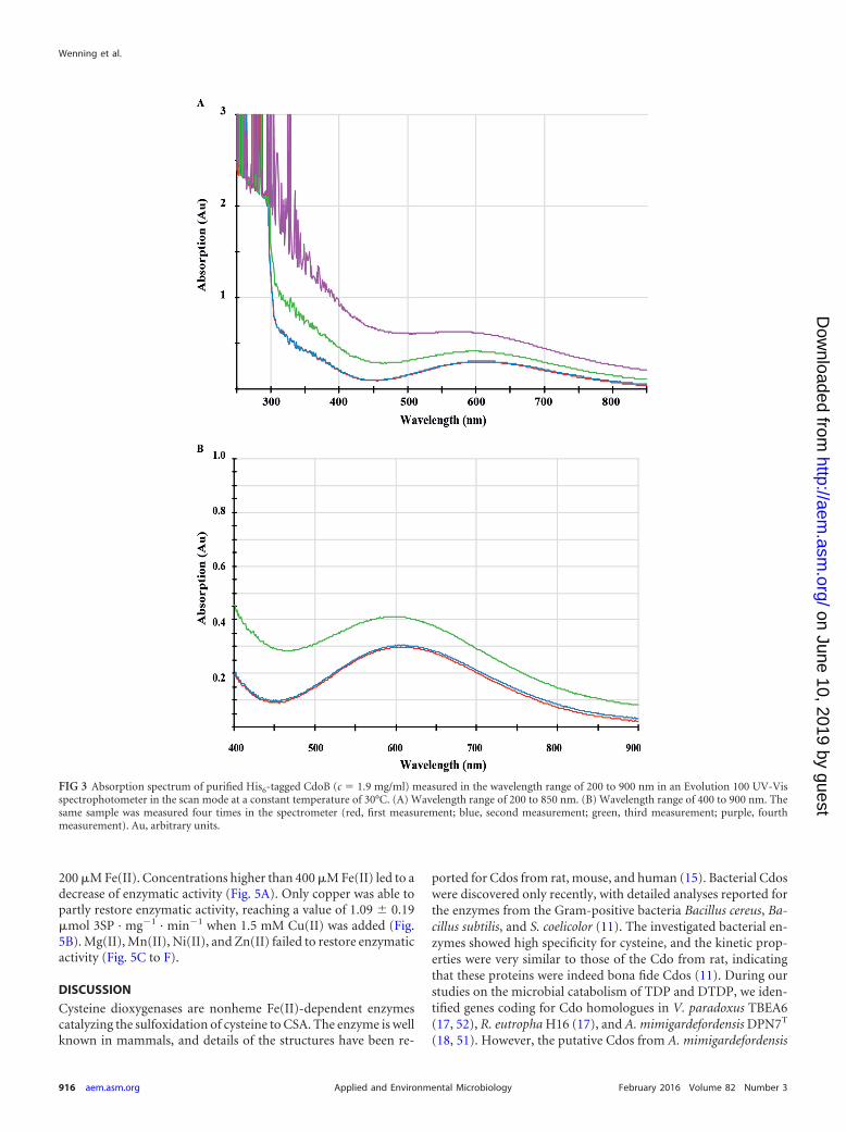
To further characterize CdoB, the reason for the blue appear-ance of the enzyme was investigated. For this, the absorption spec-trum of the purified enzyme (c 1.9 mg/ml) in the wavelength (�)range of 200 to 900 nm was measured. The spectrum showed thatthe enzyme absorbs light between 450 and 850 nm, with the high-est absorption occurring at a � value of 610 nm (Fig. 3). After thesecond measurement, the absorption of the enzyme could no lon-ger be determined, since the protein started to denature in thecuvette and the solution became turbid. Calculating an extinctioncoefficient (ε) for the enzyme at a � value of 610 nm based on thefirst two measurements gave a value of 3.2 mM�1 cm�1.
Characterization of CdoA. Analysis of purified CdoA by SECshowed an experimental molecular mass of 46.8 � 0 kDa (see Fig.S5 in the supplemental material). Since the calculated molecularmass of a hexahistidine-tagged CdoA monomer is 23.5 kDa, theexperimental molecular mass showed that CdoA apparently has adimeric quaternary structure in vitro.
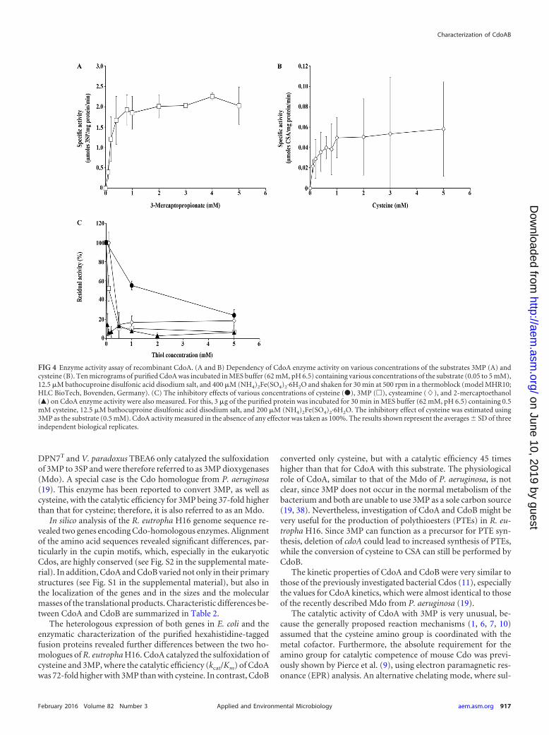
The capability of CdoA to oxidize 3MP to 3SP was previouslyrevealed by an in vivo enzyme assay cultivating cells of E. coliTop10(pBBR1MCS-3::cdoA) in M9 medium containing 3MP(17). This unusual catalysis was confirmed in the study by an invitro enzyme assay using the purified protein. In addition to 3MP,cysteine was also used as a substrate by CdoA, whereas no hypo-taurine could be detected when cysteamine was supplied as thesubstrate. For both substrates, the substrate saturation followedhyperbolic curves. The highest activities were measured at concentra-tions of 0.75 mM for 3MP and 1 mM for cysteine, whereas higherconcentrations led to substrate saturation and a constant velocity of
the enzymatic reaction (Fig. 4A and B). The maximal specific activityof CdoA for cysteine as the substrate was determined to be 0.05 �molCSA/mg protein/min; the Km, 6.7 mM; the kcat, 0.01 s�1; and thecatalytic efficiency, kcat/Km, 0.002 s�1 mM�1. When 3MP was used asa substrate, the enzyme showed a maximal specific activity of 2.2�mol 3SP/mg protein/min, a Km of 5.7 mM, a kcat of 0.82 s�1, and acatalytic efficiency, kcat/Km, of 0.144 s�1 mM�1.
The inhibitory effects of various concentrations of differentthiols on CdoA activity using cysteine as the substrate were tested.The results are illustrated in Fig. 4C and demonstrate that additionof 0.5 mM cysteamine, 3MP, or 2-mercaptoethanol led to a sig-nificant decrease of enzyme activities. The inhibitory effect of cys-teine on enzymatic formation of 3SP was also tested. In compari-son to the other thiols, the addition of cysteine resulted in a minordecrease of activity.
The optimal pH for activity was determined using a bis(2-hydroxyethyl)amino-tris(hydroxymethyl)methane/tris(hydroxy-methyl)methane (Bis-Tris/Tris) broad-range buffer system (pH5.5 to 9.0), and the highest activity occurred at pH 6.5.
An iron content of 0.38 � 0.04 mol Fe(II) per mol CdoA wasrevealed by the method of Pierce et al. (9). No Fe(III) could bedetected in CdoA.
The dependency of CdoA activity on divalent transition metalswas demonstrated by the inhibitory effect of the chelating com-pound EDTA, which resulted in a 40% decrease of activity at aconcentration of 100 �M. In contrast, sodium ethyl xanthate didnot exhibit any inhibitory effect (Table 2).
To make a reliable statement about the importance of the ironcofactor for activity of CdoA, the metal center had to be com-pletely removed. Thereafter, the apoenzyme was complementedwith different metal ions to prove the reconstruction of a func-tional enzyme. For removal of the iron cofactor, a dialysis ap-proach against 1,10-phenanthroline was used. After 30 min, itcould be observed that the H2O control stayed colorless, while the62.5 �M standard solution of FeCl2, as well as the enzyme solution(c 2 mg/ml), became red (see Fig. S6 in the supplemental ma-terial). This red coloration disappeared after 2 to 3 h. At this time,the dialysis buffer was exchanged for pure 50 mM Tris-HCl buffer(pH 7.5), in which the solution containing dialysis tubes was dia-lyzed overnight. Before the dialyzed enzyme solution was used forfurther experiments, the enzyme was concentrated and trans-ferred to 62 mM MES buffer (pH 6.5). After dialysis, the enzymeshowed an Fe(II) content of 0.11 � 0.013 mol Fe(II) per molenzyme. This is equal to a decrease of the iron content of �70%.
To prove the dependency of the CdoA activity on the ironcofactor, the dialyzed enzyme was applied in activity assays towhich different divalent metal ions Cu(II), Fe(II), Mg(II), Mn(II),Ni(II), and Zn (II) were added in a final concentration of 0.05 to1.5 mM (Fig. 5). As a reference value, the activity of the dialyzedenzyme was determined without the addition of any metal ion.The dialyzed CdoA could reach an activity of only 0.29 � 0.09�mol 3SP · mg�1 · min�1, which corresponds to 19.5% of theactivity of the nondialyzed enzyme when no divalent metal ionwas added (Fig. 5A). By addition of exogenous ferrous iron, theactivity of the dialyzed enzyme could be restored, reaching thehighest activity of 1.74 � 0.15 �mol 3SP · mg�1 · min�1 when 400�M Fe(II) was added. This corresponds to an activity 5.87-foldhigher than the activity without addition of any divalent metal ion.In the case of the nondialyzed enzyme, the highest activity of1.88 � 0.14 �mol 3SP · mg�1 · min�1 was achieved by addition of
TABLE 2 Effect of metal-acting compounds on enzyme activities ofCdoA and CdoBa
Effector
Relative activity (%)
CdoA CdoB
Sodium ethylxanthate2 �M 110 � 18.0 55 � 1.64 �M 89 � 10.8 56 � 7.0
EDTA50 �M 80 � 5.5 0100 �M 61 � 4.6 0200 �M 35 � 3.6 0
a Three to 5 �g purified CdoB was incubated for 30 min in MES buffer (62 mM, pH 6.5)containing 0.5 mM cysteine and 200 �M (NH4)2Fe(SO4)2·6H2O. CdoA activity wasobtained by 30 min of incubation of 14 �g purified CdoA in MES buffer (62 mM, pH 6.5)containing 0.5 mM 3MP and 200 �M (NH4)2Fe(SO4)2·6H2O. Each assay was alsoperformed without effectors, and the activity measured under these conditions was taken as100%. The results shown represent the averages of three independent biological replicates.
Characterization of CdoAB
February 2016 Volume 82 Number 3 aem.asm.org 915Applied and Environmental Microbiology
on June 10, 2019 by guesthttp://aem
.asm.org/
Dow
nloaded from
200 �M Fe(II). Concentrations higher than 400 �M Fe(II) led to adecrease of enzymatic activity (Fig. 5A). Only copper was able topartly restore enzymatic activity, reaching a value of 1.09 � 0.19�mol 3SP · mg�1 · min�1 when 1.5 mM Cu(II) was added (Fig.5B). Mg(II), Mn(II), Ni(II), and Zn(II) failed to restore enzymaticactivity (Fig. 5C to F).
DISCUSSION
Cysteine dioxygenases are nonheme Fe(II)-dependent enzymescatalyzing the sulfoxidation of cysteine to CSA. The enzyme is wellknown in mammals, and details of the structures have been re-
ported for Cdos from rat, mouse, and human (15). Bacterial Cdoswere discovered only recently, with detailed analyses reported forthe enzymes from the Gram-positive bacteria Bacillus cereus, Ba-cillus subtilis, and S. coelicolor (11). The investigated bacterial en-zymes showed high specificity for cysteine, and the kinetic prop-erties were very similar to those of the Cdo from rat, indicatingthat these proteins were indeed bona fide Cdos (11). During ourstudies on the microbial catabolism of TDP and DTDP, we iden-tified genes coding for Cdo homologues in V. paradoxus TBEA6(17, 52), R. eutropha H16 (17), and A. mimigardefordensis DPN7T
(18, 51). However, the putative Cdos from A. mimigardefordensis
FIG 3 Absorption spectrum of purified His6-tagged CdoB (c 1.9 mg/ml) measured in the wavelength range of 200 to 900 nm in an Evolution 100 UV-Visspectrophotometer in the scan mode at a constant temperature of 30°C. (A) Wavelength range of 200 to 850 nm. (B) Wavelength range of 400 to 900 nm. Thesame sample was measured four times in the spectrometer (red, first measurement; blue, second measurement; green, third measurement; purple, fourthmeasurement). Au, arbitrary units.
Wenning et al.
916 aem.asm.org February 2016 Volume 82 Number 3Applied and Environmental Microbiology
on June 10, 2019 by guesthttp://aem
.asm.org/
Dow
nloaded from
DPN7T and V. paradoxus TBEA6 only catalyzed the sulfoxidationof 3MP to 3SP and were therefore referred to as 3MP dioxygenases(Mdo). A special case is the Cdo homologue from P. aeruginosa(19). This enzyme has been reported to convert 3MP, as well ascysteine, with the catalytic efficiency for 3MP being 37-fold higherthan that for cysteine; therefore, it is also referred to as an Mdo.
In silico analysis of the R. eutropha H16 genome sequence re-vealed two genes encoding Cdo-homologous enzymes. Alignmentof the amino acid sequences revealed significant differences, par-ticularly in the cupin motifs, which, especially in the eukaryoticCdos, are highly conserved (see Fig. S2 in the supplemental mate-rial). In addition, CdoA and CdoB varied not only in their primarystructures (see Fig. S1 in the supplemental material), but also inthe localization of the genes and in the sizes and the molecularmasses of the translational products. Characteristic differences be-tween CdoA and CdoB are summarized in Table 2.
The heterologous expression of both genes in E. coli and theenzymatic characterization of the purified hexahistidine-taggedfusion proteins revealed further differences between the two ho-mologues of R. eutropha H16. CdoA catalyzed the sulfoxidation ofcysteine and 3MP, where the catalytic efficiency (kcat/Km) of CdoAwas 72-fold higher with 3MP than with cysteine. In contrast, CdoB
converted only cysteine, but with a catalytic efficiency 45 timeshigher than that for CdoA with this substrate. The physiologicalrole of CdoA, similar to that of the Mdo of P. aeruginosa, is notclear, since 3MP does not occur in the normal metabolism of thebacterium and both are unable to use 3MP as a sole carbon source(19, 38). Nevertheless, investigation of CdoA and CdoB might bevery useful for the production of polythioesters (PTEs) in R. eu-tropha H16. Since 3MP can function as a precursor for PTE syn-thesis, deletion of cdoA could lead to increased synthesis of PTEs,while the conversion of cysteine to CSA can still be performed byCdoB.
The kinetic properties of CdoA and CdoB were very similar tothose of the previously investigated bacterial Cdos (11), especiallythe values for CdoA kinetics, which were almost identical to thoseof the recently described Mdo from P. aeruginosa (19).
The catalytic activity of CdoA with 3MP is very unusual, be-cause the generally proposed reaction mechanisms (1, 6, 7, 10)assumed that the cysteine amino group is coordinated with themetal cofactor. Furthermore, the absolute requirement for theamino group for catalytic competence of mouse Cdo was previ-ously shown by Pierce et al. (9), using electron paramagnetic res-onance (EPR) analysis. An alternative chelating mode, where sul-
FIG 4 Enzyme activity assay of recombinant CdoA. (A and B) Dependency of CdoA enzyme activity on various concentrations of the substrates 3MP (A) andcysteine (B). Ten micrograms of purified CdoA was incubated in MES buffer (62 mM, pH 6.5) containing various concentrations of the substrate (0.05 to 5 mM),12.5 �M bathocuproine disulfonic acid disodium salt, and 400 �M (NH4)2Fe(SO4)2·6H2O and shaken for 30 min at 500 rpm in a thermoblock (model MHR10;HLC BioTech, Bovenden, Germany). (C) The inhibitory effects of various concentrations of cysteine (�), 3MP (�), cysteamine (�), and 2-mercaptoethanol(Œ) on CdoA enzyme activity were also measured. For this, 3 �g of the purified protein was incubated for 30 min in MES buffer (62 mM, pH 6.5) containing 0.5mM cysteine, 12.5 �M bathocuproine disulfonic acid disodium salt, and 200 �M (NH4)2Fe(SO4)2·6H2O. The inhibitory effect of cysteine was estimated using3MP as the substrate (0.5 mM). CdoA activity measured in the absence of any effector was taken as 100%. The results shown represent the averages � SD of threeindependent biological replicates.
Characterization of CdoAB
February 2016 Volume 82 Number 3 aem.asm.org 917Applied and Environmental Microbiology
on June 10, 2019 by guesthttp://aem
.asm.org/
Dow
nloaded from

fur and a carboxylate oxygen bind to the metal cofactor, wasconsidered by McCoy et al. (6).
Analysis of the degrees of polymerization of both enzymes bySEC showed that CdoA is a dimer and CdoB a trimer in vitro. So
far, most Cdo homologues have been detected as monomers (6,7). One exception is the investigation by Ye et al. (7), who analyzedthe human Cdo homologue and mutations thereof. They also ob-served a dimeric structure for one of the mutated enzymes, which
FIG 5 Complementation experiments with recombinant CdoA and recombinant dialyzed CdoA with 3MP as the substrate and addition of Fe(II) before (�) andafter (�) dialysis of the enzyme (A), Cu(II) (o) compared to Fe(II) (�) added to the dialyzed enzyme (B), Mg(II) (Œ) compared to Fe(II) (�) added to thedialyzed enzyme (C), Mn(II) (�) compared to Fe(II) (�) added to the dialyzed enzyme (D), Ni(II) (Œ) compared to Fe(II) (�) added to the dialyzed enzyme(E), and Zn(II) (�) compared to Fe(II) (�) added to the dialyzed enzyme (F). Ten micrograms of purified CdoA was incubated in MES buffer (62 mM, pH 6.5)containing 1 mM 3MP and various concentrations (0 to 1.5 mM) of the respective metal salt (CuCl2, FeCl2, MgCl2, MnCl2, NiCl2, and ZnCl2) and shaken for 30min at 500 rpm in a thermoblock (model MHR10; HLC BioTech, Bovenden, Germany). The results shown represent the averages � SD of three independentbiological replicates.
Wenning et al.
918 aem.asm.org February 2016 Volume 82 Number 3Applied and Environmental Microbiology
on June 10, 2019 by guesthttp://aem
.asm.org/
Dow
nloaded from

showed the same catalytic efficiency and metal incorporationcharacteristics as the monomeric enzymes. Since these describedenzymes are all of eukaryotic origin, a direct comparison is notconvenient. The previously described prokaryotic proteins havenot been examined for their degrees of polymerization (11, 15,17). The degree of polymerization could also be influenced by therecombinant host in which the enzyme is expressed.
Determination of the iron contents in CdoA and CdoB showeda submolar amount of Fe(II) present in the enzymes. This was alsodescribed for various other Cdo orthologues, e.g., from Rattusnorvegicus (12, 56), Mus musculus (6), and human (7). Generallyone would assume a ratio of 1 mol Fe(II)/mol enzyme if all enzymemolecules are present in their active forms. A reason for the sub-molar amount of iron in the enzyme might be too little iron in theculture medium, loss of iron during purification, or occupation ofthe active centers of the remaining Cdo molecules by other metals.
All previously described Cdos were characterized as strictly de-pendent on ferrous iron, and it has often been reported thatIMAC-purified Cdos showed no catalytic activity until exogenousferrous iron was added, whereas the addition of other transitionmetals failed to restore the activity (10–12). The experiments con-ducted in this study showed that the enzymes CdoA and CdoB areactive after purification by IMAC, also without addition of exog-enous divalent metal ions. Nevertheless, it could be verified thatthe enzyme activity of CdoB significantly increased after additionof FeCl2, CuCl2, NiCl2, or ZnCl2.
The inhibition studies with EDTA showed a complete loss ofactivity of CdoB even at low concentrations of EDTA (50 �M). Incontrast, CdoA still retained 80% of its original activity at thisEDTA concentration. This indicates stronger binding of metalions to CdoA than to CdoB. Since 1,10-phenanthroline is a stron-ger chelating agent than EDTA, an approach with 1,10-phenan-throline was developed. The almost complete loss of activity ofCdoA after removal of the iron by dialysis against buffer contain-ing 1,10-phenanthroline demonstrated that iron is indeed presentin the catalytic center of the enzyme. By addition of exogenousferrous iron with an optimal concentration of 200 to 400 �M, fullrestoration of activity of the dialyzed enzyme was achieved. Whenthe dialyzed enzyme was complemented with other divalent metalions, the activity was only marginally enhanced by addition ofCu(II). A reason for this observation might be that Cu(II) is theonly metal ion besides Fe(II) that is able to have 6 neighboringatoms and thereby build stable coordinated complexes with theHis residues of the protein, the substrate, and O2. Another expla-nation could be an undesired interaction of the buffer componentMES with the tested cations, which was described by Good et al. in1966 (57).
CdoB showed blue color after purification by IMAC. Becauseblue proteins are in general associated with Cu(II) content, as inplastocyanin (58), this observation is unusual for an enzyme thatis predicted to be iron dependent, like CdoB. Nevertheless, thereare some examples of other blue iron proteins in the literature,e.g., the 4-hydroxyphenylpyruvate dioxygenase of Pseudomonassp. strain P.J. 874. The color corresponded to an absorption max-imum at 595 nm and was ascribed to tyrosyl-Fe(III) charge trans-fer bands (59, 60). The blue color of CdoB most likely also resultsfrom a charge transfer complex between Fe(III) and a potentamino acid residue. CdoB showed an absorption maximum at 610nm. This is very similar to the absorption maximum observed forthe 4-hydroxyphenylpyruvate dioxygenase. The first report of
such a blue Cdo was published by Gardner et al. (61). The aerobicaddition of cysteine to an as-isolated mouse Cdo was found tochange the color of the purified protein solution from pale yellowto blue. Because the electronic absorption and the magnetic-cir-cular-dichroism spectroscopic signatures were very similar tothose of a well-characterized Fe(III) superoxide reductase, the au-thors proposed that the visible color was caused by a cysteinyl-Fe(III) charge transfer. However, further analyses of CdoB mustbe performed to reveal which amino acid residue is involved inthis charge transfer complex.
ACKNOWLEDGMENTS
The assistance of Christoph Knuf and Claudius Sandmeier in some exper-iments is gratefully acknowledged.
REFERENCES1. Joseph CA, Maroney MJ. 2007. Cysteine dioxygenase: structure and
mechanism. Chem Commun 28:3338 –3349.2. Stipanuk MH. 2004. Sulfur amino acid metabolism: pathways for pro-
duction and removal of homocysteine and cysteine. Annu Rev Nutr 24:539 –577. http://dx.doi.org/10.1146/annurev.nutr.24.012003.132418.
3. Heafield MT, Fearn S, Steventon GB, Waring RH, Williams AC, Stur-man SG. 1990. Plasma cysteine and sulphate levels in patients with motorneurone, Parkinson’s and Alzheimer’s disease. Neurosci Lett 110:216 –220. http://dx.doi.org/10.1016/0304-3940(90)90814-P.
4. Perry TL, Norman MG, Yong VW, Whiting S, Crichton JU, Hansen S,Kish SJ. 1985. Hallervorden-Spatz disease: cysteine accumulation andcysteine dioxygenase deficiency in the globus pallidus. Ann Neurol 18:482– 489. http://dx.doi.org/10.1002/ana.410180411.
5. Simmons CR, Liu Q, Huang Q, Hao Q, Begley TP, Karplus PA,Stipanuk MH. 2006. Crystal structure of mammalian cysteine dioxyge-nase: a novel mononuclear iron center for cysteine thiol oxidation. J BiolChem 281:18723–18733. http://dx.doi.org/10.1074/jbc.M601555200.
6. McCoy JG, Bailey LJ, Bitto E, Bingman CA, Aceti DJ, Fox BG, PhillipsGN. 2006. Structure and mechanism of mouse cysteine dioxygenase. ProcNatl Acad Sci U S A 103:3084 –3089. http://dx.doi.org/10.1073/pnas.0509262103.
7. Ye S, Wu X, Wei L, Tang D, Sun P, Bartlam M, Rao Z. 2007. An insightinto the mechanism of human cysteine dioxygenase. Key roles of thethioether-bonded tyrosine-cysteine cofactor. J Biol Chem 282:3391–3402.
8. Straganz GD, Nidetzky B. 2006. Variations of the 2-His-1-carboxylatetheme in mononuclear non-heme FeII oxygenases. Chembiochem7:1536 –1548. http://dx.doi.org/10.1002/cbic.200600152.
9. Pierce BS, Gardner JD, Bailey LJ, Brunold TC, Fox BG. 2007. Charac-terization of the nitrosyl adduct of substrate-bound mouse cysteine dioxy-genase by electron paramagnetic resonance: electronic structure of theactive site and mechanistic implications. Biochemistry 46:8569 – 8578.http://dx.doi.org/10.1021/bi700662d.
10. Simmons CR, Hirschberger LL, Machi MS, Stipanuk MH. 2006. Ex-pression, purification, and kinetic characterization of recombinant ratcysteine dioxygenase, a non-heme metalloenzyme necessary for regula-tion of cellular cysteine levels. Protein Expr Purif 47:74 – 81. http://dx.doi.org/10.1016/j.pep.2005.10.025.
11. Dominy JE, Simmons CR, Karplus PA, Gehring AM, Stipanuk MH.2006. Identification and characterization of bacterial cysteine dioxyge-nases: a new route of cysteine degradation for eubacteria. J Bacteriol 188:5561–5569. http://dx.doi.org/10.1128/JB.00291-06.
12. Chai SC, Jerkins AA, Banik JJ, Shalev I, Pinkham JL, Uden PC, Ma-roney MJ. 2005. Heterologous expression, purification, and characteriza-tion of recombinant rat cysteine dioxygenase. J Biol Chem 280:9865–9869.http://dx.doi.org/10.1074/jbc.M413733200.
13. Chai SC, Bruyere JR, Maroney MJ. 2006. Probes of the catalytic site ofcysteine dioxygenase. J Biol Chem 281:15774 –15779. http://dx.doi.org/10.1074/jbc.M601269200.
14. Dominy JE, Hwang J, Guo S, Hirschberger LL, Zhang S, StipanukMH. 2008. Synthesis of amino acid cofactor in cysteine dioxygenase isregulated by substrate and represents a novel post-translational regu-lation of activity. J Biol Chem 283:12188 –12201. http://dx.doi.org/10.1074/jbc.M800044200.
15. Stipanuk MH, Simmons CR, Karplus PA, Dominy JE. 2011. Thiol
Characterization of CdoAB
February 2016 Volume 82 Number 3 aem.asm.org 919Applied and Environmental Microbiology
on June 10, 2019 by guesthttp://aem
.asm.org/
Dow
nloaded from

dioxygenases: unique families of cupin proteins. Amino Acids 41:91–102.http://dx.doi.org/10.1007/s00726-010-0518-2.
16. Stipanuk MH, Londono M, Hirschberger LL, Hickey C, Thiel DJ, WangL. 2004. Evidence for expression of a single distinct form of mammaliancysteine dioxygenase. Amino Acids 26:99 –106. http://dx.doi.org/10.1007/s00726-003-0001-4.
17. Bruland N, Wübbeler JH, Steinbüchel A. 2009. 3-Mercaptopropionatedioxygenase, a cysteine dioxygenase homologue, catalyzes the initial stepof 3-mercaptopropionate catabolism in the 3,3-thiodipropionic acid-degrading bacterium Variovorax paradoxus. J Biol Chem 284:660 – 672.http://dx.doi.org/10.1074/jbc.M806762200.
18. Wübbeler JH, Bruland N, Kretschmer K, Steinbüchel A. 2008. Novelpathway for catabolism of the organic sulfur compound 3,3=-dithiodipropionic acid via 3-mercaptopropionic acid and 3-sulfinopropionicacid to propionyl-coenzyme A by the aerobic bacterium Tetrathiobacter mi-migardefordensis strain DPN7. Appl Environ Microbiol 74:4028 – 4035.http://dx.doi.org/10.1128/AEM.00422-08.
19. Tchesnokov EP, Fellner M, Siakkou E, Kleffmann T, Martin LW, AloiS, Lamont IL, Wilbanks SM, Jameson GNL. 2015. The cysteine dioxy-genase homologue from Pseudomonas aeruginosa is a 3-mercaptopropi-onate dioxygenase. J Biol Chem 290:24424 –24437. http://dx.doi.org/10.1074/jbc.M114.635672.
20. Xia Y, Wübbeler JH, Qi Q, Steinbüchel A. 2012. Employing a recombi-nant strain of Advenella mimigardefordensis for biotechnical production ofhomopolythioesters from 3,3=-dithiodipropionic acid. Appl Environ Mi-crobiol 78:3286 –3297. http://dx.doi.org/10.1128/AEM.00007-12.
21. Pohlmann A, Fricke WF, Reinecke F, Kusian B, Liesegang H, Cramm R,Eitinger T, Ewering C, Pötter M, Schwartz E, Strittmatter A, Voss I,Gottschalk G, Steinbüchel A, Friedrich B, Bowien B. 2006. Genomesequence of the bioplastic-producing “Knallgas” bacterium Ralstonia eu-tropha H16. Nat Biotechnol 24:1257–1262. http://dx.doi.org/10.1038/nbt1244.
22. Dunwell JM, Purvis A, Khuri S. 2004. Cupins: the most functionallydiverse protein superfamily? Phytochemistry 65:7–17. http://dx.doi.org/10.1016/j.phytochem.2003.08.016.
23. Lütke-Eversloh T, Bergander K, Luftmann H, Steinbuchel A. 2001.Identification of a new class of biopolymer: bacterial synthesis of a sulfur-containing polymer with thioester linkages. Microbiology 147:11–19.http://dx.doi.org/10.1099/00221287-147-1-11.
24. Lütke-Eversloh T, Steinbüchel A. 2003. Novel precursor substrates forpolythioesters (PTE) and limits of PTE biosynthesis in Ralstonia eutropha.FEMS Microbiol Lett 221:191–196. http://dx.doi.org/10.1016/S0378-1097(03)00185-X.
25. Wübbeler JH, Steinbüchel A. 2014. New pathways for bacterial polythio-esters. Curr Opin Biotechnol 29:85–92. http://dx.doi.org/10.1016/j.copbio.2014.02.017.
26. Doberstein C, Grote J, Wübbeler JH, Steinbüchel A. 2014. Polythioestersynthesis in Ralstonia eutropha H16: novel insights into 3,3=-thiodipropionic acid and 3,3=-dithiodipropionic acid catabolism. J Bio-technol 184:187–198. http://dx.doi.org/10.1016/j.jbiotec.2014.05.022.
27. Wübbeler JH, Raberg M, Brandt U, Steinbüchel A. 2010. Dihydrolipo-amide dehydrogenases of Advenella mimigardefordensis and Ralstonia eu-tropha catalyze cleavage of 3,3=-dithiodipropionic acid into 3-mercapto-propionic acid. Appl Environ Microbiol 76:7023–7028. http://dx.doi.org/10.1128/AEM.01706-10.
28. Hu H, Mylon SE, Benoit G. 2006. Distribution of the thiols glutathioneand 3-mercaptopropionic acid in Connecticut lakes. Limnol Oceanogr51:2763–2774. http://dx.doi.org/10.4319/lo.2006.51.6.2763.
29. Al-Farawati R, Van Den Berg CM. 2001. Thiols in coastal waters of thewestern North Sea and English Channel. Environ Sci Technol 35:1902–1911. http://dx.doi.org/10.1021/es000073i.
30. Zhang J, Wang F, House JD, Page B. 2004. Thiols in wetland interstitialwaters and their role in mercury and methylmercury speciation. LimnolOceanogr 49:2276 –2286. http://dx.doi.org/10.4319/lo.2004.49.6.2276.
31. Bürgmann H, Howard EC, Ye W, Sun F, Sun S, Napierala S, MoranMA. 2007. Transcriptional response of Silicibacter pomeroyi DSS-3 to di-methylsulfoniopropionate (DMSP). Environ Microbiol 9:2742–2755.http://dx.doi.org/10.1111/j.1462-2920.2007.01386.x.
32. Kiene RP, Malloy KD, Taylor BF. 1990. Sulfur-containing amino acids asprecursors of thiols in anoxic coastal sediments. Appl Environ Microbiol56:156 –161.
33. Todd JD, Rogers R, Li YG, Wexler M, Bond PL, Sun L, Curson ARJ,Malin G, Steinke M, Johnston AWB. 2007. Structural and regulatory
genes required to make the gas dimethyl sulfide in bacteria. Science 315:666 – 669. http://dx.doi.org/10.1126/science.1135370.
34. Van der Maarel MJEC, Jansen M, Hansen TA. 1995. Methanogenicconversion of 3-S-methylmercaptopropionate to 3-mercaptopropionate.Appl Environ Microbiol 61:48 –51.
35. Van der Maarel MJEC, Hansen TA. 1996. Anaerobic microorganismsinvolved in the degradation of DMS(P), p 351–360. In Kiene RP, VisscherPT, Keller MD, Kirst GO (ed), Biological and environmental chemistry ofDMSP and related sulfonium compounds. Plenum Press, New York, NY.
36. Visschert PT, Taylor BF. 1994. Demethylation of dimethylsulfoniopro-pionate to 3-mercaptopropionate by an aerobic marine bacterium. ApplEnviron Microbiol 60:4617– 4619.
37. Mopper K, Taylor BF. 1986. Biogeochemical cycling of sulfur-thiols incoastal marine sediments, p 324 –339. In Sohn M (ed), Organic marinegeochemistry. American Chemical Society, Washington, DC.
38. Lütke-Eversloh T, Steinbüchel A. 2003. Polythioesters, p 63– 80. In Mat-sumura S, Steinbüchel A (ed), Biopolymers, vol 9. Wiley-VCH, Wein-heim, Germany.
39. Kiene RP, Taylor BF. 1988. Biotransformation of organosulphur com-pounds in sediments via 3-mercaptopropionate. Nature 332:148 –150.http://dx.doi.org/10.1038/332148a0.
40. Vairavamurthy A, Mopper K. 1987. Geochemical formation of organo-sulphur compounds (thiols) by addition of H2S to sedimentary organicmatter. Nature 329:623– 625. http://dx.doi.org/10.1038/329623a0.
41. Schürmann M, Wübbeler JH, Grote J, Steinbüchel A. 2011. Novelreaction of succinyl coenzyme A (succinyl-CoA) synthetase: activationof 3-sulfinopropionate to 3-sulfinopropionyl-CoA in Advenella mim-igardefordensis s tra in DPN7 T during degradat ion of 3 ,3 =-dithiodipropionic acid. J Bacteriol 193:3078 –3089. http://dx.doi.org/10.1128/JB.00049-11.
42. Sambrook J, Fritsch EF, Maniatis T. 1989. Molecular cloning: a laboratorymanual. Cold Spring Harbor Laboratory Press, Cold Spring Harbor, NY.
43. Marmur J. 1961. A procedure for the isolation of deoxyribonucleic acidfrom micro-organisms. J Mol Biol 3:208 –218. http://dx.doi.org/10.1016/S0022-2836(61)80047-8.
44. Altschul S, Madden TL, Schäffer AA, Zhang J, Zhang Z, Miller W,Lipman DJ. 1997. Gapped BLAST and PSI-BLAST: a new generation ofprotein database search programs. Nucleic Acids Res 25:3389 –3402. http://dx.doi.org/10.1093/nar/25.17.3389.
45. Thompson J, Gibson TJ, Plewniak F, Jeanmougin F, Higgins DG. 1997.The CLUSTAL_X windows interface: flexible strategies for multiple se-quence alignment aided by quality analysis tools. Nucleic Acids Res 25:4876 – 4882. http://dx.doi.org/10.1093/nar/25.24.4876.
46. Hall T. 1999. BioEdit: a user-friendly biological sequence alignment edi-tor and analysis program for Windows 95/98/NT. Nucleic Acids Symp Ser41:95–98.
47. Saitou N, Nei M. 1987. The neighbor-joining method: a new method forreconstructing phylogenetic trees. Mol Biol Evol 4:406 – 425.
48. Aboulmagd E, Oppermann-Sanio FB, Steinbüchel A. 2000. Molecularcharacterization of the cyanophycin synthetase from Synechocystis sp.strain PCC6308. Arch Microbiol 174:297–306. http://dx.doi.org/10.1007/s002030000206.
49. Laemmli UK. 1970. Cleavage of structural proteins during the assembly ofthe head of bacteriophage T4. Nature 227:680 – 685. http://dx.doi.org/10.1038/227680a0.
50. Weber K, Osborn M. 1969. The reliability of molecular weight determi-nations by dodecyl sulfate-polyacrylamide gel electrophoresis. J BiolChem 244:4406 – 4412.
51. Wübbeler JH, Hiessl S, Schuldes J, Thürmer A, Daniel R, SteinbüchelA. 2014. Unravelling the complete genome sequence of Advenella mim-igardefordensis strain DPN7T and novel insights in the catabolism of thexenobiotic polythioester precursor 3,3=-dithiodipropionate. Microbiol-ogy 160:1401–1416. http://dx.doi.org/10.1099/mic.0.078279-0.
52. Wübbeler JH, Hiessl S, Meinert C, Schuldes J, Poehlein A, Daniel R,Steinbüchel A. 2015. The genome of Variovorax paradoxus strain TBEA6provides new understandings for the catabolism of 3,3=-thiodipropionicacid and hence the production of polythioesters. J Biotechnol 209:85–95.http://dx.doi.org/10.1016/j.jbiotec.2015.06.390.
53. Wang Y, Addess KJ, Chen J, Geer LY, He J, He S, Lu S, Madej T,Marchler-Bauer A, Thiessen PA, Zhang N, Bryant SH. 2007. MMDB:annotating protein sequences with Entrez’s 3D-structure database. Nu-cleic Acids Res 35:D298 –D300. http://dx.doi.org/10.1093/nar/gkl952.
54. Brandt U, Hiessl S, Schuldes J, Thürmer A, Wübbeler JH, Daniel R,
Wenning et al.
920 aem.asm.org February 2016 Volume 82 Number 3Applied and Environmental Microbiology
on June 10, 2019 by guesthttp://aem
.asm.org/
Dow
nloaded from
Steinbüchel A. 2014. Genome-guided insights into the versatile metaboliccapabilities of the mercaptosuccinate-utilizing �-proteobacterium Vario-vorax paradoxus strain B4. Environ Microbiol 16:3370 –3386. http://dx.doi.org/10.1111/1462-2920.12340.
55. Brandt U, Schürmann M, Steinbüchel A. 2014. Mercaptosuccinate di-oxygenase, a cysteine dioxygenase homologue, from Variovorax para-doxus strain B4 is the key enzyme of mercaptosuccinate degradation. J BiolChem 289:30800 –30809. http://dx.doi.org/10.1074/jbc.M114.579730.
56. Yamaguchi K, Hosokawa Y. 1987. Cysteine dioxygenase. Methods Enzy-mol 143:395– 403. http://dx.doi.org/10.1016/0076-6879(87)43069-3.
57. Good NE, Winget GD, Winter W, Connolly TN, Izawa S, Singh RMM.1966. Hydrogen ion buffers for biological research. Biochemistry 5:467–477. http://dx.doi.org/10.1021/bi00866a011.
58. Rydén LG, Hunt LT. 1993. Evolution of protein complexity: The bluecopper-containing oxidases and related proteins. J Mol Evol 36:41– 66.http://dx.doi.org/10.1007/BF02407305.
59. Lindstedt S, Rundgren M. 1982. Blue color, metal content, and substrate
binding in 4-hydroxyphenylpyruvate dioxygenase from Pseudomonas sp.strain P. J 874. J Biol Chem 257:11922–11931.
60. Bradley FC, Lindstedt S, Lipscomb JD, Que L, Roe AL, Rundgren M.1986. 4-Hydroxyphenylpyruvate dioxygenase is an iron-tyrosinate pro-tein. J Biol Chem 261:11693–11696.
61. Gardner JD, Pierce BS, Fox BG, Brunold TC. 2010. Spectroscopic andcomputational characterization of substrate-bound mouse cysteine di-oxygenase: nature of the ferrous and ferric cysteine adducts and mecha-nistic implications. Biochemistry 49:6033– 6041. http://dx.doi.org/10.1021/bi100189h.
62. Wilde E. 1962. Untersuchungen über Wachstum und Speicherstoffsyn-these von Hydrogenomonas. Arch Mikrobiol 43:109 –137. http://dx.doi.org/10.1007/BF00406429.
63. Bachmann BJ. 1987. Linkage map of Escherichia coli K12, p 807– 876. InNeidhardt FC (ed), Escherichia coli and Salmonella typhimurium: cellularand molecular biology, vol 2. American Society for Microbiology, Wash-ington, DC.
Characterization of CdoAB
February 2016 Volume 82 Number 3 aem.asm.org 921Applied and Environmental Microbiology
on June 10, 2019 by guesthttp://aem
.asm.org/
Dow
nloaded from


![Delay compensation using Smith predictor for wireless ... · is based on adaptive Smith predictors. In [12], communication disturbance observer (CDOB) and network disturbance (ND)](https://static.fdocuments.in/doc/165x107/601eb1ef7b8fd602336ea565/delay-compensation-using-smith-predictor-for-wireless-is-based-on-adaptive-smith.jpg)


