Sublethal effects of synthetic dyes on rainbow ... · Eight slides were obtained solutions were...
8
DISEASES OF AQUATIC ORGANISMS Dis. aquat. Org. 1 Published February l7 Sublethal effects of synthetic dyes on rainbow trout Oncorhynchus mykiss: a light and electron microscope study Laboratori de Biologia, Facultat de Veterinaria, U.A.B., E-08193 Bellaterra, Barcelona, Spain Institut dVInvestigaci6 Textil i Cooperacio Industrial de Terrassa, Barcelona, Spain ABSTRACT- Subchronic exposures to sublethal concentrations of the synthetic, metal-complex dye C.I. Acid Violet 66 and its azo compound C.I. Acid Red 217 did not cause severe histological damage to rainbow trout Oncorhynchus mykiss. Fish exposed to C.I. Acid Violet 66 for 30 d exhibited the most pronounced morphological alterations, which included spongiosis of the gill tissue and degeneration of chloride cells. Both dyes triggered adaptative responses which involved granulocytes, macrophages and granular cells in the gill, and melanomacrophages in the kidney, liver and spleen. Hepatocytes displayed altered endoplasmic reticulurn after treatment. Frequencies of rnicronucleated peripheral erythrocytes did not increase following dye treatment, suggesting that these chemicals, at least at the doses tested, are not genotoxic following a 30 d exposure. INTRODUCTION The environmental problems posed by dyes are rather moderate in severity compared with those caused by other chemicals such as pesticides, deter- gents, industrial oils, heavy metals, etc. (Anliker 1977). Dyes have been shown to be mildly to moderately toxic to aquatic organisms: 98 % of more than 3000 commer- cial dyes tested on fish by the ETAD (Ecological and Toxicological Association of the Dyestuffs Manufactur- ing Industry) exhibited 48 h values greater than 1 mg 1-' (Clarke & Anliker 1980). However, it must be taken into account that more than 100 t of dyes (50 '10 corresponding to the textile sector) are released daily into the environment, primarily dissolved or suspended in water, and evaluations of their sublethal effects on aquatic organisms are not available in the literature. Toxicity tests carried out in our laboratory demon- strated that the 48 h LCS0's of the metal-complex dye C.I. Acid Violet 66 (see Fig. lb) and the azo compound utilized in its synthesis (C.I. Acid Red 217; see Fig, la) for rainbow trout Oncorhynchus mykiss were 8.20 and 71.04 mg 1-' respectively (&va et al. 1990), revealing that toxicity was mainly due to the presence of chromium (Cr 111) in the dye molecule. It was also Addressee for correspondence O Inter-Research/Printed in Germany shown that, following treatment with sublethal concen- trations, levels of these chemicals increased in the bile and accumulation in the tissues occurred (bva 1989). In order to elucidate the morphological effects, if any, of a sublethal dose of the chromium-complex dye and its azo compound on trout, a light and electron micro- scope study was undertaken after treating the fish up to 30 d with both chemicals. Since a few dyestuffs, par- ticularly benzidine derivatives, have been shown to be mutagenic in mammals (Anliker & Steinle 1987, Arcos et al. 1988) and recent experimental data (Bianchini & Levis 1988) indicate that chronic exposures even to low doses of insoluble Cr (111) produce genotoxic conse- quences, the piscine micronucleus test (Hooftman & de Raat 1982) was used to assess the genotoxicity, if any, of both dyes. MATERIAL AND METHODS Experimental treatment. Rainbow trout Oncorhyn- chus mykiss (sexually immature, ca 10 to 15 cm long) were obtained from a local hatchery and maintained in well-aerated running freshwater (total hardness 316 mg 1-l; pH 8 f 0.1; temperature 14 +- l "C) for at least 2 wk before experiments. Fish were fed a com- mercial fish diet at a rate of 1 % live weight d-' during the acclimation period as well as during treatment.
Transcript of Sublethal effects of synthetic dyes on rainbow ... · Eight slides were obtained solutions were...
DISEASES OF AQUATIC ORGANISMS Dis. aquat. Org.
1 Published February l 7
Sublethal effects of synthetic dyes on rainbow trout Oncorhynchus mykiss: a light and electron
microscope study
Laboratori de Biologia, Facultat de Veterinaria, U.A.B., E-08193 Bellaterra, Barcelona, Spain Institut dVInvestigaci6 Textil i Cooperacio Industrial de Terrassa, Barcelona, Spain
ABSTRACT- Subchronic exposures to sublethal concentrations of the synthetic, metal-complex dye C.I. Acid Violet 66 and its azo compound C.I. Acid Red 217 did not cause severe histological damage to rainbow trout Oncorhynchus mykiss. Fish exposed to C.I. Acid Violet 66 for 30 d exhibited the most pronounced morphological alterations, which included spongiosis of the gill tissue and degeneration of chloride cells. Both dyes triggered adaptative responses which involved granulocytes, macrophages and granular cells in the gill, and melanomacrophages in the kidney, liver and spleen. Hepatocytes displayed altered endoplasmic reticulurn after treatment. Frequencies of rnicronucleated peripheral erythrocytes did not increase following dye treatment, suggesting that these chemicals, at least at the doses tested, are not genotoxic following a 30 d exposure.
INTRODUCTION
The environmental problems posed by dyes are rather moderate in severity compared with those caused by other chemicals such as pesticides, deter- gents, industrial oils, heavy metals, etc. (Anliker 1977). Dyes have been shown to be mildly to moderately toxic to aquatic organisms: 98 % of more than 3000 commer- cial dyes tested on fish by the ETAD (Ecological and Toxicological Association of the Dyestuffs Manufactur- ing Industry) exhibited 48 h values greater than 1 mg 1-' (Clarke & Anliker 1980). However, it must be taken into account that more than 100 t of dyes (50 '10 corresponding to the textile sector) are released daily into the environment, primarily dissolved or suspended in water, and evaluations of their sublethal effects on aquatic organisms are not available in the literature.
Toxicity tests carried out in our laboratory demon- strated that the 48 h LCS0's of the metal-complex dye C.I. Acid Violet 66 (see Fig. l b ) and the azo compound utilized in its synthesis (C.I. Acid Red 217; see Fig, l a ) for rainbow trout Oncorhynchus mykiss were 8.20 and 71.04 mg 1-' respectively (&va et al. 1990), revealing that toxicity was mainly due to the presence of chromium (Cr 111) in the dye molecule. It was also
Addressee for correspondence
O Inter-Research/Printed in Germany
shown that, following treatment with sublethal concen- trations, levels of these chemicals increased in the bile and accumulation in the tissues occurred ( b v a 1989). In order to elucidate the morphological effects, if any, of a sublethal dose of the chromium-complex dye and its azo compound on trout, a light and electron micro- scope study was undertaken after treating the fish up to 30 d with both chemicals. Since a few dyestuffs, par- ticularly benzidine derivatives, have been shown to be mutagenic in mammals (Anliker & Steinle 1987, Arcos et al. 1988) and recent experimental data (Bianchini & Levis 1988) indicate that chronic exposures even to low doses of insoluble Cr (111) produce genotoxic conse- quences, the piscine micronucleus test (Hooftman & de Raat 1982) was used to assess the genotoxicity, if any, of both dyes.
MATERIAL AND METHODS
Experimental treatment. Rainbow trout Oncorhyn- chus mykiss (sexually immature, ca 10 to 15 cm long) were obtained from a local hatchery and maintained in well-aerated running freshwater (total hardness 316 mg 1-l; pH 8 f 0.1; temperature 14 +- l "C) for a t least 2 wk before experiments. Fish were fed a com- mercial fish diet a t a rate of 1 % live weight d-' during the acclimation period as well as during treatment.

104 Dis. aquat. Org. 12: 103-110, 1992
C . I . R C 1 0 RED 217
b C . I . A C I D V I O L E T 66
Fig. 1. Chemical structure of the dyes used in this study
metal-complex dye). Both chemicals were supplied by Sandoz S.A. (Basel). Ten fish were used as controls and were handled in the same way as treated ones.
Histological and electron microscope study. Treated specimens were killed with tricaine methanesulfonate (MS 222, Sandoz) and samples of the liver, kidney, spleen and gills were fixed in 5 % glutaraldehyde in 0.1 M Na cacodilate buffer (pH 7.3), post-fixed in 2 O/O Os04 dehydrated through ethanol series, stained 'en block' with uranyl acetate and embedded in araldite. Semi-thin sections (1 vm) were cut, stained with toluidine blue and viewed under a light microscope. Ultrathin sections were stained on the microscopical grids with lead citrate and observed in a Hitachi H- 7000 transmission electron microscope. Several sam- ples were fixed m 10 O/o buffered formalin, embedded in paraffin wax, cut at 6 \!m and stained using the Perl's Prussian Blue method (Luna 1968). This staining proce- dure allows differentiation of the 3 pigments present within the melanomacrophages (Wolke et al. 1985): the
Individuals for experiments were placed in 20 1 tanks ferric ion of hemosiderin stains bright blue, the lipofuc- (10 fish tank-'). Metal-complex dye solutions were pre- sin pigment remains unstained and melanin, also pared by adding 4.1 mg 1-' of the commercial dye unstained, appears as dark brown granules. (Fig, l a ) . This concentration corresponds to half the Piscine micronucleus test. After severing the caudal 48 h LCS0 for trout (IZlva et al. 1990). In order to remove vein, a drop of blood was smeared on the slide, air fish excretions and to maintain a relatively constant dried, methanol fixed, and then stained in May-Gfin- concentration of the toxicant in experimental tanks, wald-Giemsa solutions. Eight slides were obtained solutions were replaced every 2 d. Fish were sacrificed from each fish. The number of micronuclei scored after after a 14 and 30 d exposure to the toxicant. In order to counting 500 mature erythrocytes per slide was compare the histopathological effects of the metal- recorded. Erythrocytes were scored blind and only cells complex dye (Fig. l b ) and its azo compound (Fig. l a ) , a with intact cell and nuclear membranes were included group of 10 fish was exposed to 2.9 mg 1-' of C.I. Acid in the counts. We only counted as micronuclei isolated Red 217 for 14 and 30 d (2.9 mg corresponds to the nuclear fragments, following the morphological criteria amount of this compound present in 4.1 mg of the final described by Tates et al. (1980) (see Fig. 2). The fre-
Fig. 2. Oncorhynchus mykiss. Mic- ronucleated erythrocyte (arrow) of rain- bow trout. Micronuclei are visible as isolated nuclear fragments May-Grun-
wald Giemsa, X 1320

Marlasca et al.- Effects of dyes on trout 105
Fig. 3. Oncorhynchus mykiss. Gill morphology of (a, b) untreated and (c to e) dye-treated rainbow trout. Note that following treatment with metal-complex dye for 30 d, lymphoid spaces (c) are dilated and invaded by granulocytes (arrows). Spongiosis of the filament epithelium (d, e) is apparent after treatment, and mitotic figures (d; arrowheads) are more frequent than in control fish. Granular cells, which in untreated fish are only located in the connective tissue (a) surrounding the cartilage, are observed invading the epithelia1 layer in treated fish (d; open arrow). C: cartilage; cc: chloride cells; af: afferent artery. Toluidine blue;
(a) x 610, (b) X 1950, (c) x 1320, (d) x 1750, (e) x 1300
lob Dis. aquat. Org. 12: 103-110, 1992
quencies of micronuclei in pooled control and expen- mental erythrocytes were compared statistically using the tables of Kastenbaum & Bowman (1970).
RESULTS
None of the fish displayed any apparent toxicity symptoms (overturning, erratic movement, increased coughing, flared operculum, etc.) during experimenta- tion. The lamellar epithelium of trout gill is composed of 2 layers of epithelial cells separated by lymphoid spaces; chloride cells are located in the interlamellar spaces of the filament and in the lamellae (Fig. 3a, b). In control fish, granular cells were mainly found in the connective layer surrounding the cartilage (Fig. 3a). Following treatment with ejther the metal-complex dye or its azo compound, gill epithelium was spongiotic and
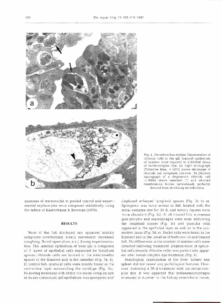
Fig. 4 . Oncorhynchus mykiss. Degeneration of chloride cells in the gill filament epithelium of rainbow trout exposed to sublethal doses of metal-complex dye. (a) Light micrograph (Toluidine blue; X 1200) shows shrinkage of chloride cell cytoplasm (arrows). (b) Electron micrograph of a degenerate chloride cell ( X 9000) shows vesicular ( ' ) and whorled membranous bodies (arrowhead), probably
derived from involuting mitochondna
displayed enlarged lymphoid spaces (Fig. 3c to e) . Spongiosis was more severe in fish treated with the metal-complex dye for 30 d, and mitotic figures were more abundant (Fig. 3d). In all treated fish, numerous granulocytes and macrophages were seen infiltrating the lymphoid spaces (Fig. 3c) and granular cells appeared in the epithelial layer as well as in the con- nective tissue (Fig. 3d, e ) . Rodl.et cells were found in the filament and in the lamellae of both control and treated fish. No differences in the number of mucous cells were detected following treatment. Degeneration of epithe- lial cells (mainly chloride cells) was particularly appar- ent after metal-complex dye treatment (Fig. 4 ) .
Histological examination of the liver, kidney and spleen did not reveal any pathological features. How- ever, following a 30 d treatment with the metal-com- plex dye, it was apparent that melanomacrophages increased in number in the kidney intertubular bssue.

- a Q) Q)
::g
-0
-J
g
sg
2
ctn
C22
' %'E:
5
U"..
0-O
k
2 61 'C
3
5 a5 %
S
W
4
mc
~
ar 0
b
PU
m
5 S$-
%"a
U-
.d
or'u
2l 2 e
w
- a-,
a 5s
so
w
- z 5
O 2 2 .$ 3 -g
gg g $5 2
SE-
f m
m
2-
2
.V X
m
2 2 2
.F=' a
-1
.2
vj .5
U
.m U
4
'5 E
$g4 m
=U
;
3;-
5 2
-c
C'
$@
Q)
U
*S
8
;z

108 Dis. aquat. Org. 12: 103-110, 1992
Fig. 7. Oncorhynchus mykiss. Electron micrograph of the rough endoplasmic reticulum of hepatocytes in (a) control ( X 21 500) and (b) treated rainbow trout ( X 21 000). Note the dilation and degranulation ( * ) of the reticulum in treated fish. N: nucleus;
m: rnitochondria
spleen and, particularly, in the liver, surrounding the higher in the group exposed to C.I. Acid Red 217, no biliary ducts (Fig. 5). Perl's Prussian Blue staining statistically significant differences (Kastenbaum & revealed that there was an increase in the amount of Bowman 1970) were found relative to control values. melanin pigment, whereas hemosiderin remained unaltered. From ultrastructural observations it was apparent that in treated fish, renal melanocytes were DISCUSSION packed with melanosomes which displayed all stages of development, whereas in control fish, melanosomes Our observations provide evidence of histological in early stages of development were rare (Fig. 6). After and ultrastructural alterations, although not severe, in 30 d exposure of fish to the metal-complex dye, fish exposed to dye solutions. Gill spongiosis, granulo- hepatocytes exhibited dilation and degranulation of cyte infiltration in the enlarged lyrnphoid spaces and the endoplasmic reticulum (Fig. 7), although the reticu- increases in the number of granular cell and mitotic lar nature of the organelle was not lost. No changes figures were observed following all treatments. How- were found in mitochondria and other hepatocyte ever, these changes were more pronounced after treat- organelles. Table 1 shows the frequencies of micro- ment of fish with the metal-complex dye for 30 d, and nuclei found in mature peripheral erythrocytes of degeneration of epithelia1 cells (mainly chloride cells) Oncorhynchus mykiss exposed to both dyes for 30 d. was particularly apparent in this group. Chloride cells Although the number of micronucleated cells was have been shown to be the most sensitive gill cell type
to a great variety of agents such as heavy metals (Crespo & Sala 1986), acid water (Tietge et al. 1988),
Table 1. Oncorhynchus mykiss. Frequencies of micronucl.ei in imbalanced diet (Bell et al. 1985) and Stress (Peters & peripheral erythrocytes of trout exposed to dyes. No statisti- Hong 1985). Our observations accord with the fact that cally significant differences were recorded when comparing the 48 h L , - ~ ~ Of the metal-complex dye for trout is one-
treated and control values (Kastenbaum & Bowmann 1970) ninth that of its azo compound (Riva et al. 1990) and
Treatment Control" C.I. Acid C.I. Acid
Violet 66b Red 21fa
Frequency (?L) 0.083 0.036 0.250
" 36 000 erythrocytes scored in control and C.J Acid Red 217 treated fish (9 fish, pooled) h 28 000 erythrocytes scored in metal-complex dye treated fish (7 fish, pooled)
suggest that dye toxic effects might be malnly due to the presence of the metal (Cr 111). Nevertheless, it must be taken into account that gill morphological altera- tions described in the present work are non-specific and have been reported previously in fish exposed to other toxicants (Mallatt 1985, Evans 1987). The
enlargement of the lyrnphoid spaces and the infiltration granulocytes and macro~hages might to
an inflammatory response to the toxicants. Although the lamellar structure was not severely altered after

Marlasca et al.: Effects of dyes on trout 109
treatment, the length of the water-blood pathway might be increased in dye-exposed animals, which might account for an unbalanced gas transfer. Bran- chial granular cells, histologically similar to the eosinophilic cells found in the submucosa of the trout intestinal tract (Bergeron & Woodward 1983), have been shown to increase in number following all dye treatments, which might be consistent with their hypothesised inflammatory role (Ferguson 1989). Cells undergoing mitosis were more abundant following dye treatment. The same observation was reported by Crespo et al. (1986) for trout intestinal epithelium after oral administration of lead and cadmium and was related to an increased renewal rate of absorptive cells.
The histological and ultrastructural examination of the ludney, liver and spleen revealed that melanomac- rophages increased in number, melanin was more abundant within cells, and melanosomes, in early stages of development, were more apparent in treated specimens, which might indicate increased melanin synthesis in these fish. Pigment-containing macro- phages are a prominent feature of piscine haemo- poietic tissues, and the role of melanin might be related to detoxification mechanisms, since this pigment is able to absorb free radicals and cations (Agius 1985). Pig- mented macrophage accun~ulations were proposed as possible indices of fish health (Wolke et al. 1984) and several authors described increased pigmented mac- rophage numbers in fish caught from polluted waters (Wolke et al. 1985) and experimentally treated with polluted waters (Poels et al. 1980). However, reduced numbers of melanomacrophage centers were reported in flounder exposed to hydrocarbon-contaminated sed- i m e n t ~ (Payne & Fancey 1989), and it was suggested that at low levels of pollution the cellular defense system would be capable of removing debris by means of increased phagocytic activity, whereas at higher levels of pollution phagocytosis might be impaired, leading to a decrease in melanomacrophage centres.
Ultrastructural observations of the liver hepatocyte showed dilation and degranulation of rough endoplas- mic reticulum following metal-complex dye exposure, although the reticular nature of the organelle was not lost. Ultrastructural changes in the endoplasmic reticulum of fish exposed to other toxicants have also been described by Braunbeck et al. (1989). It is assumed that dilation of endoplasmic reticulum can be due to an ingress of water and/or a storage of secretory products (Ghadially 1982). However, the fact that mitochondria were not swollen and the lumen of the reticulum dis- played the same electron density following treatment does not support either of these hypotheses. Rather, we suggest that these changes might correspond to the initial events leading to formation of new smooth endo- plasmic reticulum, since a great variety of drugs (includ-
ing azo compounds) have been reported to induce hypertrophy of smooth endoplasmic reticulum in mam- malian hepatocytes (Ghadially 1982).
Since the application of the micronucleus test to assess genotoxic damage to fish using the peripheral erythrocytes of the eastern mudmlnnow Umbra pygrnea (Hooftman & de Raat 1982), there have been numerous reports on the induction of micronuclei following fish exposure to genotoxic agents (Al-Sabti 1986, Das & Nanda 1986, Metcalfe 1988). However, although the micronucleus test has been used to assess environ- mental pollution (Hose et al. 1987, Scarpato et al. 1990), recent studies by Carrasco et al. (1990) point out that this assay lacks sensitivity to the presence and effects of chemical contaminants, mainly due to the fact that nuclear lesion frequencies are very low and variable. Our study shows that neither C.I. Acid Violet 66 nor C.I. Acid Red 217 induced micronuclei formation in mature peripheral erythrocytes of trout at the doses tested. It could be argued that the period of exposure was not long enough. However, Das & Nanda (1986) have described statistically significant increases in the number of micro- nucleated cells after exposing catfish Heteropneutes fossilis to paper mill effluents for the same period of time (30 d), and Metcalfe (1988) has reported the induction of micronuclei in mudminnows Umbra limi and bullheads Ictalurus nebulosus 96 h after intraperitoneal injection with ethyl methane-sulphonate and benzo-(a)-pyrene. Moreover, Al-Sabti (1986) has described increased mi- cronucleus frequencies in carp 48 h after treatment with several chemicals. As previously pointed out by Met- calfe (1988), the low incidence of micronuclei in peripheral fish erythrocytes (ranging from 0.036 to 0.25%0 in our study) creates a problem for statistical analysis. However, in the present work a large sample of erythrocytes was included in the counts: up to 28 000 cells (7 fish, pooled) and 36 000 cells (9 fish, pooled) were scored after treatment with C.I. Acid Violet 66 and C.I. Acid Red 217 respectively; even so, no differences were found when comparing these values to those for control micronuclei (36 000 cells scored; 9 fish, pooled). These results might suggest that neither dye, at least at the doses tested, is genotoxic following a 30 d exposure.
Acknowledgements. This work received financial support from the DGICYT (Direction General de Investigacion Cientifica y Tecnica del Ministerio de Educacion y Ciencia), PB 87-0180.
LITERATURE CITED
Aqus, C. (1985). The melano-macrophage centres of fish: a review. In: Manning, M. J., Tatner, M. F. (eds.) Fish Immunology. Academic Press, London, p. 85-105
Al-Sabti, K. (1986). Clastogenic effects of five carcinogenic- mutagenic chemicals on the cells of the common carp. Cypnnus carpio L. Comp. Biochem. Physiol. 85C: 5-9

110 Dis. aquat. Org. 12: 103-110, 1992
Anliker, R. (1977). Colour chemistry and the environment. Ecotoxicol. Environ. Saf. 1. 21 1-237
Anliker, R , Steinle, D (1987) Prevention of risks in the use of handling colorants. In: Proceedings of the 14th IFATCC Congress. Tampere, Finland, 3-5 June 1987. Tampere University of Technology, Tampere, p. 325-346
Arcos, J. C., Woo, Y., Lai, D. Y (1988). Database on binary combination effects of chemical carcinogens Envir. Car- cinogenesis Rev. (J. Envir. Sci. Health) 6: 1-150
Bell, M. V., Henderson, R. J . , Pirie. J . S., Sargent, J . R. (1985). Effects of dietary polyunsaturated fatty acid deficiencies on mortality, growth and gill structure in the turbot Scoph- thalmus maximus. J. Fish. Biol. 26: 181-191
Bergeron, T., Woodward, B. (1983). Ultrastructure of the small intestine of the rainbow trout (Salmo gairdneri) before and after stratum granulosum formation. Can J. 2001. 61 133-138
Bianchini, V., Levis, A. G. (1988). Review of genetic effects and mecnanisms of acuon of chromlum compounds. Sci total Environ. 71: 351-355
Braunbeck, T., Storch, V., Nagel, R. (1989). Sex-specific reac- tion of liver ultrastructure in zebra fish Brachydanio rerio after prolonged sublethal exposure to 4-nitrophenol. Aquat. Toxicol. 14. 185-203
Carrasco, K. R.. Tilbury, K. L., Myers, M. S. (1990). Assessment of the piscine micronucleus test as an in situ biological indicator of chemical contaminant effects. Can. J. Fish. Aquat. Sci. 47: 2123-2136
Clarke, E. A., Anliker, R. (1980). Organic dyes and pigments In: Hutzinger, 0. (ed.) The handbook of environmental chemistry, Vol. 3, Part A. Springer-Verlag, Berlin, p. 181-215
Crespo, S., Nonnotte, C., Colin, A., Leray, L.. Nonnotte, L., Aubree, A. (1986) Morphological and functional altera- tions induced in trout intestine by dietary cadmium and lead. J . Fish Biol. 28: 69-80
Crespo, S., Sala, R. (1986). Ultrastructural alterations of the dogfish (Scyliorhinus canicula) gill filament related to experimental aquatic zinc pollution. Dis. aquat. Org. 1. 99-104
Das, R. K., Nanda, N. K. (1986). Induction of micronuclei in peripheral erythrocytes of fish Heteropneutes fossilis by mitomycin C and paper mill effluent. Mutation Res. 175: 67-7 l
Evans, D. M. (1987). The fish gill: site of action and model for toxic effects of environmental pollutants. Environ. Hlth Perspectives 71: 47-58
Ferguson, H. W. (1989). Systemic pathology of fish. A text and atlas of comparative tissue responses in disease of teleosts. Iowa State University Press, Ames
Ghadially, F. N. (1982). Ultrastructural pathology of the cell and matrix. Butterworths, London
Hooftman, N., de Raat, W. K. (1982). Induction of nuclear anomalies (micronuclei) in the peripheral blood erythro- cytes of the eastern mudminnow Umbra pygmea by ethyl methane-sulphonate. mutation Res. 104: 147-152
Hose, J E., Cross, J . N. , Smith, S. G . , Diehl, D. (1987). Elevated
Responsible Subject Editor: G. Peters, Hamburg, Germany
circulating erythrocyte micronuclei In fishes from contami- nated sites off southern California. Mar. environ. Res. 22: 167-176
Kastenbaum, A., Bowman, K. (1970). Tables for determining the statistical significance of mutation frequencies. Muta- tion Res. 9: 527-549
Luna, L. G. (1968). Manual of histologic stainlng methods of the Armed Forces Institute of Pathology. McGraw-Hill Book Company, New York
Mallat. J . (1985). Fish gill structural changes induced by toxicants and other irritants: a statistical review. Can. J Fish. Aquat. Sci. 42: 630-648
Metcalfe, C. D. (1988). Induction of micronuclei and nuclear abnormalities in the erythrocytes of mudminnows (Umbra limi) and Brown bullheads (Ictalurus nebulosus). Bull. environ. Contam. Toxicol. 40: 489-495
Payne, J. F., Fancey, L. F. (1989). Effect of polycyclic aromatic hydrocarbons on immune responses in fish: change in Melanomacrophage Centres m Flounder (Pseudopleuro- nectes americanus) exposed to hydrocarbon-contaminated sediments. Marine environ. Res. 28: 431-435
Peters, G., Hong, L. Q. (1985). Gill structure and blood electro- lyte levels of European eels under stress. In: Ellis, A. E. (ed.) Fish and shellfish pathology. Academic Press, Lon- don, p. 183-189
Poels, C. L. M., Van Der Gaag, M. A., Van De Kerkhoff, J . F. J . (1980). An inveslgation into the long-term effects of Rhine water on rainbow trout. Wat. Res. 14: 1029-1035
Rva , M. C. (1989). Toxicidad, acumulaci6n y efectos fisio- logicos del colorante premetabilizado C.I. acid violet 66 y su base azoica C.I. acid red 217 e n la trucha arcoiris Salmo gairdneri R. Ph.D. thesis, Universitat Autbnoma de Bar- celona
Riva, M. C., Flos, R. , Crespi, M. (1990). Evaluaci6n Toxi- cologica de un colorante de complejo metalico y su base azoica en la trucha arcoiris Oncorhynchus mykiss W. y elecci6n de la dosis. Revta. Toxicol. 7: 319-328
Scarpato, R.. Migliore, L., Alfinito-Cognetti, G., Barale, R. (1990). Induction of micronuclei in gill tissue of Mytilus ga1loprovinciaLi.s exposed to polluted manne waters. Mar. Pollut. Bull. 21: 74-80
Tates, A. D., Neuteboom, I. , Hofker, M., den Engelse, L. (1980). A micronucleus technique for detecting clistogenic effects of mutagens, carcinogens (DEN, DMN) in hepatocytes of ratliver in vivo. Mutation Res. 7: 11-20
Tietge, J. E., Johnson, R. D., Bergman, H. L. (1988). Mor- phometric changes in gill secondary lamellae of brook trout (Salvelinus fonDnalis) after long-term exposure to acid and aluminum. Can. J. Fish. Aquat. Sci. 45: 1643-1648
Wolke, R. E., George, C. J., Blazer, V S. (1984). Pigmented macrophage accumulations (MMC: PMB). possible moni- tors of fish health. NOAA tech. Rep. NMFS 25: 93-97
Wolke, R. E., Murchelano. R. A., Dickstein, C. D., George, C. J . (1985). Preliminary evaluation of the use of macrophage aggregates (MA) as fish health monitors. Bull. environ. Contam. Toxicol 35: 222-227
manuscript first received: June 11, 1991 Revised verslon accepted October 21, 1991