Subjective Visual Vertical and Horizontal in Vestibular ... · namic SVV and SVH. Thus, dynamic SVV...
5
J Int Adv Otol 2017; 13(2): 254-8 • DOI: 10.5152/iao.2017.4056 Original Article INTRODUCTION The measurement of subjective visual vertical (SVV) and subjective visual horizontal (SVH) is a valid assessment of vestibular func- tion primarily of the otolith organs and/or the central graviceptive pathways. Studies have determined that the SVV and SVH in healthy individuals in an upright static position do not deviate more than ±2.5 from true vertical or horizontal [1, 2] . The tilt of SVV and SVH is a very sensitive sign of vestibular tonus imbalance in the roll plane [3] . Recently, computer software-based methods have become available, making the test easy to conduct and enjoyable for the patient [4] . Static SVV and SVH are sensitive to acute vestibular loss. Static SVV and SVH however are compensated early as compared to dy- namic SVV and SVH. Thus, dynamic SVV and SVH values hint at insults that may have occurred earlier along the otolithic pathway [5] . Vestibular migraine (VM), also called as migraine-associated vertigo or migrainous vertigo, is a well-established clinical entity. How- ever, its pathophysiological mechanisms are still under evaluation. Some reports have suggested that patients with migraine have subclinical dysfunctions in the vestibular spinal reflex system, which may be partially due to the subclinical damage to the macula [6] . The abnormal information may influence the proprioceptive cues for postural control, resulting in unsteadiness. Another prob- able theory is that migraine-induced vasospasm causes decrease in regional blood flow to the inner ear (via the internal auditory artery from the anterior inferior cerebellar artery) causing ischemia to the labyrinth, thereby resulting in transient or permanent hearing or vestibular loss [7] . Abnormal findings of central oculomotor and cerebellar functions in persons with migraine between the attacks of VM suggest subclinical continuous neuronal dysfunction in the brainstem and cerebellar nuclei [8] . A study focusing on the probable pathophysiological links between VM and vestibular mechanisms has proposed complex interactions involving the vestibular nuclei, trigeminal system, and thalamocortical pathways [9] . Subjective Visual Vertical and Horizontal in Vestibular Migraine OBJECTIVE: To assess the functional status of the otolithic pathway in vestibular migraine by comparing the results of static and dynamic sub- jective visual vertical and horizontal [subjective visual vertical (SVV) and subjective visual horizontal (SVH)] testing in patients with vestibular migraine with that of normal individuals. MATERIALS and METHODS: This hospital-based prospective study was conducted in 82 normal adults and 66 adults with vestibular migraine. The SVV and SVH angles were measured under static and dynamic conditions using a software-based test protocol. The arithmetic mean of six readings in each situation was considered. The results were further analyzed by stratifying cases and controls into two age groups 20–40 years and 41–60 years and into gender. RESULTS: The clinical profile of the patients with vestibular migraine was comparable to the available literature. The dynamic SVV and SVH in both age groups and the static SVH in the 41–60 years age group were significantly higher compared to normal individuals (p<0.05). The dynamic SVV and SVH were significantly higher in the cases compared to controls among both males and females (p<0.05). CONCLUSION: There is evidence of otolithic pathway abnormalities in individuals with vestibular migraine. The inclusion of SVV and SVH testing for the evaluation of patients with vestibular migraine may be useful in the interpretation and rehabilitation of symptoms in these patients. KEYWORDS: Vestibular function tests, subjective visual vertical, subjective visual horizontal vestibular migraine, migrainous vertigo Gaurav Ashish, Ann Mary Augustine, Amit Kumar Tyagi, Anjali Lepcha, Achamma Balraj Department of Ear Nose and Throat, Vellore Ear Nose and Throat Center, Patna, India (GA) Department of Ear Nose and Throat, Christian Medical College Hospital, Vellore, India (AMA, AKT, AL) Department of Ear Nose and Throat, Tirumalai Mission Hospital, Ranipet, India (AB) Corresponding Address: Anjali Lepcha E-mail: [email protected] Submitted: 19.05.2017 Accepted: 17.07.2017 ©Copyright 2017 by The European Academy of Otology and Neurotology and The Politzer Society - Available online at www.advancedotology.org Cite this article as: Ashish G, Augustine AM, Tyagi AK, Lepcha A, Balraj A. Subjective Visual Vertical and Horizontal in Vestibular Migraine. J Int Adv Otol 2017; 13: 254-8. 254
Transcript of Subjective Visual Vertical and Horizontal in Vestibular ... · namic SVV and SVH. Thus, dynamic SVV...

J Int Adv Otol 2017; 13(2): 254-8 • DOI: 10.5152/iao.2017.4056
Original Article
INTRODUCTIONThe measurement of subjective visual vertical (SVV) and subjective visual horizontal (SVH) is a valid assessment of vestibular func-tion primarily of the otolith organs and/or the central graviceptive pathways. Studies have determined that the SVV and SVH in healthy individuals in an upright static position do not deviate more than ±2.5 from true vertical or horizontal [1, 2]. The tilt of SVV and SVH is a very sensitive sign of vestibular tonus imbalance in the roll plane [3]. Recently, computer software-based methods have become available, making the test easy to conduct and enjoyable for the patient [4].
Static SVV and SVH are sensitive to acute vestibular loss. Static SVV and SVH however are compensated early as compared to dy-namic SVV and SVH. Thus, dynamic SVV and SVH values hint at insults that may have occurred earlier along the otolithic pathway [5].
Vestibular migraine (VM), also called as migraine-associated vertigo or migrainous vertigo, is a well-established clinical entity. How-ever, its pathophysiological mechanisms are still under evaluation. Some reports have suggested that patients with migraine have subclinical dysfunctions in the vestibular spinal reflex system, which may be partially due to the subclinical damage to the macula [6]. The abnormal information may influence the proprioceptive cues for postural control, resulting in unsteadiness. Another prob-able theory is that migraine-induced vasospasm causes decrease in regional blood flow to the inner ear (via the internal auditory artery from the anterior inferior cerebellar artery) causing ischemia to the labyrinth, thereby resulting in transient or permanent hearing or vestibular loss [7]. Abnormal findings of central oculomotor and cerebellar functions in persons with migraine between the attacks of VM suggest subclinical continuous neuronal dysfunction in the brainstem and cerebellar nuclei [8]. A study focusing on the probable pathophysiological links between VM and vestibular mechanisms has proposed complex interactions involving the vestibular nuclei, trigeminal system, and thalamocortical pathways [9].
Subjective Visual Vertical and Horizontal in Vestibular Migraine
OBJECTIVE: To assess the functional status of the otolithic pathway in vestibular migraine by comparing the results of static and dynamic sub-jective visual vertical and horizontal [subjective visual vertical (SVV) and subjective visual horizontal (SVH)] testing in patients with vestibular migraine with that of normal individuals.
MATERIALS and METHODS: This hospital-based prospective study was conducted in 82 normal adults and 66 adults with vestibular migraine. The SVV and SVH angles were measured under static and dynamic conditions using a software-based test protocol. The arithmetic mean of six readings in each situation was considered. The results were further analyzed by stratifying cases and controls into two age groups 20–40 years and 41–60 years and into gender.
RESULTS: The clinical profile of the patients with vestibular migraine was comparable to the available literature. The dynamic SVV and SVH in both age groups and the static SVH in the 41–60 years age group were significantly higher compared to normal individuals (p<0.05). The dynamic SVV and SVH were significantly higher in the cases compared to controls among both males and females (p<0.05).
CONCLUSION: There is evidence of otolithic pathway abnormalities in individuals with vestibular migraine. The inclusion of SVV and SVH testing for the evaluation of patients with vestibular migraine may be useful in the interpretation and rehabilitation of symptoms in these patients.
KEYWORDS: Vestibular function tests, subjective visual vertical, subjective visual horizontal vestibular migraine, migrainous vertigo
Gaurav Ashish, Ann Mary Augustine, Amit Kumar Tyagi, Anjali Lepcha, Achamma BalrajDepartment of Ear Nose and Throat, Vellore Ear Nose and Throat Center, Patna, India (GA)Department of Ear Nose and Throat, Christian Medical College Hospital, Vellore, India (AMA, AKT, AL)Department of Ear Nose and Throat, Tirumalai Mission Hospital, Ranipet, India (AB)
Corresponding Address: Anjali Lepcha E-mail: [email protected]
Submitted: 19.05.2017 Accepted: 17.07.2017©Copyright 2017 by The European Academy of Otology and Neurotology and The Politzer Society - Available online at www.advancedotology.org
Cite this article as: Ashish G, Augustine AM, Tyagi AK, Lepcha A, Balraj A. Subjective Visual Vertical and Horizontal in Vestibular Migraine. J Int Adv Otol 2017; 13: 254-8.
254
Although all clinical otoneurological tests are usually found to be normal, studies have suggested that approximately 25% of patients with migraine headaches had abnormal results on vestibular func-tion tests. This clearly indicates the coexistence of vestibular anom-alies in patients with migraine [10-12]. Prior studies have included sub-jects who underwent a battery of tests in the symptom-free periods but some studies conducted during the acute attack revealed find-ings that were suggestive of both central vestibular dysfunction and peripheral dysfunction [13]. An area that is however often not covered in these tests is the otolithic pathway. The otolithic pathway helps in perceiving the gravitational vertical and horizontal. Any abnormality in this pathway, which consists of the peripheral vestibular end or-gans, the utricle and saccule, and its central connections including the brainstem, thalamus, and vestibular cortex, may cause an erro-neous sense of spatial orientation [14]. A test that detects pathology in this pathway may give valuable information.
The purpose of this study was to investigate the SVV/SVH abnormal-ities in patients with VM, which may contribute to subjective imbal-ance. The clinical profile of these patients was also studied.
MATERIALS and METHODSThis was a hospital-based prospective cross sectional study of a group of normal adults and patients with VM between July 2013 and August 2014. The institutional review board and ethics committee approved the study, and it was conducted in accordance with the Helsinki Declaration. The normative group consisted of volunteers aged between 20 and 60 years with no history of vertigo; otological complaints; head trauma; headache; ototoxic drug use; ear surgeries; or systemic illnesses, such as diabetes, hypertension, hypothyroid-ism, and with no abnormalities on otoneurological examination. The relatives of patients attending the audiovestibular clinic were recruit-ed in the normative group.
The VM group consisted of consecutive patients aged between 20 and 60 years who were diagnosed with VM based on the Neuhaus-er’s classification [15] in the audiovestibular clinic of our tertiary care setup. For a target sensitivity of 90%, an alpha of 0.05, and precision of ±10%, a sample size of 36 were required in each study popula-tion in each age group; 72 normal adults and 72 patients with VM were therefore required to be recruited for the study. After obtaining informed consent, both patients and volunteers were subjected to static and dynamic SVV and SVH testing. The technician who con-ducted the test was blinded to the status of the person being tested.
Procedure of the TestThe verticality of the SVV and SVH test was calibrated using a plumb line, which served as the reference line for the gravitational vertical. The subject was made to sit in a dark room (to avoid visual cues). The height of the chair was adjusted such that the subject’s eye level corresponded to the middle of the screen. The subject was provided with a contour mask with binocular vision fitted with a set of three obturators to reduce the chance of visual cues. The vision was con-firmed as binocular and not uniocular after fitting of the contour bin-ocular spectacles.
The stimulus was projected on a large screen monitor mounted in front of the patient. The stimulus was a vertical illuminated “line” pro-
jected on the screen provided by the software from the SVV equip-ment (MUS_VS-V1.3.2.Rev B Synapsis, France).
The “line” was presented at a preset angle (between 5° and 20°). The subject was required to adjust the “line” to vertical as perceived by the subject using a joy stick (remote controlled potentiometer). For the dynamic assessment, the background was rotated clockwise and anti-clockwise.
The test was repeated six times each for static and dynamic settings and values were recorded. Varying preset angles were randomly allo-cated by the software for all the tests. The test was similarly repeated with a projected horizontal illuminated “line” to test SVH. At the end of the test, 24 values were obtained (6 each for static and dynamic SVV and SVH). These values were depicted either as positive or neg-ative according to the direction of deviation. The average was calcu-lated as the arithmetic mean irrespective of whether the value was positive or negative.
The clinical and audiovestibular profile of the patients with VM was also studied.
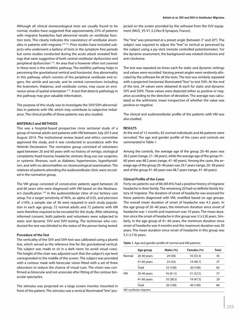
RESULTSAt the end of 12 months, 82 normal individuals and 66 patients were recruited. The age and gender profile of the cases and controls are summarized in Table 1.
Among the controls, the average age of the group 20–40 years was 28.2 years (range, 21–38 years), while the average age of the group 41–60 years was 48.2 years (range, 41–60 years). Among the cases, the av-erage age of the group 20–40 years was 29.7 years (range, 20–38 years) and of the group 41–60 years was 48.7 years (range, 41–60 years).
Clinical Profile of the CasesForty-six patients out of 66 (69.6%) had a positive history of migraine headache in their family. The remaining 20 had no definite family his-tory of migraine. The duration of onset of headache was assessed for these patients diagnosed with VM, stratified based on age groups. The overall mean duration of onset of headache was 4.3 years. In the age group of 20–40 years, the minimum duration since onset of headache was 1 month and maximum was 10 years. The mean dura-tion since the onset of headache in this group was 3 (±2.8) years. Sim-ilarly, in the age group of 41–60 years, the minimum duration since onset of headache was 4 months and the maximum duration was 30 years. The mean duration since onset of headache in this group was 5.5 (±7.6) years.
Table 1. Age and gender profile of normal and VM patients
Age group Males (%) Females (%) Total
Normal 20-40 years 29 (58) 16 (53.3) 45
41-60 years 23 (42) 14 (46.7) 37
Total 52 (100) 30 (100) 82
VM 20-40 years 16 (61.5) 21 (52.5) 37
41-60 years 10 (38.5) 19 (47.5) 29
Total 26 (100) 40 (100) 66VM: vestibular migraine
255
Ashish et al. SVV and SVH in Vestibular Migraine
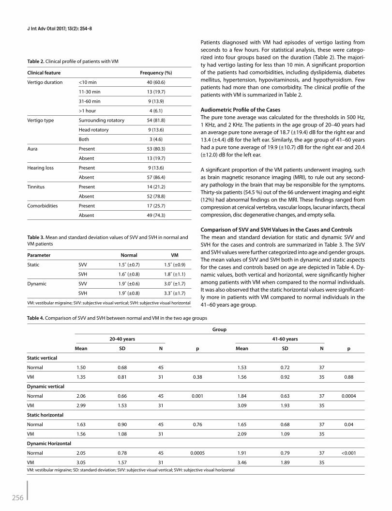
Patients diagnosed with VM had episodes of vertigo lasting from seconds to a few hours. For statistical analysis, these were catego-rized into four groups based on the duration (Table 2). The majori-ty had vertigo lasting for less than 10 min. A significant proportion of the patients had comorbidities, including dyslipidemia, diabetes mellitus, hypertension, hypovitaminosis, and hypothyroidism. Few patients had more than one comorbidity. The clinical profile of the patients with VM is summarized in Table 2.
Audiometric Profile of the CasesThe pure tone average was calculated for the thresholds in 500 Hz, 1 KHz, and 2 KHz. The patients in the age group of 20–40 years had an average pure tone average of 18.7 (±19.4) dB for the right ear and 13.4 (±4.4) dB for the left ear. Similarly, the age group of 41–60 years had a pure tone average of 19.9 (±10.7) dB for the right ear and 20.4 (±12.0) dB for the left ear.
A significant proportion of the VM patients underwent imaging, such as brain magnetic resonance imaging (MRI), to rule out any second-ary pathology in the brain that may be responsible for the symptoms. Thirty-six patients (54.5 %) out of the 66 underwent imaging and eight (12%) had abnormal findings on the MRI. These findings ranged from compression at cervical vertebra, vascular loops, lacunar infarcts, thecal compression, disc degenerative changes, and empty sella.
Comparison of SVV and SVH Values in the Cases and ControlsThe mean and standard deviation for static and dynamic SVV and SVH for the cases and controls are summarized in Table 3. The SVV and SVH values were further categorized into age and gender groups. The mean values of SVV and SVH both in dynamic and static aspects for the cases and controls based on age are depicted in Table 4. Dy-namic values, both vertical and horizontal, were significantly higher among patients with VM when compared to the normal individuals. It was also observed that the static horizontal values were significant-ly more in patients with VM compared to normal individuals in the 41–60 years age group.
Table 4. Comparison of SVV and SVH between normal and VM in the two age groups
Group
20-40 years 41-60 years
Mean SD N p Mean SD N p
Static vertical
Normal 1.50 0.68 45 1.53 0.72 37
VM 1.35 0.81 31 0.38 1.56 0.92 35 0.88
Dynamic vertical
Normal 2.06 0.66 45 0.001 1.84 0.63 37 0.0004
VM 2.99 1.53 31 3.09 1.93 35
Static horizontal
Normal 1.63 0.90 45 0.76 1.65 0.68 37 0.04
VM 1.56 1.08 31 2.09 1.09 35
Dynamic Horizontal
Normal 2.05 0.78 45 0.0005 1.91 0.79 37 <0.001
VM 3.05 1.57 31 3.46 1.89 35VM: vestibular migraine; SD: standard deviation; SVV: subjective visual vertical; SVH: subjective visual horizontal
Table 2. Clinical profile of patients with VM
Clinical feature Frequency (%)
Vertigo duration <10 min 40 (60.6)
11-30 min 13 (19.7)
31-60 min 9 (13.9)
>1 hour 4 (6.1)
Vertigo type Surrounding rotatory 54 (81.8)
Head rotatory 9 (13.6)
Both 3 (4.6)
Aura Present 53 (80.3)
Absent 13 (19.7)
Hearing loss Present 9 (13.6)
Absent 57 (86.4)
Tinnitus Present 14 (21.2)
Absent 52 (78.8)
Comorbidities Present 17 (25.7)
Absent 49 (74.3)
Table 3. Mean and standard deviation values of SVV and SVH in normal and VM patients
Parameter Normal VM
Static SVV 1.5˚ (±0.7) 1.5˚ (±0.9)
SVH 1.6˚ (±0.8) 1.8˚ (±1.1)
Dynamic SVV 1.9˚ (±0.6) 3.0˚ (±1.7)
SVH 1.9˚ (±0.8) 3.3˚ (±1.7)
VM: vestibular migraine; SVV: subjective visual vertical; SVH: subjective visual horizontal
256
J Int Adv Otol 2017; 13(2): 254-8

The mean static and dynamic values among the cases and controls based on gender are summarized in Table 5. The dynamic SVV and SVH were significantly more in patients with VM in both males and females, while the static values were not significantly different be-tween cases and controls in both gender groups.
DISCUSSIONMany methods have been suggested for the evaluation of SVV in clin-ical practice. We have used a computer software-based SVV equip-ment (MUS_VS-V1.3.2.Rev B Synapsis, France), which has a remote controlled potentiometer for recording the SVV and SVH values.
The mean values among normal individuals were 1.5° for static SVV, 1.9° for dynamic SVV, 1.6° for static SVH, and 1.9° for dynamic SVH. These values were in accordance to other studies. Previous studies have suggested that the normal values of static SVV and dynamic SVV range from ±1.5° to ±3.0° [1, 16-18].
VM is more common in females than in males [15, 19], and our study has additionally shown a female preponderance (60.1%). VM can occur at any age [19] and these patients usually have associated aura and a positive family history [19-22]. A family history of migraine was found in 69.6% of our subjects, and aura was reported in 80.3% of patients.
Studies have reported abnormal SVV findings in the Ménière’s disease, vestibular neuritis, gentamycin toxicity, and after stape-dectomy [23-26]. VM is the second most common cause of dizziness [27]. Patients can present with a spectrum of symptoms from spon-taneous room spinning vertigo, positional vertigo, or nonspecific dizziness and symptoms may last for seconds to days [15]. In our study, a majority of patients diagnosed with VM had surrounding rotatory vertigo, which lasted <10 min. This was similar to findings in previous studies [28].
A significant outcome of this study was the observed dynamic SVV and SVH values in patients with VM, which were significantly higher com-
pared to normal subjects. The static SVV values were not different from that of the controls, which implies that tonic vestibular compensation may have been achieved in these patients during the interictal phase (as these tests were conducted during the symptom-free periods). However, the fact that there is a significant difference in the static SVH (among 40–60 year olds) and dynamic SVV and SVH suggests that the dynamic compensation for the otlithic pathway defects have not been achieved in these patients. These abnormalities may also explain some of the symptoms in these patients in the context of other otoneurolog-ical examination and testing being normal.
VM is known to be the great “mimicker” of all diagnosis of vertigo [29]. The diagnosis of VM is based on patients history and diagnostic criteria have been well established [15, 30]. Further studies using SVV and SVH in individ-uals during episodes of vestibular migraine and pre- and post-treatment of VM may help explain the pathophysiology of VM better.
CONCLUSIONPatients with VM have spatial disorientation and a covert dysfunction of the otolithic pathway as shown in this study. The inclusion of SVV and SVH testing for the evaluation of patients with VM may be useful in the interpretation and rehabilitation of symptoms in these patients.
Ethical approval: Authors declared that the research was conducted accord-ing to the principles of the World Medical Association Declaration of Helsinki “Ethical Principles for Medical Research Involving Human Subjects”, (amended in October 2013).
Informed consent: Written informed consent was obtained from all individu-al participants included in the study.
Peer-review: Externally peer-reviewed.
Author Contributions: Concept - A.L., A.B.; Design- A.B.; Supervision - A.L., A.B.; Resources - G.A., A.K.T.; Materials - G.A., A.K.T.; Data Collection and/or Pro-cessing - G.A., A.K.T.; Analysis and/or Interpretation - G.A., A.M.A., A.K.T.; Liter-ature Search - G.A., A.L.; Writing Manuscript - A.M.A., A.L.; Critical Review - A.L.
Table 5. Comparison of SVV and SVH between normal and VM patients based on gender
Group
Males Females
Mean SD N p Mean SD N p
Static vertical
Normal 1.5 0.7 52 0.22
1.4 0.7 30 0.35
VM 1.3 0.6 26 1.6 1.0 40
Dynamic vertical
Normal 2.0 0.7 52 0.03
1.8 0.6 30 <0.0001
VM 2.7 2.1 26 3.3 1.5 40
Static horizontal
Normal 1.7 0.8 52 1.0
1.6 0.8 30 0.24
VM 1.7 1.1 26 1.9 1.2 40
Dynamic horizontal
Normal 2.1 0.9 52 0.02
1.8 0.6 30 <0.0001
VM 2.9 2.1 26 3.5 1.5 40VM: vestibular migraine; SVV: subjective visual vertical; SVH: subjective visual horizontal
257
Ashish et al. SVV and SVH in Vestibular Migraine

Conflict of Interest: No conflict of interest was declared by the authors.
Financial Disclosure: This research was funded by Institutional FLUID re-search grant no: 8411.
REFERENCES1. Akin FW, Murnane OD, Pearson A, Byrd S and Kelly KJ. Normative data
for the subjective visual vertical test during centrifugation. J Am Acad Audiol 2011; 22: 460-8. [CrossRef ]
2. Ashish G, Augustine AM, Tyagi AK, Lepcha A, Balraj A. Subjective visual vertical and horizontal: Normative values using a software-based test in the Indian population. Indian J Otol 2016; 22: 208-12. [CrossRef ]
3. Eghlimi B, Schaaf H and Hesse G. Measuring the subjective visual verti-cal using a portable system: a comparison with the standard darkroom method. HNO 2012; 60: 330-6. [CrossRef ]
4. Pavan TZ, Funabashi M, Carneiro JAO, Pontelli TEGDS, Tedeschi W, Colafêmina JF, et al. Software for subjective visual vertical assessment: an observational cross-sectional study. Braz J Otorhinolaryngol 2012; 78: 51-8. [CrossRef]
5. Schaaf H, Kastellis G, Hesse G. Utricular function; Correlation of three investi-gations carried out in routine practice. HNO 2013; 61: 692-8. [CrossRef]
6. Brandt T, Strupp M. General vestibular testing. Clin Neurophysiol 2005; 116: 406-26. [CrossRef ]
7. Lee H, Lopez I, Ishiyama A and Baloh RW. Can migraine damage the in-ner ear? Arch Neurol 2000; 57: 1631-4. [CrossRef ]
8. Sándor PS, Mascia A, Seidel L, de Pasqua V and Schoenen J. Subclinical cerebellar impairment in the common types of migraine: a three-dimen-sional analysis of reaching movements. Ann Neurol 2001; 49: 668-72. [CrossRef ]
9. Furman JM, Marcus DA, Balaban CD. Migrainous vertigo: development of a pathogenetic model and structured diagnostic interview. Curr Opin Neurol 2003; 16: 5-13. [CrossRef ]
10. Bir LS, Ardiç FN, Kara CO, Akalin O, Pinar HS, Celiker A. Migraine patients with or without vertigo: comparison of clinical and electronystagmo-graphic findings. J Otolaryngol 2003; 32: 234-8. [CrossRef ]
11. Harno H, Hirvonen T, Kaunisto MA, Aalto H, Levo H, Isotalo E, et al. Sub-clinical vestibulocerebellar dysfunction in migraine with and without aura. Neurology 2003; 61: 1748-52. [CrossRef ]
12. Ishizaki K, Mori N, Takeshima T, Fukuhara Y, Ijiri T, Kusumi M, et al. Stat-ic stabilometry in patients with migraine and tension-type headache during a headache-free period. Psychiatry Clin Neurosci 2002; 56: 85-90. [CrossRef ]
13. Von Brevern M, Zeise D, Neuhauser H, Clarke AH, Lempert T. Acute mi-grainous vertigo: clinical and oculographic findings. Brain J Neurol 2005; 128: 365-74. [CrossRef ]
14. Friedmann, G. The judgment of the visual vertical and horizontal with pe-ripheral and central vestibular lesions. Brain 1970; 93: 313328. [CrossRef]
15. Lempert T and Neuhauser H, Migrainous vertigo. Neurol Clin 2005; 23: 715-30. [CrossRef ]
16. Hafstrom A, Fransson P, Karlberg M, Magnusson M. Idiosyncratic com-pensation of the subjective visual horizontal and vertical in 60 patients after unilateral vestibular deafferentation. ActaOtolaryngologica 2004; 124: 165-71. [CrossRef ]
17. Tribukait A, Eiken O. Changes in the perceived head transversal plane and the subjective visual horizontal induced by Coriolis stimulation during gondola centrifugation. J Vestib Res Equilib Orientat 2006; 16: 105-16.
18. Tribukait A, Eiken O. Perception of the head transversal plane and the subjective horizontal during gondola centrifugation. Percept Psycho-phys 2005; 67: 369-82. [CrossRef ]
19. Dieterich M, Brandt T. Episodic vertigo related to migraine (90 cases): vestibular migraine? J Neurol 1999; 246: 883-92. [CrossRef ]
20. Johnson GD. Medical management of migraine-related dizziness and vertigo. The Laryngoscope 1998; 108: 1-28. [CrossRef ]
21. Ophoff RA, Terwindt GM, Vergouwe MN, van Eijk R, Oefner PJ, Hoffman SM, et al. Familial hemiplegic migraine and episodic ataxia type-2 are caused by mutations in the Ca2+ channel gene CACNL1A4. Cell 1996; 87: 543-52. [CrossRef ]
22. Reploeg MD, Goebel JA. Migraine-associated dizziness: patient char-acteristics and management options. Otol Neurotol 2002; 23: 364-71. [CrossRef ]
23. Ogawa Y, Otsuka K, Shimizu S, Inagaki T, Kondo T, Suzuki M. Subjective visual vertical perception in patients with vestibular neuritis and sudden sensorineural hearing loss. J Vestib Res Equilib Orientat 2012; 22: 205-11.
24. Shin JE, Kim CH, Park HJ. Vestibular abnormality in patients with Me-niere’s disease and migrainous vertigo. Acta Otolaryngol 2012; 133: 154-8. [CrossRef ]
25. Takai Y, Murofushi T, Ushio M, Iwasaki S. Recovery of subjective visual horizontal after unilateral vestibular deafferentation by intratympanic instillation of gentamicin. J Vestib Res Equilib Orientat 2006; 16: 69-73.
26. Tribukait A, Bergenius J. The Subjective Visual Horizontal after Stape-dotomy: Evidence for an Increased Resting Activity in Otolithic Afferents. Acta Otolaryngol 1998; 118: 299-306. [CrossRef ]
27. Bisdorff AR. Management of vestibular migraine. Ther Adv Neurol Disord 2011; 4: 183-91. [CrossRef ]
28. Casani AP, Sellari-Franceschini S, Napolitano A, Muscatello L, Dallan I. Otoneurologic dysfunctions in migraine patients with or without verti-go. Otol Neurotol 2009; 30: 961-7. [CrossRef ]
29. Kerber KA. Vertigo and Dizziness in the Emergency Department. Emerg Med Clin North Am 2009; 27: 39-50. [CrossRef ]
30. The international classification of headache disorders, 3rd edition (beta version). Cephalalgia 2013; 33: 629-808. [CrossRef ]
258
J Int Adv Otol 2017; 13(2): 254-8