Subject code: HSP102 Human Structure & Physiology
45
Human Structure & Physiology Dr. Vinicius Cruzat Senior Lecturer and Research Fellow Subject code: HSP102
Transcript of Subject code: HSP102 Human Structure & Physiology

Human Structure & Physiology
Dr. Vinicius CruzatSenior Lecturer and Research Fellow
Subject code: HSP102
COMMONWEALTH OF AUSTRALIA
Copyright Regulation 1969
WARNINGThis material has been copied and communicated to you by or on behalf of Torrens University Australia pursuant to Part VB of the
Copyright Act 1968 (the Act)
The material in this communication may be subject to copyright under the Act. Any further copying or communication of this
material by you may be the subject of copyright protection under the Act.
Do not remove this notice

Concept 1Structure and function of the small
intestine
Human Structure & PhysiologySubject code: HSP102
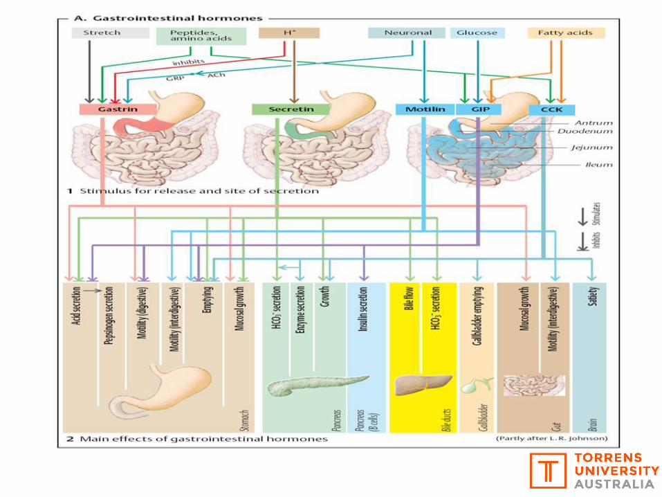

Gastrin
Secretin
MotilinCCK
GIP (Gastric inhibitory polypeptide)
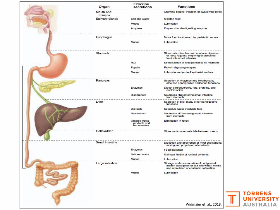
Widmaier et al., 2018.

Small intestine
Tortora & Derrickson, 2018.

Ø Intestinal juice provides a vehicle for absorption of substances from chyme as they come in contact with the villi
Ø Brush border enzymes, found on the surfaces of the microvilli of absorptive cells, break down food products
Small intestine
Tortora & Derrickson, 2018.
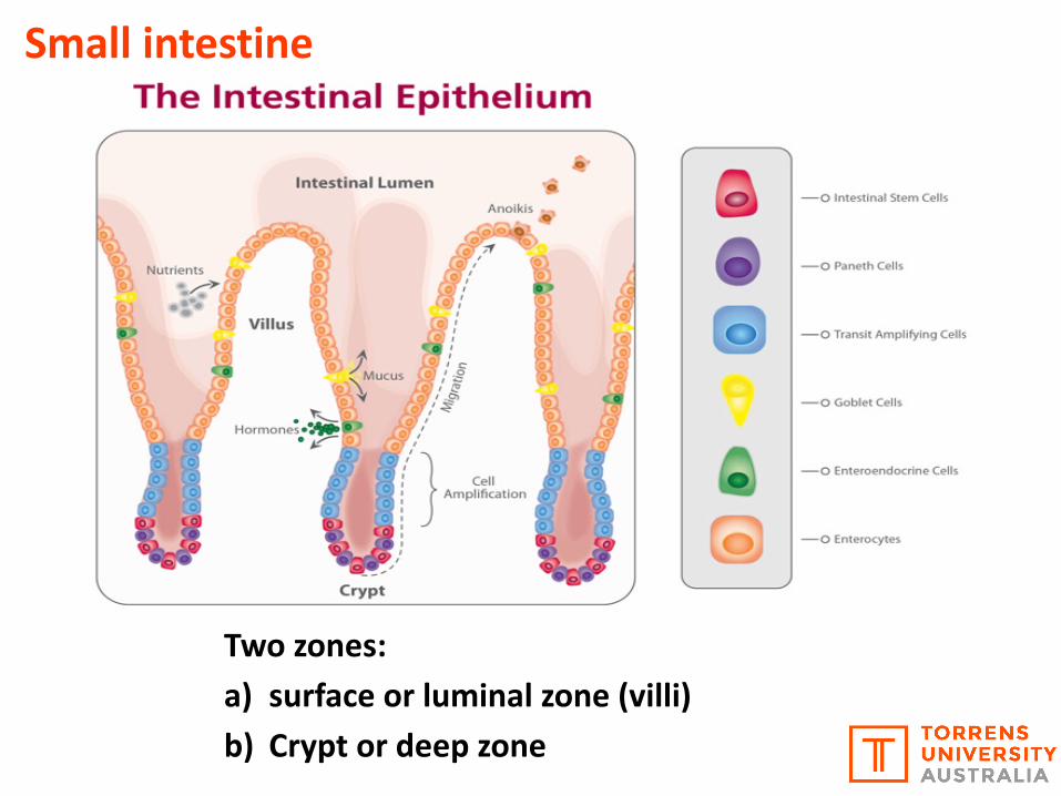
Two zones:a) surface or luminal zone (villi)b) Crypt or deep zone
Small intestine

46,800xTEM
Small intestine
Villi
Microvilli
Tortora & Derrickson, 2018.

Gastrointestinal (GI) Tract
Tortora & Derrickson, 2018.
Widmaier et al., 2018.

IleumShort and digit form villi
Ø Absence of Brunner's
gland
Ø Prominent presence of
Peyer’s patches (or
aggregated lymphoid
nodules – secrete IgA)
Ø Presence of Paneth cells
at the base of the
Lieberkühn Crypts
JejunumLong and digit form villi
Ø Presence of Paneth cells
in the base of the Crypts
of Lieberkühn
(Lysosomes)
Ø Some presence of Paneth
cells in the lamina propria
layer
DuodenumLong and short villi
Ø Brunner's gland -
Submucosa (HCO3
secretion – pH control)
Ø Sphincter of Oddi (Larger
papillary - bile duct -
Pancreatic and Biliary
secretions)
Secondary Lymphatic Organs - GALT
Histological differences between duodenum, jejunum and ileum
Tortora & Derrickson, 2018.

Overall function - absorption in the Small IntestineSmall intestine
Tortora & Derrickson, 2018.

Concept 2Structure and function of the large
intestine
Human Structure & PhysiologySubject code: HSP102

Large intestineThe last stages of digestion and absorption in the bodyØ Substances are further broken down by bacteria and some vitamins are
synthesized by bacterial actionØ Colon - tube that extends from the ileocecal part to the anus part of the gutØ Average measurment - about 125-150 cmØ Form - Inverted U shapeØ Width - about 6.5 cm diameter
Ileocecal valve - Prevents retrograde flow of fecal contents of the colon into the small intestine. Its a very thick muscular layer.
Tortora & Derrickson, 2018.
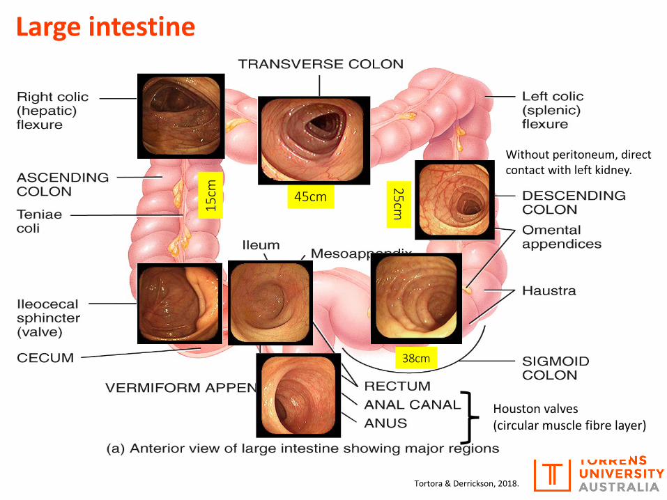
15cm 45cm
25cm
38cm
Houston valves (circular muscle fibre layer)
Without peritoneum, direct contact with left kidney.
Large intestine
Tortora & Derrickson, 2018.

Large intestine
Tortora & Derrickson, 2018.

Large intestine
Tortora & Derrickson, 2018.

Large intestineØ Haustral churning
Distension reaches a certain point and the walls of the haustra contract to squeeze contents onward
Ø PeristalsisPropulsive contractions
Ø Mass peristalsisA strong peristaltic wave that begins in the transverse colon and quickly drives the contents of the colon into the rectum
Tortora & Derrickson, 2018.

Large intestineFeces formationConsist of water, inorganic salts, sloughed-off epithelial cells, bacteria, products of bacterial decomposition, and undigested portions of food
Tortora & Derrickson, 2018.
Widmaier et al., 2018.
Brown stool type of colour = Pigment present in bile(brown stool coloration) destruction of ’old’ haemoglobin.

Large intestineDefecation reflex
1. Rectal wall distends2. Stretch receptors send sensory
nerve impulses to the sacral spinal cord
3. Motor impulses travel back to the descending colon, sigmoid colon, rectum, and anus
4. Longitudinal rectal muscles contract and the internal anal sphincter opens
If the external anal sphincter is voluntarily relaxed, defecation occurs and the feces are expelledTortora & Derrickson, 2018.
Wani & Thakur, 2016

Medical aid designed to classify the feces forms into 7 groups. Developed by Heatonand Lewis (University of Bristol, England), first published in 1997. Because the formof the stool depends on the time it spends in the colon, there is a correlationbetween the colonic transit time and the stool type.
5-7 tending towards diarrhea; dietary or
supplemental fiber can help regulate the form
of fecal waste.
The Bristol Stool Scale Types 1-2 = constipation
3 and 4 = "ideal stools" especially the 4 (easiest to pass)

Ø The appendix seems to have no function, however bacteria can proliferate inthis place which is part of the intestinal metabolism and serve as a stimulus forthe Immune system.
Ø Obstruction and inflammation in theappendix (appendicitis).Ø The faecal mass stimulates theinflammatory response, leading toappendicitis.
Large intestineAppendix
Tortora & Derrickson, 2018.

Concept 3Integration and absorption
Human Structure & PhysiologySubject code: HSP102

Splanchnic circulation
Absorption
Tortora & Derrickson, 2018.

Tortora & Derrickson, 2018.
Absorption


a) Na-Glucose Co-transporters (SGLTs)
Special transport processes bind to glucose,fructose and galactose, allowing it to cross thephospholipid bilayer.
2 families are described
b) Facilitated diffusion glucose transporters (GLUTs)
Glucose transport
Tortora & Derrickson, 2018.

a) Na-Glucose Co-transporters (SGLTs) or secondary active transport
SGLT-1
Substance is transported against the electrochemical gradient, taking advantageof the "energetic ride" of another substance (Na+) that is transported in favour ofits gradient, both transported in the same direction.ü Transport of glucose and galactoseü Symport system (2 substances same direction)ü Secondary active transport 3Na+/2K+ ATPase in the basolateral membraneü Carries glucose against its concentration gradientü Saturation: 30-50 mM glucoseü Induced by CKK and GLP-2

b) Facilitated diffusion glucose transporters (GLUTs)
Ø Has a specific combining site for the molecule being transported. Affinity for glucose, galactose and fructose
Ø "There is no ATP consumption by the cell" Transport depends on the extracellular [ ]Ø Substance moves according to the concentration gradient, > [ ] where << [ ]Ø Carry 35 mole of water / SGLT 210 mols
Kellett & Brot-Laroche, 2005.

b) Facilitated diffusion glucose transporters (GLUTs)

Protein/Amino acid transport

Amino acid Peptides1) PepT-12) Transcellular movement pathway through direct peptide penetration and
endocytosis3) Paracellular movement
Protein/Amino acid transport
Free amino acids1) Specific amino acid
transporters
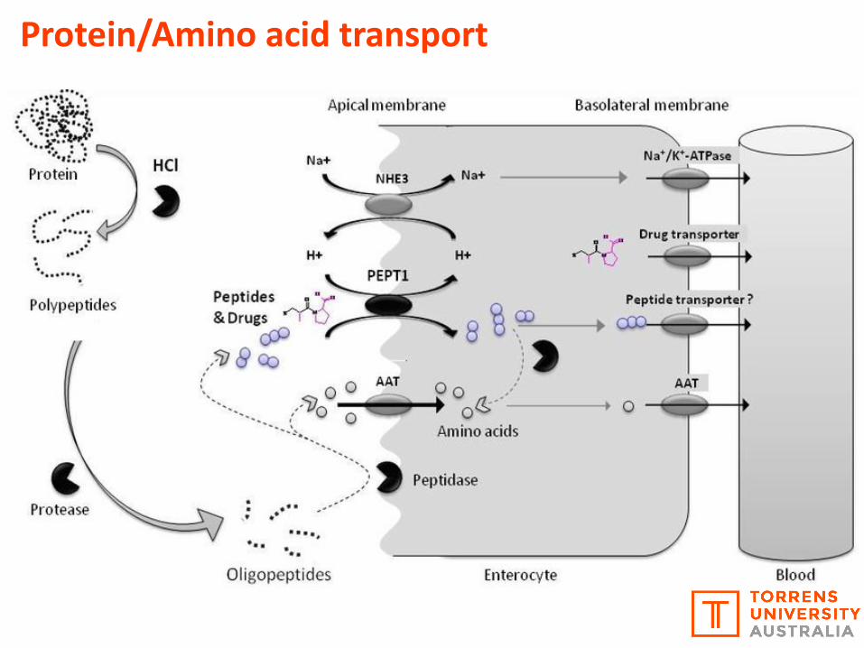
Protein/Amino acid transport

Broer, 2008.
Protein/Amino acid transport

Lipid transport

Lipid transport
A micelle is a sphere that formswhen the hydrophilic (water friendly)heads of phospholipids align to shieldhydrophobic fatty tails from thewatery environment.Vadakayil, 2010.

Lipid transport Chylomicron
Micelles

Minerals and water transport

The type and concentration ofsolute in a compartment determinesthe distribution of fluid in the othercompartments. In an equilibriumsituation, all compartments of theorganism have equal tonicity(osmolality) ~ 285 mOsmoles.
Minerals and water transport
Intracellular Extracellular
Interstitial fluid Plasma/blood
Mg++K+ Na+
HPO42-
Na+K+
Cl- Cl-
Na+
Prot -
K+
Cl- Mg++ Mg++
Ca2+ Ca2+ Ca2+ HCO3-HCO3
-HCO3-
Prot -Prot - HPO42- HPO4
2-

Plas
ma
Plas
ma
Plas
ma
Intracellular liquid
Extracellular liquid
Differences in osmolality changes H2O movement between the extra and intracellular fluids/space
↓ osmolality in the extracellular space (e.g. hyper hydration) = H2O move from the extra to the intracellular space, to equal the osmolality and cause swelling.
Similar [ ] on both sides (extracellular and
intracellular space) the osmolality will the the same.
↑ osmolality in the extracellular (e.g. dehydration) = H2O move from intra to the
extracellular space, to equal the osmolality and cause cell
dehydration.

Aquaporin increases the rate of osmosis.
Aquaporins
The function of Aquaporin is to allow the passive diffusion of water across the cell membrane. The structure of Aquaporin is a donut and water goes through the donut hole.
Minerals and water transport
”For a while scientists noticed the same thing. Water clearly efficiently enters a cell, but how?”
High H2O potentialLow solute concentration

Aquaporin protein makes a water channel through the cell (plasma) membrane.
Peter AgreJohns Hopkins
Roderick MacKinnon
Rockefeller
• Aquaporins are channel proteins that move water rapidly into the cell through facilitated diffusion.• They were discovered by these two in 1991.• They shared the 2003 Nobel Prize in Chemistry.
AquaporinsMinerals and water transport

Overviews and Animations for the Digestive System
http://www.wiley.com/college/interactions/Energy/content/Energy/dig3a/frameset2.htm
Ø Carbohydrate Digestion and Absorption
http://www.wiley.com/college/interactions/Energy/content/Energy/dig4a/frameset2.htm
Ø Protein Digestion and Absorption
http://www.wiley.com/college/interactions/Energy/content/Energy/dig5a/frameset2.htm
Ø Lipid Digestion and Absorption
http://www.wiley.com/college/interactions/Energy/content/Energy/dig7a/frameset2.htm
Ø Hormonal Control of Digestive Activities

Main and recommended referencesØ Tortora, G. and Derrickson, B. (2014). Principles of Anatomy & Physiology. 14th ed. John Wiley & Sons.
Ø Tortora, G. and Derrickson, B. (2018). Principles of Anatomy & Physiology. 2th ed. Asia-Pacific EditionHybrid. John Wiley & Sons.
Ø Hall, J. E., & Guyton, A. C. (2015). Guyton and Hall textbook of medical physiology. 13th ed.Philadelphia, PA: Saunders Elsevier.
Ø McArdle, W. D., Katch, F. I., & Katch, V. L. (2014). Exercise physiology: Energy, nutrition, and humanperformance. 8th ed. Philadelphia: Lippincott Williams & Wilkins.
Ø Alberts, B., Johnson, A., Lewis, J., Raff, M., Roberts, K., & Walter, P. (2002). Molecular biology of thecell. 15th ed. New York: Garland Science.
Ø Abbas, A. K., Lichtman, A. H., & Pillai, S. (2017). Cellular and molecular immunology. 9th ed.Philadelphia: Saunders/Elsevier.
Ø Netter, F. H. (2018). Atlas of human anatomy. 7th ed. Philadelphia, PA: Saunders/Elsevier.
Ø Widmaier, Eric P., Raff, Hershel, Strang, Kevin T. (2018). Vander's human physiology: the mechanismsof body function 15th ed. Boston: McGraw-Hill Higher Education.
Ø Silverthorn, D. U. (2018). Human physiology: An integrated approach. 8th ed. San Francisco: Harlow:Benjamin Cummings.
Ø Boron, W. F., Boulpaep, E. L. (2016). Medical physiology: a cellular and molecular approach. 3rd ed.Philadelphia: Saunders/Elsevier.