Subhasish Chandra Spectroscopic Sources Assistant...
80
Spectroscopic Sources Subhasish Chandra Assistant Professor Institute of Forensic Science, Nagpur
-
Upload
truongdieu -
Category
Documents
-
view
218 -
download
1
Transcript of Subhasish Chandra Spectroscopic Sources Assistant...

Spec t r o s c op i c Sour c e sSubhasish Chandra Assistant Professor Institute of Forensic Science, Nagpur

SpectroscopySpectroscopy is a general term for the science dealing with interactions of various types of radiation with matter. Spectrometry and spectrometric methods refer to the measurement of the intensity of radiation with a transducer. The most widely used methods are based on electromagnetic radiation, the most recognizable being visible light and radiant heat. Less obvious manifestations include gamma ray, X – ray, ultraviolet, microwave and radio – frequency radiation.
Instrumental Analysis by Skoog, Holler and Crouch2

Electromagnetic RadiationElectromagnetic radiation is produced when a charge particle accelerates. The acceleration of charge produces changing electric and magnetic field which constitute an electromagnetic wave. The formation of electromagnetic waves can be explained using Maxwell’s electrodynamics equations. The electromagnetic spectrum encompasses an enormous range of wavelengths and frequencies (and thus energies).
Concepts of Physics Part 2 by H. C. Verma3

Electromagnetic Spectrum
4

Types of Electromagnetic Radiation➢Radio Waves are formed due to acceleration of charges through
conducting wires. Ranging from more than 10 km to about 0.1 m in wavelength, they are generated by electronic devices like LC oscillators and are used in radio and television communication systems.
➢Microwaves are generated by electric circuits with oscillating current. They have wavelengths ranging from approximately 0.3 m to 0.1 mm. Because of their short wavelengths, they are well suited for radar systems and for studying the atomic and molecular properties of matter. Microwave ovens (in which the wavelength of the radiation is 0.122 m) are an interesting domestic application of these waves.
Concepts of Physics Part 2 by H. C. Verma5

➢ Infrared Waves have wavelengths ranging from 1mm to the longest wavelength of visible light, 700 nm. These waves, produced by molecules and room temperature objects, are readily absorbed by most materials. The infrared (IR) energy absorbed by a substance appears as internal energy because the energy agitates the atoms of the object, increasing their vibrational or translational motion, which results in a temperature increase. Infrared radiation has practical and scientific applications in many areas, including physical therapy, IR photography, and vibrational spectroscopy.
Fundamentals of Physics by Halliday, Resnick, Walker6
Types of Electromagnetic Radiation

➢ Visible Light is the only part of the electromagnetic spectrum that the human eye can detect. Light is produced by the rearrangement of electrons in atoms and molecules. The various wavelengths of visible light, which correspond to different colours, range from red (700 nm) to violet (400 nm). The sensitivity of the human eye is a function of wavelength, being a maximum at a wavelength of about 550 nm .
➢ Ultraviolet Waves cover wavelengths ranging from approximately 400 nm to 0.6 nm. The Sun is an important source of ultraviolet (UV) light. Various other sources such as tungsten lamp are also used to produce ultraviolet light. Although they lack the energy to ionize atoms, long – wavelength ultraviolet radiation can cause chemical reactions and can cause fluorescence.
Concepts of Physics Part 2 by H. C. Verma7
Types of Electromagnetic Radiation

➢ X – rays have wavelengths ranging from about 10 nm to 1 pm. The most common source of X – rays is the deceleration of high energy electrons bombarded on a metal target. They are used as a diagnostic tool in medicine and as a treatment for certain forms of cancer. They are also used in the study of crystal structure because their wavelengths are comparable to the atomic separation distances in solids (about 0.1 nm).
➢ Gamma Rays are electromagnetic waves emitted by radioactive nuclei (such as Co-60 and Cs-137) and are also produced during certain nuclear reactions. High energy gamma rays are a component of cosmic rays that enter the Earth’s atmosphere from space. They have wavelengths ranging from approximately 1 pm to less than 10 fm. They are highly penetrating and produce serious damage when absorbed by living tissues.
Fundamentals of Physics by Halliday, Resnick, Walker8
Types of Electromagnetic Radiation

Types of SpectroscopyType of Spectroscopy Wavelength
RangeType of Quantum Transition
Gamma ray emission 0.005 - 1Å NuclearX – ray absorption, emission, fluorescence and diffraction
0.1 - 100Å Inner electron
Vacuum ultraviolet absorption 10 – 180 nm Bonding electrons
Ultraviolet – Visible absorption, emission and fluorescence
180 – 780 nm Bonding electrons
Infrared absorption and Raman scattering
0.78 – 300 µm Rotation / vibration of molecules
Microwave absorption 0.75 – 375 mm Rotation of molecules
Electron spin resonance 3 cm Spin of electrons in a magnetic field
Nuclear magnetic resonance 0.6 – 10 m Spin of nuclei in a magnetic field
9Instrumental Analysis by Skoog, Holler and Crouch

Emission and Absorption Spectra
PHE – 15 Astronomy and Astrophysics of BSc (Physics), IGNOU10

Sources of Radiation
11Instrumental Analysis by Skoog, Holler and Crouch

➢ Radiation is produced when electricity passes through the tungsten filament encased in a glass envelope.
➢ The energy distribution is a p p r o x i m a t e l y t h a t o f a blackbody. Thus the radiation is temperature dependent.
➢ In most instruments, operating temperature of the filament is 2850 K.
➢ The wavelength corresponding to this temperature corresponds to the infrared region.
Tungsten Bulb12
Instrumental Analysis by Skoog, Holler and Crouch
Light Sources for Ultraviolet – Visible Molecular Absorption Spectroscopy

➢ The bulk of the radiation is emitted in infrared region.
➢ It is useful for the wavelength region between 300 nm to 2500 nm.
➢ In the visible region, the energy output of the lamp approximately varies as the fourth power of operating voltage. As a result voltage regulators or feedback – controlled power supplies are employed to obtain a stable radiation source.
Spectrum of Tungsten Bulb
zeiss-campus.magnet.fsu.edu/articles/lightsources/tungstenhalogen.html13
Light Sources for Ultraviolet – Visible Molecular Absorption Spectroscopy

➢ Tungsten – Halogen lamps, also called quartz – halogen lamps, contain a small quantity of iod ine wi th in the quar tz envelope that houses the tungsten filament.
➢ Quartz allows the filament to be operated at a temperature of about 3500 K, which leads to higher intensities and extends the range of the lamp into the ultraviolet region.
Tungsten Halogen Bulb
14Instrumental Analysis by Skoog, Holler and Crouch
Light Sources for Ultraviolet – Visible Molecular Absorption Spectroscopy

➢ The bulk of the radiation is emitted in visible and near infrared region.
➢ It is useful for the wavelength region between 200 nm to 2500 nm.
➢ I t e m p l o y s a H a l o g e n regenerat ive cycle which increases the life of the bulb.
Spectrum of Tungsten Halogen Bulb
zeiss-campus.magnet.fsu.edu/articles/lightsources/tungstenhalogen.html15
Light Sources for Ultraviolet – Visible Molecular Absorption Spectroscopy

➢ Once the envelope reaches a temperature of approximately 200 to 250 °C , the Tungsten atoms evaporated from the filament & react with gaseous halogen vapor and the trace levels of molecular oxygen to form tungsten oxyhalides.
➢ Instead of condensing on the hot inner walls of the envelope, the oxyhalide compounds are circulated by convection currents back to the region surrounding the filament where they decompose, leaving elemental tungsten re-deposited on the cooler regions of the filament.
Halogen Regenerative Cycle
zeiss-campus.magnet.fsu.edu/articles/lightsources/tungstenhalogen.html16
Light Sources for Ultraviolet – Visible Molecular Absorption Spectroscopy

➢ The xenon arc lamp produces intense radiation by the passage o f c u r r e n t t h r o u g h a n atmosphere of xenon.
➢ It produces a largely continuous and uniform spectrum across the entire visible spectral region.
➢ The emission profile features a c o l o r t e m p e r a t u r e o f approximately 6000 K (close to that of sunlight) and lacks prominent emission lines.
Xenon Arc Lamp17
Instrumental Analysis by Skoog, Holler and Crouch
Light Sources for Ultraviolet – Visible Molecular Absorption Spectroscopy

➢ The spectrum is a continuum over the range between about 200 to 1000 nm. The peak intensity occurs at about 500 nm.
➢ Approximately 70 percent of the output occurs at infrared wavelengths while less than 5 percent is in UV region.
➢ In visible region, approximately 85 percent of the total energy emitted resides in the continuum and the rest from the line spectrum.Spectrum of
Xenon Arc Lamp
zeiss-campus.magnet.fsu.edu/articles/lightsources/xenonarc.html18
Light Sources for Ultraviolet – Visible Molecular Absorption Spectroscopy

➢ A continuum spectrum in the ultraviolet region can be p r o d u c e d b y e l e c t r i c a l excitation of deuterium or hydrogen at low pressures.
➢ When deuterium is given electrical energy , it forms excited molecular species .
➢ This excited species dissociates to give two atomic species and an ultraviolet photon.
Deuterium Lamp
eE*2D
ν
ν
hEEEEhDDDED
DDDe
e
++==
+ʹ́+ʹ→→+
ʹ́ʹ*2
*22
19Instrumental Analysis by Skoog, Holler and Crouch
Light Sources for Ultraviolet – Visible Molecular Absorption Spectroscopy

➢ The output is in the range of 160 to 800 nm.
➢ In the ultraviolet region a continuum spectrum exists.
➢ At longer wavelengths, the spectra consists of emission lines and bands superimposed on a weak continuum.
➢ Quartz windows is provided to extend the output in the UV region to 190 nm.
Spectrum of Deuterium Lamp
20Instrumental Analysis by Skoog, Holler and Crouch
Light Sources for Ultraviolet – Visible Molecular Absorption Spectroscopy

➢ An LED is a PN – junction device that produces radiant energy (light) when forward biased.
Light Emitting Diodes (LEDs)
Gallium Aluminum Arsenide (GaAlAs)
λmax = 900 nm
Gallium Arsenic Phosphide (GaAsP)
λmax = 650 nm
Gallium Phosphide (GaP)
λmax = 550 nm
Gallium Nitride (GaN)λmax = 465 nm
Indium Gallium Nitride (InGaN)
λmax = 450 nm
21Instrumental Analysis by Skoog, Holler and Crouch
Light Sources for Ultraviolet – Visible Molecular Absorption Spectroscopy

➢ LEDs produce a spectral continuum over a narrow wavelength range.
➢ The typical (Full Width at Half Maximum) of an LED is about 20 to 50 nm.
➢ LEDs can be used as semi – monochromatic sources or in conjunction with interference filters to further narrow down the spectral output.
➢ The LEDs can be operated in continuous or in pulsed mode.
Spectrum of Light Emitting Diodes (LEDs)
zeiss-campus.magnet.fsu.edu/articles/lightsources/leds.html22
Light Sources for Ultraviolet – Visible Molecular Absorption Spectroscopy
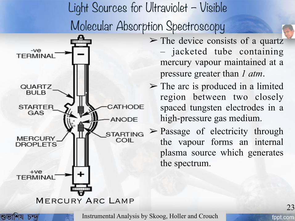
➢ The device consists of a quartz – jacketed tube containing mercury vapour maintained at a pressure greater than 1 atm.
➢ The arc is produced in a limited region between two closely spaced tungsten electrodes in a high-pressure gas medium.
➢ Passage of electricity through the vapour forms an internal plasma source which generates the spectrum.
Mercury Arc Lamp23
Instrumental Analysis by Skoog, Holler and Crouch
Light Sources for Ultraviolet – Visible Molecular Absorption Spectroscopy

➢ There emission profile has several prominent emission lines (365 nm, 405 nm, 436 nm, 546 nm & 579 nm) present in v is ib le region which are significantly brighter than the continuous background.
➢ There are significant number of spectral lines in the ultraviolet region between 250 and 350 nm.
➢ Approximately 45 percent of the radiant output falls between 350 to 700 nm.
Spectrum of Mercury Arc Lamp
zeiss-campus.magnet.fsu.edu/articles/lightsources/mercuryarc.html24
Light Sources for Ultraviolet – Visible Molecular Absorption Spectroscopy

➢ The spectrum has several lesser lines in the infrared wavelengths exceeding 1000 nm.
➢ The spectral emission region between 600 and 1000 nm is relatively continuous and no brighter in output than xenon arc lamps.
Spectrum of Mercury Arc Lamp
zeiss-campus.magnet.fsu.edu/articles/lightsources/mercuryarc.html25
Light Sources for Infrared Spectroscopy

➢ The majority of the emitted energy (up to 85 percent) lies in the infrared and near-infrared regions of the spectrum.
➢ It is a convenient source for the near – infrared region of 0.78 to 2.5 µm.
Spectrum of Tungsten Halogen Bulb
zeiss-campus.magnet.fsu.edu/articles/lightsources/tungstenhalogen.html26
Light Sources for Infrared Spectroscopy

➢ Tightly wound nichrome wire heated to about 1100 K by an electric current is a good source of infrared radiation.
➢ The radiation is less intense than other sources but are nearly maintenance free and requires no cooling.
➢ Process analyzers often use these as radiation sources.
➢ Rhodium wire heater sealed in a ceramic cylinder has similar properties but is more expensive and not used much.
Incandescent Wire 27
Instrumental Analysis by Skoog, Holler and Crouch
Light Sources for Infrared Spectroscopy

➢ It is composed of rare oxides (Zirconium (Zr), Yttrium (Y), Erbium (Er)) formed into a cylinder having a diameter of 1 to 3 mm and a length of 2 to 5 cm.
➢ Platinum leads are sealed to the ends of the cylinder for electrical connectivity. These leads act as the resistive heating element.
➢ O n b e i n g p r o v i d e d w i t h electricity, the temperature increases to between 1200 to 2200 K .
Nernst Glower28
Instrumental Analysis by Skoog, Holler and Crouch
Light Sources for Infrared Spectroscopy

➢ It has a large negative temperature coefficient of electrical resistance.
➢ The glower needs to be heated externally to dull red before the current is large enough to maintain the desired temperature.
➢ The overall spectral output is that of a blackbody in between 1 and 15 µm.
➢ The maximum output peaks in the infrared region at about 1.4 µm.
Spectrum of Nernst Glower
29Instrumental Analysis by Skoog, Holler and Crouch
Light Sources for Infrared Spectroscopy

➢ It is a silicon carbide rod, about 5 cm in length and 5 mm in diameter. It is electrically heated to about 1300 to 1500 K.
➢ It has a positive coefficient of resistance.
➢ The spectral output is in the infrared region of 1 to 50 µm and peaks at about 2 µm.
➢ Combined with a interference filter, it produces radiation having wavelengths of 4 to 15 µm.
Globar30
Instrumental Analysis by Skoog, Holler and Crouch
Light Sources for Infrared Spectroscopy

➢ A tunable carbon dioxide laser is used as an infrared source for monitoring the concentration of certain atmospheric pollutants and fo r de t e rmin ing the absorbing species in aqueous solutions.
➢ It produces a band of radiation in the range of 9 to 11 µm consisting of about 100 closely spaced lines with 10.6 µm the most prominent one.
Carbon Dioxide Laser
31Instrumental Analysis by Skoog, Holler and Crouch
Light Sources for Infrared Spectroscopy

➢ The range of wavelengths is l i m i t e d b u t t h e r e g i o n fortunately is rich in absorption b a n d s a r i s i n g f r o m t h e interactive stretching modes of carbon dioxide.
➢ It is useful in quantitative determination of a number of impor tant molecules v iz . ammonia, butadiene, benzene, ethanol, nitrogen dioxide and trichloroethyne.
Carbon Dioxide Laser
32Instrumental Analysis by Skoog, Holler and Crouch
Light Sources for Infrared Spectroscopy

➢ It consists of a tungsten anode and a cylindrical cathode sealed in a glass tube filled with neon or argon at low pressures.
➢ The cathode is constructed of the metal whose spectrum is desired.
➢ Ionization of the inert gas occurs when a potential difference of about 300 V is applied.
➢ The electrons migrate to the electrodes due to this ionization.
Hollow Cathode Lamps
Light Sources for Atomic Absorption Spectroscopy
33Instrumental Analysis by Skoog, Holler and Crouch

➢ When the voltage becomes sufficiently large, the gaseous cations acquire enough kinetic energy to dislodge some of the metal atoms from the cathode surface and produce an atomic cloud in a process called sputtering.
➢ A portion of the sputtered metal atoms are in excited states and thus emit their characteristic radiation as they return to the ground state.
Spectrum of Hollow Cathode Lamps
34Instrumental Analysis by Skoog, Holler and Crouch
Light Sources for Atomic Absorption Spectroscopy

➢ It is constructed from a sealed quartz tube containing an inert gas such as argon at low pressures and a small quantity of a metal or its salt whose spectrum is of interest.
➢ It contains no electrode and is energized by an intense field of radio frequency or microwave radiation.
➢ Argon ionizes and the ions are accelerated by the high frequency component of the field until they gain sufficient energy to excite the atoms of the metal whose spectrum is sought.
Electrodeless Discharge Lamps
35Instrumental Analysis by Skoog, Holler and Crouch
Light Sources for Atomic Absorption Spectroscopy

Coolidge Tube
Concepts of Physics Part 2 by H. C. Verma36
X – rays

➢ One portion of the spectrum consists of the X – rays emitted in all possible wavelengths having a minimum wavelength (cut – off wavelength, λmin). This part of the s p e c t r u m i s d u e t o b r e m s s t r a h l u n g ( b r a k i n g radiation) and is known as Continuous X – ray.
➢ The other portion is due to electron jumps in the shells of the atoms of the target and are known as Characteristic X – ray.
X – ray Spectrum
Fundamentals of Physics by Halliday, Resnick, Walker37
X – rays

➢ Bremsstrahlung is a German word which means Braking Radiation (bremsen: to brake, strahlung: radiation).
➢ The accelerated electrons strike the target and penetrate deep into the atoms.
➢ The electrons lose their kinetic energy when they come in contact with the Coulomb field of the nuclei.
➢ The electrons either stop completely or deviate from their original path.
Continuous X – rays
A Textbook of Engineering Physics by Avadhanalu, Kshirsagar
uwat
erlo
o.ca
38
X – rays

➢ The deviated electrons make several collisions before coming to rest.
➢ A part of this lost kinetic energy is converted into photons of electromagnetic radiation.
➢ The remaining part increases the kinetic energy of the colliding particle of the target. The energy received by the target goes into heating the target.
Continuous X – rays
Engineering Physics by Dattu R Joshi
uwat
erlo
o.ca
39
X – rays

➢ A t h i g h e r a c c e l e r a t i o n potentials, maximum intensity of the X – rays shifts toward lower wavelengths.
➢ The emitted radiation has continuous spectral distribution.
➢ For any potential, a sharply defined minimum wavelength is present. This minimum wavelength is called as cut – off wavelength.
Continuous X – rays Spectrum
Concepts of Physics Part 2 by H. C. Verma
hype
rphy
sics
.phy
-ast
r.gsu
.edu
40
X – rays

Maximum frequency corresponds to minimum wavelength and the above equation thus becomes,
Therefore,
Fundamentals of Physics by Halliday, Resnick, Walker
The electrons are accelerated through a potential V. The energy gained by the electrons is eV. According to Planck’s hypothesis the energy of the X – ray photons is given as hν, where h is the Planck’s constant and ν is the frequency of the emitted X – rays. Let us consider the case where the electron loses all its energy during its collision with the nucleus. The X – ray photon thus produced will have maximum energy. Then,
eVh =maxν
eVhc
eVhc
=
=
min
min
λ
λ
Jsh 341063.6 −×= smc /103 8×=Ce 19106.1 −×=
Å12400
1024.1
min
6
min
V
mV
=
−
=×
λ
λ
Duane – Hunt Formula 41
X – rays

➢ An atom radiates energy when an electron jumps from one allowed orbit to another allowed orbit.
➢ When a high energy electron knocks off an electron from the inner orbit, an electron from an outer orbit jumps to fill the vacancy in the inner orbit.
➢ Such a transition causes the emission of a high energy photon i.e. the X – ray photon.
Characteristic X – rays
A Textbook of Engineering Physics by Avadhanalu, Kshirsagar
uwat
erlo
o.ca
42
X – rays

➢ The transition of electrons giving X–rays of wavelengths
➢ The group of lines formed due to these transitions are called the K–series.
➢ L–series are produced when vacancies in L–shell are filled by electrons from next higher shells.
Characteristic X – rays
γβα
γ
β
α
KKK EEEKKNKKMKKL
>>
⇒→
⇒→
⇒→
Engineering Physics by Dattu R Joshi43
X – rays

➢ The spectrum consists of a series of discrete lines.
➢ These lines are characteristic of the target used and are hence called the Characteristic X – rays.
➢ This spectrum is also known as the line spectrum.
➢ It consists of discrete spectral l i n e s w h i c h c o n s t i t u t e K,L,M,N,… series.
Characteristic X – rays Spectrum
hype
rphy
sics
.phy
-ast
r.gsu
.edu
Concepts of Physics Part 2 by H. C. Verma44
X – rays

➢ K–series are the most energetic and constitute the hard X – rays whereas L,M,N,… series form the soft X – rays.
➢ When the atomic number of the target is increased, there is a regular shift towards the shorter wavelength.
➢ The spectrum is strikingly similar for all elements.
➢ The spectrum for an element remains the same irrespect ive of the compounds that the element forms.
Characteristic X – rays Spectrum
hype
rphy
sics
.phy
-ast
r.gsu
.edu
Fundamentals of Physics by Halliday, Resnick, Walker45
X – rays

➢ The spectrum emitted by each element was exactly similar but t h e l i n e s h a d d i f f e r e n t wavelengths.
➢ Moseley found that frequency of any particular line in a series varied regularly from one element to the next one in the periodic table.
➢ He found that the plot between the square root of the frequency and the atomic number of the element is a straight line. Such a plot is known as the Moseley’s plot.
Moseley’s Law
hype
rphy
sics
.phy
-ast
r.gsu
.edu
A Textbook of Engineering Physics by Avadhanalu, Kshirsagar46
X – rays
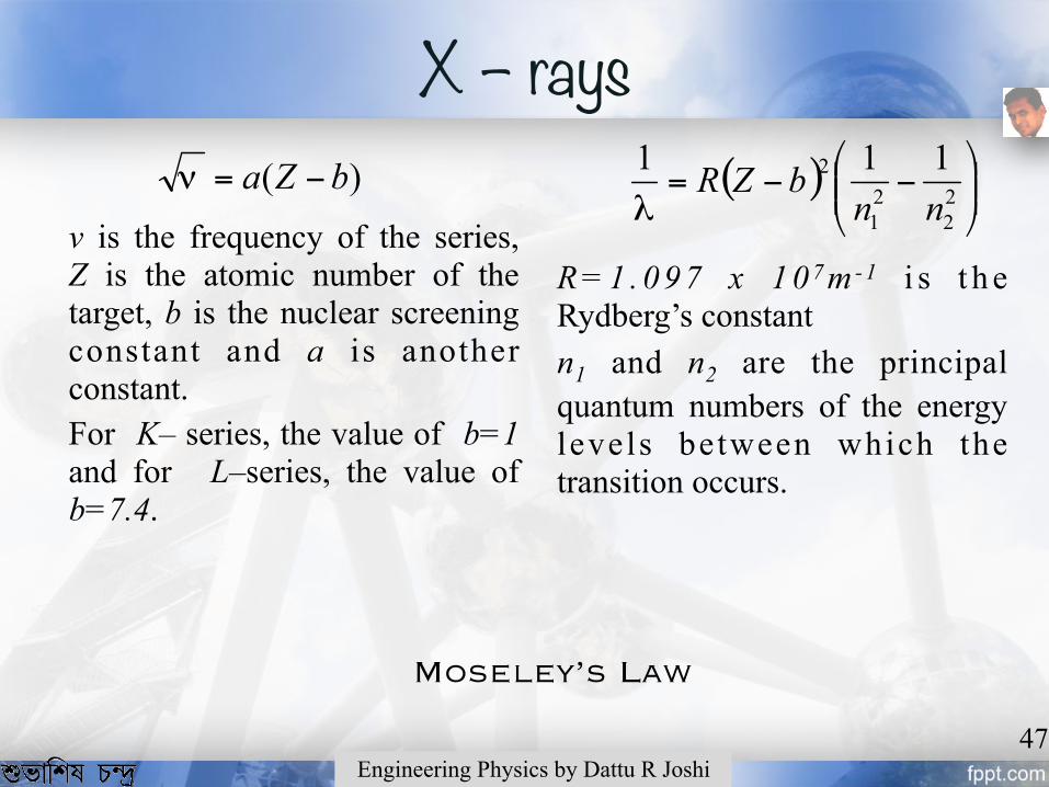
R = 1 . 0 9 7 x 1 0 7 m - 1 i s t h e Rydberg’s constant n1 and n2 are the principal quantum numbers of the energy levels between which the transition occurs.
Moseley’s Law
47Engineering Physics by Dattu R Joshi
)( bZa −=ν
ν is the frequency of the series, Z is the atomic number of the target, b is the nuclear screening constant and a is another constant. For K– series, the value of b=1 and for L–series, the value of b=7.4.
( ) ⎟⎟⎠
⎞⎜⎜⎝
⎛−−= 2
221
2 111nn
bZRλ
X – rays

➢ X – rays are produced by the retardation of electric charges which predicts that they are electromagnetic waves.
➢ They are unaffected by electric and magnetic fields. ➢ They affect a photographic plate. ➢ They can be reflected, refracted and diffracted. ➢ They can produce ionization of gases through which they pass and
are also able to influence the electrical properties of solids and liquids.
➢ They produce fluorescence and phosphorescence in many substances like platinocyanides and Cesium sulphide.
Concepts of Physics Part 2 by H. C. Verma48
Properties of X - rays
X – rays

➢ They can penetrate through many solid substances which are normally opaque to visible light. The penetrating power is different for different substances depending upon their density.
➢ Photoelectric effect is observed due to impact of X – rays on a metal surface. The emitted photoelectrons have high velocities of the order of 6 x 10⁵ m/s to 8 x 10⁵ m/s .
➢ They can damage and kill living cells and thus cause reddening of skin, produce deep – seated burns and unpredictable chemical changes.
49Fundamentals of Physics by Halliday, Resnick, Walker
Properties of X - rays
X – rays

➢ When X – rays fall upon a material, they produce secondary X – rays which are of two types,
❑ Scattered X – rays: For gases and other substances having low atomic numbers, the secondary rays are produced due to elastic scattering and hence the penetrating power and the wavelength of the secondary X – rays is same as the incident X – rays. Only the direction of the incident rays change. If there are free electrons in the substance, the impact of X – rays with them modifies the incident rays and the scattered rays have lesser energy.
Properties of X - rays50
A Textbook of Engineering Physics by Avadhanalu, Kshirsagar
X – rays

❑ Characteristic X – rays: For the substances having higher atomic numbers, the secondary X – rays have less wavelength and penetrating power. The rays become more homogeneous and are grouped into several series known as K,L,M,… series. The penetrating power decreases as we move from K–series to M–series and onwards. Also, the K– series of higher atomic number elements is more penetrating than the K–series of lower atomic number.
51Engineering Physics by Dattu R Joshi
Properties of X - rays
X – rays

➢ Industrial Applications ❑ To detect hair cracks in the internal structure of a body, imperfections in
moulds, forgings and foundry castings. ❑ To test gross structures, welds for homogeneity. X – ray photographs may
reveal defects in the manufactured heavy steel plates used in bridge construction
❑ To detect cracks and blowholes in metal plates, flaws in the wings and fuselages of airplanes and parts of other fast moving machines.
❑ To analyze the structure of alloys and other composite bodies by determining the crystal structure in an ingot with the help of diffraction.
❑ To detect and photograph defects of internal structure of a body viz. machine parts intended for withstanding high pressures, cracks in wood, porcelain and other insulators, defects in diamonds and other precious stones.
❑ To examine the age of rocks and its infrastructure in geology.
Concepts of Physics Part 2 by H. C. Verma52Applications of X - rays
X – rays

➢ Medical Applications ❑ Radiography: For diagnosis purposes, X – rays are widely used to detect
fractures, tumours, kidney stones, presence of foreign matter like bullets, diseased organs in the human body.
The digestive tract can be made visible by mixing a harmless compound of a heavy element like bismuth with food. Some salts are injected or given orally to render certain regions of the body opaque to the rays and thus enabling photography of different organs.
❑ Röntgen Therapy: The diseased tissues are more susceptible to destruction than the surrounding healthy issue. This fact is used to cure cancers, tumours, skin diseases and malignant sores by controlled exposure to X – rays. Long exposures of human body to hard X – rays results in the loss of white blood corpuscles, causes sterility and other genetic changes.
❑ X – rays are also used for the identification of cells and tissues and for production of genetic mutations.
53Applications of X - raysFundamentals of Physics by Halliday, Resnick, Walker
X – rays

➢ Scientific Research ❑ Investigation of structure of atom ❑ The identification of chemical elements, determination of their
atomic number, energy levels in the extra nuclear part of the atoms ❑ The investigation of the structure of crystalline solids and alloys ❑ Analyzing the structure of complex organic molecules examining
X – ray diffraction patterns.
54Applications of X - raysA Textbook of Engineering Physics by Avadhanalu, Kshirsagar
X – rays

➢ When radiation crosses an interface between media that differ in refractive indices, reflection always occurs.
➢ The fraction of reflected radiation becomes greater with increasing differences in refractive indices.
➢ For a beam entering an interface at right angles, the reflected fraction is given as,
I0 is the intensity of the incident beam, Ir is the intensity of the reflected beam n1 and n2 are the refractive indices of the two media.
55Reflection of RadiationInstrumental Analysis by Skoog, Holler and Crouch
Interaction of Radiation with Matter
( )( )212
212
0 nnnn
IIr
+
−=

➢ Much of the radiation that is incident on a material is transmitted without loss of intensity. But at selected wavelengths, radiation intensity is attenuated. This attenuation in which these certain wavelengths are removed is called absorption.
➢ This happens due to transfer of electromagnetic energy to atoms, ions or molecules composing the material. The general requirements for absorption of electromagnetic radiation are, ❑ There is an interaction between the electromagnetic radiation and the material.
For UV – Visible, the interaction is with electronic energy of the valence electrons. For infrared, the vibrational energy of the chemical bonds is altered.
❑ The energy of the electromagnetic radiation must exactly correspond to the energy of transition in the material i.e. the difference in energy between the material’s quantized energy states.
❑ If there are no quantized energy levels matching the quantum energy of radiation, the material will appear transparent to the given radiation.
56Absorption and Transmission of RadiationInstrumental Analysis by Skoog, Holler and Crouch
Interaction of Radiation with Matter

➢ Atomic Absorption: The passage of radiation through a medium that consists of monoatomic particles results in the absorption of a few well – defined wavelengths. Such spectra are very simple due to the small number of possible energy states for the absorbing particles.
Ultraviolet and visible radiation has enough energy to cause transitions of the outermost electrons only. X – rays on the other hand have energies that allows them to interact with innermost electrons.
➢ Molecular Absorption: Absorption spectra for polyatomic molecules are considerably more complex than atomic spectra because the number of energy states of molecules is generally enormous when compared with the number of energy states for isolated atoms. The energy E of a molecule is made up of three components,
E = Eelectronic + Evibrational + Erotational
Eelectronic is the energy due to the energy states of the bonding electrons, Evibrational is the energy due to interatomic vibrations, Erotational is the energy due to the various rotational motions within the molecule.
57Absorption of RadiationInstrumental Analysis by Skoog, Holler and Crouch
Interaction of Radiation with Matter

58Instrumental Analysis by Skoog, Holler and Crouch
➢ Transmittance: The transmittance T of the medium is the fraction of incident radiation transmitted by the medium.
T = P/P0 (value from 0 to 1) P0 is the incident power of the beam and P is the power of the beam
after absorbed by the sample. Transmittance is often expressed as a percentage, %T = P/P0 x 100 % (value from 0 to 100)
➢ Absorbance: The absorbance A of a medium is defined by the equation,
A = -1og10 T = -log10 (P/P0 ) = log10 (P0/P ) = log10 (1/T )
Absorption and Transmission of Radiation
Interaction of Radiation with Matter

59Instrumental Analysis by Skoog, Holler and Crouch
hype
rphy
sics
.phy
-ast
r.gsu
.edu
Interaction of Radiation with Matter

➢ When a photon interacts with atom, it may or may not impart some energy to it.
➢ The photon may be deflected with no energy transfer.
➢ This process is called Rayleigh scattering and is most probable for very low-energy photons.
Rayleigh Scattering
The Essential Physics of Medical Imaging by Bushberg et. al60
Interaction of Radiation with Matter

➢ It is usually the predominant type of interaction for medium energy photons, 0.3 to 3 MeV.
➢ The photon interacts with an atomic electron sufficiently to eject it from orbit, the photon retains a portion of its original energy and continues moving in a new direction.
➢ The effect has an absorption component and scattering component.
Compton Effect
The Essential Physics of Medical Imaging by Bushberg et. al61
Interaction of Radiation with Matter

➢ The scattered photon will interact again, but since its energy has decreased , i t becomes more probable that it will enter into a photoelectric or Rayleigh interaction.
➢ The free electron produced by the Compton process may be quite energetic and behave like a beta particle of similar energy, producing secondary ionization and excitation before coming to rest.
Compton Effect
The Essential Physics of Medical Imaging by Bushberg et. al62
Interaction of Radiation with Matter

➢ The most probable fate of a photon having energy slightly higher than the binding energy o f a t o m i c e l e c t r o n s i s photoelectric absorption.
➢ The photon transfers all of its energy to the electron and its own existence terminates.
➢ The electron will escape its orbit with a kinetic energy equal to the difference between the photon energy and its own binding energy.
Photoelectric Effect
The Essential Physics of Medical Imaging by Bushberg et. al63
Interaction of Radiation with Matter

➢ It is important for photons below 0.1 MeV if the absorbing medium is water or biological tissue.
➢ In high atomic mass number materials such as lead, this process is important for photons up to 1 MeV .
➢ Photoelectron, the initiated secondary radiation, produce a d d i t i o n a l i o n i z a t i o n a n d excitation of orbital electrons.
➢ Filling of the electron vacancy left by the photoelectron results in characteristic X-rays.Photoelectric Effect
The Essential Physics of Medical Imaging by Bushberg et. al64
Interaction of Radiation with Matter

➢ Photons with energy greater than 1.024 MeV, under the influence of electromagnetic field of a nucleus, may be converted into electron and positron.
➢ At least 1.024 MeV of photons energy are required for pair production, because the energy equivalent of the rest mass of the electron and positron is 0.51 MeV each.
➢ Pair production is not very probable until the photon energy exceeds about 5 MeV. Pair Production
The Essential Physics of Medical Imaging by Bushberg et. al65
Interaction of Radiation with Matter

➢ Pauli exclusion principle states that no two electrons in an atom can have the same set of four quantum numbers. Thus no more than two electrons can occupy an orbital and both are required to have opposed spin states. The spins in this condition are said to be paired.
➢ A molecular electronic state in which all electrons are paired is called a singlet state. When such a molecule is subjected to a magnetic field, the electronic energy levels do not split. When one of a pair of electrons of a molecule is excited to a higher energy level, singlet or a triplet state is formed.
➢ The excitation is governed by the selection rule, 2S+1, where S is the spin multiplicity.
Ground Singlet State
The Essential Physics of Medical Imaging by Bushberg et. al66
Fluorescence and Phoshphorescence
Excited Singlet State
Excited Triplet State
Interaction of Radiation with Matter

Let the up – spin of an electron be denoted as +1/2 and down – spin be denoted by -1/2.
❑ If the excited electron is still paired with the ground state electron, then the selection rule gives us,
This state is known as singlet state. ❑ If the excited electron is not paired with the ground
state electron, then the selection rule gives us,
This state is known as triplet state.
Ground Singlet State
The Essential Physics of Medical Imaging by Bushberg et. al67
Fluorescence and Phoshphorescence
Excited Singlet State
Excited Triplet State
Interaction of Radiation with Matter
1121
21212 =+⎥
⎦
⎤⎢⎣
⎡⎟⎠
⎞⎜⎝
⎛−+⎟⎠
⎞⎜⎝
⎛×=+S
3121
21212 =+⎥
⎦
⎤⎢⎣
⎡⎟⎠
⎞⎜⎝
⎛+⎟⎠
⎞⎜⎝
⎛×=+S

➢ A singlet – triplet transition (or reverse) involves a change in the electronic state and is a significantly less probable event than the singlet – singlet transition.
➢ The average lifetime of an excited triplet state may range from 10-4 to several seconds while the average lifetime of an excited singlet state is about 10-8 seconds.
➢ Radiation induced excitation of a ground state molecule to an excited triplet state has a low probability of occurring and the absorption bands are several orders of magnitude less intense than the analogous singlet – singlet absorption.
Ground Singlet State
The Essential Physics of Medical Imaging by Bushberg et. al68
Fluorescence and Phoshphorescence
Excited Singlet State
Excited Triplet State
Interaction of Radiation with Matter

The Essential Physics of Medical Imaging by Bushberg et. al69
Fluorescence and Phoshphorescence
Interaction of Radiation with Matter

The rate of photon absorption is very rapid and takes about 10-14 to 10-15 seconds. An excited molecule can return to its ground state by a combination of several steps.
❑ Vibrational Relaxation: A molecule may be excited to any of the several vibrational levels. Collisions between molecules of the excited species and those of the solvent lead to rapid energy transfer with miniscule increase in temperature of the solvent. The vibrationally excited molecule has an average lifetime of 10-12 seconds or less. This time is significantly lesser than the average lifetime of an electronically excited state. This process is normally non – radiational.
❑ Internal Conversion: It is an intermolecular process in which a molecule passes to a lower energy electronic state without emission of radiation. It is a crossover between two states of the same multiplicity i.e singlet – singlet or triplet – triplet. It is particularly efficient when two electronic levels are sufficiently close for an overlap in vibrational energy levels. This can also happen between an excited singlet state and the ground state.
70Instrumental Analysis by Skoog, Holler and Crouch
Interaction of Radiation with Matter
Fluorescence and Phoshphorescence

❑ External Conversion: It is the interaction and energy transfer between excited molecule and the solvent or other solutes. It is also a radiation less process.
❑ Intersystem Crossing: It is the crossover between electronic states of different multiplicity. The most common transition is from a singlet to a triplet state. The probability of transition increases if vibrational levels of the two states overlap. This radiation less transition is most common in molecules having heavy atoms like iodine or bromine.
❑ Fluorescence: When the molecule deactivates from a excited singlet state (say S2), radiation is emitted. This radiation occurs in 10-5 to 10-10 seconds. This emission of radiation is known as fluorescence.
❑ Phosphorescence: When the molecule deactivates from a triplet state to the ground state, radiation is emitted in about 10-4 to few seconds. Emission from such a transition may persist for some time after irradiation has ceased. This emission of radiation is known as phosphorescence.
71Instrumental Analysis by Skoog, Holler and Crouch
Interaction of Radiation with Matter
Fluorescence and Phoshphorescence

Spectroscopists use the interaction of radiation with matter to obtain information about a sample. The analyte is predominantly in its ground state. On being applied the stimulus, some of the analyte species undergoes a transition to a higher energy or excited state. The information about the analyte can be acquired by,
➢ measuring the electromagnetic radiation emitted as the analyte returns to the ground state.
➢ measuring the amount of electromagnetic radiation absorbed or scattered as a result of excitation.
72Spectroscopic SignificanceInstrumental Analysis by Skoog, Holler and Crouch
Interaction of Radiation with Matter

➢ Emission Spectroscopy: Analyte is stimulated by heat or electrical energy and the radiant power, as the analyte returns to ground state, is measured. The results of such measurements are often expressed graphically by a spectrum. A spectrum is a plot of the emitted radiation as a function of frequency or wavelength.
➢ Chemiluminescence Spectroscopy: The mechanism is same as emission spectroscopy but the stimulus is a chemical reaction.
➢ Absorption Spectroscopy: The analyte absorbs a quantum of energy provided to it. The amount of light absorbed is measured as a function of wavelength.
73Types of Spectroscopic methodsInstrumental Analysis by Skoog, Holler and Crouch
Interaction of Radiation with Matter

➢ Photoluminescence Spectroscopy: The analyte emits photons after absorbing energy. This emission of photons is measured. The two types are fluorescence and phosphorescence spectroscopy.
➢ Elastic Scattering: In elastic scattering, the wavelength of the scattered radiation is the same as the source radiation. This method helps in nephelometry and particle sizing.
➢ Inelastic Scattering: In inelastic scattering, the wavelength of the scattered radiation is not same as the source radiation. In Raman spectroscopy, this scattering produces a vibrational spectrum of molecules and the intensity of the scattered radiation is recorded as a function of the frequency shift of the incident radiation.
74Types of Spectroscopic methodsInstrumental Analysis by Skoog, Holler and Crouch
Interaction of Radiation with Matter

75Types of Spectroscopic TechniquesInstrumental Analysis by Skoog, Holler and Crouch
Type of Energy Transfer Region of EM Spectrum
Spectroscopic Technique
Absorption γ – rays MossbauerX – rays X – ray AbsorptionUV – Visible UV – Visible
Atomic AbsorptionInfrared Infrared
RamanMicrowaves Microwave
Electron Spin Resonance (ESR)Radio waves Nuclear Magnetic Resonance (NMR)
Emission UV – Visible Atomic EmissionPhotoluminescence X – rays X – ray Fluorescence
UV – Visible FluorescencePhosphorescenceAtomic Fluorescence
Interaction of Radiation with Matter

76Types of Spectroscopic TechniquesInstrumental Analysis by Skoog, Holler and Crouch
T y p e o f Interaction
Region of EM Spectrum
Spectroscopic Technique
Diffraction X – rays X – ray diffraction
Refraction UV – Visible Refractometry
Scattering UV – Visible NephelometryTurbidimetry
Dispersion UV – Visible Optical Rotary Dispersion
Interaction of Radiation with Matter

➢ Microwave Spectra ❑ Determination of abundance of isotope. ❑ Determination of bond length. ❑ Determination of structure of molecule.
➢ Infrared Spectra ❑ Detection of functional groups of organic compounds. ❑ Conformational studies. ❑ Determination of geometry of molecule.
➢ Visible Spectra ❑ Determination of geometry of molecule. ❑ Study of isomers. ❑ Study of degree of hydrolysis. ❑ Constitution of organic compounds.
77Instrumental Analysis by Skoog, Holler and Crouch
Applications of Spectroscopy

➢ UV – Visible Spectra ❑ Qualitative analysis. ❑ Identification of unknown compounds. ❑ Extent of conjugation and identification of configuration of geometrical
isomers.
➢ Radiofrequency spectra ❑ Identification of structural isomers. ❑ Detection of hydrogen bonding, aromaticity, bond character etc. ❑ Structural diagnoses.
➢ ESR spectra ❑ Structural determination. ❑ Study of reaction velocities and mechanism.
78Instrumental Analysis by Skoog, Holler and Crouch
Applications of Spectroscopy

➢ Instrumental Analysis by Skoog, Holler and Crouch ➢ Engineering Physics by Dattu R Joshi ➢ A Textbook of Engineering Physics by Avadhanalu, Kshirsagar ➢ Concepts of Physics Part 2 by H. C. Verma ➢ Fundamentals of Physics by Halliday, Resnick, Walker ➢ University Physics by Sears, Zeemansky ➢ The Essential Physics of Medical Imaging by Bushberg, Seibert,
Leidholdt, Boone ➢ Physics Problems for West Bengal Joint Entrance Examination ➢ zeiss-campus.magnet.fsu.edu/articles/lightsources ➢ hyperphysics.phy-astr.gsu.edu
79Instrumental Analysis by Skoog, Holler and Crouch
References

For You It Begins, For Me It Ends ! Rabindranath Tagore