Study of the interaction of antiplasmodial strychnine … · 1 Study of the interaction of...
28
1 Study of the interaction of antiplasmodial strychnine derivatives with the glycine receptor Geneviève Philippe a,* , Laurent Nguyen b,# , Luc Angenot a , Michel Frédérich a , Gustave Moonen b,c , Monique Tits a and Jean-Michel Rigo b,d a Natural and Synthetic Drugs Research Center, Laboratory of Pharmacognosy, University of Liège, Avenue de l’Hôpital 1, B36, B-4000 Liège, Belgium b Center for Cellular and Molecular Neurobiology, University of Liège, B-4020 Liège, Belgium c Department of Neurology, University of Liège, C.H.U., B35, B-4000 Liège, Belgium d Hasselt University, BIOMED Research Institute, Agoralaan, Gebouw D, B-3590 Diepenbeek, Belgium. * Corresponding author. Tel.: +32 4 366 4336; fax: +32 4 366 4332. E-mail address: [email protected] (G. Philippe) # Present address : Division of Molecular Neurobiology, National Institute for Medical Research, The Ridgeway, Mill Hill, London, NW7 IAA, UK
Transcript of Study of the interaction of antiplasmodial strychnine … · 1 Study of the interaction of...
1
Study of the interaction of antiplasmodial strychnine derivatives
with the glycine receptor
Geneviève Philippea,*, Laurent Nguyenb,#, Luc Angenota, Michel Frédéricha, Gustave
Moonenb,c, Monique Titsa and Jean-Michel Rigob,d
a Natural and Synthetic Drugs Research Center, Laboratory of Pharmacognosy, University of
Liège, Avenue de l’Hôpital 1, B36, B-4000 Liège, Belgium
b Center for Cellular and Molecular Neurobiology, University of Liège, B-4020 Liège,
Belgium
c Department of Neurology, University of Liège, C.H.U., B35, B-4000 Liège, Belgium
d Hasselt University, BIOMED Research Institute, Agoralaan, Gebouw D, B-3590
Diepenbeek, Belgium.
* Corresponding author. Tel.: +32 4 366 4336; fax: +32 4 366 4332.
E-mail address: [email protected] (G. Philippe)
# Present address : Division of Molecular Neurobiology, National Institute for Medical
Research, The Ridgeway, Mill Hill, London, NW7 IAA, UK

2
Abstract
Strychnos icaja Baill. (Loganiaceae) is a liana found in Central Africa known to be an
arrow and ordeal poison but also used by traditional medicine to treat malaria. Recently, many
dimeric or trimeric indolomonoterpenic alkaloids with antiplasmodial properties have been
isolated from its rootbark. As Since these alkaloids are derivatives of strychnine, it was
important, in view of their potential use as antimalarial drugs, to assess their possible
convulsant strychnine-like properties. In that regard, their interaction with the strychnine-
sensitive glycine receptor was investigated by whole-cell patch clamp recordings on glycine-
gated currents in mouse spinal cord neurons in culture and by [3H]strychnine competition
assays on membranes from adult rat spinal cord. These experiments were carried out on
sungucine (leading compound of the chemical class) and on the antiplasmodial
strychnogucine B (dimeric) and strychnohexamine (trimeric). In comparison with strychnine,
all compounds interact with a very poor efficacy and only at concentrations > 1 µM with both
[3H]strychnine binding and glycine-gated currents. Furthermore, the effects of strychnine and
protostrychnine, a monomeric alkaloid (without antiplasmodial activity) also isolated from
Strychnos icaja and differing from strychnine only by a cycle opening, were compared in the
same way. The weak interaction of protostrychnine confirms the importance of the G cycle
ring structure in strychnine for its binding to the glycine receptor and its antagonist properties.
Keywords :
Strychnine; Glycine receptor; Patch-clamp; [3H]strychnine binding; Antimalarial drug

3
1 Introduction
Nowadays, malaria is still the major parasitic infectionous disease in many tropical and
subtropical regions. During the last years, the situation has worsened in many ways, mainly
due to parasites becoming increasingly resistant to the currently most currently used drugs
(Greenwood, 2004). In our search for new antimalarial agents, we have been brought
toinitiated a study on Strychnos icaja Baill. (Loganiaceae), a species close to Strychnos nux
vomica which provides strychnine. This plant is an African liana mainly used as an ordeal
poison, but also occasionally used in African traditional medicine to treat malaria
(Neuwinger, 1996). The roots of this species are rich in indolomonoterpenic alkaloids which
belong to the strychnane group. Monomeric, dimeric and even trimeric alkaloids were
isolated, examples of which are shown in figure 1. Among these, some bisindolic alkaloids,
which all derive from sungucine (Kambu, Coune et al., 1979) and possess a particular 5’-23
linkage between the two constitutive moieties, show potent and selective antiplasmodial
properties (Frederich, De Pauw et al., 2000; Frederich, De Pauw et al., 2001). In in vitro
assays against chloroquino-resistant line of Plasmodium falciparum, their half-maximal
inhibitory concentrations (IC50) ranges from 0.085 µM for strychnogucine B to 10 µM for
sungucine. Strychnohexamine, the first naturally occuring trimeric indolomonoterpenic
alkaloid recently isolated from this same plant also possesses antiplasmodial properties
(Philippe, Prost et al., 2002). Moreover, Strychnos icaja contains a series of monomeric
alkaloids, devoid of antiplasmodial properties, consisting of strychnine (whose toxicity is
well-known) and its derivatives such as protostrychnine, hydroxystrychnine, icajine and
vomicine. The convulsant effects of most of these monomeric derivatives have been described
on mice many years ago (Sandberg and Kristianson, 1970).
Indeed, the latter compounds, i.e. strychnine and some of its analogs, such as
pseudostrychnine (Rajendra, Lynch et al., 1997), are better known as potent and highly

4
selective antagonists of the strychnine-sensitive glycine receptor which is the predominant
carrier of fast inhibitory neurotransmission at synapses in the vertebrate spinal cord and brain
stem. The glycine receptor belongs to the family of ligand-gated ion channels. Functional
glycine receptors are pentameric protein complexes which consist either of homomers of α
subunits or of heteromers of 2α and 3β subunits (Grudzinska, Schemm et al., 2005). Four α
(α1-α4) and one β subunit have been so far described (Legendre, 2001). In the spinal cord, a
developmental swith In glycine receptor composition occurs that lead to the replacement of
immature α2 homomers to mature α1/β heteromers (Singer and Berger, 2000). This switch is
also known to occur during in vitro maturation (Tapia and Aguayo, 1998; Withers and St
John, 1997; Mangin, Nguyen et al., 2005). Its activation by glycine, its main agonist, induces
the opening of an anion-selective channel, thereby allowing influx of Cl- into the cytoplasm.
The resulting hyperpolarization of the postsynaptic membrane stabilizes the resting potential
of the cell and thus inhibits neuronal firing. Hence, a strong antagonism of glycine receptors
can cause motor disturbance and increased muscle tone, what can lead to convulsions and, at
high doses, even to death (Breitinger and Becker, 2002; Laube, Maksay et al., 2002).
Since the above-mentioned bisindolic and trisindolic alkaloids include in their structure
moieties identical or very similar to strychnine, it could be hypothesized that they also could
antagonize glycine-induced currents. This could lead to a convulsant effect, a potential
deleterous side-effect that should be studied before using these compounds as antimalarial
drugs. Cytotoxic activities on human cancer lines have been previously evaluated and the
antiprotozoal selectivity index has indicated a good selectivity (Frederich, De Pauw et al.,
2001), so that our major concern remains to check wether these alkaloids are devoid of
strychnine-like properties.
As it has previously been demonstrated that the affinity of strychnine analogs to
strychnine binding sites highly correlates with their convulsant and lethal pharmacological

5
activities (Mackerer, Kochman et al., 1977), we decided to investigate the interaction of
sungucine, strychnogucine B and strychnohexamine with the glycine receptor. Moreover, in
order to complete the establishment of a structure-activity relationship, we have also tested
the monomeric protostrychnine, isolated concurrently, whose toxicity is unknown. First, we
have carried out [3H]strychnine displacement binding experiments on membranes from adult
rat spinal cord. Then, to tackle the functional aspect of the interaction, we have performed
whole-cell patch-clamp recordings on in vitro-cultured embryonic mouse spinal cord neurons.

6
2 Materials and methods
2.1 Tested drugs
The four tested alkaloids were isolated and purified in our laboratory from Strychnos
icaja root bark as previously described (Frederich, De Pauw et al., 2000; Frederich, De Pauw
et al., 2001; Philippe, De Mol P. et al., 2003; Philippe, Prost et al., 2002). Unwanted
strychnine impurities in these alkaloids samples were searched by means of an already
described high- pressure liquid chromatography (HPLC) method (Gadi Biala, Tits et al.,
1996) that we have slightly modified. According to the obtained chromatograms (data not
shown), sungucine, strychnohexamine, and protostrychnine samples were completely devoid
of strychnine. The sample of strychnogucine B, however, appeared to contain strychnine
impurities. That could result from a degradation of strychnogucine B whichthat includes the
strychnine moiety in its structure but, more likely, a bit small amount of strychnine, one of the
most abundant alkaloids found in Strychnos icaja, was isolated at the same time as
strychnogucine B. Since the latter was obtained from the plant in very small quantities, no
further purification could be carried out. These traces of strychnine in the sample were taken
in consideration in the next experiments.
2.2 Binding assays
2.2.1 Membrane preparation
All procedures for preparing membranes were carried out at 4 °C. Spinal cords from
adult rats were dissected. Then, 3 g of fresh or frozen (-80 °C) tissue were homogenized in 10
volumes of phosphate buffered saline (PBS; 0.15 M NaCl in 0.01 M sodium phosphate buffer,
pH 7.20) with an Ultra-Turrax homogenizer for 60 s. The homogenate was centrifuged for 10
min at 50,000 g. The supernatant was discarded and the pellet was resuspended by
homogenization as before. The homogenate was centrifuged a second time and the final pellet

7
was resuspended by rehomogenization in 5 volumes PBS buffer. Determination of protein
content was performed according to the method described by Bradford (1976). The
homogenate was kept frozen at –70 °C until use.
2.2.2 Binding assays
All experimental points were done in triplicate. The binding assay was performed in a
final volume of 1 ml of PBS buffer. The radioligand [3H]strychnine (23.7 Ci/mmol, NEN)
was diluted in PBS and used at a final concentration of 2-4 nM. The membrane preparation
was resuspended in PBS to give a final protein concentration in the binding assay of 0.3-1
mg/ml. Non-specific binding was defined using 10-4 M strychnine.
After addition of the tested alkaloids, the radiolabelled ligand and finally the membrane
suspension to the incubation tubes, the samples were thoroughly mixed and incubated at 4 °C
for 30 min. Protein content of each tube was accurately evaluated according to Bradford
(1976). After incubation, the samples were poured directly onto Whatman GF/B glass fiber
filters with vacuum applied and immediately washed with 3 x 4 ml cold buffer. Filters were,
then, transferred into vials containing 10 ml scintillation fluid (Ecoscint-a, National
diagnostics, Atlanta, GA, USA). Radioactivity on the filters was determined by conventional
liquid scintillation counting using a Beckman LS3801 counter.
2.3 Electrophysiology
2.3.1 Cell culture
Spinal cord neurons in culture were obtained from 14-days-old mouse embryos using
an adaptation of a method fully described by Withers and St John (1997). Briefly, pregnant
mice obtained from our breeding rodent facility were killed following National Institute of
Health animal welfare guidelines. All animal husbandry and handling conditions were in
Commentaire [JMR1] : Ajouter ville et pays.
8
accord with the Belgian regulation (“Arrêté Royal du 14 novembre 1993 relatif à la protection
des animaux d’expérience”). Embryo The spinal cords were carefully dissected and freed of
meninges and attached dorsal root ganglia. This was followed by a wash with their culture
medium which consisted of Dulbecco’s modified minimum essential medium (Invitrogen,
Gent, Belgium) supplemented with glucose (6 g/l, final concentration), 5% (V/V) fetal calf
serum (FCS, Invitrogen), 10% (V/V) horse serum (HS, Invitrogen) and the N1 supplement
(insulin 5 μg/ml; transferrin 5 μg/ml; progesterone 20 nM; putrescine 100 μM, selenium 30
nM) (Bottenstein and Sato, 1979). Cell dissociation was achieved by up and down aspirations
through the large tip of a 5 ml plastic pipette put on the bottom of a conical glass tube. The
resulting cell suspension was filtered through a 40 μm nylon sieve. Fifty microliters of the cell
suspension was seeded on polyornithine (0.1 mg/ml in distilled water) coated glass coverslips
(10 mm diameter) in the centre of 35 mm plastic Petri dishes (NUNC, Roskilde, Denmark) at
a concentration of 1.25 x 10⁶ cells per ml. The medium was renewed once weekly and cells
were used for electrophysiological recordings after 7-14 days-in-vitro (DIV).
2.3.2 Whole-cell recordings and drugs
Coverslips containing cultured spinal cord neurons were transferred to the stage of an
inverted Zeiss interferential contrast microscope. They were maintained at room temperature
(20-25 °C) in a recording chamber which was continuously perfused with a physiological
saline solution containing (in mM): NaCl, 116 ; D-glucose, 11.1 ; KCl, 5.4 ; CaCl2.2H2O, 1.8
; MgCl2.6H2O, 2.0 ; HEPES, 10.0; pH 7.2. Glycine was purchased from UCB (Brussels;
Belgium). Strychnine and SR-95531 (gabazine) were purchased from Sigma (USA). All the
drugs were applied by means of a local microperfusion system (SPS-8, List Medical).
Borosilicate patch-clamp recording electrodes (5-10 MΩ) were made using a Flaming-Brown
microelectrode puller (P97, Sutter Instrument Co). Micropipettes were filled with an

9
intracellular-like solution containing (in mM): KCl, 130.0; CaCl2.2H2O, 1.0; D-glucose, 11.1;
EGTA, 10.0; Na2-ATP, 2.5; Mg-ATP, 2.5; HEPES, 10.0, pH 7.4. Standard whole-cell
recordings (Hamill et al., 1981) were performed with a Bio-Logic RK400 patch-clamp. Series
resistances (10-20 MΩ) were electronically compensated (80 to 85%) and current traces were
filtered at 3 kHz, acquired and digitized at 0.5 kHz, and stored on a personal computer
system. Control of drug application and data acquisition were achieved using an ITC-16
acquisition board (Instrutech Corporation) and the TIDA for Windows software (HEKA
Elektronik Lombrecht/Pfolz, Germany).
2.4 Data analysis
For binding experiments, “n” represented the number of experiments (usually 4)
resulting from 3 replicates for a given drug concentration and a given experiment. Counting
results were corrected for the protein content of the samples and, after substracting non-
specific binding, subsequently normalized to binding without any added drugs (100%). Drug
concentration-response profiles were fitted either to ‘one site competition’ equation of the
form :
50log101 ICX
BottomTopBottomY −+−
+=
or to ‘two sites competition’ equation of the form:
2log1log 5050 101)11(
1011 ICXICX
BottomTopFractionBottomTopFractionBottomY −− +−
×−++
−×+=
where Y = percent of strychnine specific binding, X = log (drug concentration), IC50 is the
half-maximal inhibitory drug concentration and Fraction1 is the fraction of all sites that have
affinity 1 (IC501).
For whole-cell recordings, “n” represented the number of recorded cells. Peak currents
in the different experimental conditions were measured and subsequently normalized to the

10
preceding and the following responses (100%) in control conditions. Drug concentration-
response profiles were fitted to the following equation:
Hnctrl
drugICI
I
][1
150+
=
where I and Ictrl respectively represent the glycine-induced current at a given concentration of
the drug and the control current induced by glycine in the absence of any drug. IC50 is the
half-maximal inhibitory drug concentration and nH is the Hillslope.
For all experiments, a statistical analysis was performed using either unpaired two-
tailed Student’s t-test between control and experimental conditions or one-way analysis of
variance (ANOVA-1) followed by a Dunnett’s multiple comparison post-tests when
significance was reached or least sum-of-squares F-test to compare fits of concentration-
response curves (GraphPad Prism software, version 4.00, San Diego, CA).
Except when stated otherwise, the results are expressed as mean and standard error of the
mean (SEM).

11
3 Results
3.1 Binding assays
The interaction of protostrychnine, sungucine, strychnogucine B and strychnohexamine
with the strychnine-sensitive glycine receptor was first studied by [3H]strychnine binding
displacement experiments. As opposed to [3H]glycine which also binds to strychnine-
insensitive sites, i.e. N-methyl-D-aspartate (NMDA) receptors, [3H]strychnine specifically
binds to glycine receptors. The interaction of glycine and strychnine at glycine receptors
involves overlapping, but conformationally distinct, recognitions sites (O'Connor, Phelan et
al., 1996). The competitive nature of strychnine inhibition of agonist action at glycine
receptors may rely on its steric inhibition of agonist binding (Rajendra, Lynch et al., 1997).
Since our aim was to study strychnine-related alkaloids, cold strychnine was preferred to
glycine for the determination of non-specific binding to address both glycine-sensitive and –
insensitive sites (Cimino, Marini et al., 1996).
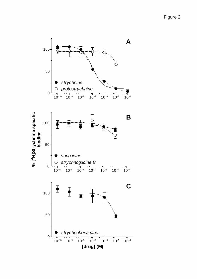
Figure 2 displays the competition binding curves of the four tested alkaloids and of
unlabelled strychnine used as a reference. Unlabelled strychnine potently displaced
[3H]strychnine binding to adult rat spinal cord membranes with an IC50 of 84.6 ± 17.7 nM
(mean ± standard deviation; n = 4 experiments; figure 2A). The experimental data could
slightly better be fitted with a two-site competition curve (P = 0.0378, least sum-of-squares F-
test) yielding an IC50 of 63.3 ± 23.3 nM for 79.6 ± 8.1% of the binding (site 1) and of 5.5 ±
2.2 µM for the remaining (site 2). Interestingly, the fraction of site 1 nicely correlated with the
inhibition of [3H]strychnine binding by 10 mM glycine (74.2 ± 7.2%; n = 4 experiments).
As shown in figures 2A to 2C, the four tested alkaloids were very weak at displacing
[3H]strychnine: none of them showed a significant effect up to 1 µM and their inhibition at
10 µM was rather modest: 33.5 ± 14.0% for protostrychnine, 13.4 ± 8.11% for sungucine,
34.7 ± 17.4 % for strychnogucine B and 52.0 ± 7.4% for strychnohexamine.

12
3.2 Patch-clamp recordings
Our binding experiments suggest that the four tested strychnine-related alkaloids very
weakly interact, if at all, with the strychnine binding to glycine receptors. This does not rule
out an interaction of those compounds with glycine-triggered functional responses. Therefore,
we compared the effects of the four alklaloids with the one of strychnine on glycine-induced
currents evoked in cultured spinal cord neurones cultured for 7-14 days in vitro and recorded
using the patch-clamp technique in the whole-cell configuration and in the voltage-clamp
mode. At this stage of in vitro maturation, glycine receptors are mainly constituted by α1/β
heteromers (Tapia and Aguayo, 1998; Withers and St John, 1997; Mangin, Nguyen et al.,
2005) and, thus, correspond to the mature form of the receptor that was studied in our
reported binding experiments.
In our recording conditions, the spinal cord neurones cultured for 7-14 days in vitro
responded to bath application of glycine by inward currents (see figure 3A) which reversed
around 0 mV, a value close to the Nernst equilibrium potential for chloride (0.6 mV; data not
shown). Those glycine currents were unaffected by 10 µM gabazine, showing that there was
no cross-activation of type A GABA receptors, and poorly inhibited by picrotoxin, suggesting
that they were mainly carried by heteromeric glycine receptors, as already described for
mature spinal cord neurone glycine receptors (heteromeres of 2 α1 subunits for 3 β subunits)
(Grudzinska et al., 2005).showing that they were mainly carried by heteromeric glycine
receptors (Mangin, Nguyen et al., 2005; Tapia and Aguayo, 1998).
Figures 3 to 5 show the inhibition profiles of the four tested alkaloids compared to
strychnine on currents induced by 100 µM glycine (a concentration close to its EC50
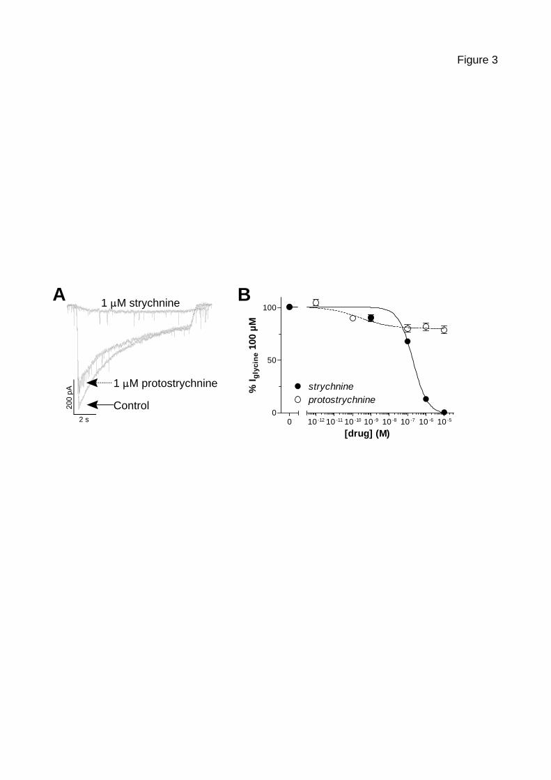
(Rajendra, Lynch et al., 1997)) in cultured spinal cord neurones. Strychnine (figure 3)
inhibited glycine-evoked currents with an IC50 of 196.9 ± 18.1 nM (n = 5-62) and a Hill

13
coefficient of 1.1 ± 0.1. As in the binding experiments, the strychnine-related alkaloids only
showed weak interactions with GlyR-mediated responses. The maximal inhibition of glycine
currents was 19.9 ±1.8% (n = 7-14) for protostrychnine (figure 3), 13.6 ± 0.9% (n = 7-27) for
sungucine (figures 4A and 4B), 32.9 ± 12.0% (n = 11-19) for strychnogucine B (figures 4C
and 4D) and 10.0 ± 8.4 (n = 9) for strychnohexamine (figure 5). Due to its low availability,
strychnohexamine could only be tested at 1 µM.

14
4 Discussion
The present study reports that strychnogucine B and strychnohexamine, new alkaloids
isolated from Strychnos icaja which are able to inhibit the growth of malaria parasites,
interact, as sungucine, with a very poor efficacy and only at concentrations > 1 µM with both
[3H]strychnine binding on rat spinal cord membranes and glycine responses in whole-cell
patch- and voltage-clamped cultured mouse spinal neurones. Although we cannot completely
rule out a pro-convulsant effect, which would be associated to the use of these drugs close to
strychnine as antimalarials, the data reported here are, to our knowledge, the first attempts to
evaluate their acute toxicity.
Two types of assays were used in the present study to address the putative analogy
between bisindolic and trisindolic alkaloids and strychnine, both at the biochemical or
structural level (binding) and at the functional level (patch-clamp). For sungucine and
strychnogucine B, both types of experiments allowed us to draw similar conclusions, i.e. a
poor efficacy of these compounds in antagonizing strychnine binding or glycine-gated
currents and the high concentration (> 1 µM) needed to achieve this effect. Nevertheless, at
first sight, strychnogucine B, which is the most promising antimalarial derived from
Strychnos icaja, seems to interact with the glycine receptor with a greater efficacy than
sungucine. However, this effect could likely be attributed to contaminating strychnine in our
preparation. Indeed, a small peak corresponding to strychnine could be observed by HPLC in
the strychnogucine B sample (data not shown). The amount of strychnine impurities was
estimated as not exceeding 1% (w/w), as appreciated from binding competition curves as well
as from glycine current inhibition curves.If the effects of strychnogucine B on [3H]strychnine
binding and on glycine-induced currents were te be attributed to contaminating strychnine,
this would imply a 0.5 % strychnine contamination, as inferred from binding curves, and 1 %
15
from glycine current inhibition curves. This does not exceed the maximal contamination
estimated for HPLC measures and, hence, could suggest that our observed effects of
strychnogucine B could be attributed to contaminating strychnine. Anyhow, this would at
least imply still less that the strychnine-like effects of strychnogucine B itself are far over-
estimated.
As opposed to the dimeric compounds, no clear correlation was found between binding
and electrophysiology results in the case of strychnohexamine. Two hypotheses could help
explaining this discrepancy. First, the different experimental conditions of binding and
electrophysiology could account for the differences. FirstSecond, since we have observed that
~20% of the strychnine binding was insensitive to glycine (Cimino, Marini et al., 1996),
strychnohexamine could have a higher affinity for these glycine-insensitive sites, hence
explaining its absence of effect on responses mediated by the glycine receptor. One could also
speculate A second possibility is that strychnohexamine well interacts with the strychnine site
of the glycine receptor, but does not hinder glycine binding and the subsequent
conformational change leading to channel opening, thus acting as a ‘null’ antagonist. Such
‘null’ antagonists are known at other ligand-gated channels related to glycine receptors, e.g.
flumazenil at the benzodiazepine site of type A GABA receptors (Millan, 2003). Further
experiments (strychnine binding competition by strychnohexamine in the presence of glycine
and strychnine antagonism of glycine-gated currents in the presence of strychnohexamine) are
needed to fully addres these issuesthis issue.
Measuring the interaction between alkaloids derived from Strychnos icaja and the glycine
receptor has also allowed us to study the influence of some chemical modifications in the
structure of strychnine on the interaction with the glycine receptor. First, ‘dimerization’ seems
to strongly reduce the intensity of the interaction. Indeed, strychnogucine B, the lower part of

16
which being a moeity strychnine bound to a moiety isotrychnine II, was at least a thousand
times less potent in interacting with the glycine receptor, what could be the consequence of a
too large sterical hindrance.
On the other hand, protostrychnine, which has been only recently found in this plant, but
was already also previously described in Strychnos nux vomica (Baser, Bisset et al., 1979),
possesses, in comparison with strychnine, an opening of the ether bond belonging to the G
cycle (see figure 1). No data about the strychnine-like properties of protostrychnine were
available, but the results obtained in this work had been previously somewhat hypothesized.
Indeed, isostrychnine I, an alkaloid structurally close to protostrychnine (the hydroxyle in 17
is replaced in isostrychnine I by an insaturation in 16-17), had been assayed in binding
experiments on membrane preparations obtained from the central nervous system of the rat
(Mackerer, Kochman et al., 1977) and of the pigeon (LeFort, Henke et al., 1978) and was
quite ineffective in both studies. Moreover, in the seventies, Swedish researchers had already
hypothesized that ether oxygen between the carbons 17 and 18 may be involved in the fit to
the receptor (Sandberg and Kristianson, 1970). Our in vitro assays thus confirm the
importance of the ether bound of the G cycle in strychnine derivatives structures for their
binding to the glycine receptor and for their antagonistic effect on glycine responses.
In conclusion, our data provide the first evidence that promising antimalarial alkaloid
agents derived from Strychnos icaja are devoid of strychnine-related properties, at least in
vitro. Further studies will imply in vivo assays aimed at confirming the absence of pro-
convulsant or toxic effects of these compounds together with examining their in vivo
antimalarial potency.

17
Acknowledgements
The authors wish to thank Mrs. Patricia Ernst, Mrs. Arlette Brose, and Ms Patricia
Piscicelli for skilful technical assistance and also Professor P. Hubert (Laboratory of
Analytical Chemistry, University of Liège) and Mr J.-N. Wauters (Laboratory of
Pharmacognosy) for HPLC kind advice. M.F. is a research associate from the Belgian
National Fund for Scientific Research (FNRS). Financial support from the Fondation Léon
Fredericq is gratefully acknowledged.

18
References
1. Baser,K.H.C., Bisset,N.G., Hylands,P.J., 1979. Protostrychnine, a new alkaloid from
Strychnos nux vomica. Phytochemistry 18, 512-514.
2. Bottenstein,J.E., Sato,G., 1979. Growth of rat neuroblastoma cell line in serum-free
supplemented medium. Proc.Natl.Acad.Sci.USA 76, 514-517.
3. Bradford,M.M., 1976. A rapid and sensitive method for the quantification of microgram
quantities of protein utilizing the principal of protein-dye binding. Anal.Biochem. 72,
248-255.
4. Breitinger,H.G., Becker,C.M., 2002. The inhibitory glycine receptor-simple views of a
complicated channel. Chembiochem. 3, 1042-1052.
5. Cimino,M., Marini,P., Cattabeni,F., 1996. Interaction of thiocolchicoside with
[3H]strychnine binding sites in rat spinal cord and brainstem. Eur.J.Pharmacol. 318, 201-
204.
6. Frederich,M., De Pauw,M.C., Llabres,G., Tits,M., Hayette,M.P., Brandt,V., Penelle,J.,
De,M.P., Angenot,L., 2000. New antimalarial and cytotoxic sungucine derivatives from
Strychnos icaja roots. Planta Med. 66, 262-269.
7. Frederich,M., De Pauw,M.C., Prosperi,C., Tits,M., Brandt,V., Penelle,J., Hayette,M.,
DeMol,P., Angenot,L., 2001. Strychnogucines A and B, two new antiplasmodial
bisindole alkaloids from Strychnos icaja. J.Nat.Prod. 64, 12-16.

19
8. Gadi Biala,R., Tits,M., Wauters,J.N., Angenot,L., 1996. A new HPLC method for the
assay of alklaloids in Strychnos nux-vomica and Strychnos igantii. Fitoterapia 67, 163-
165.
9. Greenwood,B., 2004. Between hope and a hard place. Nature 430, 926-927.
10. Grudzinska,J., Schemm,R., Haeger,S., Nicke,A., Schmalzing,G., Betz,H., Laube,B.,
2005. The beta subunit determines the ligand binding properties of synaptic glycine
receptors. Neuron 45, 727-739.
11. Kambu,K., Coune,C., Angenot,L., 1979. Nouveaux alcaloïdes des racines du Strychnos
icaja. Planta Med. 37, 161-164.
12. Laube,B., Maksay,G., Schemm,R., Betz,H., 2002. Modulation of glycine receptor
function: a novel approach for therapeutic intervention at inhibitory synapses? Trends
Pharmacol.Sci. 23, 519-527.
13. LeFort,D., Henke,H., Cuenod,M., 1978. Glycine specific [3H]strychnine binding in the
pigeon CNS. J.Neurochem. 30, 1287-1291.
14. Legendre,P., 2001. The glycinergic inhibitory synapse. Cell Mol.Life Sci. 58, 760-793.
15. Mackerer,C.R., Kochman,R.L., Shen,T.F., Hershenson,F.M., 1977. The binding of
strychnine and strychnine analogs to synaptic membranes of rat brainstem and spinal
cord. J.Pharmacol.Exp.Ther. 201, 326-331.
16. Mangin,J.M., Nguyen,L., Gougnard,C., Hans,G., Rogister,B., Belachew,S., Moonen,G.,
Legendre,P., Rigo,J.M., 2005. Developmental Regulation of {beta}-Carboline-Induced
Inhibition of Glycine-Evoked Responses Depends on Glycine Receptor {beta} Subunit
Expression. Mol.Pharmacol. 67, 1783-1796.
20
17. Neuwinger,H.D., 1996. Loganiaceae: Strychnos icaja. In: Neuwinger,H.D. (Ed.),
African Ethnobotany: Poisons and Drugs, Chemistry, Pharmacology, Toxicology.
Chapman & Hall, London, pp. 569-578.
18. O'Connor,V., Phelan,P.P., Fry,J.P., 1996. Interactions of glycine and strychnine with
their receptor recognition sites in mouse spinal cord. Neurochem.Int. 29, 423-434.
19. Philippe,G., De Mol P., Zeches-Hanrot,M., Nuzillard,J.M., Tits,M.H., Angenot,L.,
Frederich,M., 2003. Indolomonoterpenic alkaloids from Strychnos icaja roots.
Phytochemistry 62, 623-629.
20. Philippe,G., Prost,E., Nuzillard,J.M., Zeches-Hanrot,M., Tits,M., Angenot,L.,
Frederich,M., 2002. Strychnohexamine from Strychnos icaja, a naturally occuring
trimeric indolomonoterpenic alkaloid. Tetrahedron Lett. 43, 3387-3390.
21. Rajendra,S., Lynch,J.W., Schofield,P.R., 1997. The glycine receptor. Pharmacol.Ther.
73, 121-146.
22. Sandberg,F., Kristianson,K., 1970. A comparative study of the convulsant effects of
strychnos alkaloids. Acta Pharm.Suec. 7, 329-336.
23. Singer,J.H., Berger,A.J., 2000. Development of inhibitory synaptic transmission to
motoneurons. Brain Res.Bull. 53, 553-560.
24. Tapia,J.C., Aguayo,L.G., 1998. Changes in the properties of developing glycine
receptors in cultured mouse spinal neurons. Synapse 28, 185-194.
25. Withers,M.D., St John,P.A., 1997. Embryonic rat spinal cord neurons change expression
of glycine receptor subtypes during development in vitro. J.Neurobiol. 32, 579-592.

21
Figure legends
Figure 1 – Structures of tested strychnine derivatives
Chemical structures of strychnine (with classical numerotation) and tested alkaloids
Figure 2 – Interaction of strychnine derivatives with strychnine specific
binding
Effect of increasing concentrations of strychnine (A;●), protostrychnine (A;○), sungucine
(B;●), strychnogucine B (B;○) and strychnohexamine (C;●) on [3H]strychnine specific
binding to spinal cord membranes from adult rats. Results (expressed in percent of
[3H]strychnine specific binding in control buffer) are mean and SEM (n = 4) of four
independent bindings for each condition (triplicates for each condition in a single binding).
For strychnine, data were fitted both with one site (solid line) and with two sites (dotted line)
competition curve (see Materials and Methods).
Figure 3 – Effect of protostrychnine on glycine-induced currents in cultured
spinal cord neurons
A The same cultured spinal cord neuron was voltage-clamped at a holding potential of
-70 mV using the whole-cell patch-clamp technique (see Materials and Methods) and
currents induced by a 10 s perfusion of 100 µM glycine alone (Control) or in
combination with 1 µM strychnine or protostrychnine were recorded. Strychnine and
protostrychnine were perfused for 15 s before being co-applied with glycine. A 60 s
period was allowed for the washout of drugs.
B Currents evoked by 100 µM glycine in cultured spinal cord neurons were recorded in
the presence of increasing strychnine (black filled circles) and protostrychnine (empty

22
circles) concentrations. Results are expressed as percentage of glycine-induced current
peak amplitudes in the absence of drugs (mean ± SEM, n = 5-62 for strychnine and 7-
14 for protostrychnine).
Figure 4 – Effect of sungucine and strychnogucine B on glycine-induced
currents in cultured spinal cord neurons
A,C Cultured spinal cord neurons were voltage-clamped at a holding potential of -70 mV
using the whole-cell patch-clamp technique (see Materials and Methods) and currents
induced by a 10 s perfusion of 100 µM glycine alone (Control) or in combination with
0.1 µM or 10 µM sungucine (A) or strychnogucine B (C) were recorded. Sungucine
and strychnogucine B were perfused for 15 s before being co-applied with glycine. A
60 s period was allowed for the washout of drugs.
B,D Currents evoked by 100 µM glycine in cultured spinal cord neurons were recorded in
the presence of increasing sungucine (B) and strychnogucine B (D) concentrations.
Results are expressed as percentage of glycine-induced current peak amplitudes in the
absence of drugs (mean ± SEM, n = 7-27 for sungucine and 11-19 for
strychnogucine B).
Figure 5 – Effect of strychnohexamine on glycine-induced currents in
cultured spinal cord neurons
A The same cultured spinal cord neuron was voltage-clamped at a holding potential of
-70 mV using the whole-cell patch-clamp technique (see Materials and Methods) and
currents induced by a 10 s perfusion of 100 µM glycine alone (Control) or in
combination with 1 µM strychnine or strychnohexamine were recorded. Strychnine

23
and strychnohexamine were perfused for 15 s before being co-applied with glycine. A
60 s period was allowed for the washout of drugs.
B Currents evoked by 100 µM glycine in cultured spinal cord neurons were recorded in
the presence of 1 µM strychnohexamine. Results are expressed as percentage of
glycine-induced current peak amplitudes in the absence of strychnohexamine (mean ±
SEM, n = 9-18).

Figure 1
NH
H1
2
3
45
6
78
9
10
11
1213
14
15
16
17
18
19
20
21
2223
H
N
O
HN
H
H
OH
N
H
HHOO O
NH
H
N
H
O
N H
N
H
O
H
H
NH
H
N
H
O
N H
N
H
O
H
H
HO
OH
NH
H
N
H
O
N H
N
H
H
NH
N
H
strychnine protostrychnine
sungucine
strychnogucine B
strychnohexamine
10 -10 10 -9 10 -8 10 -7 10 -6 10 -5 10 -40
50
100
strychnineprotostrychnine
10 -10 10 -9 10 -8 10 -7 10 -6 10 -5 10 -40
50
100
sungucinestrychnogucine B%
[3 H]S
tryc
hnin
e sp
ecifi
cbi
ndin
g
10 -10 10 -9 10 -8 10 -7 10 -6 10 -5 10 -40
50
100
strychnohexamine
[drug] (M)
A
B
C
Figure 2
00
50
100
10 -12 10 -11 10 -10 10 -9 10 -8 10 -7 10 -6 10 -5
strychnineprotostrychnine
[drug] (M)
% I g
lyci
ne 1
00 µ
M
A B
Control2 s
1 :M protostrychnine
1 :M strychnine
Figure 3

00
50
100
10-12 10 -11 10 -10 10 -9 10 -8 10 -7 10 -6 10 -5
[sungucine] (M)
% I g
lyci
ne 1
00 µ
M
00
50
100
10 -12 10 -11 10 -10 10 -9 10 -8 10 -7 10 -6 10 -5
[strychnogucine B] (M)
% I g
lyci
ne 1
00 µ
M
C
A B
D
10 :M sungucine
Control3 s
0.1 :M sungucine
Figure 4
10 :M strychnogucine B
Control3 s
0.1 :M strychnogucine B

Contro
l
Strych
nohe
xamine
0
5 0
1 0 0
% I gl
ycin
e 100
µM
A B
Control2 s
1 :M strychnohexamine
1 :M strychnine
Figure 5