Study of Marginal Microleakage in Root Caries Restored ... NAZEM 4 17.pdf · Restoration of the...
8
http://www.revmaterialeplastice.ro MATERIALE PLASTICE ♦54♦No. 4 ♦2017 740 Study of Marginal Microleakage in Root Caries Restored with Resin Composites, Giomers and Glass Ionomer Cements DAWOD NAZEM 1 , CORNELIA FLORENTINA BICLESANU 1 *, DOINA PALUTA 1 , STEFAN MANEA 1 , MONICA BANITA 2 , VIOLETA HANCU 1 , ANAMARIA FLORESCU 1 1 Titu Maiorescu University of Bucharest, Faculty of Dentistry, Department of Dental Specialities, 67A Gheorghe Petrascu Str., 031593, Bucharest, Romania 2 University of Medicine and Pharmacy of Craiova, Faculty of Dental Medicine, 2 Petru Rares Str., 200349, Craiova, Romania Root decay is a complex and multifactorial disease. The aim of this study is to assess in vitro the sealing capacity of common aesthetic materials used to restore root caries as well as the size of the hybrid layer formed by SE-1 step vs 2-step ER adhesive techniques. For this study, 45 extracted teeth were used. After cleaning, near the enamel-cement junction, but at the level of the cementum, cavities were prepared and restored with resin composite, giomer and glass-ionomer cements. The teeth were covered with nail polish leaving uncovered the restoration area and 2mm around it; teeth were kept for 24 h , in 2% methylene blue. To assess the degree of marginal adaptation, SEM analysis was further performed. The association of 2-step ER adhesive system with giomers and resin composite shows the maximum adhesion efficiency by forming a uniform, thick layer and dentinal tubules obliteration compared to SE-1 step adhesive system associated with the same restoration materials. Key words root decay, adhesive systems, resin composite, SEM analysis Root decays are considered a major oral health problem in the elderly. This hypothesis is argued by the increase in the average life of the population and the persistence on the oral arch of the teeth for a longer time associated with the periodontal recession process which is considered one of the triggering causes of the root decays. Root decay is a complex and multifactorial disease. A more recent review of the literature has identified the following factors, organized in four areas, as responsible for root caries production: socio-demographic factor (age, gender, race/ethnicity), systemic health status (dementia), parameters in the intra oral area (number of teeth, bacterial plaque, bacterial species, dental caries index, loss of periodontal insertion, gingival recession, salivary flow) and behavioral ones (dental hygiene, brushing, smoking) [1]. The therapeutic management of root caries is based on the application of methods of prevention and remineralization of damaged dental structures to the detriment of the actual restoration techniques. The use of atraumatic restorative therapy (ART) is considered a viable solution due to its advantages of using hand tools without anesthesia and lesion restoration with a glass ionomer cement [2,3]. The restoration is done only when the cavity is formed and the age and the patient’s health status are taken into account. Due to local anatomical features, various restorative materials and preparing techniques can be imployed. Thus, when restoration is done with non- adhesive materials, the preparation requires retention both on the cervical and apical walls; if restoration is done with resin composite, it is only performed in the apical wall, and when restoring with the glass ionomer cement it does not require physical retention. In this way, the clinical success appreciated through a correct marginal adaptation is in correlation with several factors such as morphological particularities, the preparation pattern and the category of restorative material used [4]. The aim of this study is to assess in vitro the sealing capacity of common aesthetic materials used to restore * email: [email protected]; Phone: +40722636335 root caries as well as the size of the hybrid layer formed by SE-1 step vs ER 2-step adhesive techniques. Experimental part Material and method T ooth preparation For this study, 45 extracted teeth for orthodontic or periodontal reasons were used. After extraction, the teeth were cleaned of debris and then stored in physiological serum until the experiment was performed. Near the enamel-cement junction, but at the level of the cementum, cavities were prepared according to the minimally invasive principles, using a high-speed diamond bur, under water cooling and a low speed round bur. The approximate dimensions of the cavities were 3 mm mid- distal, 4 mm occluso-cervical and 1.5 mm in depth. Tooth restoration Depending on the restoration material and the type of adhesive system, the teeth were divided into 3 groups (A, B, C), with subdivision of the batches into 3 subgroups, in total resulted 9 groups with 5 teeth each and 10 restorations in each group (table 1). The materials used in the study and in their composition are presented in table 2. The protocol for applying restoration materials was as follows (table 3): (1) 2-step ER adhesive system protocol: Adper Single bond (3M Espe); After the preparation of the cavity, etching acid (Scotchbond) was applied, which was kept on the tooth for 15 s, then it was removed by washing for 10 s. Next, excess water was removed and two consecutive Adper Single bond (3M Espe) adhesive layers were applied, which were gently dried for five seconds to evaporate the solvents and lightcured for 10 s. (2) 1-step SE adhesive system protocol: G-Bond (GC); The adhesive was applied to the cavities, kept on the tooth for 10 s, then dried for 5 s and 10 s of lightcuring was achieved.
Transcript of Study of Marginal Microleakage in Root Caries Restored ... NAZEM 4 17.pdf · Restoration of the...

http://www.revmaterialeplastice.ro MATERIALE PLASTICE ♦ 54♦ No. 4 ♦ 2017740
Study of Marginal Microleakage in Root Caries Restored with ResinComposites, Giomers and Glass Ionomer Cements
DAWOD NAZEM1, CORNELIA FLORENTINA BICLESANU1*, DOINA PALUTA1, STEFAN MANEA1, MONICA BANITA2,VIOLETA HANCU1, ANAMARIA FLORESCU1
1 Titu Maiorescu University of Bucharest, Faculty of Dentistry, Department of Dental Specialities, 67A Gheorghe Petrascu Str.,031593, Bucharest, Romania2 University of Medicine and Pharmacy of Craiova, Faculty of Dental Medicine, 2 Petru Rares Str., 200349, Craiova, Romania
Root decay is a complex and multifactorial disease. The aim of this study is to assess in vitro the sealingcapacity of common aesthetic materials used to restore root caries as well as the size of the hybrid layerformed by SE-1 step vs 2-step ER adhesive techniques. For this study, 45 extracted teeth were used. Aftercleaning, near the enamel-cement junction, but at the level of the cementum, cavities were prepared andrestored with resin composite, giomer and glass-ionomer cements. The teeth were covered with nail polishleaving uncovered the restoration area and 2mm around it; teeth were kept for 24 h , in 2% methylene blue.To assess the degree of marginal adaptation, SEM analysis was further performed. The association of 2-stepER adhesive system with giomers and resin composite shows the maximum adhesion efficiency by forminga uniform, thick layer and dentinal tubules obliteration compared to SE-1 step adhesive system associatedwith the same restoration materials.
Key words root decay, adhesive systems, resin composite, SEM analysis
Root decays are considered a major oral health problemin the elderly. This hypothesis is argued by the increase inthe average life of the population and the persistence onthe oral arch of the teeth for a longer time associated withthe periodontal recession process which is considered oneof the triggering causes of the root decays.
Root decay is a complex and multifactorial disease. Amore recent review of the literature has identified thefollowing factors, organized in four areas, as responsiblefor root caries production: socio-demographic factor (age,gender, race/ethnicity), systemic health status (dementia),parameters in the intra oral area (number of teeth, bacterialplaque, bacterial species, dental caries index, loss ofperiodontal insertion, gingival recession, salivary flow) andbehavioral ones (dental hygiene, brushing, smoking) [1].
The therapeutic management of root caries is based onthe application of methods of prevention andremineralization of damaged dental structures to thedetriment of the actual restoration techniques. The use ofatraumatic restorative therapy (ART) is considered a viablesolution due to its advantages of using hand tools withoutanesthesia and lesion restoration with a glass ionomercement [2,3].
The restoration is done only when the cavity is formedand the age and the patient’s health status are taken intoaccount. Due to local anatomical features, variousrestorative materials and preparing techniques can beimployed. Thus, when restoration is done with non-adhesive materials, the preparation requires retention bothon the cervical and apical walls; if restoration is done withresin composite, it is only performed in the apical wall, andwhen restoring with the glass ionomer cement it does notrequire physical retention. In this way, the clinical successappreciated through a correct marginal adaptation is incorrelation with several factors such as morphologicalparticularities, the preparation pattern and the category ofrestorative material used [4].
The aim of this study is to assess in vitro the sealingcapacity of common aesthetic materials used to restore
* email: [email protected]; Phone: +40722636335
root caries as well as the size of the hybrid layer formed bySE-1 step vs ER 2-step adhesive techniques.
Experimental partMaterial and methodTooth preparation
For this study, 45 extracted teeth for orthodontic orperiodontal reasons were used. After extraction, the teethwere cleaned of debris and then stored in physiologicalserum until the experiment was performed.
Near the enamel-cement junction, but at the level of thecementum, cavities were prepared according to theminimally invasive principles, using a high-speed diamondbur, under water cooling and a low speed round bur. Theapproximate dimensions of the cavities were 3 mm mid-distal, 4 mm occluso-cervical and 1.5 mm in depth.
Tooth restorationDepending on the restoration material and the type of
adhesive system, the teeth were divided into 3 groups (A,B, C), with subdivision of the batches into 3 subgroups, intotal resulted 9 groups with 5 teeth each and 10 restorationsin each group (table 1).
The materials used in the study and in their compositionare presented in table 2.
The protocol for applying restoration materials was asfollows (table 3):
(1) 2-step ER adhesive system protocol: Adper Singlebond (3M Espe);
After the preparation of the cavity, etching acid(Scotchbond) was applied, which was kept on the toothfor 15 s, then it was removed by washing for 10 s. Next,excess water was removed and two consecutive AdperSingle bond (3M Espe) adhesive layers were applied, whichwere gently dried for five seconds to evaporate the solventsand lightcured for 10 s.
(2) 1-step SE adhesive system protocol: G-Bond (GC); The adhesive was applied to the cavities, kept on the
tooth for 10 s, then dried for 5 s and 10 s of lightcuring wasachieved.

http://www.revmaterialeplastice.roMATERIALE PLASTICE ♦ 54♦ No. 4 ♦ 2017 741
Table 1STUDY
BATCHES
Table 2 MATERIALS AND COMPOSITION
http://www.revmaterialeplastice.ro MATERIALE PLASTICE ♦ 54♦ No. 4 ♦ 2017742
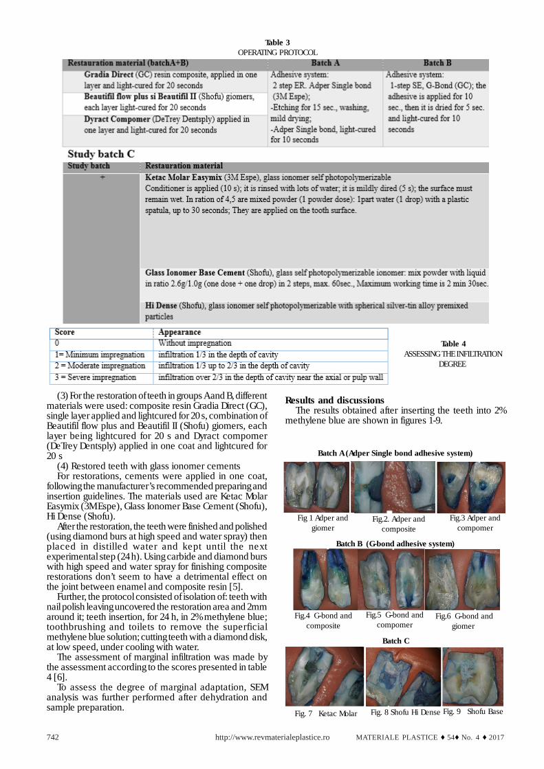
(3) For the restoration of teeth in groups A and B, differentmaterials were used: composite resin Gradia Direct (GC),single layer applied and lightcured for 20 s, combination ofBeautifil flow plus and Beautifil II (Shofu) giomers, eachlayer being lightcured for 20 s and Dyract compomer(DeTrey Dentsply) applied in one coat and lightcured for20 s
(4) Restored teeth with glass ionomer cementsFor restorations, cements were applied in one coat,
following the manufacturer’s recommended preparing andinsertion guidelines. The materials used are Ketac MolarEasymix (3M Espe), Glass Ionomer Base Cement (Shofu),Hi Dense (Shofu).
After the restoration, the teeth were finished and polished(using diamond burs at high speed and water spray) thenplaced in distilled water and kept until the nextexperimental step (24 h). Using carbide and diamond burswith high speed and water spray for finishing compositerestorations don’t seem to have a detrimental effect onthe joint between enamel and composite resin [5].
Further, the protocol consisted of isolation of: teeth withnail polish leaving uncovered the restoration area and 2mmaround it; teeth insertion, for 24 h, in 2% methylene blue;toothbrushing and toilets to remove the superficialmethylene blue solution; cutting teeth with a diamond disk,at low speed, under cooling with water.
The assessment of marginal infiltration was made bythe assessment according to the scores presented in table4 [6].
To assess the degree of marginal adaptation, SEManalysis was further performed after dehydration andsample preparation.
Results and discussionsThe results obtained after inserting the teeth into 2%
methylene blue are shown in figures 1-9.
Table 3 OPERATING PROTOCOL
Table 4 ASSESSING THE INFILTRATION
DEGREE
Batch A (Adper Single bond adhesive system)
Batch B (G-bond adhesive system)
Batch C
Fig 1 Adper andgiomer
Fig.2. Adper andcomposite
Fig. 7 Ketac Molar
Fig.3 Adper andcompomer
Fig.4 G-bond andcomposite
Fig.5 G-bond andcompomer
Fig.6 G-bond andgiomer
Fig. 8 Shofu Hi Dense Fig. 9 Shofu Base
http://www.revmaterialeplastice.roMATERIALE PLASTICE ♦ 54♦ No. 4 ♦ 2017 743
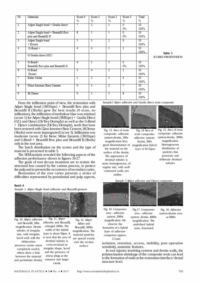
From the infiltration point of view, the restoration withAdper Single bond (3M Espe) + Beautifil flow plus andBeautifil II (Shofu) gave the best results (0 score, noinfiltration), the infiltration of methylene blue was minimal(score 1) for Adper Single bond (3M Espe) + Gradia Direct(GC) and Dyract (DeTrey Dentsply) as well as the G-Bond+ Dyract combination (DeTrey Dentsply), teeth that havebeen restored with Glass Ionomer Base Cement, Hi Dense(Shofu) were more impregnated (score 3). Infiltration wasmoderate (score 2) for Ketac Molar Easymix (3M Espe)and G-Bond + Beautifil flow plus and Beautifil II (Shofu)only in the root area.
The batch distribution on the scores and the type ofmaterial is presented in table 5.
The SEM analysis revealed the following aspects of theadhesion performance shown in figures 10-27.
The goals of root decays treatment are to restore thestructural loss caused by the carious process, to protectthe pulp and to prevent the occurrence of secondary caries.
Restoration of the root caries presents a series ofdifficulties represented by periodontal and pulp aspects,
isolation, retention, access, visibility, post operativesensitivity, anatomic features.
In root injuries involving cement and dentin walls, thepolymerization shrinkage of the composite resin can leadto the formation of voids at the restoration interface/ dentalstructure level.
Table 5SCORES PRESENTATION
Batch ASample 1. Adper Single bond adhesive and Beautifil giomers
Sample2 Adper adhesive and Gradia direct resin composite
Fig. 10. Adper adhesiveand Beautifil, 500x
magnification. Dentintubules of irregularsize, with irregular,laced wall, with the
obliterationpresence (some seem
completely sealed,others show a halo
between the materialand peritubular dentin)
Fig. 11. Adperadhesive and Beautifil,
enlarged 2000x. Thewidth of the hybrid
layer is about 50µm. Itis seen that the area of
dentinal tubules iscross-sectional in
irregular shape, lacedand the presence ofsmear plugs at the
entrance into largercanals
Fig. 12. AdperAdher and
Beautifil, 5000xmagnification. Thematerial particlesare spread evenlyover the section
surface
Fig. 13. Area of resincomposite- adhesivesystem-dentin, 200xmagnification.Very
good dissemination ofthe material on the
surface of the dentin.The appearance ofdentinal tubules is
more homogeneous, ofregular size, with well-
contoured walls, net outline.
Fig. 14 Area ofresin composite-adhesive system-
dentin, 500xmagnification Hybrid layer of 40-50µm.
Fig. 15. Area of resincomposite- adhesivesystem-dentin, 2000x
magnification.Homogeneousdistribution ofparticles thatpenetrate and
obliterate dentinaltubules
Sample 3 Adper adhesive and Dyract compomer
Fig. 16. Compomerarea - adhesivesystem, 2000x
magnification. Weobserve the
formation of a hybridlayer of adhesive-compomer approx.
2-3 µm
Fig.17. Compomerarea - adhesive
system- dentin, 4000xmagnification. Theundefined hybrid
zone, festooned.
Fig. 18. Adhesivesystem-dentin area
at 5000x

http://www.revmaterialeplastice.ro MATERIALE PLASTICE ♦ 54♦ No. 4 ♦ 2017744
Fig. 19. Giomer -G bond adhesivesystem- hybridized smear layer-
dentin area, magnified 500 x.
Fig. 20. G-bond adhesive and Beautifilgiomer, magnified 1000x. It can be noticedthe presence of the continuation solution
between the two interfaces
Fig. 21. Area of adhesive system- hybridized smear layer– dentin, magnified at 2000x. It can be noticed the
presence of pores in the adhesive system. It seems tobe a low adhesive area, with deep crevasses and
branched out of the adhesive system pores. It can alsobe a technique error with too strong dehydration
Fig. 23. Dentin- hybridized smearlayer - adhesive system - compositeresin area at 500x. On the left there
is the dentin with good particle covering
Fig. 24. G-bond adhesive andGradia direct composite, 1000x
magnification.
Fig. 22. Dentin area - bonding system Gbond - composite resin, 400x
magnification. At the adhesive layer level,pores of variable sizes are observed. The
presence of dentinal tubules and theirbranches with partial obliteration with
modest particles is evidenced
Batch BSample 4 G-bond adhesive and Beautifil giomers
Fig. 25. Dentin- hybridized smearlayer- adhesive system- compomer,1000x magnification. It seems to be a
well-organized hybrid layer,thickness~20 µm.
Fig. 26. Dentin- hybridized smearlayer - adhesive system - compomer at 1000x.
Fig. 27. Dentin- hybridized smear layer - adhesivesystem - compomer at 5000x. The material looks very
well and homogeneously structured in particles ofapproximately equal size, which seem to seal the
dentinal tubules in the lower part of the image.Demineralization of peritubular dentin.
Sample 6 G-bond adhesive and Dyract compomer
Sample 5 G-bond adhesive and Gradia direct composite
Batch C
Fig. 28. CIS HiDense Shofu. Themixed layer seems fractured onboth sides, modest size, under
10 µm.
Fig. 29. Non-homogeneousdentin may be an area of
Tomes granular layer
Fig. 30. CIS Base, 200 xsurface dentin magnification
with dentinal tubules andcollateral branches
Fig. 31. CIS Base, dentin 1000xmagnification with dentinal
tubules and collateralbranches, small particles of
material inside

http://www.revmaterialeplastice.roMATERIALE PLASTICE ♦ 54♦ No. 4 ♦ 2017 745
Fig. 32. CIS Base, magnification2000x.
Fig. 35. CIS Ketac Molar,magnification 800x. Largefragments of material thatappear to be superficially
located and small fragments inthe dentinal tubules in the
longitudinal section.
Fig. 33. CIS Ketac Molar, 200xmagnification. The preparation is
fractured, the mixed area 20-30µm
Fig. 34. CIS Ketac Molar,magnification 400x.
In this study, teeth restored with composite resins,giomers and compomers in combination with 2- step ERand 1- step SE adhesives as well as self polymerizableglass ionomer cement were used for the evaluation ofmarginal adhesion and microleakage.
The test by which the sealing capacity of a restorationmaterial is checked is maintaining marginal adaptation.To determine the degree of marginal microleakage in thisstudy we used 2% methylene blue. This colorant is highlywater-soluble and penetrates the dental structures, plus ithas a low molecular weight (about 0.52 nm2) comparedto the diameter of the dentinal canals (1-4µ) and thebacteria (2-4µ) and easily penetrates dentinal canalsimitating the passage of bacterial toxins into them [7].
In the present study, marginal microleakage was presentto some extent in almost all the restoration materials used,the glass ionomer exhibiting a maximum infiltrationfollowed by composites, giomers and compomers whichshowed better results with minimal infiltration. These dataare consistent with the study by Sayed HYEl et al. whichshowed that the marginal microleakage of glass ionomercement, composite and compomer restorations wasevident in all restoration materials [8].
Glass ionomer cement has a lower sealing capacity andamong the three cements used in this study, Ketac Molarhas a slight superiority compared to the other two cements(Glass Ionomer Base Cement, Hi Dense).
Glass ionomer cement has many qualities thatrecommend it as the ideal material for the restoration ofthe root caries because it performs a chemical retentionwith the dental structure, releases fluoride throughout theretention period [3].
However, glass ionomer cement has poor sealingcapacity when used to prevent marginal microleakage.The presence of smear layer as well as the polymerizationshrinkage may affect the sealing capacity of the glassionomer cement [9]. Higher infiltration capacity at the levelof the teeth restored with glass ionomer cement could beattributed to inappropriate condensation with theincorporation of air bubbles. The glass ionomer cementsused in the study are self-polymerizable, so preparation isdone extemporaneously by combining a powder with aliquid. This preparation method cannot accurately measurethe quantities of the mixture while the encapsulatedpreparations are not subject to such errors. This may bethe explanation for glass ionomer cements showing a lesstight marginal adaptation compared to Dyract [10].
In addition, water loss from the composition of glassionomer cement results in a porous aspect withmicroscopic cracks. To prevent this, it is important that thecement is protected with varnish or grease (petroleumjelly). In this context, studies show that the failure of aglass ionomer cement occurs inside it rather than at theglass ionomer cement /tooth interface. So, determiningthe adhesion force of glass ionomer cement is actually
measuring the resistance to cement stretching. This forceis relatively low in freshly prepared cement, but increasesin time [3].
Other authors reported frequent voids and fractures atthe interface with gingival wall as well as cohesivefractures, concluding that all types of failures are presentin glass ionomers cements, when using them in open-sandwich technique [11].
In this study, microleakage was minimal when AdperSingle bond (3M Espe) + Dyract (DeTrey Dentsply) as wellas the G-Bond + Dyract combination (DeTrey Dentsply)were used. But, in class V compomer-filled cavities, Rominushowed that microleakeage at the gingival interface couldbe significantly reduced by aditional silane treatmentofpreviously acid etched dentin [12].
Resin composites are the first restoration materials thatmaintain marginal integrity over a clinically acceptableperiod [13].
The advantage of using composites to restore root cariesis the ability to adhere without mechanical retention, andthe disadvantages are the need for good isolation, goodvisibility, furthermore the adhesion to dentin is inferior toadhesion to enamel.
Acid etching, the application of a low viscosity bondingagent and the provision of perfect insulation should beroutine procedures for all composite resin restorations [13].The composite material used in this study is Gradia Direct(GC). Gradia Direct is a lightcuring hybrid composite thatcontains pre-polymerized micro-particles, a coupling agentand dimethacrylate urethane as a matrix. This productoffers significant advantages in aesthetics, finishing, wearand tear resistance as well as a lower susceptibility topolymerization shrinkage. All of these factors cancontribute to the good performance of composite materialsin this study.
The giomers are new, effective restorative and adhesivedental materials with a high capacity of both fluoriderelease and recharge with it, biocompatible and aesthetic.For these reasons, they are the ideal material for restoringthe defects of the root area. The giomer concept is basedon pre-reacted glass ionomer (PRG) technology.
Adhesive agents in the study used are 2-step ER AdperSingle bond (3M Espe) and SE 1-step, G-Bond. Adper Singlebond (3M Espe) is a fifth generation adhesive system. Itremoves the smear layer, enlarges the dentinal tubules anddemineralizes the intratubular dentin. Primer moistens thecollagen fibers, which leads to the formation of the hybridlayer.
G-Bond is a 7th generation (single component) adhesivesystem that combines acid and adhesive technology withsuperior chemical and mechanical adhesion. Self-etching
http://www.revmaterialeplastice.ro MATERIALE PLASTICE ♦ 54♦ No. 4 ♦ 2017746
adhesives do not require a separate etching phase becausethey contain an acid monomer in a solution of water orHEMA-based adhesive. The separate tooth rinse phase iseliminated because these systems contain water that isnever completely removed from the tooth.
For these reasons, the marginal adaptation of arestoration depends, in addition to the type of material ortechniques of polymerization, on the use of differentadhesive systems. The composite material is associatedwith a different microleakage depending on the adhesivetechnique.
In this study, SEM analysis shows that ER Adper Singlebond (3M Espe) and Gradia Direct (GC) resin compositeexhibits maximum adhesion efficiency.
SEM analysis conducted in their study by Qi CZ and et al.shows that ER adhesive achieves a better bond to enamelcompared to SE adhesives, but the bond to dentin andcement did not show any significant difference [14].
In this context, in another study, the authors concludedthat marginal integrity of enamel increased significantlywhen ER adhesive systems and composite resin were used;the marginal integrity of dentin increased significantly incomposite restorations using SE. Both the marginal seal ofenamel and dentin increased significantly by using an SEadhesive system instead of the glass ionomer cementconditioner [6].
In the present study, the association of Adper Single bond(3M Espe) with the Beautifil flow plus and Beautifil II(Shofu) is the most beneficial, the study shows that thereis no marginal microleakage. The result is consistent withSantos et al. which in their study showed that the ERadhesive system presented the best resistance to dentin,concluding that these adhesive systems are superior to SEadhesives [15].
This result is, however, inconsistent with some literaturestudies that show that AdperTM Single Bond adhesivesystem provides a lesser marginal adaptation to the greaterpresence of marginal microleakage compared to SE 1 stepGBond. In this context it is supported the idea that there isa higher degree of microleakage at the gingival margincompared to occlusal one [16, 17].
In this study, the association of G-Bond (GC) withcomposites or compomers has been shown to have inferiorresults compared to the results obtained by associatingthe same materials with the ER 2 step technique. However,in this association the study showed that infiltration waspresent only at enamel-cementum jonction.
The results are in agreement with other studies thatsuggest that the SE 1-step technique (G-Bond (GC)) hasless marginal sealing capacity compared to the ER 2 stepAdper Single bond (3M Espe) technique [18, 19] .
Although the ER adhesive system has shown bettervalues in a meta-analysis by Masarwa N et al., comparedto the SE system, the authors did not find any significantdifference in the longevity of the restorations made withthe two types of adhesive techniques [20].
The adhesive layer should act as an elastic pad againstvarious stresses, with many factors affecting its stiffness/elasticity. These include the modulus of elasticity of itscomponents, the thickness of the hybrid layer and thedegree of interaction between its components [21] andthe type of monomer it makes up. The presence ofbisphenol glycidyl methacrylate (BIS-GMA) makes theadhesive more rigid due to its high molecular weightcompared to other monomers, such as urethanedimethacrylate (UDMA) and triethylene glycoldimethacrylate (TEGDMA).
Thus, the absence of BIS-GMA from GC Bond makesthe adhesive layer more elastic and more tolerant of stresscompared to Adper adhesive system.
The results of Carrera’s recent research have shown thatthe mean adhesion strength with SE (14.9 ± 1.9 MPa) wassignificantly higher than ER Adper Single Bond (12.9 ± 3.0MPa) (p <0.05); Most specimens (91.3%) presented apattern of adhesion failure and fractures along the resincomposite-dentine interface. SE failed mainly at thedentine-adhesive interface, while ER Adper Single Bond atthe composite-adhesive interface [22].
Due to the high degree of hydrophilicity, SE 1-step light-curing adhesives have been shown to act as permeablemembranes, allowing water to pass through the adhesivelayer.
For SE 1-step adhesives without HEMA (G bond) acomplex phase separation process was reported in whichwater is separated from other adhesive components. Thisprocess is determined by gradual evaporation of the organicsolvent (usually ethanol and acetone) with bubbleformation in the adhesive layer. It is obvious that theincorporation of the bubbles may contribute to thedegradation of adhesion, but persistence of the water inthe adhesive layer can also negatively affect the adhesion[23, 24].
In the present study, the association of Adper Single bond(3M Espe) with Beautifil flow plus and Beautifil II (Shofu),but also with Gradia direct composite, resulted in theformation of a strong hybrid layer of approx. 50 µm, withthe uniform dissemination of the particulate material onthe dentin surface, as well as the obliteration of the dentinaltubules.
Instead, associating G-bond with the same restorationmaterials formed a much smaller hybrid layer of about 20µm with the presence of pores and partial obliteration ofdentinal tubules.
In agreement with this, Albaladejo concludes that ERadhesive systems have thicker layers than those formedin self-etched adhesive systems but both were continuousand uniform in thickness, and the all-in-one adhesive usedin his study showed the formation of droplets between theadhesive and composite resin; they may occur as a resultof the absorption of dentine water through osmosis andmay interfere with the polymerization process of the resin[23].
Other studies, that used Adper Single Bond as an adhesivein combination with the Filtek Z250 composite resin,revealed an average thickness of the 8-12 µm in the hybridlayer [25].
Studies show that the hybrid layer formed by SE 1 stepmeasures 3 - 5µm and resin tags of approximately 2 - 3µm. Both the length of the resin tags in the dentine as wellas the thickness of the hybrid layer produced by the 7thgeneration adhesives are lower compared to the fifth-generation adhesives. This can be determined by the weaketching used in SE techniques (GC Bond) [21].
The adhesion after use of the ER system is much deepercompared to the effect produced by the 7th generation ofSE adhesives and the adhesive interface is more uniformin SE containing the acidic monomer which dissolves thehybrid layer [26,27].
As far as the compomers are concerned, in this studyAdper adhesive and Dyract compomer formed an adhesive-compomer hybrid layer of approx. 2-3 µm indefinitelydelimited, festooned. Instead, the G-bond adhesiveassociated with the same compomer formed a well-organized, homogeneous hybrid layer, ~ 20 µm thick, withapproximately homogeneous particles that seem to sealthe dentinal tubules well.

http://www.revmaterialeplastice.roMATERIALE PLASTICE ♦ 54♦ No. 4 ♦ 2017 747
It is true that self-etching adhesives exhibit loweradhesion resistance values, but the advantages ofsimplicity of the technique and elimination of rinsing anddrying steps cannot be ignored. In this way, the possibilityof over-wetting or over-drying, which are detrimental tothe integrity of the interface, is reduced. Future clinicalstudies are needed to confirm that these characteristicsare important to the longevity of restoration [28].
ConclusionsThe present study was conducted to assess the
performance of root restorations in vitro by analyzing theadhesion resistance achieved with aesthetic restorativematerials.
The association of Adper Single bond (3M Espe) withBeautifil flow plus and Beautifil II (Shofu) and Gradia Direct(GC) composite shows the maximum adhesion efficiencyby forming a uniform, thick layer and dentinal tubulesobliteration compared to G-Bond adhesive (GC) associatedwith the same restoration materials.
Simple techniques, moderate etching and lowsensitivity, in addition to its performance, make the clinicalchoice of 7th generation adhesives an advantage.
References1. RITTER, AV., SHUGARS, DA., BADER, JD., Community Dent OralEpidemiol.,38, nr. 5, 2010, p.383–972. EC, L., LUO, Y., TAN, HP., DYSON, JE., CORBET, EF., J Dent Res., 85,2006, p. 929–932.3. SIDHU, SK., NICHOLSON, JW., J Funct Biomater.,7, nr.3, 2016, p. 2-15.4. DONALD, LC., SHYUE, C., Decisions in dentistry, 03, 2016, p. 24-75. IOVAN, G., STOLERIU, S., PANCU, G., NICA, I., SANDU, AV., ANDRIAN,S., TANCULESCU, O., Mat. Plast., 54, no.2, 2017, p.3756. KHOROUSHI, M., KARVANDI, TM., KAMALI, B., MAZAHERI, H., IndianJ Dent Res.,23, 2012, p.378-837. HELENO, JFG. et all., Dental Press Endod., 2, nr.2, 2012, p.30-6.8. SAYED, H., ABDALLA, AI., SHALBY, ME., Tanta Dental Journal, 11,nr.3, 2014, p.180-1889. WATSON, TF. et all., Dental materials, 30, 2014, p.50-61.
10. PRABHAKAR, AR., MADAN, M., RAJU, OS., J. Indian Soc. Pedo.Prev. Dent.,21, nr.2, 2003, p.45–48.11. GHIORGHE, CA., IOVAN, G., ANDRIAN, S., NICA, I, TOPOLICEANU,C., PANCU, G., Rev. Chim. (Bucharest), 68, no.8, 2017, p.189012. ROMINU, M., FLORITA, Z., ROMINU RO., SINESCU, C., NEGRUTIU,ML., PETRESCU, EL., POP, DM., ENESCU, M., TUDOR, A., Rev. Chim.(Bucharest), 62, no.8, 2011, p. 84913. SHETTY, K., et all. Dental Science, 7, nr. 6, 2015, p. 607-611.14. QI, CZ., JIANG, Y., LI, SY., LIN, Y., FAN, XM., YU, Q., Shanghai KouQiang Yi Xue, 20, nr.3, 2011, p. 260-4.15. SANTOS, RA., et all., RGO, Rev Gauch Odontol, Porto Alegre.,62,nr.4, 2014, p.365-370.16. GUPTA, A., et all. J Clin Diagn Res.,11, nr.4, 2017, p. ZC53–ZC56.17. TABARI, M., ESMAEILI, B., ALIMOHAMMADI, M., BEJER MIR, AP.,GHAREKHANI, S., HAJIAHMADI, M., et all., Caspian J Dent Res., 3,2014, p.14–19.18. VAN LANDUYT, KL., DE MUNCK, J., MINE, A., CARDOSO, MV.,PEUMANS, M., VAN MEERBEEK, B.,J Dent Res., 89, 2010, p.1045-50.19. SARR, M., KANE, AW., VREVEN, J., MINE, A., VAN LANDUYT, KL.,PEUMANS, M., et all., Oper Dent, 35, 2010, p.94-104. 20. MASARWA, N., et all., Journal of Evidence Based Dental Practice,16, nr.2, 2016, p.96-10621. ELIGUZELOGLU, E., ERASLAN, O., OMURLU, H., ESKITASCIOGLU,G., BELLI, S., Eur J Dent., 4, nr.2, 2010, p.160-5.22. CARRERA, CA., CHEN, YC., LI, Y., RUDNEY, J., APARICIO, C., FOK,A., Journal of Dentistry, 52, 2016, p.37-44.23. ALBALADEJO, A., OSORIO, R.,TOLEDANO, M., FERRARI, M., MedOral Patol Oral Cir Bucal, 15, nr.1, 2010, p. e112-8.24. SAURO, S., PASHLEY, DH., MANNOCCI, F., TAY, FR., PILECKI, P.,SHERRIFF, M., et all., Eur J Oral Sci., 116, 2008, p.184-93.25. SAVEANU, CI., DANILA, I., Journal of Romanian Medical Dentistry,14,nr.2, 2010, p.148-51.26. GANGADARAN, V., PALANISWAMY, M., BALASUBRAMANIAN, M., JIndian Acad Dent Spec Res., 2, nr. 1, 2015, p.5-727. TOUMA, L., YASSIN, O., Iran J Public Health, 44, nr.6, 2015, p.885-88628. MARTINS, GC., et all. Eur J Dent., 6, nr.2, 2012, p.169-177
Manuscript received: 26.10.2017