Studies Toward the Biosynthesis of Chimonanthine...
97
Studies Toward the Biosynthesis of Chimonanthine in Chimonanthus praecox by Soo Hur B.Sc. (Spec.), University of Alberta, 2012 Thesis Submitted in Partial Fulfillment of the Requirements for the Degree of Master of Science in the Department of Chemistry Faculty of Science Soo Hur SIMON FRASER UNIVERSITY Spring 2016
Transcript of Studies Toward the Biosynthesis of Chimonanthine...

Studies Toward the Biosynthesis of
Chimonanthine in Chimonanthus praecox
by
Soo Hur
B.Sc. (Spec.), University of Alberta, 2012
Thesis Submitted in Partial Fulfillment of the
Requirements for the Degree of
Master of Science
in the
Department of Chemistry
Faculty of Science
Soo Hur
SIMON FRASER UNIVERSITY
Spring 2016

ii
Approval
Name: Soo Hur
Degree: Master of Science (Chemistry)
Title: Studies Toward the Biosynthesis of Chimonanthine in Chimonanthus praecox
Examining Committee: Chair: Dr. Michael H. Eikerling Professor
Dr. Robert A. Britton Senior Supervisor Professor
Dr. David Vocadlo Co-Supervisor Professor
Dr. Erika Plettner Supervisor Professor
Dr. Roger Linington Internal Examiner Associate Professor
Date Defended/Approved: January 18, 2016

iii
Abstract
Chimonanthine is the building block of a series of natural products found in terrestrial
plants including members of Psychotria in the family of Rubiaceae. Studies have shown
that alkaloids containing the chimonanthine core display interesting analgesic, inhibition
of melanogenesis, and anti-cancer activities. The goal of this study is to explore the
precursor directed biosynthesis of chimonanthine and the enzymes involved in the
biosynthesis of chimoanthine as well as to identify these enzymes for potential use as
biocatalysts that can generate libraries of modified natural products. Herein we report
the identification of a suitable plant containing these enzymes and demonstrate the
feasibility of new assays by showing that feeding of plants with synthetic precursors
leads to the production of labelled chimonanthine.
Keywords: chimonanthine; precursor-directed biosynthesis; calycanthaeceous alkaloid, pyrrolidinoindoline alkaloids;

iv
Acknowledgements
First of all, I would like to thank Prof. Robert Britton, and Prof. David Vocadlo for allowing
me to work in their labs in the last two and half years. The project they gave me was
very interesting because I was able to explore various science including chemistry,
biochemistry, and some biology, all at the same time. Their thoughtful ideas, and
detailed instructions led me to develop initial steps to start research, and their patience
and attitude shown to me have made my time at Simon Fraser University enjoyable.
I am also thankful for having Prof. Erika Plettner as my supervisory committee member.
Her knowledge in biosynthesis of natural products gave me a lot of great idea to try new
experiments, which eventually led me to obtain the data I wanted. Thank you for giving
me time to discuss the research progress, and providing me various ideas to initiate new
experiments.
Also, the time I spent with all of the members of the Britton group, and the Vocadlo
group was enjoyable, and it was good time to discuss science with them. I would like to
say special thank you to Dr. Stanley Chang for giving me opportunities to work on
sysnthetic organic chemistry for total synthesis of ascospiroketal A. I learned a lot of
interesting chemistry under your supervision. Another special thanks to Dr. Milan
Bergeron-Brlek and Michael Holmes for giving me various ideas on my research, and
sacrificing their time to help with my scientific writing. Also, Dr. Milan Bergeron-Brlek
provided me with a neat, warm and nice bed, and lots of great food on one of the
weekends during Basic Military Officer Qualification training. The food you provided I will
never forget. I’d like to thank Dr. Robby Zhai, Dr. Ajay Naidu, Dr. Matt Nodwell, Dr. Jake
Goodwin-Tindall, Dr. Shira Halperin, Jason Draper, Hope Fan, Vijay Dhand, Matt Taron,
Abhi Bagai, Chris Adamson, Daniel Kwon, Micahel Meanwell and Venugopal Rao Challa
for making the working lab environment fun and productive.
Lastly, I appreciate the unlimited support and love from my family. They always
motivated me whenever I feel discouraged. I do not think that I would make it without
their support and love.

v
Table of Contents
Approval .......................................................................................................................... ii Abstract .......................................................................................................................... iii Acknowledgements ........................................................................................................ iv Table of Contents ............................................................................................................ v List of Tables ................................................................................................................. vii List of Figures................................................................................................................ viii List of Schemes ............................................................................................................... x List of Acronyms ............................................................................................................. xi
Chapter 1. Introduction ............................................................................................. 1 1.1. Enzymes in Nature and in Chemistry ...................................................................... 1 1.2. Calycanthaceous Alkaloids ..................................................................................... 6 1.3. Chimonanthine ....................................................................................................... 9
1.3.1. Background Information ............................................................................. 9 1.3.2. Early Syntheses of Chimonanthine .......................................................... 11 1.3.3. First Enantioselective Syntheses of Chimonanthine ................................ 13 1.3.4. Additional Total Syntheses of Chimonanthine. . ....................................... 16 1.3.5. Biosynthetic Studies of Chimonanthine .................................................... 17
1.4. Proposed Biosynthesis of Chimonanthine............................................................. 18 1.5. Main Goals of This Study ...................................................................................... 19
Chapter 2. Studies towards the Biosynthesis of (-)- and meso-Chimonanthine ...................................................................................... 21
2.1. Background Information ........................................................................................ 21 2.2. Preparation of Precursor Analogues ..................................................................... 25 2.3. In Planta Experiment (Feeding Experiment) ......................................................... 26
2.3.1. Screening Precursor Analogues .............................................................. 26 2.3.2. Isolation of Unnatural Products, (-)-5,5’-Difluoro-chimonanthine and
meso-5,5’-Difluoro-chimonanthine ........................................................... 31 2.4. In Vitro Assay ....................................................................................................... 35
2.4.1. Background Information ........................................................................... 35 2.4.2. Cytoplasmic Protein Extraction from Leaf ................................................ 36 2.4.3. Cytoplasmic Protein Extraction from Root................................................ 39 2.4.4. In vitro Assay with Protoplast ................................................................... 42 2.4.5. Proteins from Cell Walls .......................................................................... 43
2.5. Summary .............................................................................................................. 49 2.6. Future Direction .................................................................................................... 50 2.7. Experimental ........................................................................................................ 52
2.7.1. General Considerations ........................................................................... 52 2.7.2. Experimental Information for Tryptamine Analogues ............................... 54 2.7.3. Experimental Information for Nb-methyltryptamine analogues .................. 59 2.7.4. Synthetic Standards of D6-chimonanthines .............................................. 63 2.7.5. Feeding Precursors in planta and Screening Precursors by LC-MS......... 65 2.7.6. Isolation of Fluorinated Unnatural Products ............................................. 65

vi
2.7.7. Plant Protein Extraction with P-PERTM Plant Protein Extraction Kit and in vitro Assay .................................................................................... 67
2.7.8. Plant Protein Extraction from Leaves Using Blender and in vitro Assay ...................................................................................................... 68
2.7.9. Plant Protein Extraction From Roots Using Blender and in vitro Assay [83] .................................................................................................. 69
2.7.10. Root protein separation by Ammonium Sulfate Precipitation.................... 69 2.7.11. Preparation of Protoplasts [84]................................................................... 70 2.7.12. In Vitro Assay with Protoplasts ................................................................ 70 2.7.13. Preparation of Cell Wall[88] ....................................................................... 70 2.7.14. Preparation of Cell Wall proteins[89] .......................................................... 71
References ................................................................................................................ 72 Appendix A. Total Synthesis of Ascospiroketal A Through a AgI-Promoted
Cyclization Cascade ............................................................................................. 79

vii
List of Tables
Table 2.1. Results after feeding experiment ............................................................ 28
Table 2.2. Isolation of chimonanthines and unnatural chimonanthines by HPLC ..................................................................................................... 32
Table 2.3 Localization of Biosynthesis of Chimonanthine ....................................... 36
Table 2.4 In vitro assay with protein extracts by P-PER™ Plant Protein Extraction Kit .......................................................................................... 37
Table 2.5. In vitro assay with protein extracts from homogenized plant leaf samples after blending ........................................................................... 38
Table 2.7. In vitro assay with 113 and protein extracts from homogenized plant root samples after blending and ammonium sulfate precipitation ............................................................................................ 41
Table 2.8. LC methods on LC-MS analysis ............................................................. 65
Table 2.9. HPLC eluent system to isolate difluorochimonanthines .......................... 66
Table 2.10. Extraction buffers and reaction buffers used in in vitro assay ................. 68

viii
List of Figures
Figure 1.1. Examples of well-known alkaloids; morphine (6), cocaine (7), and nicotine (8) ............................................................................................... 3
Figure 1.2 Examples of (bis)indoline calycanthaceous alkaloids. .............................. 7
Figure 1.3. Chimonanthine homologues: hodgkinsine (55) and quadrigemine C (56) ....................................................................................................... 9
Figure 1.4. Structure of (-)-chimonanthine (29), (+)-chimonanthine (30), and meso-chimonanthine (31). ........................................................................ 9
Figure 1.5. Illustration of tail-flick model with mouse................................................ 10
Figure 1.6. Kirby’s Radiolabelled Precursors and Radiolabelled Chimonanthine products from Chimonanthus praecox ........................... 17
Figure 2.1. Illustration of unnatural product synthesis by PDB[70,71] ........................... 21
Figure 2.2. Precursor directed biosynthesis of nostocarboline analogues ................ 23
Figure 2.3. Illustration of experimental scheme. (a) in planta assay (b) in vitro assay. .................................................................................................... 24
Figure 2.4. Precursor analogues to study PDB of chimonanthine ............................. 26
Figure 2.5. Feeding precursor analogues into leaves of chimonanthus praecox .................................................................................................. 27
Figure 2.6. Target unnatural chimonanthine analogues after feeding Chimonanthus praecox with 108: (-)-5,5’-fluorochimonanthine (128), and meso-5,5’-fluorochimonanthine (129) .................................... 31
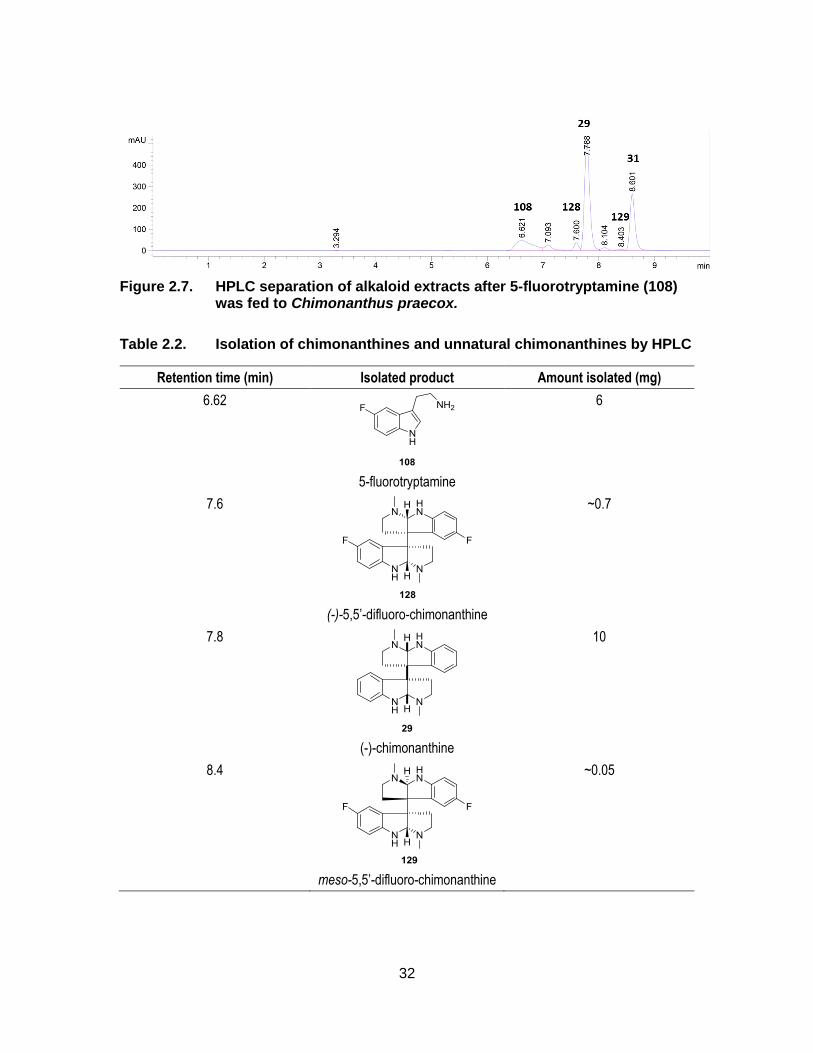
Figure 2.7. HPLC separation of alkaloid extracts after 5-fluorotryptamine (108) was fed to Chimonanthus praecox. ........................................................ 32
Figure 2.8. NMR spectra of various temperature NMR experiments on 1H and 19F NMR of isolated meso-5,5’-difluoro-chimonanthine (129) ................. 34
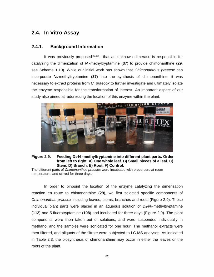
Figure 2.9. Feeding D3-Nb-methyltryptamine into different plant parts. Order from left to right. A) One whole leaf. B) Small pieces of a leaf. C) Stem. D) Branch. E) Root. F) Control. .................................................... 35
Figure 2.10. Image of roots of Chimonanthus praecox washed with distilled water. ..................................................................................................... 40
Figure 2.11. Fluorescent microscopy images of plant cells. Native plant cells (left) and a protoplast after enzymatic digestion (Right). ......................... 42
Figure 2.12. Images of ground leaves in liquid nitrogen in a mortar (a), cell wall powders after removing cellular components (b) and cell wall proteins on SDS-PAGE (c) ..................................................................... 45
Figure 2.13. Extracted ion chromatograms of m/z of 383.2042 ± 0.002 in the time interval of 4.4 min to 5.2 min after incubation of precursor with different amount of cell walls ........................................................... 46

ix
Figure 2.14. LC-MS EIC chromatograms. .................................................................. 48
Figure 2.15. O’Connor’s mutasynthesis in Catharanthus roseus. ............................... 51
x
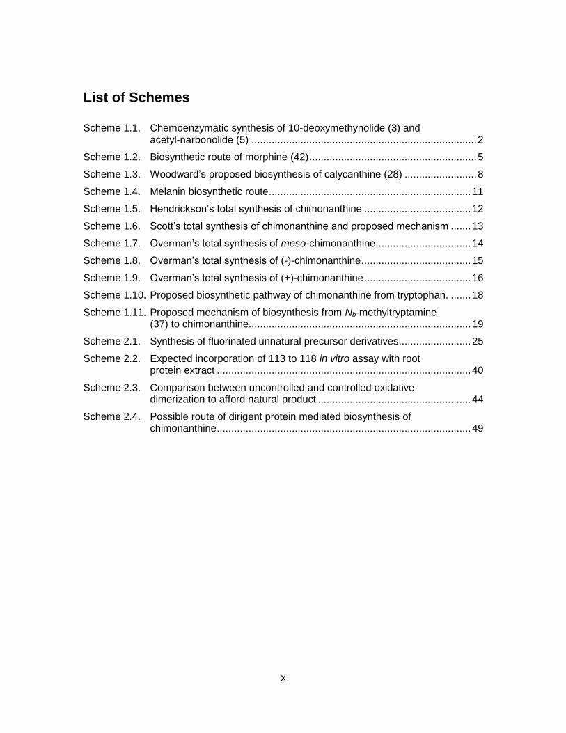
List of Schemes
Scheme 1.1. Chemoenzymatic synthesis of 10-deoxymethynolide (3) and acetyl-narbonolide (5) .............................................................................. 2
Scheme 1.2. Biosynthetic route of morphine (42) .......................................................... 5
Scheme 1.3. Woodward’s proposed biosynthesis of calycanthine (28) ......................... 8
Scheme 1.4. Melanin biosynthetic route ...................................................................... 11
Scheme 1.5. Hendrickson’s total synthesis of chimonanthine ..................................... 12
Scheme 1.6. Scott’s total synthesis of chimonanthine and proposed mechanism ....... 13
Scheme 1.7. Overman’s total synthesis of meso-chimonanthine ................................. 14
Scheme 1.8. Overman’s total synthesis of (-)-chimonanthine ...................................... 15
Scheme 1.9. Overman’s total synthesis of (+)-chimonanthine ..................................... 16
Scheme 1.10. Proposed biosynthetic pathway of chimonanthine from tryptophan. ....... 18
Scheme 1.11. Proposed mechanism of biosynthesis from Nb-methyltryptamine (37) to chimonanthine............................................................................. 19
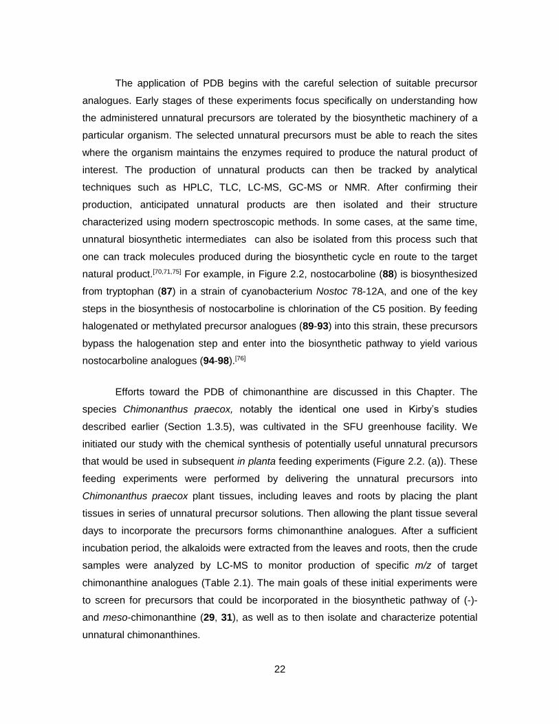
Scheme 2.1. Synthesis of fluorinated unnatural precursor derivatives ......................... 25
Scheme 2.2. Expected incorporation of 113 to 118 in vitro assay with root protein extract ........................................................................................ 40
Scheme 2.3. Comparison between uncontrolled and controlled oxidative dimerization to afford natural product ..................................................... 44
Scheme 2.4. Possible route of dirigent protein mediated biosynthesis of chimonanthine ........................................................................................ 49

xi
List of Acronyms
2,2-DMP 2,2-dimethoxypropane
4’OMT 4-O-methyltransferase
6OMT 6-O-methyltransferase
AcOH Acetic acid
ACP Acyl carrier protein
AT Acyl transferase
BCl3 Boron trichloride
BSA Bovine serum albumin
CH2Cl2 Dichloromethane
CH2O Formaldehyde
CNMT Coclaurine N-methyltransferase
CODM Codeine O-demethylase
COR Codeine reductase
CSA Camphosulfonic acid monohydrate
DCM Dichloromethane
DCT Dopachrome tautomerase
DEAD Diethyl azocarboxylate
DH Dehydratase
DIPA Diisopropylamine
DMF Dimethylformamide
DMPU 1,3-Dimethyl-3,4,5,6-tetrahydro-2-pyrimidinone
DOPA Dihydroxyphenylalanine
DRR 1,2-Dehydroreticuline reductase
DRS 1,2-Dehydroreticuline synthase
DTT Dithiothreitol
EDTA Ethlyenediaminetetraacetic acid
EIC Extracted Ion Chromatogram
EMCCD Electron multiplying charge coupled device
ER Enoyl reductase
Et2O Diethyl ether
Et3N Triethyl amine

xii
EtOH Ethanol
FAD Flavin adenine dinucleotide
FeCl3 Iron (III) chloride
G6P Glucose-6-phosphate
G6PDH Glucose-6-phosphate dehydrogenase
GC Gas chromatography
GC-MS Gas chromatography-mass spectrometry
HMPA Hexamethylphosphoramide
HN3 Hydrazoic acid
HPLC High performance liquid chromatography
HR High resolution
I2 Iodine
INMT Indoleethylamine N-methyltransferase
JRES J-resolved spectroscopy
KR Ketoreductase
KS Ketosynthase
LC Liquid chromatography
LC-MS Liquid chromatography-mass spectrometry
LDA Lithium diisopropylamide
L-DOPA L-3,4-dihydroxyphenylalanine
LiAlD4 Lithium aluminium deuteride
LiAlH4 Lithium aluminium hydride
LiCl Lithium chloride
Me3Al Trimethyl aluminium
MeMgI Methyl magnesium iodide
MeOH Methanol
MES 2-(N-Morpholino)ethanesulfonic acid
MM-NAC Methylmalonyl N-acetylcysteramine
MOPS 3-(N-Morpholino)propanesulfonic acid
MPE Maximum possible effect
mRNA Messenger ribonucleic acid
MS Mass spectrometry
Na Sodium

xiii
NaBH(OAc)3 Sodium triacetoxyborohydride
NaBH4 Sodium borohydride
NADH Nicotinamide adenine dinucleotide
NADP Nicotinamide adenine dinucleotide phosphate
NaH Sodium hydride
NaHMDS Sodium bis(trimethylsilyl)amide
NCS Norcoclaurine synthase
NH3 Ammonia
NMR Nuclear magnetic resonance
PAH Phenylalanine hyroxylase
Pb(OAc)4 Lead (IV) acetate
PBS Phosphate buffered saline
PDB Precursor-directed biosynthesis
PEG Polyethyleneglycol
Ph3P Triphenylphosphine
PhH Benzene
PVPP Poly(vinylpolypyrrolidone)
Red-Al Sodium bis(2-methoxyethoxy)aluminumhydride
RNA Ribonucleic acid
SalAT 7-O-Acetyltransferase
SalR Salutaridine reductase
SDS-PAGE Sodium dodecyl sulfate – polyacrylamide gel electrophoresis
SmI2 Samarium (II) iodide
T6ODM Thebaine 6-O-demethylase
TDC Tryptophandecarboxylase
TE Thioesterase
TFA Trifluoroacetic acid
TH-1 Tryptophan hydroxynase 1
THF Tetrahydrofuran
TLC Thin layer chromatography
TRP-1 Tyrosinase related protein 1
TRP-2 Tyrosinase related protein 2
TYDC Tyrosine decarboxylase

xiv
Zn Zinc

1
Chapter 1. Introduction
1.1. Enzymes in Nature and in Chemistry
In Nature, living organisms (e.g. mammals, plants) produce useful natural
products via numerous biosynthetic processes that rely on enzymes, which possess
several unique characteristics such as high substrate specificity and enantioselectivity.[1–
4] A large number of these enzymes have now been identified and structurally
characterized. For example, polyketide biosynthesis has been the subject of intense
research and is now largely understood to rely on collections of enzymes that form
“modules” which are responsible for adding one more acetate or propionate unit to a
growing polyketide chain and effect subsequent functional group transformations (e.g.,
reduction or dehydration). The biosynthesis of polyketides starts with a loading module,
and ends with a thioesterase which removes the polyketide natural product from the
biosynthetic machinery.[5–7] Understanding this biosynthetic pathway has allowed
scientists to exploit this molecular machinery in the synthesis of many unnatural
polyketides. An example of this is shown in Scheme 1.1, where Sherman and co-
workers carried out the chemoenzymatic synthesis of 10-deoxymethynolide (3) and
acetyl-narbonolide (5). The substrate 2 was prepared in 11 steps from the Roche ester
(1) then further elaborated and lactonized by the actions of PikAIII-TE to afford 3 or two
modules of PikAIII/PikAIV to afford the related macrolactone 5.[8] This rapid synthesis of
these complicated polyketides illustrates the power of using biosynthetic pathways to
compliment total syntheses.
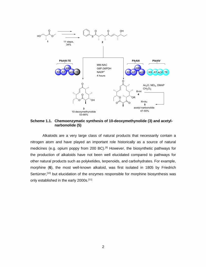
2
Scheme 1.1. Chemoenzymatic synthesis of 10-deoxymethynolide (3) and acetyl-narbonolide (5)
Alkaloids are a very large class of natural products that necessarily contain a
nitrogen atom and have played an important role historically as a source of natural
medicines (e.g. opium poppy from 200 BC).[9] However, the biosynthetic pathways for
the production of alkaloids have not been well elucidated compared to pathways for
other natural products such as polyketides, terpenoids, and carbohydrates. For example,
morphine (6), the most well-known alkaloid, was first isolated in 1805 by Friedrich
Sertürner,[10] but elucidation of the enzymes responsible for morphine biosynthesis was
only established in the early 2000s.[11]

3
Figure 1.1. Examples of well-known alkaloids; morphine (6), cocaine (7), and nicotine (8)
The difficulties in elucidating pathways involved in alkaloid biosynthesis is in part
related to the fact that there exist numerous structurally distinct families of alkaloids
whose natural production initiates with a unique starting material and consequently each
biosynthetic step requires discrete enzymes. For example, the biosynthesis of morphine
(6) is depicted in Scheme 1.2.[11–13] This pathway initiates with decarboxylation of L-
DOPA (9) by tyrosine decarboxylase (TYDC) to afford dopamine (10). Norcoclaurine
synthase (NCS) utilizes substrates 10 and 4-hydroxyphenylacetaldehyde (11) to yield
(S)-norcoclaurine (12), which is then methylated by norclaurine 6-O-methyltransferase
(6OMT) to obtain (S)-coclaurine (13). Subsequent methylation by coclaurine N-
methyltransferase (CNMT) affords (S)-N-methylcoclaurine (14). Then, a selective
oxidation of an aromatic ring by N-methylcoclaurine-3-hydroxylase (Cyp80B3) affords
(S)-3’-hydroxy-N-methylcoclaurine (15), which undergoes selective methylation on
hydroxyl group by 3’-hydroxy-N-methylcoclaurine 4-O-methyltransferase (4’OMT) to
obtain (S)-reticuline (16). Iminium ion mediated epimerization of substrate 16 to (R)-
reticuline (18) is performed by two enzymes: 1,2-dehydroreticuline synthase (DRS) and
1,2-dehydroreticuline reductase (DRR). Salutaridine synthase (Cyp719B1) cyclizes 18 to
yield salutaridine (19). Salutaridine reductase (SalR) reduces ketone function in 19 to
produce salutaridinol (20). Salutaridinol 7-O-acetyltransferase (SalAT) specifically
acetylates the C7 alcohol function to provide salutaridinol-7-O-acetate (21), which
spontaneously cyclizes to thebaine (22).
There are two possible routes from thebaine (22) to morphine (6). The first
pathway is from 22 to neopinone 23 via removal of the methyl group at the 6 position by
thebaine 6-O-demethylase (T6ODM). Then, 23 spontaneously undergoes
rearrangement to codeinone (24). The reduction of the enone to an allylic alcohol by
codeine reductase (COR) yields codeine (25). The last step in the first pathway involves

4
demethylation by codeine O-demethylase (CODM) to obtain morphine (6). The second
route from 22 to 6 involves demethylation CODM to afford oripavine (26), which
undergoes another demethylation by T6ODM to yield mophinone (27). Lastly, 27
undergo reduction by COR to obtain morphine (6).

5
Scheme 1.2. Biosynthetic route of morphine (42)

6
(Scheme 1.2 – continued from previous page) TYDC: tyrosine decarboxylase, NCS: norcoclaurine synthase, 6OMT: 6-O-methyltransferase, CNMT: coclaurine N-methyltransferase, Cyp80B3: N-methylcoclaurine 3 –hydroxylase, 4’OMT: 3’-hydroxy-N-methylcoclaurine 4-Omethyltransferase, DRS: 1,2-dehydroreticuline synthase, DRR: 1,2-dehydroreticuline reductase, Cyp719B1: salutaridine synthase, SalR: salutaridine reductase, SalAT: 7-O-acetyltransferase, T6ODM: thebaine 6-O-demethylase, COR: codeine reductase, CODM: codeine O-demethylase
Thus, biosynthetic studies on alkaloids are complicated by their unique and
complex structures. However, there is no doubt that further studies towards the
identification of biosynthetic pathways are useful as they would support the large scale
syntheses of these potentially important natural products as well as analogues. In this
thesis, the biosynthesis of one member of the family of calycanthaceous alkaloids,
chimonanthine, is studied. In the following section, the history, bioactivity, and synthetic
and biosynthetic efforts towards chimonanthine will be discussed.
1.2. Calycanthaceous Alkaloids
The study on calycanthaceous alkaloids started with a letter written in November,
1887 by Mr. J. H. H. Boyd. [14]
“Hundreds of cattle and sheep have died here in the past five years from ‘bubby’ (the eccentric name of Calycanthus glaucus). The seeds only are poisonous. When a brute gets a sufficient dose, from five to ten well-filled pods, it makes for the nearest water and often falls dead while drinking, or it may live three or four weeks and then die. The symptoms are like those of a man extremely drunk, except that any noise frightens it. Stamp the ground hard close to a brute poisoned with ‘bubby,’ and it will jump and jerk and tremble for several minutes. That is our method of telling when they have taken it. The eyes turn white and glassy, and while lying they throw back the head and look as if dead already. ‘Bubby’ does not seem to hurt a brute so much if it cannot get water. Our best remedy is apple brandy, strong coffee and raw eggs poured down as soon as possible after finding. It is certain that ‘bubby’ is the most poisonous of any shrub or weed in existence here, from the fact that when brutes have once eaten it they will take it every time they can get it. It grows on every hillside, along all branches (creeks), in every fence corner and almost everywhere.”
In 1888, Eccles studied the poisonous “bubby” seeds (Calycanthus glaucus) in
order to elucidate the toxic components from the extracts, and was the first to report a

7
new compound, calycanthine (28) which represents the first entry of a new class of
alkaloids which have become known as the calycanthaceous alkaloids (Figure 1.2).[15]
Interestingly, during this study it was reported that alkaloid 28 was not toxic. The isolated
sample of (+)-calycanthine also allowed for the detailed description of several physical
properties, including specific bad odour, poor solubility in water and good solubility in
diethylether or chloroform. Furthermore, it was noted that the colour of the alkaloids
change after coming into contact with several acids. For example, when 28 was treated
with concentrated sulphuric acid, the initial mixture turned into a yellow solution. In the
presence of concentrated ‘muriatic acid’ (HCl), the solution turned yellow initially and
then turned olive green. A combination of concentrated sulphuric acid and sugar made a
“lovely” pink red colour. Also, Eccles reported that (+)-calycanthine (28) made up 2% of
the total mass of the seed. One year after this initial discovery, Wiley determined that the
alkaloid has the molecular formula C18H40N2O11.[14]
Figure 1.2 Examples of (bis)indoline calycanthaceous alkaloids.
Nearly twenty years after these initial studies, Gordin reported that there was no
oxygen atom in (+)-calycanthine (28) and determined that the molecular formula was

8
C11H14N2 by measuring the amount of CO2 and volume of N2 released from calycanthine
(28).[16,17] A further twenty years later, in 1925, the molecular formula was doubled by
Späth and Stroh to C22H28N4.[18] In a subsequent study by Barger, the molecular formula
of (+)-calycanthine (28) was further revised to C22H26N4 in 1939. [19] However, the
molecular structure was not elucidated until 1960 when Woodward, Clark and Katz
determined the structure of (+)-calycanthine (28) by degradation methods.[20] They also
proposed a potential biosynthetic pathway (Scheme 1.3) to (+)-calycanthine (28)
beginning with the β,β’-oxidation of Nb-methyltryptamine (37). The same year, the
structure of (+)-calycanthine (28) was confirmed by Hamor via X-ray crystallography of
the alkaloid.[21]
Scheme 1.3. Woodward’s proposed biosynthesis of calycanthine (28)
The discovery of a number of new calycanthaceous alkaloids was reported
following the isolation of (+)-calycanthine.[19,22–26] (Figure 1.2) Additionally, several
homologues of (+)-calycanthine (28) produced by medicinal terrestrial plants (e.g.
Psychotria sp.) were identified that contain a central meso-chimonanthine unit as part of
their structure (highlighted in blue, Figure 1.3).[27–32]
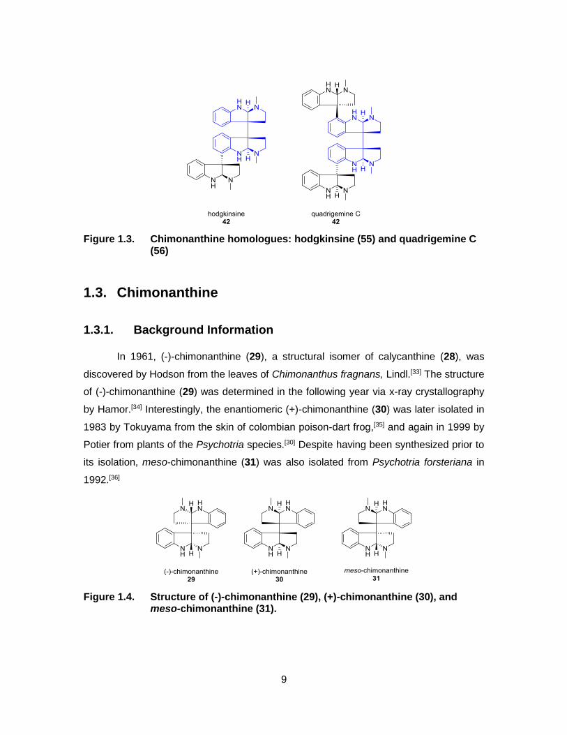
9
Figure 1.3. Chimonanthine homologues: hodgkinsine (55) and quadrigemine C (56)
1.3. Chimonanthine
1.3.1. Background Information
In 1961, (-)-chimonanthine (29), a structural isomer of calycanthine (28), was
discovered by Hodson from the leaves of Chimonanthus fragnans, Lindl.[33] The structure
of (-)-chimonanthine (29) was determined in the following year via x-ray crystallography
by Hamor.[34] Interestingly, the enantiomeric (+)-chimonanthine (30) was later isolated in
1983 by Tokuyama from the skin of colombian poison-dart frog,[35] and again in 1999 by
Potier from plants of the Psychotria species.[30] Despite having been synthesized prior to
its isolation, meso-chimonanthine (31) was also isolated from Psychotria forsteriana in
1992.[36]
Figure 1.4. Structure of (-)-chimonanthine (29), (+)-chimonanthine (30), and meso-chimonanthine (31).

10
In 2002, Verotta performed a comparative study on the analgesic properties of
chimonanthines to morphine (6), and found that the former family of natural products
(29-31) exhibit strong binding affinities toward the μ-opioid receptor.[37] In the tail-flick
mouse assay (Figure 1.5), Verotta compared (-)- and (+)-chimonanthines (29 and 30)
with morphine (6) by measuring the change in the response time after causing pain to
the mouse.[38] While morphine (6) displayed 100% maximum possible effect (MPE) at 6
mg/kg, (-)-chimonanthine (29) displayed 40% MPE at 10 mg/kg and (+)-chimonanthine
(30) displayed 66% MPE at 5 mg/kg. In the capsaicin induced pain model, Verotta
reported that a dosage of 0.25 mg/kg of (-) and (+)-chimonanthines (29, 30), the mice
reduced licking at the site where capsaicin was injected, and they observed that
inhibition of licking diminished by 47% and 38% when administered with (-)-
chimonanthine (29) and (+)-chimonanthine (30), respectively. Furthermore, (-)-
chimonanthine (29), (+)-chimonanthine (30) and meso-chimonanthine (31) displayed
strong binding affinity (Ki) to μ-opioid receptor with 271 ± 85 nM, 652 ± 159 nM and 341
± 29 nM, respectively. As a comparison, morphine (6) displayed 0.76 ± 0.04 nM binding
affinity towards the same receptor. These results suggested that chimonanthines could
be good lead candidates as analgesics.
Figure 1.5. Illustration of tail-flick model with mouse
Further investigations into the biological activity of the chimonanthines revealed
that (-)-chimonanthine (29) inhibits melanogenesis, in vitro (see Scheme 1.4). This
pathway is responsible for the generation of melanin in the body. The study reported that
(-)-chimonanthine (29) showed inhibition of melanin production in B16 melanoma 4A5
cells (IC50 = 1.4 μM), and compared favourably to the commercially available tyrosinase
inhibitor arbutin (IC50 = 174 μM). However, the inhibition mechanism associated with
chimonanthine has not been reported yet.[39–41]

11
Scheme 1.4. Melanin biosynthetic route The blue arrows in chemical structures of eumelanin and pheomelanin indicate where elongation occurs, and the COOH groups in parenthesis can be substituted with H.
1.3.2. Early Syntheses of Chimonanthine
The interesting biological activity and intriguing structural characteristics of
chimonanthine has inspired the development of several synthetic routes to these

12
compounds. The major challenge in these syntheses relates to the controlled
introduction of the two adjacent quaternary all-carbon stereocentres, for which several
new methods were devised. In 1962, Hendrickson reported the first total synthesis of
rac-chimonanthine (rac-29) and meso-chimonanthine (31) (Scheme 1.5).[42,43] This
biomimetic total synthesis of chimonanthine relied on an oxidative dimerization of 55 to
afford the key C-C bond. This biomimetic sequence supported the previously proposed
biosynthesis of calycanthine by Woodward (see Scheme 1.3).
Scheme 1.5. Hendrickson’s total synthesis of chimonanthine
Two years later, Scott reported the second total synthesis of chimonanthine via
oxidative dimerization of Nb-methyltryptamine (37) (Scheme 1.6).[44] This synthesis
utilized a common biological building block 37 and was achieved in a single step with an
improved yield compared to that reported by Hendrickson. The proposed mechanism for
this key transformation is depicted in Scheme 1.6. Thus, deprotonation of the indole
nitrogen by methylmagnesium iodide affords the resonance-stabilised anion 58. The
anion then undergoes a single electron oxidation with iron (III) chloride to afford a C3
radical which undergoes radical recombination with an equivalent coupling partner to
provide the key C3-C3’ bond to yield chimonanthine (rac-29, meso-31).

13
Scheme 1.6. Scott’s total synthesis of chimonanthine and proposed mechanism
1.3.3. First Enantioselective Syntheses of Chimonanthine
While the racemic syntheses of the chimonanthines were achieved with few
synthetic steps, their enantioselective synthesis presented a major challenge. Overman
and coworkers completed the first total synthesis of meso, (+)- and (-)-chimonanthine
through a series of enantioselective syntheses that proved to be landmarks in
asymmetric synthesis. For meso-chimonanthine, the readily available isoindigo 59 was
converted to 60 via samarium mediated reductive dialkylation (Scheme 1.7).[45]
Reduction by Red-Al afforded hexacyclic intermediate 61, which was dihydroxylated and
cleaved to afford diol 62. Subsequent Mitsunobu reaction, azide reduction and exposure
to trimethylaluminum provided bis(pyrroloindoline) 63. Finally, methylation and
deprotection afforded desired product meso-31.

14
Scheme 1.7. Overman’s total synthesis of meso-chimonanthine
In 1999, Overman reported an enantioselective total synthesis of (-)-
chimonanthine (29, Scheme 1.8).[46] The key reaction in the total synthesis of 29 is an
intramolecular double Heck reaction cascade of 69 to 70. In this single step, the vicinal
quaternary all-carbon stereocentres were introduced in high yield. Following synthetic
steps that included cleavage of the cyclohexene ring to provide diol 71. Further reduction
and a Mitsunobu reaction afforded 72, the heating of which in methanol and subsequent
bis-methylation yielded bispyrroloindoline 73. Lastly, removal of benzyl group from 73
provided the desired product (-)-chimonanthine (29).

15
Scheme 1.8. Overman’s total synthesis of (-)-chimonanthine
Following his successful synthesis of (-)-chimonanthine (43), Overman reported a
modified synthesis of (+)-chimonanthine (30, Scheme 1.9) in 2000.[47] An interesting
highlight in Overman’s synthesis of 30 involves dialkylation of dihydroisoindigo 74 to
afford bisoxyindole 75. In a single step, the desired all-carbon quaternary stereocentres

16
were introduced in excellent yield. The remaining synthetic steps to (+)-30 were identical
to those employed in the total synthesis of (-)-30.
Scheme 1.9. Overman’s total synthesis of (+)-chimonanthine
1.3.4. Additional Total Syntheses of Chimonanthine. .
Following Overman’s elegant enantioselective syntheses of meso, (-) and (+)-
chimonanthines (31, 29, 30), numerous total syntheses of chimonanthine have been
reported. In 2002, Takayama achieved a two-step synthesis of rac-chimonanthine (rac-
29) and meso-chimonanthine (31).[48] In 2007, Movassaghi reported the total synthesis of
(+)-chimonanthine (30) via the Co-mediated homodimerization of tryptophan after
benzylic bromination.[49] In 2012, Matunaga developed an enantioselective synthesis of
(+)-chimonanthine (30) in six steps through the use of a Schiff base.[50] In 2013, Ma

17
reported a highly enantioselective synthesis of (-)-chimonanthine (29) in three steps via
bromocyclization of tryptamine.[51] Due in part to these efforts, there have been
significant advances in achieving the C3-C3’ dimerization of indole derivatives leading to
the synthesis of several chimonanthine analogues.[52–61]
1.3.5. Biosynthetic Studies of Chimonanthine
While there have been multiple syntheses of chimonanthine and related
analogues reported, only one study relating to the biosynthesis of this unique alkaloid
has been reported.[62,63]
Figure 1.6. Kirby’s Radiolabelled Precursors and Radiolabelled Chimonanthine products from Chimonanthus praecox
The radiolabelled precursors were fed to Chimonanthus praecox, and the percentage of incorporation rate was determined by the ratio of radioactivity of isolated chimonanthine (counts per minute) over weight of isolated chimonanthine.
In this single study, Kirby synthesized radiolabelled tryptamine derivatives (80-
82, Figure 1.6) and then fed solutions of these compounds to the leaves of
Chimonanthus fragnans, a terrestrial plant from southern China. After seven days, the
alkaloids were isolated by extraction, and the incorporation of radiolabelled precursors

18
into chimonanthine was evaluated. From the radioactivity data, it was determined that
the radiolabelled tryptophan (80), tryptamine (81) and Nb-methyltryptamine (82) were
incorporated to the isolated sample of chimonanthine at a rate of 3.6%, 11.1% and 0.1%,
respectively. These results indicated that tryptamine in particular was a biological
starting point for the biosynthesis of chimonanthine. The low incorporation rate for the
radiolabelled Nb-methyltryptamine (82) compared to that of tryptamine (81) was
postulated to result from the low solubility of this compound in water.
Unfortunately, since Kirby’s study 45 years ago, there have been no further
studies on the biosynthesis of chimonanthine.
1.4. Proposed Biosynthesis of Chimonanthine
The biosynthetic pathway proposed by Kirby involves the steps tryptophan (85)
→ tryptamine (86) → Nb-methyltryptamine (37) → chimonanthine. (Scheme 1.10) It is
reasonable that tryptophan decarboxylase (TDC) is involved in the removal of
carboxylate group from tryptophan (85) to yield tryptamine (86).[64–66] Then,
indolethylamine N-methyltransferase (INMT),[67–69] a well-characterized enzyme, could
effect methylation of the primary amine to afford Nb-methyltryptamine (37). The final
dimerization step of Nb-methyltryptamine (37) into chimonanthine involves unidentified
enzyme(s).
Scheme 1.10. Proposed biosynthetic pathway of chimonanthine from tryptophan.
The unknown dimerase that carries out the conversion of 37 into chimonanthine
was proposed to be responsible for the oxidation of 37 required for recombination of the
resultant radicals to form the two adjacent quaternary all-carbon stereocentres (Scheme
1.11).

19
Scheme 1.11. Proposed mechanism of biosynthesis from Nb-methyltryptamine (37) to chimonanthine.
1.5. Main Goals of This Study
Since the discovery of (-)-chimonanthine (29) approximately 55 years ago, the
chemical syntheses of chimonanthines and their derivatives have attracted considerable
interest from the synthetic community owing to their interesting molecular structures and
potentially useful biological activities. However, the biosynthesis of chimonanthine is not
well established. More specifically, the key enzyme responsible for the dimerization of
Nb-methyltryptamine (37) to provide chimonanthine is unknown. Based on Kirby’s
proposal in 1969, the protein is postulated to have two important functions: i) oxidation of
a reactive indole intermediate to form a radical, and ii) promoting an enantioselective
carbon-carbon bond formation reaction with an equivalent radical intermediate.
Importantly, identification of this dimerase could allow for the chemoenzymatic synthesis
of chimonanthines and chimonanthine analogues using cheap and commercially
available starting materials.
The goal of the thesis was to develop methods to investigate the unknown
enzyme responsible for the oxidative dimerization of Nb-methyltryptamine (37). The
following chapter of this thesis includes a discussion of in planta experiments. Similar to
Kirby’s study, halogenated or isotope labelled tryptamine derivatives were prepared (e.g.

20
fluorinated tryptamine derivative, deuterated tryptamine derivative), and these
precursors were fed into a chimonanthine-producing plant, Chimonanthus praecox.
Once suitable biosynthetic precursors were identified, in vitro experiments were initiated.
Then, the results from protein extractions, cell component preparation (protoplast and
cell wall) are presented, and examination reaction buffers with an ultimate goal of
producing chimonanthine analogues using cell extracts.

21
Chapter 2. Studies towards the Biosynthesis of (-)- and meso-Chimonanthine
2.1. Background Information
Precursor-directed biosynthesis (PDB) is a method used to exploit existing
biosynthetic pathways to natural products by feeding unnatural precursors that can be
transformed though these pathways into structurally related unnatural products.[70,71] As
illustrated in Figure 2.1, PDB relies on the administration of unnatural precursor of the
natural biosynthetic precursor into a target tissue or organism. In Nature, only natural
precursors are used to synthesize a natural product. However, when a synthetic
unnatural precursor analogue is administered to a particular organism, this unnatural
substrate can, in some cases, be assimilated into the biosynthetic pathways of the
natural product, leading to the formation of an unnatural product that is structurally
related to the natural product. The biosynthetic machinery (e.g., enzymes) is then
exploited to produce analogues of the targeted natural products.[72–74]
Figure 2.1. Illustration of unnatural product synthesis by PDB[70,71]
22
The application of PDB begins with the careful selection of suitable precursor
analogues. Early stages of these experiments focus specifically on understanding how
the administered unnatural precursors are tolerated by the biosynthetic machinery of a
particular organism. The selected unnatural precursors must be able to reach the sites
where the organism maintains the enzymes required to produce the natural product of
interest. The production of unnatural products can then be tracked by analytical
techniques such as HPLC, TLC, LC-MS, GC-MS or NMR. After confirming their
production, anticipated unnatural products are then isolated and their structure
characterized using modern spectroscopic methods. In some cases, at the same time,
unnatural biosynthetic intermediates can also be isolated from this process such that
one can track molecules produced during the biosynthetic cycle en route to the target
natural product.[70,71,75] For example, in Figure 2.2, nostocarboline (88) is biosynthesized
from tryptophan (87) in a strain of cyanobacterium Nostoc 78-12A, and one of the key
steps in the biosynthesis of nostocarboline is chlorination of the C5 position. By feeding
halogenated or methylated precursor analogues (89-93) into this strain, these precursors
bypass the halogenation step and enter into the biosynthetic pathway to yield various
nostocarboline analogues (94-98).[76]
Efforts toward the PDB of chimonanthine are discussed in this Chapter. The
species Chimonanthus praecox, notably the identical one used in Kirby’s studies
described earlier (Section 1.3.5), was cultivated in the SFU greenhouse facility. We
initiated our study with the chemical synthesis of potentially useful unnatural precursors
that would be used in subsequent in planta feeding experiments (Figure 2.2. (a)). These
feeding experiments were performed by delivering the unnatural precursors into
Chimonanthus praecox plant tissues, including leaves and roots by placing the plant
tissues in series of unnatural precursor solutions. Then allowing the plant tissue several
days to incorporate the precursors forms chimonanthine analogues. After a sufficient
incubation period, the alkaloids were extracted from the leaves and roots, then the crude
samples were analyzed by LC-MS to monitor production of specific m/z of target
chimonanthine analogues (Table 2.1). The main goals of these initial experiments were
to screen for precursors that could be incorporated in the biosynthetic pathway of (-)-
and meso-chimonanthine (29, 31), as well as to then isolate and characterize potential
unnatural chimonanthines.

23
Figure 2.2. Precursor directed biosynthesis of nostocarboline analogues

24
Figure 2.3. Illustration of experimental scheme. (a) in planta assay (b) in vitro assay.
(a) in planta experiment: unnatural precursors screening and isolation of unnatural products. (b) in vitro experiment: studies toward the identification of a dimerase catalyzing the production of chimonanthine. Analytical tools such as NMR and/or LC-MS allow detection of targeted unnatural product(s)
Following the identification of suitable unnatural precursors, this study focused on
the extraction of protein and components of cells from Chimonanthus praecox followed
by in vitro assays on the biosynthesis of chimonanthine (Figure 2.2. (b)). Specifically, the
extracted protein or cell components are incubated with unnatural precursors shown to
be competent in the PDB studies, then the presence of chimonanthine analogues is
assessed using LC-MS analyses. Following these studies, the proteins or cells are
further separated by either protein size or cell fractionation, respectively. These different
types of fractions can then be analyzed for their ability to produce the unnatural
products. The ultimate goal of this study is to isolate the unknown enzyme(s) that is(are)
25
responsible for the C-C bond formation/dimerization between two Nb-methyl-tryptamines
(37) that yields chimonanthine.
2.2. Preparation of Precursor Analogues
As described in Section 1.3.5., Kirby’s study led to the proposal for a biosynthetic
pathway involving the conversion of tryptophan (85) to tryptamine (86), which is then
converted to Nb-methyl-tryptamine (37) and finally dimerized to provide chimonanthine
(e.g. 29) (Scheme 1.10). In order to select suitable unnatural precursor candidates for
our feeding experiments, we began our investigation by evaluating commercially
available and synthetically accessible analogues of tryptamine and Nb-methyl-
tryptamine.
Scheme 2.1. Synthesis of fluorinated unnatural precursor derivatives
In Scheme 2.1, 5-fluoroindole (100) undergoes alkylation to afford nitroalkene
101. Then, without purification of 101, the nitro group is reduced using NaBH4 to yield 5-
fluorotryptamine (102). Addition of chloroethylformate to 102 yields carbamate 103,
which after reduction using LiAlH4 or LiAlD4 furnishes the desired unnatural precursors
104 or 105. The same chemistry was applied to prepare compounds 37, 110, 112, and
115 (Figure 2.4, see experimental for details). The other precursor analogs used in this
study were purchased from AK Scientifics and/or Alfa Aesar.

26
The unnatural precursors have unique molecular weight distinguishable from
natural precursor, and also have unique characteristics, for example, the fluorinated
precursors 108, 109, 113, and 114 can be tracked by 19F-NMR, and brominated
precursors (111 and 115) display specific isotope patterns in MS analysis (Figure 2.4).
Figure 2.4. Precursor analogues to study PDB of chimonanthine
2.3. In Planta Experiment (Feeding Experiment)
2.3.1. Screening Precursor Analogues
The precursor analogues were dissolved independently in weakly acidic water
(pH = 4) and fed to the leaves of Chimonanthus praecox by standing the stems of leaves
in the water solution as described by Kirby.[63] This process required a five-day
incubation period in a greenhouse and, after this time, the leaves were collected and
ground using a mortar and pestle with freezing in liquid nitrogen. The powdered green
leaves were suspended in methanol and stirred overnight. The solvent was collected by
filtration to remove insoluble plant debris and then concentrated in vacuo. The crude
alkaloid samples were then obtained by acid/base and chloroform extractions (see
Experimental 2.7.6). The resulting crude mixtures were submitted for LC-MS analyses to
detect the presence of potential unnatural products.

27
Figure 2.5. Feeding precursor analogues into leaves of chimonanthus praecox
In our studies, we used the extracted ion chromatogram (EIC) function for our
high resolution-liquid chromatography-mass spectrometry experiments. Specifically, EIC
allows the detection of the specific m/z of interest from the entire data acquired in
chromatographic run. The use of this technique is critical for the detection of small
amounts of unnatural products in the presence of larger amounts of the natural products
and other small molecules. In Table 2.1, the expected unnatural products from feeding
experiments involving either one or two unnatural precursors are presented.
From the results of LC-MS analyses the formation of unnatural products was
preliminarily established by monitoring their unique m/z. Administration of a single
precursor or two precursors simultaneously were performed. The relative level of
incorporation was estimated by calculating by the ratio of areas of m/z of unnatural
product over areas of m/z of (-)-chimonanthine (29) observed in the LC chromatograms
using the MS as a detector in EIC mode. The LC-MS analysis in EIC mode indicated that
precursor analogues 107, 108, 109, 112, 113, and 114 were incorporated into the
expected unnatural chimonanthine analogues (117, 118, 119, 122, and 123).
Furthermore, 6-methyltryptamine 106, 5-chlorotryptamine (110), 5-bromotryptamine
(111), and 5-bromo-Nb-methyltryptamine (115) did not provide the m/z expected for the
potential chimonanthine analogues (116, 120, and 121). Interestingly, after feeding with
7-methyltryptamine (107), LC-MS analysis detected the expected m/z of the
corresponding chimonanthine analogue (117). As shown in Table 2.1, two other
precursor analogues also gave rise to the anticipated m/z of their corresponding
chimonanthine analogues. Based on these preliminary results suggesting that C5 and

28
C6 substituted products were not formed, we postulate that somewhat larger
substituents, such as Cl, Br. And CH3, at these positions may not be tolerated by the
enzymes in the biosynthetic pathway leading to chimonanthine.
Table 2.1. Results after feeding experiment
Entry Precursors Expected chimonanthine
analogue
Exact m/z of chimonanthin
e analogue
Detection of the m/z of
chimonanthine analogue
Incorporation
(%)
1
375.2543 No NI
2
375.2543 Yes, 373.2545 1.97
3
383.2042 Yes, 383.2043 9.34
4
383.2042 Yes, 383.2038 17.1
5
415.1451 No NI

29
6
503.0440 No NI
7
353.2607 Yes, 353.2614 0.54
8
383.2042 Yes, 383.2043 1.1
9
389.2418 Yes, 389.2413 2.12
10
503.0441
505.0421
507.0400
No NI
11
+
368.2324
Yes, 378.2325
0.79

30
12
+
368.2324
Yes, 368.2325
1.12
13
+
368.2324
Yes, 368.2319
1.06
14
+
371.2512
Yes, 371.2508
0.79
15
+
365.2136
Yes, 365.2137
1.30
16
+
365.2136
Yes, 365.2144
1.11
31
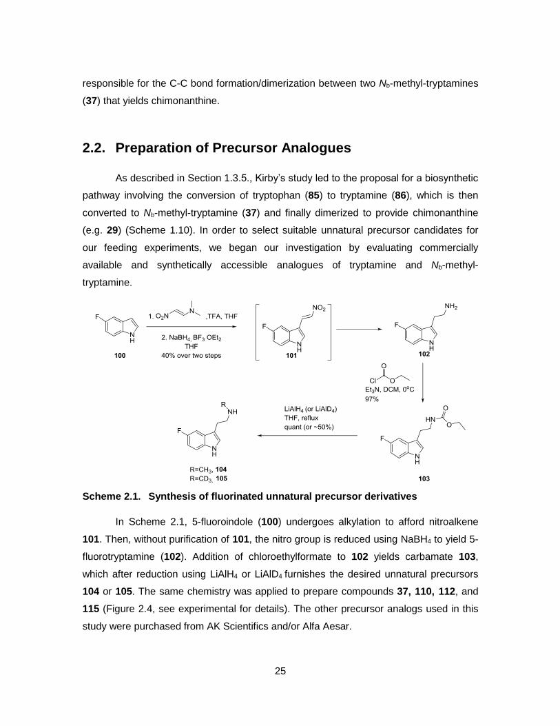
NI: No incorporation
2.3.2. Isolation of Unnatural Products, (-)-5,5’-Difluoro-chimonanthine and meso-5,5’-Difluoro-chimonanthine
After identifying several putative precursor analogues, we decided to utilize 5-
fluorotryptamine (108) as our model precursor analogue for our in planta experiments.
This choice was largely driven by the generation of a strong m/z signal associated with
that of the target unnatural product by LC-MS analyses (entry 3, Table 2.1) but also by
the ability to monitor formation of this product by 19F NMR spectroscopy. The feeding
experiments were therefore scaled-up in order to isolate sufficient quantities of the
unnatural products 128 and 129 (Figure 2.6) that would enable proper characterization
to confirm results from LC-MS analyses.
Figure 2.6. Target unnatural chimonanthine analogues after feeding Chimonanthus praecox with 108: (-)-5,5’-fluorochimonanthine (128), and meso-5,5’-fluorochimonanthine (129)
The extracted crude alkaloids (40 mg) obtained from plant tissues after the
feeding experiments were solubilized in methanol prior to purification by preparative
HPLC (Figure 2.7).
32
Figure 2.7. HPLC separation of alkaloid extracts after 5-fluorotryptamine (108) was fed to Chimonanthus praecox.
Table 2.2. Isolation of chimonanthines and unnatural chimonanthines by HPLC
Retention time (min) Isolated product Amount isolated (mg)
6.62
5-fluorotryptamine
6
7.6
(-)-5,5’-difluoro-chimonanthine
~0.7
7.8
(-)-chimonanthine
10
8.4
meso-5,5’-difluoro-chimonanthine
~0.05

33
8.6
meso-chimonanthine
6
Only the characterized products are reported in the table. The other fractions were not fully characterized.
Table 2.2 summarizes the characterization of the isolated alkaloids. Among the
40 mg of the crude alkaloid preparation, 6 mg (15%) was the administered compound
108. The amounts of the known natural products, (-)-chimonanthine (29) and meso-
chimonanthine (31) were 10 mg (25%), and 6 mg (15%), respectively. The desired
chimonanthine analogues, 128, and 129 were collected, and the amounts were
approximately 0.7 mg (1.75%) and 0.05 mg (0.1%), respectively. Some remaining
material may be present in the additional peaks observed in the chromatogram but these
have not been characterized.
After separation, each fraction was submitted for LC-MS analysis. The fractions
with the expected m/z were combined and characterized by 1H-NMR and 19F-NMR
spectroscopy. (-)-5,5’-Difluorochimonanthine (128, Figure 2.6) showed the desired m/z
when analyzed by LC-MS and eluted at a retention time of 1.5 minutes. As expected, the
1H-NMR spectrum displayed three characteristic aromatic proton resonances (δ 6.4 to
6.9 ppm) and a single fluorine resonance (δ 127.1 ppm) in the 19F-NMR spectrum. This
provided good evidence for the incorporation of fluorine into the unnatural product 128.
While the characterization of (-)-5,5’-difluorochimonanthine (128) was not
particularly challenging, the characterization of meso-5,5’-difluorochimonanthine (129)
presented several interesting challenges. Based on the LC-MS analyses of meso-5,5’-
difluorochimonanthine (129), we observed the desired m/z signal corresponding to the
unnatural product at a retention time of 4.2 minutes. As depicted in Figure 2.8, 1H-NMR
and 19F-NMR analyses performed at room temperature were not enough to allow proper
characterization. The resonances from the 1H-NMR spectra were very broad and J-
couplings could not be determined. The 19F-NMR spectrum included two fluorine

34
resonances at room temperature (δ 129.2 and 129.4 ppm), which did not match with the
expected single fluorine resonance.
We speculated that the broad signals may stem from the presence of slowly
interconverting atropisomers. Accordingly, we were able to resolve this issue by
increasing the temperature of the sample being analyzed by NMR spectroscopy up to 80
°C. As indicated in Figure 2.6, an increase in temperature during acquisition of 1H-NMR
spectra resulted in spectra with much sharper resonances, presumably due to the more
rapid interconversion of two atropisomers. Additionally, the corresponding 19F-NMR
spectra recorded at higher temperatures also indicated coalescence of two separate
fluorine resonances into a single resonance.
Figure 2.8. 1H and 19F NMR spectra of isolated meso-5,5’-difluoro-chimonanthine (129) acquired at various temperatures
The NMR experiments were performed at four different temperatures, 25, 40, 60 and 80 °C).
Based on these NMR spectroscopic experiments, we were able to characterize
meso-5,5’-difluorochimonanthine (129). It indicated meso-129 is not formally meso at
room temperature, but rather is a mixture of two atropisomers. Also, at higher
temperature, the atropisomers rapidly interconvert such that the atropisomers display a
spectrum characteristic of a meso compound.[77–82]. It is expected that an energy barrier
must be overcome in order to rotate the C3-C3’ bond; thus, at room temperature there
are two possible conformations of meso-5,5’-difluorochimonanthine (129).
35
2.4. In Vitro Assay
2.4.1. Background Information
It was previously proposed[20,62] that an unknown dimerase is responsible for
catalyzing the dimerization of Nb-methyltryptamine (37) to provide chimonanthine (29,
see Scheme 1.10). While our initial work has shown that Chimonanthus praecox can
incorporate Nb-methyltryptamine (37) into the synthesis of chimonanthine, it was
necessary to extract proteins from C. praecox to further investigate and ultimately isolate
the enzyme responsible for the transformation of interest. An important aspect of our
study also aimed at addressing the location of this enzyme within the plant.
Figure 2.9. Feeding D3-Nb-methyltryptamine into different plant parts. Order from left to right. A) One whole leaf. B) Small pieces of a leaf. C) Stem. D) Branch. E) Root. F) Control.
The different parts of Chimonanthus praecox were incubated with precursors at room temperature, and stirred for three days.
In order to pinpoint the location of the enzyme catalyzing the dimerization
reaction en route to chimonanthine (29), we first selected specific components of
Chimonanthus praecox including leaves, stems, branches and roots (Figure 2.9). These
individual plant parts were placed in an aqueous solution of D3-Nb-methyltryptamine
(112) and 5-fluorotryptamine (108) and incubated for three days (Figure 2.9). The plant
components were then taken out of solutions, and were suspended individually in
methanol and the samples were sonicated for one hour. The methanol extracts were
then filtered, and aliquots of the filtrate were subjected to LC-MS analyses. As indicated
in Table 2.3, the biosynthesis of chimonanthine may occur in either the leaves or the
roots of the plant.

36
Table 2.3 Localization of Biosynthesis of Chimonanthine
Entry Plant Part Detection of (118)
m/z 383.2042
Detection of (122)
m/z 353.2607
1 Leaf Yes Yes
2 Stem No No
3 Branch No No
4 Root Yes Yes
5 Control (No plant part) No No
m/z of 118: 383.2042, of 122: 353.2607
2.4.2. Cytoplasmic Protein Extraction from Leaf
The initial attempts to extract the proteins involved in this transformation were
performed using commercially available plant protein extraction kits in combination with
mechanical extraction methods. For our experiments, we opted to use the P-PER™
Plant Protein Extraction Kit, which has the advantage of being fast and providing pure
protein that can be used directly for enzymatic reactions. Specifically, this kit is designed
for the protein extraction up to 80 mg of plant sample but can only extract cytoplasmic
proteins.
Following the extraction protocol on leaves as described by the manufacturer, the
extracted sample was incubated with Nb-methyltryptamine derivatives 112 and 113
overnight under several conditions. Table 2.4 summarizes the experimental data. The
samples were then filtered through Amicon® 3K centrifuge tubes and aliquots from each
sample were subjected to LC-MS analyses. Unfortunately, the products having desired
m/z were not detected in any of the samples. The lack of success from these
experiments may be due to the use of an inappropriate buffer during the in vitro
enzymatic reaction or protein extraction.

37
Table 2.4 In vitro assay with protein extracts by P-PER™ Plant Protein Extraction Kit
Entry Precursor Buffer Cofactor pH Temperature
(°C)
1 5-F-Nb-methyltryptamine A N/A 7.4 rt No
2 5-F-Nb-methyltryptamine A N/A 7.4 4 No
3 5-F-Nb-methyltryptamine A N/A 7.4 30 No
4 5-F-Nb-methyltryptamine A N/A 4 rt No
5 5-F-Nb-methyltryptamine A N/A 9 rt No
6 5-F-Nb-methyltryptamine A Vitamin C 7.4 rt No
7 5-F-Nb-methyltryptamine A Mg 7.4 rt No
8 5-F-Nb-methyltryptamine A FeCl3 7.4 rt No
9 5-F-Nb-methyltryptamine A NADH 7.4 rt No
10 5-F-Nb-methyltryptamine A FAD 7.4 rt No
11 5-F-Nb-methyltryptamine A NAD 7.4 rt No
12 5-F-Nb-methyltryptamine B N/A 7.8 rt No
13 5-F-Nb-methyltryptamine B N/A 7.8 4 No
14 5-F-Nb-methyltryptamine B N/A 7.8 30 No
15 5-F-Nb-methyltryptamine B MgBr2 7.8 rt No
16 5-F-Nb-methyltryptamine B FeCl3 7.8 rt No
17 5-F-Nb-methyltryptamine B FeSO4 7.8 rt No
18 5-F-Nb-methyltryptamine B NADH 7.8 rt No
19 5-F-Nb-methyltryptamine B FAD 7.8 rt No
20 5-F-Nb-methyltryptamine B NAD 7.8 rt No
Precursor Buffer Cofactor pH Temperature
(°C)
Detection of m/z of 353.2607
21 5-F-Nb-methyltryptamine A N/A 4 rt No
22 5-F-Nb-methyltryptamine A N/A 7.4 rt No
23 5-F-Nb-methyltryptamine A N/A 9 rt No
24 5-F-Nb-methyltryptamine A N/A 7.4 4 No
25 5-F-Nb-methyltryptamine A N/A 7.4 30 No
26 5-F-Nb-methyltryptamine A Vitamin C 7.4 rt No
27 5-F-Nb-methyltryptamine A MgBr2 7.4 rt No
28 5-F-Nb-methyltryptamine A FeCl3 7.4 rt No

38
29 5-F-Nb-methyltryptamine A NADH 7.4 rt No
30 5-F-Nb-methyltryptamine A FAD 7.4 rt No
31 5-F-Nb-methyltryptamine A NAD 7.4 rt No
32 5-F-Nb-methyltryptamine B N/A 7.8 rt No
33 5-F-Nb-methyltryptamine B N/A 7.8 4 No
34 5-F-Nb-methyltryptamine B N/A 7.8 30 No
35 5-F-Nb-methyltryptamine B MgBr2 7.8 rt No
36 5-F-Nb-methyltryptamine B FeCl3 7.8 rt No
37 5-F-Nb-methyltryptamine B FeSO4 7.8 rt No
38 5-F-Nb-methyltryptamine B NADH 7.8 rt No
39 5-F-Nb-methyltryptamine B Vitamin C 7.8 rt No
40 5-F-Nb-methyltryptamine B FAD 7.8 rt No
41 5-F-Nb-methyltryptamine B NAD 7.8 rt No
Buffer A: PBS. Buffer B: 0.2M Tris-HCl, pH 7
The second attempt to obtain protein extracts used homogenized extracts of
leaves. In a cold room (4 °C), 20 g of freshly removed leaves of Chimonanthus praecox
were placed in a blender with 150 ml of various cold buffers (Table 2.5). The leaves
were homogenized in a blender for 4 × 10 seconds pulses. The plant debris were filtered
over cheesecloth and the filtrate was centrifuged at 14,000 rpm for 30 min at 4 °C. The
resulting supernatants were collected and concentrated by using Amicon® centrifuge
tubes. The protein samples were then incubated with precursor analogues 103 and 104
in various conditions (Table 2.5), but the enzymatic reactions were not conclusive.
Table 2.5. In vitro assay with protein extracts from homogenized plant leaf samples after blending
Entry Precursor Extraction
Buffer
pH Temperature
(°C)
1 5-F-Nb-methyltryptamine
A 7.4 4, rt, 37 No
2 5-F-Nb-methyltryptamine
B 7.8 4, rt, 37 No
3 5-F-Nb-methyltryptamine
C 7.4 rt No

39
4 5-F-Nb-methyltryptamine
D 7.1 rt No
5 5-F-Nb-methyltryptamine
E 7.0 rt No
6 5-F-Nb-methyltryptamine
F 4.3, 5.3, 6.4, 7.3,
8.3, 9.1,10
rt No
Entry Precursor Extraction
Buffer
pH Temperature
(°C)
7 D3-Nb-methyltryptamine
A 4, 7.4, 9 4, rt, 37 No
8 D3-Nb-methyltryptamine
B 7.8 4, rt, 37 No
9 D3-Nb-methyltryptamine
C 7.4 rt No
10 D3-Nb-methyltryptamine
D 7.1 rt No
11 D3-Nb-methyltryptamine
E 7.0 rt No
12 D3-Nb-methyltryptamine
F 4.3, 5.3, 6.4, 7.3,
8.3, 9.1,10
4, rt, 37 No
Buffer A: PBS. Buffer B: 0.2M Tris-HCl pH of 7.8. Buffer C: PBS, 1 mM PEG, 5% (w/v) PVPP, 0.01% Triton X-100. Buffer D: 25mM Hepes-Na, 0.5 mM EDTA, 8 mM MgCl2, 8 mM DTT. Buffer E: 0.2M MOPS, 5% w/v PVPP, 1% Triton X-100 (v/v), 10% glycerol, 2 mM DTT. Buffer E: 50 mM Tris-HCl, 10 mM DTT, 0.5 M sucrose. Buffer F: 0.45 M Mannitol, 50 mM sodium phosphate, 2 mM EDTA rt: room temperature
2.4.3. Cytoplasmic Protein Extraction from Root
Following the failure to isolate the enzymes responsible for the biosynthetic
pathway from the leaves of C. praecox, the next target was the root of the plant (Figure
2.10). The roots of C. praecox were collected and subjected to the same extraction
procedure as that previously described for the leaves, with the notable exception that a
pH 4.5 sodium acetate buffer was used because we assumed that in lower pH the

40
precursors have better solubility in water.[83] Further purification and separation of root
proteins was performed by ammonium sulfate precipitations at concentrations of 0-20%,
20-40%, 40-60%, 60-80% and 80-95%, and the proteins from each precipitation were
collected after dialysis in pH 4.5 sodium acetate buffer.
Figure 2.10. Image of roots of Chimonanthus praecox washed with distilled water.
The crude protein samples were concentrated by Amicon® centrifugation tubes
followed by ammonium sulfate precipitation. The crude root protein samples were
incubated with D3-Nb-methyltryptamine (112) or 5-fluoro-Nb-methyltryptamine (113)
(Scheme 2.2).
Scheme 2.2. Expected incorporation of 113 to 118 in vitro assay with root protein extract
Unfortunately, these experiments were unsuccessful (Table 2.6). In light of the
failures encountered in the protein extract-based experiments, we suspended further
investigation into isolating the key enzyme from the roots, primarily due to the
detrimental effects on the plant as a result of removing its roots for our studies.

41
Table 2.6. In vitro assay with 118 and protein extracts from homogenized plant root samples after blending and ammonium sulfate precipitation
Entry Cofactor (s) Ammonium sulfate (%)
1 N/A crude No
2 FAD crude No
3 NADP+, NADPH crude No
4 NADP+, NADPH, FeCl3, FeSO4
crude No
5 N/A 0-20 No
6 FAD 0-20 No
7 NADP+, NADPH 0-20 No
8 NADP+, NADPH, FeCl3, FeSO4
0-20 No
9 N/A 20-40 No
10 FAD 20-40 No
11 NADP+, NADPH 20-40 No
12 NADP+, NADPH, FeCl3, FeSO4
20-40 No
13 N/A 40-60 No
14 FAD 40-60 No
15 NADP+, NADPH 40-60 No
16 NADP+, NADPH, FeCl3, FeSO4
40-60 No
17 N/A 60-80 No
18 FAD 60-80 No
19 NADP+, NADPH 60-80 No
20 NADP+, NADPH, FeCl3, FeSO4
60-80 No
21 N/A 80-95 No
22 FAD 80-95 No
23 NADP+, NADPH 80-95 No
24 NADP+, NADPH, FeCl3, FeSO4
80-95 No

42
Buffer: 50 mM sodium acetate, pH 4.5, reaction at room temperature overnight. m/z of difluorinated chimonanthine is 383.2042
2.4.4. In vitro Assay with Protoplast
The cell wall that surrounds the plasma membrane is a distinct cellular
component of plant cells. Our lack of success in the preceding experiments led us to
believe that the cell wall may hinder the homogenization of plant protein samples during
blending. Thus, we next removed the cell wall to obtain the protoplasts, which are plant
cells without a cell wall. The protoplast preparation was adapted from Sheen’s
methods[84] (for detailed procedures, see the experimental section 2.7.11). The cell wall
was digested enzymatically by incubation of leaves of Chimonanthus praecox with two
enzymes: cellulase and pectinase (Figure 2.11). After preparation of protoplasts, 5-
fluorotryptamine (108) was added to the protoplast samples and incubated overnight at
room temperature. The samples were filtered through Amicon® ultra centrifugal tubes.
Unfortunately, these experiments did not produce the desired difluorochimonanthine.
Figure 2.11. Fluorescent microscopy images of plant cells. Native plant cells (left) and a protoplast after enzymatic digestion (Right).
The images were taken with WaveFX spinning disc confocal microscopy with 40x objective and Hammamatsu 9100 EMCCD camera. The native plant cells (left) are in shape in the presence of cell wall, but the protoplast (right) after enzymatic digestion of cell wall in native cells becomes rounded.
The scale bars indicate 10 m

43
2.4.5. Proteins from Cell Walls
The cell wall was then considered as a possible site for this elusive enzymatic
reaction, since no chimonanthine analogues were produced during our cytoplasmic
protein extracts and protoplasts feeding experiments. The plant cell wall is known to
protect cells from insects, and provide rigidity to maintain the shape of the cell as well as
to help it endure internal osmotic pressure. Also, it is mainly composed of carbohydrates
including cellulose, hemicellulose, and pectin. Other compositions of cell walls are lignin,
suberin, waxes and proteins. However, the protein and enzyme content of cell walls is
not well known.[85–87]
In 1997, however, the Lewis group at Washington State University reported a
new kind of protein involved in the synthesis of (+)-pinoresinol called ‘dirigent protein’
found in the cell wall of Forsythia suspensa.[88–93] The meaning of dirigent is ‘to align’,
and the main function of the dirigent protein is to provide only the desired stereoselective
product by avoiding undesired C-C bond formation by aligning two substrates with
specific orientation. The enzyme was also proposed to stabilize one of the radical
intermediates involved in the reaction. Their study examined the biosynthesis of (+)-
pinoresinol (135, Scheme 2.3), which is a dimer of (E)-coniferyl alcohol (130). Lewis and
coworkers postulated that an enzyme is responsible for coupling two (E)-coniferyl (130)
alcohols selectively to form (+)-pinoresinol. They reported that pure (E)-coniferyl alcohol
(130) spontaneously led to several different isomers (132-134). However, the ‘dirigent
protein’ offered region-and stereospecific control in the production of (+)-pinoresinol in
the presence of an oxidant. Several years later, another dirigent protein which afforded (-
)-pinoresinol (136) from (E)-coniferyl alcohol (130) was discovered in 2010 (Scheme
2.3).[94,95]
The evidence for dirigent proteins in the cell wall of F. suspensa provided an
important precedent for the investigation of enzymes embedded in the cell wall. With this
in mind, we decided to investigate the cell wall of C. praecox to determine if it contained
the elusive dimerase we were seeking. Several important biosynthetic similarities exist in
regards to the formation of the downstream metabolites present in F. suspense and
Chimonanthus praecox. For example, the dimeric nature of pinoresinol is similar to that
observed in chimonanthine, albeit synthesized from different monomers. Furthermore,

44
the biosynthesis of pinoresinol occurs via an oxidative dimerization reaction, which is
also postulated to occur during the biosynthesis of chimonanthine. Based on these
topical similarities, we set out to isolate the cell wall proteins from C. praecox with the
rationale being that the enzyme involved in the formation of chimonanthine might be
present in this cellular component.
Scheme 2.3. Comparison between uncontrolled and controlled oxidative dimerization to afford natural product

45
The procedure used for the cell wall preparation was that described by Lewis
(see experimental section 2.7.13 for details) (Figure 2.12). Following the preparation of
cell wall material, 0 mg, 100 mg, 200 mg, 300 mg and 400 mg of this cell wall
preparation was incubated with an aqueous solution of 1 mL of 50 mM potassium
phosphate buffer (pH 7), 5 mM 5-fluoro-Nb-methyltryptamine (113), and 2.5 µM
ammonium persulfate overnight at 30 °C. The samples were then filtered through 3K
Amicon® centrifugal tubes, to remove proteins and insoluble materials, and then
submitted for analyses by LC-MS.
Figure 2.12. Images of ground leaves in liquid nitrogen in a mortar (a), cell wall
powders after removing cellular components (b) and cell wall proteins on SDS-PAGE (c)
A green power was obtained after grinding leaves of C. praecox in liquid nitrogen using a mortar and pestle (a). Mild detergent and acetone works to remove cytoplasmic components by opening up the cell membrane to afford pale-yellow cell wall materials (b). Proteins were then extracted from the crude cell wall materials and separated by SDS-PAGE (c).
We were delighted to find that the crude cell wall preparation appeared to
promote the dimerization of our fluorinated precursors as demonstrated by the presence
of m/z signal of 383.2042 ± 0.002 corresponding to difluorinated chimonanthine
analogue (128) (Figure 2.13). This observation provides good evidence for the presence
of the dimerase within the cell walls of Chimonanthus praecox.
Figure 2.13 displays the EIC for the m/z signal of 383.2042 ± 0.002. Notably the
intensity of the signals at a retention time of 4.7 min becomes stronger as the amount of
cell wall materials used in the assays is increased. This data indicates that more product

46
was formed in the reaction mixture containing the cell wall components. One reservation,
however, is that the retention time observed is not the same as that observed for the
isolated difluorochimonanthines. Furthermore, we did not perform a control experiment
in which no precursor was added to test for the presence of molecules having the
expected mass within the cell wall fraction. Nevertheless, this preliminary observation
provided some impetus to investigate further these cell wall materials as a potential
source of the putative dimerase.
Figure 2.13. Extracted ion chromatograms of m/z of 383.2042 ± 0.002 in the time interval of 4.4 min to 5.2 min after incubation of precursor with different amount of cell walls
To further test for the presence of the putative dimerase enzyme in cell walls, the
cell wall proteins were extracted with 1M NaCl from the pale yellow crude cell walls
(Figure 2.12. (b)). After concentrating the extracted protein samples by 3K Amicon®
centrifugal tubes, 200 µL of the resulting crude protein sample was obtained having a
protein concentration of 1.8 mg/mL as determined by using DC Protein Assay (Bio-Rad).
Incubation of the resulting extracted proteins (0 µL, 15 µL, 30 µL, 45 µL and 60 µL of 1.8
µg/µL protein solution) were added to the same reaction buffer that was previously
developed (ammonium persulfate in potassium phosphate buffer) and the 5-fluoro-Nb-

47
methyltryptamine precursor was added. The mixtures were incubated overnight at 30 °C
and the samples were then filtered through 3k Amicon ® centrifugal tubes. As shown in
Figure 2.14a, EIC analysis indicated the presence of a compound having an m/z of
383.2042 ± 0.002, which corresponds to the predicted m/z for 128 (m/z = 383.2042). We
were able to further confirm the identity of this species as 5,5’-difluorochimonanthine by
overlaying the chromatograms of (-)-5,5’-difluorochimonanthine (Figure 2.14.(b)) and
meso-5,5’-difluorochimonanthine (Figure 2.14.c) with that obtained from our incubation
experiments. In the case of (-)-5,5’-difluorochimonanthine (128, tR = 1.4-1.5 min) and
meso-5,5’-difluorochimonanthine (129, tR = 4.2-4.4 min), the peak area and peak height
also increased with larger amounts of the extracted cell wall proteins. These results
provide good support for our hypothesis that an enzyme associated with the cell wall is
responsible for the dimerization of 5-fluoro-Nb-methyltryptamine (113).

48
Figure 2.14. LC-MS EIC chromatograms. (a) EIC of m/z 383.204 ± 0.002 in the region of 1.1 min to 4.8 min. (b) EIC overlays with difluorochimonanthine isolated from Chimonanthus praecox (time interval: 1.2 min to 2.2 min) (c) EIC overlays with difluorochimonanthine isolated from Chimonanthus praecox (time interval: 3.8 min to 4.6 min).
Notably, these signals did not appear in the absence of an oxidant or cell wall
preparation. This observation suggests that both an oxidant and cell wall proteins are
required for the biosynthesis of chimonanthine.

49
2.5. Summary
Although the identification and characterization of the unknown chimonanthine
dimerase has not yet been achieved, the experiments described in this thesis provide an
advanced starting point for solving this problem. First, we identified that Chimonanthus
praecox is able to use several precursor analogues for the biosynthesis of the
corresponding unnatural chimonanthines. Also, feeding experiments followed by LC-MS
analyses indicated that larger substituents on the C5 and C6 position of tryptamine
hindered the incorporation of precursor analogues into the active site during the
biosynthesis of unnatural chimonanthines. Then, through incubating different plant parts
with a suitable precursor analogue (5-fluoro-Nb-methyltryptamine), we identified that the
biosynthesis of chimonanthine should take place either in the leaves or the roots of
Chimonanthus praecox. During our attempts to purify proteins from different components
of plant cells, we were delighted to find good data supporting the presence of protein(s)
in the cell wall that catalyze the biosynthesis of chimonanthine in the presence of an
oxidant such as ammonium persulfate.
Scheme 2.4. Possible route of dirigent protein mediated biosynthesis of chimonanthine
We propose that the biosynthesis of chimonanthine in C. praecox involves a
dirigent protein. This may occur by the oxidation of starting compound 37 to radical
species 38, upon which an unknown dirigent protein stabilizes the radical on C3, and
aligns two Nb-methyltryptamine together to afford (-)-chimonanthine, but not (+)-
chimonanthine. There exist, however, alternative possible mechanisms that may be
addressed through detailed studied of the enzyme once purified.

50
2.6. Future Direction
The short-term goal of future work should be the investigation of the cell wall
fractions in more detail in order to isolate and characterize the putative dimerase. Even
though we were able to demonstrate the production of unnatural products through
incubation with cell wall protein extracts, we encountered difficulties in subsequent
reproduction of the experiments. An improvement of the extraction or reaction protocols
through buffer screening would represent the next logical step. After the development of
a robust protocol, our next objective would require fractionating the extracts by
separation techniques including ion exchange, size exclusion or precursor attached-
affinity chromatography,[96–99] native gel running,[100–102] ammonium sulfate
precipitation[103] or Rotofor, a preparatory isoelectric focusing apparatus[104,105] to search
a fraction where the target protein is present. We expect to be able to purify and isolate
the target protein through well-established protein separation techniques. Finally, we
propose using tryptic digestion of the protein in tandem with MS to help identify protein
sequence.[106–108]
Another possible approach is via mutasynthesis,[70,71,75] which would first involve
a viable leaf cell culture of Chimonanthus praecox[109] followed by silencing tryptamine
biosynthesis in the cell.[110] Through such a method, the producing organism cannot use
tryptamine as a feedstock for chimonanthine biosynthesis, which means unnatural
chimonanthine analogues would be the major product as only unnatural precursors
would be available to the plant cells. The advantage of this technique is that the
separation of unnatural products and chimonanthine analogues would be more facile in
the absence of large quantities of the natural product, chimonanthine. This technique
has previously been successfully implemented by O’Connor in producing fluorinated
unnatural products (138-141) from Catharanthus roseus hairy root culture (Figure 2.15)
In this study, she successfully silenced the tryptophan decarboxylase in hairy root of C.
roseus which resulted in the absence of tryptamine biosynthesis and the production of
unnatural products.[110]

51
Figure 2.15. O’Connor’s mutasynthesis in Catharanthus roseus. The administration of unnatural precursor, 5-fluorotryptamine, into the hairy root of C. roseus causes production of fluorinated alkaloids, fluorinated-ajmalicine (138), fluorinated-serpentine (139), fluorinated-catharanthine (140), and fluorinated-tabersonine (141) in hairy root culture
The long-term goal of this project will be the recombinant expression of the
dimerase enzyme in vitro. Specifically, the identification of complete protein sequence of
the key enzyme would allow for the direct synthesis of the corresponding mRNA.[111]
More likely, some protein sequence would allow identification of the gene by genome
sequencing or through traditional cloning methods. The resulting gene sequence would
be used to recombinantly express the target protein in microbiological systems or
convenient plant systems to facilitate scale-up production of the enzyme.[112–114] Once
enough enzyme is obtained, chemoenzymatic reactions could be performed to prepare
various chimonanthine analogues in one step from Nb-methyltryptamine and other
alkaloid precursors. Additionally, the chemoenzymatic activity tolerance will be tested in
reactions designed to generate quaternary carbon centre(s) in the final downstream
52
products, which would potentially allow rapid access to structurally complex and diverse
natural products.
2.7. Experimental
2.7.1. General Considerations
All reactions described were performed under an atmosphere of dry nitrogen
using oven dried glassware unless otherwise specified. Flash chromatography was
carried out with 230-400 mesh silica gel (SiliCycle, SiliaFlash® P60). Concentration and
removal of trace solvents was done via a Büchi rotary evaporator using dry ice/acetone
condenser, and vacuum applied from an aspirator or Büchi V-500 pump.
All reagents and starting materials were purchased from Sigma Aldrich, Alfa
Aesar, TCI America, and/or Strem, and were used without further purification. All
solvents were purchased from Sigma Aldrich, AK Scientific, EMD, Anachemia, Caledon,
Fisher, or ACP and used without further purification, unless otherwise specified.
Diisopropylamine (DIPA) and CH2Cl2 were freshly distilled over calcium hydride.
Tetrahydrofuran (THF) was freshly distilled over Na metal/benzophenone.
Cold temperatures were maintained by use of the following conditions: 5 °C,
fridge (True Manufacturing, TS-49G); 0 °C, ice-water bath; −40 °C, acetonitrile-dry ice
bath; −78 °C, acetone-dry ice bath; temperatures between −78 °C and 0 °C required for
longer reaction times were maintained with a Neslab Cryocool Immersion Cooler (CC-
100 II) in a EtOH/2-propanol bath.
Nuclear magnetic resonance (NMR) spectra were recorded using chloroform-d3
(CDCl3) or methanol-d4 (CD3OD) or dimethyl sulfoxide-d6 (CD3SOCD3) as solvents.
Signal positions (δ) are given in parts per million from tetramethylsilane (δ 0) and were
measured relative to the signal of the solvent (1H NMR: CDCl3: δ 7.26, CD3OD: δ 3.31,
CD3SOCD3: δ 2.50; 13C NMR: CDCl3: δ 77.16, CD3OD: δ 49.15, CD3SOCD3: δ 39.51).

53
Coupling constants (J values) are given in Hertz (Hz) and are reported to the nearest 0.1
Hz. 1H NMR spectral data are tabulated in the following order: multiplicity (s, singlet; d,
doublet; t, triplet; q, quartet; quint, quintet; m, multiplet), coupling constants,
experimental integration providing the number of protons. NMR spectra were recorded
on a Bruker Avance 600 equipped with a QNP or TCI cryoprobe (600 MHz), Bruker 500
(500 MHz), or Bruker 400 (400 MHz). Assignments of 1H and 13C NMR spectra are
based on analysis of 1H- 1H COSY, HSQC, HMBC spectra, where applicable.
Optical rotations were measured using a PerkinElmer 341 polarimeter at a
wavelength of 589 nm.
High resolution mass spectra were performed using a Bruker MaXis Impact TOF
LC/MS. The HRMS was calibrated with internal standard sodium formate.
High performance liquid chromatography (HPLC) was performed using an Agilent
1200 Series equipped with a variable wavelength UV-Vis detector (λ = 254 nm) and
XBridgeTM PREP C18 5µm 10x150mm column.
The plant, Chimonanthus praecox, was purchased from Flora Excotica, QC,
Canada, and then was cultivated in SFU greenhouse facility all the time. They have
been watered twice a week to maintain the plant.
In planta feeding experiments were performed in a temperature-controlled
greenhouse (SFU greenhouse facility). Plant protein samples were concentrated by
using 10 kDa Amicon® centrifugation tubes (15 mL, 0.5 mL). They were centrifuged at
4,000 g (15 mL) or 14,000 g (0.5 mL) for 60-minute at 4 °C. The filtrates are discarded
and the concentrated samples are all collected, and repeat this procedure again to
prepare concentrated protein sample.
Fluorescence microscopy was performed using a WaveFX spinning disc confocal
system equipped with a Yogogawa CSU-10 confocal head, lasers with λ = 441, 491,
561, 647 nm, and a Hammamatsu 9100 EMCCD camera. An objective lens of 40x
magnification was used.

54
2.7.2. Experimental Information for Tryptamine Analogues
Preparation of 5-fluorotryptamine (108)
To a heterogeneous mixture of 5-fluoroindole (100, 1.5g, 11.1 mmol) and 1-
dimethylamino-2-nitroethylene (1.4 g, 12.2 mmol) was added trifluoroacetic acid (20 ml,
0.55 M) and the reaction mixture was stirred for two hours. After this time, the reaction
mixture turned black and was then diluted with EtOAc (50 mL) and 10 % aqueous
Na2CO3 (150 mL). The phases were separated and the aqueous phase was washed
with EtOAc (4 x 50 mL). The combined organic phases were then washed with brine
(150 mL), dried over MgSO4 and the solvent was removed in vacuo. The crude mixture
was then suspended in hot Et2O, filtered through Celite® and the solvent was removed
in vacuo to give the crude product (2.1 g) as a yellow solid.
To a stirred flask containing THF (75 mL) at 0 °C was added sodium borohydride
(1.26 g, 33.4 mmol). BF3OEt2 (6.3 g, 44.5 mmol, 4 equiv) was then added slowly to the
reaction mixture and stirred for 15 minutes at room temperature. The crude product from
the first step was then added dropwise as a solution in THF (20 mL). The reaction
mixture was heated at reflux for two hours, then cooled to room temperature and
acidified to pH 1 by the addition of 1N HCl solution (~25 mL). The reaction mixture was
refluxed for an additional two hours, then cooled to room temperature and washed with
Et2O (100 ml x 4). The combined organic phases were dried over MgSO4 and filtered,
then the solvent was removed in vacuo. Purification of the crude material by flash
chromatography (10% ammonia solution (7N in MeOH), 90% CH2Cl2) to afford 5-
fluorotryptamine (108, 1.05 g, 51 %) as a brown solid.
1H NMR (400 MHz, CD3OD) δ: 7.28 (dd, J= 4.4, 8.6 Hz, 1H), 7.20 (dd, J= 2.5,
10.0 Hz, 1H), 7.14 (s, 1H), 6.85 (dt, J = 2.5, 9.1 Hz, 1H), 2.93 (dt, J = 2.4, 6.6 Hz, 2H),
2.85 (dt, J = 2.4, 6.6 Hz, 2H)
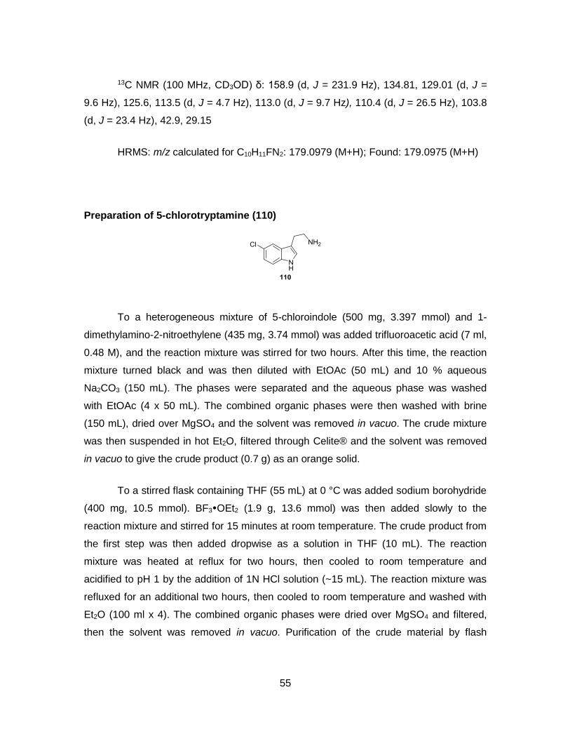
55
13C NMR (100 MHz, CD3OD) δ: 158.9 (d, J = 231.9 Hz), 134.81, 129.01 (d, J =
9.6 Hz), 125.6, 113.5 (d, J = 4.7 Hz), 113.0 (d, J = 9.7 Hz), 110.4 (d, J = 26.5 Hz), 103.8
(d, J = 23.4 Hz), 42.9, 29.15
HRMS: m/z calculated for C10H11FN2: 179.0979 (M+H); Found: 179.0975 (M+H)
Preparation of 5-chlorotryptamine (110)
To a heterogeneous mixture of 5-chloroindole (500 mg, 3.397 mmol) and 1-
dimethylamino-2-nitroethylene (435 mg, 3.74 mmol) was added trifluoroacetic acid (7 ml,
0.48 M), and the reaction mixture was stirred for two hours. After this time, the reaction
mixture turned black and was then diluted with EtOAc (50 mL) and 10 % aqueous
Na2CO3 (150 mL). The phases were separated and the aqueous phase was washed
with EtOAc (4 x 50 mL). The combined organic phases were then washed with brine
(150 mL), dried over MgSO4 and the solvent was removed in vacuo. The crude mixture
was then suspended in hot Et2O, filtered through Celite® and the solvent was removed
in vacuo to give the crude product (0.7 g) as an orange solid.
To a stirred flask containing THF (55 mL) at 0 °C was added sodium borohydride
(400 mg, 10.5 mmol). BF3OEt2 (1.9 g, 13.6 mmol) was then added slowly to the
reaction mixture and stirred for 15 minutes at room temperature. The crude product from
the first step was then added dropwise as a solution in THF (10 mL). The reaction
mixture was heated at reflux for two hours, then cooled to room temperature and
acidified to pH 1 by the addition of 1N HCl solution (~15 mL). The reaction mixture was
refluxed for an additional two hours, then cooled to room temperature and washed with
Et2O (100 ml x 4). The combined organic phases were dried over MgSO4 and filtered,
then the solvent was removed in vacuo. Purification of the crude material by flash

56
chromatography (10% ammonia solution (7N in MeOH), 90% CH2Cl2) to afford 5-
chlorotryptamine (110, 0.21 g, 32 %) as a brown solid.
1H NMR (600 MHz, CD3OD) δ: 7.52 (dd, J = 0.5, 2.0 Hz, 1H), 7.30 (dd, J =0.5,
8.6 Hz, 1H), 7.12 (s, 1H), 7.05 (dd, J = 2.0, 8.6 Hz, 1H), 2.92 (dt, J = 1.6, 7.1 Hz, 2H),
2.85 (dt, J = 1.6, 7.1 Hz, 2H)
13C NMR (150 MHz, CD3OD) δ: 136.6, 129.8, 125.4, 125.3, 122.5, 118.7, 113.5,
113.2, 42.9, 29.0
HRMS: m/z calculated for C10H11ClN2: 195.0684 (M+H); Found: 195.0682 (M+H)
Preparation of 5-bromotryptamine (111)
5-bromotrytapmine hydrochloride salt (1.0 g, 3.6 mmol) was dissolved in
water (30 mL) and basified with 15% NaOH in water (7 mL). The aqueous phase was
extracted with dichloromethane (20 ml x 3). The combined organic phases were dried
over MgSO4 and filtered, then the solvent was removed in vacuo to afford 5-
bromotryptamine (111) as a brown solid (0.77 g, 3.2 mmol, 89 %)
1H NMR (600 MHz, CD3OD) δ: 7.68 (dd, J = 1.7 Hz, 1H), 7.25 (dd, J = 8.6 Hz,
1H), 7.17 (dd, J = 1.8, 8.6 Hz, 1H), 7.10 (s, 1H), 2.91 (t, J = 6.6 Hz, 2H), 2.85 (t, J = 6.6
Hz, 2H)
13C NMR (150 MHz, CD3OD) δ: 136.8, 130.6, 125.1, 125.0, 121.9, 113.9, 113.2,
112.8, 43.0, 29.2
HRMS: m/z calculated for C10H11BrN2: 239.0178, 241.0158 (M+H); Found:
239.0182, 241.0163 (M+H)

57
Preparation of carbamate 142
To a stirred solution of tryptamine (88, 1.0 g, 6.2 mmol) in CH2Cl2 (16 mL) was
added triethylamine (0.87 mL, 12 mmol) then cooled to 0 °C. Ethyl chloroformate (0.62
mL, 6.5 mmol) was then added dropwise then the reaction mixture was warmed to room
temperature then diluted with H2O (25 mL) and the phases were separated. The organic
phase was washed with 1N HCl (15 mL), 5% aqueous NaHCO3 (15 mL), H2O (15 mL)
and brine (15 mL) then dried (MgSO4) and filtered and the solvent removed in vacuo.
Purification of the crude material by flash chromatography (EtOAc-hexanes 30:70)
provided carbamate 142. (1.35 g, 5.8 mmol, 93%) as a yellow oil.
1H NMR (400 MHz, CD3OD) δ: 7.56 (d, J = 7.9 Hz, 1H), 7.32 (dt, J = 0.8, 8.1 Hz,
1H), 7.08 (dt, J = 1.0, 7.1 Hz, 1H), 7.04 (s, 1H), 6.99 (dt, J = 1.0, 7.1 Hz, 1H), 4.05 (q, J =
7.1 Hz, 2H), 3.38 (m, 2H), 2.91 (t, J = 7.5 Hz, 2H), 1.21 (t, J = 7.1 Hz, 3H)
13C NMR (150 MHz, CD3OD) δ: 159.2, 138.1, 128.8, 123.3, 122.2, 119.5, 119.3,
113.3, 112.2, 61.6, 42.8, 26.8, 15.0
HRMS: m/z calculated for C13H16N2O2: 255.1104 (M+Na); Found: 255.1103
(M+Na)
Preparation of 5-fluorocarbamate 103

58
To a stirred solution of 5-fluorotryptamine (108, 180 mg, 1.0 mmol) in CH2Cl2 (10
mL) was added triethylamine (0.15 mL, 1.1 mmol) then cooled to 0 °C. Ethyl
chloroformate (0.10 mL, 1.1 mmol) was then added dropwise then the reaction mixture
was warmed to room temperature then diluted with H2O (15 mL) and the phases were
separated. The organic phase was washed with 1N HCl (15 mL), 5% aqueous NaHCO3
(15 mL), H2O (15 mL) and brine (15 mL) then dried (MgSO4) and filtered and the solvent
removed in vacuo. Purification of the crude material by flash chromatography (EtOAc-
hexanes 30:70) provided 5-fluorocarbamate 103. (0.24 g, 0.97 mmol, 97%) as a yellow
oil.
1H NMR (500 MHz, CD3OD) δ: 7.56 (d, J = 7.9 Hz, 1H), 7.32 (dt, J = 0.8, 8.1 Hz,
1H), 7.08 (dt, J = 1.0, 7.1 Hz, 1H), 7.04 (s, 1H), 6.99 (dt, J = 1.0, 7.1 Hz, 1H), 4.05 (q, J =
7.1 Hz, 2H), 3.38 (m, 2H), 2.91 (t, J = 7.5 Hz, 2H), 1.21 (t, J = 7.1 Hz, 3H)
13C NMR (125 MHz, CD3OD) δ: 159.3, 158.8 (d, J = 231.9 Hz), 134.6, 129.1 (d, J
= 9.4 Hz), 125.4, 113.6 (d, J = 5.0 Hz), 112.9 (d, J = 9.7 Hz), 110.3 (d, J = 26.5 Hz),
103.9 (d, J = 23.4 Hz), 61.6, 42.8, 26.8, 15.0
HRMS: m/z calculated for C13H15N2FO2: 273.1010 (M+Na); Found: 273.1009
(M+Na)
Preparation of 5-bromocarbamate 143
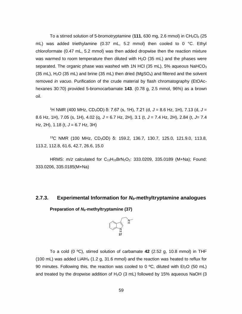
59
To a stirred solution of 5-bromotryptamine (111, 630 mg, 2.6 mmol) in CH2Cl2 (25
mL) was added triethylamine (0.37 mL, 5.2 mmol) then cooled to 0 °C. Ethyl
chloroformate (0.47 mL, 5.2 mmol) was then added dropwise then the reaction mixture
was warmed to room temperature then diluted with H2O (35 mL) and the phases were
separated. The organic phase was washed with 1N HCl (35 mL), 5% aqueous NaHCO3
(35 mL), H2O (35 mL) and brine (35 mL) then dried (MgSO4) and filtered and the solvent
removed in vacuo. Purification of the crude material by flash chromatography (EtOAc-
hexanes 30:70) provided 5-bromocarbamate 143. (0.78 g, 2.5 mmol, 96%) as a brown
oil.
1H NMR (400 MHz, CD3OD) δ: 7.67 (s, 1H), 7.21 (d, J = 8.6 Hz, 1H), 7.13 (d, J =
8.6 Hz, 1H), 7.05 (s, 1H), 4.02 (q, J = 6.7 Hz, 2H), 3.1 (t, J = 7.4 Hz, 2H), 2.84 (t, J= 7.4
Hz, 2H), 1.18 (t, J = 6.7 Hz, 3H)
13C NMR (100 MHz, CD3OD) δ: 159.2, 136.7, 130.7, 125.0, 121.9.0, 113.8,
113.2, 112.8, 61.6, 42.7, 26.6, 15.0
HRMS: m/z calculated for C13H15BrN2O2: 333.0209, 335.0189 (M+Na); Found:
333.0206, 335.0185(M+Na)
2.7.3. Experimental Information for Nb-methyltryptamine analogues
Preparation of Nb-methyltryptamine (37)
To a cold (0 ºC), stirred solution of carbamate 42 (2.52 g, 10.8 mmol) in THF
(100 mL) was added LiAlH4 (1.2 g, 31.6 mmol) and the reaction was heated to reflux for
90 minutes. Following this, the reaction was cooled to 0 ºC, diluted with Et2O (50 mL)
and treated by the dropwise addition of H2O (3 mL) followed by 15% aqueous NaOH (3

60
mL) and a further addition of H2O (9 mL). MgSO4 was then added and the reaction
mixture was stirred for 15 minutes then filtered and the solvent was removed in vacuo to
afford Nb-methyltryptamine (7) as a yellow solid (1.58 g, 9.1 mmol, 84%)
1H NMR (400 MHz, CD3OD) δ: 7.53 (td, J = 0.9, 8.0 Hz, 1H), 7.32 (td, J =0.5, 8.1
Hz, 1H), 7.08 (ddd, J = 1.1, 7.1, 8.1 Hz, 1H), 7.03 (s, 1H), 6.99 (ddd, J = 1.1, 7.1, 8.1 Hz,
1H), 2.91 (m, 2H), 2.81 (m, 2H), 2.33 (s, 3H)
13C NMR (150 MHz, CD3OD) δ: 138.2, 128.6, 123.4, 122.4, 119.6, 119.2, 113.4,
112.3, 53.0, 35.9, 26.0
HRMS: m/z calculated for C11H14N2: 175.1230 (M+H); Found: 175.1236 (M+H)
Preparation of D3-Nb-methyltryptamine (112)
To a cold (0 ºC), stirred solution of carbamate 42 (0.65 g, 2.8 mmol) in THF (50
mL) was added LiAlD4 (0.46 g, 2.8 mmol) and the reaction was heated to reflux for 16
hours. Following this, the reaction was cooled to 0 ºC, diluted with Et2O (50 mL) and
treated by the dropwise addition of H2O (0.5 mL) followed by 15% aqueous NaOH (0.5
mL) and a further addition of H2O (1.5 mL). MgSO4 was then added and the reaction
mixture was stirred for 15 minutes then filtered and the solvent was removed in vacuo.
Purification of the crude material by flash chromatography (10% ammonia solution (7N in
MeOH), 90% CH2Cl2) to afford D3-Nb-methyltryptamine (112) as a yellow solid (0.22 g,
1.2 mmol, 44%).
1H NMR (400 MHz, CD3OD) δ: 7.55 (td, J = 0.9, 8.0 Hz, 1H), 7.33 (td, J =0.5, 8.1
Hz, 1H), 7.09 (ddd, J = 1.1, 7.1, 8.1 Hz, 1H), 7.08 (s, 1H), 7.00 (ddd, J = 1.1, 7.1, 8.1 Hz,
1H), 2.98 (m, 2H), 2.92 (m, 2H)

61
13C NMR (150 MHz, CD3OD) δ: 138.3, 128.6, 123.5, 122.4, 119.7, 119.2, 112.9,
112.3, 52.6, 34.8 (m), 25.6
HRMS: m/z calculated for C11H11D3N2: 178.1418 (M+H); Found: 178.1420 (M+H)
Preparation of 5-fluoro-Nb-methyltryptamine (113)
To a cold (0 ºC), stirred solution of 5-fluorocarbamate 103 (0.10 g, 0.40 mmol) in
THF (10 mL) was added LiAlH4 (46 mg, 1.2 mmol) and the reaction was heated to reflux
for two hours. Following this, the reaction was cooled to 0 ºC, diluted with Et2O (25 mL)
and treated by the dropwise addition of H2O (50 L) followed by 15% aqueous NaOH
(50 L) and a further addition of H2O (150 L). MgSO4 was then added and the reaction
mixture was stirred for 15 minutes then filtered and the solvent was removed in vacuo to
afford 5-fluoro-Nb-methyltryptamine (113) as a brown oil (81.9 mg, 9.1 mmol, 84%)
1H NMR (500 MHz, CD3OD) δ: 7.28 (dd, J =4.4, 8.7 Hz, 1H), 7.21 (dd, J =2.4, 9.9
Hz, 1H), 7.12 (s, 1H), 6.85 (dt, J = 2.6, 9.2 Hz, 1H), 2.90 (m, 2H), 2.85 (m, 2H), 2.4 (s,
3H)
13C NMR (125 MHz, CD3OD) δ: 158.8 (d, J = 231.9 Hz), 134.8, 128.9 (d, J = 9.5
Hz), 125.5, 113.4 (d, J = 4.6 Hz), 113.0 (d, J = 9.6 Hz), 110.4 (d, J = 26.4 Hz), 103.8 (d,
J = 23.5 Hz), 52.7, 35.8, 25.7
HRMS: m/z calculated for C11H14FN2: 193.1136 (M+H); Found: 193.1136 (M+H)
Preparation of 5-fluoro-D3-Nb-methyltryptamine (114)

62
To a cold (0 ºC), stirred solution of 5-fluorocarbamate 103 (0.10 g, 0.40 mmol) in
THF (10 mL) was added LiAlD4 (0.10 g, 2.4 mmol) and the reaction was heated to reflux
for 16 hours. Following this, the reaction was cooled to 0 ºC, diluted with Et2O (25 mL)
and treated by the dropwise addition of H2O (100 L) followed by 15% aqueous NaOH
(100 L) and a further addition of H2O (300 L). MgSO4 was then added and the reaction
mixture was stirred for 15 minutes then filtered and the solvent was removed in vacuo.
Purification of the crude material by flash chromatography (10% ammonia solution (7N in
MeOH), 90% CH2Cl2) to afford 5-fluoro-D3-Nb-methyltryptamine (114) as a brown solid
(0.22 g, 1.2 mmol, 44%).
1H NMR (600 MHz, CD3OD) δ: 7.28 (dd, J =4.4, 8.7 Hz, 1H), 7.21 (dd, J =2.4, 9.9
Hz, 1H), 7.11 (s, 1H), 6.85 (dt, J = 2.5, 9.1 Hz, 1H), 2.90 (m, 2H), 2.83 (m, 2H)
13C NMR (150 MHz, CD3OD) δ: 158.8 (d, J = 231.9 Hz), 134.8, 128.9 (d, J = 9.5
Hz), 125.5, 113.5 (d, J = 4.7 Hz), 113.0 (d, J = 9.6 Hz), 110.4 (d, J = 26.5 Hz), 103.8 (d,
J = 23.4 Hz), 52.7, 35 (hept, J = 20.5 Hz), 25.8
HRMS: m/z calculated for C11H14D3FN2: 196.1324 (M+H); Found: 196.1326
(M+H)
Preparation of 5-bromo-Nb-methyltryptamine (115)
To a cold (0 ºC), stirred solution of 5-bromocarbamate 143 (0.81 g, 2.6 mmol) in
THF (25 mL) was added LiAlH4 (380 mg, 10 mmol) and the reaction was heated to reflux

63
for two hours. Following this, the reaction was cooled to 0 ºC, diluted with Et2O (30 mL)
and treated by the dropwise addition of H2O (0.4 mL) followed by 15% aqueous NaOH
(0.4 mL) and a further addition of H2O (1.2 mL). MgSO4 was then added and the reaction
mixture was stirred for 15 minutes then filtered and the solvent was removed in vacuo to
afford 5-bromo-Nb-methyltryptamine (115) as a brown oil (650 mg, 2.5, 99%)
1H NMR (400 MHz, CD3OD) δ: 7.68 (d, J = 1.7 Hz, 1H), 7.25 (d, J = 8.6 Hz, 1H),
7.16 (dd, J = 1.7, 8.6 Hz, 1H), 7.09 (s, 1H), 2.90 (m, 2H), 2.83 (m, 2H), 2.34 (s, 3H)
13C NMR (100 MHz, CD3OD) δ: 136.8, 130.5, 125.1, 125.0, 121.8, 113.9 113.2,
112.8, 52.9, 35.9, 25.7
HRMS: m/z calculated for C11H14BrN2: 253.0335, 255.0315 (M+H); Found:
253.0338, 255.0318 (M+H)
2.7.4. Synthetic Standards of D6-chimonanthines
Preparation of D6-chimonanthine 144 and 145
This procedure was adapted from Takayama’s chimonanthine synthesis.
To a cold (-30 ºC), stirred solution of carbamate 142 (1.5 g, 6.35 mmol) in
trifluoroethanol (8 mL) was added [bis(trifluoroacetoxy)iodo]benzene (1.8 g, 4.1 mmol)
was added over 3 hours by the addition of 0.3 g portions every 30 minutes, and the
reaction was stirred at -30 ºC for two hours. Following this, the reaction was warmed to
room temperature, and the solvent was removed in vacuo. The crude mixture was
dissolved in freshly distilled THF (60 mL), and the reaction mixture was cooled to 0 ºC.
To a cold (0 ºC), stirred solution was added LiAlD4 (0.80 g, 19 mmol) and the reaction
was heated to reflux for 16 hours. Following this the reaction was cooled to 0 ºC, diluted
with Et2O (100 mL) and treated by the dropwise addition of H2O (0.8 mL) followed by
15% aqueous NaOH (0.8 mL) and a further addition of H2O (2.4 mL). MgSO4 was then
added and the reaction mixture was stirred for 15 minutes then filtered and the solvent
was removed in vacuo. Purification of the crude material by flash chromatography (10%

64
ammonia solution (7N in MeOH), 90% CH2Cl2) to afford D6-rac-144 (5 mg, 1%) and D6-
meso-155 (trace)
1H NMR (600 MHz, CD3OD) δ: 7.23 (broad s, 2H), 6.98 (broad s, 2H), 6.63
(broad s, 2H), 6.54 (broad s, 2H), 2.70-2.09 (m, 10H)
2H NMR (600 MHz, CH2Cl2) δ: 2.39 (s)
HRMS: m/z calculated for C22H20D6N4: 353.2607 (M+H); Found: 353.2606 (M+H)
1H NMR (600 MHz, CD3Cl) δ: 7.04 (m, 4H), 6.49 (d, J = 7.9 Hz, 4H), 6.63 (broad
s, 2H), 2.60-2.35 (m, 6H), 2.06 (m, 2H)
HRMS: m/z calculated for C22H20D6N4: 353.2607 (M+H); Found: 353.2604 (M+H)

65
2.7.5. Feeding Precursors in planta and Screening Precursors by LC-MS
Fresh stems with several leaves were collected from Chimonanthus praecox,
then the tips of stems were placed to the solutions of each synthetic precursor or two-
precursor mixed (106-115, 5 mM, H2O-DMSO 90:10, 10 mL, pH 4-5, see Table 2.1) and
incubated in greenhouse overnight. Then, a leaf from each solution were collected, and
the leaf was cut into small pieces individually. The small pieces were placed in MeOH
(0.5 mL) in 1.5 mL Eppendorf® tubes, then, the samples were sonicated for one hour.
The green methanol solutions was filtered and submitted to LC-MS.
LC-MS was run over 10 minutes with the eluent system (Table 2.7). MS was
calibrated with internal standard, sodium formate. Then, the LC-MS chromatograms
performed EIC analysis at m/z of target unnatural products.
Table 2.7. LC methods on LC-MS analysis
Time (min) Solvent: A (%) Solvent: B (%)
0 90 10
1 90 10
8 40 60
9 0 100
10 0 100
10.1 90 10
12 90 10
Solvent A: H2O with 0.1 % formic acid. Solvent B: acetonitrile with 0.1 % formic acid. Column temperature: 30 ºC. Initial flow rate: 0.3 mL/min, ramp to 0.5 mL/min in 8min and Hold till 12 min. Total run time: 12 min
2.7.6. Isolation of Fluorinated Unnatural Products
The scale-up experiments commenced with the feeding of 50 mg of 5-
fluorotryptamine (109) to 20 g of fresh leaves of Chimonanthus praecox over 5 days.
After 5 days, the leaves were collected and cut into small pieces. The small pieces of
leaves were suspended in MeOH (250 mL) and stirred overnight to extract alkaloids,
then filtered. The solvent was removed in vacuo, and the plant extracts were dissolved in
chloroform (75 mL). The mixture was acidified with 1M HCl (75 mL), and the phases
were separated. The organic phase was washed with 15% NaOH (75 mL), then the

66
phases were separated again. The combined organic phases were then washed with
brine (100 mL), dried over MgSO4 and the solvent was removed in vacuo to afford brown
plant extracts (40 mg).
The plant extracts were dissolved in methanol (1 mL) and placed in fridge
overnight, then filtered to remove solid impurities. The filtered sample was submitted to
HPLC to purify the alkaloids. After HPLC isolation, each fraction was submitted to LC-
MS to monitor if the desired m/z of 383.2042 was present or not. The fractions that
contained the desired m/z of 383.2042 were collected and concentrated under vacuum
to afford 128 (~0.7 mg) and 129 (~0.05 mg).
Table 2.8. HPLC eluent system to isolate difluorochimonanthines
Time (min) Solvent: A (%) Solvent: B (%)
0 70 30
6 15 85
8 15 85
8.5 70 30
10 70 30
Solvent A: H2O with 0.1 % formic acid, Solvent B: methanol with 0.1 % formic acid. Injection volume 200 µL.
Characterization of isolated alkaloid (-)-128
1H NMR (600 MHz, CD3Cl) δ: 6.86 (broad s, 2H), 6.66 (broad s, 2H), 6.33 (broad
s, 2H), 2.64-2.44 (m, 8H), 2.34 (s, 6H), 2.07 (broad s, 2H)
13C NMR (150 MHz, CD3Cl) δ: 157.1 (d, J = 239.7 Hz), 146.7, 135.1 (d, J = 7.2
Hz), 129.9, 114.5 (d, J = 22 Hz), 110.0, 86.1, 53.6, 52.6, 37.3, 35.8

67
HRMS: m/z calculated for C22H24F2N4: 383.2042 (M+H); Found: 383.2049 (M+H)
[α]D20: -35 (c: 0.06, CHCl3)
Characterization of isolated alkaloid meso-129
1H NMR (600 MHz, (CD3)2SO) δ: 6.70 (m, 2H), 6.34 (d, J = 0.5, 8.5 Hz, 2H), 6.33
(d, J = 8.5 Hz, 2H), 2.28 (m, 10H), 2.11 (s, 6H)
HRMS: m/z calculated for C22H24F2N4: 383.2042 (M+H); Found: 383.2038 (M+H)
2.7.7. Plant Protein Extraction with P-PERTM Plant Protein Extraction Kit and in vitro Assay
A small leaf (~80 mg) was collected from Chimonanthus praecox, and protein
extraction procedures were followed as described in P-PERTM Plant Protein Extraction
Kit. 100 L of protein samples were used in the in vitro assay in combination with
precursor analogues in the following reaction buffer system (1mL, 1 mM precursor, 0.1
mM cofactor as indicated in Table 2.4).
Reaction Buffer
Buffer A: PBS
Buffer B: 0.2 M Tris-HCl

68
After overnight incubation at the specified temperatures, the samples were
filtered in 3K Amicon® centrifugation tube, and the filtrate was submitted for LC-MS
analysis.
2.7.8. Plant Protein Extraction from Leaves Using Blender and in vitro Assay
Fresh leaves (~20-50 g) were collected from Chimonanthus praecox. These
leaves were frozen and then ground into a powder using a mortar and pestle cooled in
liquid nitrogen. The powdered leaves were then placed in a blender (Oster® ) and cold (4
ºC) extraction buffer (200 mL) and protease inhibitor (Roche® cOmplete, Mini, one
tablet) were added in a cold (4 ºC) room. The blender was run for 15 seconds and this
blending was repeated five times. After the blending, the homogenized samples were
filtered through 4-layers of cheese cloth, then centrifuged at 14,000 rpm for 30 minutes
at 4 °C to remove cellular debris and large cellular components. The supernatant was
concentrated by using Amicon® centrifugal tubes as described above and the
concentrated supernatant sample was then transferred into pre-cooled 2 mL Eppendorf
tubes.
The collected protein samples were then added into the reaction buffers specified
below and precursor analogues 112 or 113 (1 mM). After overnight incubation at the
specified temperatures, the samples were filtered through 3K Amicon® centrifugal tubes,
and finally subjected to LC-MS analysis.
Table 2.9. Extraction buffers and reaction buffers used in in vitro assay
Entry Extraction Buffer Reaction Buffer
1 PBS PBS
2 0.2 M Tris-HCl 0.2 M Tris-HCl
3 PBS, 1 mM PEG, 5% (w/v) PVPP, 0.01% Triton X-100
PBS
4 25 mM Hepes-Na, 0.5 mM EDTA, DTT, 8 mM MgCl2
25 mM Hepes-Na, 8 mM MgCl2
5 50 mM Tris-HCl, 10 mM DTT, 0.5 M sucrose.
50 mM Tris-HCl, 0.5 M sucrose.
6 0.45 M Mannitol, 50 mM sodium phosphate, 2 mM EDTA
1 M Mannitol, 0.2 M sodium phosphate

69
2.7.9. Plant Protein Extraction From Roots Using Blender and in vitro Assay [83]
Fresh roots (~100 g) were collected from Chimonanthus praecox. The roots were
washed thoroughly with distilled water (1 L X 5) to remove any residual soil. These roots
were ground to provide a powder using a mortar and pestle cooled with liquid nitrogen.
The powdered roots were placed in a blender (Oster® ) and cooled (4 ºC) extraction
buffer (500m L) and protease inhibitor (Roche® cOmplete, Mini, 1 tablet) were added to
the sample in a cold (4 ºC) room. The blender was run for 15 seconds intervals and
repeated 5 times. After blending, the homogenized samples were filtered through 4-
layers of cheesecloth, then centrifuged at 14,000 rpm for 30 minutes at 4 ºC to remove
cellular debris and large cellular components. The supernatants were then collected and
subjected to ammonium sulfate precipitation to separate and concentrate protein
samples.
2.7.10. Root protein separation by Ammonium Sulfate Precipitation
To a beaker of plant protein supernatant sample, in a cold (4 ºC) room, was
slowly added ammonium sulfate to obtain a 20% solution, which was then stirred for 12
hours. After this time the sample was centrifuged at 14,000 rpm for one hour at 4 ºC and
the solids were collected. More ammonium sulfate was then added to the filtrate to
increase the concentration to 40% and the resulting mixture was stirred for a further 12
hours and then centrifuged at 14,000 rpm for one hour at 4 ºC and the solids were
collected. This procedure was repeated with ammonium sulfate concentrations of 60%,
80%, and 95% to afford the ammonium sulfate plant protein fractions.
Each solid fraction was later dissolved in sodium acetate buffer (10 mL, 50 mM,
pH 4.5), and the ammonium sulfate was removed by dialysis against this same buffer
over 16 hours. The protein solution was then centrifuged at 15,000 for 30 minutes to
remove residual solids and afford a solution of the protein extracts.
70
2.7.11. Preparation of Protoplasts [84]
Fresh leaves (~3-5 g) were collected from Chimonanthus praecox, and they were
cut into 0.5-1 mm strips using a razor blade. The short strips were placed in digestive
enzyme solution (20 mM MES (pH 5.7) containing 1.5% (w/v) cellulose R10, 0.4 %
(wt/vol) macrozyme R10, 0.4 M mannitol and 20 mM KCl) in a 50 mL Erlenmeyer flask.
The solution was kept in the dark by wrapping the flask with aluminium foil and the
solution was gently shaken (100 rpm) for 4 hours at room temperature. The solution was
then transferred into a 15 mL Falcon® centrifuge tube and centrifuged at 2,000 g. The
solids were collected and washed with washing buffer (0.5 M mannitol, 4 mM MES-KOH,
pH 5.5, 20 mM KCl, and 3 x 20 mL). The washed protoplasts were directly used for
assays and microscopy.
2.7.12. In Vitro Assay with Protoplasts
The collected protoplasts (~0.5 g) were suspended in PBS (1.5 mL) in a 2 mL
Eppendorf® tube. Then 5-fluorotryptamine (108, 20 L, 0.2 M) was added and the
mixture was incubated at 30 ºC overnight. MeOH (0.5 mL) was added after which the
sample was sonicated for one hour. The sample was filtered using a 3K Amicon®
centrifugal tube and then subjected to analysis by LC-MS.
2.7.13. Preparation of Cell Wall[88]
Leaves of Chimonanthus praecox were collected (15 g), frozen in liquid nitrogen
and ground into powder using a cooled mortar and pestle. The resulting green powder
was transferred into a 500 mL-beaker containing potassium phosphate buffer (250 mL,
50 mM, and pH 7.0) with 1 % (v/v) of Triton X-100 and stirred for 4 hours at 4 ºC. The
mixture was then filtered through one layer of cheesecloth, the insoluble materials were
then washed with cold distilled water (1 L) and squeezed in the cheesecloth to remove
any residual water. The insoluble materials were washed overnight with 0.5 M of NaCl
(250 mL) and filtered through one layer of cheesecloth. The insoluble materials were
washed with cold distilled water (1 L) and squeezed in cheesecloth to remove residual
water. 4 g of the cell wall materials were collected in this manner.

71
2.7.14. Preparation of Cell Wall proteins[89]
25 g of leaves were collected and frozen in liquid nitrogen. They were then
ground using a cooled mortar and pestle into a fine green powder. The green powder
was homogenized in 0.1 M potassium phosphate buffer (pH 7.0, 300 mL) containing 5
mM DTT and then filtered through four-layers of cheese cloth. The insoluble materials
were collected, and shaken sequentially with pre-chilled (-20 ºC) acetone (3 x 300 mL,
30 min each), solution A (0.1 M potassium phosphate buffer (pH 6.5) with 0.1 % beta-
mercaptoethanol, 4 ºC 300 mL, 30 min), solution A containing 1% (v/v) Triton X-100 (4
ºC, 300 mL, 4 hours), and finally with solution A to remove remaining detergent (4 ºC,
300 mL, 16 hours). Between each procedure, the homogenate was filtered through 4
layers of cheesecloth and the insoluble materials were collected.
The collected insoluble materials (pale yellow) were placed into a cooled (4 ºC)
solution of 200 mL of 1 M NaCl solution (4 hours) and stirred with a magnetic stir bar to
extract the proteins from the insoluble residues. The homogenate was decanted and
filtered through 4-layers of cheesecloth and the yellow solution (protein sample) was
concentrated using 15 mL 10K Amicon® centrifugation tubes (4000 g, 45 mins) and then
concentrated again using 0.5 mL 10K Amicon® centrifugation tubes (14,000 g, 45 mins).
The protein concentration of the resulting samples was determined by Bradford assay
(BioRad), using BSA as a protein standard, to afford 200 µL of yellow protein sample
(1.8 mg / mL).
The protein extracts (0, 15, 30, 45, and 60 µL) were assayed in a solution of 0.1
M potassium phosphate (pH 7) containing 5 mM of 5-fluoro-Nb-methyltryptamine and 2.5
µL of ammonium persulfate in 250 µL overnight at 30 ºC. After the incubation the
samples were filtered through 3K Amicon® centrifugation tubes and the filtrates were
submitted for analysis by LC-MS.
72
References
[1] B. Jones, R. J. Kazlauskas, Nat. Chem. 2014, 7, 11–12.
[2] C. E. Hornsby, R. S. Paton, Nat. Chem. 2014, 6, 88–9.
[3] J. M. Winter, Y. Tang, Curr. Opin. Biotechnol. 2012, 23, 736–743.
[4] N. Tibrewal, Y. Tang, Annu. Rev. Chem. Biomol. Eng. 2014, 5, 347–66.
[5] B. Shen, Curr. Opin. Chem. Biol. 2003, 7, 285–295.
[6] J. Staunton, K. J. Weissman, Nat. Prod. Rep. 2001, 18, 380–416.
[7] L. Katz, S. Donadio, Annu. Rev. Microbiol. 1993, 47, 875–912.
[8] D. a. Hansen, C. M. Rath, E. B. Eisman, A. R. H. Narayan, J. D. Kittendorf, J. D. Mortison, Y. J. Yoon, D. H. Sherman, J. Am. Chem. Soc. 2013, 135, 11232–11238.
[9] M. Hesse, Alkaloids: Nature’s Curse or Blessing?, Wiley-VCH, 2002.
[10] R. Schemitz, Pharm. Hist. 1985, 27, 61–74.
[11] E. J. Dimise, S. D. Bruner, Nat. Chem. Biol. 2010, 6, 251–252.
[12] J. M. Hagel, P. J. Facchini, Nat. Chem. Biol. 2010, 6, 273–275.
[13] J. Ziegler, P. J. Facchini, Annu. Rev. Plant Biol. 2008, 59, 735–769.
[14] H. W. Wiley, Am. Chem. J. 1889, 11, 557.
[15] R. G. Eccles, Proceeding Am. Pharm. Assoc. 1888, 382.
[16] H. M. Gordin, J. Am. Chem. Soc. 1905, 27, 144.
[17] H. M. Gordin, J. Am. Chem. Soc. 1909, 1305.
[18] E. Späth, W. Stroh, Berichte Der Dtsch. Chem. Gesellschaft 1925, 59, 2131.

73
[19] G. Barger, J. Madinaveitia, P. Streuli, J. Chem. Soc. 1939, 510–517.
[20] R. Woodwards, C. Yang, T. Katz, V. Clark, J. Harley-Mason, R. Ingleby, N. Sheppard, Proc. Chem. Soc. 1960, 76–78.
[21] T. A. Hamor, J. M. Robertson, H. N. Shrivastava, J. V. Silverton, Proc. Chem. Soc. 1960, 78–80.
[22] G. Barger, A. Jacob, J. Madinaveitia, Recl. des Trav. Chim. des Pays-Bas 1938, 57, 548–554.
[23] D. G. O’Donovan, M. F. Keogh, J. Chem. Soc. 1966, 1570–1572.
[24] T. R. Govindachari, K. Nagarajan, S. Rajappa, J. Sci. Industirial Res. 1962, 21, 455.
[25] H. F. Hodson, G. F. Smith, J. Chem. Soc. 1957, 1877–1880.
[26] G.-L. Ma, G.-X. Yang, J. Xiong, W.-L. Cheng, K.-J. Cheng, J.-F. Hu, Tetrahedron Lett. 2015, 56, 4071–4075.
[27] Y. Adjibade, H. Saad, T. Sevenet, B. Kuballa, J. C. Quirion, R. An ton, Planta Med. 1990, 56, 212–215.
[28] T. a Amador, L. Verotta, D. S. Nunes, E. Elisabetsky, Phytomedicine 2001, 8, 202–206.
[29] E. Elisabetsky, T. a. Arnador, R. R. Albuquerque, D. S. Nunes, a. Do CT Carvalho, J. Ethnopharmacol. 1995, 48, 77–83.
[30] V. Jannic, F. Guéritte, O. Laprévote, L. Serani, M. T. Martin, T. Sévenet, P. Potier, J. Nat. Prod. 1999, 62, 838–843.
[31] L. Verotta, T. Pilati, M. Tato, E. Elisabetsky, T. a. Amador, D. S. Nunes, J. Nat. Prod. 1998, 61, 392–396.
[32] L. Verotta, F. Peterlongo, E. Elisabetsky, T. a. Amador, D. S. Nunes, J. Chromatogr. A 1999, 841, 165–176.
[33] H. Hodson, B. Robinson, G. Smith, Proc. Chem. Soc. 1961, 465–466.
[34] I. J. Grant, T. A. Hamor, J. M. Robertson, G. . Sim, Proc. Chem. Soc. Chem. Soc. 1962, 148–149.
[35] T. Tokuyama, J. W. Daly, Tetrahedron 1983, 39, 41–47.

74
[36] Y. Adjibade, B. Weniger, J. Quirion, B. Kuballa, P. Cabalion, R. Anton, Phytochemistry 1992, 31, 317–319.
[37] L. Verotta, F. Orsini, M. Sbacchi, M. a. Scheildler, T. a. Amador, E. Elisabetsky, Bioorganic Med. Chem. 2002, 10, 2133–2142.
[38] K. Ramabadran, M. Bansinath, H. Turndorf, M. M. Puig, J. Pharmacol. Methods 1989, 21, 21–31.
[39] T. Morikawa, Y. Nakanishi, K. Ninomiya, H. Matsuda, S. Nakashima, H. Miki, Y. Miyashita, M. Yoshikawa, T. Hayakawa, O. Muraoka, J. Nat. Med. 2014, 68, 539–549.
[40] T. S. Chang, V. C. H. Lin, Int. J. Mol. Sci. 2011, 12, 8787–8796.
[41] J. M. Gillbro, M. J. Olsson, Int. J. Cosmet. Sci. 2011, 33, 210–221.
[42] J. B. Hendrickson, R. Rees, R. Göschke, Proc. Chem. Soc. 1962, 383–384.
[43] J. B. Hendrickson, R. Göschke, R. Rees, Tetrahedron 1964, 20, 565–579.
[44] A. . Scott, F. McCapra, E. S. Hall, J. Am. Chem. Soc. 1964, 86, 302–303.
[45] J. T. Link, L. E. Overman, J. Am. Chem. Soc. 1996, 118, 8166–8167.
[46] L. E. Overman, D. V. Paone, B. a. Stearns, J. Am. Chem. Soc. 1999, 121, 7702–7703.
[47] L. E. Overman, J. F. Larrow, B. a. Stearns, J. M. Vance, Angew. Chemie - Int. Ed. 2000, 39, 213–215.
[48] H. Ishikawa, H. Takayama, N. Aimi, Tetrahedron Lett. 2002, 43, 5637–5639.
[49] M. Movassaghi, M. a. Schmidt, Angew. Chemie - Int. Ed. 2007, 46, 3725–3728.
[50] H. Mitsunuma, M. Shibasaki, M. Kanai, S. Matsunaga, Angew. Chemie - Int. Ed. 2012, 51, 5217–5221.
[51] W. Xie, G. Jiang, H. Liu, J. Hu, X. Pan, H. Zhang, X. Wan, Y. Lai, D. Ma, Angew. Chemie - Int. Ed. 2013, 52, 12924–12927.
[52] S. Ghosh, S. Chaudhuri, A. Bisai, Org. Lett. 2015, 17, 1373–1376.
[53] K. Liang, X. Deng, X. Tong, D. Li, M. Ding, A. Zhou, C. Xia, Org. Lett. 2015, 17, 206–209.

75
[54] Y.-X. Li, H.-X. Wang, S. Ali, X.-F. Xia, Y.-M. Liang, Chem. Commun. 2012, 48, 2343.
[55] S. Tadano, Y. Mukaeda, H. Ishikawa, Angew. Chemie - Int. Ed. 2013, 52, 7990–7994.
[56] J. Kim, J. a Ashenhurst, M. Movassaghi, Science 2009, 324, 238–241.
[57] J. Kim, M. Movassaghi, J. Am. Chem. Soc. 2010, 132, 14376–14378.
[58] Y. Li, W.-H. Wang, S.-D. Yang, B.-J. Li, C. Feng, Z.-J. Shi, Chem. Commun. (Camb). 2010, 46, 4553–4555.
[59] S. Tadano, H. Ishikawa, Synlett 2014, 25, 0157.
[60] H. Ishikawa, Yakugaku Zasshi 2015, 135, 383–390.
[61] J. a. May, B. Stoltz, Tetrahedron 2006, 62, 5262–5271.
[62] G. W. Kirby, S. W. Shah, E. J. Herbert, J. Chem. Soc. Perkin 1 1969, 14, 1916–1919.
[63] E. J. Herbert, Studies in Alkaloids Biosyntheis, 1964.
[64] D. D. Songstad, V. De Luca, N. Brisson, W. G. Kurz, C. L. Nessler, Plant Physiol. 1990, 94, 1410–1413.
[65] J. C. Thomas, D. C. Adams, C. Nessler, J. K. Brown, H. J. Bohnert, Plant Physiol. 1995, 7–10.
[66] I. Islas, V. M. Loyola-Vargas, M. de Lourdes Miranda-Ham, Vitr. Cell. Dev. Biol. - Plant 1994, 30, 81–83.
[67] M. a Thompson, R. M. Weinshilboum, J. Biol. Chem. 1998, 273, 34502–34510.
[68] Y. H. Kim, Joo, Exp. Mol. Med. 2001, 33, 23–28.
[69] M. a Thompson, E. Moon, U. J. Kim, J. Xu, M. J. Siciliano, R. M. Weinshilboum, Genomics 1999, 61, 285–297.
[70] R. Thiericke, J. Rohr, Nat. Prod. Rep. 1993, 10, 265–289.
[71] J. Kennedy, Nat. Prod. Rep. 2008, 25, 25–34.
[72] B. R. Clark, S. O’Connor, D. Fox, J. Leroy, C. D. Murphy, Org. Biomol. Chem. 2011, 9, 6306–6311.

76
[73] S. Brown, S. E. O’Connor, ChemBioChem 2015, 16, 2129–2135.
[74] S. E. O’Connor, T. P. Brutnell, Curr. Opin. Plant Biol. 2014, 19, iv–v.
[75] S. Weist, R. D. Süssmuth, Appl. Microbiol. Biotechnol. 2005, 68, 141–150.
[76] C. Portmann, C. Prestinari, T. Myers, J. Scharte, K. Gadermann, ChemBioChem 2009, 10, 889–895.
[77] P. Espinet, A. C. Albéniz, J. a. Casares, J. M. Martínez-Ilarduya, Coord. Chem. Rev. 2008, 252, 2180–2208.
[78] O. Ritzeler, G. Klein, J.-L. Reymond, Phosphorus. Sulfur. Silicon Relat. Elem. 1999, 144, 243–246.
[79] S. R. Laplante, P. J. Edwards, L. D. Fader, A. Jakalian, O. Hucke, ChemMedChem 2011, 6, 505–513.
[80] G. H. Christie, J. Kenner, J. Chem. Soc. Trans. 1922, 121, 614–620.
[81] G. Bringmann, A. J. P. Mortimer, P. a. Keller, M. J. Gresser, J. Garner, M. Breuning, Angew. Chemie - Int. Ed. 2005, 44, 5384–5427.
[82] G. Bringmann, T. Gulder, T. a M. Gulder, M. Breuning, Chem. Rev. 2011, 111, 563–639.
[83] F. Hiibel, E. Beck, Plant Physiol. 1996, 112, 1429–1436.
[84] S.-D. Yoo, Y.-H. Cho, J. Sheen, Nat. Protoc. 2007, 2, 1565–1572.
[85] K. H. Caffall, D. Mohnen, Carbohydr. Res. 2009, 344, 1879–1900.
[86] D. J. Bradley, P. Kjellbom, C. J. Lamb, Cell 1992, 70, 21–30.
[87] J. E. Varner, S. Louis, Cell 1989, 56, 231–239.
[88] L. B. Davin, D. L. Bedgar, T. Katayama, N. G. Lewis, Phytochemistry 1992, 31, 3869–3874.
[89] L. B. Davin, H. B. Wang, A. L. Crowell, D. L. Bedgar, D. M. Martin, S. Sarkanen, N. G. Lewis, Science 1997, 275, 362–366.
[90] L. B. Davin, N. G. Lewis, Plant Physiol. 2000, 123, 453–462.
[91] V. Burlat, M. Kwon, L. B. Davin, N. G. Lewis, Phytochemistry 2001, 57, 883–897.

77
[92] L. B. Davin, N. G. Lewis, Curr. Opin. Biotechnol. 2005, 16, 398–406.
[93] K. W. Kim, S. G. A. Moinuddin, K. M. Atwell, M. A. Costa, L. B. Davin, N. G. Lewis, J. Biol. Chem. 2012, 287, 33957–33972.
[94] B. Pickel, M. A. Constantin, J. Pfannstiel, J. Conrad, U. Beifuss, A. Schaller, Angew. Chemie - Int. Ed. 2010, 49, 202–204.
[95] B. Pickel, A. Schaller, Appl. Microbiol. Biotechnol. 2013, 97, 8427–8438.
[96] K. S. Ham, S. C. Wu, A. G. Darvill, P. Albersheim, Plant J. 1997, 11, 169–179.
[97] G. a Rosenthal, Proc. Natl. Acad. Sci. U. S. A. 1992, 89, 1780–1784.
[98] Z. Minic, C. Rihouey, C. T. Do, P. Lerouge, L. Jouanin, Plant Physiol. 2004, 135, 867–878.
[99] H. Korthout, P. Van Der Hoeven, M. J. Wagner, E. Van Hunnik, a. H. De Boer, Plant Physiol. 1994, 105, 1281–1288.
[100] J. Rivoal, C. R. Smith, T. F. Moraes, D. H. Turpin, W. C. Plaxton, Anal. Biochem. 2002, 300, 94–99.
[101] D. H. Lee, C. B. Lee, Plant Sci. 2000, 159, 75–85.
[102] D. Wu, F. E. Regnier, Anal. Chem. 1993, 65, 2029–2035.
[103] R. R. Burgess, Protein Precipitation Techniques, Elsevier Inc., 2009.
[104] R. O. Musser, D. F. Cipollini, S. M. Hum-Musser, S. a. Williams, J. K. Brown, G. W. Felton, Arch. Insect Biochem. Physiol. 2005, 58, 128–137.
[105] a Ayala, J. Parrado, a Machado, Appl. Biochem. Biotechnol. 1998, 69, 11–16.
[106] A. Shevchenko, M. Wilm, O. Vorm, M. Mann, Anal. Chem. 1996, 68, 850–858.
[107] B. Domon, R. Aebersold, Science (80-. ). 2006, 312, 212–217.
[108] a J. Link, J. Eng, D. M. Schieltz, E. Carmack, G. J. Mize, D. R. Morris, B. M. Garvik, J. R. Yates, Nat. Biotechnol. 1999, 17, 676–682.
[109] B. Kozomara, B. Vinterhalter, L. Radojević, D. Vinterhalter, Vitr. Cell. Dev. Biol. - Plant 2008, 44, 142–147.
[110] W. Runguphan, J. J. Maresh, S. E. O’Connor, Proc. Natl. Acad. Sci. U. S. A. 2009, 106, 13673–13678.

78
[111] a Roach, K. Boylan, S. Horvath, S. B. Prusiner, L. E. Hood, Cell 1983, 34, 799–806.
[112] A. Nakagawa, H. Minami, J.-S. Kim, T. Koyanagi, T. Katayama, F. Sato, H. Kumagai, Nat. Commun. 2011, 2, 326.
[113] H. Minami, J.-S. Kim, N. Ikezawa, T. Takemura, T. Katayama, H. Kumagai, F. Sato, Proc. Natl. Acad. Sci. U. S. A. 2008, 105, 7393–7398.
[114] K. M. Hawkins, C. D. Smolke, Nat. Chem. Biol. 2008, 4, 564–573.

79
Appendix A. Total Synthesis of Ascospiroketal A Through a AgI-Promoted Cyclization Cascade
Reprinted (adapted) with permission from Angewandte Chemie International Edition. Copyright 2015 John Wiley and Sons.
Chang, S.; Hur, S.; Britton, R. Angew. Chem. Int. Ed. 2015, 54, 211

80

81
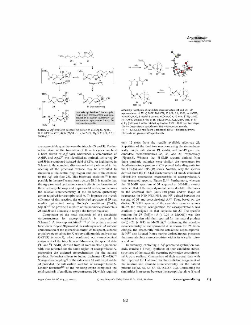
82
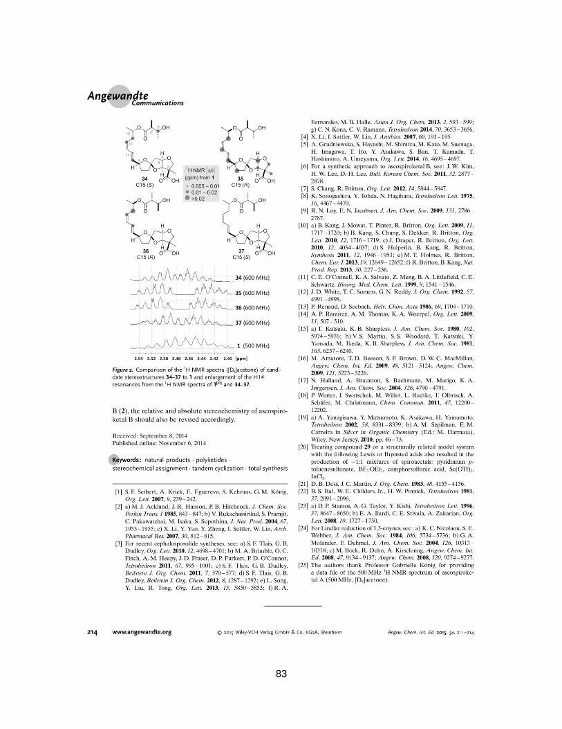
83