Studies on the practical production of … for use Title Studies on the practical production of...
141
Instructions for use Title Studies on the practical production of docosahexaenoic acid using thraustochytrid microorganisms Author(s) Bin Haji Mohd Taha, Ahmad Iskandar Citation 北海道大学. 博士(環境科学) 甲第11348号 Issue Date 2014-03-25 DOI 10.14943/doctoral.k11348 Doc URL http://hdl.handle.net/2115/58150 Type theses (doctoral) File Information Ahmad_Iskandar_Bin_Haji_Mohd_Taha.pdf Hokkaido University Collection of Scholarly and Academic Papers : HUSCAP
Transcript of Studies on the practical production of … for use Title Studies on the practical production of...
Instructions for use
Title Studies on the practical production of docosahexaenoic acid using thraustochytrid microorganisms
Author(s) Bin Haji Mohd Taha, Ahmad Iskandar
Citation 北海道大学. 博士(環境科学) 甲第11348号
Issue Date 2014-03-25
DOI 10.14943/doctoral.k11348
Doc URL http://hdl.handle.net/2115/58150
Type theses (doctoral)
File Information Ahmad_Iskandar_Bin_Haji_Mohd_Taha.pdf
Hokkaido University Collection of Scholarly and Academic Papers : HUSCAP
PhD Dissertation
Studies on the practical production of docosahexaenoic acid
using thraustochytrid microorganisms
Graduate School of Environmental Science
Hokkaido University
Ahmad Iskandar Bin Haji Mohd Taha
2014
I
Acknowledgement
Bismillahirahmanirrahim, al-hamdulillahi rabbilalamin, I would like to
take this opportunity to convey my highest appreciation first of all to my
supervisor, Professor Hidetoshi Okuyama for his kind attention, patience and
guidance for the past 3 years throughout my PhD research. He has given me
many opportunities to write in many publications, try various experiments, visit
various institutions for collaborative research and encouraged me to attend to so
many prestigious academic meetings. This will also not be possible without the
support from Hokkaido University, which has provided a great environment to
study and also the Japanese government for the scholarship through the Japan
Ministry of Education, Culture, Sports, Science and Technology (MEXT). I have
many plans to apply my experience, knowledge, skills and networks made during
my study, not just for the further development of my country but also to
contribute back to Japan as well. I also would like to record my sincere
appreciation to Dr. Mamiko Sato and Takako Kaneko from Japan Women
University for kindly teaching me the TEM cryofixation technique, professor
Yoshinobu Nodasaka and Natsumi Ushijima from the dental faculty of Hokkaido
University for training me on the transmission and scanning electron microscope
work, professor Tojo Motoaki from Osaka Prefectural University for his kind
collaboration in the mitochondrial DNA work, Dr. Naoki Morita from Advanced
Industrial Science and Technology Hokkaido (AIST) and Dr. Akio Ueno from
Horonobe Research Institute for the Subsurface Environment (H-RiSE) for their
kind collaboration in using their lab and equipment for experiments. I also would
like to say many thanks to my wonderful lab members and many good Japanese
friends in and out of the university who have helped me in so many ways during
my student life. A special thanks also goes to my dear friend Kak Widya, who has
been very kind and helpful throughout my stay in Sapporo. Finally I would like to
thank my wonderful parents Hj Mohd Taha Bin Mat Yassin and Hjh Latiffah Bte
Hj Abdullah, as well as my family and friends at home to whom my gratitude can
never be put simply into words for their endless support. I hope the findings and

II
writings from this work will be insightful and beneficial to other students and
biotechnologists for application and development of the bioindustry.
III
Abstract
Docosahexaenoic acid (DHA) is one of the essential fatty acids required for the
maintenance of good health. It is found in high amounts in the brain and retina as
part of the phospholipid component of neurons and therefore is necessary for
good functioning of the brain as well as in treating illnesses such as Alzheimer
disease and depression. As consumption of fish, which is a major source of DHA
in the diet, decreases because of the modern lifestyle, DHA can be consumed
directly as a food supplement. Fish oil is the main source of commercial DHA;
however, the industry is currently facing some problems such as decreasing fish
catch and accumulation of heavy metals because of marine pollution. Microbial
oil is a potential alternative to fish oil such as that from marine thraustochytrid
microorganisms. These microorganisms normally require seawater for
cultivation because of the requirement of NaCl for growth; however, the chloride
ion corrodes industrial equipment, thereby resulting in high maintenance costs.
This study focuses on the cultivation of thraustochytrid strain 12B without the
use of seawater by investigating the impact of major salts present in the
seawater to come up with a medium containing low NaCl that can produce cells
with high DHA yield. The taxonomic attributes of strain 12B were also studied in
relation to other thraustochytrids as well as in relation to DHA production. The
possible role of DHA accumulated in the thraustochytrid was also investigated.
NaCl in the culture medium was able to be reduced to 0.1% (w/v), and high cell
yield was obtained when MgSO4 was also supplemented in the culture medium.
The NM medium, which contains 0.1% (w/v) NaCl and 1% (w/v) MgSO4, was
found to produce a higher cell yield and DHA productivity compared with the
conventional seawater containing F medium, and cells grown in the NM medium
produced a 18.8 mg/ml cell yield with DHA productivity of 2.4 mg/ml/day,
compared with cells grown in the F medium, which produced a 11.4 mg/ml cell
yield and 0.7 mg/ml/day DHA, respectively. The low 0.1% (w/v) NaCl content in
the culture medium is very favorable, as it does not lead to the corrosion of metal
industrial equipment. Using inexpensive industrial-grade peptone, glucose and
yeast extract, production cost could be reduced by 30% compared with the F
IV
medium. The DHA yield was also increased in cells grown in the industrial-grade
medium (INM) compared with cells grown in the NM medium. Based on the
sequence analysis of the mitochondrial cytochrome oxidase subunit II gene and
the 18S rRNA gene, together with data from morphological and physiological
features, strain 12B is tentatively considered to be a new thraustochytrid species
under the genus Aurantiochytrium.
V
Table of contents
Acknowledgement I
Abstract III
Table of contents V
List of symbols, acronyms and abbreviations IX
List of tables XI
List of figures XIII
References
Published papers attached(参考文献)
Page
General introduction
A. Food and diet 1
B. Benefit of DHA to health 3
C. Sources of DHA 4
CHAPTER 1: Cultivation of thraustochytrid strain 12B in media containing low
NaCl concentration
1.1. Summary 6
1.2. Introduction
1.2.1. Cultivation of thraustochytrids for DHA production 8
1.2.2. Culture medium and seawater 9
1.2.3. Medium with simple composition and low NaCl concentration 10
1.3. Materials and methods
1.3.1. Microorganisms and cultural media 12
1.3.2. Cultivation, growth measurement, and harvesting of cells 12
1.3.3. Lipid extraction 14
1.3.4. Preparation and analysis of fatty acid methyl esters 14
1.3.5. Analysis of glucose 15
VI
Page
1.3.6. Microscopic observation 15
1.3.6.1. Scanning electron microscopy 16
1.3.6.2. Transmission electron microscopy 16
1.3.7. Protein analysis 17
1.4. Results
1.4.1. Effect of salt composition of the culture media on growth, lipid and DHA
yields of thraustochytrid strain 12B 19
1.4.1.1. Effect of NaCl and MgSO4 concentrations 19
1.4.1.2. Effect of CaCO3 and KHSO4 supplementation 22
1.4.1.3. Effect of different molecular forms of potassium compounds on
cultivation 22
1.4.1.4. Application of NM medium to other thraustochytrid strain 24
1.4.1.5. Distribution of DHA in lipid classes 27
1.4.1.6. Application of industrial grade ingredients and up-scaling to jar
cultivation to increase DHA yield 27
1.4.1.7. Effect of the concentration of glucose in NM medium on the growth and
DHA yield in strain 12B 30
1.4.2. Microscopic observation 32
1.4.2.1. Scanning electron micrography 32
1.4.2.2. Transmission electron micrography 36
1.5. Discussion
1.5.1. NaCl and cell productivity 39
1.5.2. Effect of magnesium and other compounds to cell productivity 41
1.5.3. Possible effect of limiting compounds 43
1.5.4. Applicability of the NM medium 45
1.6. Conclusions 48
CHAPTER 2: Tentative identification of thraustochytrid strain 12B
2.1 Summary 49
VII
2.2. Introduction
Page
2.2.1. Taxonomic overview of thraustochytrid microorganisms 51
2.2.2. Characterization of thraustochytrid microorganisms 52
2.3. Materials and methods
2.3.1. Microorganisms and their cultivation 55
2.3.2. DNA extraction, PCR, and sequencing the mitochondrial COXII gene 56
2.3.3. Observation of a life cycle of strain 12B 59
2.4. Results
2.4.1. Phenotypic and lifecycle study of strain 12B 59
2.4.2. Phylogenetic study based on the 18S rRNA gene sequence 66
2.4.3. Phylogenetic study based on mitochondrial COXII gene 67
2.5. Discussion
2.5.1. Phenotypic characteristics and life cycle of strain 12B 69
2.5.2. Molecular phylogenetic studies of 12B 72
2.6. Conclusions 74
CHAPTER 3: Possible functions of DHA and its accumulation in thraustochytrid
microorganisms
3.1. Summary 75
3.2. Introduction
3.2.1. Role of polyunsaturated fatty acid in adaptation 76
3.2.2. Possible role of DHA as energy source 76
3.2.3. Role of DHA in antioxidative stress 78
3.3. Materials and methods
3.3.1. Microorganisms 80
3.3.2. Culture media and cultivation conditions 80
3.3.2.1. Culture condition during nutrient starvation 80
3.3.2.2. Culture condition for UV exposure 81
3.3.3. Sampling and fatty acid analysis 81
3.4. Results
VIII
Page
3.4.1. Effect of starvation of strain 12B cells on its dry cell weight, fatty acid
amount and composition 83
3.4.2. Effect of UV irradiation on the growth and fatty acid composition of strain
12B 87
3.5. Discussion
3.5.1. Role of DHA as energy source 91
3.5.2. Effect of UV irradiation on DHA-producing microorganism 92
3.6. Conclusion 97
4. References 98
IX
List of symbols, acronyms and abbreviations
BHT: Butylated hydroxyl toluene
C16:0: Hexadecanoic acid
C15:0: Pentadecanoic acid
C18:0: Octadecanoic acid
C18:1: Oleic acid
C18:3: Lenolenic
C20:5: Docosapentadecanoic acid
C22:6: Docosahexaenoic acid
CoA: Coenzyme A
COX: Cytochrome oxidase
CW: Cell wall
EPA: Eicosapentaenoic acid
DCW: Dry cell weight
DHA: Docosahexaenoic acid
Dob: Dark oil body
FAME: Fatty acid methyl ester
F.a.: Fatty acid
ITS: Internal transcribed spacer
INM: Industrial grade NM medium containing industrial grade glucose, peptone and
yeast extract.
INMK: INM medium supplemented with 0.05% (w/v) K2SO4
Lob: Light oil body
M: Medium supplemented with MgSO4 but deficient in NaCl
mt: Mitochondrium
N: Medium supplemented with NaCl but deficient in MgSO4
n: Cell nucleus
NM: Medium supplemented with 0.1% NaCl and 1% MgSO4
OD: Optical density through absorbance value at 600 nm
PB: Potassium phosphate buffer
PCR: Polymerase chain reaction
PUFA: Polyunsaturated fatty acid
X
ROS: Reactive oxygen species
r.t.: Room temperature
SEM: Scanning electron microscope
SWP: Seawater containing pollen
TEM: Transmission electron microscope
TG: Triacylglycerol
TLC: Thin layer chromatography
UV: Ultraviolet radiation.
w/v: Weight by volume
v/v: Volume by volume
XI
List of Tables
Page
Table 1.1. Effect of culture media F and NM to DCW and DHA yield (mg/ml)
and percentage composition of lipid, fatty acid and DHA of strain
12B in comparison to A. limacinum SR21 26
Table 1.2. Effect of medium on lipid and DHA contents of lipid groups of
thraustochytrid strain 12B 28
Table 1.3. Effect of culture medium and cultivation method on the dry cell
weight, lipid, fatty acid and DHA yield of thraustochytrid strain
12B 29
Table 2.1. Primer sequence used for the amplification of COX genes 58
Table 2.2. Morphological features of strain 12B and other Aurantiochytrium
species 61
Table 2.3. Duration of major life stages observed in the thraustochytrid strain
12B in NM medium, F medium and seawater 65
Table 3.1. Percentage composition of fatty acid of strain 12B grown in 1.5%
(w/v) NaCl 86
Table 3.2: Percentage fatty acid composition of cells grown in F and N
medium in the absence or presence of UV 89
XII
Page
Table 3.3. Effect of UV exposure and non-exposure on the dry cell weight,
lipid, fatty acid and DHA yield of strain 12B cultivated in F and NM
media 90
XIII
List of Figures
Page
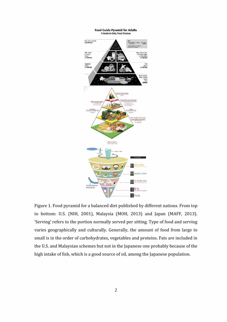
Figure 1. Food pyramid for balance diet published by different nations 2
Figure 1.1. Effect of NaCl (A), MgSO4 (B), CaCO3 (C), and KHSO4 (D)
concentrations in medium on cell and lipid yields of
thraustochytrid strain 12B 21
Figure 1.2. Effect of different forms of potassium compounds supplementation
in in the NM medium on the dry cell weight yield of
thraustochytrid strain 12B 23
Figure 1.3. Growth curve of thraustochytrid strain 12B and A. limacinum SR21
in F and NM medium 25
Figure 1.4. Effect of different glucose concentration on the dry cell weight,
lipid, DHA yield (mg/ml) and culture optical density in the NM
culture medium 31
Figure 1.5. Observation of the cell surface of thraustochytrid strain 12B grown
in F, NM, N and M media under SEM 33
Figure 1.6. Effect of medium composition on the size of sporangium in
thraustochytrid strain 12B 34
Figure 1.7. Abnormal cell shapes seen from thraustochytrid strain 12B cells
grown in M medium 35
Page
XIV
Figure 1.8. Cross sectional observation of thraustochytrid strain 12B grown in
F, NM, N and M medium under TEM 37
Figure 1.9. TEM of thraustochytrid strain 12B in F, NM and N medium
sampled by ultrarapid cryofixation technique 38
Figure 2.1. Growth profile of thraustochytrid strain 12B in F and NM media
62
Figure 2.2. Generalized life cycle of thraustochytrid strain 12B observed in
cells grown in F medium, NM medium and actual seawater 64
Figure 2.3. Phylogeny of thraustochytrid strain 12B to other thraustochytrid
species based on 18S rRNA gene analysis 67
Figure 2.4. Sequence similarity of COXII mitochondrial DNA between
thraustochytrid strain 12B, Aurantiochytrium limacinum SR21 and
Thraustochytrium aureum 68
Figure 3.1. Amount of dry cell weight obtained from cultivation of
thraustochytrid strain 12B in 1.5% (w/v) NaCl medium without
glucose, peptone and yeast extract supplementation 84
Figure 3.2: Effect of starvation on the absolute amount of major fatty acid
component of strain 12B 85
Figure 3.3. Growth curve of thraustochytrid strain 12B cells in A) F medium
B) NM medium, in the presence or absence of UV exposure 88
1
General introduction
A. Food and diet
Food is one of the essential keys for life by providing energy for growth and
for physiological maintenance. Human beings require a balanced diet consisting of
45–65% carbohydrate, 10–35% proteins and less than 30% fats within the total
daily calories (NIH, 2001). The amount of food is further determined by factors
such as age, gender, and activity (resting or exercising), and body conditions, such
as injury, sickness, and pregnancy. The environment, including both geography and
climate, can become a factor determining the amount and kind of food consumed,
and mental states, temperature, and humidity can affect body physiology. The
environment also determines what sources of food will be available in any
particular geographic location, which shapes the food culture and is reflected in the
differences in the servings within the balanced diet food pyramid published by
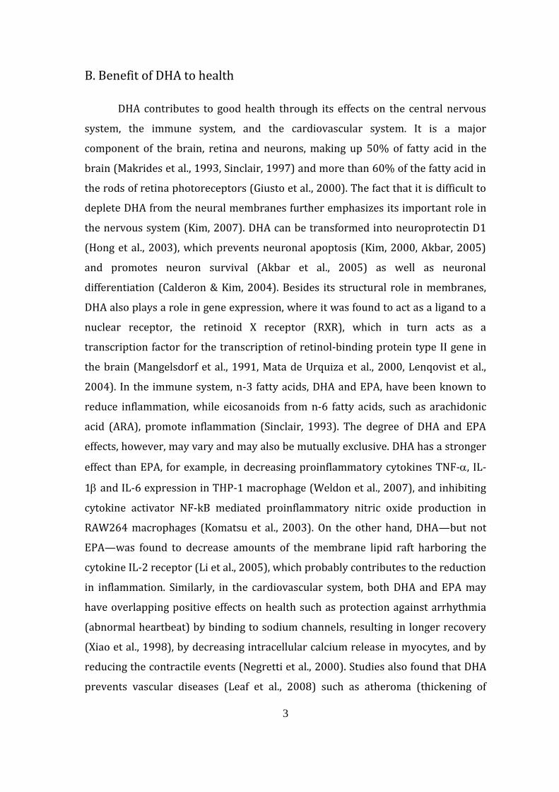
different nations around the world (Fig. 1). The recommended dietary allowance
for Japanese has been determined and published by the Japan Society of Nutritional
and Food Science. The estimated requirement for an average adult (18–49-year-
old) Japanese man is 2650 kcal/day (Tabata et al., 2013), out of which 20–30% of
the total energy should come from fat.
The type of fatty acid can be divided into saturated fatty acids, n-6 and n-3
unsaturated fatty acids, for which an estimated dietary goal has been determined to
be 4.5–7%, <10% and <2.2%, respectively, in order to prevent the onset of lifestyle-
related diseases (Ezaki et al., 2013). The consumption of eicosapentaenoic acid
(EPA) and docosahexaenoic acid (DHA), which are n-3 fatty acids, are 0.36 and 0.63
g/day, respectively, in Japan, which is the highest in the world (Elmadfa &
Kornsteiner, 2009). However, it is still below the maximum recommended daily
intake for combined EPA-DHA of 2 g/day (WHO, 2010).
2
Figure 1. Food pyramid for a balanced diet published by different nations. From top
to bottom: U.S. (NIH, 2001), Malaysia (MOH, 2013) and Japan (MAFF, 2013).
‘Serving’ refers to the portion normally served per sitting. Type of food and serving
varies geographically and culturally. Generally, the amount of food from large to
small is in the order of carbohydrates, vegetables and proteins. Fats are included in
the U.S. and Malaysian schemes but not in the Japanese one probably because of the
high intake of fish, which is a good source of oil, among the Japanese population.
3
B. Benefit of DHA to health
DHA contributes to good health through its effects on the central nervous
system, the immune system, and the cardiovascular system. It is a major
component of the brain, retina and neurons, making up 50% of fatty acid in the
brain (Makrides et al., 1993, Sinclair, 1997) and more than 60% of the fatty acid in
the rods of retina photoreceptors (Giusto et al., 2000). The fact that it is difficult to
deplete DHA from the neural membranes further emphasizes its important role in
the nervous system (Kim, 2007). DHA can be transformed into neuroprotectin D1
(Hong et al., 2003), which prevents neuronal apoptosis (Kim, 2000, Akbar, 2005)
and promotes neuron survival (Akbar et al., 2005) as well as neuronal
differentiation (Calderon & Kim, 2004). Besides its structural role in membranes,
DHA also plays a role in gene expression, where it was found to act as a ligand to a
nuclear receptor, the retinoid X receptor (RXR), which in turn acts as a
transcription factor for the transcription of retinol-binding protein type II gene in
the brain (Mangelsdorf et al., 1991, Mata de Urquiza et al., 2000, Lenqovist et al.,
2004). In the immune system, n-3 fatty acids, DHA and EPA, have been known to
reduce inflammation, while eicosanoids from n-6 fatty acids, such as arachidonic
acid (ARA), promote inflammation (Sinclair, 1993). The degree of DHA and EPA
effects, however, may vary and may also be mutually exclusive. DHA has a stronger
effect than EPA, for example, in decreasing proinflammatory cytokines TNF-, IL-
1 and IL-6 expression in THP-1 macrophage (Weldon et al., 2007), and inhibiting
cytokine activator NF-kB mediated proinflammatory nitric oxide production in
RAW264 macrophages (Komatsu et al., 2003). On the other hand, DHA—but not
EPA—was found to decrease amounts of the membrane lipid raft harboring the
cytokine IL-2 receptor (Li et al., 2005), which probably contributes to the reduction
in inflammation. Similarly, in the cardiovascular system, both DHA and EPA may
have overlapping positive effects on health such as protection against arrhythmia
(abnormal heartbeat) by binding to sodium channels, resulting in longer recovery
(Xiao et al., 1998), by decreasing intracellular calcium release in myocytes, and by
reducing the contractile events (Negretti et al., 2000). Studies also found that DHA
prevents vascular diseases (Leaf et al., 2008) such as atheroma (thickening of
4
artery walls) by reducing the expression of vascular and intercellular adhesion
molecules VCAM-1 and ICAM-1, respectively (De Caterina et al., 1994, 2000).
Other health benefits of DHA and EPA are their effects of lowering
triacylglycerol (TG) level (Mozaffarian & Wu, 2012) and enhancing the viability of
insulin-secreting cells (Suresh & Das, 2001). DHA also has antioxidative potential
(Okuyama et al., 2008b), lowering the risk to colorectal cancer and breast cancer
with an intake of 500 mg/day (WHO, 2010). Furthermore, the bioconverted DHA
also has antibacterial (Shin, 2007) as well as antifungal properties (Bajpai et al.,
2008). DHA also has been shown to be safe for consumption. At high concentration
(2 g per kg body weight per day), a slight increase in liver and spleen weight
(extramedullary hematopoiesis) to accommodate the large dietary lipid load
without any accompanying histopathological symptoms was observed, and the only
significant pathological symptom with very high DHA consumption (7.5% of diet) is
lipogranuloma; i.e., the formation of yellow spots in the abdominal adipose tissue
(Kroes et al., 2003).
C. Sources of DHA
There are two major sources for DHA synthesis in the human body: the liver
(Scott & Bazan, 1989) and the astrocyte glial cells (Moore et al., 1991); however,
DHA can also be directly derived from the diet. DHA (C22:6) is made up of 22
carbon chain fatty acids with six cis-unsaturated double bonds (at 4, 7, 10, 13, 16,
19). Inside the body, it is synthesized in low quantities from n-3 lenolenic acid
(C18:3), which is an essential fatty acid. It can be synthesized through three
possible pathways: the ‘aerobic 4 desaturation-independent’, the ‘aerobic 4
desaturation-dependent’ (Sprecher pathway) and the ‘anaerobic polyketide
synthase’ pathways (Qiu, 2003). The 4 desaturation-independent pathway occurs
in mammals through desaturation and elongation where successive elongation and
desaturation from C18:3, to C22:5 occur in the microsome, while C22:5 to C22:6
(DHA) occurs in the peroxisome. The aerobic 4 desaturation-dependent pathway,
on the other hand, occurs in thraustochytrid microorganisms, where DHA synthesis
5
only involves the microsome by direct desaturation of C22:5 to C22:6. The
anaerobic polyketide synthase pathway slightly resembles the fatty acid synthase
cycle. The fatty acid chain is increased by the condensation of acyl-ACP with
malonyl-ACP, producing a 3-ketoacyl-ACP, which is then reduced forming 3-
hydroxyacyl-ACP followed by dehydration to enoyl-ACP and another reduction to
acyl-ACP. For DHA synthesis, however, double bonds are formed by the
condensation of enoyl-ACP with malonyl-ACP instead of undergoing another
reduction to acyl-ACP.
DHA in the diet is mainly obtained from eating fish and eggs; however, DHA
can also be consumed directly in the form of supplements, with the added benefit
of purity and information on its concentration. Presently, DHA is mostly extracted
from marine fish, which accumulate DHA through feeding on microalgae, as they
are unable to synthesize DHA from C18:3 (Sargent et al., 1999). Conversely,
freshwater fish are able to synthesize DHA from essential fatty acids using the same
pathways as mammals (Bell et al., 2003), and by taking advantage of the possibility
of large-scale aquaculture, this is an alternative source that could solve the problem
of dwindling marine fish stock. However, fish oil still presents various problems,
such as undesirable taste, odor and a complex fatty acid composition, which is
costly to purify (Wen et al., 2003). Higher terrestrial plants generally do not
produce DHA (Gill & Valivety, 1997). Some bacteria, particularly those from marine
habitats, also produce DHA; however, it is incorporated in the cell membrane and is
rarely stored in the cell as triacylglycerol (Alvarez & Steinbüchel, 2002); therefore,
cellular levels of DHA in bacteria are normally very low. The other main alternative
sources of DHA are marine microalgae, such as the golden algae (Grima et al., 1992),
green algae (Yongmanitchai & Ward, 1989, Renaud et al., 1999) and diatoms
(Yongmanitchai & Ward, 1989, Renaud et al., 1999); however, their DHA content is
not very high. The most promising source of DHA is now a heterotrophic
dinoflagellate Cryptocodenium cohnii (Mendes et al., 2009) and thraustochytrid
microorganisms.
6
Chapter 1: Cultivation of thraustochytrid strain 12B
in media containing low NaCl concentration
1.1. Summary
Thraustochytrid strain 12B produces a higher amount of DHA in the NM
medium containing 0.1% (w/v) NaCl and 1% (w/v) MgSO4 compared with the F
medium containing 50% (v/v) seawater, both of which were supplemented with
8% (w/v) glucose, 1% (w/v) peptone and 1% (w/v) yeast extract. The DHA
productivities of cells grown in NM and F media were 0.8 and 0.2 mg/ml/day,
respectively. Supplementation of NaCl at 0.1% (w/v) and MgSO4 at 1% (w/v),
which are the most necessary inorganic compounds, resulted in a high
accumulation of large oil bodies inside the cell. The source of DHA in the cell was
mostly triacylglycerol (TG), which was approximately 71% of the total DHA when
the cells were grown in the F medium and 81% in the NM medium. DHA, however,
only makes up 26% of the total fatty acid in the TG in F-cultured cells and 21% in
NM-cultured cells. The addition of other salts, CaCO3, KHSO4, K2SO4, K2HPO4 and
KH2PO4, in the NM medium did not substantially increase the DHA yield of strain
12B. The cells grown in the NM medium observed using transmission electron
microscopy (TEM) and scanning electron microscopy (SEM) were found to have
thin cell walls and sloughing of the cell surface, which indicate prolific growth and
cell maturity. The walls of cells grown in the NM medium were as thin as those of
cells grown in the F medium, being 0.08 and 0.1 m thick, respectively, while cells
grown in N and M media were thicker, 0.24 and 1.5 m thick, respectively.
Preparation of cells using the TEM cyrofixation technique, which preserves cellular
structures better than the standard TEM preparations, shows a much thicker cell
wall, 2.14 m, for cells cultured in N, indicating the fragility of the wall of cells
grown in the N medium in the absence of MgSO4. The growth profile based on the
optical density showed earlier entry into the stationary phase by cells grown in the
7
NM medium compared with that of cells grown in the F medium, which may also be
one reason for the high lipid accumulation, as cells normally accumulate lipid more
during the stationary phase. Besides the simple medium composition, cells grown
in the NM medium have higher glucose utilization efficiency, at less than 8% (w/v)
glucose compared with previous studies, with high DHA yield obtained starting
from 5% (w/v) glucose and highest at 7% (w/v) glucose, being 2.7 mg/ml and 3.1
mg/ml respectively. The NM medium was also applicable to another
thraustochytrid species, Aurantiochytrium limacinum SR21. NaCl may have a role in
cell division, as cells grew abnormally large and have abnormal shapes in NaCl-
deficient M medium, which contains 8% (w/v) glucose, 1% (w/v) peptone, 1%
(w/v) yeast extract and 1% (w/v) MgSO4. On the other hand, MgSO4 may have a
role in osmotic regulation, as some cells appear to shrink in MgSO4-deficient N
medium, which contains 8% (w/v) glucose, 1% (w/v) peptone, 1% (w/v) yeast
extract and 0.1% (w/v) NaCl. The yield of DHA was increased when strain 12B was
cultured in NM medium using industrial-grade glucose, peptone and yeast extract
from 2.4 to 4.6 mg/ml during flask cultivation and was further increased through
jar cultivation from 4.6 to 11.7 mg/ml. The high DHA yield from the use of the NM
medium is therefore very desirable for large-scale industrial application in the
cultivation of thraustochytrid microorganisms for DHA production.
8
1.2. Introduction
1.2.1. Cultivation of thraustochytrids for DHA production
Thraustochytrid microorganisms are generally referred to as microalgae
despite being heterotrophic microorganisms, yet these microorganisms do have
some similar features such as being unicellular, having biflagellate zoospores and
being taxonomically placed under the same phylum as algae, Heterokonta. A
number of thraustochytrid strains with high DHA productivity have been reported,
such as Schizochytrium sp. SR21 (Yaguchi et al., 1997), Schizochytrium sp. (Qu et al.,
2013), thraustochytrid ATCC PTA-9695 (Apt et al., 2012) and Aurantiochytrium sp.
KRS101 (Hong et al., 2013), with DHA productivities of 3.3, 3.4, 4.1 and 5.6 g/l/day,
respectively. The strain 12B used in this study has also been reported to have high
DHA productivity, as high as 5.4 g/l/day so far under jar cultivation (Okuyama et al.,
2008a). The strain has been used to produce not just DHA-rich triacylglycerol (TG)
but also phospholipid, particularly DHA-rich phosphotidylserine (Okuyama et al.,
2007, Okuyama et al., 2012, Bin Haji Mohd Taha et al., 2012). Parameters such as
temperature, salinity, pH, oxygen level, culture mode and age of culture as well as
various aspects of the culture composition such as carbon source, nitrogen source,
carbon-to-nitrogen ratio, macroelements such as Na+, microelements (trace metals)
and vitamins have been known to affect the growth and culture yield of
thraustochytrids (Raghukumar, 2008b). Different thraustochytrid species from
different genera do not have the same growth rate in any one medium with the
same composition (Lewis et al., 1999). In a chemically defined culture medium,
some thraustochytrid species have an obligate requirement for vitamin B
supplementation (Goldstein, 1973), and even the presence of specific trace metals
such as manganese and copper results in a large increase or decrease in dry cell
weight (DCW) yield depending on the thraustochytrid species (Nagano et al., 2013).
9
1.2.2. Culture medium and seawater
The conventional cultivation method for thraustochytrids including strain
12B normally uses a medium containing half the concentration of seawater because
thraustochytrids are marine microorganisms. In large-scale industrial cultivation,
the use of seawater imposes several problems such as the cost of transporting
seawater, dependability on sites close to the sea, and corrosion of cultivation
facilities because normal tank fermenters (N type SS316) can withstand less than
0.1% chloride concentration at 30°C (Grundfos, 2012).
Seawater is also not very suitable for thraustochytrid cultivation because of
the presence of various chemical and heavy metal pollutants. The use of artificial
seawater made with distilled water is preferable as it avoids the introduction of
heavy metals such as mercury into the biomass as shown in a cultivation involving
S. limacinum (Pyle et al., 2008), while cultivation using seawater may introduce
mercury into oils such as those extracted from fish caught from the sea (Foran et al.,
2003). The study of pollution of thraustochytrid microorganisms is very limited,
and the only study reported on Schizochytrium sp. shows deformation of the cell
structure, formation of holes in the cell wall, and shrinkage of cells when 32 ppm
zinc and copper heavy metals were added during cultivation, while cell lysis
occurred when higher concentration of 256–384 ppm were added (Lin et al., 2010).
Despite the decrease of heavy metal pollutants to conform to the minimum
environmental quality standard throughout 1971 to 1997, the organic pollutant
level of the sea around Japan in the same period did not improve (MOE, 2013).
Unfortunately, recent data are unavailable. On the other hand, a survey on chemical
substances suspected to have long-term toxicity to humans has detected 357
pollutants out of 1069 chemicals tested in both aquatic and marine surface water
around Japan (MOE, 2011), a small number relative to the total 19,247 existing
chemical substances registered under the chemical substances control law (NITE,
2013).
A bigger concern nowadays, however, is the presence of radioactive isotopes
in the sea surrounding Japan because of the accident at the Fukushima nuclear
10
power plant, which is thought to have released about 5.5 1015 becquerel (Bq) of
radioactive isotope cesium 137 (137Cs) during the first few days of the accident
(Estournel et al., 2012). 137Cs has a half-life of about 31 years (Unterweger, 2002),
and increased concentration from 1.4 to 6.0 (Bq kg–1) in the migratory Pacific
bluefin tuna Thunnus orientalis caught in waters off California has already been
detected because of the Fukushima accident (Madigan et al., 2012). There are no
studies yet on the effect of radioactive isotopes on thraustochytrids; however, the
uptake of 137Cs by some phytoplankton species has already been demonstrated
(Adam and Garnier-Laplace, 2003, Heldal et al., 2001).
1.2.3. Medium with simple composition and low NaCl concentration
Artificial seawater (ASW), with defined chemical components and a
concentration of NaCl almost the same as seawater at about 3% (w/v), has been
used instead of seawater in some investigations on the cultivation of
thraustochytrids (Nagano et al., 2009, Min et al., 2012). The effectiveness of the
ASW compared with actual seawater has been the subject of a review paper
(Berges and Franklin, 2001), which reported inconsistencies of superiority
compared with seawater depending on marine species. The composition of ASW
for particular species such as thraustochytrids has probably been tailored for this
group of microorganisms and is slightly different from the general ASW mentioned.
For thraustochytrid microorganisms, there is a necessity for sodium ions
(Raghukumar, 2008b) but not for chloride ions, leading to experimentation in
reducing or excluding them completely from the culture medium, as they are also
unfavorable for culture equipment.
There are very few papers that have reported chloride-independent medium
alternatives such as those made by substituting NaCl with Na2SO4 (Barclay, 1994);
however, the culture yield was found to be very low. Another study that uses
Na2SO4 (Prabu et al., 2012) has also been published; however, the absolute DCW
yield data from which the DHA yield was calculated in terms of gram per gram
were not disclosed and therefore open to speculation. Mannitol and sucrose are
also alternatives; however, similarly, studies have only concentrated on cell growth

11
based on optical density (Shabala et al., 2009) or percentage fatty acid without
disclosing the absolute DHA yield and productivity (Shabala et al., 2013). Apart
from this, media with complex ingredients such as M-5 medium, FFM medium
(Barclay et al., 1994) and medium 1–6 (Singh and Ward, 1997) with various trace
elements as supplementation have commonly been used. There is normally a
preference for using an artificial and chemically defined medium over a natural
medium for the cultivation of microbes because every component in the chemically
defined medium is known. The use of peptone and yeast extract in culture medium
may categorize it as a natural medium because of its complex composition, and the
exact constituents of some components such as trace elements are not fully
quantified by manufacturers. Natural media are considered to have poor
reproducibility (Arora, 2013); however, both peptone and yeast extract available
nowadays are produced using a standard procedure, and therefore high
reproducibility in cell yield during cultivation is normally achieved. The use of
natural ingredients consequently makes the supplementation of trace amounts of
minerals and vitamins unnecessary. The aim of this study is to compose a simple
culture medium that is not corrosive to industrial equipment by having a low NaCl
content. This is an important aspect for large-scale industrial DHA production, as it
will reduce manufacturing cost and time, as preparation will become
uncomplicated, practical and efficient.
12
1.3. Materials and methods
1.3.1. Microorganisms and cultural media
The thraustochytrid microorganism strain 12B (strain 12B; NITE P-68) was
mainly used throughout this work. Strain 12B was previously isolated from a
mangrove ecosystem in Okinawa, Japan (Perveen et al., 2006). Another
thraustochytrid microorganism, Aurantiochytrium limacinum SR21, was isolated
from a coral reef area of the Yap Islands, Federated States of Micronesia (Nakahara
et al., 1996) and was originally identified as Schizocytrium limacinum SR21 (IFO
32693). It was kindly provided by the Institute of Fermentation, Osaka (IFO), with
the permission of Dr. Yokochi Toshihiro, National Institute of Advanced Science and
Technology (AIST).
1.3.2. Cultivation, growth measurement, and harvesting of cells
All media contain the same base organic composition of 1% (w/v) yeast
extract (Nacalai Tesque, special reagent grade), 1% (w/v) peptone (Kyokuto,
laboratory grade) and 8% (w/v) glucose (Kanto Chemical, guaranteed reagent for
JIS grade). The basic culture media for the cultivation of the thraustochytrids were
BY+ agar or liquid media as well as F medium. Both BY+ and F media are composed
of 50% filtrated seawater. The growth-optimized condition of strain 12B under
liquid cultivation was determined by investigating the effect of major target
compounds supplemented at different concentrations on the growth of cells. The
target inorganic compounds investigated were NaCl at 0, 0.1, 0.2, 0.3 and 0.5%
(w/v), MgSO4•7H2O at 0, 0.05, 0.1, 0.5, 1, 1.5, 2, 2.5 and 3% (w/v), CaCO3 at 0, 0.05,
0.1, 0.5% (w/v) and KHSO4 at 0, 0.05, 0.1, 0.5, 1, 1.5, 2, 2.5 and 3% (w/v). To assess
the influence of types of potassium-containing compounds on the growth of cells,
determined as increase in DCW, K2SO4, KH2PO4, and K2HPO4 were used at 0, 0.01,
0.05, 0.1, 0.5, 1, 1.5 and 3% (w/v). The best medium—i.e., the NM medium—was
only supplemented with 0.1% NaCl (w/v) and 1% MgSO4•7H2O (w/v). Cells were
13
also grown in N medium, which was only supplemented with 0.1% NaCl (w/v) and
M medium, which was only supplemented with MgSO4•7H2O (w/v) to see the effect
of the deficiency of either inorganic compound on culture yield and cell
morphology.
In order to simulate an industrial cultivation, analytical-grade glucose, yeast
extract and peptone were replaced with industrial-grade substitute yeast extract
Koucho P-81 (Cosmo Foods Co. Ltd) and peptone Proyield® Soy SE50MK Friesland
Campina (Oriental Yeast Co. Ltd). The INM cultures were not supplemented with
0.1% NaCl (w/v) because the industrial-grade yeast extract already contained 15%
salt. All cultivations, except for the jar cultivation, were done in 10 ml medium in a
50 ml flask with shaking at 150 rpm in the dark for 3 days at 30°C. Upscaling of the
experiment using an industrial NM medium (INM) was carried out in a jar
fermenter (MBF, EYELA, Tokyo Rikakikai Co. Ltd, Tokyo, Japan). The inoculum was
prepared by inoculating one loop full of 12B cells from plate agar culture into 30 ml
F medium in a 300 ml flask and incubated with shaking at 160 rpm in the dark for 3
days at 30°C. The jar was filled with 1.5 l INM medium, and 15 ml liquid inoculum
was then added. The jar cultivation was carried out for 5 days under the following
conditions: incubation temperature at 30°C, mixing speed at 300 rpm, with air
purged into the medium at 1.4–1.6 l/min.
Cells were harvested by centrifugation (RX-200, TOMY SEIKO Co. Ltd, Tokyo,
Japan) at 6000 rpm for 10 min, which is equivalent to 10,000 g, washed with
0.8% NaCl, recentrifuged and freeze-dried for 24 hr at –4°C, –0.092 MPa (Labconco,
Kansas, US) with an external vacuum pump (SVP-100S, Sato Vacuum Machinery
Industrial Co. Ltd, Tokyo, Japan). The DCW was determined after freeze-drying, and
the sample was stored at –80°C until used.
1.3.3. Lipid extraction
Lipid was extracted from the dried cells using the Bligh and Dyer method
(Bligh and Dyer, 1959) with modification in the ratio of solvent used. Freeze-dried
sample was initially mixed with a mixture of water, chloroform and methanol in the
14
ratio of 4:5:10 instead of 1:1:1. After vortexing for 30 s, water was again added to
the mixture in the ratio of 5:19 instead of 1:3. In addition, chloroform was again
added to the new mixture in the ratio of 1:6, and the chloroform layer was collected
by centrifugation at 2000 rpm (1856 g) for 5 min (KN-70, Kubota, Osaka, Japan),
and chloroform was added two more times by repeating this step.
Extracted lipid was then dried with N2 and redissolved to 50 mg/ml by
adding chloroform:methanol (2:1, v/v) solution containing 1% (w/v) butylated
hydroxy toluene (BHT). Separation of lipid into triacylglycerol (TG), polar (polar)
and other lipid classes was done by thin-layer chromatography (TLC). An aliquot of
10 l (500 g) of lipid was pipetted on to a silica glass plate (silica gel 60; Merck,
Damstadt, Germany) and allowed to develop in solvent containing hexane, diethyl
ether and acetic acid in the ratio of 50:50:1. Separated lipid classes were observed
by first spraying the plate with an acetone and water (4:1) solution containing
0.01% (w/v) primulin and then exposing the plate to ultraviolet radiation (UV).
Spots of lipid classes were scrapped and collected into screw-capped test tubes and
methanolyzed as described below.
1.3.4. Preparation and analysis of fatty acid methyl esters
Methanolysis of lipid was carried out by adding 10 l lipid, 10 l
heneicosanoic acid internal standard (2 mg/ml in benzene) and 1 ml methanol
containing 10% acetyl chloride into a screw-capped test tube and heating at 100°C
for 1 hr (Dry Block Bath MG-2, Torika Corp., Nagoya, Japan). The resulting fatty acid
methyl esters (FAMEs) were extracted with 2 ml of hexane three times. After
concentrating the FAME fractions through evaporation under N2, the samples were
subjected to gas–liquid chromatography (GLC) and gas chromatography–mass
spectrometry (GC–MS).
The FAME fraction was analyzed by gas chromatography (GC) using a GC-
353B unit (GL Sciences, Tokyo, Japan) equipped with a capillary column BPX70 (25
m 0.22 mm i.d., SGE, Yokohama, Japan) and flame ionization detection (FID) with
nitrogen as carrier gas. The column temperature was programmed to increase at
15
5°C/min from 150°C to 240°C and then maintained for 2 min while the injector and
detector temperatures were set at 240°C. Samples were also analyzed by a gas
chromatograph CP-3800 (Varian, Tokyo, Japan) with an ion-trap mass
spectrophotometer Saturn 2200 (Varian, Tokyo, Japan) equipped with the same
column. The column temperature was programmed to increase from 80°C to 240°C
with a split ratio of 20:1, helium carrier gas with a flow rate of 0.9 ml/min, and the
injector temperature set at 240°C. GC-MS was used to confirm the identification of
peaks corresponding to specific fatty acids, while gas chromatography–flame
ionization detection (GC–FID) was used for routine calculation of fatty acid yield
including DHA.
1.3.5. Analysis of glucose
The depletion of glucose in culture media was determined by pipetting 10 l
of culture sample onto silica gel, developed by using solvent containing 1-butanol,
2-propanol, H2O, and acetic acid (7:5:4:2, v/v) and then detected by spraying with
anisaldehyde (anisaldehyde, conc. H2SO4, and 95% ethanol, 1:1:18, %v/v) followed
by heating in an oven at 120°C for 10 min.
1.3.6. Microscopic observation
In order to determine any possible link between culture conditions, lipid
accumulation and changes in the external and internal cell structures of strain 12B,
SEM and TEM were performed. Cultures were grown in F, NM, N and M media in a
50 ml flask as mentioned above. Sampling of cells was done by centrifuging the
cultures at 5000 rpm (8000 g) for 5 min at 4°C, followed by washing with 0.1 M
potassium phosphate buffer (PB) at pH 7.4. The initial fixation and dehydration
steps are similar in SEM and TEM.
1.3.6.1. Scanning electron microscopy
Cells were harvested, washed in 0.1 M PB and cut into 1.5 1.5 0.5 mm
sections while submerged in PB. Prefixation was carried out in 5 ml PB containing
16
2% parafolmaldehyde (Wako Pure Chemical) and 2.5% glutaraldehyde (Nisshin
EM), and rotated for 2 hr (RT-50, Taiyo, Musashi, Japan) followed by washing thrice
with PB and rotating for 10 min each time. Postfixation was carried out by mixing
2% OsO4 (Nisshin EM) and 0.2 M PB (1:1), and incubating at 4°C for 2 hr. The
dehydration of cell samples for SEM was done by increasing ethanol concentration
from 70, 80, 90, 95 to 100%, with 15 min at each step, at room temperature (rt),
except for 70% ethanol, which was done overnight. Further drying was carried out
by exposing the cells to isoamyl acetate (Kanto Chemical) for 10 min and finally
with liquid CO2 for 2 hr (Critical point dryer HCP-1, Hitachi, Japan). Dried samples
were then fixed onto a metal plate and coated with platinum–palladium (Ion
sputter 1030, Hitachi, Tokyo) followed by observation under a scanning electron
microscope (S-4000, Hitachi, Tokyo, Japan).
1.3.6.2. Transmission electron microscopy
The cell preparations were essentially the same as for SEM until the
dehydration stage, after which cells were infiltrated by increasing the
concentration of EPON (EPON 812, TAAB Laboratories Equipment Ltd), by
immersing the cells in 5 ml of 100% QY-1 solvent, QY-1:EPON (2:1, v/v), and QY-
1:EPON (1:2, v/v) successively for 15 min each, and finally in 100% EPON
overnight. Prior to casting, the EPON used overnight was discarded and replaced
with fresh EPON, and rotated for another 4 hr. Samples were then embedded in
casting plates and allowed to harden at 60°C for 2 days. Ultrathin sections of
samples were cut by an Ultra Diamond Knife with a 2.5 mm, 35° blade (DiATOME,
Nisshin EM Co. Ltd, Tokyo, Japan) and placed on copper grids. Samples were
stained with 2% uranyl acetate (Wako Pure Chemical) and then lead citrate (Kanto
Chemicals) for 15 min and then 5 min, respectively, before being observed under a
transmission electron microscope (JEM 1400, JEOL, Tokyo, Japan).
Another TEM preparation method—i.e., the ultrarapid cryofixation
technique—was also tried for samples cultivated in a 50 ml flask for 7 days in 10
ml F, NM and N media. This technique offers better preservation of cellular
structures compared with the conventional TEM method. After washing and
centrifuging the cell in PB, about 1 l of cell was transferred onto a copper plate
17
and covered by another plate. The grid was then dropped into a liquid nitrogen
tank (Reichert KF80, Reichert Inc., New York, US) to preserve the cellular
structures, followed by freeze substitution using 2% OsO4 in acetone from –80°C, –
40°C, –20°C, 4°C to room temperature (rt) for 3 days, 2 hr, 2 hr and 1 hr,
respectively. Samples were then washed and infiltrated with increasing QY-2
solvent from 100% acetone to acetone:QY-2 (1:1) to 100% QY-2 for 15 min each.
Solvent substitution was then carried out by increasing the concentration of quetol
812 from 10, 30, 50, 70, 80, 90 to 100% for 30, 40, 60, 60 min, overnight, 2 hr and 2
hr, respectively. Samples were then embedded in EPON and allowed to solidify at
60°C for 2 days before being cut and stained for observation under the electron
microscope.
1.3.7. Protein analysis
The influence of compounds in the culture medium on the cellular
metabolism of thraustochytrid strain 12B such as in protein and enzyme
expression could probably be seen through differences in its protein expression.
Thraustochytrid strain 12B grown in 10 ml F, NM, N and M media was cultivated in
a 50 ml flask as mentioned previously, and the cells were centrifuged, washed and
stored at –80°C until analyzed. For protein extraction, the cells were resuspended
in a small amount of PB and then ground using a sterilized pestle and crucible. The
suspension was centrifuged (4°C, 10,000 g, 15 min) to collect the supernatant
containing cellular proteins and was then concentrated by adding 100% acetone at
four times the volume of sample. The mixture was then vortexed and incubated at –
20°C for 10 min. Precipitated protein was collected by centrifugation (12,000 g, 5
min), and the acetone supernatant was discarded. The protein was redissolved in
100 l PB, and the protein concentration was measured and equalized to other
samples (Qubit 2.0 Fluorometer, Invitrogen, Carlsbad, California, US). Equal
volumes of protein sample and SDS sample buffer were mixed, and a volume from
this was taken and further treated with 2-mercaptoethanol (1% v/v) and heated
for 5 min at 100°C. Samples were run in gel containing 5–20% denaturant (e-Pagel
E-R520L, ATTO, Tokyo, Japan) in a tank containing SDS running buffer at 200 V, 20
18
mA, 4.5 W for 80 min. Observation of the protein banding profile was made by
silver staining (silver stain II kit, Wako, Osaka, Japan).
19
1.4. Results
1.4.1. Effect of the salt composition of the culture media on growth, and
lipid and DHA yields of thraustochytrid strain 12B
Identification of the most relevant compounds and the concentrations
leading to the formulation of the NM medium that yields high cell yield, lipid, fatty
acid and ultimately high DHA yield was obtained by investigating the effect of NaCl,
MgSO4, CaCO3, potassium compounds KHSO4 and K2SO4, and phosphorus-
containing potassium compounds KH2PO4 and K2HPO4. The phosphorus-containing
compounds were also investigated to see any possible additional effect of
phosphorus as a potential limiting compound in the thraustochytrid cultivation.
1.4.1.1. Effect of NaCl and MgSO4 concentrations
Strain 12B is normally cultivated in F medium, in which 1% peptone, 1%
yeast extract, and 8% glucose are dissolved in 50% seawater. To investigate the
necessity of NaCl in the cultivation of strain 12B, modified F media containing
various concentrations of NaCl, instead of 50% seawater, were prepared. During 3
days’ cultivation, the thraustochytrid strain 12B was unable to grow in the absence
of NaCl (Fig. 1.1A). The DCW and lipid yields decreased from 4.3 to 3.1 mg/ml and
from 1.3 to 0.6 mg/ml, respectively, with decreasing NaCl concentration from 0.5 to
0.2% (w/v). However, both DCW and lipid yields suddenly increased again to 4.1
and 1.1 mg/ml, respectively, at 0.1% (w/v) NaCl. The modified F medium with
0.1% (w/v) NaCl was then used in the subsequent experiments.
The magnesium ion is the fourth most abundant ion in seawater (Brown et
al., 1989). In this experiment, MgSO4 and not MgCl2 was used because of the
corrosive nature of Cl─. The addition of a small amount of MgSO4 to 0.1% NaCl-
containing modified F medium considerably increased the DCW and lipid yields of
strain 12B (Fig. 1.1B). The highest amounts of DCW and lipid at 18.8 and 11.2
mg/ml, respectively, were obtained when 1% (w/v) MgSO4 was added to the
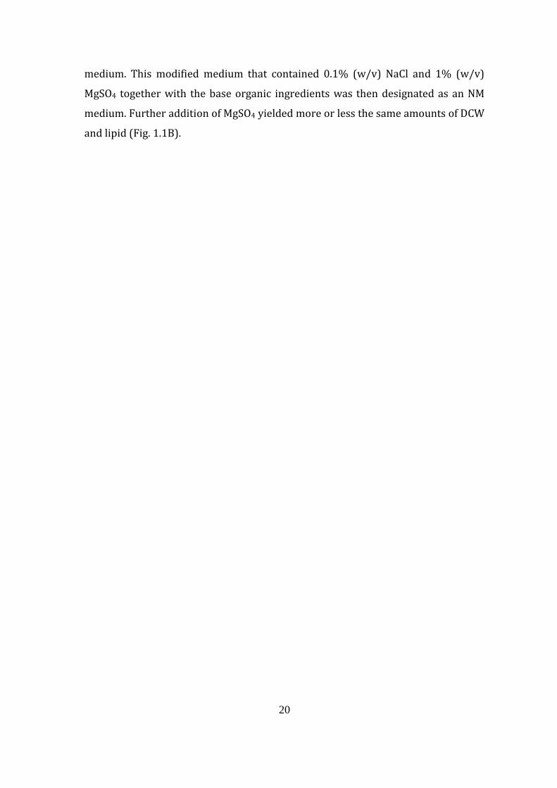
20
medium. This modified medium that contained 0.1% (w/v) NaCl and 1% (w/v)
MgSO4 together with the base organic ingredients was then designated as an NM
medium. Further addition of MgSO4 yielded more or less the same amounts of DCW
and lipid (Fig. 1.1B).
21
Figure 1.1. Effect of NaCl (A), MgSO4 (B), CaCO3 (C), and KHSO4 (D) concentrations
in medium on cell and lipid yields of thraustochytrid strain 12B. (A) Reduction in
NaCl concentration from 0.5% to 0.2% (w/v) initially reduced the DCW and lipid
yields and the percentage lipid per DCW. All three values increased again at 0.1%
NaCl; however, cells were not able to grow at zero NaCl concentration. (B)
Supplementing the N medium containing 0.1% NaCl with MgSO4 drastically
increased the DCW and lipid yield, with the highest yield being obtained at 1%
(w/v), beyond which the yield remained more or less stationary. (C)
Supplementing the NM medium containing 0.1% (w/v) NaCl and 1% (w/v) MgSO4
with CaCO3 initially decreased both the DCW and lipid yields when CaCO3
concentration was increased from 0 to 0.1% (w/v). An increase of DCW was
observed at 0.5% (w/v) CaCO3; however, the % lipid per DCW was low. (D)
Supplementation with KHSO4 decreased the DCW and lipid as well as the % lipid
per DCW, with yields reaching zero at 1% (w/v) and higher concentrations. All
media contained a similar organic base composition of 1% (w/v) yeast extract, 1%
(w/v) peptone and 8% (w/v) glucose. A colony from a 2-day BY+ agar plate was
used as an inoculum, and all cultures were incubated in a 50 ml flask containing 10
ml culture medium in the dark for 3 days at 30°C and 150 rpm before cells were
harvested, freeze-dried and the lipid extracted.
22
1.4.1.2. Effect of CaCO3 and KHSO4 supplementation
The effect of combined addition of calcium carbonate and potassium
hydrogen phosphate on the growth, lipid and DHA yields of strain 12B was
examined in the NM medium. Although both DCW and lipid yields gradually
decreased with increasing concentration of CaCO3 from 0% to 0.1% (w/v), these
parameters suddenly increased at 0.5% (w/v) CaCO3. However, the percentage
lipid per DCW was almost unchanged (Fig. 1.1C). The decrease in DCW and lipid
yields was also seen when potassium hydrogen sulfate was added to the NM
medium, and both yields became zero when 1% (w/v) KHSO4 or more was added
(Fig. 1.1D). When both CaCO3 and KHSO4 were added together in the absence or
presence of 1% (w/v) MgSO4, no difference in yield was obtained, and positive
yield was obtained only because of MgSO4 supplementation (data not shown).
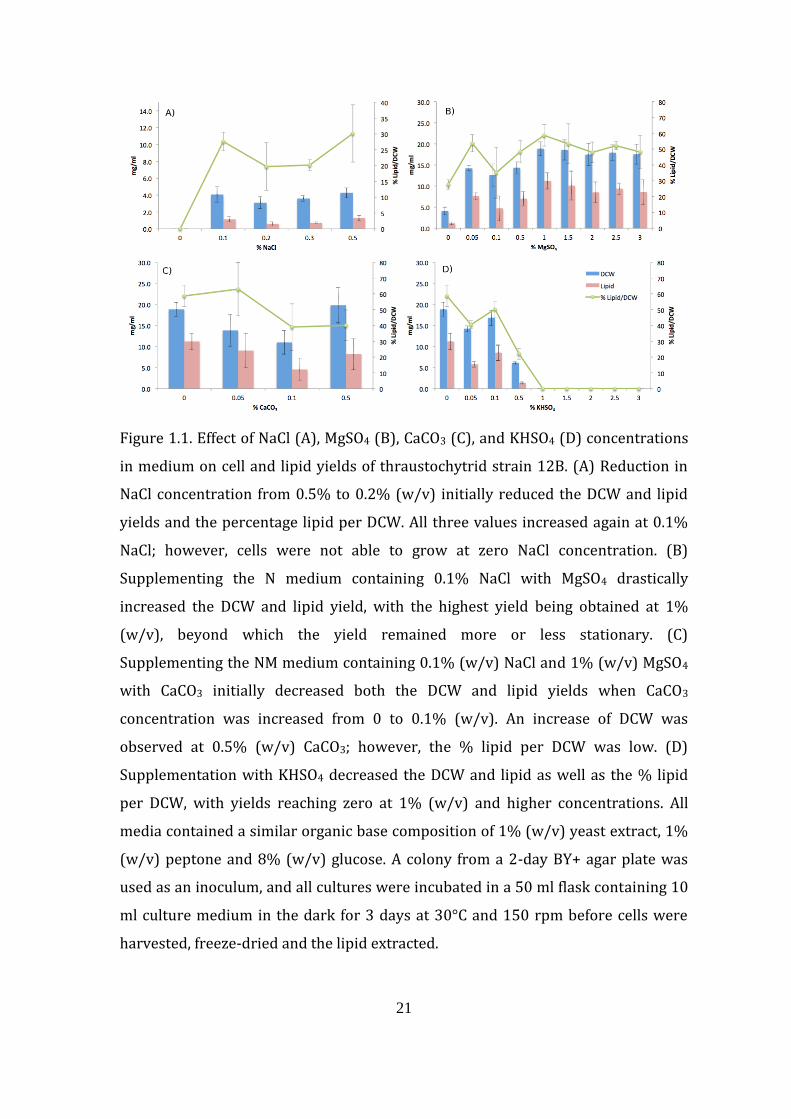
1.4.1.3. Effect of different molecular forms of potassium compounds on
cultivation
The addition of potassium generally decreased the DCW yield with
increasing concentration. However, a slight increase in DCW yield was observed
when the amount added was below 0.1% (w/v), except for KHSO4 (Fig. 1.2). Each
compound specifically increased one type of yield and not the other, with K2SO4 as
the best compound to increase DCW, K2HPO4 for the overall lipid, and KH2PO4 for
the overall fatty acid, although the difference in DHA yield is not considerably
different compared with the NM (data not shown). In terms of percentage fatty acid
composition, the addition of potassium compounds also resulted in a higher
amount of octadecanoic acid (oleic acid; C18:1) from 0.6% in cells grown in NM
medium to more than 20% when potassium compounds were added. The
percentage of pentadecanoic acid (C15:0) was low for all potassium-containing
media except KHSO4. The percentage composition of DHA (C22:6), on the other
hand, appears to be more or less the same with or without the addition of
potassium (data not shown).
23
Figure 1.2. Effect of supplementing the NM medium with different forms of
potassium compound on the DCW yield of thraustochytrid strain 12B. KHSO4 and
K2SO4 were used as representative potassium compounds without phosphorus,
while KH2PO4 and K2HPO4 were used as representative potassium compounds
containing phosphorus. There was generally a decrease in DCW with increasing
concentration of potassium compounds; however, a slight spike in DCW was
obtained at very low potassium compound concentrations. All media were
inoculated with a colony from a 2-day BY+ agar plate, and incubation was carried
out in a 50 ml flask containing 10 ml of culture medium in the dark for 3 days at
30°C.
0.0
5.0
10.0
15.0
20.0
25.0
30.0
0 0.2 0.4 0.6 0.8 1 1.2 1.4 1.6 1.8 2 2.2 2.4 2.6 2.8 3 3.2
DC
W (
mg
/m
l)
% concentration (w/v)
KHSO4K2SO4KH2PO4K2HPO4
24
1.4.1.4. Application of NM medium to other thraustochytrid strain
The A. limacinum SR21 is a well-known thraustochytrid strain that
accumulates one of the highest amounts of DHA during cultivation in artificial
seawater containing about 1.5% NaCl (w/v) (Yaguchi et al., 1997). The application
of the NM medium to the cultivation of A. limacinum SR21 showed an almost
similar growth pattern to that of thraustochytrid strain 12B. There was steady
growth from zero time onwards, and the stationary phase started at around 3–5
days during cultivation. The number of days for glucose to be depleted from the
culture media varied between thraustochytrid strain 12B and A. limacinum SR21.
Glucose was depleted by thraustochytrid strain 12B after 7 days’ cultivation in both
F and NM, while for A. limacinum SR21, glucose depletion was even faster, at 6 and
5 days, respectively (data not shown). Generally, there were higher DCW,
percentage lipid, percentage fatty acid and percentage DHA yields in NM compared
with the F culture, especially for thraustochytrid strain 12B after 3 days’ cultivation
(Table 1.1). The DCW and DHA yields of cells grown in the NM medium after 3 days’
cultivation were 18.8 and 2.4 mg/ml, respectively, for strain 12B, while the yields
from cells grown in F medium were 10.4 and 0.5 mg/ml, respectively. In the case of
A. limacinum SR21, the DCW and DHA yields from cells grown in NM and F media
were almost the same. Higher DCW and DHA yields were obtained from strain 12B
compared with A. limacinum SR21 when each was cultivated in NM medium. The
DCW of strain 12B was 18.8 mg/ml compared with 12.9 mg/ml for A. limacinum
SR21, while the DHA yield was also higher for strain 12B at 2.4 mg/ml compared
with 1.1 mg/ml for A. limacinum SR21.
25
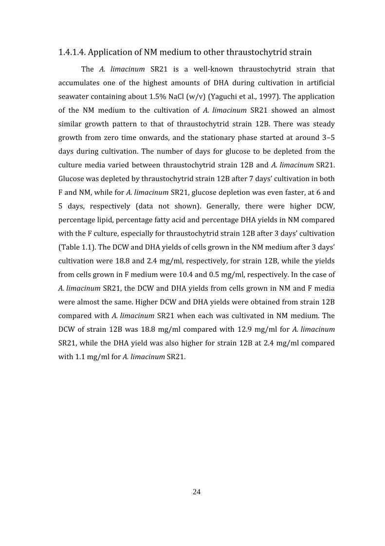
Figure 1.3. Growth curves of thraustochytrid strain 12B and A. limacinum SR21 in F
and NM media. Growth patterns were more or less the same for both strains 12B
and SR21 in the F and NM media for up to 3 days’ cultivation. Longer cultivation
resulted in the stationary phase for the strains in the NM culture, while growth
appeared to continue up to the 5th day in the F medium before entering the
stationary phase. Both F and NM media contain the same organic composition
consisting of 8% (w/v) glucose, 1% (w/v) yeast extract and 1% (peptone). F
medium is also composed of 50% seawater, while NM medium was made by using
distilled water supplemented with 0.1% (w/v) NaCl and 1% (w/v) MgSO4•7H2O. A
colony from a 2-day BY+ agar plate was used as an inoculum, and incubation was
carried out in a 50 ml flask containing 10 ml culture medium incubated in the dark
at 30°C for up to 8 days. Triplicate sampling was taken from a 10 ml culture each
day.
0
5
10
15
20
25
30
35
0 2 4 6 8 10
A6
00
nm
Days
12B-F
12B-NM
SR21-F
SR21-NM
26
Table 1.1. Effect of F and NM culture media on DCW and DHA yields (mg/ml) and
percentage composition of lipid, fatty acid and DHA of strain 12B compared with A.
limacinum SR21.*
A higher yield was obtained after 3 days’ cultivation in NM compared with the F
medium. A. limacinum SR21 yields are generally higher compared with strain 12B
grown in the F medium, and the opposite result was obtained when the strains
were grown in the NM medium. A colony from 12B or SR21 cells grown for 2 days
on a BY+ agar plate was used to inoculate a 10 ml culture in a 50 ml flask and
incubated at 30°C for 3 days.
*Average ± standard deviation from three independent cultures
27
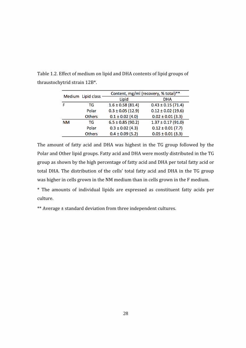
1.4.1.5. Distribution of DHA in lipid classes
Fatty acids including DHA were mostly stored as TG in the thraustochytrid
strain 12B (Okuyama et al., 2007). In this study, the composition of TG, polar lipid,
and other lipid classes was expressed as the amount of their constituent fatty acids
after TLC separation. The percentage of TG from cells grown 3 days in the F
medium was 81.4%, while that in the NM medium was 90.2% (Table 1.2). The
amount of DHA constituting the fatty acid in TG was 26% in F and 21% in NM
media-cultured cells; however, the DHA from TG contributed to 71% of the total
DHA in cells from the F culture and 91% in cells grown from the NM culture. About
20% of the fatty acid was lost through TLC analysis compared with lipid that was
directly methanolyzed (data not shown).
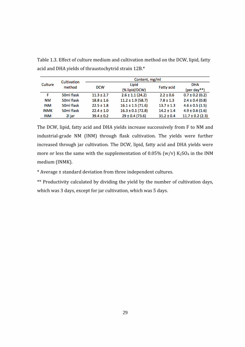
1.4.1.6. Application of industrial-grade ingredients and upscaling to jar
cultivation to increase DHA yield
To examine the usefulness of industrial-grade ingredients in the
productivity of DHA, strain 12B was cultivated in a flask and jar fermenter using
NM medium that had been prepared using industrial-grade organic ingredients
(yeast extract, peptone, and glucose), then called INM medium. There is a gradual
increase of DHA yield from 0.7 to 2.4 mg/ml between cells grown in F and NM
media during flask cultivation. A further increase was seen in the DHA yield from
2.4 to 4.6 mg/ml between analytical-grade NM medium and industrial-grade INM
medium. The DHA yield was again increased from 4.6 to 11.7 mg/ml between flask
and jar cultivation in INM medium (Table 1.3).
28
Table 1.2. Effect of medium on lipid and DHA contents of lipid groups of
thraustochytrid strain 12B*.
The amount of fatty acid and DHA was highest in the TG group followed by the
Polar and Other lipid groups. Fatty acid and DHA were mostly distributed in the TG
group as shown by the high percentage of fatty acid and DHA per total fatty acid or
total DHA. The distribution of the cells’ total fatty acid and DHA in the TG group
was higher in cells grown in the NM medium than in cells grown in the F medium.
* The amounts of individual lipids are expressed as constituent fatty acids per
culture.
** Average ± standard deviation from three independent cultures.
29
Table 1.3. Effect of culture medium and cultivation method on the DCW, lipid, fatty
acid and DHA yields of thraustochytrid strain 12B.*
The DCW, lipid, fatty acid and DHA yields increase successively from F to NM and
industrial-grade NM (INM) through flask cultivation. The yields were further
increased through jar cultivation. The DCW, lipid, fatty acid and DHA yields were
more or less the same with the supplementation of 0.05% (w/v) K2SO4 in the INM
medium (INMK).
* Average ± standard deviation from three independent cultures.
** Productivity calculated by dividing the yield by the number of cultivation days,
which was 3 days, except for jar cultivation, which was 5 days.
30
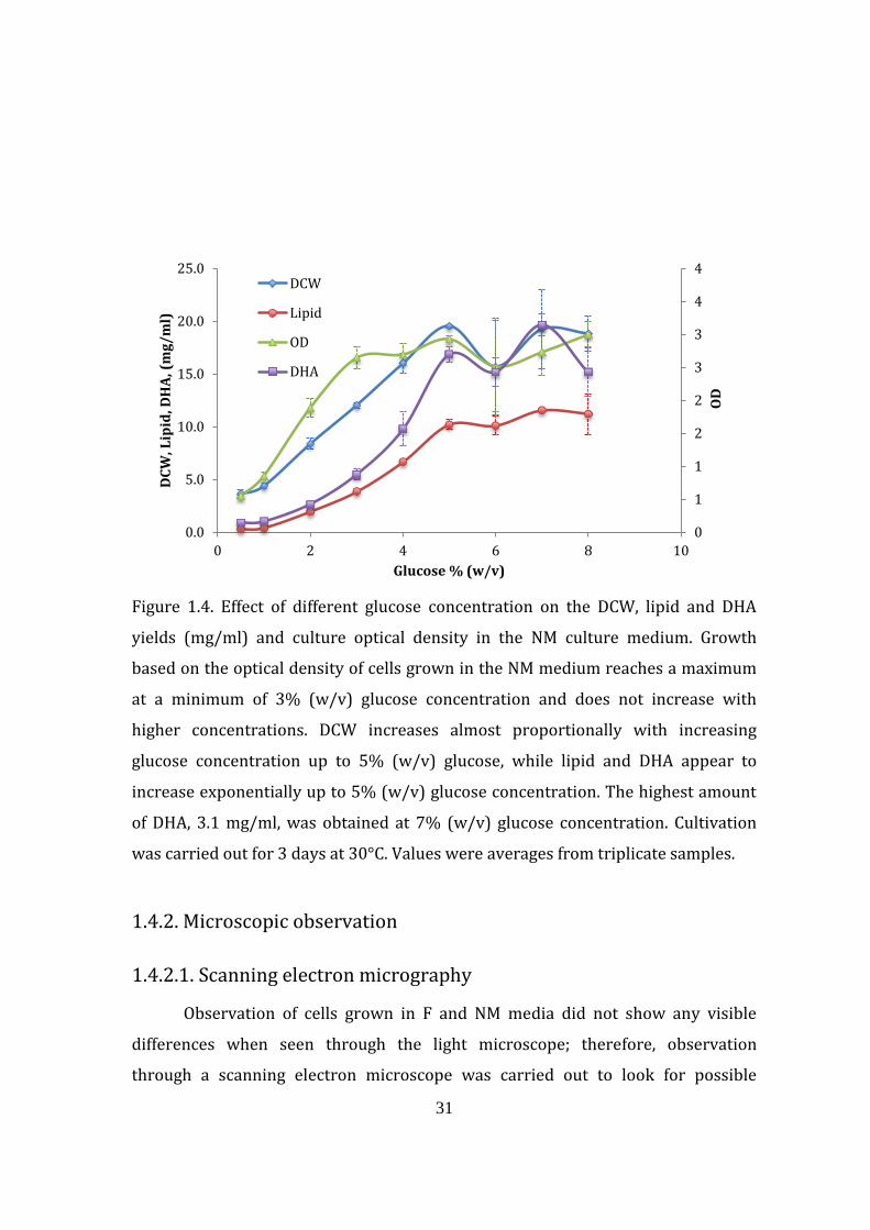
1.4.1.7. Effect of the concentration of glucose in NM medium on the growth and
DHA yield in strain 12B
In order to see the influence of glucose concentration on the DCW, lipid,
fatty acid and DHA yields in the NM medium, strain 12B was grown in 10 ml
medium in a 50 ml flask, supplemented with 0.1, 1, 2, 3, 4, 5, 6, 7 and 8% (w/v)
glucose, and incubated in the dark at 30°C and 150 rpm for 3 days. The efficiency of
thraustochytrid strain 12B in converting glucose to biomass (DCW) in the NM
medium was found to be highest when the glucose concentration in the medium
was 1 to 5% (w/v). Within this range of concentration, the increase of DCW was
found to be proportional to the glucose concentration, while lipid and DHA yields
increased exponentially. The highest DCW yield at 5% (w/v) glucose was 19.6
mg/ml, while the highest lipid and DHA yields were 11.6 and 3.1 mg/ml,
respectively. Cell optical density, however, reached a maximum at 3% (w/v) NaCl
without further increase at higher glucose concentrations, and the highest amount
of DHA (3.1 mg/ml) was obtained when the glucose concentration was 7% (w/v)
(Fig. 1.4).
31
Figure 1.4. Effect of different glucose concentration on the DCW, lipid and DHA
yields (mg/ml) and culture optical density in the NM culture medium. Growth
based on the optical density of cells grown in the NM medium reaches a maximum
at a minimum of 3% (w/v) glucose concentration and does not increase with
higher concentrations. DCW increases almost proportionally with increasing
glucose concentration up to 5% (w/v) glucose, while lipid and DHA appear to
increase exponentially up to 5% (w/v) glucose concentration. The highest amount
of DHA, 3.1 mg/ml, was obtained at 7% (w/v) glucose concentration. Cultivation
was carried out for 3 days at 30°C. Values were averages from triplicate samples.
1.4.2. Microscopic observation
1.4.2.1. Scanning electron micrography
Observation of cells grown in F and NM media did not show any visible
differences when seen through the light microscope; therefore, observation
through a scanning electron microscope was carried out to look for possible
0
1
1
2
2
3
3
4
4
0.0
5.0
10.0
15.0
20.0
25.0
0 2 4 6 8 10
OD
DC
W, L
ipid
, DH
A, (
mg
/m
l)
Glucose % (w/v)
DCW
Lipid
OD
DHA
32
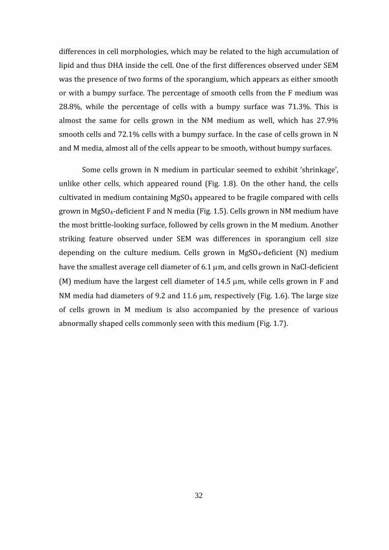
differences in cell morphologies, which may be related to the high accumulation of
lipid and thus DHA inside the cell. One of the first differences observed under SEM
was the presence of two forms of the sporangium, which appears as either smooth
or with a bumpy surface. The percentage of smooth cells from the F medium was
28.8%, while the percentage of cells with a bumpy surface was 71.3%. This is
almost the same for cells grown in the NM medium as well, which has 27.9%
smooth cells and 72.1% cells with a bumpy surface. In the case of cells grown in N
and M media, almost all of the cells appear to be smooth, without bumpy surfaces.
Some cells grown in N medium in particular seemed to exhibit ‘shrinkage’,
unlike other cells, which appeared round (Fig. 1.8). On the other hand, the cells
cultivated in medium containing MgSO4 appeared to be fragile compared with cells
grown in MgSO4-deficient F and N media (Fig. 1.5). Cells grown in NM medium have
the most brittle-looking surface, followed by cells grown in the M medium. Another
striking feature observed under SEM was differences in sporangium cell size
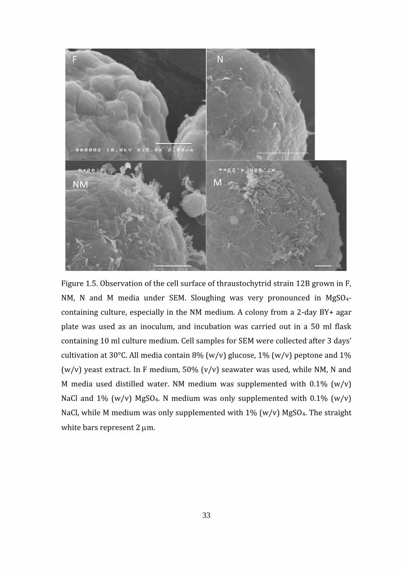
depending on the culture medium. Cells grown in MgSO4-deficient (N) medium
have the smallest average cell diameter of 6.1 m, and cells grown in NaCl-deficient
(M) medium have the largest cell diameter of 14.5 m, while cells grown in F and
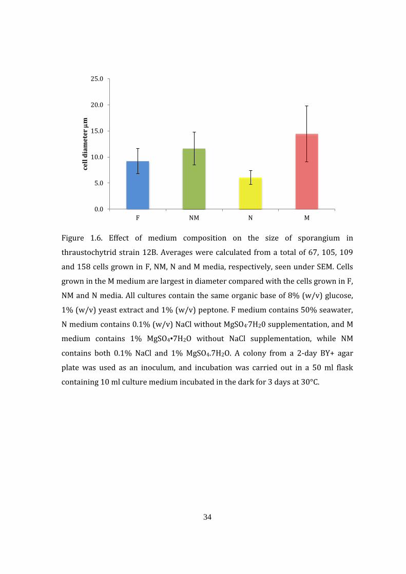
NM media had diameters of 9.2 and 11.6 m, respectively (Fig. 1.6). The large size
of cells grown in M medium is also accompanied by the presence of various
abnormally shaped cells commonly seen with this medium (Fig. 1.7).
33
Figure 1.5. Observation of the cell surface of thraustochytrid strain 12B grown in F,
NM, N and M media under SEM. Sloughing was very pronounced in MgSO4-
containing culture, especially in the NM medium. A colony from a 2-day BY+ agar
plate was used as an inoculum, and incubation was carried out in a 50 ml flask
containing 10 ml culture medium. Cell samples for SEM were collected after 3 days’
cultivation at 30°C. All media contain 8% (w/v) glucose, 1% (w/v) peptone and 1%
(w/v) yeast extract. In F medium, 50% (v/v) seawater was used, while NM, N and
M media used distilled water. NM medium was supplemented with 0.1% (w/v)
NaCl and 1% (w/v) MgSO4. N medium was only supplemented with 0.1% (w/v)
NaCl, while M medium was only supplemented with 1% (w/v) MgSO4. The straight
white bars represent 2 m.
34
Figure 1.6. Effect of medium composition on the size of sporangium in
thraustochytrid strain 12B. Averages were calculated from a total of 67, 105, 109
and 158 cells grown in F, NM, N and M media, respectively, seen under SEM. Cells
grown in the M medium are largest in diameter compared with the cells grown in F,
NM and N media. All cultures contain the same organic base of 8% (w/v) glucose,
1% (w/v) yeast extract and 1% (w/v) peptone. F medium contains 50% seawater,
N medium contains 0.1% (w/v) NaCl without MgSO4.7H20 supplementation, and M
medium contains 1% MgSO4•7H2O without NaCl supplementation, while NM
contains both 0.1% NaCl and 1% MgSO4.7H2O. A colony from a 2-day BY+ agar
plate was used as an inoculum, and incubation was carried out in a 50 ml flask
containing 10 ml culture medium incubated in the dark for 3 days at 30°C.
0.0
5.0
10.0
15.0
20.0
25.0
F NM N M
cell
dia
me
ter
m
35
Figure 1.7. Abnormal cell shapes seen from thraustochytrid strain 12B cells grown
in M medium. Some cells from the M culture show irregular cell shapes from the
normal round sporangium seen in F and NM culture. The M medium is composed of
1% (w/v) MgSO4.7H2O without NaCl supplementation but contains an organic base
of 8% (w/v) glucose, 1% (w/v) yeast extract and 1% (w/v) peptone. The culture
was carried in a 50 ml flask by using 10 ml culture solution. The culture was grown
for 3 days at 30°C in the dark. The straight white bars represent 2 m.
36
1.4.2.2. Transmission electron micrography
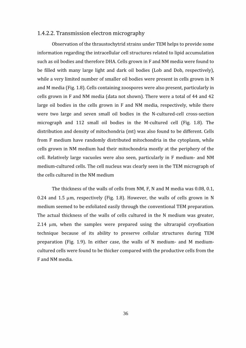
Observation of the thraustochytrid strains under TEM helps to provide some
information regarding the intracellular cell structures related to lipid accumulation
such as oil bodies and therefore DHA. Cells grown in F and NM media were found to
be filled with many large light and dark oil bodies (Lob and Dob, respectively),
while a very limited number of smaller oil bodies were present in cells grown in N
and M media (Fig. 1.8). Cells containing zoospores were also present, particularly in
cells grown in F and NM media (data not shown). There were a total of 44 and 42
large oil bodies in the cells grown in F and NM media, respectively, while there
were two large and seven small oil bodies in the N-cultured-cell cross-section
micrograph and 112 small oil bodies in the M-cultured cell (Fig. 1.8). The
distribution and density of mitochondria (mt) was also found to be different. Cells
from F medium have randomly distributed mitochondria in the cytoplasm, while
cells grown in NM medium had their mitochondria mostly at the periphery of the
cell. Relatively large vacuoles were also seen, particularly in F medium- and NM
medium-cultured cells. The cell nucleus was clearly seen in the TEM micrograph of
the cells cultured in the NM medium
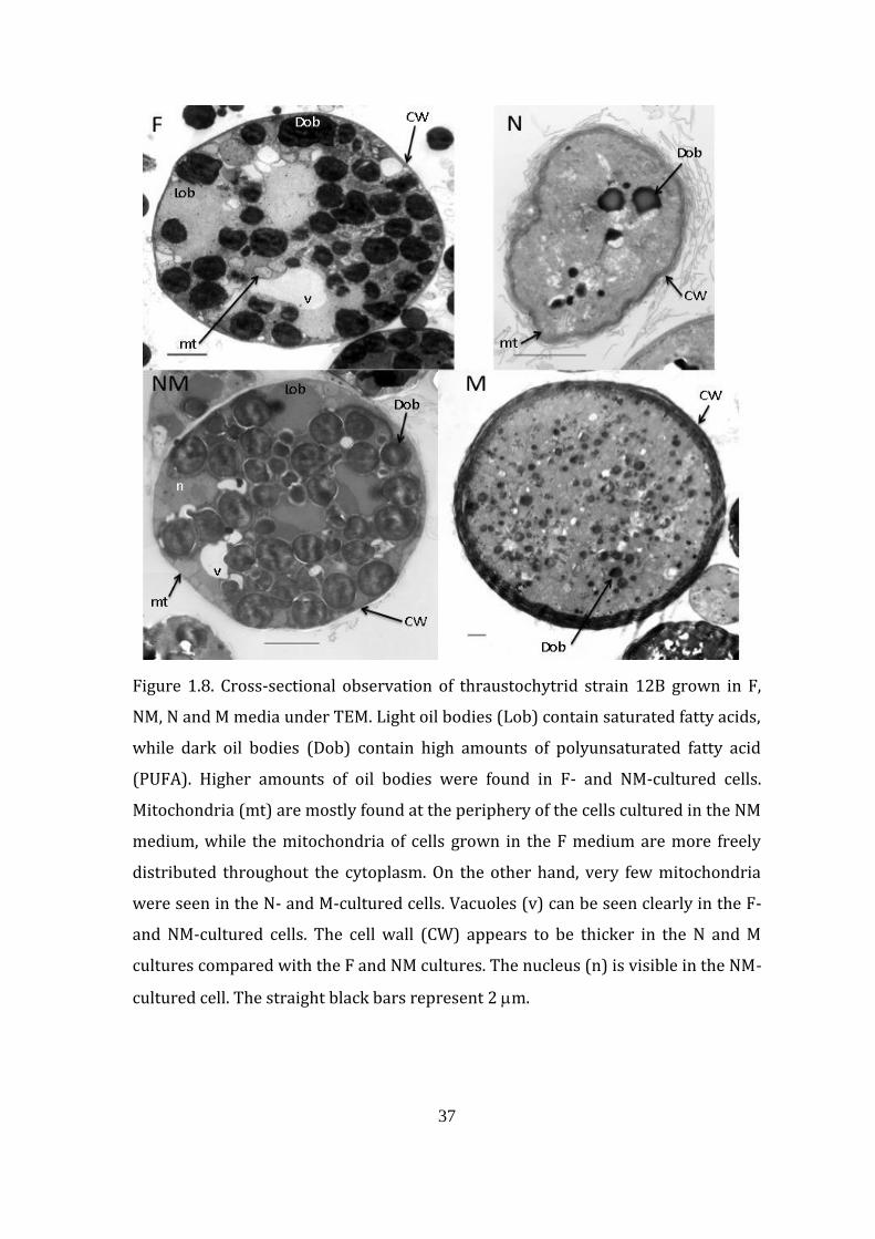
The thickness of the walls of cells from NM, F, N and M media was 0.08, 0.1,
0.24 and 1.5 m, respectively (Fig. 1.8). However, the walls of cells grown in N
medium seemed to be exfoliated easily through the conventional TEM preparation.
The actual thickness of the walls of cells cultured in the N medium was greater,
2.14 m, when the samples were prepared using the ultrarapid cryofixation
technique because of its ability to preserve cellular structures during TEM
preparation (Fig. 1.9). In either case, the walls of N medium- and M medium-
cultured cells were found to be thicker compared with the productive cells from the
F and NM media.
37
Figure 1.8. Cross-sectional observation of thraustochytrid strain 12B grown in F,
NM, N and M media under TEM. Light oil bodies (Lob) contain saturated fatty acids,
while dark oil bodies (Dob) contain high amounts of polyunsaturated fatty acid
(PUFA). Higher amounts of oil bodies were found in F- and NM-cultured cells.
Mitochondria (mt) are mostly found at the periphery of the cells cultured in the NM
medium, while the mitochondria of cells grown in the F medium are more freely
distributed throughout the cytoplasm. On the other hand, very few mitochondria
were seen in the N- and M-cultured cells. Vacuoles (v) can be seen clearly in the F-
and NM-cultured cells. The cell wall (CW) appears to be thicker in the N and M
cultures compared with the F and NM cultures. The nucleus (n) is visible in the NM-
cultured cell. The straight black bars represent 2 m.
38
Figure 1.9. TEM image of thraustochytrid strain 12B in F, NM and N media sampled
using the ultrarapid cryofixation technique. Many oil bodies were found in F- and
NM-cultured cells. Light oil bodies (Lob) indicate higher amounts of saturated fatty
acids, while dark oil bodies (Dob) indicate a higher composition of unsaturated
fatty acids. Peripheral distribution of mitochondria (mt) for cells grown in NM is
indicated. The original thick cell wall (CW) was preserved in the N-cultured cell by
using the ultrarapid cryofixation technique. The walls of cells grown in F and NM
media, however, remained thin and similar to when the cells were prepared using
the standard noncryofixation TEM method. The strain 12B observed here was
cultured in media containing the same organic base of 8% (w/v) glucose, 1% (w/v)
yeast extract and 1% (w/v) peptone. Only the F culture is composed of 50% (v/v)
seawater, while the N medium contains 0.1% (w/v) NaCl without MgSO4.7H20
supplementation, the M medium contains 1% MgSO4•7H2O without NaCl
supplementation, and the NM medium contains both 0.1% NaCl and 1%
MgSO4•7H2O. A colony from a 2-day BY+ agar plate was used as an inoculum, and
incubation was carried out in a 50 ml flask containing 10 ml culture medium
incubated in the dark for 3 days at 30°C. The white bars represent 2 m.
39
1.5. Discussion
1.5.1. NaCl and cell productivity
Thraustochytrids are marine microorganisms and require minerals that
exist in seawater for growth. The major inorganic compounds present in seawater
are ions of chloride (Cl–), sodium (Na+), sulfate (SO42–), magnesium (Mg2+), calcium
(Ca2+), potassium (K+) and bicarbonate (HCO3–) at concentrations of 19.0, 10.6, 2.6,
1.3, 0.4, 0.4 and 0.1 mg/ml, respectively (Brown et al., 1989). It is considered that
one or more combinations of these ions could be necessary for optimal growth and
high DHA production by microorganisms including thraustochytrids, and for this
reason, NaCl, MgSO4, KHSO4, and CaCO3 were used as representative compounds to
screen for the most essential inorganic supplement using thraustochytrid strain
12B. To assess this, initially the amount of lipid was used as an indirect indicator
for DHA production, as higher lipid production would also lead to an increase in the
absolute amount of DHA (data not shown).
As shown in Figure 1.1A, strain 12B exhibited no growth in a NaCl-deficient
medium, indicating an obligate requirement of NaCl for growth. This is in
agreement with another report on the obligate requirement for NaCl in T. aureum
(Garrill et al., 1992). This is probably because both the sodium cation and the
chloride anion in thraustochytrids and other marine microorganisms have an
important role in osmotic regulation (Garrill et al., 1992, Wethered & Jennings,
1985). According to Perveen et al. (2006), strain 12B grew in a medium in which
50% (v/v) seawater was replaced with water containing only glucose, peptone and
yeast extract without NaCl. The growth of strain 12B observed in the absence of
NaCl in that experiment was probably due to the transfer of 0.1 ml inoculum from a
1 day preculture of strain 12B in F medium, which contains 50% (v/v) seawater,
thus giving rise to approximately 0.02% (w/v) NaCl in the culture medium. The use
of a cell colony directly for inoculation makes the present study more accurate, as
no carry-over of salts occurred, and strain 12B was indeed unable to grow in the
absence of NaCl. However, the fact that the strain was able to grow in medium
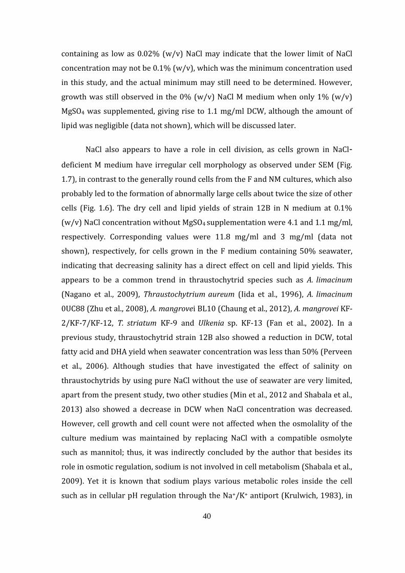
40
containing as low as 0.02% (w/v) NaCl may indicate that the lower limit of NaCl
concentration may not be 0.1% (w/v), which was the minimum concentration used
in this study, and the actual minimum may still need to be determined. However,
growth was still observed in the 0% (w/v) NaCl M medium when only 1% (w/v)
MgSO4 was supplemented, giving rise to 1.1 mg/ml DCW, although the amount of
lipid was negligible (data not shown), which will be discussed later.
NaCl also appears to have a role in cell division, as cells grown in NaCl-
deficient M medium have irregular cell morphology as observed under SEM (Fig.
1.7), in contrast to the generally round cells from the F and NM cultures, which also
probably led to the formation of abnormally large cells about twice the size of other
cells (Fig. 1.6). The dry cell and lipid yields of strain 12B in N medium at 0.1%
(w/v) NaCl concentration without MgSO4 supplementation were 4.1 and 1.1 mg/ml,
respectively. Corresponding values were 11.8 mg/ml and 3 mg/ml (data not
shown), respectively, for cells grown in the F medium containing 50% seawater,
indicating that decreasing salinity has a direct effect on cell and lipid yields. This
appears to be a common trend in thraustochytrid species such as A. limacinum
(Nagano et al., 2009), Thraustochytrium aureum (Iida et al., 1996), A. limacinum
0UC88 (Zhu et al., 2008), A. mangrovei BL10 (Chaung et al., 2012), A. mangrovei KF-
2/KF-7/KF-12, T. striatum KF-9 and Ulkenia sp. KF-13 (Fan et al., 2002). In a
previous study, thraustochytrid strain 12B also showed a reduction in DCW, total
fatty acid and DHA yield when seawater concentration was less than 50% (Perveen
et al., 2006). Although studies that have investigated the effect of salinity on
thraustochytrids by using pure NaCl without the use of seawater are very limited,
apart from the present study, two other studies (Min et al., 2012 and Shabala et al.,
2013) also showed a decrease in DCW when NaCl concentration was decreased.
However, cell growth and cell count were not affected when the osmolality of the
culture medium was maintained by replacing NaCl with a compatible osmolyte
such as mannitol; thus, it was indirectly concluded by the author that besides its
role in osmotic regulation, sodium is not involved in cell metabolism (Shabala et al.,
2009). Yet it is known that sodium plays various metabolic roles inside the cell
such as in cellular pH regulation through the Na+/K+ antiport (Krulwich, 1983), in
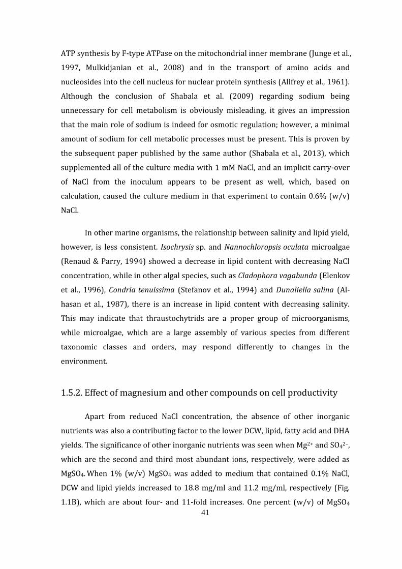
41
ATP synthesis by F-type ATPase on the mitochondrial inner membrane (Junge et al.,
1997, Mulkidjanian et al., 2008) and in the transport of amino acids and
nucleosides into the cell nucleus for nuclear protein synthesis (Allfrey et al., 1961).
Although the conclusion of Shabala et al. (2009) regarding sodium being
unnecessary for cell metabolism is obviously misleading, it gives an impression
that the main role of sodium is indeed for osmotic regulation; however, a minimal
amount of sodium for cell metabolic processes must be present. This is proven by
the subsequent paper published by the same author (Shabala et al., 2013), which
supplemented all of the culture media with 1 mM NaCl, and an implicit carry-over
of NaCl from the inoculum appears to be present as well, which, based on
calculation, caused the culture medium in that experiment to contain 0.6% (w/v)
NaCl.
In other marine organisms, the relationship between salinity and lipid yield,
however, is less consistent. Isochrysis sp. and Nannochloropsis oculata microalgae
(Renaud & Parry, 1994) showed a decrease in lipid content with decreasing NaCl
concentration, while in other algal species, such as Cladophora vagabunda (Elenkov
et al., 1996), Condria tenuissima (Stefanov et al., 1994) and Dunaliella salina (Al-
hasan et al., 1987), there is an increase in lipid content with decreasing salinity.
This may indicate that thraustochytrids are a proper group of microorganisms,
while microalgae, which are a large assembly of various species from different
taxonomic classes and orders, may respond differently to changes in the
environment.
1.5.2. Effect of magnesium and other compounds on cell productivity
Apart from reduced NaCl concentration, the absence of other inorganic
nutrients was also a contributing factor to the lower DCW, lipid, fatty acid and DHA
yields. The significance of other inorganic nutrients was seen when Mg2+ and SO42–,
which are the second and third most abundant ions, respectively, were added as
MgSO4. When 1% (w/v) MgSO4 was added to medium that contained 0.1% NaCl,
DCW and lipid yields increased to 18.8 mg/ml and 11.2 mg/ml, respectively (Fig.
1.1B), which are about four- and 11-fold increases. One percent (w/v) of MgSO4
42
seems to be saturated for strain 12B because further addition of this salt has no
further effects on the cell yield in the flask cultivation. The importance of
magnesium lies in its various roles in cell proliferation, apoptosis and protein
synthesis (Wolf & Cittadini, 1999) as well as in diverse enzyme reactions (Jahnen-
Dechnet & Ketteler, 2012). In the presence of a low NaCl concentration of 0.1%
(w/v), the addition of magnesium increased both DCW and lipid yields; however, in
the absence of NaCl, a negligible amount of lipid was extracted (data not shown).
The dependence on sodium is probably because it is involved in the transport of
magnesium into the cell through the sodium–magnesium antiport mechanism
(Flatman, 1991). This has been observed in Schizochytrium mangrovei, in which a
decrease in intracellular sodium concentration was seen together with an increase
in magnesium concentration (Unagul et al., 2006). However, as mentioned above,
thraustochytrids, including strain 12B, have an obligate requirement for sodium,
resulting in a slight growth of 1.1 mg/ml DCW from cells cultivated in the M
medium containing 1% (w/v) MgSO4 and no NaCl supplementation. Based on the
cell morphology from SEM and TEM observation with low 0.1% (w/v) NaCl and the
absence of MgSO4, some cells exhibit shrinkage (Fig. 1.8), suggesting that MgSO4
may have some role in osmotic regulation. Because one of the important roles of
NaCl is in osmotic adjustment (Shaballa et al., 2009), MgSO4 may have partly
fulfilled this role during NaCl deficiency. Another structural feature shown by
strain 12B cells grown in the absence of either NaCl or MgSO4 was the presence of
thicker cell walls as seen under TEM (Fig. 1.8). Thick-walled thraustochytrids have
been associated with low cell proliferation (Alderman et al., 1974) and therefore
poor growth during cultivation. This is reflected by the low amounts of DCW from
cells grown in NaCl-deficient M medium and MgSO4-deficient N medium, which are
4.1 and 1.1 mg/ml, respectively, as well as by the low lipid yields, which are 1.1
mg/ml and negligible, respectively.Thraustochytrid microorganisms normally
grow well in a medium containing 50% (v/v) seawater, which contains about 1.5%
(w/v) NaCl. When the cells are grown in medium containing no NaCl, such as the M
medium, or a low NaCl concentration, such as the N medium (0.1% NaCl, w/v), the
hydrostatic pressure inside the cell is expected to be lower than outside the cell.
This would result in an osmotic influx of water into the cell through the cell
43
membrane, which is somehow permeable to water and consequently could cause
the cell to burst (Lodish et al., 2000). For eukaryotic cells that have a cell wall such
as the thraustochytrids, an increase in cell volume because of osmotic inflow of
water is normally prevented, and an increase in intracellular pressure occurs
instead. However, unlike plant cells, which have a rigid cell wall, the cell wall of
thraustochytrids is flexible and lacks any rigid fibrillar structural components
(Darley et al., 1973). One way to increase the tensile strength of the cell wall is
therefore by increasing its thickness, as in the case of algae that have high turgor
pressure (Bisson, 1995). In addition, the thraustochytrid cell wall is also known to
be hydrophobic to some extent (Raghukumar et al., 2000); therefore, a thicker cell
wall would increase cell hydrophobicity, which therefore might restrict the osmotic
inflow of water into the cell. Thraustochytrid cells that were grown in a medium
with optimal osmolality would therefore have thinner cell walls, as seen in the
high-DHA-productive cells grown in the F and NM media.
1.5.3. Possible effect of limiting compounds
Apart from nitrogen, phosphorus is also considered to be a limiting nutrient
in seawater (Downing, 1997). Because KHSO4 has a corrosive property, the effect of
both noncorrosive phosphorus and non-phosphorus-containing potassium
compounds KH2PO4, K2HPO4 and K2SO4 was also investigated. The chemical nature
of the potassium-containing compounds varies as the pH of the culture medium
changes when the NM medium is supplemented by individual compounds. The pH
of K2HPO4 tends to increase, and that of KHSO4 and KH2PO4 tends to decrease,
while the pH of K2SO4 remains stable with increasing concentration (data not
shown). A slight increase in DCW was obtained initially at very low potassium
compound concentrations, but in general, the addition of potassium compounds
decreases DCW yield with increasing concentration (Fig. 1.2). Further analysis was
carried out at low potassium compound concentrations, and a slight increase in the
lipid, fatty acid and DHA absolute yields was obtained (data not shown). When the
culture medium was only composed of NaCl and K2SO4 as inorganic nutrients, the
DCW yield was only one-third that of NM medium, while the lipid yield was
44
approximately 19 times far lower than that of NM medium. Furthermore, the effect
on the yields was almost the same regardless of the absence (INM medium) or
presence (INMK medium) of K2SO4 in the medium with DHA productivity of 1.5 and
1.6 mg/ml/day, respectively (Table 1.3). This indicates that the magnesium ion, not
the sulfate ion, is the most essential ion in MgSO4 contrary to the previous
suggestion by Nagano et al. (2009). The cell wall is thought to be made up of
circular scales synthesized by the Golgi apparatus, which are transferred to the cell
surface, resulting in the laminated structure of the cell wall as seen in
thraustochytrid species T. aggregatum (Darley et al., 1973) and Althornia crouchii
(Alderman et al., 1974). In Aplochytrium sp. 2400/57, the cell wall is composed of
protein, carbohydrate and sulfate at 27.4%, 20% and 5.3%, respectively, of the
total cell wall matter (Ulken et al., 1985), while in Thraustochytrium sp., this varies
from 30% to 43% protein and from 21% to 36% carbohydrate. The other half of
the cell wall composition is not yet known (Ulken et al., 1985). Because sulfate
makes up 5.3% of the cell wall (Ulken et al., 1985), it is therefore still a necessary
inorganic component in the culture media.
The exclusion of potassium compounds from the culture medium can be
considered as advantageous as it will help to reduce industrial cost with simple
medium composition. Analysis of the fatty acid composition also shows that the
percentage of DHA per total fatty acid is generally higher in the absence of
potassium compound (data not shown). The percentage composition of C18:1 is
also lower, at 1.4% instead of about 29%, when the medium is not supplemented
with potassium; therefore, the extracted oil has higher quality and lower
complexity for DHA purification when potassium compounds are not added to the
culture medium. On the other hand, the addition of calcium and carbonate ions also
tended to reduce the DCW and lipid yields (Fig. 1.1C, 1.1D), proving it to be
unnecessary for strain 12B and possibly for other thraustochytrids as well. The
concentration needed for these nutrients is probably low and could be obtained
from the organic nutrients such as the yeast extract.
The characteristic genus-specific, fatty acid composition of strain 12B
additionally was found not to be affected by the culture medium. The ARA
45
percentage composition was unaffected at less than 3%, which can be used to
distinguish it from the genus Sicyoidochytrium, which normally has no AA, and this
also is the case for the genus Aplanochytrium (Yang et al., 2010), while
Schizochyrium, Ulkenia, botryochytrium and parietichytrium all have AA at more
than 3% per total fatty acid (Laby Base, 2009a). The strain 12B n-6
docosapentaenoic acid (DPA) was also unaffected, and the absence of n-3 (DPA)
eliminates the probability of it being under the genus Oblongichytrium (Laby Base,
2009b). Apart from the lack of lipid information for the genera Althornia and
Japonochytrium, and the absence of a unifying fatty acid characteristic for
Thraustochytrium, it is therefore most likely that thraustochytrid strain 12B
belongs to the genus Aurantiochytrium, based on the information of its fatty acid
composition.
1.5.4. Applicability of the NM medium
Apart from being noncorrosive to culture equipment at low NaCl
concentration, the NM medium’s DHA yield is also about five times higher than that
of the F medium after 3 days’ cultivation, hence exhibiting a faster lipid
accumulation (Table 1.1). DHA was mostly stored in the form of triacylglycerol
(TG). The NM medium was also applicable to other thraustochytrid strains such as
the Aurantiochytrium sp. SR 21 strain, which has a higher efficiency of glucose
utilization in the NM medium compared with the F medium, which becomes
depleted after 5 days instead of 6 days (data not shown). The NM medium also
requires less than 8% (w/v) glucose (Fig. 1.4) as opposed to the optimal
concentration previously reported (Perveen et al., 2006). The faster lipid
accumulation is probably related to growth as both the 12B and the SR21
thraustochytrid strains enter the stationary phase earlier during cultivation in the
NM medium (Fig. 1.3). This is an important feature because cells in the stationary
phase tend to have a high amount of triacylglycerol (Ratha & Prasanna, 2012). Cells
grown in medium containing MgSO4, especially in NM medium, show sloughing of
the cell surface (Fig. 1.5), which is a characteristic feature of mature cells (Harrison
& Jones, 1974) and might be related to the early stationary growth (Fig. 1.3).
46
The thraustochytrid strain 12B cells appeared to have a surface that was
either relatively smooth or bumpy, with the latter being most common, especially
in cells grown in F and NM media. The bumpy surface is most likely due to the
presence of oil bodies inside the cell, suggesting a soft or flexible nature of the cell
wall, and therefore can be used as an indirect indication of high lipid production.
The flexible nature of the cell wall has been reported by Darley et al. (1973) in T.
aggregatum, whereby the cell wall obtained from sonication was easily folded or
wrinkled during whole mount preparation because of the absence of rigid fibrillar
structural components. Oil bodies appear as light, grayish or dark regions inside
the cells, which occurs through the reduction of EM reagent OsO4 to the black
colored OsO2, especially in lipid (Hayes et al., 1963), particularly in fatty acids with
cis-unsaturated bonds (Adams et al., 1967) with increased in the intensity with the
increase in the number of double bonds as has been demonstrated previously using
C18:1, C18:2 and DHA (Cheng et al., 2009). Some oil bodies also have gradation of
intensity, which is probably due to the formation of a semicrystalline
triacylglycerol state, forming alternate dark PUFA layers and light saturated-
monounsaturated fatty acid layers (Ashford et al., 2000). Hollow membrane-like
structures can also be seen inside some of the smaller oil bodies (data not shown),
which is thought to be associated with lipid synthesis, which become internalized
during lipid deposition (Weete et al., 1997) and disappear in larger oil bodies
(Morita et al., 2006).
Compared with previous studies, the 0.2 g/l/day DHA productivity from the
flask cultivation in F medium from this experiment was lower than that in earlier
experiments, which reported a productivity of 2.8 g/l/day (Perveen et al., 2006)
and 3.1 g/l/day (Okuyama et al., 2008a), which is probably due to differences in
inoculation. The DHA productivity of strain 12B in the NM medium increased
successively from cultivation using laboratory-grade to industrial-grade
ingredients and from flask to jar fermentation (Table 1.3). However, when the
industrial-grade medium (IN) was not supplemented with magnesium,
productivity was comparatively very low, with DCW and lipid yields being 4.6 and
2.7 mg/ml, respectively; therefore, the presence of possible contaminating
47
compounds is still not as crucial as magnesium supplementation (data not shown).
Alternatively, the strain probably favors plant-based nitrogen sources, as the
laboratory-grade peptone used was animal based (casein pancreatin digest), while
the industrial-grade peptone was plant based (made from soy).
The preference for plant-based nitrogen sources may be related to the
ecology of thraustochytrids, as these microorganisms generally live on plant
detritus in nature (Raghukumar, 2002). In particular, one of the major amino acids
produced from metabolism of ammonia in plant leaves is glutamic acid (Miflin &
Lea, 1982). This is a major amino acid component of the cell wall, accounting for
28.5% of the total amino acids in T. aggregatum (Darley et al., 1973). It is also
among the most efficient nitrogen sources for thraustochytrid growth (Singh &
Ward, 1997) and has been reported to elicit strong attraction responses in S.
mangrovei zoospores (Fan et al., 2002). Although the information on the amount of
glutamic acid in the peptone was not disclosed by the manufacturer, a high amount
of glutamic acid has been known to be present in soybean (Wolf et al., 1982). The
use of plant-based ingredients is also advantageous for industrial application as it
will be able to comply with the halal food industry requirements. The increase in
DCW, lipid, fatty acid and DHA yields when the cultivation method was changed
from flask to jar is thought to be due to the better aeration and mixing in the jar
fermenter. The highest DHA productivity from the jar fermenter for strain 12B
reported so far is 5.4 g/l/day (Okuyama, 2008a); therefore, further optimization of
large-scale cultivation parameters is possible when using the NM medium to
increase the DHA yield from the current 2.3 g/l/day productivity.
48
1.6. Conclusions
A noncorrosive NM medium consisting of 8% glucose, 1%(w/v) yeast
extract and 1% (w/v) peptone supplemented with 0.1% (w/v) NaCl and 1% (w/v)
MgSO4, resulted in high DCW, lipid and DHA yields of 18.8, 11.2 and 2.4 mg/ml,
respectively, after 3 days’ cultivation, which is therefore superior to the F medium
containing 50% seawater. The DHA yield was further increased through jar
cultivation using industrial-grade chemicals producing 11.7 mg/ml DHA or a
productivity of 2.3 mg/ml/day. DHA in TG contributed to 71% of total DHA when
strain 12B was grown in F medium and 81% in the NM medium. NaCl and MgSO4
are the most important inorganic compounds for the cultivation of thraustochytrid
strain 12B, while other compounds—CaCO3, phosphorous and non-phosphorous-
containing potassium compounds KHSO4, K2SO4, KH2PO4 and K2HPO4—either
reduce the DHA yield or decrease the DHA purity by increasing the percentage
composition of C18:1. Cells grown in the NM medium have high amounts of oil
bodies and exhibit thin cell walls and sloughing of the cell surface, indicating
prolific growth and rapid cell maturity. This is supported by the growth profile,
which shows cells from the NM culture entering the stationary phase earlier than
cells from the F culture. Cells from the NM medium also have thin cell walls, which
indicate prolific growth compared with the case when either NaCl or MgSO4 is
deficient (N and M media). The absence of NaCl results in abnormally large cells
and unusual cell morphology, which may indicate the role of NaCl in cellular
division. The absence of MgSO4, on the other hand, results in shrinking of some
cells, indicating its possible role in osmotic regulation. The NM medium also has
higher glucose utilization efficiency, with glucose being depleted faster than in F
medium, and less than 8% (w/v) glucose is required for high DCW, lipid and DHA
yields. The NM medium was also applicable to the thraustochytrid species
Aurantiochytrium limacinum SR21 and possibly to other thraustochytrid species as
well.
49
Chapter 2: Tentative identification of
thraustochytrid strain 12B
2.1 Summary
Molecular and morphological taxonomic data indicated that the
thraustochytrid strain 12B belongs to the genus Aurantiochytrium. Strain 12B
exhibited the presence of an amoebic stage, the disintegration of the cell wall
during zoospore release, a lack of naked protoplasm and the rare presence of an
ectoplasmic network, all indicating that it belongs to the genus Aurantiochytrium.
Moreover, phylogenetic analysis using the 18S rRNA gene sequence showed that
strain 12B groups together well with other Aurantiochytrium species. This is
further supported by mitochondrial DNA analysis, which shows a very high
sequence similarity to Aurantiochytrium limacinum SR21 compared with a strain
from another genus, Thraustochytrium aureum. The cytochrome oxidase II (COXII)
gene probably has a high taxonomic value at the genus level for thraustochytrid
microorganisms. The proposed cutoff value for the percentage 18S rRNA sequence
similarity of 98.8% for a species was higher than the 98.5% similarity found
between strain 12B and A. limacinum, which suggests that strain 12B and A.
limacinum may be closely related but are two different species. This is further
supported by the differences in physiology and phenotype between strain 12B and
A. limacinum SR21, which showed differences seen in terms of coloration on agar
culture, rate of glucose utilization, differences in DCW, lipid, fatty acid and DHA
yields during cultivation as well as differences in the zoospore flagellum structure.
It has also been reported previously that fusion between cells was observed in
strain 12B, which has never been mentioned in other thraustochytrid species. The
presence of multiple 18S rRNA gene copy in the previous study of strain 12B is also
almost unique among thraustochytrid microorganisms. Comparison of the
structure of tubular hair on the anterior flagellum between strain 12B and SR21
50
also indicates an apparent difference, providing further support of their status as
separate species. In addition, from the life-cycle study, when grown in the NM
medium, strain 12B was observed to have shorter motile stages and longer
immotile stages compared with cells grown in the F medium containing 50%
seawater, which could explain why cells grown in the NM medium accumulated
lipid faster and more through conservation of oil bodies than those grown in the F
medium.
51
2.2. Introduction
2.2.1. Taxonomic overview of thraustochytrid microorganisms
Thraustochytrid microorganisms have been grouped together with brown
algae (Phaeophyceae), yellow-green algae (Xanthophyceae), golden algae
(Chrysophyceae) and diatoms (Bacillariophyta) under the same phylum,
Heterokonta (Cavalier-smith, 1994, NCBI taxonomy, 2013). Some authors use the
term ‘thraustochytrids’ to refer to genera or species from both families of
thraustochytriaceae and labyrinthulaceae (Gupta et al., 2012, Metz et al., 2010),
while others use the term specifically for genera or species under the
thraustochytriaceae family only (Raghukumar, 2008b, Tsui and Vrijmoed, 2012). In
this study, the meaning of ‘thraustochytrids’ used by Raghukumar (2008b) and
Tsui and Vrijmoed (2012) is followed. Various taxonomic changes were made for
the thraustochytrid group, with the latest taxonomic classification being domain
Eukaryota, kingdom Chromalveolata, phylum Heterokonta/Stramenopiles, class
Labyrinthulomycetes, order Labyrinthulales and under the family
Thraustochytriaceae, while Labyrinthulids, on the other hand, belong to the same
class but under the family Labyrinthulaceae (Honda, 2009, Gupta et al., 2012, NCBI,
2013). Thraustochytrid strains form 11 genera: Althornia (1), Aplanochytrium (8),
Aurantiochytrium (2), Botryochytrium (1), Japonochytrium (1), Oblongichytrium (3),
Parietichytrium (1), Schizochytrium (1), Sicyoidochytrium (1), Thraustochytrium
(17) and Ulkenia (3) species, with the number of known species in brackets (Laby
Base, 2009c). Most thraustochytrid species are considered to have proper scientific
names; however, there are hundreds of thraustochytrid strains that have been
isolated but are still without proper species names and are waiting to be studied.
2.2.2. Characterization of thraustochytrid microorganisms
The unique characteristic of thraustochytrid microorganisms is that they all
have PUFAs, particularly DHA, in high quantity, and the percentage per total fatty
52
acid varies depending on genus (Huang et al., 2003, Yokoyama et al., 2007,
Yokoyama & Honda, 2007, Yang et al., 2010). The thraustochytrid strain 12B, for
example, was reported to contain DHA at 43% of total fatty acid, producing 2.8
mg/ml/day DHA (Perveen et al., 2006). The labyrinthulid species too may contain
DHA (Sakata et al., 2000, Kumon et al., 2002, 2006) but probably not in all species
(Kumon et al., 2003). Fatty acid composition has been used as a taxonomic tool for
differentiation of thraustochytrids at the genus level (Laby Base, 2009a, b, Yang et
al., 2010) as mentioned in Chapter 1. Taxonomic studies on strain 12B have been
carried out in the past. The zoospore of strain 12B has previously been observed to
have two different laterally inserted flagella, with the anterior flagellum being lined
with tubular hairs (Perveen et al., 2006). The presence of a multilayered cellulosic
cell wall, mitochondria with tubular cristae, an ectoplasmic network and the
presence of the amoebic form have been documented by Ando (2005) and were
also seen in this study, some of which are unique characteristics of thraustochytrid
microorganisms. Phagocytosis or fusion among thraustochytrid cells has never
been reported in the literature but was observed in strain 12B (Perveen and
Okuyama, unpublished data). Molecular characterization by using the 18S rRNA
gene was also undertaken for strain 12B, and direct sequencing of the PCR product
from the 18S rRNA amplification was not possible unless the product was cloned
into Escherichia coli first. It was found that as many as 32 multiple sequences for
the 1727 base pair (bp) 18S rRNA gene existed for strain 12B (Ando, 2005).
Although the presence of several 18S rDNA units in tandem is common in
eukaryotes (Long and Bawid, 1980), reports of this in thraustochytrids appears to
be limited to only one paper, regarding Schizochytrium sp. TIO01 (Cheng et al.,
2011); therefore, this might be a unique characteristic only for some
thraustochytrid species including strain 12B. Definitive taxonomic identification,
however, has not yet been made for strain 12B because of the confusion in the
taxonomic classification at that time. Major strides in improving the taxonomic
classification of thraustochytrids have been made and are still going on; therefore,
reprocessing of the accumulated information on strain 12B to cope with the
changes in taxonomy, and further studies, are still necessary for its identification.
53
Traditional taxonomic studies on thraustochytrids normally focus on
phenotypic characterization and life cycle for strain identification (Harrison &
Jones, 1974, Alderman et al., 1974), and these are still used today. A biochemical
method using acriflavine and ThrFL1 probe has been available to identify
thraustochytrids from other protists through florescence (Gupta et al., 2012). For
intergenus taxonomic characterization, apart from morphology, fatty acid
composition analysis (Huang et al., 2003, Yokoyama et al., 2007a, Yokoyama &
Honda, 2007, Yang et al., 2010) and carotenoid composition (Yokoyama et al., 2007,
Yokoyama & Honda, 2007) have already been used. Molecular methods are
increasingly used together with morphological information to reinforce the species
taxonomic identification. The first attempt to use the molecular method in the
phylogenetic study of thraustochytrids was probably by Porter and Kochert (1978)
using rRNA molecular weight, and over the past decade, an increasing number of
studies have incorporated the molecular approach. Molecular markers that have
been used in the phylogenetic study of thraustochytrids include genes of 5S rRNA
(Mannella et al., 1987), 18S rRNA (Cavalier-Smith et al., 1994, Leipe et al., 1996,
Honda et al., 1999, Mo et al., 2002, Yokoyama et al., 2007, Yokoyama & Honda, 2007,
Tsui et al., 2009), SSU rRNA, 5.8S rRNA (Stokes et al., 2002, Qian et al., 2007),
internal transcribed spacers 1 and 2 (ITS 1 & 2, Qian et al., 2007), actin, tubulin and
elongation factor 1-alpha (Tsui et al., 2009), as well as DNAs amplified by the
randomly amplified polymorphic DNA (RAPD) method (Oclarit & Hepowit, 2007).
Mitochondrial DNA is also a useful molecular marker and may help to reinforce the
phylogeny data from the 18S rRNA gene, which is the most widely used molecular
marker for phylogenetic relationship in eukaryotes. Mitochondrial genes such as
cytochrome oxidase I (COXI) and NADH dehydrogenase subunit 5 (ND5 have been
used for phylogenetic studies of various organisms including, but not restricted to,
flies (Bajpai & Tewari, 2010), cytochrome oxidase subunit II (COXII) and
cytochrome b in butterflies (Torres et al., 2001) and mammals (Honeycutt et al.,
1995, Piaggio & Spicer, 2001), cytochrome oxidase III (COXIII) in protozoa (Tian et
al., 2013), and ND2 and ATPase6 in fish (Mu et al., 2012). Phylogenetic analysis
using mitochondrial DNA COXI has also been used on thraustochytrids (Qian et al.,
2007).
54
Mitochondrial COXI DNA sequence has become a standard molecular tool for
species identification in eukaryotic organisms (Ratnasingham and Hebert, 2007).
Cytochrome oxidase functions as the final enzyme in the respiratory chain (Denis,
1985) and also as a proton pump that generates transmembrane electrochemical
gradient for ATP synthesis (Richter and Ludwig, 2003). In the mature enzyme
complex, the center is made up of subunits I, II and III, which are encoded by the
mitochondrion (Richter and Ludwig, 2003). Like the commonly used nuclear DNA
molecular tool, the 18S rRNA gene, the important role of COX in the fundamental
functioning of the cell results in the conservation of the DNA sequence. However,
compared with the 18S rRNA gene, the COX gene sequence is more variable
because of its location in the mitochondria, as it is predisposed to damage from
reactive oxygen species produced during mitochondrial oxidative phosphorylation
(Yakes and Houten, 1997) and because there are fewer DNA repair mechanisms in
the mitochondria compared with the nucleus (Larsen et al., 2005). Compared with
nuclear DNA, mitochondrial DNA is also a favorable molecular tool for several
reasons including the presence of multiple copies allowing easy amplification and
the general absence of introns (Galtier et al., 2009). Moreover, the short length of
about 600 bp pairs of the COX gene facilitates rapid sequencing but contains
adequate nucleotide variation for species identification (IBOL, 2014). The
phylogenetic analysis of the COXII gene in the pseudofungus Pythium species under
the class Oomycete, which was the same class to which thraustochytrids previously
belonged (Sparrow, 1960), has shown good resolution of species and forms
groupings that agree with 28S rRNA gene analysis (Martin, 2000) or in some cases
the phylogenetic analysis of the COXII gene is better than that of the ribosomal
internal transcribed spacer (Robideau et al., 2011). The interspecies resolution of
COXII also helped to differentiate between two morphologically similar Pythium
species, P. oligandrum and P. hydrosporum (Villa, 2006), while COXI was also used
to identify new Pythium species (Long et al., 2013). The use of COXI in microalgae
(Evans and Mann, 2009) and algae (Robba et al., 2006, Boo et al., 2011) has also
been reported.
55
The aim of this chapter is to investigate the taxonomic status of strain 12B,
which previously has been poorly characterized, and to apply the findings in life-
cycle and other characterization studies to help explain further the accumulation of
lipid and DHA in the cells grown in NM medium that contain 0.1% NaCl and 1.0%
MgSO4 but not seawater (see Chapter 1).
56
2.3. Materials and methods
2.3.1. Microorganisms and their cultivation
Two thraustochytrid microorganisms were cultivated, the thraustochytrid
strain 12B (strain 12B; NITE P-68) and Aurantiochytrium limacinum SR21. Strain
12B was previously isolated from a mangrove ecosystem in Okinawa, Japan
(Perveen et al., 2006), while A. limacinum SR21, isolated from a coral reef area of
the Yap Islands, Federated States of Micronesia and originally identified as
Schizocytrium limacinum SR21 (IFO 32693) (Nakahara et al., 1996), was kindly
provided by the IFO, with permission of Dr. Yokochi Toshihiro, National Institute of
Advanced Science and Technology (AIST). For DNA extraction, both strains were
cultivated in NM medium containing 8% (w/v) glucose, 1% (w/v) yeast extract, 1%
(peptone), 0.1% (w/v) NaCl and 1% (w/v) MgSO4 for 3 days at 30°C. F medium was
also used for cultivation. The composition of F medium is almost the same as NM
medium except that in place of NaCl and MgSO4, 50% (v/v) seawater was used.
2.3.2. DNA extraction, PCR, and sequencing the mitochondrial COX gene
In this study, the 18S rRNA gene sequence, which had been determined by
Ando (2005), was reanalyzed with other thraustochytrid sequences from GenBank
by choosing representative species from every thraustochytrid genus based on the
latest taxonomic classification. In order to know the phylogenetic relationship
between strain 12B and Schizochytrium (Aurantiochytrium) limacinum SR21, which
is considered to be the closest organism to strain 12B Ando (2005), mitochondrial
DNA COXI, COXII, and COXII including ITS (COXII+ITS) gene sequences of these two
strains were determined.
Fresh genomic DNA was extracted from strain 12B and A. limacinum SR21
cells using the DNeasy Plant Mini Kit (Qiagen, Tokyo, Japan). Three sets of primers
were used for the amplification of COXI, COXII, and COXII+ITS. Primers FM55 and
FM52R (Long et al., 2013) were used to amplify the COXI gene, and primers FM58
57
and FM66 (Villa et al., 2006) were used to amplify the COXII gene, while primers
FM35 and FMPhy-10b (Martin, 2000) were used to amplify the COXII+ITS gene
(Table 2.1). The PCR mix is composed of 21.75 l H2O, 25 l PCR buffer, 1 l
forward and reverse primers (6.4 pmol), 0.25 l MightyAmp DNA polymerase and
1 l template DNA (10 ng/l). The PCR temperature program for COXI was: 95°C
for 3 min initial denaturation, 35 set cycles of 95°C for 30 sec denaturation, 54°C
for 30 sec annealing, 72°C for 30 sec extension, and a final 72°C for 5 min extension
step, while the annealing temperature for COXII and COXII+ITS was set to 56°C
(GeneAmp 2720 Thermal Cycler, Applied Biosystems, Tokyo, Japan). The amplified
gene segment was confirmed by electrophoresis (Mupid, Advance Co. Ltd, Tokyo,
Japan) against a known 100 bp molecular marker (Takara, Shiga, Japan). PCR
products were purified for sequencing using the GenElute PCR Clean-Up Kit
(Sigma-Aldrich, Tokyo, Japan). Samples for sequencing were prepared by mixing 1
l PCR product, 1 l primer (6.4 pmol) and 12 l distilled water. A total of six
reaction mixtures per sample, one for each primer (Table 2.1), were sent to Operon
Biotechnology Corp. and sequenced (ABI 3730XL, Applied Biosystems, Tokyo,
Japan). Sequences obtained were then aligned, edited and run in the NCBI BLAST
database search (Discontiguous MegaBLAST) for cross-species sequence similarity
analysis. For sequence identity/similarity analysis of multiple sequences, the
sequences were submitted online to http://www.ebi.ac.uk/Tools/msa/clustalw2/
(European Bioinformatics Institute, Cambridge, UK) for pairwise alignment and
DNA weight matrix analysis with ClustalW. The short length of the sequence at the
start and end containing the ‘N’ unknown nucleotides, which occurred because of
sequencing error, were not included in the sequence similarity calculation.
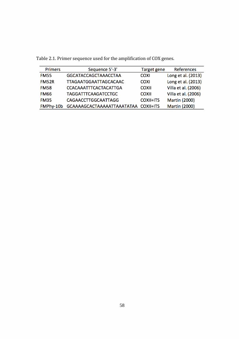
58
Table 2.1. Primer sequence used for the amplification of COX genes.
59
2.3.3. Observation of a life cycle of strain 12B
For the direct observation of life cycle, a small amount of cells was
transferred from a 2-day agar culture, by touching the colony with the tip of a
sterilized toothpick, into an Eppendorf tube containing 1 ml F medium, NM
medium or seawater. About 5–20 l of the suspension was then pipetted into the
well of a sterilized glass slide and covered with a coverslip. The well on the glass
slide was made by using vinyl tape (Nichiban, Tokyo, Japan), with one part of the
tape being discontinuous to provide ventilation. Observation of the life cycle of
thraustochytrid strain 12B was carried out using a light microscope (BX 51TF,
Olympus, Tokyo, Japan) under a UPlanf1 40/0.75 objective, with the light set to
between 4 and 6. An eyepiece camera (MacWin MW130, Science-eye, Niiza, Japan)
was inserted in the microscope observation tube and connected directly to a
computer, which recorded a continuous or snapshot video of 1 frame every 15, 30
or 60 sec (DinoCapture 1.0 software, AnMo Electronics Corporation, Taipei,
Taiwan).
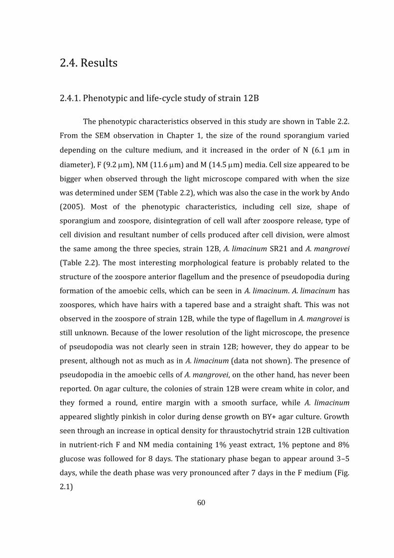
60
2.4. Results
2.4.1. Phenotypic and life-cycle study of strain 12B
The phenotypic characteristics observed in this study are shown in Table 2.2.
From the SEM observation in Chapter 1, the size of the round sporangium varied
depending on the culture medium, and it increased in the order of N (6.1 m in
diameter), F (9.2 m), NM (11.6 m) and M (14.5 m) media. Cell size appeared to be
bigger when observed through the light microscope compared with when the size
was determined under SEM (Table 2.2), which was also the case in the work by Ando
(2005). Most of the phenotypic characteristics, including cell size, shape of
sporangium and zoospore, disintegration of cell wall after zoospore release, type of
cell division and resultant number of cells produced after cell division, were almost
the same among the three species, strain 12B, A. limacinum SR21 and A. mangrovei
(Table 2.2). The most interesting morphological feature is probably related to the
structure of the zoospore anterior flagellum and the presence of pseudopodia during
formation of the amoebic cells, which can be seen in A. limacinum. A. limacinum has
zoospores, which have hairs with a tapered base and a straight shaft. This was not
observed in the zoospore of strain 12B, while the type of flagellum in A. mangrovei is
still unknown. Because of the lower resolution of the light microscope, the presence
of pseudopodia was not clearly seen in strain 12B; however, they do appear to be
present, although not as much as in A. limacinum (data not shown). The presence of
pseudopodia in the amoebic cells of A. mangrovei, on the other hand, has never been
reported. On agar culture, the colonies of strain 12B were cream white in color, and
they formed a round, entire margin with a smooth surface, while A. limacinum
appeared slightly pinkish in color during dense growth on BY+ agar culture. Growth
seen through an increase in optical density for thraustochytrid strain 12B cultivation
in nutrient-rich F and NM media containing 1% yeast extract, 1% peptone and 8%
glucose was followed for 8 days. The stationary phase began to appear around 3–5
days, while the death phase was very pronounced after 7 days in the F medium (Fig.
2.1)
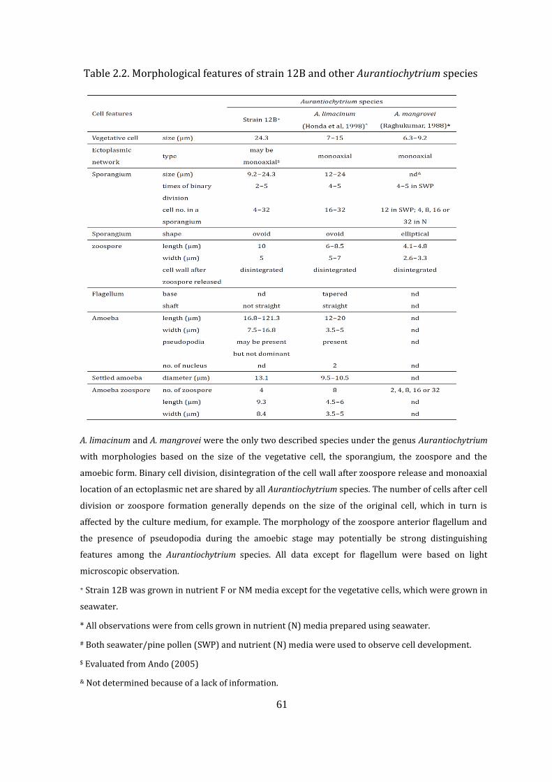
61
Table 2.2. Morphological features of strain 12B and other Aurantiochytrium species
A. limacinum and A. mangrovei were the only two described species under the genus Aurantiochytrium
with morphologies based on the size of the vegetative cell, the sporangium, the zoospore and the
amoebic form. Binary cell division, disintegration of the cell wall after zoospore release and monoaxial
location of an ectoplasmic net are shared by all Aurantiochytrium species. The number of cells after cell
division or zoospore formation generally depends on the size of the original cell, which in turn is
affected by the culture medium, for example. The morphology of the zoospore anterior flagellum and
the presence of pseudopodia during the amoebic stage may potentially be strong distinguishing
features among the Aurantiochytrium species. All data except for flagellum were based on light
microscopic observation.
+ Strain 12B was grown in nutrient F or NM media except for the vegetative cells, which were grown in
seawater.
* All observations were from cells grown in nutrient (N) media prepared using seawater.
# Both seawater/pine pollen (SWP) and nutrient (N) media were used to observe cell development.
$ Evaluated from Ando (2005)
& Not determined because of a lack of information.
62
Figure 2.1. Growth profile of thraustochytrid strain 12B in F and NM media. The
12B cells grown in the NM medium appeared to enter the stationary phase earlier
than in the F medium, at 4 days and 5 days respectively. Cells were cultivated in a
50 ml flask containing 10 ml liquid medium and incubated at 30°C and 150 rpm.
Optical densities of cultures were measured photometrically at 600 nm. Triplicate
sampling was performed every day from new 10 ml culture up to 8 days. Both the F
and NM media contained the same organic composition, consisting of 8% (w/v)
glucose, 1% (w/v) yeast extract and 1% (peptone). F medium was also composed
of 50% seawater, while NM medium was made using distilled water and
supplemented with 0.1% (w/v) NaCl and 1% (w/v) MgSO4•7H2O, and a colony
from a 2-day BY+ agar plate was used as an inoculum.
The life cycle of thraustochytrid strain 12B was observed through a light
microscope, and a continuous video was recorded for up to 72 hours. The complex
life cycle of strain 12B is shown in Figure 2.2 and could probably be generalized
into three main stages: (1) the sporangium stage, (2) the amoebic stage, and (3) the
zoospore stage, with various transition stages in between the main stages. The
characteristic of the sporangium stage was its rounded shape and immovability.
The amoebic cell had an elongated and flexible body and could move around slowly.
The zoospore, on the other hand, moved very quickly, appearing to be ovoid under
0
5
10
15
20
25
30
35
0 2 4 6 8 10
A6
00
nm
Days
12B-F
12B-NM
63
the light microscope, and was the smallest compared with the other stages. Settled
zoospores were chosen as the starting cell for the observation. Initially, the
zoospore increased in size to become a sporangium, and a total of 8.4 and 22 hr
was taken for the initial fission stage to complete in F and NM media, respectively.
This was followed by the settling stage, a period when no observable changes
occurred, which lasted for 1.4 and 1.6 hr in F and NM media, respectively, followed
by a transition to the amoebic stage for a period of 2.3 and 1 hr, respectively, before
settling again to form a rounded cell for 1 and 1.7 hr, respectively. The cell then
divided, forming zoospores, which moved actively for 0.1 and 0.2 hr, respectively,
before settling down and either reforming into the amoebic form or back into a
sporangium. The zoospore was the smallest and fastest-moving stage and was
exceptionally fast when suspended in seawater deficient in nutrients. The
vegetative cell is characteristic, with its ectoplasmic network seen as white streaks
radiating out from the cell. The amoebic form could exist at any time during the life
cycle, as shown by the arrows radiating out of the core cycle. The final fate of the
cell, shown as B, C and D, was not yet determined. Point A may have come from D
or directly from the zoospore (3). The sporangium and the vegetative cell may be
linked through pathway E.
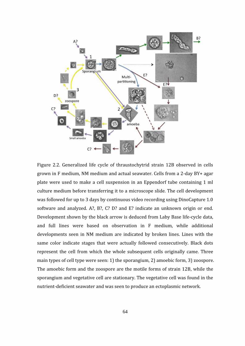
64
Figure 2.2. Generalized life cycle of thraustochytrid strain 12B observed in cells
grown in F medium, NM medium and actual seawater. Cells from a 2-day BY+ agar
plate were used to make a cell suspension in an Eppendorf tube containing 1 ml
culture medium before transferring it to a microscope slide. The cell development
was followed for up to 3 days by continuous video recording using DinoCapture 1.0
software and analyzed. A?, B?, C? D? and E? indicate an unknown origin or end.
Development shown by the black arrow is deduced from Laby Base life-cycle data,
and full lines were based on observation in F medium, while additional
developments seen in NM medium are indicated by broken lines. Lines with the
same color indicate stages that were actually followed consecutively. Black dots
represent the cell from which the whole subsequent cells originally came. Three
main types of cell type were seen: 1) the sporangium, 2) amoebic form, 3) zoospore.
The amoebic form and the zoospore are the motile forms of strain 12B, while the
sporangium and vegetative cell are stationary. The vegetative cell was found in the
nutrient-deficient seawater and was seen to produce an ectoplasmic network.
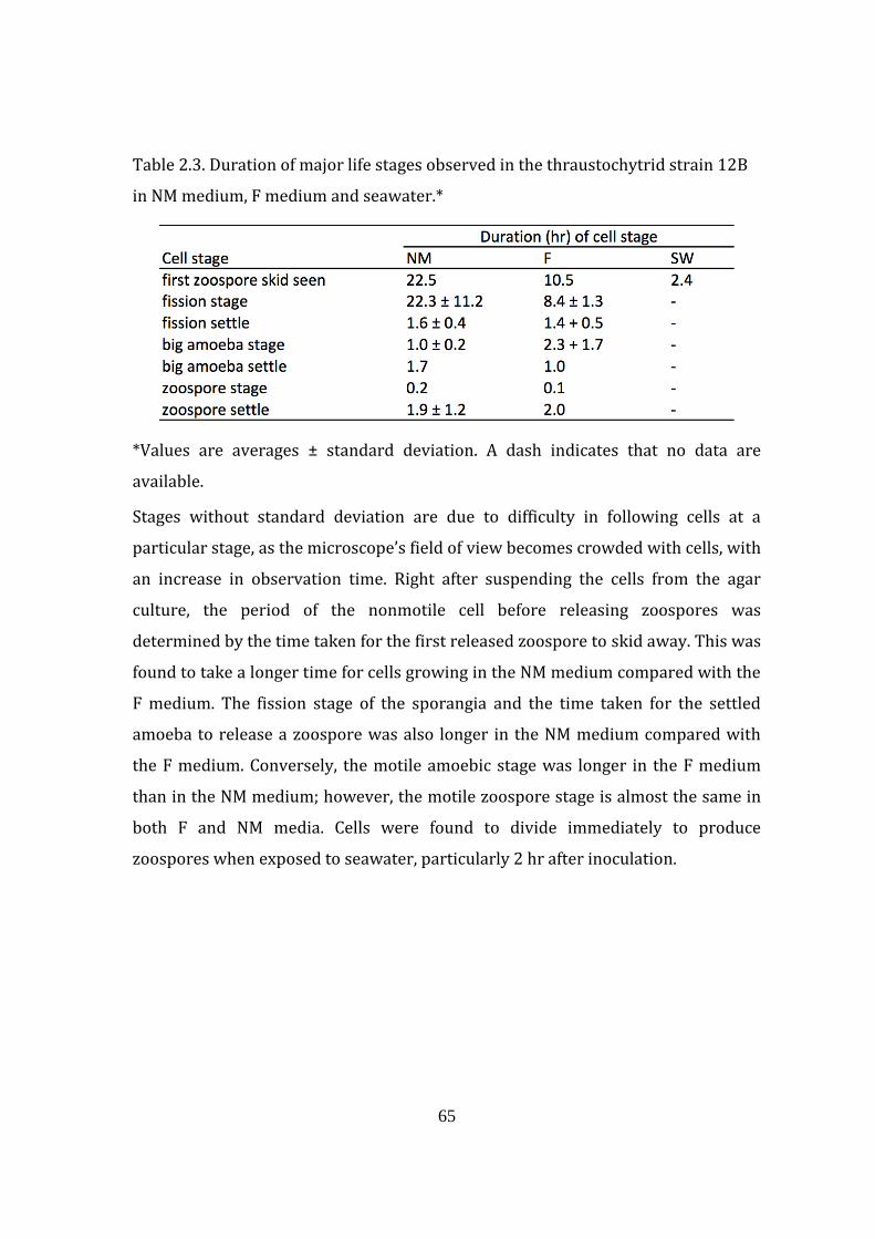
65
Table 2.3. Duration of major life stages observed in the thraustochytrid strain 12B
in NM medium, F medium and seawater.*
*Values are averages ± standard deviation. A dash indicates that no data are
available.
Stages without standard deviation are due to difficulty in following cells at a
particular stage, as the microscope’s field of view becomes crowded with cells, with
an increase in observation time. Right after suspending the cells from the agar
culture, the period of the nonmotile cell before releasing zoospores was
determined by the time taken for the first released zoospore to skid away. This was
found to take a longer time for cells growing in the NM medium compared with the
F medium. The fission stage of the sporangia and the time taken for the settled
amoeba to release a zoospore was also longer in the NM medium compared with
the F medium. Conversely, the motile amoebic stage was longer in the F medium
than in the NM medium; however, the motile zoospore stage is almost the same in
both F and NM media. Cells were found to divide immediately to produce
zoospores when exposed to seawater, particularly 2 hr after inoculation.
66
2.4.2. Phylogenetic study based on the 18S rRNA gene sequence
In the previous phylogenetic study on thraustochytrid strain 12B based on
the 18S rRNA gene, both Schizochytrium (Aurantiochytrium) limacinum SR21 and
Thraustochytriidae sp. A5-20 were found to be very closely positioned on the
phylogenetic tree (Ando, 2005). However, many genera were not monophyletic,
particularly in Thraustochytrium, Schizochytrium and Ulkenia. Various taxonomic
changes have also occurred ever since the study by Ando (2005), giving rise to the
new genera Aurantiochytrium, Oblongichytrium, Botryochytrium, Parietichytrium
and Sicyoidochytrium (Yokoyama and Honda, 2007, Yokoyama et al., 2007). By
comparing sequences from nine representative thraustochytrid genera from the
latest taxonomic classification, strain 12B was found to be grouped well in the
genus Aurantiochytrium, with high bootstrap values (Fig. 2.3). The 18S rRNA gene
sequence similarity decreases from the species level to the genus level and the
family level, with a percentage similarity of 98.6%, 83.4% and 78.6%, respectively
(data not shown). Strain 12B had a higher sequence similarity to A. limacinum
(98.5%) than to A. mangrovei (96.6%). The average sequence similarity of the 18S
rRNA gene between two species of the 17 A. limacinum isolates as a sample
organism was calculated to be 98.8% (data not shown), and this value of similarity
could probably be used as the cutoff value for species identification of
Aurantiochytrium species in thraustochytrid microorganisms.
2.4.3. Phylogenetic study based on mitochondrial COXII gene
COXII gene sequences of strain 12B and A. limacinum SR21 were aligned
together to analyze their sequence similarity/identity to each other. At the same
time, COXII sequences from Thraustochytrium aureum were also analyzed to
provide a comparison between different thraustochytrids at the genus level (Fig.
2.4). There was a high percentage identity or similarity of 98.6% between strain
12B and SR21, while a very low sequence similarity of about 59% was obtained for
both strain 12B and SR21 compared with T. aureum.
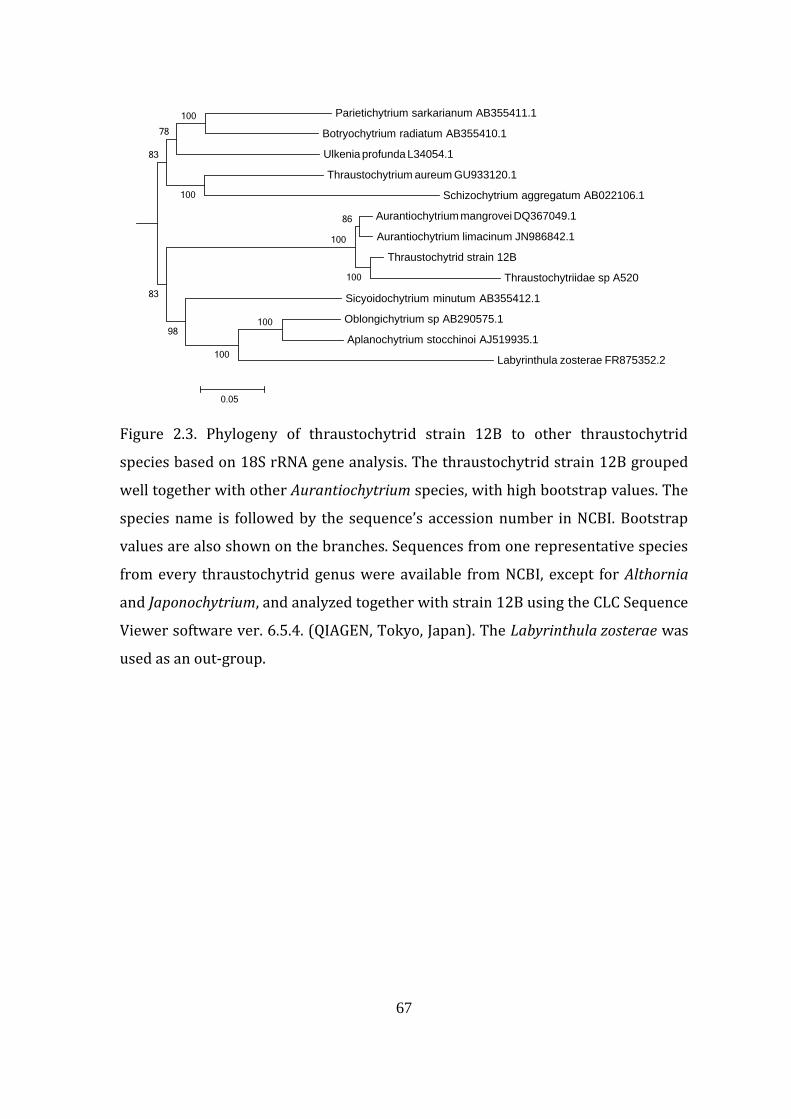
67
Figure 2.3. Phylogeny of thraustochytrid strain 12B to other thraustochytrid
species based on 18S rRNA gene analysis. The thraustochytrid strain 12B grouped
well together with other Aurantiochytrium species, with high bootstrap values. The
species name is followed by the sequence’s accession number in NCBI. Bootstrap
values are also shown on the branches. Sequences from one representative species
from every thraustochytrid genus were available from NCBI, except for Althornia
and Japonochytrium, and analyzed together with strain 12B using the CLC Sequence
Viewer software ver. 6.5.4. (QIAGEN, Tokyo, Japan). The Labyrinthula zosterae was
used as an out-group.
Parietichytrium sarkarianum AB355411.1
Botryochytrium radiatum AB355410.1
Ulkenia profunda L34054.1
Thraustochytrium aureum GU933120.1
Schizochytrium aggregatum AB022106.1
Aurantiochytrium mangrovei DQ367049.1
Aurantiochytrium limacinum JN986842.1
Thraustochytrid strain 12B
Thraustochytriidae sp A520
Sicyoidochytrium minutum AB355412.1
Oblongichytrium sp AB290575.1
Aplanochytrium stocchinoi AJ519935.1
Labyrinthula zosterae FR875352.2
100
100
98
86
100
100
83
100
78
100
83
0.05
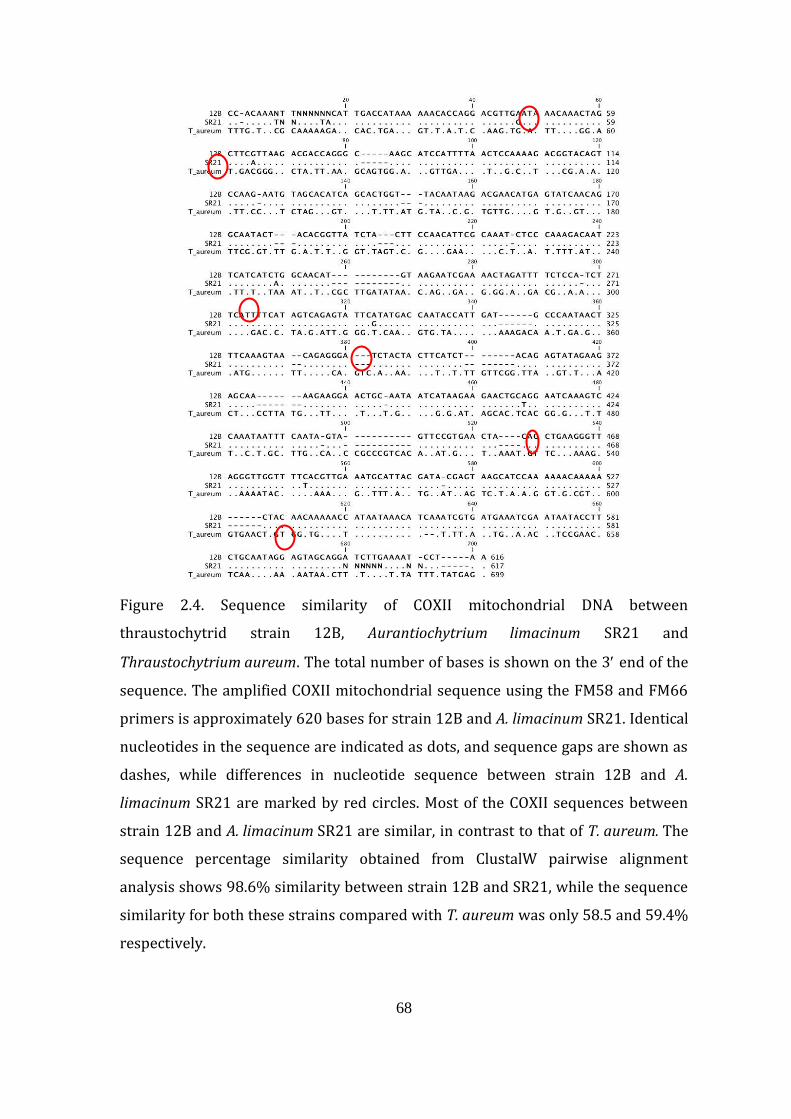
68
Figure 2.4. Sequence similarity of COXII mitochondrial DNA between
thraustochytrid strain 12B, Aurantiochytrium limacinum SR21 and
Thraustochytrium aureum. The total number of bases is shown on the 3 end of the
sequence. The amplified COXII mitochondrial sequence using the FM58 and FM66
primers is approximately 620 bases for strain 12B and A. limacinum SR21. Identical
nucleotides in the sequence are indicated as dots, and sequence gaps are shown as
dashes, while differences in nucleotide sequence between strain 12B and A.
limacinum SR21 are marked by red circles. Most of the COXII sequences between
strain 12B and A. limacinum SR21 are similar, in contrast to that of T. aureum. The
sequence percentage similarity obtained from ClustalW pairwise alignment
analysis shows 98.6% similarity between strain 12B and SR21, while the sequence
similarity for both these strains compared with T. aureum was only 58.5 and 59.4%
respectively.
69
2.5. Discussion
2.5.1. Phenotypic characteristics and life cycle of strain 12B
Microscopic observations under light and electron microscopes were
carried out to see the effect of culture medium on cell structure and life cycle in
order to provide some explanation for the increase of cell yield and DHA
productivity in the NM medium. Changes brought about in the cell may affect the
taxonomic value of the features observed; however, at the same time, they are also
considered to be important (Goldstein, 1973). All thraustochytrids share some
morphological features that are characteristic of species belonging to
Thraustochytriaceae, such as the presence of a noncellulosic scaly cell wall—the
sagenogenetosome—an intracellular organelle that produces an ectoplasmic net
(Huang et al., 2003), and the formation of biflagellate zoospores, one smooth and
another with tripartite tubular hairs (Metz et al., 2010). The flagella on the strain
12B zoospore have already been shown in a previous study (Perveen et al., 2006),
and the characteristic cell wall of strain 12B observed under TEM is shown in
Figure 1.8.
There are various differences seen between strain 12B and SR21 in terms of
phenotype and physiology, such as colony morphology and rate of glucose
utilization (data not shown), and in terms of DCW, lipid, fatty acid and DHA yields
during cultivation in F and NM media (Table 1.1). The colony morphology of 12B
and SR21 appeared to be almost the same, with a round, smooth margin and a
surface with a cream white color, although that of SR21 was slightly pinkish in
color in a dense plate culture. As mentioned in Chapter 1, the number of days for
the strains to deplete glucose in the culture medium varied. The SR21 culture
appeared to deplete glucose faster, at 6 days in F culture and 5 days in NM culture,
while strain 12B took 7 days in both F and NM (data not shown). The DCW, lipid,
fatty acid and yields were also different between the two strains (Table 1.1). After 3
days’ cultivation, the DCW yield in NM medium for strain 12B and SR21 was 18.7
and 12.9 mg/ml, respectively; the percentage lipid per DCW was 55.5 and 41.5%,
70
respectively; the percentage fatty acid per total lipid was 75% and 59.7%,
respectively, and there was a slight difference in percentage DHA per total fatty
acid of 31.2 and 33.5%, respectively. There were also differences in the fatty acid
composition between the two, particularly when they were cultured in the F
medium; however, the fatty acid composition was almost the same when they were
grown in the NM medium (data not shown). The structure of the tubular hair on the
anterior flagella, which does not appear to have a tapered base and a smooth shaft
(Perveen et al., 2006), both of which are observable in A. limacinum (Honda et al.,
1998), further supports the notion that they are probably two different species.
Perveen et al. (2006) has also observed fusion among the strain 12B cells.
Phagocytosis of bacteria by thraustochytrid microorganisms has been documented
(Raghukumar, 1992, Yokochi et al., 2001); however, no fusion or phagocytosis
between thraustochytrid cells has ever been reported in the literature, which
therefore makes this a unique feature for strain 12B.
Observation of the life cycle through a light microscope shows that
thraustochytrid strain 12B is generally made up of three main stages, the
sporangium, amoebic and zoospore stages, with a period of ‘settlement’ in between
transformations. However, there are still many questions regarding the
transformation preference of strain 12B, as it was seen to be able to transform into
the amoebic form directly from the sporangium state (course B in Fig. 2.2), or to
transform into an even smaller amoebic form after the settling of the big amoebic
stage (course C), or to transform from an undeveloped zoospore (course D) or from
a developed zoospore stage (Fig. 2.2). The eventual outcome from all these courses,
however, might be the reformation of the zoospore or the sporangium. The
presence of the amoebic stage is a characteristic feature of the genera Ulkenia,
Parietichytrium, Sicyoidochytrium and Botryochytrium (Yokoyama, 2007a);
however, there are also exceptions for some species of Aurantiochytrium, in
particular A. limacinum and A. mangrovei, which were also found to have an
amoebic stage (Yokoyama & Honda, 2007). The characteristic feature is a naked
protoplasm where the cell suddenly becomes ‘invisible’ in Ulkenia, Parietichytrium,
Sicyoidochytrium and Botryochytrium; however, such a stage was not seen in strain
71
12B. An ectoplasmic network was also very rarely seen among strain 12B cells, and
together with the disintegration of the cell wall during the release of the zoospore,
this further emphasizes that strain 12B belongs to the genus Aurantiochytrium.
Thraustochytrid strain 12B grew to abnormal sizes in the M culture (Fig.
1.7), as seen in Chapter 1. The repeating sporangium–amoeba transformations
(course B in Fig. 2.2) might be how the strain 12B cells in NaCl-deficient M medium
acquire a bigger cell size. The possible effect of NaCl deficiency in the disruption of
the cell division mechanism is probably linked with zoospore formation. In this
experiment, the zoospore was seen earliest in seawater containing no nutrients,
followed by cells grown in glucose-rich F medium containing 50% seawater, and
latest in 0.1% NaCl glucose-rich NM medium (Table 2.3). Assuming that F medium
contains 1.5% NaCl, while the seawater contains 3% NaCl, zoospore release seems
to be related to increasing salinity for strain 12B. The thraustochytrid strain 12B
may have to undergo transformation through course B (Fig. 2.2), probably because
it cannot form zoospores in the absence of NaCl. In a different study, the opposite
was seen, whereby decreasing salinity from 2.3% to 0.75% increased zoospore
production in Ulkenia sp. and Aurantiochytrium mangrovei (Tsui et al., 2012). The
study did not include lowering the NaCl concentration to 0% salinity, and therefore
the high zoospore production at 0.75% salinity probably indicates the optimum
concentration for these two strains. This is because a normal distribution pattern
in zoospore production with salinity was evident for the Schizochytrium sp. KF-1 at
30°C, which shows the presence of an optimal concentration for salinity (Fan et al.,
2002).
There was long settling stage for the sporangium and amoebic form of cells
grown in the NM culture compared with the F culture. This might lead to better
accumulation of lipid and thus DHA inside the cell, and reduced utilization of
storage TG as an energy source. The time taken for a sporangium to divide (fission
stage) right after inoculation probably takes a long time only at the beginning
because of adaptation to the culture medium. However, cells appear to be growing
almost immediately as shown in Figure 2.1. Further subsequent fission stages,
however, were observed to have a shorter duration (data not shown). Unlike the
72
observation in F and NM media, growth in nutrient-deficient seawater was not so
dynamic, except for the numerous zoospores skidding at extremely high speed
within the medium. The size of the zoospore in seawater was also relatively smaller
compared with zoospores found in F and NM cultures (data not shown).
2.5.2. Molecular phylogenetic studies of 12B
Previous phylogenetic study (Ando, 2005) has shown that many
thraustochytrid genera were not monophyletic; however, various taxonomic
changes have occurred, and currently only the genus Thraustochytrium is still
found scattered throughout the phylogenetic tree, while other thraustochytrid
genera have become monophyletic (Yokoyama et al., 2007, Yokoyama & Honda,
2007b). Strain 12B is closely related to A. limacinum SR21, Thraustochytrid sp. A5-
20 and Schizochytrium mangrovei, based on phylogenetic analysis of the 18S rRNA
gene (Fig. 2.3). In terms of percentage sequence similarity, out of the only two
described Aurantiochytrium species, A. mangrovei and A. limacinum, strain 12B has
a higher similarity with A. limacium at 98.5%. However, considering the percentage
sequence similarity cutoff value for species at 98.8%, strain 12B could probably be
considered to be a closely related, but different, species. The assignment of strain
12B to a new species was further supported by the physiological and phenotypic
differences mentioned above.
Amplification of the mitochondrial DNA was performed using primers that have
been developed for the Pythium genus, which is also under the phylum
Heterokonta/Stramenopiles (Long et al., 2013), with some species under this genus
also producing PUFAs such as EPA by P. irregular (Wu et al., 2013). Amplification of
COXI and COXII+ITS was not successful, resulting in poor sequence quality,
indicating unsuitability of the PCR primers (data not shown). There is also a
probability that many introns exist in both the COXI and COXII+ITS genes, as this
rare occurrence has been associated with low amplification success in fungi (Seifert,
2009). However, the amplification of the COXII gene from the thraustochytrid
strain 12B and SR21 was successful, resulting in the production of 620 base
amplicons. There were some differences in the COXII sequence between 12B and
73
SR21, with a sequence similarity of 98.6%. A higher sequence variation was
observed when the sequences of both strain 12B and A. limacinum SR21 were
compared with that of Thraustochytrium aureum, with a sequence similarity of 58.5
and 59.4%, respectively (Fig. 2.4, data not shown). This indicates that both strain
12B and A. limacinum SR21 are closely related, and the COXII sequence is probably
conserved for species from the same genus. The conservative nature of the
mitochondrial DNA sequence between closely related thraustochytrid species has
also been seen in QPX isolates (Qian et al., 2007), which exhibited a higher
variation when the nuclear ITS of SSU was used, and therefore is useful for
intraspecies differentiation (Qian et al., 2007). The number of base pair differences
has been documented for intraspecies and interspecies variation. A total of 0 to 4
base pair differences in the 539 bp COXI sequence in algae are normally present as
intraspecies variation, while 28 to 148 bp differences were seen to be present
between species (Robba et al., 2006). There might be some significance in the 6 bp
difference in the COXII gene sequence between strain 12B and A. limacinum SR21;
however, more comparison is necessary, such as by investigating the intraspecies
COXII sequence variation, such as by using A. limacinum isolates as a possible
model organism.

74
2.6. Conclusions
Thraustochytrid strain 12B belongs to the genus Aurantiochytrium, based on
morphology through the absence of naked protoplasm, the rarity of an ectoplasmic
network produced by the cell, and the disintegration of the cell wall during
zoospore release as well as the 18S rRNA gene phylogenetic grouping. There is a
high 98.6% sequence similarity in the mitochondrial COXII gene sequence between
the two Aurantiochytrium species, strain 12B and A. limacinum SR21, and a very
low sequence similarity of less than 60% when both strains are compared with
another thraustochytrid genus, Thraustochytrium aureum. The mitochondrial COXII
gene therefore may have a high taxonomic value for differentiating the genus level
for thraustochytrids. Based on the 18S rRNA analysis, the lower sequence
similarity of 98.5% to the proposed 98.8% cutoff value between strain 12B and A.
limacinum sequences may indicate that strain 12B is a different species from A.
limacinum. This is further supported by the physiological and phenotypic
differences seen in terms of coloration of algal culture, rate of glucose utilization,
and differences in DCW, lipid, fatty acid and DHA yields during cultivation, as well
as differences in the zoospore flagellum structure. Fusion among thraustochytrid
cells and the presence of multiple 18S rRNA gene copy in the previous study of
strain 12B is also not commonly found or reported in other thraustochytrid
microorganisms, making it a unique strain. Additionally, strain 12B has a complex
life cycle in nutrient-rich F and NM media. In the life-cycle study, the stationary
phases were relatively longer, while the duration for the mobile amoebic form
phase was shorter when strain 12B was cultured in NM medium compared with F
medium. This probably contributes to the conservation of oil bodies inside the cell,
resulting in higher cell, lipid, and DHA yields in the NM medium.
75
Chapter 3: Possible functions of DHA and its
accumulation in thraustochytrid microorganisms
3.1. Summary
One of the possible reasons for the high accumulation of lipid and
particularly DHA in thraustochytrid strain 12B is its role in energy antioxidation.
During starvation, the percentage composition of pentadecanoic acid (C15:0) and
hexadecanoic acid (C16:0) decreased from 31% to 15% and from 22% to 14%,
respectively, while the percentage composition of DHA increased from 25% to 42%
after 7 days’ cultivation in nutrient-deficient 1.5% (w/v) NaCl solution. The
preferential use of C15:0 was also seen in the absolute yield data for C15:0, which
shows a continuous decrease with time during starvation. The absolute amount of
DHA, on the other hand, was the highest among all of the major fatty acids
throughout the experiment. This shows that other fatty acids such as the saturated
fatty acid C15:0 was preferentially used as a source of energy over DHA, which
suggests that DHA may have other important functions apart from being an energy
source. During exposure to UV-C, the strain 12B cells’ growth profile was more or
less similar compared with cultivation in the dark. The percentage DHA
composition per total fatty acid was slightly decreased; however, the absolute cell,
lipid and DHA yields were higher when strain 12B was cultured under UV,
increasing from 0.5 to 3.4 mg/ml. DHA may have been involved in the intracellular
antioxidative mechanisms, allowing the cell to survive for 3 days’ cultivation under
UV. At the same time, the exposure to UV is thought to affect the zoospores’ flagella
development, resulting in lower motility and hence to contribute to further
preservation of intracellular lipid reserves. This approach may have an important
application in industrial production of DHA.
76
3.2. Introduction
3.2.1. Role of PUFA in adaptation
Microorganisms are considered to synthesize PUFAs to adapt to the
environment, where PUFAs such as EPA and DHA play a certain role in their
survival. One of the functions of PUFA is believed to be its contribution to providing
fluidity to the cell membrane at low temperature or high pressure (Hazel &
Williams, 1990), referred to as homeoviscous adaptation. This is defined as a
homeostatic process that regulates the viscosity of membrane lipids to achieve a
constant fluidity at temperature of growth by varying the fatty acid composition of
phospholipids (Sinensky, 1974). Homeoviscous adaptation is important for
maintaining the function of the membrane as a barrier and for maintaining the
function of proteins in the membrane involved in various enzyme reactions for
energy, signaling and intra- or extracellular transport of molecules. The activity of
membrane proteins is highly affected by the membrane structure (Jensen &
Mouritsen, 2004, Andersen & Koeppe, 2007), which is influenced by thickness,
curvature and membrane elasticity (Phillips et al., 2009). Although PUFAs could act
as a substitute for monounsaturated fatty acids (Allen et al., 1999), it is actually the
monounsaturated fatty acids that are more affected by changes in temperature or
pressure compared with PUFAs (Hazel & Williams, 1990). The accumulation of
PUFAs such as DHA therefore probably serves other important functions inside the
cell.
3.2.2. Possible role of DHA as energy source
Triacylglycerol (TG) is a common form of energy and carbon storage in
eukaryotes such as plants, yeast, and animals (Coleman, 2004), and even in some
bacteria under the group actinomycetes (Alvarez, 2002). DHA is probably used as a
source of energy in DHA-producing thraustochytrid microorganisms, because these
microorganisms also accumulate a high amount of DHA as TG (Okuyama et al.,
77
2007, Morita et al., 2006). This was shown earlier in Chapter 1, whereby strain 12B
was able to be grown in a low-NaCl-concentration NM medium, resulting in a high
DHA yield of up to 11.7 mg/ml after 5 days in jar cultivation (Table 1.3). The TG of
the thraustochytrid strain 12B grown in the NM medium in flasks was found to
make up to 47% of the total DCW in which up to 21% of the TG fatty acid is
composed of DHA (data not shown). However, cells grown in F and NM media have
almost the same percentage lipid per DCW, 25.8 and 27.8% respectively (data not
shown). The presence of DHA in TG may predispose it to be a feedstock for
respiration during periods of nutrient deficiency (of organic carbon and nitrogen),
as in the case of the thraustochytrid strain NIOS-1 (Jain et al., 2007). TG was
accumulated by the NIOS-1 strain and was used as an energy source in that strain,
as the amount of oil bodies or storage lipid, which are typically TG, was found to
decrease when the cell changed to motile amoebic forms as seen directly by Nile
blue staining. Hexadecanoic acid (C16:0) and DHA are the main fatty acids in the
strain, and the analysis of fatty acids after transferring cells that have accumulated
a high amount of TG into nutrient-deficient medium shows a reduction in absolute
DHA yield and also a decrease in percentage DHA composition, while the
percentage of C16:0 increases during starvation (Jain et al., 2007). On the other
hand, TG is also known to be a feedstock for phospholipid production (Coleman,
2004), and the conversion of DHA-rich TG into DHA-rich phospholipid was
previously achieved by cultivating a thraustochytrid strain in a nitrogen-rich
medium that was devoid of glucose as a carbon source (Okuyama et al., 2007). The
use of TG as an energy source in thraustochytrid strain 12B grown in glucose-
deficient medium was supported by the presence of numerous mitochondria in the
cytoplasm (Bin Haji Mohd Taha et al., 2012). From this, it was previously
hypothesized that DHA was channeled to the production of DHA-rich phospholipid
inside mitochondrial membranes as many mitochondria were synthesized. It is
therefore still not clear whether the accumulation of DHA is to serve as an energy
source preferentially over other fatty acids or the opposite. However, it is more
reasonable for the thraustochytrid microorganisms to utilize the saturated fatty
acid preferentially because the beta-oxidation process for saturated fatty acids is
shorter, bypassing two reactions involving enoyl-CoA isomerase and 2, 4-dienoyl-
78
CoA reductase (D’Andrea, 1994), and the synthesis of PUFA is more costly. The
preferential use and degradation of saturated fatty acids as an energy source would
consequently be one of the possible reasons for DHA accumulation in
thraustochytrid microorganisms.
3.2.3. Role of DHA in antioxidative stress
DHA also has a direct and indirect antioxidative role in cells. In bacteria,
DHA is thought to increase the hydrophobicity of the cell membrane, preventing
the entry of hydrophilic extracellular reactive oxygen species (ROS) such as
hydrogen peroxide into the cell (Okuyama et al., 2008b). The hypothesis is
probably also relevant to eukaryotes, as the general structure of all biological
membranes is shared (Alberts et al., 2002). Most DHA-producing organisms
including protozoa, algae and bacteria are marine in origin and are rarely
terrestrial or freshwater organisms (Okuyama et al., 2008b). Human beings are
also able to produce DHA in the astrocyte; however, they are incapable of
producing it in significant amounts (Moore et al., 1991). The production of DHA by
marine organisms may have arisen as a protective strategy against H2O2, which is
present in a stable concentration, has a long lifetime in seawater and is
photochemically produced through UV absorption by organic matter in the
seawater or by hydrothermal vents (Lesser, 2006).
Thraustochytrid microorganisms are naturally ‘marine’ (Raghukumar,
2008a), while those with a terrestrial habitat normally are brackish or saline in
nature (Raghukumar, 2008b). The thraustochytrid strain 12B is also from a marine
source, originally isolated from a mangrove river in Okinawa (Perveen et al., 2006).
Unlike bacteria, most DHA are stored in TG in thraustochytrids (Okuyama et al.,
2007, Morita et al., 2006), including strain 12B, and it was suggested that one of the
reasons for this is its function as an intracellular antioxidant (Jain et al., 2007).
Apart from exogenous sources, ROS could also be intracellularly produced by the
exposure of cells to UV, resulting in peroxidation of PUFA (Catalá, 2009).
‘Photosensitizers’ in the cell could generate ROS by absorbing energy from UV
(Bossi, 2008). The damaging effect of UV to key enzymes in the respiration
79
pathway may also promote ROS formation (He, 2002). The four types of UV (UV-V,
UV-C, UV-B, and UV-A) are defined by their wavelength range of 100–200 nm, 200–
280 nm, 280–315 nm and 315–400 nm, respectively (Koutchma, 2009). The solar
UV on earth is made up of 95% UV-A and 5% UV-B, while UV-C does not penetrate
the earth’s atmosphere (Santos et al., 2013). However, there is less UV in marine
than in terrestrial environments. The amount of UV-B that penetrates water bodies
is 10% of the amount at the water surface at depths ranging from 0.1 m to 40 m,
depending on the water column properties, the air–water interface properties and
the incident irradiance (Booth, 1997). DHA in a mixture with other PUFAs, as it
would be in nature, has a high tendency to be oxidized (Miyashita et al., 1983). The
low UV penetration coupled with the presence of H2O2 may have produced a
natural selective pressure for DHA-producing microorganisms in marine
environments.
The objective of this chapter is to investigate the function of DHA in PUFA-
producing thraustochytrid strain 12B to understand further the factors affecting
DHA production for possible application in industrial practices.
80
3.3. Materials and methods
3.3.1. Microorganisms
Strain 12B (NITE P-68), which is a high-DHA-accumulating thraustochytrid
eukaryotic microorganism, was isolated from a mangrove in Okinawa, Japan
(Perveen et al., 2006) and was used for both starvation and UV–antioxidation
experiments.
3.3.2. Culture media and cultivation conditions
The strain 12B working cultures were maintained on BY+ agar plates, and
agar cultures used as inoculum were incubated in the dark at 30°C for 2 days. The
BY+ agar was prepared by dissolving glucose in seawater, while yeast extract,
peptone and agar were dissolved in distilled water to a final concentration of 0.5,
0.1, 0.1 and 1.5% (w/v), respectively. The solutions were autoclaved separately to
avoid precipitation and browning of glucose, and were mixed afterwards, resulting
in a 50% seawater solution. The preparation of the F liquid medium is similar to
that of the BY+ medium except that the concentrations of glucose, yeast extract and
peptone were 8%, 1% and 1% (w/v), respectively, without the addition of agar.
The NM medium is composed of 8% (w/v) glucose, 1% (w/v) yeast extract, 1%
(w/v) peptone, 0.1% (w/v) NaCl and 1% (w/v) MgSO4•7H2O dissolved in distilled
water.
3.3.2.1. Culture condition during nutrient starvation
TG rich cells for the starvation experiment were prepared by culturing
strain 12B in the dark for 3 days at 30°C and 150 rpm, by inoculating a colony from
a 2-day BY+ agar culture into a 10 ml F medium in a 50 ml flask. To investigate the
potential use of DHA as a preferential source of energy, strain 12B cells, which had
accumulated DHA-rich TG, were gathered by centrifugation (6000 rpm, 4°C, 10
min), washed aseptically with 1.5% NaCl and resuspended in 10 ml of the same
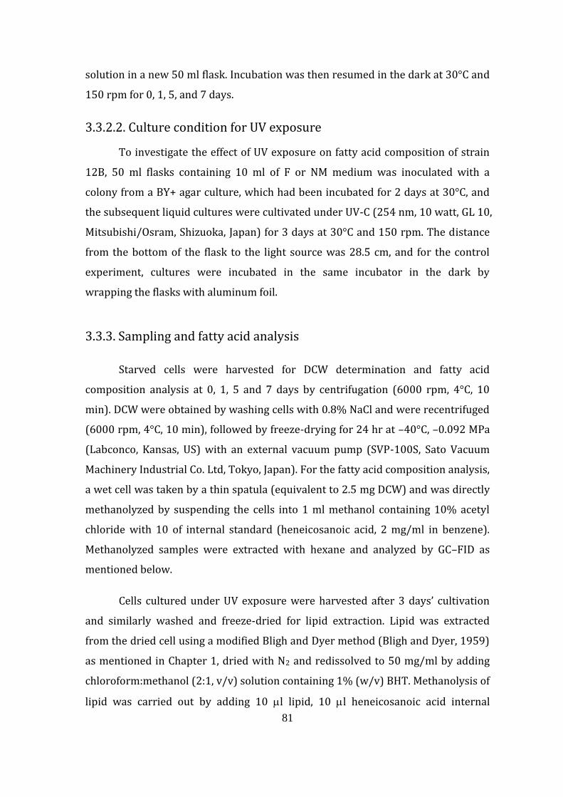
81
solution in a new 50 ml flask. Incubation was then resumed in the dark at 30°C and
150 rpm for 0, 1, 5, and 7 days.
3.3.2.2. Culture condition for UV exposure
To investigate the effect of UV exposure on fatty acid composition of strain
12B, 50 ml flasks containing 10 ml of F or NM medium was inoculated with a
colony from a BY+ agar culture, which had been incubated for 2 days at 30°C, and
the subsequent liquid cultures were cultivated under UV-C (254 nm, 10 watt, GL 10,
Mitsubishi/Osram, Shizuoka, Japan) for 3 days at 30°C and 150 rpm. The distance
from the bottom of the flask to the light source was 28.5 cm, and for the control
experiment, cultures were incubated in the same incubator in the dark by
wrapping the flasks with aluminum foil.
3.3.3. Sampling and fatty acid analysis
Starved cells were harvested for DCW determination and fatty acid
composition analysis at 0, 1, 5 and 7 days by centrifugation (6000 rpm, 4°C, 10
min). DCW were obtained by washing cells with 0.8% NaCl and were recentrifuged
(6000 rpm, 4°C, 10 min), followed by freeze-drying for 24 hr at –40°C, –0.092 MPa
(Labconco, Kansas, US) with an external vacuum pump (SVP-100S, Sato Vacuum
Machinery Industrial Co. Ltd, Tokyo, Japan). For the fatty acid composition analysis,
a wet cell was taken by a thin spatula (equivalent to 2.5 mg DCW) and was directly
methanolyzed by suspending the cells into 1 ml methanol containing 10% acetyl
chloride with 10 of internal standard (heneicosanoic acid, 2 mg/ml in benzene).
Methanolyzed samples were extracted with hexane and analyzed by GC–FID as
mentioned below.
Cells cultured under UV exposure were harvested after 3 days’ cultivation
and similarly washed and freeze-dried for lipid extraction. Lipid was extracted
from the dried cell using a modified Bligh and Dyer method (Bligh and Dyer, 1959)
as mentioned in Chapter 1, dried with N2 and redissolved to 50 mg/ml by adding
chloroform:methanol (2:1, v/v) solution containing 1% (w/v) BHT. Methanolysis of
lipid was carried out by adding 10 l lipid, 10 l heneicosanoic acid internal
82
standard (2 mg/ml in benzene) and 1 ml methanol containing 10% acetyl chloride
into a screw-capped test tube and heated at 100°C for 1 hr (Dry Block Bath MG-2,
Torika corp., Nagoya, Japan). The resulting FAMEs were extracted with 2 ml of
hexane thrice. After concentrating the FAME fractions through evaporation under
N2, the samples were subjected to GLC and GC–MS. The FAME fraction was
analyzed with a gas chromatograph GC-353B (GL sciences, Tokyo, Japan) equipped
with a capillary column BPX70 (25 m 0.22 mm i.d., SGE, Yokohama, Japan) and
FID with nitrogen as carrier gas. The column temperature was programmed to
increase at 5°C/min from 150°C to 240°C and then maintained for 2 min while the
injector and detector temperatures were set at 240°C. Samples were also analyzed
with an ion-trap gas chromatograph CP-3800 (Varian, Tokyo, Japan) and a mass
spectrophotometer Saturn 2200 (Varian, Tokyo, Japan) equipped with a capillary
column TC-70 (30 m 0.25 mm i.d., GL Sciences Inc., Tokyo, Japan). The column
temperature was programmed to increase from 80°C to 240°C, with a split ratio of
20:1, helium carrier gas with a flow rate of 0.9 ml/min and the injector
temperature set at 240°C. GC-MS was used to confirm the identity of peak
components corresponding to specific fatty acids, while GC–FID was used for
routine calculation of fatty acid yield including DHA.
83
3.4. Results
3.4.1. Effect of starvation of strain 12B cells on its DCW, fatty acid
amount and composition
Cultivation of the thraustochytrid strain 12B under nutrient starvation
resulted in a decrease in DCW, with a marked decrease from 30 to 8 mg/ml after 3
days' starvation (Fig. 3.1). In the same period, drastic changes in the absolute
amount of major fatty acid in 12B were also found, particularly for DHA, which
increased from 69.3 to 128.7 mg/g DCW (Fig. 3.2). The yield of fatty acids of C16:0,
heptadecanoic acid (C17:0) and DPA during the 7 days’ starvation experiment was
almost the same as that of DHA, which initially increased after 3 days’ incubation
then suddenly decreased after the 5th day and again increased slightly after 7 days’
incubation in nutrient-deficient solution. Only the yield for pentadecanoic acid
(C15:0) showed a continuous gradual decrease from 57.5 to 23.4 mg/g DCW after 7
days’ starvation. Changes in fatty acid composition, however, generally showed a
decrease in the composition of short-chain fatty acids from C14 to C18:1 and an
increase in longer-chain fatty acids from C19:0 to DHA (Table 3.1). A major
decrease was seen especially in C15:0 and C16:0 fatty acids, from 31% to 12% and
from 22% to 16%, respectively, while a major increase was seen in DPA and DHA,
from 6% to 10% and from 25% to 43%, respectively. Both the major increase and
decrease occurred during the first 72 hr of incubation.
84
Figure 3.1. Amount of DCW obtained from cultivation of thraustochytrid strain 12B
in 1.5% (w/v) NaCl medium without glucose, peptone and yeast extract
supplementation. There was a marked decrease in DCW during the first 3 days’
incubation, which appears more or less the same afterwards. Strain 12B cells were
initially grown in the F medium containing 8% (w/v) glucose, 1% (w/v) yeast
extract and 1% (w/v) peptone, to allow the cell to accumulate lipid and DHA for 3
days. The cells were then washed and resuspended in an equal volume of 10 ml
1.5% (w/v) NaCl solution and incubated at 30°C and 150 rpm. For DCW
determination, cells were harvested after 3, 5 and 7 days’ incubation.
0
5
10
15
20
25
30
35
40
45
50
0 72 120 168
Dry
ce
ll w
eig
ht
(mg
/m
l)
Duration of starvation (hr)
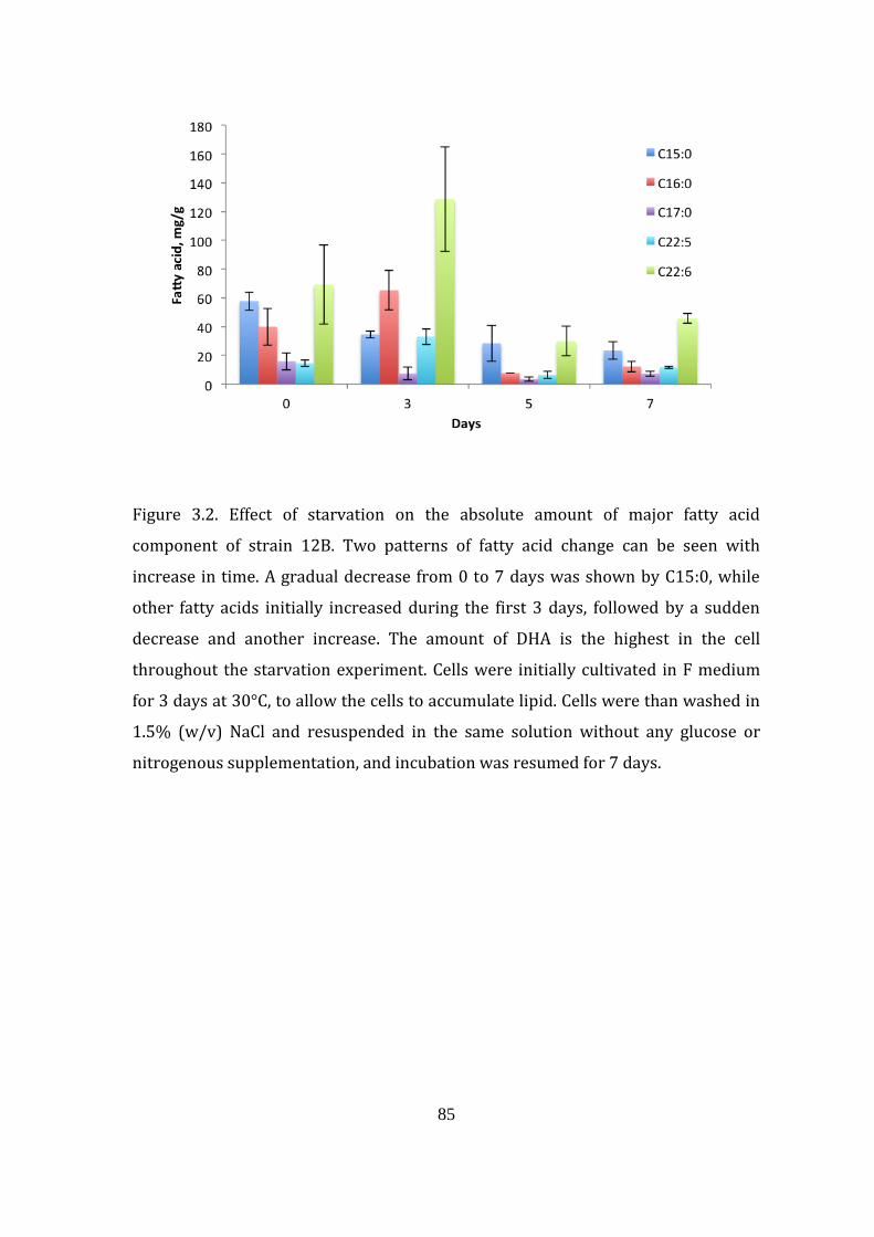
85
Figure 3.2. Effect of starvation on the absolute amount of major fatty acid
component of strain 12B. Two patterns of fatty acid change can be seen with
increase in time. A gradual decrease from 0 to 7 days was shown by C15:0, while
other fatty acids initially increased during the first 3 days, followed by a sudden
decrease and another increase. The amount of DHA is the highest in the cell
throughout the starvation experiment. Cells were initially cultivated in F medium
for 3 days at 30°C, to allow the cells to accumulate lipid. Cells were than washed in
1.5% (w/v) NaCl and resuspended in the same solution without any glucose or
nitrogenous supplementation, and incubation was resumed for 7 days.
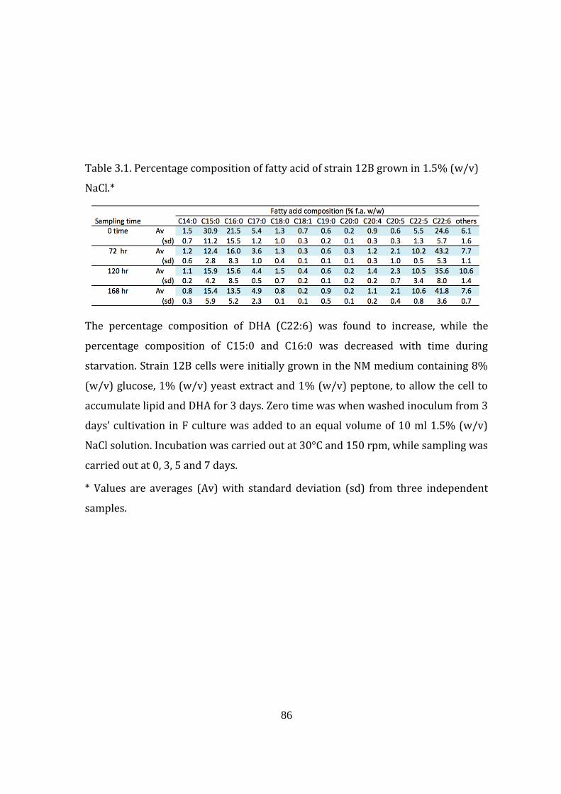
86
Table 3.1. Percentage composition of fatty acid of strain 12B grown in 1.5% (w/v)
NaCl.*
The percentage composition of DHA (C22:6) was found to increase, while the
percentage composition of C15:0 and C16:0 was decreased with time during
starvation. Strain 12B cells were initially grown in the NM medium containing 8%
(w/v) glucose, 1% (w/v) yeast extract and 1% (w/v) peptone, to allow the cell to
accumulate lipid and DHA for 3 days. Zero time was when washed inoculum from 3
days’ cultivation in F culture was added to an equal volume of 10 ml 1.5% (w/v)
NaCl solution. Incubation was carried out at 30°C and 150 rpm, while sampling was
carried out at 0, 3, 5 and 7 days.
* Values are averages (Av) with standard deviation (sd) from three independent
samples.
87
3.4.2. Effect of UV on the growth and fatty acid composition of strain
12B
The effect of UV on the growth pattern of 12B appeared to be almost
identical when the strain was cultivated in both F and NM media in the absence of
UV (Fig. 3.3). When the cultures of strain 12B were exposed to UV, glucose in the
NM media was depleted faster than in the F medium. In the F medium, the time for
glucose depletion was reduced from 7 to 6 days, and from 7 to 4 days in the NM
medium (data not shown). Exposure of strain 12B cultures to UV led to changes in
cellular fatty acid composition. After 3 days’ cultivation, an increase in percentage
composition when exposed to UV was seen for C16:0, octadecanoic acid (C18:0),
EPA (C20:5) and DPA (C22:5), and large differences were seen for C16:0, which
increased from 17% to 35% in F medium but less in NM medium, with only a slight
increase from 42% to 45%. On the other hand, a decrease was found in C15:0,
C17:0, C18:1 and C22:6, especially in C15:0, with a marked decrease from 31% to
16% in the F medium and a smaller decrease from 10% to 6% in the M culture after
being exposed to UV for 3 days (Table 3.2). The exposure of thraustochytrid strain
12B to UV unexpectedly resulted in an increase in the absolute amount of DCW,
lipid, fatty acid and DHA, from 13.6, 4.7, 1.7, 0.5 mg/ml to 24, 16.1, 12.2, 3.4 mg/ml,
respectively, compared with when 12B was cultivated in the dark after 3 days’
cultivation in F medium, while in the NM medium, the increase was from 18.7, 10.4,
7.8 and 2.4 mg/ml to 24.7, 12.9, 10.2, 3.8 mg/ml, respectively (Table 3.3).
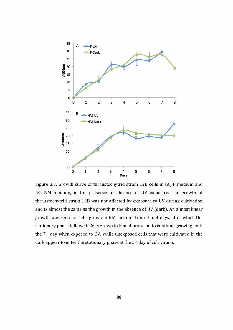
88
Figure 3.3. Growth curve of thraustochytrid strain 12B cells in (A) F medium and
(B) NM medium, in the presence or absence of UV exposure. The growth of
thraustochytrid strain 12B was not affected by exposure to UV during cultivation
and is almost the same as the growth in the absence of UV (dark). An almost linear
growth was seen for cells grown in NM medium from 0 to 4 days, after which the
stationary phase followed. Cells grown in F medium seem to continue growing until
the 7th day when exposed to UV, while unexposed cells that were cultivated in the
dark appear to enter the stationary phase at the 5th day of cultivation.
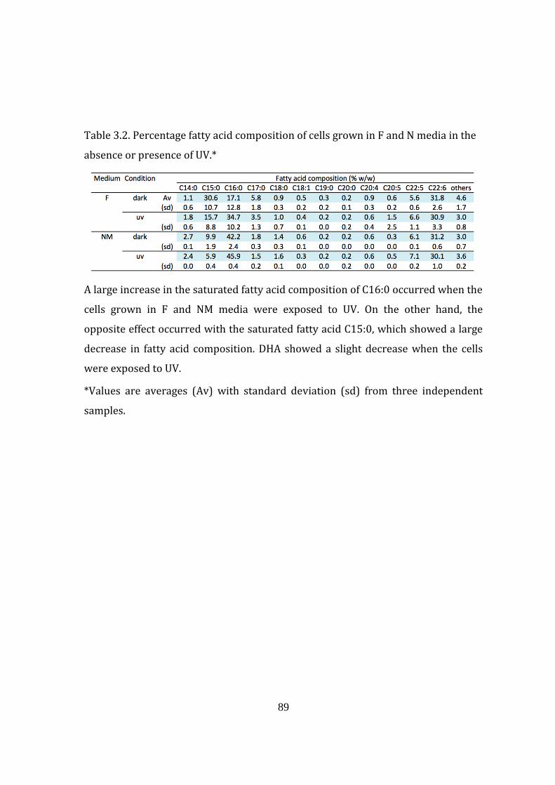
89
Table 3.2. Percentage fatty acid composition of cells grown in F and N media in the
absence or presence of UV.*
A large increase in the saturated fatty acid composition of C16:0 occurred when the
cells grown in F and NM media were exposed to UV. On the other hand, the
opposite effect occurred with the saturated fatty acid C15:0, which showed a large
decrease in fatty acid composition. DHA showed a slight decrease when the cells
were exposed to UV.
*Values are averages (Av) with standard deviation (sd) from three independent
samples.
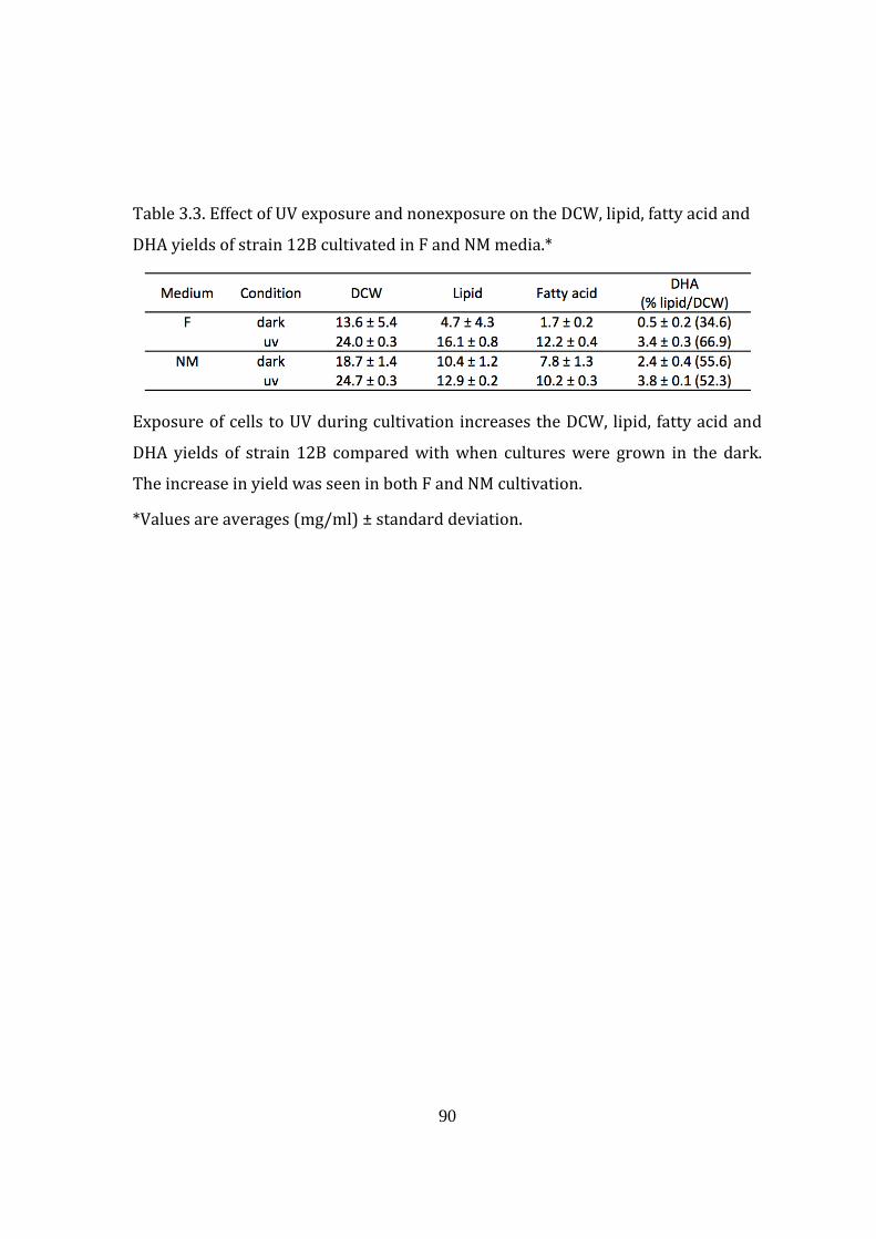
90
Table 3.3. Effect of UV exposure and nonexposure on the DCW, lipid, fatty acid and
DHA yields of strain 12B cultivated in F and NM media.*
Exposure of cells to UV during cultivation increases the DCW, lipid, fatty acid and
DHA yields of strain 12B compared with when cultures were grown in the dark.
The increase in yield was seen in both F and NM cultivation.
*Values are averages (mg/ml) ± standard deviation.
91
3.5. Discussion
3.5.1. Role of DHA as an energy source
During the period of nutrient deprivation, the TG reserve can be
metabolized as a source of energy. This will be done by the hydrolysis of TG into
free fatty acids and diacylglycerol, monoacylglycerol and glycerol successively by
the action of lipase. Glycerol will be converted into dihydroxyacetone phosphate
and will enter the glycolysis pathway, while the free fatty acids will be esterified to
coenzyme A (CoA). In eukaryotic cells, fatty acids are transported into the
mitochondria and then oxidized via the -oxidation system. From one cycle of the
-oxidation of acyl-CoA, two carbons are sequentially removed, forming acetyl-CoA,
NADH, and FADH2. The former acetyl-CoA and the latter NADH and FADH2 are used
through the TCA cycle and electron transport chain, respectively, to synthesize ATP
(Garret and Grisham, 2007).
The thraustochytrid strain 12B cells grown in the F medium for 3 days
contained about 16% TG per DCW (data not shown), and therefore full utilization
of TG is expected to result in a loss of about 16% of the DCW. However, during the
first 72 hr of incubation, about 70% of DCW was lost (Fig. 3.1). This could imply
that one of the reasons for the loss of DCW is probably through cell death, leaving
only the persistent vegetative form to survive. Clumping of cells was observed in
the culture, which further supports the presence of the vegetative form, which is
known to have ectoplasmic networks allowing cells to aggregate (data not shown).
Major changes in the absolute yield of major fatty acids also occurred during the
first 3 days of incubation (Fig. 3.2). During a few days of cultivation in glucose-
deficient medium, changes in intracellular structures have been reported for strain
12B whereby a small amount of oil bodies were found and a large number of
mitochondria were seen inside the cell (Bin Haji Mohd Taha, 2012). Considering all
the facts—that is, the medium was nutrient deficient, large amounts of DCW were
lost, and cell activity was assumed to be high—there might be a possibility that the
92
high amount of DHA (mg DHA/g DCW) was obtained through phagocytosis or cell
fusion. This behavior has been observed previously (unpublished), as mentioned in
Chapter 2. Based on the absolute yield data, only C15:0 seems to be preferentially
used as an energy source throughout the starvation period, while other major fatty
acids were also used, particularly between the 3rd and 5th day of the incubation.
DHA was not preferentially used as an energy source but instead was accumulated
from the 5th to the 7th day of incubation. The amount of DHA was also highest
among all the major fatty acids throughout the starvation experiment. This implies
that DHA may play some other important role inside the cell.
In terms of fatty acid composition, DHA and DPA generally were found to
increase with incubation time, with the highest increase during the first 72 hr from
6% to 10% and from 25% to 43%, respectively (Table 3.1). A reduction was instead
seen in the other two major fatty acids C15:0 and C16:0, which further supports the
absolute yield data; that is, saturated fatty acids were preferentially used as an
energy source. This supports the previous study by Okuyama et al. (2007),
whereby the percentage DHA per total fatty acid was found to increase in glucose-
deficient and nitrogen-rich medium, where DHA-rich TG was converted to DHA-
rich phospholipid, and the absolute amount of lipid and thus TG decreased,
indicating starvation. The opposite was reported for the thraustochytrid strain
NIOS-1, which shows a reduction in the percentage DHA and an increased
percentage of C16:0 during starvation (Jain et al., 2007), with the absolute DHA
decreasing from about 90 to 25 g/g DCW, while C16:0 remains more or less at
about 225 g/g DCW after 20 days’ starvation, indicating a possible species-specific
preference.
3.5.2. Effect of UV on DHA-producing microorganism
In nature, organisms are frequently exposed to various forms of stress
including UV, which is part of sunlight. UV has been reported to trigger stress in
various ways. UV-A, for example, has been reported to effect membrane transport
and tRNA function by producing an adduct at the 13th position in the tRNA, which
93
consequently alters its ability to load amino acids (Jagger, 1981). UV-B, on the other
hand, was reported to reduce the amount of ATP by 75% in marine planktonic
assemblages, indicating the effect of UV by reducing metabolic activity (Vosjan et al.,
1990), while UV-C has a direct effect on DNA through the formation of cyclobutane
dimers and breaks in the sugar phosphate backbone (Santos et al., 2013). The effect
of UV on the structure and number of intracellular organelles has also been
reported (Meindl and Lütz, 1996, Holzinger and Lütz, 2006, Steinhoff et al., 2007),
for which one of the many reasons is the formation of disulfide bonds between
tubulin dimers preventing proper microtubule polymerization and attainment of
the correct conformation, which in turn affects the functioning of the cell
cytoskeleton (Meindl and Lütz, 1996). The deleterious effect of UV on
microorganisms led to the widespread use of UV-C in food and medical industry for
sterilization, as effective doses have been published for different microorganisms
including viruses, bacteria, yeast, fungi, protozoa and algae (Koutchma, 2009,
Chang, 1985).
The typical characteristics of stress on cell growth could normally be
detected through the presence of an extended lag phase and a reduced maximum
cell amount at the exponential phase as well as a rapid death phase. All these
features, however, were not seen from the growth profile of the thraustochytrid
strain grown in the presence of UV-C compared with the control in the dark, in both
F and NM media (Fig. 3.3). The exposure of strain 12B cells cultivated in F and NM
media to UV showed a similar growth profile without showing any sign of ‘typical’
stress. Absorption of 50% of UV-A and UV-B by the culture medium has been
reported in zoospore cultivation of brown algae Alaria esculenta, which prevents
overburdening of the cell repair mechanism (Wiencke et al., 2007). Similarly, the
cultivation medium may have absorbed UV to some extent allowing a lesser
amount to reach the cell.
In terms of fatty acid composition, an increase or decrease in some fatty
acids was observed from both saturated and unsaturated fatty acids (Table 3.2).
The major saturated fatty acid C15:0 was found to decrease from 31% to 16% in F
culture and from 10% to 6% in cells grown in NM medium, while C16:0 increased
94
from 17% to 35% in cells grown in F medium and from 42% to 46% in cells grown
in NM medium when the culture was exposed to UV compared with cultivation in
the dark. On the other hand, only a slight increase in PUFAs of C20:5 and C22:5 was
found in cells grown in both F and NM media because of UV exposure compared
with the dark, while a slight decrease in percentage was found for C22:6 (DHA)
after 3 days’ cultivation with less than 1% difference. There are probably no
studies to date regarding the effect of UV on fatty acid composition of
thraustochytrid microorganisms. Most studies have investigated the effect of UV-B
exposure to phytoplankton such as algae, higher plants and also bacteria. The high
increase in C16:0 saturated fatty acid composition was also seen together with an
increase of 18:1 in the phytoplankton Tetraselmis sp. (Goes et al., 1994), microalga
Isochrysis galbana and dinoflagellate Prorocentrum micans (Wang and Chai, 1994),
and in Haptophyte alga Phaeocystis antarctica (Skerratt et al., 1998). However,
unlike the slight decrease in the percentage composition of DHA seen in strain 12B,
the Tetraselmis sp, I. galbana and P. micans all showed a large decrease in PUFAs
C18:3 and C20:5, C18:4 and C22:6, and C22:6, respectively. Conversely, no
observable trend in fatty acid change was observed when the diatoms Odontella
weissflogii and Chaetoceros simplex were exposed to UV-B, which seems to be
unaffected. There is therefore some species-specific response to UV depending on
the organism; however, the differences in the type of UV used may also be another
factor for the more or less constant DHA percentage composition in strain 12B,
although the mechanism is still not clear.
Despite the slight decrease in percentage DHA composition in
thraustochytrid strain 12B when exposed to UV, the absolute amount of DHA was
increased because of the increase in DCW and lipid yields. One of the possible
reasons that there was an increase in DCW and lipid yields is the reduction in the
duration of the amoebic and zoospore stages and an increase in the settling stage
because of UV exposure, allowing lipid to be accumulated, as it is known that motile
thraustochytrid strain NIOS-1 would utilize its lipid store during the motile
amoebic stage (Jain et al., 2007). This speculation can be supported by the finding
that the shorter the duration of the amoebic stage of strain 12B cells grown in NM
95
medium compared with that of the amoebic-stage cells in F medium, the higher the
amount of lipid accumulated in the cells (see Table 2.3 and 1.1). UV has been
reported to result in the loss of flagella in the green algae Chlamydomonas
reinhardtii (Donk and Hessen, 1995) and also in the loss of pili in bacteria Proteus
mirabilis, Escherichia coli and Neisseria gonorrhoeae (Silverblatt, 1979). The same
effect may possibly occur in the flagella of thraustochytrids, affecting the motility of
the zoospores and allowing lipid to be further accumulated in the cell, contrarily to
actively swimming zoospores such as those of Pterygophora californica algae,
which consequently utilize a high amount of their intracellular lipid reserve (Reed
et al., 1999).
The relationship between the optical density (OD) and cell number (in CFU)
of strain 12B is governed by the equation: OD = (0.0082 CFU) + 0.3878. The OD at
A600 nm increases proportionally with cell number (CFU), with a high correlation
R2 value of 0.998, given that the OD is less than 2.0. Equal cell number, however,
does not necessarily translate to equal biomass, as seen in this experiment where
higher DCW was obtained from UV-exposed culture compared with cells cultivated
in the dark, although the growth profile was almost the same (Fig. 16). This is
probably due to the accumulation of lipid inside the cell, because a similar
observation was also reported in a microalgae Chlorella sp. (Hsieh and Wu, 2009),
where an increase in biomass due to lipid accumulation was recorded, although the
number of cells was found to be constant. As the strain 12B samples were freeze-
dried and their weight was measured in this experiment, cells that accumulate lipid
and those that do not may have a different biomass because the water in the
cytoplasm of the latter was thought to evaporate, leaving lighter cells.
DHA was found to have the highest oxidative stability in vivo among all
PUFAs, (Miyashita et al., 1983); however, the tendency for DHA to be oxidized
depends on the amount of DHA bonded to the positions in TG. Only the free fatty
acid form of DHA, or DHA bound to all three fatty acid positions in TG, results in a
high tendency for oxidation (Lyberg et al., 2005). The fact that DHA made up 26%
of the fatty acid of TG in thraustochytrid strain 12B cell grown in F medium in the
dark for 3 days (data not shown) indicates that not all the OH groups of TG are
96
being filled by DHA, hence reducing the tendency for DHA to be oxidized. This may
allow DHA to play an important role in antioxidation by indirectly interacting with
specific or nonspecific ligands instead, which would bind to related transcription
factors, increasing the transcription of antioxidant enzymes CuZn-superoxide
dismutase and Mn-superoxide dismutase (Huangfu et al., 2013). DHA was also
found to suppress indirectly the formation of enzymatically derived intracellular
ROS from cyclooxygenase-2 and xanthine oxidase by increasing enzymatic
antioxidant such as the reduced glutathione and others (Kim & Chung., 2007). DHA
therefore may have indirectly protected strain 12B from oxidative DNA damage
from UV-generated ROS during the 3 days’ cultivation period. In the case of the
zoospores of non-DHA-producing brown algae Alaria esculenta (Wiencke et al.,
2007), the cellular repair mechanism was much shorter, at less than 16 hr of
exposure to UV. The protective antioxidative function of DHA, together with the
side effect of UV in reducing the duration of the mobile amoebic and zoospore stage,
consequently increases lipid accumulation and ultimately increases the absolute
amount of DHA in thraustochytrid strain 12B, which may have industrial
applications.
97
3.6. Conclusion
The accumulation of lipid and DHA in thraustochytrid strain 12B may serve
as a source of energy and more importantly as a factor in its role in antioxidation.
The thraustochytrid strain 12B appears to prefer the use of saturated fatty acid
over DHA as a source of energy, as the absolute amount of C15:0 was found to
decrease as starvation continued, while the absolute amount of DHA remained high
compared with the other major fatty acids throughout the experiment. The
percentage composition of saturated fatty acids pentadecanoic acid (C15:0) and
hexadecanoic acid (C16:0) was also found to decrease from 31% to 15% and from
22% to 14%, respectively, during starvation, while the percentage composition of
DHA was found to increase from 25% to 42% after 7 days. Other fatty acids were
therefore preferentially used over DHA as a source of energy, and DHA may serve
other important roles in the cell. During exposure to UV, DHA may have allowed the
strain 12B cell to survive during the 3 days’ cultivation under UV, as the percentage
composition of DHA was found to decrease slightly. UV exposure is considered to
affect the motility of zoospores of strain 12B, resulting in the conservation of the
intracellular lipid store, which may explain the increase from 13.6, 4.7, 1.7, 0.5
mg/ml to 24, 16.1, 12.2, 3.4 mg/ml in the absolute DCW, lipid, fatty acid and DHA
yields, respectively.
98
4. References
Adam, C. & Garnier-Laplace, J. (2003). Bioaccumulation of silver-110m, cobalt-60,
cesium-137, and manganese-54 by the freshwater algae Scenedesmus
obliquus and Cyclotella meneghiana and by suspended matter collected
during a summer bloom event. Limnology and Oceanography, 48(6), 2303-
2313.
Adams C. W. M., Abdulla, Y. H. & Bayliss, O. B. (1967). Osmium tetroxide as a
histochemical and histological reagent. Histochemie, 9, 68-77.
Al-hasan, R. H., Ghannoum, M. A., Sallal, A-K., Abu-Elteen, K. H. & Radwan, S. S.
(1987). Correlative changes of growth, pigmentation and lipid composition
of Dunaliella salina in response to halostress. Journal of General Microbiology,
133, 2607-2616.
Alberts, B., Johnson, A., Lewis, J., Raff, M., Roberts, K. & Walter, P. (2002). Molecular
Biology of the Cell (4th ed.). New York: Garland Science.
Allen, E. E., Facciotti, D. & Bartlett, D. H. (1999). Monounsaturated but not
polyunsaturated fatty acids are required for growth of the deep sea
bacterium Photobacterium profundum SS9 at high pressure and low
temperature. Applied and Environmental Microbiology, 65(4), 1710-1720.
Allfrey, V. G., Meudt, R., Hopkins, J. W., & Mirsky, A. E. (1961). Sodium dependent
transport reactions in the cell nucleus and their role in the protein and
nucleic acid synthesis. Proceedings of the National Academy of Sciences of the
United States of America, 47(7), 907-932.
99
Akbar, M., Calderon, F., Wen, Z. & Kim, H. Y.(2005). Docosahexaenoic acid: A
positive modulator of Akt signaling in neuronal survival. Proceedings of the
National Academy of Sciences of the United States of America, 102, 10858 –
10863.
Alderman, D. J., Harrison, J. L., Bremer, G. B. & Jones, E. B. G. (1974). Taxonomic
revisions in the marine biflagellate fungi: The ultrastructural evidence.
Marine Biology, 25, 345-357.
Alvarez, H. M. & Steinbüchel, A. (2002). Triacylglycerols in prokaryotic
microorganisms. Applied Microbiology and Biotechnology, 60, 367-376.
Andersen, O. S. & Koeppe, R. E. (2007). Bilayer thickness and membrane protein
function: An energetic perspective, Annual Review of Biophysics and
Biomolecular Structure, 36(1), 107-130.
Ando, H. (2005). ドコサヘキサエン酸(DHA)を蓄積する新規微生物の単離と同
定および DHAの生理機能に関する研究. Master’s thesis, Graduate School
of Environmental Earth Science, Hokkaido University, Japan.
Apt, K. E., Pfeifer, J. W., Hansen, J. M., Behrens, P. W., Zirkle, R. & Stahl, T. L. (2012).
Thraustochytrids, fatty acid compositions, and methods of making and uses
thereof. Patent No. US 2012/0088831 A1. Washington, DC: US. Patent and
Trademark Office.
Arora, M. (2013). Cell culture media: A review. Materials and Methods.
http://dx.doi.org/10.13070/mm.en.3.175
Ashford, A., Barclay, W. R., Weaver, C. A., Giddings, T. H. & Zeller, S. (2000). Electron
microscopy may reveal structure of docosahexaenoic acid-rich oil within
Schizochytrium sp. Lipids, 35(12), 1377-1387.
100
Bajpai, N. & Tewari, R. R. (2010). Mitochondrial DNA sequence-based phylogenetic
relationship among flesh flies of the genus Sarcophaga (Sarcophagidae:
Diptera). Journal of Genetics, 89(1), 51-54.
Bajpai, V. K., Shin, S. Y., Kim, H. R. & Kang, S. C. (2008). Anti-fungal action of
bioconverted eicosapentaenoic acid (bEPA) against plant pathogens.
Industrial Crops and Products, 27, 136-141.
Barclay, W. R. (1994). Process for growing thraustochytrium and schizochytrium
using non-chloride salts to produce a microfloral biomass having ω- 3 highly
unsaturated fatty acids. U.S. Patent No. 5,340,742. Washington, DC: US.
Patent and Trademark Office.
Bell, M. V., Dick, J. R. & Porter, A. E. A. (2003). In vivo assays of docosahexaenoic
acid biosynthesis in fish. In H. I. Browman & A. B. Skiftesvik (Eds.), The Big
Fish Bang. Proceedings of the 26th Annual Larval Fish Conference. (pp. 229-
237). Bergen, Norway: Institute of Marine Research.
Berges, J. A. & Franklin, D. J. (2001). Evolution of an artificial seawater medium,
improvements in enriched seawater, artificial water over the last two
decades. Journal of Phycology, 37, 1138-1145.
Bin Haji Mohd Taha, A. I., Ahmed, R. Z., Motoigi, T., Watanabe, K., Kurosawa, N. &
Okuyama, H. (2013). Lipids in cold-adapted microorganisms. In I. Yumoto
(Ed.), Cold adapted microorganisms. United Kingdom: Caister Academic
Press.
Bin Haji Mohd Taha, A. I., Sato, M., Motoaki, T., Kanda, H., Kimoto, T., Teruyuki, K. &
Okuyama, H. (2012). Fermentative production of long-chain
polyunsaturated fatty acid-containing phospholipids by the two-step
cultivation method using microorganisms. New Food Industry, 54(10), 1-8.
101
Bisson, M. A. (1995). Osmotic acclimation and turgor pressure regulation in algae.
Naturwissenschaften, 82, 461-471
Bligh, E. G. & Dyer, W. J. (1959). A rapid method of total lipid extraction and
purification, Canadian Journal of Biochemistry and Physiology, 37, 911-917
Booth, C. R. & Morrow, J. H. (1997). The penetration of UV into natural waters.
Photochemistry and Photobiology, 65(2), 224-257.
Bossi, O., Gartsbein, M., Leitges, M., Kuroki, T., Grossman, S. & Tennenbaum, T.
(2008). UV irradiation increases ROS production via PKCδ signaling in
primary murine fibroblasts. Journal of Cellular Biochemistry, 105, 194-207.
Brown, E., Colling, A., Park D., Phillips, J., Rothery, D. & Wright, J. (1989). Seawater:
Its composition, properties and behavior (2nd ed.). Oxford: Butterworth-
Heinemann.
Calderon, F. & Kim, H. Y. (2004). Docosahexaenoic acid promotes neurite growth in
hippocampal neurons. Journal of Neurochemistry, 90, 979–988.
Catalá, A. (2009). Lipid peroxidation of membrane phospholipids generates
hydroxyl-alkenals and oxidized phospholipids active in physiological and/or
pathological conditions. Chemistry and Physics of Lipids, 157, 1-11.
Cavalier-smith, T., Allsopp, M. T. E. P. & Chao, E. E. (1994). Thraustochytrids are
chromists, not fungi: 18S rRNA signatures of Heterokonts. Philosophical
Transactions: Biological Sciences, 346(1318), 387-397.
Chaung, K., Chu, C. Y. & Chen, Y. M. (2012). Effect of culture conditions on growth,
lipid content, and fatty acid composition of Aurantiochytrium mangrovei
strain BL10. AMB Express, 2(42), 1-11.
102
Cheng, J., Fujita, A., Ohsaki, Y., Suzuki, M., Shinohara, Y. & Fujimoto, T. (2009).
Quantitative electron microscopy shows uniform incorporation of
triglycerides into existing lipid droplets. Histochemistry and Cell Biology, 132,
281-291.
Coleman, R. A. & Lee, D. P. (2004). Enzymes of triacylglycerol synthesis and their
regulation. Progress in Lipid Research, 43, 134-176.
D’Andrea, G. (1994). Β-Oxidation of polyunsaturated fatty acids. Biochemical
Education, 22(2), 89-91.
Darley, W. M., Porter, D. & Fuller, M. S. (1973). Cell wall composition and synthesis
via golgi-directed scale formation in the marine eukaryote, Schizochytrium
aggregatum, with a note on Thraustochytrium sp. Arch. Mikrobiol, 90, 89-106.
De Caterina, R., Cybulsky, M. I., Clinton, S. K., Gimbrone, M. A., Jr., & Libby, P. (1994).
The omega-3 fatty acid docosahexaenoate reduces cytokine-induced
expression of proatherogenic and proinflammatory proteins in human
endothelial cells. Arteriosclerosis, Thrombosis, and Vascular Biology, 14(11),
1829−1836.
De Caterina, R., Liao, J. K., & Libby, P. (2000). Fatty acid modulation of endothelial
activation. The American Journal of Clinical Nutrition, 71(1 Suppl),
213S−223S.
Denis, M. (1985). Structure and function of cytochrome-c oxidase. Biochimie, 68,
459-470.
Donk, E. V. & Hessen, D. O. (1995). Loss of flagella in the green alga Chlamydomonas
reinhardtii due to in situ UV-exposure. Scientia Marina, 60(Supl.1), 107-112.
103
Downing, J. A. (1997). Marine nitrogen: Phosphorus stoichiometry and the global
N:P cycle. Biogeochemistry, 37, 237-252.
Elenkov, I., Stefanov, K., Dimitrova-Konaklieva, S. & Popov, S. (1996). Effect of
salinity on lipid composition of Cladophora vagabunda. Phytochemistry,
42(1), 39-44.
Elmadfa, I. & Kornsteiner, M. (2009). Fats and fatty acid requirements for adults. In
B. Burlingame, C. Nishida, R. Uauy, R. Weisell (Eds). Fats and fatty acids in
human nutrition, joint FAO/WHO expert consultation. Annals of Nutrition &
Metabolism, 55(1-3), 2009.
Estournel, C., Bosc, E., Bocquet, M., Ulses, C., Marsaleix, P., Winiarek, V., Osvath, I.,
Nguyen, C., Duhaut, T., Lyard, F., Michaud, H. & Auclair, F. (2012).
Assessment of the amount of cesium-137 released into the Pacific Ocean
after the Fukushima accident and analysis of its dispersion in Japanese
coastal waters. Journal of Geophysical Research, 117, 1-13.
Ezaki, O., Miyake, Y., Sato, S. & Iso, H. (2013). Dietary reference intakes for Japanese
2010: Fat. Journal of Nutritional Science and Vitaminology , 59, S44-S52.
Evans, K. M. & Mann, D. G. (2009). A proposed protocol for nomenclaturally
effective DNA barcoding of microalgae. Phycologia, 48(1), 70-74.
Fan, K. W., Vrijmoed, L. L. P. & Jones, E. B. G. (2002). Physiological studies of
subtropical mangrove thraustochytrids. Botanica Marina, 45, 50-57.
Flatman, P. W. & Smith L. M. (1991). Sodium-dependent magnesium uptake by
ferret red cells. Journal of Physiology, 443, 217-230.
104
Foran, S. E., Flood, J. G. & Lewandrowski, K. B. (2003). Measurement of mercury
levels in concentrated over-the-counter fish oil preparations. Is fish oil
healthier than fish? Archives of Pathology & Laboratory Medicine, 127, 1603-
1605.
Galtier, N., Nabholz, B., Glémin, S. & Hurst, G. D. D. (2009). Mitochondrial DNA as a
marker of molecular diversity, a reappraisal. Molecular Ecology, 18, 4541-
4550.
Garret R. H., & Grisham C. M. (2007). Biochemistry (3rd ed). Belmont, CA: Thomson
Brooks/Cole.
Garrill, A., Clipson, N. J. W., Jennings, D. H. (1992). Preliminary observations on the
monovalent cation relations of Thraustochytrium aureum, a fungus requiring
sodium for growth. Mycological Research, 96(4), 295-304.
Gill, I. & Valivety, R. (1997). Polyunsaturated fatty acids, part 1: Occurrence,
biological activities and applications. Trends in Biotechnology, 15(10), 401-
409.
Giusto, N. M., Pasquare, S. J., Salvador, P. I., Roque, M. E. & Ilincheta de Boschero, M.
G. (2000). Lipid metabolism in vertebrate retinal rod outer segments,
Progress in Lipid Research. 39, 315–391.
Goes, J. I., Handa, N., Taguchi., S. & Hama, T. (1994). Effect of UV-B radiation on the
fatty acid composition of the marine phytoplankter Tetraselmis sp.:
relationship to cellular pigments. Marine Ecology Progress Series, 114, 259-
274.
Goldstein, R. A. (1973). Amino-acid interactions in psychrophiles, mesophiles,
thermophiles, and hyperthermophiles: Insights from the quasi-chemical
approximation. Protein Science, 16, 1887-1895.
105
Grima, E. M., Perez, J. A. S., Sanchez, J. L. G., Camacho, F. G. & Alonso, D. L. (1992).
EPA from Isochrysis galbana. Growth conditions and productivity. Process
Biochemistry, 27, 299–305.
Grundfos. (2012). Corrosion resistance chart, aggressive water. Retrieved August 31,
2012, from http://nsf.kavi.com/apps/group_public/download.php/ 2055/
Chlorides%20Stainless%20Resistance.pdf.
Gupta, A., Barrow, C. J. & Puri, M. (2012). Omega-3 biotechnology: Thraustochytrids
as a novel source of omega-3 oils. Biotechnology Advances, 30(6), 1746-1747.
Harrison, J. L. & Jones, E. B. G. (1974). Ultrastructural aspects of the marine fungus
Japonochytrium sp. Archives of Microbiology, 96, 305-317.
Hayes, T. L., Lindgreen, F. T. & Gofman, J. W. (1963). A quantitative determination of
the osmium tetroxide-lipoprotein interaction. The Journal of Cell Biology,
19(1), 251-255.
Hazel, J. & Williams, E. E. (1990). The role of alterations in membrane lipid
composition in enabling physiological adaptation of organisms to their
physical environment. Progress in Lipid Research, 29, 167-227.
He, Y. Y. & Häder, D. P. (2002). UV-B-induced formation of reactive oxygen species
and oxidative damage of the cyanobacterium Anabaena sp.: protective
effects of ascorbic acid and N-acetyl-L-cysteine. Journal of Photochemistry
and Photobiology B: Biology, 66, 115-124.
Heldal, H. E., Stupakoff, I. & Fisher, N. S. (2001). Bioaccumulation of 137Cs and 57Co
by five marine phytoplankton species. Journal of Environmental
Radioactivity, 57, 231-236.
106
Holzinger, A. & Lütz, C. (2006). Algae and UV irradiation: Effects on ultrastructure
and related metabolic functions. Micron, 37, 190-207.
Honda, D., Yokochi, T., Nakahara, T., Erata, M. & Higashihara T. (1998).
Schizochytrium limacinum sp. nov., a new thraustochytrid from a mangrove
area in the west Pacific ocean. Mycological Research, 102(4), 439-448.
Honda, D., Yokochi, T., Nakahara, T., Raghukumar, S., Nakagiri, A., Schaumann, K. &
Higashihara, T. (1999). Molecular Phylogeny of Labyrinthulids and
Thraustochytrids based on the sequencing of 18s ribosomal RNA gene.
Journal of Eukaryotic Microbiology, 46(6), 637-647.
Honeycutt, R. L., Nedbal, M. A., Adkins, R. M. & Janecek, L. L. (1995). Mammalian
Mitochondrial DNA Evolution: A Comparison of the Cytochrome b and
Cytochrome c Oxidase II Genes. Journal of Molecular Evolution, 40, 260-272.
Hong, S., Gronert, K., Devchand, P. R., Moussignac, R. L., & Serhan, C. N. (2003).
Novel docosatrienes and 17S-resolvins generated from docosahexaenoic
acid in murine brain, human blood, and glial cells, autocoids in anti-
inflammation. The Journal of Biological Chemistry, 278, 14677–14687.
Hong, W., Yu, A., Oh, B., Park, J. M., Kim, C. H., Sohn, J., Kondo, A., & Seo, J. (2013).
Large-scale production of microalgal lipids containing high levels of
docosahexaenoic acid upon fermentation of Aurantiochytrium sp. KRS101.
Food and Nutrition Sciences, 4, 1-5.
Hsieh, C. H. & Wu, W. T. (2009). Cultivation of microalgae for oil production with a
cultivation strategy of urea limitation. Bioresource Technology 100(17),
3921-3926.
Huang, J., Aki, T., Yokochi, T., Nakahara, T., Honda, D., Kawamoto, S., Shigeta, S., Ono,
K. & Suzuki, O. (2003). Grouping newly isolated docosahexaenoic acid-
107
producing thraustochytrids based on their polyunsaturated fatty acid
profiles and comparative analysis of 18S rRNA genes. Marine Biotechnology,
5, 450-457.
Huangfu, J., Liu J., Peng, C., Suen Y. L., Wang, M., Jiang, Y., Chen, Z. Y. & Chen, F.
(2013). DHA-rich marine microalga Schizochytrium mangrovei possesses
anti-ageing effects on Drosophila melanogaster. Journal of Functional Foods,
5, 888-896.
IBOL (2014). What is DNA Barcoding? Retrieved January 4th, 2014, from
http://ibol.org/about-us/what-is-dna-barcoding/
Jagger, J. (1981). Near-UV radiation effects on microorganisms. Photochemistry and
Photobiology, 34, 761-768.
Jahnen-Dechent, W. & Ketteler, M. Magnesium basics. (2012). Clinical Kidney
Journal, 5(suppl 1), i3-i14.
Jain, R., Raghukumar, S., Sambaiah, K., Kumon, Y. & Nakahara, T. (2007).
Docosahexaenoic acid accumulation in thraustochytrids: search for the
rationale. Marine Biology, 151, 1657-1664.
Jensen, M. Ø. & Mouritsen, O. G. (2004). Lipids do influence protein function- the
hydrophobic matching hypothesis revisited. Biochimica Et Biophysica Acta,
1666, 205-226.
Junge, W., Lill, H. & Engelbrecht, S. (1997). ATP synthase: an electrochemical
transducer with rotatory mechanics. Trends in Biochemical Sciences, 22(11),
420-423.
Kim, H. (2007). Novel metabolism of docosahexaenoic acid in neural cells. The
Journal of Biological Chemistry, 282(26), 18661-18665.
108
Kim, H. Y., Akbar, M., Lau, A. & Edsall, L. (2000). Inhibition of neuronal apoptosis by
docosahexaenoic acid (22:6n-3), role of phosphatidylserine in antiapoptotic
effect. The Journal Biological Chemistry. 275, 35215–35223.
Kim, Y. J. & Chung, H. Y. (2007). Antioxidative and anti-inflammatory actions of
docosahexaenoic acid and eicosapentaenoic acid in renal epithelial cells and
macrophages. Journal of Medicinal Food, 10(2), 225-231.
Komatsu, W., Ishihara, K., Murata, M., Saito, H. & Shinohara, K. (2003).
Docosahexaenoic acid suppresses nitric oxide production and inducible
nitric oxide synthase expression in interferon-gamma plus
lipopolysaccharide-stimulated murine macrophages by inhibiting the
oxidative stress. Free Radical Biology and Medicine, 34(8), 1006−1016.
Koutchma, (2009). Advances in ultraviolet light technology for non-thermal
processing of liquid foods. Food and Bioprocess Technology, 2, 138-155.
Kroes, R., Schaefer, E. J., Squire, R. A. & Williams, G. M. (2003). A review of the safety
of DHA45-oil. Food and Chemical Toxicology, 41, 1433-1446.
Krulwich, T. A. (1983). Na+/H+ antiporters. Biochimica et Biophysica Acta, 726, 245-
264.
Kumon, Y., Yokoyama, R., Haque, Z., Yokochi, T., Honda, D. & Nakahara, T. (2006). A
new labyrinthulid isolate that produces only docosahexaenoic acid. Marine
Biotechnology, 8, 170-177.
Kumon, Y., Yokochi, T., Nakahara, T. & Yamaoka, M. (2002). Production of long-
chain polyunsaturated fatty acids by monoxenic growth of labyrinthulids on
109
oil-dispersed agar medium. Applied Microbiology and Biotechnology, 60, 275-
280.
Kumon, Y., Yokoyama, R., Yokochi, T., Honda, D. & Nakahara. (2003). A new
labyrinthulid isolate, which solely produces n-6 docosapentaenoic acid.
Applied Microbiology and Biotechnology, 63, 22-28.
Laby Base (2009a). 広義の Schizochytrium 属の PUFA 組成比較, Retrieved
September 6, 2013, from http://syst.bio.konanu.ac.jp/labybase
/schizochytrium_sl_pufa.html.
Laby Base (2009b). 広義の Ulkenia 属の PUFA 組成比較. Retrieved September 6,
2013, from http://syst.bio.konan-u.ac.jp/labybase/.
Laby Base (2009c). Classification with strain information. Retrieved September 6,
2013, from http://syst.bio.konan-u.ac.jp/labybase/
Larsen, N. B., Rasmussen, M. & Rasmussen, L. J. (2005). Nuclear and mitochondrial
DNA repair, similar pathways. Mitochondrion, 5, 89-108.
Leaf, A., Kang, J. X. & Xiao, Y. F. (2008). Fish oil fatty acids as cardiovascular drugs.
Current Vascular Pharmacology, 6(1), 1-12.
Leipe, D. D., Tong S. M., Goggin, C. L., Slemenda, S. B., Pieniazek, N. J. & Sogin, M., M.
(1996). 16S-like rDNA sequences from Developayella elegans,
Labyrinthuloides haliotidis, and Proteromonas lacertae confirm that the
stramenopiles are a primarily heterotrophic group. European Journal of
Protistology, 32, 449-458.
110
Lengovist, J., Mata de Urquiza, A., Bergman, A. C., Willson, T. M., Sjövall, J., Perlmann,
T. & Griffiths, W. J. (2004). Polyunsaturated fatty acids including
docosahexaenoic and arachidonic acid bind to the Retinoid X Receptor α
ligand-binding domain. Molecular & Cellular Proteomics, 3, 692-703.
Lesser, M. P. (2006). Oxidative stress in marine environments: Biochemistry and
physiological ecology. Annual Review of Physiology, 68, 253-278.
Lewis, T. E., Nichols, P. D. & McMeekin, T. A. (1999). The biotechnological potential
of thraustochytrids. Marine Biotechnology, 1, 580-587.
Li, Q., Wang, M., Tan, L., Wang, C., Ma, J., Li, N. (2005). Docosahexaenoic acid
changes lipid composition and interleukin-2 receptor signaling in
membrane rafts. The Journal of Lipid Research, 46(9), 1904−1913.
Lin, Y. C. Leaňo, E. M., Pang, K. L. (2010). Effects of Cu(II) and Zn(II) on growth and
cell morphology of thraustochytrids isolated from fallen mangrove leaves in
Taiwan. Botanica Marina, 53, 2010.
Lodish, H., Berk, A., Zipursky, L., Matsudaira, P., Baltimore, D. & Darnell, J. (2000).
Molecular cell biology. (4th ed). New York: W. H. Freeman.
Long, Y. Y., Sun, X., Wei, J. G., Sun, X., Wei, J. J., Deng, H. & Guo, L. D. (2013). Two new
species, Pythium agreste and P. wuhanense, based on morphological
characteristics and DNA sequence data. Mycological Progress, DOI
10.1007/s11557-013-0901-y.
Lyberg, A. M., Fasoli, E. & Adlercreutz, P. (2005). Monitoring the oxidation of
docosahexaenoic acid in lipids. American Oil Chemists’ Society, 40(9), 969-
979.
111
Madigan, D. J., Baumann, Z. & Fisher, N. S. (2012). Pacific bluefin tuna transport
Fukushima-derived radionuclides from Japan to California. Proceedings of
the National Academy of Sciences, 109(24), 9483-9486.
MAFF (2013). 食事バランスガイド早わかり. Retrieved October 21st 2013, from
http://www.maff.go.jp/j/syokuiku/zissen_navi/balance/guide.html
Makrides, M., Simmer, K. & Goggin, M. (1993). Erythrocyte docosahexaenoic acid
correlates with the visual response of healthy, term infants. Pediatric
Research, 33(4), 425-427.
Mangelsdorf, D. J., Umesono, K., Kliewer, S. A., Borgmeyer, U., Ong, E. S. & Evans, R.
M. (1991). A direct repeat in the cellular retinol-binding protein type II gene
confers differential regulation by RXR and RAR. Cell, 66, 555-561.
Mannella, C. A., Frank, J. & Delihas, N. (1987). Interrelatedness of 5S RNA sequences
investigated by correspondence analysis. Journal of Molecular Evolution, 24,
228-235.
Martin, F. N. (2000). Phylogenetic relationships among some Pythium species
inferred from sequence analysis of the mitochondrially encoded cytochrome
oxidase II gene. Mycologia, 92(4), 711-727.
Mata de Urquiza, A., Liu, S., Sjovall, J. & Perlmann, T. (2000). Docosahexaenoic acid a
ligand for the retinoid X receptor in mouse brain. Science, 290, 2140–2144.
Meindl, U. & Lütz, C. (1996). Effects of UV irradiation on cell development and
ultrastructure of the green alga Micrasterias. Journal of Photochemistry and
Photobiology B: Biology, 36, 285-292.
112
Mendes, A., Reis, A., Vasconcelos, R., Guerra, P. & da Silva, T. L. (2009).
Crypthecodinium cohnii with emphasis on DHA production: a review.
Journal of Applied Phycology, 21, 199-214.
Metz, J. G., Weaver, C. A. & Kuner, J. (2010). Schizochytrium fatty acid synthase
(FAS) and products and methods related thereto. US Patent No. 7,820,407 B2.
Washington, DC: US. Patent and Trademark Office.
Miflin, B. J. & Lea, P. J. (1982). Ammonia assimilation and amino acid metabolism.
Encyclopedia of Plant Physiology, 14/A, 5-64.
Min, K. H., Lee, H. H., Anbu, P., Chaulagain, B. P. & Hur, B. K. (2012). The effects of
culture condition on the growth property and docosahexaenoic acid
production from Thraustochytrium aureum ATCC 34304. Korean Journal of
Chemical Engineering, 29(9), 1211-1215.
Miyashita, K., Nara, E. & Ota, T. (1983). Oxidative stability of polyunsaturated fatty
acids in an aqueous solution. Bioscience, Biotechnology, and Biochemistry,
57(10), 1638-1640.
Mo C., Douek, J. & Rinkevich, B. (2002). Development of a PCR strategy for
thraustochytrid identification based on 18S rDNA sequence. Marine Biology,
140, 883-889.
MOE (2011). Outline of results of the environmental survey and monitoring of
chemicals in FY 2010. Retrieved December 27th 2013, from www.env.go.jp/
chemi/kurohon/en/http2011e/pdf/02 Outline.pdf
MOE (2013). State of Japan’s environment at a Glance: Water pollution. Retrieved
December 27th 2013, from http://www.env.go.jp/en/water/ wq/pollution/
index.html
113
MOH (2013). Diet. Retrieved October 21st 2013, from www.moh.gov.my/images/
gallery/Garispanduan/diet/introduction.pdf
Moore, S. A., Yoder, E., Murphy, S., Dutton, G. R. & Spector, A. A. (1991). Astrocytes,
not neurons, produce docosahexaenoic acid (22:6ω-3) and arachidonic acid
(20:4ω-6). Journal of Neurochemistry, 56, 518 –524.
Morita, E., Kumon, Y., Nakahara, T., Kagiwada, S. & Noguchi, T. (2006).
Docosahexaenoic acid production and lipid-body formation in
Schizochytrium limacinum SR21. Marine Biotechnology, 9, 319-327.
Mozaffarian, D. & Wu, J. H. Y. (2012). (n-3) Fatty acids and cardiovascular health:
Are effects of EPA and DHA shared or complementary? The Journal of
Nutrition. doi:10.3945/ jn.111.149633.
Mu, X. D., Wang, X. J., Song, H. M., Yang, Y. X., Luo, D., Gu., D. E., Xu, M., Liu, C, Luo, J. R.,
Hu, Y. C. (2012). Mitochondrial DNA as effective molecular markers for the
genetic variation and phylogeny of the family Osteoglossidae. Gene, 511,
320-325.
Mulkidjanian, A Y., Dibrov, P., Galperin, M. Y. (2008). The past and present of
sodium energetics: May the sodium-motive force be with you. Biochimica et
Biophysica Acta, 1777, 985-992.
Nagano, N., Taoka Y., Honda, D. & Hayashi, M. (2009). Optimization of culture
conditions for growth and docosahexaenoic acid production by a marine
thraustochytrid, Aurantiochytrium limacinum mh0186. Journal of Oleo
Science, 58(12), 623-628.
Nagano, N., Taoka, Y., Honda, D. & Hayashi, M. (2013). Effect of trace elements on
growth of marine eukaryotes, thraustochytrids. Journal of Bioscience and
Bioengineering, 116(3), 337-339.
114
Nakahara, T., Yokochi, T., Higashihara, T., Tanaka, S., Yaguchi, T. & Honda, T. (1996).
Production of docosahexaenoic and docosapentaenoic acids by
Schizochytrium sp. isolated from Yap islands. Journal of the American Oil
Chemist’s Society, 73(11), 1421-1426.
NCBI (2013). Stramenopiles. Retrieved September 6, 2013, from NCBI taxonomy
browser database.
Negretti, N., Perez, M. R., Walker, D. & O'Neill, S. C. (2000). Inhibition of
sarcoplasmic reticulum function by polyunsaturated fatty acids in intact,
isolated myocytes from rat ventricular muscle. The Journal of Physiology,
523(Pt 2), 367−375.
NIH (2001). Information about energy balance. Retrieved October 21 2013, from
http://science.education.nih.gov/supplements/nih4/energy/guide/info-
energy-balance.htm.
NITE (2013). Chemical substances control law: Existing chemical substances. Data
Description. Retrieved December 27th 2013, from
http://www.safe.nite.go.jp/
english/sougou/doc/html/hazard_help_e.html#jp_cscl
Oclarit, J. M. & Hepowit, N. L. (2007). DNA Amplicons using arbitrary primers
distinguish polymorphic loci among mangrove thraustochytrid genomes.
OCEANS 2007-Europe IEEE, 1-7.
Okuyama, H., Ando, H., Ueno, A., Zakia, P., Kogame, K., Yamamoto, Y., Ito, M., Yamada,
Y. (2008a). ドコサヘキサエン酸(DHA)生産性の高い新規ラビリンチュラ類
微生物およびその利用. Japanese Patent No. 4047354. Sapporo, Hokkaido:
Japan Patent Office.
115
Okuyama, H., Orikasa, Y. & Nishida, T. (2007). In vivo conversion of triacylglycerol
to docosahexaenoic acid-containing phospholipids in a thraustochytrid-like
microorganism, strain 12B. Biotechnology Letters, 29, 1977-1981.
Okuyama, H., Orikasa Y. & Nishida, T. (2008b). Significance of antioxidative
functions of eicosapentaenoic and docosahexaenoic acids in marine
microorganisms. Applied and Environmental Microbiology, 74(3), 570-574.
Okuyama, H., Sakaguchi, T., Wakahama, T., Hori, R., Bin Haji Mohd Taha, A. I., Sato,
M., Kaneko, K., Shimizu, Y. & Kimoto, T. (2012). Microbial fermentation-
based production of phospholipids containing long-chain polyunsaturated
fatty acids as their constitutents. Japanese Patent No. JP2012152130.
Okayama, Japan.
Perveen, Z., Ando, H., Ueno, A., Ito, Y., Yamamoto, Y., Yamada, Y., Takagi, T., Kaneko,
T., Kogame, K. & Okuyama, H. (2006). Isolation and characterization of a
novel thraustochytrid-like microorganism that efficiently produces
docosahexaenoic acid. Biotechnology Letters, 28, 197-202.
Phillips, R., Ursell, T., Wiggins, P. & Sens, P. (2009). Emerging roles for lipids in
shaping membrane-protein function. Nature, 459, 379-385.
Piaggio, A. J. & Spicer, G. S. (2001). Molecular Phylogeny of the Chipmunks Inferred
from Mitochondrial Cytochrome b and Cytochrome Oxidase II Gene
Sequences. Molecular Phylogenetics and Evolution, 20(3), 335-350.
Porter, D. & Kochert, G. (1978). Ribosomal RNA molecular weights and the
phylogeny of Labyrinthula. Experimental Mycology, 2, 346-351.
116
Prabu, R., Raksha, S. & Karuppuchamy, S. (2012). Effect of sodium sulphate salinity
for production of docosahexaenoic acid (DHA) by Thraustochytrids aureum
RAK-21.Asian Biomedicine, 6(5), 693-701.
Pyle, D. J., Garcia, R. A. & Wen, Z. (2008). Producing docosahexaenoic acid (DHA)-
rich algae from biodiesel-derived crude glycerol, effects of Impurities on
DHA production and algal biomass composition. Journal of Agricultural and
Food Chemistry, 56, 3933-3939.
Qian, H., Liu, Q., Allam, B. & Collier, J. L. (2007). Molecular genetic variation within
and among isolates of QPX (Thraustochytridae), a parasite of the hard clam
Mercenaria mercenaria. Diseases of Aquatic Organisms, 77, 159-168.
Qiu, X. (2003). Biosynthesis of docosahexaenoic acid (DHA, 22:6-4, 7,10,13,16,19):
Two distinct pathways. Prostaglandins, Leukotrienes and Essential Fatty
Acids, 68, 181-186.
Qu, L., Ren, L. & Huang, H. (2013). Scale-up of docosahexaenoic acid production in
fed-batch fermentation by Schizochytrium sp. based on volumetric oxygen-
transfer coefficient. Biochemical Engineering Journal, 77, 82-87.
Raghukumar, C. (2008a). Marine fungal biotechnology: An ecological perspective.
Fungal Diversity, 31, 19-35.
Raghukumar, S. (1988). Schizochytrium mangrovei sp. nov, a thraustochytrid from
mangroves in India. Transactions of the British Mycological Society, 90(4),
627-631.
Raghukumar, S. (1992). Bacterivory: a novel dual role for thraustochytrids in the
sea. Marine Biology, 113, 165-169.
117
Raghukumar, S., Anis, A. C., Khandeparker, L. & Patil, J. S. (2000). Thraustochytrid
protists as a component of marine microbial films. Marine Biology, 136, 603-
609.
Raghukumar, S. (2002). Ecology of the marine protists, the Labyrinthulomycetes
(Thraustochytrids and Labyrinthulids). European Journal of Protistology, 38,
127-145.
Raghukumar, S. (2008b). Thraustochytrid marine protists: Production of PUFAs
and other emerging technologies. Mar Biotechnol. 10, 631-640.
Raghukumar, S., Anil, A. C., Khandeparker, L. & Patil, J. S. (2000). Thraustochytrid
protists as a component of marine microbial films. Marine Biology, 136, 603-
609.
Ratha, S. K. & Prasanna, R. (2012). Bioprospecting microalgae as potential sources
of “green energy”— Challenges and perspectives (Review). Applied
Biochemistry and Microbiology, 48(2), 109-125.
Ratnasingham, S. & Hebert, P. D. N. (2007). The barcode of life data system.
Molecular Ecology Notes, 7, 355-364.
Reed, D. C., Brzezinski, M. A., Coury, D. A., Graham, W. M. & Petty, R. L. (1999).
Neutral lipids in macroalgal spores and their role in swimming. Marine
Biology, 133, 737-744.
Renaud, S. M. & Parry, D. L. (1994). Microalgae for use in tropical aquaculture II:
Effect of salinity on growth, gross chemical composition and fatty acid
composition of three species of marine microalgae. Journal of Applied
Phycology, 6, 347-356.
118
Renaud, S. M., Thinh, L.V. & Parry, D. L. (1999). The gross chemical composition and
fatty acid composition of 18 species of tropical Australia microalgae for
possible use in mariculture. Aquaculture, 170, 147–59.
Richter, O. M. H & Ludwig, B. (2003). Cytochrome c oxidase – structure, function,
and physiology of a redox-driven molecular machine. Reviews of Physiology,
Biochemistry and Pharmacology, 147, 47-74.
Robba, L., Russell, S. J., Barker, G. L. & Brodie, J. (2006). Assessing the use of the
mitochondrial cox1 marker for use in DNA barcoding of red algae
(Rhodophyta). American Journal of Botany, 93(8), 1101-1108.
Robideau, G. P., Cock, A. W. A. M., Coffey, M. D., Voglmayr, H., Brouwer, H., Bala, K.,
Chitty, D. W., Désaulniers, N., Eggertson, Q. A., Gachon, C. M. M., Hu, C. H.,
Küpper, F. C., Rintoul, T. L.,, Sarhan, E., Vertappen, E. C. P., Zhang, Y., Bonants,
P. J. M., Ristaino, J. B. & Lévesque, C. A. (2011). DNA barcoding of oomycetes
with cytochrome c oxidase subunit I and internal transcribed spacer.
Molecular Ecology Resources, 11, 1002-1011.
Sakata, T., Fujisawa, T. & Yoshikawa, T. (2000). Colony formation and fatty acid
composition of marine labyrinthulid isolates grown on agar media. Fisheries
Science, 66, 84-90.
Santos, A. L., Moreirinha, C., Lopes, D., Esteves, A. C., Henriques, I., Almeida, A.,
Domingues, M. R. M., Delgadillo, I., Correia, A. & Cunha, A. (2013). Effects of
UV radiation on the lipids and proteins of bacteria studied by mid-infrared
spectroscopy. Environmental Science and Technology, 47, 6306-6315.
Sargent, J., McEvoy, L., Estevez, A., Bell, G., Bell, M., Henderson, J. & Douglas, T.
(1999). Lipid nutrition of marine fish during early development: current
status and future directions. Aquaculture, 179(1-4), 217-229.
119
Scott, B. L. & Bazan, N. G. (1989). Membrane docosahexaenoate is supplied to the
developing brain and retina by the liver. Proceedings of the National
Academy of Sciences of the United States of America, 86, 2903–2907.
Seifert, K. A. (2009). Progress towards DNA barcoding of fungi. Molecular Ecology
Resources, 9(Suppl. 1), 83-89.
Shabala, L., McMeekin, T. & Shabala, S. (2009). Osmotic adjustment and
requirement for sodium in marine protest thraustochytrid. Environmental
Microbiology, 11(7), 1835-1843.
Shabala, L., McMeekin, T. & Shabala, S. (2013). Thraustochytrids can be grown in
low-salt media without affecting PUFA production. Marine Biotechnology, 15,
437-444.
Shin, S. Y., Bajpai, V. K., Kim, H. R. & Kang, S. C. (2007). Antibacterial activity of
bioconverted eicosapentaenoic (EPA) and docosahexaenoic acid (DHA)
against foodborne pathogenic bacteria. International Journal of Food
Microbiology, 113, 233-236.
Silverblatt, F. J. (1979). Ultraviolet irradiation disrupts somatic pili structure and
function. Infection and Immunity, 25(3), 1060-1065.
Sinclair, A. J. (1993). The nutritional significance of omega-3 polyunsaturated fatty
acids for humans. ASEAN Food Journal, 8(1), 3-13.
Sinclair, A. J. (1997). Neural function following dietary n-3 fatty acid depletion. In S.
Yehuda & D. I. Mostofsky (Eds). Handbook of essential fatty acid biology:
biochemistry, physiology and behavioral neurobiology. New York: Humana
Press.
120
Singh, A. & Ward, O. P. (1996). Production of high yields of docosahexaenoic acid by
Thraustochytrium roseum ATCC 28210. Journal of Industrial Microbiology, 16,
370-373.
Sinensky, M. (1974). Homeoviscous adaptation- a homeostatic process that
regulates the viscosity of membrane lipids in Escherichia coli. Proceedings of
the National Academy of Sciences, 71(2), 522-525.
Singh, A. & Ward, O. P. (1997). Microbial production of docosahexaenoic acid (DHA,
C22:6). Advances in Applied Microbiology, 45, 27-311
Sparrow Jr, F. K. (1960). Aquatic Phycomycetes. Ann Arbor: University of Michigan
Press.
Skerratt, J. H., Davidson, A. D., Nichols, P. D. & McMeekin, T. A. (1998). Effect of UV-B
on lipid content of three Antarctic marine phytoplankton. Phytochemistry,
49(4), 999-1007.
Stefanov, K., Seizova, K., Elenkov, I., Kuleva, L., Popov, S. & Dimitrova-Komaklieva, S.
(1994). Lipid composition of the red alga Chondria tenuissima (Good et
Wood.) Ag., inhabiting waters with different salinities. Botanica Marina, 37,
445-447.
Steinhoff, F. S., Wiencke, C., Müller R. & Bischof K. (2007). Effects of ultraviolet
radiation and temperature on the ultrastructure of zoospores of the brown
macroalga Laminaria hyperborean. Plant Biology, 10, 388-397.
Stokes, N. A., Carvo, L. M. R., Reece, K. S. & Burreson, E. M. (2002). Molecular
diagnostics, field validation, and phylogenetic analysis of Quahog Parasite
Unknown (QPX), a pathogen of the hard clam Mercenaria mercenaria.
Diseases of Aquatic Organisms, 52, 233-247.
121
Suresh, Y. & Das, U. N. (2001). Protective action of arachidonic acid against alloxan-
induced cytotoxicity and diabetes mellitus. Prostaglandins, Leukotrienes and
Essential Fatty Acids, 64(1), 37−52.
Tabata, I., Ebine, N., Kawashima, Y., Ishikawa-Takata, K., Tanaka, S., Higuchi, M. &
Yoshitake, Y. (2013). Dietary reference intake for Japanese 2010: Energy.
Journal of Nutritional Science and Vitaminology, 59, S26-S35.
Tian, Z., Liu, G., Yin, H., Luo, J., Guan, G., Xie, J., Luo, J., Zheng, J., Tian, M., Yuan, X.,
Wang, F., Chen, R. & Wang, H. (2013). Cytochrome c oxidase subunit III
(COX3) gene, an informative marker for phylogenetic analysis and
differentiation of Babesia species in China. Infection, Genetics and Evolution,
18, 13-17.
Torres, E., Lees, D. C., Vane-Wright, R. I., Kremen, C., Leonard, J. A. & Wayne, R. K.
(2001). Examining Monophyly in a Large Radiation of Madagascan
Butterflies (Lepidoptera: Satyrinae: Mycalesina) Based on Mitochondrial
DNA Data. Molecular Phylogenetics and Evolution, 20(3), 460-473.
Tsui, C. K. M., Fan, K. W., Chow, R. K. K., Jones, E. B. G. & Vrijmoed, L. L. P. (2012).
Zoospore production and motility of mangrove thraustochytrids from Hong
Kong under various salinities. Mycoscience, 53, 1-9.
Tsui, C. K. M., Marshall W., Yokoyama, R., Honda, D., Lippmeier, J. C., Craven, K. D.,
Peterson, P. D. & Berbee, M. L. (2009). Labyrinthulomycetes phylogeny and
its implications for the evolutionary loss of chloroplasts and gain of
ectoplasmic gliding. Molecular Phylogenetics and Evolution, 50, 129-140.
Tsui, C. K. M. & Vrijmoed, L. L. P. (2012). A re-visit to the evolution and
ecophysiology of the Labyrinthulomycetes. In Antonio C. (Ed.), Marine
ecosystems (pp. 161-176). Rijeka, Croatia: InTech.
122
Ulken, A., Jäckle, I. & Bahnweg, G. (1985). Morphology, nutrition and taxonomy of
an Aplanochytrium sp. from the Sargasso sea. Marine Biology, 85, 89-95.
Unagul, P., Assantachai, C., Phadungruengluij, S., Pongsuteeragul, T., Suphantharika,
M. & Verduyn, C. (2006). Biomass and docosahexaenoic acid formation by
Schizochytrium mangrovei Sk-02 at low salt concentrations. Botanica Marina,
49, 182-190.
Unterweger, M. P. (2002). Half-life measurements at the National Institute of
Standards and Technology. Applied Radiation and Isotopes, 56, 125-130.
Villa, N. O., Kageyama, K. & Suga, H. (2006). Phylogenetic relationships of Pythium
and Phytophthora species based on ITS rDNA, cytochrome oxidase II and β-
tubulin gene sequences. Mycologia, 98(3), 410-422.
Vosjan, J. H., Döhler, G. and Nieuwland, G. (1990). Effect of UV-B irradiance on the
ATP content of microorganisms of the Weddell sea (Antarctica). Netherlands
Journal of Sea Research, 25(3), 391-393.
Wang, K. S. & Chai, T. (1994). Reduction in omega-3 fatty acids by UV-B irradiation
in microalgae. Journal of Applied Phycology, 6, 415-421.
Weete, J. D., Kim, H., Gandhi, S. R., Wang, Y. & Dute, R. (1997). Lipids and
ultrastructure of Thraustochytrium sp. ATCC 26185. Lipids, 32(8), 839-845
Weldon, S. M., Mullen, A. C., Loscher, C. E., Hurley, L. A. & Roche, H. M. (2007).
Docosahexaenoic acid induces an anti-inflammatory profile in
lipopolysaccharide- stimulated human THP-1 macrophages more effectively
than eicosapentaenoic acid. The Journal of Nutritional Biochemistry, 18(4),
250−258.
123
Wen, Z. & Chen, F. (2003). Heterotrophic production of eicosapentaenoic acid by
microalgae. Biotechnology Advances, 21, 273-294.
Wethered, J. M. & Jennings, D. H. (1985). Major solutes contributing to solute
potential of Thraustochytrium aureum and T. roseum after growth in media
of different salinities. Transactions of the British Mycological Society, 85(3),
439-446.
WHO (2010). Diet, nutrition and the prevention of chronic diseases. WHO technical
report series, 916. Switzerland: World Health Organization.
Wiencke, C., Lüder, U. H. & Roleda, M. Y. (2007). Impact of ultraviolet radiation on
physiology and development of zoospores of the brown alga Alaria esculenta
from Spitsbergen. Physiologia Plantarum, 130, 601-612.
Wolf, F. I. & Cittadini, A. (1999). Magnesium in cell proliferation and differentiation.
Frontiers in Bioscience. 4, 607-617.
Wolf, R. B., Cavins, J. R., Kleiman, R. and Black, L. T. (1982). Effect of temperature on
soybean seed constituents: Oil, protein, moisture, fatty acids, amino acids
and sugars. Journal of the American Oil Chemists’ Society, 59(5), 230-232.
Wu, L., Roe, C. L. & Wen, Z. (2013). The safety assessment of Pythium irregulare as a
producer of biomass and eicosapentaenoic acid for use in dietary
supplements and food ingredients. Applied Microbiology and Biotechnol,
97(17), 7579-7585.
Xiao, Y. F., Wright, S. N., Wang, G. K., Morgan, J. P. & Leaf, A. (1998). Fatty acids
suppress voltage-gated Na+ currents in HEK293t cells transfected with the
alpha-subunit of the human cardiac Na+ channel. Proceedings of the National
Academy of Sciences of the United States of America, 95(5), 2680−2685.
124
Yaguchi, T., Tanaka, S., Yokochi, T., Nakahara, T. & Higashihara, T. (1997).
Production of high yields of docosahexaenoic acid by Schizochytrium sp.
strain SR21. Journal of the American Oil Chemists’ Society, 74, 1431-1434.
Yakes, F. M. & Houten, B. V. (1997). Mitochondrial DNA damage is more extensive
and persists longer than nuclear DNA damage in human cells following
oxidative stress. Proceedings of the National Academy of Science of the United
States of America, 94, 514-519.
Yang, H. L., Lu, C. K., Chen, S. F., Chen, Y. M. & Chen, Y. M. (2010). Isolation and
characterization of taiwanese heterotrophic microalgae: Screening of strains
for docosahexaenoic acid (DHA) production. Marine Biotechnologyl, 12, 173-
185.
Yokochi, T., Nakahara, T., Higashihara, T., Yamaoka, M. & Kurane, R. (2001). A new
isolation method for labyrinthulids using a bacterium, Psychrobacter
phenylpyruvicus. Marine Biotechnology, 3, 68-73.
Yokoyama, R. & Honda, D. (2007). Taxonomic rearrangement of the genus
Schizochytrium sensu lato based on morphology, chemotaxonomic
characteristics, and 18S rRNA gene phylogeny (Thraustochytriaceae,
Labyrinthulomycetes): Emendation for Schizochytrium and erection of
Aurantiochytrium and Oblongichytrium gen. nov. Mycoscience, 48, 199-211.
Yokoyama, R., Salleh, B. & Honda, D. (2007). Taxonomic rearrangement of the
genus Ulkenia sensu lato based on morphology, chemotaxonomical
characteristics, and 18S rRNA gene phylogeny (Thraustochytriaceae,
Labyrinthulomycetes): Emendation for Ulkenia and erection of
Botryochytrium, Parietichytrium, and Sicyoidochytrium gen. nov. Mycoscience,
48, 329-341.
125
Yongmanitchai, W. & Ward, O. P. (1989). Omega-3 fatty acids: Alternative sources
of production. Process Biochemistry, 24, 117 – 25.
Zhu, L., Zhang, X., Ren, X. & Zhu, Q. (2008). Effects of culture conditions on growth
and docosahexaenoic acid production from Schizochytrium limacinum.
Journal of Ocean University of China, 7(1), 83-88.