Studies on the Mechanism of Action of Chloramphenicol · THE JOURNAL OP BIOLOGICAL CKEMI~TRY Vol....
12
THE JOURNAL OP BIOLOGICAL CKEMI~TRY Vol. 238, No. 7, July 1963 Printed in U.S.A. Studies on the Mechanism of Action of Chloramphenicol I. THE CONFORMATION OF CHLORAMPHENICOL IN SOLUTION* OLEG JARDETZKY From the Department of Pharmacology, Harvard Medical School, Boston 16, Massachusetts (Received for publication, August 31, 1962) Chloramphenicol (D( -)-three-2 - dichloracetamido - 1 - p - nitro - phenyl-1,3-propanediol) (Fig. la) is well known to exert its antibiotic action by specifically inhibiting the synthesis of bacterial protein, without directly affecting a large number of other metabolic processes (l-3). This effect critically depends on the steric configuration and conformation of the molecule and, particularly, of its propanol moiety. Thus, the three stereoisomers of the antibiotic (Fig. 1, b to d) can be assumed not to act by the same mechanism, since they do not inhibit protein synthesis, except at comparatively high concentrations, at which they also interfere with other cellular functions (3, 4). Further- more, the investigation of more than 2000 analogues of chlor- amphenicol carried out by Shemyakin et al. (5, 6), as well as others (7-9), has shown that virtually any alteration of the propanol moiety leads to a loss of antibiotic activity, whereas alterations of other parts of the molecule have much smaller effects. A priori, the number of conformations which the chloram- phenicol molecule could assume in solution is very large and the evidence for the existence of a preferred conformation is scant and indirect. Thus, proposing a crystal structure for brom- amphenicol, Dunitz (10) suggested the existence of an intra- molecular hydrogen bond between the 2 hydroxyl oxygens in this compound (and, by analogy, in chloramphenicol), on the grounds that in the crystal the corresponding interatomic distance is shorter by 0.6 A than the sum of van der Waals’ radii for oxygen. In addition, Fodor, Kiss, and Sallay (11) have observed N -+ 0-transacetylation in threo but not in erythro isomers of l-phenyl-2-N-acetylamino-1,3-propanediol, and have concluded from this that the Co) hydroxyl oxygen and the amide nitrogen atoms must lie in a ci.s configuration in the three and a trans configuration in the erythro isomers. The present study was carried out in an attempt to derive further information on the conformation of chloramphenicol and its isomers from the nuclear magnetic resonance (NMR) and Raman spectra of their solutions. EXPERIMENTAL PROCEDURE The NMR spectra were obtained with a Varian Associates V 4300B high resolution NMR spectrometer operating at a fre- quency of 60 megacycles1 The experimenta procedure was the * This work has been supported by Grant RG-5963 from the United States Public Health Service and by Grants G-9563 and G-19296 from the National Science Foundation. 1 A preliminary study with a 40.megacycle spectrometer has been carried out previously and reported (12). The calculations made from the 40.megacycle spectra are in qualitative agreement same as described previously (13). Chloramphenicol, its isomers and analogues were crystalline products of Parke, Davis and Company.2 Deuterated solvents were obtained from Volk Radiochemical Company and used without further purification if the spectrum of the pure solvent showed no contaminant peaks. The solutes were washed with benzene, ethylene dichloride, carbon tetrachloride, and chloroform (spectral grade products of Merck and Company) and w-ere recrystallized or lyophilized (in the cases in which removal of all residual moisture was critical) one or more times from doubly distilled Hz0 or DzO. Shifts were measured by direct side band superposition for the large peaks and by interpolation in the case of the numerous smaller lines with benzene and cyclohexane as external standards, and the solvent peak (CH, of acetone or methanol) as an internal standard. An average of at least six measurements was used for assigning the position of each line with an over-all accuracy given by the average deviation of +0.2 to 0.3 c.p.s. Bulk diamagnetic susceptibility corrections were neglected as they did not exceed 3 to 4 c.p.s., and all shifts were referred to benzene as an external standard. Areas were determined with a Varian model V 4521 integrator and by counting squares, both with an estimated error of about 10%. The Raman spectra were ob- tained with a Cary model 81 Raman spectrometer3 on samples prepared by the same procedures as for NMR spectroscopy. RESULTS AND DISCUSSION Analysis of Spectra For each pair of enantiomers the NMR spectra were found to be identical, although the spectra of the threo isomers proved to be different from those of the erythro isomers. Therefore, to avoid duplication, only the spectra of chloramphenicol and of its L( +)-erythro isomer will be discussed separately with the un- derstanding that the analysis also applies to the respective enantiomers. Chlorumphenicol (and Its L( +)-threo Isomer)-The simplest spectrum of chloramphenicol (Fig. 2~) is obtained by exchanging the hydroxyl and amide hydrogens for deuterium and dissolving the compound in a solvent free of readily exchangeable protons. with those reported here. However, because of the poorer resolu- tion and greater overIap of lines, not all lines could be assigned with certainty, with the result that somewhat higher values for the Cc2,H-C(3)H coupling constants were obtained. 2 The author is indebted to Dr. H. M. Crooks of Parke, Davis and Company for generous gifts of the materials. 3 The author is indebted to Dr. John T. Edsall for permission to use the spectrometer and to Dr. S. Ghazanfar for instruction in its operation. 2498 by guest on May 10, 2019 http://www.jbc.org/ Downloaded from
Transcript of Studies on the Mechanism of Action of Chloramphenicol · THE JOURNAL OP BIOLOGICAL CKEMI~TRY Vol....

THE JOURNAL OP BIOLOGICAL CKEMI~TRY Vol. 238, No. 7, July 1963
Printed in U.S.A.
Studies on the Mechanism of Action of Chloramphenicol
I. THE CONFORMATION OF CHLORAMPHENICOL IN SOLUTION*
OLEG JARDETZKY
From the Department of Pharmacology, Harvard Medical School, Boston 16, Massachusetts
(Received for publication, August 31, 1962)
Chloramphenicol (D( -)-three-2 - dichloracetamido - 1 - p - nitro - phenyl-1,3-propanediol) (Fig. la) is well known to exert its antibiotic action by specifically inhibiting the synthesis of bacterial protein, without directly affecting a large number of other metabolic processes (l-3). This effect critically depends on the steric configuration and conformation of the molecule and, particularly, of its propanol moiety. Thus, the three stereoisomers of the antibiotic (Fig. 1, b to d) can be assumed not to act by the same mechanism, since they do not inhibit protein synthesis, except at comparatively high concentrations, at which they also interfere with other cellular functions (3, 4). Further- more, the investigation of more than 2000 analogues of chlor- amphenicol carried out by Shemyakin et al. (5, 6), as well as others (7-9), has shown that virtually any alteration of the propanol moiety leads to a loss of antibiotic activity, whereas alterations of other parts of the molecule have much smaller effects.
A priori, the number of conformations which the chloram- phenicol molecule could assume in solution is very large and the evidence for the existence of a preferred conformation is scant and indirect. Thus, proposing a crystal structure for brom- amphenicol, Dunitz (10) suggested the existence of an intra- molecular hydrogen bond between the 2 hydroxyl oxygens in this compound (and, by analogy, in chloramphenicol), on the grounds that in the crystal the corresponding interatomic distance is shorter by 0.6 A than the sum of van der Waals’ radii for oxygen. In addition, Fodor, Kiss, and Sallay (11) have observed N -+ 0-transacetylation in threo but not in erythro isomers of l-phenyl-2-N-acetylamino-1,3-propanediol, and have concluded from this that the Co) hydroxyl oxygen and the amide nitrogen atoms must lie in a ci.s configuration in the three and a trans configuration in the erythro isomers.
The present study was carried out in an attempt to derive further information on the conformation of chloramphenicol and its isomers from the nuclear magnetic resonance (NMR) and Raman spectra of their solutions.
EXPERIMENTAL PROCEDURE
The NMR spectra were obtained with a Varian Associates V 4300B high resolution NMR spectrometer operating at a fre- quency of 60 megacycles1 The experimenta procedure was the
* This work has been supported by Grant RG-5963 from the United States Public Health Service and by Grants G-9563 and G-19296 from the National Science Foundation.
1 A preliminary study with a 40.megacycle spectrometer has been carried out previously and reported (12). The calculations made from the 40.megacycle spectra are in qualitative agreement
same as described previously (13). Chloramphenicol, its isomers and analogues were crystalline products of Parke, Davis and Company.2 Deuterated solvents were obtained from Volk Radiochemical Company and used without further purification if the spectrum of the pure solvent showed no contaminant peaks. The solutes were washed with benzene, ethylene dichloride, carbon tetrachloride, and chloroform (spectral grade products of Merck and Company) and w-ere recrystallized or lyophilized (in the cases in which removal of all residual moisture was critical) one or more times from doubly distilled Hz0 or DzO. Shifts were measured by direct side band superposition for the large peaks and by interpolation in the case of the numerous smaller lines with benzene and cyclohexane as external standards, and the solvent peak (CH, of acetone or methanol) as an internal standard. An average of at least six measurements was used for assigning the position of each line with an over-all accuracy given by the average deviation of +0.2 to 0.3 c.p.s. Bulk diamagnetic susceptibility corrections were neglected as they did not exceed 3 to 4 c.p.s., and all shifts were referred to benzene as an external standard. Areas were determined with a Varian model V 4521 integrator and by counting squares, both with an estimated error of about 10%. The Raman spectra were ob- tained with a Cary model 81 Raman spectrometer3 on samples prepared by the same procedures as for NMR spectroscopy.
RESULTS AND DISCUSSION
Analysis of Spectra
For each pair of enantiomers the NMR spectra were found to be identical, although the spectra of the threo isomers proved to be different from those of the erythro isomers. Therefore, to avoid duplication, only the spectra of chloramphenicol and of its L( +)-erythro isomer will be discussed separately with the un- derstanding that the analysis also applies to the respective enantiomers.
Chlorumphenicol (and Its L( +)-threo Isomer)-The simplest spectrum of chloramphenicol (Fig. 2~) is obtained by exchanging the hydroxyl and amide hydrogens for deuterium and dissolving the compound in a solvent free of readily exchangeable protons.
with those reported here. However, because of the poorer resolu- tion and greater overIap of lines, not all lines could be assigned with certainty, with the result that somewhat higher values for the Cc2,H-C(3)H coupling constants were obtained.
2 The author is indebted to Dr. H. M. Crooks of Parke, Davis and Company for generous gifts of the materials.
3 The author is indebted to Dr. John T. Edsall for permission to use the spectrometer and to Dr. S. Ghazanfar for instruction in its operation.
2498
by guest on May 10, 2019
http://ww
w.jbc.org/
Dow
nloaded from
July 1963 0, Jardetxky 2499
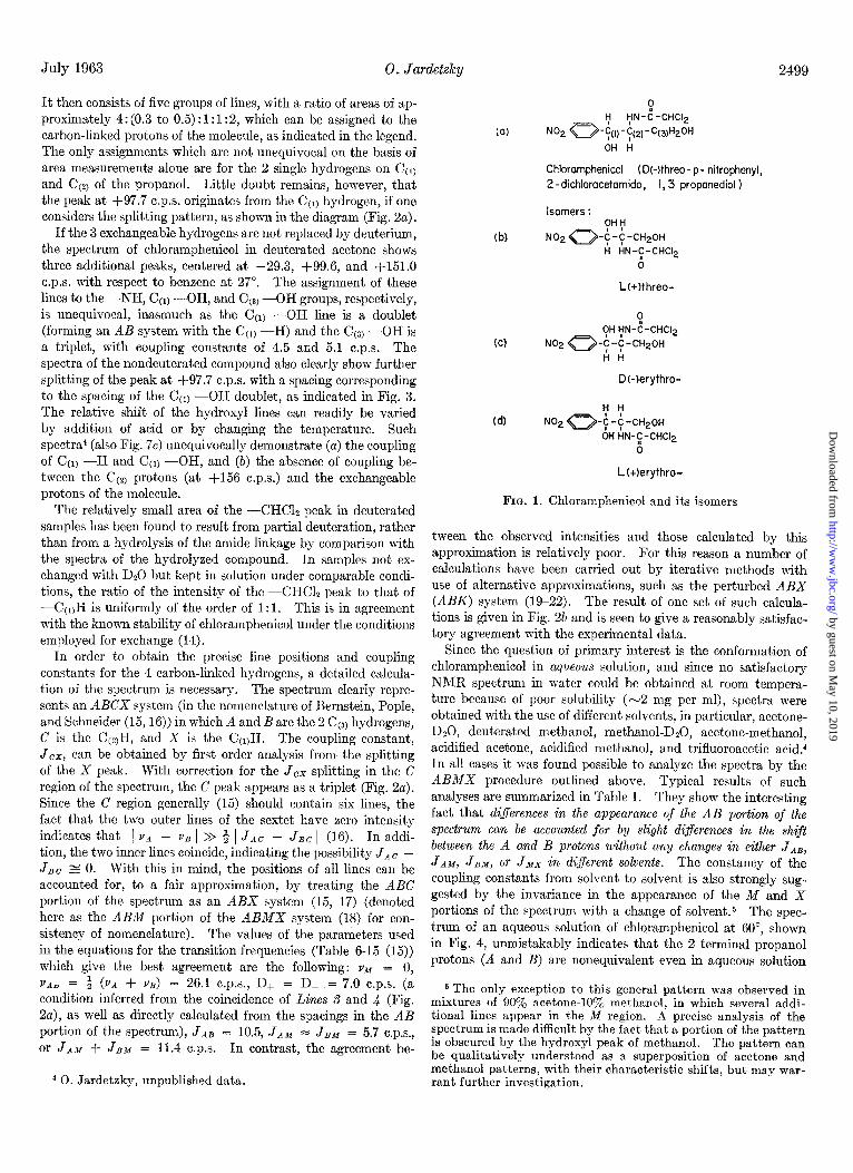
It then consists of five groups of lines, with a ratio of areas of ap- proximately 4: (0.3 to 0.5) : 1: 1:2, which can be assigned to the carbon-linked protons of the molecule, as indicated in the legend. The only assignments which are not unequivocal on the basis of area measurements alone are for the 2 single hydrogens on Ccl) and Cc?) of the propanol. Little doubt remains, however, that the peak at +97.7 c.p.s. originates from the Ccl) hydrogen, if one considers the splitting pattern, as shown in the diagram (Fig. 2~).
I f the 3 exchangeable hydrogens are not replaced by deuterium, the spectrum of chloramphenicol in deuterated acetone shows three additional peaks, centered at -29.3, +99.6, and +151.0 c.p.s. with respect to benzene at 27”. The assignment of these lines to the -NH, Ccl) -OH, and C(3) -OH groups, respectively, is unequivocal, inasmuch as the C(1) -OH line is a doublet (forming an AB system with the Ccl) -H) and the Cc3) -OH is a triplet, with coupling constants of 4.5 and 5.1 c.p.s. The spectra of the nondeuterated compound also clearly show further splitting of the peak at +97.7 c.p.s. with a spacing corresponding to the spacing of the Ccl) -OH doublet, as indicated in Fig. 3. The relative shift of the hydroxyl lines can readily be varied by addition of acid or by changing the temperature. Such spectra4 (also Fig. 7c) unequivocally demonstrate (a) the coupling of Ccl) -H and C(1) -OH, and (b) the absence of coupling be- tween the Cc2) protons (at f156 c.p.s.) and the exchangeable protons of the molecule.
The relatively small area of the -CHCl, peak in deuterated samples has been found to result from partial deuteration, rather than from a hydrolysis of the amide linkage by comparison with the spectra of the hydrolyzed compound. In samples not ex- changed with D20 but kept in solution under comparable condi- tions, the ratio of the intensity of the -CHC12 peak to that of --C~QH is uniformly of the order of 1: 1. This is in agreement with the known stability of chloramphenicol under the conditions employed for exchange (14).
In order to obtain the precise line positions and coupling constants for the 4 carbon-linked hydrogens, a detailed calcula- tion of the spectrum is necessary. The spectrum clearly repre- sents an ABCX system (in the nomenclature of Bernstein, Pople, and Schneider (15,16)) in which A and B are the 2 Cc31 hydrogens, C is the C&I, and X is the C(l)H. The coupling constant, Jcx, can be obtained by first order analysis from the splitting of the X peak. With correction for the Jcx .splitting in the C region of the spectrum, the C peak appears as a triplet (Fig. 2~). Since the C region generally (15) should contain six lines, the fact that the two outer lines of the sextet have zero intensity indicates that 1 vA - vB 1 >> $ 1 Jac - JBC 1 (I 6). In addi- tion, the two inner lines coincide, indicating the possibility JAC - JBc zz 0. With this in mind, the positions of all lines can be accounted for, to a fair approximation, by treating the ABC portion of the spectrum as an ABX system (15, 17) (denoted here as the ABM portion of the ABMX system (18) for con- sistency of nomenclature). The values of the parameters used in the equations for the transition frequencies (Table 6-15 (15)) which give the best agreement are the following: vM = 0, VAo = 3 (vA + vg) = 26.1 c.p.s., D+ = D- = 7.0 c.p.s. (a condition inferred from the coincidence of Lines S and 4 (Fig. 2a), as well as directly calculated from the spacings in the AB portion of the spectrum), JAB = 10.5, JAM = JBM = 5.7 c.p.s., or JAM + JBM = 11.4 c.p.s. In contrast, the agreement be-
4 0. Jardetzky, unpublished data.
(a)
(b)
(C)
(d)
Chloramphenicol (DWhreo- p- nitrophenyl, 2-dichloracetomido, I, 3 proponediol )
Isomers : ?H1;’
NO~~-C-C-W~~FI
ri ti~-~~-CtiCt~
c;
L(+)threo-
FI ?H I;IN-C-CHC12
No2 0-c -C-CHZO~ ItA
Dkjerythro-
‘;‘r NO2 a-$ -$ -CHzOH
OH HN-C-CHC12 6
L(+)erythro-
FIG. 1. Chloramphenicol and its isomers
tween the observed intensities and those calculated by this approximation is relatively poor. For this reason a number of calculations have been carried out by iterative methods with use of alternative approximations, such as the perturbed ABX (ABK) system (19-22). The result of one set of such calcula- tions is given in Fig. 26 and is seen to give a reasonably satisfac- tory agreement with the experimental data.
Since the question of primary interest is the conformation of chloramphenicol in aqueous solution, and since no satisfactory NMR spectrum in water could be obtained at room tempera- ture because of poor solubility (-2 mg per ml), spectra were obtained with the use of different solvents, in particular, acetone- DzO, deuterated methanol, methanol-DaO, acetone-methanol, acidified acetone, acidified methanol, and trifluoroacetic acid.4 In all cases it was found possible to analyze the spectra by the ABMX procedure outlined above. Typical results of such analyses are summarized in Table I. They show the interesting fact that diferences in the appearance of the AB portion of the spectrum can be accounted for by slight diflerences in the shift between the A and B protons without any changes in either JAB, JAM, JB~, or JMX in digerent solvents. The constancy of the coupling constallts from solvent to solvent is also strongly sug- gested by the invariance in the appearance of the M and X portions of the spectrum with a change of solvent.5 The spec- trum of an aqueous solution of chloramphenicol at 60”, shown in Fig. 4, unmistakably indicates that the 2 terminal propanol protons (A and B) are nonequivalent even in aqueous solution
5 The only exception to this general pattern was observed in mixtures of 90% acetone-107, methanol, in which several addi- tional lines appear in the M region. A precise analysis of the spectrum is made difficult by the fact that a portion of the pattern is obscured by the hydroxyl peak of methanol. The pattern can be qualitatively understood as a superposition of acetone and methanol patterns, with their characteristic shifts, but may war- rant further investigation.
by guest on May 10, 2019
http://ww
w.jbc.org/
Dow
nloaded from

24 6910 12
VD VX vM vB% v,
I I
I357811
(b)
X FIG. 2. a, 60-megacycle NMR spectrum of 0.75 M chloramphen-
icol, lyophilized from DzO, in deuterated acetone. Line positions: 9R = center of the nitrobenzene AzBz quartet; 9~ = -CHCl*; 9x = C(,,H; YM = CczjH; VA and 9~ = Cta,H2 of the propanol side chain; 9s = acetone methyl. Line positions are given in cycles per second with respect to benzene as an external standard. The splitting pattern of the ABMX system with the coupling constants
9.8 13 571jlOr2 24 6 :: II
--- MB A JdB, Jnx, Jsx, and JMX is given in first order for the purpose o orientation only, the actual values being obtained from the cal- culated spectrum. b, Calculated spectrum of the ABMX portion of chloramphenicol, with the use of the ABC approximation (17) and the values of shifts and coupling constants given in Table I, with by > Q > QM > ux.
UN VD I
-293 +99.6 +151.0 +220 (varl , I I I I 0
-50 0 +50 100 150 200
FIG. 3. Sixty-megacycle NMR spectrum of 0.75 Y chloramphenicol lyophilized from Hz0 in deuterated acetone. Same scale and designation of lines as Fig. 2a with the additional lines YN = NH, ~01 = CcI,OH, VW, = Cca,OH.
2500
by guest on May 10, 2019
http://ww
w.jbc.org/
Dow
nloaded from

July 1963 0. Jar&t& 2501
and illustrates the fact that this nonequivalence persists even at elevated temperatures. The invariance of the coupling con- stants with change of solvent strongly suggests that the conforma- tion of the molecule is the same in different solvents, GKZU&~~J water.
TABLE I Relative shifts and coupling constants for propanol
ABMX system in chloramphenicol The values of coupling constants in all of the solvent mixtures
studied (see text) are identical with those shown below.
In acetone (60 megacycles)
YAa - VBb 10.2 f 0.2 vg - v.d 21.3 f 0.2 vb! - VXd 59.0 f 0.2
1 JAB 1 10.5 zk 0.2 1 JAM 1 5.7 f 0.2 1 JBM 1 5.7 It 0.2 IJMxI 2.4 i 0.2
In methanol (60 megacycles)
13.3 f 0.2 20.8 f 0.2 58.2 f 0.2
10.2 f 0.2 lo-11 5.8 rt 0.2 5-6 5.8 f 0.2 5-6 2.6 f 0.2 2-3
In Da0 at 60° (60 me@zycles)
13-14 20-21 53-60
0 Midpoint between Lines 10 and 11 (Fig. 2). b Midpoint of Lines 6 and 7. c Center of doublet (Lines B and 5). d Center of doublet X.
VR I
ML tl56
-50 +40 +I09 cl18 +I70
I , +I84 -50 0 +50 loo 150
FIG. 4. Spectrum of chloramphenicol in DzO at 60”. Designa- tion of lines with approximate shifts is the same as in Fig. 2.
erythro Isomers-The spectra of the D( -)-erythro and the L(+)-erythro isomers are found to be similar in general appear- ance to the spectra of the two threo isomers, so that the assign- ment of the peaks can be made by analogy. However, it is apparent from the spectrum of the L(+)-eqthro isomer, shown in Fig. 5a, that the values of the coupling constants and shifts in
+162.7 - 48.1
l i75. + 39.5 tlo6.3 + 88.8 ‘i t275.8 I I I I ! I
-50 0 +50 IO0 150 200 250
lb)
Ill //I I I 98
I 3 5 idI0 12 246 ‘1 II
-VI 4 X MB A
FIG. 5. a, 60-megacycle NMR spectrum of the L(+)-erythro isomer of chloramphenicol in deuterated acetone. The exchangeable hydrogens have been removed by lyophilixation from D20 in order to simplify the spectrum. Designations of lines, shifts, and coupling constants are the same as in Fig. 2~. b, Calculated spectrum of the ABMX portion, with the use of the values of shifts and
coupling constants given in Table II.
by guest on May 10, 2019
http://ww
w.jbc.org/
Dow
nloaded from

2502 Studies on Mechanism of Action of Chloramphenicol. I Vol. 238, No. 7
TABLE II
Shifts and coupling constants for propanol ABMX system of erythro isomers of chloramphenicol
Same approximations and conventions as in Table I are used.
In acetone (60 megacycles) In methanol (60 megacycles)
PA - VB 13.2 f 0.3 15.1 f 0.5 VB - VM 12.9 f 0.2 13.5 f 0.5 Y,u - vx 56.4 f 0.3 58.5 f 0.5
1 JAB 1 11.0 f 0.3 10.0 f 0.5
1;,7 4.5 f 0.3 4.5 f 0.5
IJ::I 5.2 f 0.3 4.5 f 0.5 6.0 f 0.2 6.0 f 0.2
TABLE III Shifts and coupling constants for the propanol ABMX system of
D(-)-th~eo-i-p-amino-2-dichloracetamido-i ,b-propanediol in D20
c.p.s.
VA - VB 10.4 f 1 YB - YM 19.6 f 1 vhf - vx 58.1 * 1
/ JAB 1 9 - 10
I JAM I 4-5 /JBM I 4-5 1 JMX 1 5.3 f 0.3
the ABM region of the spectrum are appreciably different from those obtained for the three isomers. The assignment of lines indicated in the figure, and chosen because of its similarity to the assignment for the three isomers, is considerably less certain because of the greater overlap of individual peaks. A calculated spectrum, based on the same approximations as those used in Fig. 2b, is given in Fig. 56, showing that an interpretation similar to that used above is at least consistent with the observed spec- trum. The values of shifts and coupling constants are given in Table II. It is not possible to differentiate with certainty be- tween the possibilities JAM = JBM and JAM # JBM in this case. However, the marked asymmetry of the M triplet, and the spac- ing of the lines, suggests that JBM - JAM LZ 1 c.p.s. An ex- amination of a series of spectra in different solvents4 shows the same comparative invariance of the coupling constants with a change of solvent that appears to hold true for the three isomers.
p-Amino Analogue-The spectrum of D( -)-threo-l-p-amino- 2-dichloracetamido-l , 3-propanediol was also of some interest. Since its aliphatic side chain is identical with that of chlor- amphenicol, it might be expected to behave in aqueous solution in a manner similar to the side chain of the antibiotic, providing corollary evidence for the conformation of the antibiotic in water. At room temperature, the spectrum of the compound (-0.1 M
in DzO) proves again to be of the ABCX type, with shifts and coupling constants obtainable by the ABMX approximation, as shown in Table III. The individual values are somewhat less certain than in the case of chloramphenicol and its isomers, because the number of samples was limited by the very small amount of available material. The value of the MX coupling constant is, however, somewhat larger than in the case of chlor-
amphenicol. This may reflect either a slight difference in con- formation or a difference in the distribution of electron density in the adjacent aromatic ring.
Hydrogen Bonding
The position of the hydroxyl resonance lines of chloramphenicol (Fig. 3) and its isomers (not shown, but found to be identical with those in Fig. 3) in acetone strongly suggests that both alcohol groups are involved in hydrogen bonds. Thus the values of +99.6 c.p.s. and +151.0 c.p.s. correspond to shifts of roughly -4.3 p.p.m. and -3.3 p.p.m. with respect to the position of the alcohol hydroxyls at infinite dilution in CC!&, usually attributed to the non-hydrogen-bonded alcohol monomers. In contrast, the positions of the two peaks taken relative to water or alcohol (which are largely hydrogen-bonded) is of the order of 0 to +0.8 p.p.m. Part of this shift must be accounted for on the basis of the diamagnetic anisotropy of the aromatic ring. Since the position of the 2 hydroxyl protons with respect to the center of the ring is uncertain because of the flexibility of the aliphatic side chain, an accurate theoretical estimate of the diamagnetic anisotropy is not possible. Nevertheless, by use of the correc- tion factors of Johnson and Bovey (23) or Fijrsen and Nilson (24), it is possible to arrive at a range of -0.7 to -1.5 p.p.m. for the unshielding of C(r)OII and -0.2 to - 1.5 p.p.m. for CcoOH over the entire range of possible positions for the 2 protons. Even taking the largest values of the correction factor, there remains a shift of the order of -2 to -3 p.p.m. to be explained by hydrogen bonding.
Additional evidence for hydrogen bonding comes from the Raman spectrum of chloramphenicol in acetone, which clearly shows that the narrow line in the region of 3600 to 3650 cm-1 characteristic of the O-H stretching frequency of nonbonded hydroxyls is absent. Instead, a broad peak is found in the region 3400 to 3500 cm-l, which is characteristic of the stretching vibration of hydrogen-bonded hydroxyls, although part of the peak may be due to t,he N-H stretching vibration of the amide. There appears to be no evidence for the participation of the amide proton in a hydrogen bond, from either the Raman or the NMR data. Thus a shift of the N-H stretching vibration -3300 cm-r and a shift of the amide proton resonance of the order of an additional -1 to -2 p.p.m. would have been ex- pected in that case, and neither is found.
Three types of hydrogen bonds involving hydroxyl groups of chloramphenicol are possible in this system: (a) intramolecular bonds to oxygens on the same molecule (intramolecular bonding to the amide nitrogen, chlorine, and the ?r electrons of the aro- matic ring are theoretically to be considered, but can almost certainly be ruled out because of the lower electronegativity of the acceptor groups (25,26) and unfavorable steric relationships), (b) intermolecular bonds to the solvent, and (c) intermolecular bonds to the oxygens of other chloramphenicol molecules. The last possibility is made improbable by the comparative concen- tration independence of the chemical shift of the 2 hydroxyl pro- tons, shown in Table IV.‘j
6 It could be argued that the lowest concentrations reached are still high enough to allow the solute to exist largely in dimer or polymer form and that a concentration dependence would become apparent if lower concentrations could be examined. However, if the concentrations are expressed in mole fractions and the shifts are compared to the shifts of other alcohols in corresponding con- centrations (27-29), it becomes apparent that the concentration dependence of the shift is very marked in this range in all cases
by guest on May 10, 2019
http://ww
w.jbc.org/
Dow
nloaded from

July 1963 0. Jardetxky 2503
On the other hand, the concentration independence cannot be used to rule out the possibility of hydrogen bonding to the solvent, since at all accessible concentrations the solvent is present in excess, so that a concentration dependence would not become apparent. Considering that the existing correlations between chemical shift, infrared and thermodynamic data on hydrogen bonds are far from being completely consistent (25,27, 28, 30, 31), an unequivocal distinction between intramolecular bonding and bonding to the solvent cannot be made on the basis of chemical shift data published thus far. Nevertheless, corol- lary findings on simpler dials’ allow the conclusion that the existence of a relatively stable intramolecular bond is more probable in the present case.
Thus, the chemical shifts of monohydroxyl alcohols, extrap- olated to infinite dilution in acetone, fall into the range of f280 to +310 c.p.s. with respect to benzene at 60 megacycles, or just above the methyl group of acetone. The water peak appears at +285 c.p.s., at infinite dilution in acetone. The concentration dependence of the shift in the range 0 to 2 M is very marked. In contrast, the shift of dihydroxyl alcohols extrapolates to the significantly lower values of +200 to +250 c.p.s. at infinite dilution in acetone, although at high concentrations the shifts fall into the same range with the monohydroxyl alcohols. Similar observations have been reported by Kiihler, Pettig, and Fischer (32), who find that shifts in cyclopentanediols capable of forming intramolecular hydrogen bonds occur to lower fields than shifts in cyclopentanediols in which the formation of such bonds is made impossible by steric hindrance. The same trend becomes apparent on detailed examination of the shifts of differ- ent alcohols in CDC& solution measured by Bhacca, Johnson, and Shoolery (33). There appears at present little reason to doubt that water and alcohol hydroxyls are hydrogen-bonded to acetone. Thus, the chemical shifts are approximately 1.0 p.p.m. lower at infinite dilution in acetone than at infinite dilution in Ccl, or CDC& (correcting for differences in bulk diamagnetic susceptibility). The finding of a shift to lower fields in the case of dihydroxy alcohols (the position of the observed line probably representing an average between the inter- and intramolecular bond), then, suggests a greater strength of such bonds. The alternative possibility that the difference in the shift is to be attributed entirely to the diamagnetic anisotropy of the C=O as compared to the C-OH group, both acting as acceptors, is not likely in view of the finding that in the case of strong intra- molecular bonds (25, 27, 34) the shifts of hydrogens bonded to the two groups are of comparable magnitude.
Quantitative estimates of the relative strength of the two kinds of bonds can still be made only with great reservations. However, the careful infrared studies of Becker (35) indicate that the -0-H. . .O=C bond to acetone has a AH E 3.5 kcal. A somewhat smaller value of -2.8 kcal would be obt,ained from the resonance shift versus infrared shift correlation proposed by Reeves, Allan, and Stremme (31) and by using the Badger- Bauer (36) relationship. If the shift of t,he intramolecular bond is estimated to be of the order of -1 to -2 p.p.m. relative to the intermolecular hydroxyl-acetone bond (assuming one intra- molecular bond for every intermolecular bond at infinite dilution
except when intramolecular bonding is possible. Unless one as- sumes the presence of unusually stable dimers or higher polymers, one is led to the conclusion that the observed hydrogen bonds do not connect different chloramphenicol molecules.
7 To be presented in a separate communication.
TABLE IV
Chemical shifts of R hydroxyl protons of chloramphenicol in deuterated acetone at different concentrations
These shifts were referred to benzene as an external standard and corrected for differences in bulk diamagnetic susceptibility.
Concentration CwOH I
‘&OH
Jf
0.08 0.1 0.2 0.5 1.0
103.1 f 0.3 152.5 f 0.3 102.2 Z!Z 0.3 152.3 f 0.3
99.6 f 0.3 151.0 f 0.3 100.1 f 0.3 151.2 f 0.3
99.5 f 0.3 151.7 * 0.3 I
in acetone), one obtains a AHintra - AHinter LZ 2 kcal from the cited correlations or a AHintrs, E! 5 kcal. The shift observed in the Raman spectrum (-150 cm-‘) is consistent with both the observed chemical shift and this estimate. Tenuous as such estimates are at present, taken together with the smaller entropy term for intramolecular bonds (37), they tend to support the notion that intramolecular bonds can have greater stability than intermolecular bonds, even in strongly hydrogen-bonding solvents.
Structural Considerations
The foregoing analysis of the proton resonance spectra of chloramphenicol and its isomers suggests three conclusions.
1. The rotation about both single carbon-carbon bonds of the propanol side chain is restricted.
2. To a first approximation only one conformer of each com- pound is preferred (less than 5% of another conformer could have remained undetected).
3. Within the limited range of solvents examined, the stability of this conformer is not sensitive to a change of solvent; i.e.
the conformation in aqueous solution is essentially the same as that in acetone or methanol.
The first conclusion is based on two features of the spectra: (a) the smallness of the coupling constant JMX = 2.6 c.p.s. in the case of chloramphenicol and its L(+)-three isomer, and (b) the nonequivalence of the 2 hydrogens on C&l, i.e. V~ # vg, in all cases, and is consistent with the likelihood of an intramolecular hydrogen bond.8
8 It should be emphasized that the justification for the inference of restricted rotation from the nonequivalence of the 2 terminal hvdrogens is by no means self-evident. In principle. an ABC spectrum can arise in asymmetrically substituted molecules of the tvne CHZCHYZ because of differences in the averaeine of the i and Bshifts by rapid rotation, i.e. from a rotationarnonequiv- alence. Several suggestions along these lines have been made for the fluorocarbon series (15, 17, 38, 39, 40). However, as pointed out by Waugh and Cotten (41), the expectation of rotational non- equivalence is based on a symmetry argument alone and “in prac- tice it is unlikely, that the asymmetry with respect to internal rotation required to produce magnetic nonequivalence would not also be reflected in some angular dependence of potential energy.” In fact, rotational nonequivalence is not observed in the proton resonance spectra of many asymmetrically substituted molecules in which “free” rotation can be inferred from the magnitude of the coupling constants, such as amino acids (42), nucleosides and nucleotides (43,44), and catecholamines (45). The latter series of compounds is especially noteworthy, since the asymmetric centers in question resemble the asymmetric centers of chloramphenicol. The weight of the available evidence thus favors the view that the contribution of neighbor asymmetry per se to the magnetic non-
by guest on May 10, 2019
http://ww
w.jbc.org/
Dow
nloaded from

2504 Studies on Mechanism of Action of Chloramphenicol. I Vol. 238, No. 7
The postulate of a single conformer rests on the fact that all features of the observed spectra can be satisfactorily understood on this basis, and cannot readily be brought in agreement with those predicted on the assumption of a mixture of two or more conformers. Accepting the nonequivalence of protons A and B as evidence for restricted rotation, the choice of possible con- formers can be narrowed by using the type of relationship for the dependence of the coupling constants on the dihedral angle as formulated by Karplus (48, 49).g Without any attempt at precise numerical correlation, thii relation allows the inference that the observed conformation at the terminal carbon cor- responds to either
I or
II
(or to a mixture of equal amounts of both, in case the unlikely additional assumptions va(r) = vA(rr) and ve(rr) = YE hold).
A similar argument can be advanced in favor of a restriction of rotation about C&---C~~~ to a single conformer. If more than one conformer were stable in this case, additional lines would have been expected in both the M and the X region of the spec- trum, since both carbons are asymmetrically substituted. The possibility of averaging by free rotation can, on the other hand, be definitely ruled out from the magnitude of the coupling con- stant JMx = 2.6 c.p.s. (15, 18, 51). Of the four orientations about Co+&) compatible with the observed value of the coupling constant, two are eclipsed and may be excluded as being less stable (52, 53) in comparison with the two staggered conformations,
H , ml
T H iM)
__-’ _.= =.__
*-..
III
equivalence of the protons of a CH, group is negligible compared to the contributions of conformational nonequivalence. This conclusion is in agreement with the recent findings of Shafer et al. (46) and especially Whitesides et al. (47).
9 The essential validity of this relationship showing two maxima and a minimum for the coupling constants at or around values of the dihedral angle of O’, 180°, and 90”, respectively, is not in ques- tion, even though there is considerable doubt concerning the pres- ently used values of the coefficients (50).
or
H WI I
H mf,
“‘;-’
,A’ ’ ..\ ,/’ ‘\
‘\\
IV
The analysis of the proton resonance spectra of chloram- phenicol thus suggests that the compound exists in one of the following conformations, equally compatible with the observed values of coupling constants and the observed spectral patterns: I + III, I + IV, II + III, and II + IV. A reasonable choice between these possibilities can be made by assuming an intra- molecular hydrogen bond. If the possibility of forming intramolec- ular bonds is examined with the aid of molecular models, it is found that conformations of Type I allow the formation of a hydrogen bond between the two hydroxyl groups, whereas conformations of Type II allow the formation of a hydrogen bond from either of the hydroxyls to the carbonyl group. It also becomes apparent, however, that in the latter case the restric- tion of rotation about both carbon-carbon bonds is not satis- factorily accounted for. Thus the formation of a hydrogen bond Co) -OH. . . O=C would allow completely free rotation about C(z)--Co), and the formation of a bond C(3) -OH.. .O=C would allow almost free rotation about Co+&), although in this case rotation could be restricted in certain configurations by the interference of the ring with the motion of the amide group. If, however, one imposes the additional requirement that the planarity of the amide group should be preserved (allow- ing for deviations not exceeding ~~10-15” (26)), it becomes impossible to build structures of the type Co) -OH...O=C. Although the universal occurrence of planar amide groups has been seriously questioned, it is worth noting that deviations from a planar configuration have been observed only in the solid state, in which the strain energy, calculated by Pauling (26) to be of the order of 1 kcal per lo”, can be supplied at the expense of the lattice energy. It is extremely unlikely that a similar distortion would occur in solution. It therefore appears reasona- ble to rule out conformations of the type C(3) -OH. . .O=C on the grounds that they are not compatible either with the planarity of the amide group or with the occurrence of a single staggered conformer of the Co+&) region.
Similarly, conformations of the type Co) -OH. . .O=C would not account for the hindered rotation about C&--CQ), leaving structures of the Type I + III, or I + IV, as being more probable.” A decision between these two conformations is more diicult. However, taking into account the studies of Fodor, Kiss, and Sallay (ll), one finds that conformations of the Type I + III are consistent with a cis orientation of C(1) -0 and Co) -N, whereas those of Type I + IV are not. It may also be noted that I + III resembles more closely the conforma- tion assumed by bromamphenicol in the crystalline state than does I f IV. It is therefore concluded that a conformation of the type I + III is in best agreement with all of the observa- tions to date.
10 Alternative hydrogen-bonded structures can also be built. They are not further discussed here, since the values of the cou- pling constants that one would predict from them are in serious disagreement with those observed.
by guest on May 10, 2019
http://ww
w.jbc.org/
Dow
nloaded from

0. Jardetxky 2505
FIG. 6. Proposed conformations of chloramphenicol (a) and its L(+)-erythro isomer (b) ; comparison of the structure of chloram- phenicol with that of uridylic acid (c) .
By an entirely analogous sequence of arguments, one arrives at the conclusion that the most probable average conformation of the L( +)-three isomer is a mirror image of the conformation of chloramphenicol and that the two erythro isomers exist in hydrogen-bonded conformations which are also mirror images of each other. The corresponding conformations are similarly staggered about C(1)-C&, however, with the 2 hydrogens Ccl,H and Cc,,H in an almost trans orientation in the erythro case. The proposed conformations of the isomers are illustrated in Fig. 6, a and b. The conformation of the p-amino analogue of chloramphenicol is probably similar to that of chloramphenicol, with the reservations mentioned above.
The conformation shown in Fig. 6a has one further feature of interest. As can be seen in Fig. 6c, it bears a striking resem-
blance to a pyrimidine ribonucleotide (but not a deoxyribonu- cleotide) in size, orientation of individual moieties, and the distribution of electronegative groups. If it is assumed that this similarity has a bearing on the mode of action of the com- pound, certain features of the structure-activity relationships in the chloramphenicol series can be understood. Thus, any alteration of the propanol moiety would destroy the similarity to the ribose ring and any change in the size of the dichlor- acetamide side chain would alter the resemblance to a phosphate. Furthermore, the inactivity of the L( +)-threo and the erythro series of isomers would follow from the fact that they cannot form nucleotide-like conformations. The inactivity of p-sub- stituted compounds that cannot serve as hydrogen bond ac-
by guest on May 10, 2019
http://ww
w.jbc.org/
Dow
nloaded from

2506 Studies on Mechanism of Action of Chloramphenicoi. I
&I VD vx I
yh4 yf3F
I
YLYENT lMpwllT”
(01
VD uX
% vM VA vs I II I
UR uN VD VXVOI I I I I
VM vo3
&v/l, II I I
uH20 I
Vol. 238, No. 7
-50 0 +50 100 150 200
FIG. 7. Spectra of chloramphenicol in deuterated acetone as a function of temperature; a and b, deuterated compound at -30” and +85”: C. nondeuterated compound at +95”. showing shifts of exchangeable hydrogens. Designation of lines and chemical shifts are the same as in Fig. 2.
ceptors, which is apparent from the extensive data gathered by Shemyakin (6), could also be explained.
Temperature Studies
The inference concerning the stability of the conformer in different solvents, drawn in the preceding section, is justified primarily by the invariance of the coupling constants with changes of solvent. Additional information on the stability of a given conformation is sometimes obtainable from the tempera- ture dependence of the proton resonance spectra. For this reason the spectra of chloramphenicol in deuterated acetone and in DzO were studied over the temperature ranges of -70” to +95” and +50” to +95”, respectively.” Typical results are
11 Somewhat higher temperatures were undoubtedly reached but could not be accurately measured. The high vapor pressure in sealed tubes for the most part accounts for the possibility of rais- ing the temperature above the boiling point of acetone.
shown in Fig. 7. At temperatures below -30” the marked broadening of all peaks precluded the detection of possible minor changes with any degree of certainty, although the progressive transition from well resolved to broad lines would have been disturbed by the appearance of a series of new peaks. At temperatures above - 30” the spectra are certainly temperature- independent, although above 90” a slight decrease in the AB shift is noticeable. The data are consistent with the notion that the activation energy for breaking the intramolecular hydrogen bonds and overcoming the barrier to free rotation has not been reached. This conclusion is supported by the shifts of the ex- changeable hydrogens in acetone solution. As the temperature is increased, the resonance peaks move to higher fields by ap- proximately 0.4 c.p.s. per degree, which compares to the tempera- ture coefficient of the shift of the hydrogen-bonded water protons (0.38 c.p.s. per degree). The lines in each -OH multiplet begin to merge about 80”, indicating an increasing rate of exchange.
by guest on May 10, 2019
http://ww
w.jbc.org/
Dow
nloaded from

July 1963 0. Jardetxky 2507
However, even at the highest temperatures reached, the indi- vidual peaks retain their identity, allowing an upper limit for the exchange rate to be placed at about 3 x lo2 see-i. In solvents containing -OH groups and in water, the exchange is sufficiently rapid, even at room temperature, so that the indi- vidual hydroxyl peaks are not observed. It should be mentioned that this, in itself, does not speak against the persistence of an intramolecular hydrogen bond, since the only requirement for the latter is that the lifetime of a hydrogen bond be long com- pared to the period during which the bond remains broken. (Succinic acid (34) may constitute another case in point.) Fusion of resonance lines, on the other hand, means that the lifetime of a proton in the hydrogen bond be short compared to 1/(2a(vn - eon)] where vn is a shift of the hydrogen bond and ~,,n its shift in a hydroxyl (which may or may not be also hy- drogen-bonded). These two time intervals are not identical. Since the two shifts are not accurately known, an accurate estimate of the minimal exchange rate is not possible. Con- sidering, however, the observed line positions (Fig. 3), it is likely that rates as low as 30 see-r could lead to a fusion of the lines. Such rates are easily compatible with the existence of thermodynamically stable hydrogen bonds. A more detailed examination of exchange rates of chloramlihenicol in acetone- methanol and acetone-water mixturcs4 substantiates this con- clusion. The weight of the evidence thus suggests that the conformations assumed by chloramphenicol or its isomers in solution possess considerable stability.r2
SUMMARY
High resolution proton resonance spectra of chloramphenicol and its isomers are analyzed and interpreted in terms of pre- ferred conformations. Evidence is presented to show that these conformations are unique and stable. The structural similarity between the proposed preferred conformation of chlorampheni- co1 and a pyrimidine nucleotide is pointed out.
Aclcnowledgments--The author is very much indebted to Miss Norma Wade for assistance in the later part of the work, to Dr. Peter Pappas for obtaining some of the spectra, and to Dr. J. N. Shoolery of Varian Associates for several stimulat,ing dis- cussions of the problem.
REFERENCES
1.
2. 3.
4.
5.
G.
7.
GALE, E. F., AND FOLKES, J. P. B&hem. J., 63, 493 (1953) BROCK. T. D.. Bacterial. Rev.. 26, 32 (1961). HOACL~ND, 6I. B., in E. C~HARGAFF AND J. N. DAVIXON
(Editors), The nucleic acids, Vol. ZZZ, Academic Press, Inc., New York, 1960, p. 309.
MAXWELL, R. E., AND NICKEL, V. S., Antibiotics and Chemo- therapy, 4, 289 (1954).
SHEMYAKIN, M. M., BAMUAS, E. M., VINOGRADOVA, E. L., KARAPETYAN. M. G., Kor,ossov, M. N., KHOKHLOV, A. S., SHVETSOV, Y. B., AND SHCHUKINA, L. ‘A., Doklady’ Akad: Nauk S. S. S. R.. 86. 565 (1952): 94. 257 (1954).
/ , ~ I
SHEMYAKIN, M. M., Khimia Anizfbiotikov, Vol. Z (review of 1295 references on chloramphenicol, in Russian), Academy of Sciences, U. S. S. R., Moscow, 1961, p. 337.
REBSTOCK, M. C., CROOKS, H. M., CONTROULIS, J., AND BARTZ, Q. R., J. Am. Chem. Sot., 71, 2458 (1949).
12 Note added in proof: An analysis of the 100.megacycle spec- tra of chloramphenical and its L(f)-erythro isomer leads to values of coupling constants identical with those reported here. The author is grateful to Mr. Le Roy Johnson of Varian Associates for
42. JARDETZKY, O., AND JARDETZKY, C. D., J. Biol. Chem., 233, 383 (1958).
43. JARDETZKY, C. D., AND JARDETZKY, O., J. Am. Chem. Sot., 81, 222 (1960).
44. JARDETZKY, C. D., J. Am. Chem. Sot., 84, 62 (1962).
obtaining these spectra. 45. JARDETZKY, O., Adv. Chem. Phys., in press.
8.
9.
10. 11. 12.
13.
14.
15.
16.
17.
18.
19.
20.
21.
22.
23.
24.
25.
26.
27.
28.
29.
30. 31
HAHN, F. E., HAYES, J. E., WISSEMAN, C. L., JR., HOPPS, H. E., AND SMAJIEI., J. E., Antibiotics and Chemotherapy, 6, 531 (1956).
COLLINS, R. J., ELLIS, B., HANSEN, S. B., MACKENZIE, H. S., MONALIM, R. J., PETROW, V., STEPHENSON, O., AND STUR- GEON, B., J. Pharm. and Pharmacol., 4,693 (1952).
DUNITZ, J. D., J. Am. Chem. Sot., 74, 995 (1952). FODOR, G., KISS, J., AND SALLAY, I., J. Chem. Sot., 1858 (1951). JARDETZKY, O., AND JARDETZKY, C. D., Abstracts of papers of
the 136th meeting of the American Chemical Society, Ameri- can Chemical Society, Washington, 1959, p. 20C.
JARDETZKY, O., AND JARDETZKY, C. D., in D. GLICK (Editor), Methods of biochemical analysis, Vol. IX, Intcrscicnce Pub- lishers, Inc., New York, 1962, p. 235.
HIGUCHI, T., MARCUS, A. D., AND BIAS, C., J. Am. Pharm. Assoc., Sci. Ed., 35, 129 (1954).
POPLE, J. A., SCI-~NEIDER, W. G., AND BERNSTEIN, H. J., High resolution nuclear magnetic resonance, McGraw-Hill Book Company, New York, 1959.
BERNSTEIN, H. J., POPI~E, J. A., AND SCHNEIIIER, W. G., Can. J. Chem., 35, 65 (1957).
GUTOWSKY, H. S., HOLM, C. H., SAIKA, A., AND WILLIAMS, G. A., J. Am. Chem., Sot. 79, 4596 (1957).
ROBERTS, J. D., An introduction to the analysis of spin-spin splitting in high-resolution nuclear magnetic resonance spec- tra, W. A. Benjamin, Inc., New York, 1961, p. 10.
REILLY, C. A., AND SWALEN, J. D., J. Chem. Phys. 32, 1378 (1960).
ABRAHAM, R. J., AND BERNSTEIN, H. J., Can. J. Chem., 39,
216 (19Gl). FESSENDEN, R. W., AND WAUGH, J. S., J. Chem. Phys., 31, 996
(1959). CASTELLANO, S., AND WAUGH, J. S., J. Chem. Phys., 34, 295
(1961). JOHNSON, C. E., JR., AND BOVEY, F. A., J. Chem. Phys., 29,
1012 (1958). FORS~N, S., AND NILSS~N, M., ilcta Chem. &and., 13, 1383
(1959). PIMENTEL, G., ANI) MCCLELT~AN, A. L., The hydrogen bond,
W. H. Freeman and Company, San Francisco, 19GO. PAULING, L., The nature of the chemical bond, Ed. 3, Cornell
University Press, Ithaca, 1960. HUGOINS, C. M., PIMENTEL, G. C., AND SHOOLERY, J. N., J.
Chem. Phys., 23, 1244 (1955). HUGGINS, C. M., PIMENTEI,, G. C., AIYD SHOOLERY, J. N., J.
Phys. Chem., 60, 1311 (1956). BECKER, E. D., LIIXIEL, U., ANI) SHOOI~ERY, J. N., J. Molecu-
lar Spectroscopy, 2, 1 (1958). REEVES, I,. W., Can. J. Chem., 38, 748 (1960). REEVES, L. W., AI~LAN, E. A., AND STR$MME, K. O., Can. J.
Chem., 38, 1249 (1960). 32. K~HLER, H. J., PETTIG, M., ANII FISCHER, F., Bull. Group
Amp&e, Compt. rend. 10” colloque, Leipzig, 1961, p. 411. 33. BHACCA, N. S., JOHNSON, L. F., AND SHOOLERY, J. N., NMR
spectra catalog, Varian Associates, 1962. 34. EBERSON, L., AND F~RSEN, S., J. Phys. Chem., 64,767 (1960). 35. BECKER, E. D., Spectrochim. Acta, 17, 436 (1961). 36. BADGER, R. M., AND BAUER, S. H., J. Chem. Phys., 5, 839
(1937). 37. JAFFE, H. H., J. Am. Chem. Sot., 79, 2373 (1957). 38. POPLE, J. A., J. Molecular Phys., 1, 1 (1958). 39. NAIR, P. M., AND ROBERTS, J. D., J. Am. Chem. Sot., 79, 4565
(1957). 40. SHOOLERY, J. N., ANI) CRAWFORD, B., J. Molecular Spectros-
copy, 1, 270 (1957). 41. WAUGH, 3. S., AND COTTON, F. A., J. Phys. Chem., 65, 562
(1961).
by guest on May 10, 2019
http://ww
w.jbc.org/
Dow
nloaded from

2508 Studies on Mechanism of Action of Chloramphenicol. I Vol. 238, No. 7
46. SHAFER, P. R., DAVIS, D. It., VOGEL, M., NAGARAJAN, K., 50. GUTOWK~Y, H. S., MOCHEL, V. D., AND SOMERS, B. G., J. AND ROBERTS, J. D., Proc. Natl. Acad. Sci. U. S., 47, 49 Chem. Phys., 36, 1153 (1962). (1961). 51. GLICK, R. E., AND BOTHNER-BY, A. A., J. Chem. Phys., 25,
47. WHITESIDES, G. M., KAPLAN, F., NAGARAJAN, K., AND ROB- 362 (1956). ERTS, J. D., Proc. Natl. Acad. Sci. U. S., 48, 1112 (1962). 52. PITZER, K. S., Discussions Faraday Sot., 10, 66 (1950).
48. KARPLUS, M., AND ANDERSON, D. H., J. Chem. Phys., 30, 6 53. MIZUSHIMA, S., in E. HUTCHINSON AND P. VAN RYSSELBERGHE (1959). (Editors), Physical chemistry, Vol. II, Academic Press,
49. KARPLUS, M., J. Chem. Phys., 30, 11 (1959). Inc., New York, 1954.
by guest on May 10, 2019
http://ww
w.jbc.org/
Dow
nloaded from

Oleg JardetzkyCONFORMATION OF CHLORAMPHENICOL IN SOLUTIONStudies on the Mechanism of Action of Chloramphenicol: I. THE
1963, 238:2498-2508.J. Biol. Chem.
http://www.jbc.org/content/238/7/2498.citation
Access the most updated version of this article at
Alerts:
When a correction for this article is posted•
When this article is cited•
to choose from all of JBC's e-mail alertsClick here
http://www.jbc.org/content/238/7/2498.citation.full.html#ref-list-1
This article cites 0 references, 0 of which can be accessed free at
by guest on May 10, 2019
http://ww
w.jbc.org/
Dow
nloaded from