Studies on the Effects of Estrogen Stimuli on Osteogenesis
50
Studies on the Effects of Estrogen Stimuli on Osteogenesis Hyejin Kim Department of Biological Science and Technology, Faculty of Engineering, The University of Tokushima March 2014
Transcript of Studies on the Effects of Estrogen Stimuli on Osteogenesis
Studies on the Effects of
Estrogen Stimuli on Osteogenesis
Hyejin Kim
Department of Biological Science and Technology,
Faculty of Engineering,
The University of Tokushima
March 2014

TABLE OF CONTENTS
CHAPTER I. General introduction
1. Bone development and remodeling -------------------------------------------------------1
2. Role of estrogen in osteoporosis ---------------------------------------------------------- 2
3. Characterization of subtilisin-like proprotein convertases!SPCs"------------------3
CHAPTER II. Estrogen stimuli promote osteoblastic
differentiation via PACE4 in MC3T3-E1 cells
1. Introduction ---------------------------------------------------------------------------------6
2. Materials and methods
2-1. Cells culture ----------------------------------------------------------------------------7
2-2. Differentiation culture with estrogen stimuli --------------------------------------8
2-3. Alcian blue staining and alizarin red-S staining ---------------------------------10
2-4. Reverse transcription PCR-polymerase chain reaction!RT-PCR" and # # # # #
quantitative real-time PCR!qRT-PCR" ---------------------------------------------10
2-5. PACE4 knockdown in MC3T3-E1 cells ------------------------------------------15

3. Results
3-1. Differentiation of MC3T3-E1 cells induced by estrogen stimuli -------------15
3-2. Effect of estrogen stimuli on the expression patterns of bone-related
genes in MC3T3-E1 cells ----------------------------------------------------------------19
3-3. Requirement of PACE4 for the differentiation of MC3T3-E1 cells
------------------------------------------------------------------------------------------------24
4. Conclusion -------------------------------------------------------------------------------27
ABBREVIATIONS --------------------------------------------------------------------33
ACKNOWLEDGEMENTS -------------------------------------------------------34
REFERENCES ---------------------------------------------------------------------------35
LIST OF PUBLICATIONS -------------------------------------------------------47
1
CHAPTER I
General introduction
1. Bone development and remodeling
Two ossification processes occur during the fetal development of the mammalian
skeletal system, intramembranous ossification and endochondral ossification. During
intramembranous ossification, mesenchymal cells differentiate directly into bone and
form the flat bones of the skull. In this process, mesenchymal cells first differentiate
into osteoblasts, which are a type of bone-forming cells. Next, the osteoblasts begin to
deposit unmineralized osteoid. Because calcium phosphate is deposited by osteoblasts,
the osteoid forms new bone [Bradley et al., 2011].
During endochondral ossification, mesenchymal cells give first rise to a cartilaginous
frame and are then ossified and replaced, leading to the formation of long bones such as
limbs (Fig. 1). In particular, mesenchymal cells differentiate into chondrocytes to
form a temporary cartilage model. The cartilage model grows, the chondrocytes
mature and become hypertrophic, and the growing cartilage model then starts to calcify.
Chondrocytes undergo apoptosis due to their distance to blood vessels and limited
uptake of nutrients, etc. Osteoprogenitor cells and blood vessels from the periosteum
invade this area; the osteoprogenitor cells proliferate and differentiate into osteoblasts,
which then begin to lay down a bone matrix. Osteoclasts, formed from macrophages,
assist in the removal of cartilage matrix. In the fetus, primary ossification develops
first in the diaphysis and a secondary ossification center forms later in the epiphysis

2
[Gerver et al., 2000, Mackie et al., 2008, Gerver et al., 2000].
During fetal and postnatal life, continuous bone remodeling occurs in a coordinated
action involving bone-forming osteoblasts and bone-resorbing osteoclasts to maintain
the bone mass.
Fig.1 Endochondral ossification
2. Role of estrogen in osteoporosis
Osteoporosis is a disease caused by low bone mass due to increased osteoclast
activity and/or decreased osteoblast activity. This disease is thought to be due to a
multi-factorial condition such as low calcium intake, low vitamin D levels [Gennari,
2001], and low sex hormone levels [Albright et al., 1940]. In particular,
postmenopausal osteoporosis is caused primarily by estrogen deficiency.
With regard to its effects on bone resorption, estrogen has been shown to induce
apoptosis in bone-resorbing osteoclasts [Kousteni et al., 2002], and estrogen receptor
(ER) knockout mice have an increased total number of osteoclasts due to the absence of

3
estrogen-induced osteoclast apoptosis [Parikka et al., 2005]. ER! expression in
osteoclasts is required for apoptosis via estrogen and expression of the Fas ligand in
bones leads to estrogen-induced apoptosis [Nakamura et al., 2007]. Moreover,
estrogen affects osteoclast survival through upregulation of the Fas ligand in osteoblasts
[Krum et al., 2008].
During bone formation, estrogen increases osteoblast proliferation and decreases
osteoblast and osteocyte apoptosis [Kousteni et al., 2002]. However, the detailed
molecular mechanisms, e.g., identification of the peptide/protein factors or enzymes
associated with osteoblast differentiation and the signal network—from the stimulation
by estrogen to the expression of genes participating in bone differentiation—have not
been clarified yet.
3. Characterization of subtilisin-like proprotein convertases (SPCs)
Many bioactive proteins such as peptide hormones and growth factors, including
bone morphogenetic proteins (BMPs), are biosynthesized as inactive precursor proteins
that convert into the mature form through partial cleavage by specific processing
proteases at the C-terminal tail of basic amino acids with the recognition motif RXXR;
the RXK/RR motifs provide an optimum processing site [Molloy et al., 1992] (Table 1).
The partial cleavage process is required for the biosynthesis of bioactive proteins.
Homologues of kexin, which is a processing protease first isolated from yeast, were
subsequently isolated in mammals in the following order: Furin [Fuller et al., 1989],
PC1/PC3 and PC2 [Smeekens et al., 1990], PACE4 [Kiefer et al., 1991], PC4
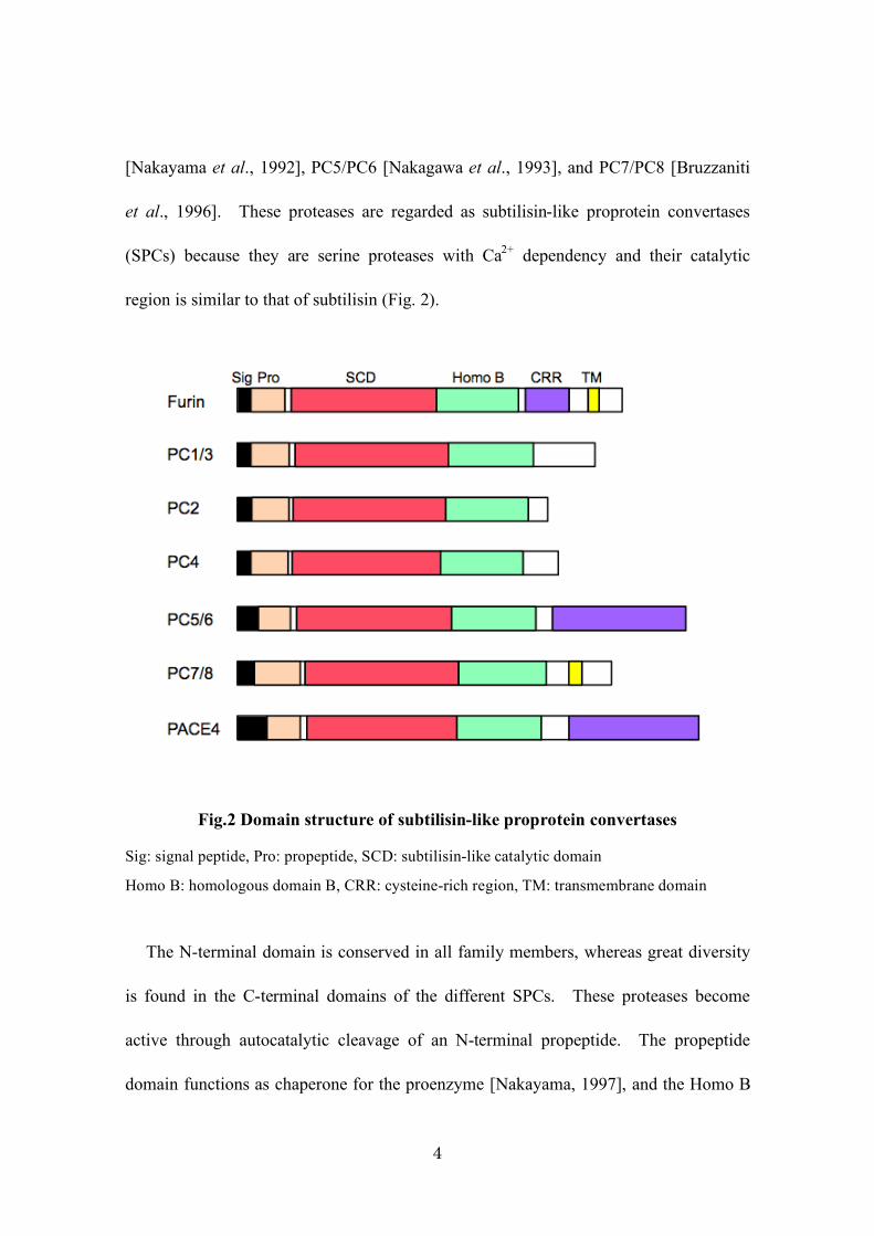
4
[Nakayama et al., 1992], PC5/PC6 [Nakagawa et al., 1993], and PC7/PC8 [Bruzzaniti
et al., 1996]. These proteases are regarded as subtilisin-like proprotein convertases
(SPCs) because they are serine proteases with Ca2+
dependency and their catalytic
region is similar to that of subtilisin (Fig. 2).
Fig.2 Domain structure of subtilisin-like proprotein convertases
Sig: signal peptide, Pro: propeptide, SCD: subtilisin-like catalytic domain
Homo B: homologous domain B, CRR: cysteine-rich region, TM: transmembrane domain
The N-terminal domain is conserved in all family members, whereas great diversity
is found in the C-terminal domains of the different SPCs. These proteases become
active through autocatalytic cleavage of an N-terminal propeptide. The propeptide
domain functions as chaperone for the proenzyme [Nakayama, 1997], and the Homo B

5
domain, as well as the catalytic domain, are required for autocatalytic activation
[Takahashi et al., 1995]. PACE4 and PC5/6 as extracellular matrix-bound enzymes
have a heparin-binding region in the cysteine-rich region [Tsuji et al., 2003]. PACE4
is conserved in humans and mice, and the knockout mouse of this gene has been
previously shown to have bone-morphogenesis defects [Constam et al., 2000] (Table 2).
Table.1: Sequence around the cleavage site of potential precursor proteins in
mouse bones
Arrows indicate the predicted cleavage sites.
Table.2: Comparison of human and mouse PACE4
Chromosomal
localization
Identity Gene
length
Exon
number
Tissue
distribution Knockout mouse
human mouse
15q26 7
86% >250 kb 22 nervous
system, heart,
liver, bone
75% viable phenotype
with bone morphogenesis
defects, cyclopia
Precursor proteins Sequences at the cleavage site
Osteocalcin
ChM-1
PTHrP
FGF23
BMP family
BMP1
BMP2
BMP3
BMP4
BMP5
BMP6
BMP7
BMP8
BMP9
BMP10
BMP15
VNRLRR"YL
IQRERR"EV
SRRLKR"AV
PRRHTR"SA
RPRSRR"AA
HKREKR"GA
KKARRK"GW
RRRAKR"SP
LLRSVR"AA
HVRTTR"SA
HLRSIR"ST
PVRAPR"AA
LARRKR"ST
SARIRR"NA
LMRSVR"GA

6
CHAPTER II
Estrogen stimuli promote osteoblastic differentiation via
PACE4 in MC3T3-E1 cells
1. Introduction
Bone mass is constantly maintained through the so-called bone remodeling process,
which consists of bone formation by osteoblasts and bone resorption by osteoclasts.
However, bone mass decreases with age, especially in postmenopausal women with low
blood estrogen levels [Albright et al., 1940]. These women are predisposed to
osteoporosis due to the collapse of the balance between bone formation and resorption,
which results in net bone resorption [Jasani et al., 1965; Riggs et al., 2002]. Because
osteoporosis-related fractures are very problematic for elderly people [Kanis et al.,
2004], endochondral ossification is required for fracture healing and where a cartilage
frame formed by chondrocytes is replaced with bone, estrogen replacement therapies for
the treatment and prevention of osteoporosis are particularly important [Riggs et al.,
2002; Scammell et al., 1996]. For example, the soybean isoflavone daidzein (Diz) is a
phytoestrogen with estrogenic activity and its intake is effective for the maintenance of
bone mass in postmenopausal women [Ma et al., 2008]. Moreover, equol (Eq), which
is a metabolite of Diz, has been shown to have greater estrogenic activity than Diz.
[Setchell et al., 2002].

7
It has been reported that estrogen induces apoptosis in osteoclasts [Kameda et al.,
1997], and the number of osteoclasts has been found to increase in ovariectomized mice
[Kousteni et al., 2002]. While estrogen suppresses bone resorption, it was also shown
to attenuate apoptosis through activation of the Src/Shc/ERK signaling pathway in
osteoblasts [Kousteni et al., 2001]. Furthermore, estrogen stimuli, including
phytoestrogens such as Diz, enhanced bone mineralization of a murine osteoblastic cell
line [Kanno et al., 2004]. Thus, understanding the mechanism of how estrogen stimuli
facilitate bone formation is important to develop treatment and prevention therapies for
osteoporosis. However, the detailed molecular mechanisms underlying bone
formation triggered by estrogen stimuli have not been clarified yet. It is well known
that BMPs are highly linked with ossification and bone formation via Smad signaling.
It is thought that several factors related to ossification, including BMPs, have to
undergo proteolytic processing to convert to the active form; furthermore, the SPC
family, which are serine endoproteases, play key roles in activating the precursors of
BMPs with a consensus cleavage motif for SPCs [Constam et al., 1999]. Recently, it
has been reported that an SPC regulates the hypertrophic conversion of murine
chondrocytes through activation of BMP6 [Yuasa et al., 2012]. Therefore, in the
present study, I investigated the mechanism underlying the effect of estrogen stimuli on
osteoblasts, with focus on the role of SPCs.
2. Materials and methods
2-1. Cell culture

8
ATDC5, a mouse chondrogenic cell line derived from embryonal carcinoma cells,
was provided by Drs. Yuji Hiraki and Chisa Shukunami of Kyoto University [Yuasa et
al., 2012]. Cells were cultured in growth medium consisting of Dulbecco’s modified
Eagle’s medium:Ham’s F12 (1:1) containing 5% fetal bovine serum (FBS) (inactivated
by treatment at 56°C), 10 µg/mL transferrin, 30 nM sodium selenite, 100 U/mL
penicillin G potassium, and 100 µg (titer)/mL streptomycin sulfate. MC3T3-E1, an
osteoblast cell line from C57BL/6 mouse calvaria, was obtained from the RIKEN Cell
Bank (No. RCB1126). MC3T3-E1 cells were cultured in Minimum Essential Medium
Alpha (Wako) containing 10% FBS, 100 U/mL penicillin G potassium, and 100 µg
(titer)/mL streptomycin sulfate. Cell culture of both cell lines was carried out in a 5%
CO2-air incubator at 37°C.
2-2. Differentiation culture with estrogen stimuli
ATDC5 cells and MC3T3-E1 cells were seeded at 1 # 104 cells/cm
2 into wells of
24-well culture plates containing 0.5 mL/well or in 35-mm culture dishes containing 2
mL/dish of the respective growth medium. When the cells reached confluence, the

9
medium was replaced with the same volume of differentiation medium containing FBS
or charcoal-dextran-treated FBS (CD-FBS). CD-FBS was prepared by treatment of
FBS with dextran-coated charcoal (Sigma) to reduce the amount of endogenous
estrogen, as described previously [Kanno et al., 2004]. Briefly, 10 g of dextran-coated
charcoal and 200 mL of FBS were mixed at 37°C for 1 h with constant stirring. The
treated FBS was centrifuged at 2500 # g for 20 min, and the supernatant was sterilized
by filtration through a 0.2-µm filter. Differentiation medium for ATDC5 cells was
prepared by addition of 10 µg/mL insulin and that for MC3T3-E1 cells was prepared by
addition of 5 mM $-glycerophosphate and 0.2 mM L-ascorbic acid to the growth
medium. Estrogenic agents such as 17$-estradiol (E2) and the soybean isoflavones
Diz and the S-form of Eq were dissolved in dimethyl sulfoxide and added to the
differentiation medium containing CD-FBS. To evaluate the dependency of the effect
of estrogenic agents on the estrogen receptor (ER), the ER inhibitor fulvestrant (Sigma)
was added to the differentiation medium at 1 µM and incubated as described above.

10
2-3. Alcian blue staining and alizarin red S staining
After incubation in the differentiation medium for the designated time, the
differentiation of ATDC5 cells and MC3T3-E1 cells was monitored by alcian blue
staining and alizarin red S staining, respectively. ATDC5 samples were washed three
times with phosphate-buffered saline and then fixed with 99% methanol at room
temperature. After 20 min, the cells were stained with alcian blue for 1 day and then
washed with dH2O. MC3T3-E1 samples were washed three times with
phosphate-buffered saline and then fixed with ice-cold 70% ethanol for 1 h on ice.
Subsequently, the cells were washed with distilled H2O and stained with alizarin red S
for 20 min at room temperature. To quantify the relative differentiation, their
absorbance at 520 nm was measured in a spectrometer (Tecan Infinite M200) after
several washes with distilled H2O.
2-4. Reverse transcription-polymerase chain reaction (RT-PCR) and quantitative
real-time PCR (qRT-PCR)

11
Total RNA was extracted from MC3T3-E1 cells during the time course using the
Qiagen RNeasy mini kit (Qiagen). Subsequently, cDNA was synthesized by reverse
transcription using the High Capacity cDNA Reverse Transcription Kit (Applied
Biosystems). Both steps were performed according to the manufacturer’s instructions.
RT-PCR performed using 1 µL of template solution containing cDNA equivalent to 100
ng mRNA was performed for 15, 22, and 25 cycles for glyceraldehyde-3-phosphate
dehydrogenase (for the data in Figs. 6 and 7) and all other genes, respectively, by using
a gene-specific primer set: an initial step at 95°C for 2 min, followed by 95°C for 1 min,
50°C or 53°C for 1 min, and 72°C for 1 min (Table 3). Each primer set corresponding
to the target genes was designed to amplify a partial region of the entire cDNA (Fig. 3).
qRT-PCRs were performed using the Power SYBR Green PCR Master Mix (Applied
Biosystems) according to the manufacturer’s protocol with primer sets for
glyceraldehyde-3-phosphate dehydrogenase, ER!, ER$, and PACE4 (Table 3); those
for furin [Hwang et al., 2006] and PC7 [Marchesi et al., 2011] were performed using
the ABI Prism 7000 Sequence Detection System.

12
Table 3: Primer sets for RT-PCR/qRT-PCR
Gene Primer Sequence
ER$ Fw: 5’-cagctcaacagcgtgtcgccta-3’
Rev: 5’-gtttcctttctcgttactgctgg-3’
ER% Fw: 5’-atggccaacttctggacacctc-3’
Rev!5’-cttggcgcttggactagtaa-3’
GR Fw!5’-acctcaataggtcgaccagc-3’
Rev!5’-cccgccaaaggagaaagcaag-3’
SOX5 Fw!5’-cttggtgctgccgtgtctcctacc-3’
Rev!5’-atagcctattgtgctaactcttgc-3’
SOX9 Fw: 5’-gcaaagttgatctgaagcgagagg-3’
Rev!5’-ccagtgtaggtgacctggccgt-3’
RUNX2 Fw!5’-tcgcctcagtgatttagggcgca-3’
Rev!5’-gtggcagtgtcatcatctgaaatac-3’
Osterix Fw!5’-tccctacccagcgccccacctct-3’
Rev!5’-ctgtgaatgggcttcttcctcagc-3’
Osteopontin Fw!5’-gatgaatctgacgaatctcacc-3’
Rev!5’-ctcagaagctgggcaacagggat-3’
Osteocalcin Fw!5’-ccctgagtctgacaaagccttca-3’
Rev!5’-tactggtctgatagctcgtc-3’
FGF23 Fw!5’-gagaatggctatgacgtctacttg-3’
Rev!5’-gctcgcgagagcaggataca-3’
FGFR1 Fw!5’-gttcaagcagttggtggaagtcc-3’
Rev!5’-cagcgccgtttgagtccactg-3’
FGFR2 Fw!5’-gaggaatacttggatctcacccag-3’
Rev!5’-aacactgccgtttatgtgtggatac-3’
FGFR3 Fw!5’-cgcatcctcactgtgacatcaacc-3’
Rev!5’-cgttactgggtggacctgggg-3’
Col-I Fw!5’-gagaagtctcaagatggtggc-3’
Rev!5’-gcggggtcggagccctcgctt-3’
Col-II Fw!5’-ccaccccgagtggaagagcggaga-3’
Rev!5’-cagccatccttcagggcagtgtat-3’
Col-X Fw!5’-ggcagcagcattacgacccaag-3’
Rev!5’-gcattgggcaattggagccatacc-3’
ALP Fw!5’-cactcgggtgaaccacgccaca-3’
Rev!5’-ctgatgagatccagaccatctagcc-3’
Gene Primer Sequence
BMP1 Fw!5’-gccgaggaaggctatggcgtgga-3’
Rev!5’-gcttgtgtaccgcaggtggaagc-3’
BMP2 Fw!5’-ccaggagcgcccgccccagacc-3’
Rev!5’-tggttggtgtgtccctgtgt-3’
BMP3 Fw!5’-ggtcatctgtctgtagatgtgg-3’
Rev!5’-caagggcagcaagatcccagtag-3’
BMP4 Fw!5’-cgctggacccgggaaaagcaac-3’
Rev!5’-gcgacggcagttcttattcttctt-3’
BMP5 Fw!5’-ctcatcagaggaggcattacaaaga-3’
Rev!5’-ctgcacagagctgtaagcccaaa -3’
BMP6 Fw!5’-ggcggtgcgtccccactgacta-3’
Rev!5’-gccccatgttgtgctgcggtgt-3’
BMP15 Fw!5’-catcaaaccaggtagcatacg-3’
Rev!5’-gagtagcaagaaggcaacatccaag-3’
ChM-1 Fw!5’-gaactcgctgatttgggtggccgt-3’
Rev!5’-ctggtggtaaggattgtcagggt-3’
PTHrP Fw!5’-gatcgcggagatccacacagcc-3’
Rev!5’-cgcttctttttctcctgttctc-3’
furin Fw!5’-gagaatgatgtggagatcatccgtg-3’
Rev!5’-tctgagtccgatgggcactcct-3’
PC1 Fw!5’-cctcggaggtcccgaagaagc-3’
Rev!5’-gtgtgattccactccaagccatca-3’
PC2 Fw!5’-acctggagcacgtccaagctgtc-3’
Rev!5’-ccgtgaagcatcagggtccattc-3’
PC4 Fw!5’-cactcactactgggatgagga-3’
Rev!5’-cagctggcctgtccttcggt-3’
PC6 Fw!5’-gtcttcagggatcccgctgttcg-3’
Rev!5’-gaatcctggcccattgcatgtc-3’
PC7 Fw!5’-ctggatggagtggattcagagc -3’
Rev!5’-agcaaatctgcccgctcttccc -3’
PACE4 Fw!5’-accgggtacctacttcgattca-3’
Rev!5’-tcgcagctcaggcagttctc-3’
GAPDH Fw!5’-gggtggagccaaacgggtc-3’
Rev!5’-ggagttgctgttgaagtcgca-3’

13

14

15
2-5. PACE4 knockdown in MC3T3-E1 cells
MC3T3-E1 cells were grown in 35-mm dishes to 50% confluence and then
transfected with 2 µg of a PACE4 or green fluorescent protein (GFP)
shRNA-expressing vector [Yuasa et al., 2009] using the FuGENE® 6 Transfection
Reagent (Roche) according to the manufacturer’s protocol. PACE4 and GFP
shRNA-expressing vectors were generated by ligation of the sequence corresponding to
shRNA against mPACE4 (G429
CGAAGTGACTCTCTTTATT448
) and the GFP target
sequence (as recommended by the manufacturer) into the vector pSilencer 3.1-H1 neo
(Ambion). After 24 h of cultivation, the used medium was replaced with fresh growth
medium containing 400 µg/mL G418 (Nacalai Tesque). This selection step was
continued for 2 weeks; the medium was changed every 3 days. The cells were then
applied to the cell differentiation assay and subjected to qRT-PCR.
3. Results
3-1. Differentiation of MC3T3-E1 cells induced by estrogen stimuli
To confirm the pharmacological effects of estrogen stimuli on ossification and to
investigate their effect on the prevention of osteoporosis, the ATDC5 chondrogenic cell

16
line and the MC3T3-E1 osteoblastic cell line were used as typical model cells
responsible for bone formation. Cells were cultured with the endogenous estrogen E2
or the phytoestrogen Diz and the S-form of Eq in differentiation medium containing
CD-FBS. First, CD-FBS was prepared by CD treatment to reduce the amount of
endogenous estrogens. The E2 concentration was less than the detection limit (<10
pg/mL); CD-FBS was used for cell cultivation to mimic the estrogen-poor condition
observed in postmenopausal women. E2 is the predominant estrogen, and Diz is a
typical soybean isoflavone with phytoestrogen activity. Diz is metabolized to Eq by
intestinal bacteria such as Lactococcus garvieae [Uchiyama et al., 2004]; Eq is known
to be a potent soybean isoflavone. Because the structures of Diz and Eq are similar to
that of E2 (Fig. 4), these compounds are able to bind to both ER! and ER$ [Usui et al.,
2006]. As shown in Figure 5, the presence of estrogen stimuli did not significantly
affect chondrogenic differentiation of ATDC5 cells (Fig. 5a). On the other hand, in
MC3T3-E1 cells, the cultivation period-dependent mineralization was significantly
decreased in culture medium containing CD-FBS compared to that containing normal
FBS. Moreover, the mineralization ability was restored by treatment with Eq, Diz, or

17
E2, even on culture in a medium containing CD-FBS (Fig. 5b). The differentiation of
MC3T3-E1 cells by these estrogen stimuli was dose-dependent, and Eq was
approximately 10-fold more effective than Diz (Fig. 5c). Furthermore, these inductive
effects were inhibited by addition of the selective ER downregulator fulvestrant, which
could bind to both ER! and ER$ [Tremblay et al., 1997, Paige et al., 1999] (Fig. 5d).
As expected, these results suggest that the induction effect of isoflavones is dependent
on the ER.
Fig. 4: Chemical structures of estradiol, daidzein, and equol.

18
Fig. 5: Effects of the estrogenic agents equol, daidzein, and estradiol on the
differentiation of ATDC5 cells and MC3T3-E1 cells.
Both cell lines were cultured in the respective differentiation medium containing
charcoal-dextran-treated fetal bovine serum (CD-FBS) with no estrogen stimulus, 10-5
M equol
(Eq), 10-5
M daidzein (Diz), or 10-9
M estradiol (E2). After each cultivation period, the
differentiation of ATDC5 and MC3T3-E1 cells was detected by alcian blue staining (a) and
alizarin red S staining (b), respectively. c To investigate the dose response of the estrogen
stimuli in MC3T3-E1 cells, the cells were cultured in differentiation medium containing FBS
only, CD-FBS only, CD-FBS with Eq (10-7
, 10-6
, or 10-5
M), CD-FBS with Diz (10-7
, 10-6
, or
10-5
M), or CD-FBS with E2 (10-11
, 10-10
, or 10-9
M) (upper picture). Subsequently,
MC3T3-E1 cells were stained by alizarin red S to confirm mineralization, and the staining
intensity was quantified by measuring the absorbance at 520 nm (n = 2) (lower graph). d To
investigate the effect of fulvestrant (Ful) on the differentiation induced by estrogenic agents,
MC3T3-E1 cells were co-cultured for 4 weeks with each estrogenic agent (10-7
M Eq, 10-7
M
Diz, and 10-11
M E2) in differentiation medium containing CD-FBS and 10-6
M Ful.

19
3-2. Effect of estrogen stimuli on the expression patterns of bone-related genes in
MC3T3-E1 cells
The molecular effects of estrogen stimuli on osteoblast differentiation were
investigated by analyzing the expression patterns of bone-related genes in MC3T3-E1
cells. Bone-related genes include the following factors: ER and glucocorticoid
receptors responsible for steroid hormone signaling (ER!, ER$, and GR), collagens that
are a type of differentiation markers of osteoblasts (type I collagen [Col-I], type II
collagen [Col-II], and type X collagen [Col-X]), transcription factors related to
mesenchymal stem cell differentiation (SOX5, SOX9, RUNX2, and osterix),
extracellular matrix proteins (osteopontin and osteocalcin), fibroblast growth factors
(FGFR1, FGFR2, FGFR3, and FGF23), factors associated with the differentiation of
osteoclasts (PTHrP, RANKL, and osteoprotegerin), BMPs (BMP1, BMP2, BMP3,
BMP4, BMP5, BMP6, BMP7, and BMP15), SPCs (furin, PC1, PC2, PC4, PC6, PACE4,
and PC7), chondromodulin-1 (ChM-1), which promotes chondrocyte differentiation,
and the bone differentiation marker alkaline phosphatase (ALP). Transcription levels
of several genes changed during the differentiation of MC3T3-E1 cells (Figs. 6a-6d).
For example, the transcription of Col-II decreased, while those of osteocalcin and

20
BMP4 increased as differentiation progressed. Most genes were not affected by
estrogen stimuli. However, I found that the transcription of PACE4 was upregulated
by estrogen stimuli as differentiation progressed (Fig. 6e). Other SPCs such as furin
and PC7 were also expressed in MC3T3-E1 cells, but their expression did not depend
on estrogen stimuli. No significant expression of other SPCs was observed.
Subsequently, I quantitatively estimated the relative transcription level of the PACE4
gene in a time course study in the presence and absence of estrogen stimuli (Fig. 7a).
As shown in Figure 7a, PACE4 gene transcription was significantly induced by Diz, Eq,
and E2 at the late stage of osteoblast differentiation. Interestingly, sequencing of the
amplicon of the PACE4 gene fragment revealed that the PACE4 isoform expressed in
MC3T3-E1 cells was PACE4A-II, the expression of which has been confirmed in
placenta and HepG2 cells [Mori et al., 1999] (GenBank Acc. No. NM_138319.2).
Moreover, because I found that the ER! mRNA levels tended to increase in response to
the lack of estrogen stimuli (Fig. 6e), I quantitatively analyzed the mRNA levels of ER!
and ER$ (Figs. 7b and 7c). ER! expression was upregulated over time in the absence
of estrogen stimuli. However, in medium containing endogenous estrogen or in the

21
presence of the exogenous estrogens E2, Eq, and Diz, its expression level in MC3T3-E1
cells was unchanged or decreased as osteoblast differentiation progressed. However,
the mRNA level of ER$ did not show such a response to the presence of estrogens.

22
Fig. 6: Time course of the transcriptional expression patterns of bone-related
genes.
MC3T3-E1 cells were cultured in differentiation medium containing charcoal-dextran-treated
fetal bovine serum with no estrogen stimulus, 10-5
M equol (Eq), 10-5
M daidzein (Diz), or
10-9
M estradiol (E2). Over a period of 4 weeks, total RNA was isolated from cultured cells
once a week, and the mRNA levels of bone-related genes were analyzed by reverse
transcription-polymerase chain reaction: a 1 week; b 2 weeks; c 3 weeks; and d 4 weeks.
e The results of two genes that showed remarkable changes depending on the estrogen stimuli
and those of the estrogen receptor $ (ER$) gene (for comparison) are shown separately. The
endogenous housekeeping gene glyceraldehyde-3-phosphate dehydrogenase (GAPDH) was
used as control.

23
Fig. 7: Time course of the expression patterns of PACE4, ER!, and ER" in the
presence or absence of estrogen stimuli.
MC3T3-E1 cells were cultured in differentiation medium containing fetal bovine serum (FBS)
only, charcoal-dextran-treated FBS (CD-FBS) only, CD-FBS with 10-5
M equol (Eq), CD-FBS
with 10-5
M daidzein (Diz), or CD-FBS with 10-9
M estradiol (E2) for 4 weeks. The
transcriptional levels of PACE4 (a) (n = 3) and those of estrogen receptor ! (ER!) (b) and ER$
(c) were analyzed by reverse transcription-polymerase chain reaction (RT-PCR) and
quantitative RT-PCR(n = 2) using cDNA prepared at each indicated time point as templates

24
3-3. Requirement of PACE4 for the differentiation of MC3T3-E1 cells
PACE4 is a member of the SPCs, which are required for the maturation/activation
of bone formation factors such as BMPs [Constam et al., 1999, Akamatsu et al., 1999]
and osteocalcin [Viegas et al., 2013]. To examine whether the expression of PACE4 is
only the result of osteoblast differentiation or if it plays an important role during
differentiation and mineralization, I prepared PACE4-knockdown MC3T3-E1 cells.
The PACE4- or GFP-specific shRNA expression vector was transfected into
MC3T3-E1 cells, and selection culture with G418 was carried out for 2 weeks.
GFP-specific shRNA-expressing MC3T3-E1 cells were used as mock cells.
G418-resistant cells were cultured in differentiation medium containing FBS, CD-FBS,
CD-FBS with Eq, CD-FBS with Diz, or CD-FBS with E2 until the late differentiation
stage. Then, we quantitatively monitored the transcriptional levels of PACE4, furin,
and PC7 expression in MC3T3-E1 cells cultured in differentiation medium containing
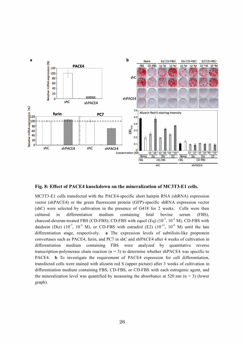
FBS (Fig. 8a). The mRNA level of PACE4 was clearly downregulated in PACE4
shRNA-expressing MC3T3-E1 cells compared to that in mock cells. The shRNA
targeting PACE4 used in this experiment was confirmed to be specific for the mRNA of
the PACE4 gene because PACE4-specific shRNA caused almost no significant

25
suppression of the transcription of furin or PC7. In the mineralization assay of
PACE4-knockdown cells and mock cells cultured in the presence or absence of estrogen
stimuli using alizarin red S staining, PACE4-knockdown cells showed a strong decrease
in mineralization, even in the presence of estrogen stimuli, whereas the differentiation
of mock cells had significantly progressed in the presence of FBS or the estrogen
stimuli Eq, Diz, and E2 with CD-FBS at the late differentiation stage, similar to normal
MC3T3-E1 cells (Fig. 8b). This result strongly suggests that PACE4 is required for
the differentiation of osteoblasts.
26
Fig. 8: Effect of PACE4 knockdown on the mineralization of MC3T3-E1 cells.
MC3T3-E1 cells transfected with the PACE4-specific short hairpin RNA (shRNA) expression
vector (shPACE4) or the green fluorescent protein (GFP)-specific shRNA expression vector
(shC) were selected by cultivation in the presence of G418 for 2 weeks. Cells were then
cultured in differentiation medium containing fetal bovine serum (FBS),
charcoal-dextran-treated FBS (CD-FBS), CD-FBS with equol (Eq) (10-7
, 10-5
M), CD-FBS with
daidzein (Diz) (10-7
, 10-5
M), or CD-FBS with estradiol (E2) (10-11
, 10-9
M) until the late
differentiation stage, respectively. a The expression levels of subtilisin-like proprotein
convertases such as PACE4, furin, and PC7 in shC and shPACE4 after 4 weeks of cultivation in
differentiation medium containing FBS were analyzed by quantitative reverse
transcription-polymerase chain reaction (n = 3) to determine whether shPACE4 was specific to
PACE4. b To investigate the requirement of PACE4 expression for cell differentiation,
transfected cells were stained with alizarin red S (upper picture) after 3 weeks of cultivation in
differentiation medium containing FBS, CD-FBS, or CD-FBS with each estrogenic agent, and
the mineralization level was quantified by measuring the absorbance at 520 nm (n = 3) (lower
graph).

27
4. Conclusion
With aging and decreases in the blood estrogen level, osteoporosis can adversely
affect the quality of life of elderly people, especially postmenopausal women.
Estrogen replacement therapy is regarded as an effective treatment for preventing
osteoporosis. Moreover, the intake of soybeans containing isoflavones, which are
phytoestrogens that mimic the action of estrogen, is an effective and attractive way to
further increase endogenous estrogen levels. Recently, it has been suggested that
people in whom Diz is metabolized to Eq by intestinal bacterial flora have better bone
health owing to the maintenance of bone mass, compared to those in whom intestinal
bacteria do not produce Eq [Wu et al., 2007]. It is well known that most stages of the
bone formation process depend on endochondral ossification, where the cartilage
formed by chondrocytes is replaced with bone derived from osteoblasts. Therefore,
we investigated which step of the cell differentiation process is regulated by estrogen
stimuli, i.e., the induction efficiency of cell differentiation by estrogen stimuli was
compared between ATDC5 and MC3T3-E1 cells. As shown in Figures 5a and 5b,
compared to ATDC5 cells, differentiation of MC3T3-E1 cells was clearly under the
control of estrogen stimuli, i.e., it was induced by E2 and the phytoestrogens Diz and

28
Eq. I also investigated the dose-dependent effect of E2 and phytoestrogens on the
differentiation of MC3T3-E1 cells. As shown in Figures 5c and 5d, Eq facilitated
osteoblast differentiation more effectively than Diz via the ER; however, E2 was a
significantly stronger inducer than these phytoestrogens. Among the different ERs,
ER! has been reported to have a role in osteoblast mechanotransduction via activation
of the $-catenin-mediated Wnt signaling pathway, which is involved in bone formation
[Armstrong et al., 2007]. As shown in Figure 6, the mRNA level of ER! was higher
than that of ER$. Furthermore, the expression of ER! was more sensitive to the
absence of estrogen stimuli, i.e., in the absence of estrogen stimuli, ER! levels
increased over time in MC3T3-E1 cells cultured in medium containing CD-FBS.
However, the expression of ER$ was not affected by the presence of estrogen stimuli
(Figs. 7b and 7c). Thus, my experimental results also suggest that ER! is the primary
receptor responsible for estrogen stimuli to facilitate bone formation. Moreover,
although both Diz and Eq showed higher affinity to ER$ than to ER!, the binding
affinities of Eq to both ER! and ER$ were higher than those of Diz [Usui et al., 2006,
Muthyala et al., 2003, Morito et al., 2001]. These findings may provide an

29
explanation for the results shown in Figure 5c, which shows that Eq is more effective
than Diz in inducing differentiation of MC3T3-E1 cells.
I also investigated the time course of the expression of bone formation-related
genes in MC3T3-E1 cells by RT-PCR. The expression levels of several genes such as
Bmp4 and osteocalcin increased during cell differentiation, but their expression was not
responsive to estrogen stimuli. Of these genes, the expression of PACE4, a member of
the SPC family, changed considerably in response to estrogen stimuli and increased as
osteoblast differentiation progressed; however, the transcription of other SPCs such as
furin and PC7 was independent of estrogen stimuli (Fig. 6 and Fig. 7a). Unlike furin,
which is a ubiquitously and constitutively expressed membrane-bound SPC, PACE4 is
localized in the extracellular matrix and its expression levels change markedly during
development and differentiation [Yuasa et al., 2009]. SPCs can cleave the precursors
of BMPs, which regulate skeletogenesis, to convert them to the active forms. For
example, furin and/or PC6 proteolytically activate BMP4 [Cui et al., 1998] and PACE4
activates BMP6 [Yuasa et al., 2012]. In addition, it has been reported that the
transcription of BMP2 is increased by treatment with E2 after 24 h in mouse

30
mesenchymal stem cells that differentiated into osteoblasts [Zhou et al., 2003];
furthermore, furin has been shown to convert proBMP2 to mature BMP2 [Felin et al.,
2010]. In the present study, mRNA expression of BMP2 and BMP6 in MC3T3-E1
cells was confirmed at trace levels; however, their expression levels were not linked
with the cell differentiation induced by estrogen stimuli. Although the mRNA
expression of BMP4 significantly increased as differentiation progressed, the increase
was not a response to estrogen stimuli. Therefore, it is difficult to explain the
correlation between osteoblast differentiation and estrogen stimuli only on the basis of
the SPC/BMP substrate relationship identified to date. As shown in Figures 6 and 7a,
among the SPCs expressed in MC3T3-E1 cells, only the increase in the expression of
PACE4 can be linked to osteoblast differentiation. In addition, knockdown of PACE4
resulted in delayed osteoblast differentiation (Fig. 8b). PACE4 plays a key role in
chondrogenic cell differentiation via processing of BMP6 [Yuasa et al., 2012]; therefore,
it is reasonable that PACE4 also regulates the differentiation of osteoblasts related to
bone formation. However, the differentiation of chondrogenic cells was not affected
by estrogen stimuli, as shown in Figure 5a. Moreover, the PACE4 isoform expressed

31
in osteoblastic cells was isoform A-II, which is a rare isoform of PACE4; to date, it has
been confirmed to be present in HepG2 cells and the placenta. Therefore, the
expression of PACE4 seems to be regulated by different independent systems in
chondrogenic cells and osteoblastic cells, and PACE4 may control osteoblast
differentiation via combinations of substrate(s) that are different from those in
chondrogenic cells. After binding of the estrogenic ligand to its receptor to regulate
the expression of target genes, the ER-ligand complex translocates to the nucleus, where
it activates transcription directly by binding to the response element or indirectly via
other DNA-binding proteins [Björnström et al., 2005]. The ER can bind to DNA in
less than 1 h after ligand binding [Métivier et al., 2005]. However, it took several
weeks to show a significant increase in the transcription of PACE4 in response to
estrogen stimuli; furthermore, there are no typical ER response elements in the upstream
region of the PACE4 gene. Therefore, it is thought that PACE4 may be indirectly
regulated by some other gene(s) that is directly targeted by estrogen stimuli, or by
post-transcriptional regulation, e.g., mRNA stabilization/destabilization, governed by
RNA-binding proteins [Lal et al., 2004]. Among the SPCs, a significant correlation
32
between ER content and the gene expression of PACE4 has only been observed in
human breast cancer [Cheng et al., 1997]; however, details of the correlation between
the dynamic states of ER and PACE4 are still unclear. Therefore, further studies are
required to explain the mechanism underlying the transcriptional regulation of PACE4
by estrogen stimuli, which was identified in this work. Moreover, the substrate(s)
leading to cell differentiation after activation by PACE4 in the presence of estrogen
stimuli has not yet been identified in osteoblastic cells. One candidate of interest is
osteocalcin, an important marker for differentiated osteoblasts; its expression increased
during the time course, similar to the expression of PACE4 at the late stage of cell
differentiation (Fig. 6). It is also known that osteocalcin requires cleavage at the SPC
recognition motif (Table 1) in order to convert into the mature active form. Therefore,
further investigations are necessary to clarify the role of PACE4 in osteoblast
differentiation.
In conclusion, the results from my present study strongly suggest that estrogen
stimuli, including physiological estrogen and phytoestrogens such as Diz and Eq,
induce osteoblast differentiation through upregulation of PACE4 mediated by the ER.

33
ABBREVATIONS
ALP, alkaline phosphatase; BMPs, bone morphogenetic proteins
CD-FBS, charcoal/dextran-treated FBS; ChM-1, chondromodulin-1
Col-& , type & collagen; Col-' , type ' collagen; Col-( , type ( collagen
Diz, daidzein; E2, 17%-estradiol; ER, estrogen receptor; Eq, equol
FBS, fetal bovine serum; FGFR, fibroblast growth factor receptor
Ful, fulvestrant; GFP, green fluorescent protein; GR, glucocorticoid receptors
MSC, mesenchymal stem cell; PBS, phosphate-buffered saline
RT-PCR, reverse transcription PCR; shRNA, short hairpin RNA
SPC, subtilisin-like proprotein convertase; qRT-PCR, quantitative real-time PCR

34
ACKNOWLEDGEMENTS
I would like to express my deep appreciation to Prof. Hideaki Nagamune for the
great support provided for this research and my graduate education. Furthermore, I
would like to convey deep gratitude to Associate Prof. Toshifumi Tomoyasu and
Assistant Prof. Atsushi Tabata for their helpful experimental suggestions and the
discussions regarding this study.
I am also especially grateful to Prof. Akihito Tsuji and Associate Prof. Keizo Yuasa
for providing invaluable advice and donating materials for the experiments, for example,
ATDC5 cells and the constructs for PACE4 knockdown.
I am grateful to Dr. H. Ohkuni for the help with the measurement of the E2
concentration in sera. I wish to express my gratitude to the members of my committee,
Prof. Akihiko Tsuji and Prof. Hitoshi Matsuki, for their comments on this work.
Finally, great thanks to my research partner, Ms. Kotomi Sakamoto for the valuable
assistance and all the collaborators in my department, especially the members of Prof.
Hideaki Nagamune’s laboratory.
35
REFERENCES
Akamatsu T, Matsuda Y, Tsumura K, Tada J, Parvin MN, Kanamori N, Hosoi K
(1999) Subtilisin-like proprotein convertase PACE4(SPC4) is a candidate processing
enzyme of bone morphogenetic proteins during tooth formation. Dev Dyn 216:#
481-488
Albright F, Bloomberg E, Smith PH!1940"Postmenopausal osteoporosis. Trans Assoc
Am Phys 55:298-305
Armstrong VJ, Muzylak M, Sunters A, Zaman G, Sazon LK, Price JS, Lanyon LE
!2007"Wnt/beta-catenin signaling is a component of osteoblastic bone cell early
responses to load-bearing and requires estrogen receptor alpha. J Biol Chem 282:
20715-20727
Björnström L, Sjöberg M!2005"Mechanisms of estrogen receptor signaling)
convergence of genomic and nongenomic actions on target genes. Molecular

36
Endocrinology 19:833-842
Bradley EW, McGee-Lawrence ME, Westendorf JJ!2011"Hdac-mediated control of
endochondral and intramembranous ossification. Crit Rev Eukaryot Gene Expr
21:101-113
Bruzzaniti A, Goodge K, Jay P, Taviaux SA, Lam MH, Berta P, Martin TJ, Moseley JM,
Gillespie MT!1996"PC [corrected], a new member of the convertase family. Biochem
J 314:727-731
Cheng M, Watson PH, Paterson JA, Seidah N, Chrétien M, Shiu RP (1997) Pro-protein
convertase gene expression in human breast cancer. Int J cancer 71:966-971
Constam DB, Robertson EJ!1999"Regulation of bone morphogenetic protein actibity
by prodomains and proprotein convertases. J Cell Biol 144:139-149
Costam DB, Robertson EJ!2000"SPC4/PCE4 regulates a TGF% signaling network
during axis formation. Genes & Dev 14:1146-1155
Cui Y, Jean F, Thomas G, Christian JL!1998"BMP4 is proteolytically activated by

37
furin and/or PC6 during vertebrate embryonic development. EMBO J 17:4735-4743
Felin JE, Mayo JL, Loos TJ, Jensen JD, Sperry DK, Gaufin SL, Meinhart CA, Moss JB,
Bridgewater LC!2010"Nuclear variants of bone morphogenetic proteins. BMC Cell
Biol 11:20-34
Fuller RS, Brake AJ, Thorner J!1989"Intracellular targeting and structural conservation
of a prohormone-processing endoprotease. Science 246:482-486
Gennari C!2001"Calcium and vitamin D nutrition and bone disease of the elderly.
Public Health Nutr 4:547-559
Gerber HP, Ferrara N!2000"Angiogenesis and bone growth. Trends Cardiovasc Med
10:223-228
Hwang E, Kim S, Sohn J, Lee J, Kim Y, Kim Y, Mook-Jung I!2006"Furin is an
endogenous regulator of $-secretase associated APP processing. Biochemical and
Biophysical Research Communications 654-659
Jasani C, Nordin BEC, Smith DA, Swanson I!1965"Spinal osteoporosis and the

38
menopause. Pros R Soc Med 58:441-444
Kameda T, Mano H, Yuasa T, Mori Y, Miyazawa K, Shiokawa M, Nakamaru Y, Hiroi
E, Hiura K, Kameda A, Yang NN, Hakeda Y, Kumegawa M!1997"Estrogen inhibits
bone resoption by directly inducing apoptosis of the bone resorbing osteoclasts. J Exp
Med 186:489-495
Kanis JA, Johnell O, Oden A, Borgstrom F, Zethraeus N, De Laet C, Jonsson B!2004"
The risk and burden of vertebral fractures in Sweden. Osteoporosis Int 15:20-26
Kanno S, Hirano S, Kayama F!2004"Effects of phytoestrogens and environmental
estrogens on osteoblastic differentiation in MC3T3-E1 cells. Toxicology 196:137-145
Kiefer MC, Tucker JE, Joh R, Landsberg KE, Saltman D, Barr PJ!1991"Identification
of a second human subtilisin-like protease gene in the fes/fps region of chromosome
15. DNA cell Biol 10:757-769
Kousteni S, Bellido T, Plotkin LI, O’Brien CA, Bodenner DL, Han L, DiGregorio GB,
Katzenellenbogen JA, Katzenellenbogen BS, Roberson PK, Weinstein RS, Jilka RL,

39
Manolagas SC!2001"Nongenotropic, sex-nonspecific signaling through the estrogen
or androgen receptors: Dissociation from transcriptional activity. Cell 104:719-730
Kousteni S, Chen JR, Bellido T, Han L, Ali AA, O’Brien CA, Plotkin L, Fu Q, Mancino
AT, Wen Y, Vertino AM, Powers CC, Stewart SA, Ebert R, Parfitt AM, Weinstein
RS, Jilka RL, Manolagas SC!2002"Reversal of bone loss in mice by nongenotropic
signaling of sex steroids. Science 298:843-846
Krum SA, Miranda-Carboni GA, Hauschka PV, Carroll JS, Lane TF, Freedman LP,
Brawn M!2008"Estrogen protects bone by inducing Fas ligand in osteoblasts to
regulate osteoclast survival. EMBO J 27:535-545
Lal A, Mazan-Mamczarz K, Kawai T, Yang X, Martindale JL, Gorospe M (2004)
Concurrent versus individual binding of HuR and AUF1 to common labile target
mRNAs. EMBO J 23:3092-3102
Ma DF, Qin LQ, Wang PY, Katoh R!2008"Soy isoflavone intake inhibits bone
resoption and stimulates bone formation in penopausal women)meta-analysis of
randomized controlled trials. European journal of Clinical Nutrition 62:155-161
40
Mackie EJ, Ahmed YA, Tatarczuch L, Chen KS, Mirams M!2008"Endochondral
ossification: how cartilage is converted into bone in the developing skeleton. Int J
Biochem Cell Biol 40:46-62
Marchesi C, Essalmani R, A.Lemarié C, Leibovitz E, Ebrahimian T, Paradis P, Seidah
N, L.Schiffrin E, Prat A!2011"Inactivation of endothelial proprotein convertase 5/6
decreases collagen deposition in the cardiovascular system: role of fibroblast
autophagy J Mol Med 89:1103-1111
Métivier R, Penot G, Hübner MR, Reid G, Brand H, Kos M, Gannon F!2003"Estrogen
receptor-alpha directs ordered, cyclical, and combinatorial recruitment of cofactors on
a natural target promoter. Cell 115:751-763
Molloy SS, Bresnahan PA, Leppla SH, Klimpel KR, Thomas G!1992"Human Furin is
a calcium-dependent serine endoprotease that recognizes the sequence Arg-X-X-Arg
and efficiently cleaves anthrax toxin protective antigen. J Biol Chem
267:16396-16402

41
Mori K, Imamaki A, Nagata K, Yonetomi Y, Kiyokage-Yoshimoto R, Martin TJ,
Gillespie MT, Nagahama M, Tsuji A, Matsuda Y (1999) Subtilisin-like proprotein
convertases, PACE4 and PC8, as well as furin, are endogenous proalbumin
convertases in HepG2 cells. J Biochem 125:627-633
Morito K, Hirose T, Kinjo J, Hirakawa T, Okawa M, Nohara T, Ogawa S, Inoue S,
Muramatsu M, Masamune Y!2001"Interaction of phytoestrogens with estrogen
receptors $and %. Biol Pharm Bull 24:351-356
Muthyala RS, Ju YH, Sheng S, Williams LD, Doerge DR, Katzenellenbogen BS,
Helferich WG, Katzenellenbogen JA!2003"Equol, a natural estrogenic metabolite
from soy isoflavones: convenient preparation and resolution of R- and S-equols and
their differing binding and biological activity through estrogen receptors alpha and
beta. Bioorg Med Chem 12:1559-1567

42
Nakamura T, Imai Y, Matsumoto T, Sato S, Takeuchi K, Igarashi K, Harada Y, Azuma
Y, Yamamoto Y, Nishina H, Takeda S, Takayanagi H, Metsger D, Kanno J, Takaoka
K, Martin TJ, Chambon P, Kato S!2007"Estrogen prevents bone loss via estrogen
receptor alpha and induction of Fas ligand in osteoclasts. Cell 130:811-823
Nakagawa T, Hosaka M, Torii S, Watanabe T, Murakami K, Nakayama K!1993"
Identification and functional expression of a new member of the mammalian
Kex2-like processing endoprotease family: its striking structural similarity to PACE4.
J Biochem 113:132-135
Nakayama K, Kim WS, Torii S, Hosaka M, Nakagawa T, Ikemizu J, Baba T, Murakami
K!1992"Identification of the fourth member of the mammalian endoprotease family
homologous to the yeast Kex2 protease. Its testis-specific expression. J Biol Chem
267:5897-5900
Nakayama K!1997"Furin: a memmalian subtilisin/Kex2p-like endoprotease involved in

43
processing of a wide variety of precursor proteins. Biochem J 327:625-635
Paige LA, Christensen DJ, Gron H, Norris JD, Gottlin EB, Padilla KM, Chang CY,
Ballas LM, Hamilton PT, McDonnell DP, Fowlkes DM (1999) Estrogen receptor
(ER) modulators each induce distinct conformational changes in ER alpha and ER beta.
Proc Natl Acad Sci USA 96:3999-4004
Parikka V, Peng Z, Hentunen T, Risteli J, Elo T, Väänänen H, Härkönen P!2005"
Estrogen responsiveness of bone formation in vitro and altered bone phenotype in
aged estrogen receptor-$ -deficient male and female mice. Eur J Endocrinol
152:301-314
Riggs BL, Khosla S, Melton LJ!2002"Sex steroids and the construction and
conservation of the adult skeleton. Endocr Rev 23:279-302
Scammell B, Roach H!1996"A New Role for the Chondrocyte in Fracture Repair)
Endochondral ossification Includes Direct Bone Formation by Former Chondrocytes.
44
J Bone Miner Res 11:737-745
Setchell KD, Brown NM, Lydeking-Olsen E!2002"The clinical importance of the
metabolite equol-a clue to the effectiveness of soy and its isoflavones. J.Nutr
132:3577-3584
Smeekens SP, Steiner DF!1990"Identification of a human insulinoma cDNA enoding
a novel mammalian protein structurally related to the yeast dibasic processing
protease Kex2. J Biol Chem 265:2997-3000
Takahashi S, Nakagawa T, Kasai K, Banno T, Duguay S, Van de Ven WJ, Murakami K,
Nakayama K!1995"A second mutant allele of furin in the processing-incompent cell
line, Lovo. Evicence for involvement of the Homo B domain in autocatalytic
activation. J Biol Chem 270:26565-26569
Tremblay GB, Tremblay A, Copeland NG, Gilbert DJ, Jenkins NA, Labrie F,
Giguere V (1997) Cloning, chromosomal localization, and functional analysis of the

45
murine estrogen receptor beta. Mol Endocrinol 11:353-365
Tsuji A Sakurai K, Kiyokaga E, Yamazaki T, Koide S, Toida K, Ishimura K, Matsuda
Y!2003"Secretory proprotein convertases PACE4 and PC6A are heparin-binding
proteins which are localized in the extracellular matrix. Potential role of PACE4 in the
activation of proproteins in the extracellular matrix. Biochim Biophys Acta
1645:95-104
Uchiyama S, Ueno T, Kumemura M, Imaizumi K, Masaki K, Shimizu S, inventors;
Otsuka Pharmaceutical Co, Ltd, assignee (2004) Streptococcus and isoflavone-
containing composition. US patent 6,716,424 B1
Usui T!2006"Molecular and Pharmacological Analysis of Genistein, Daidzein and Its
Metabolite Equol, and Their Clinical Significance. Soy Protein Research, Japan
9:153-157
Viegas CS, Simes DC, Williamson MK, Cavaco S, Laizé V, Price PA, Cancela ML
(2013) Sturgeon osteocalcin shares structural features with matrix Gla protein:
evolutionary relationship and functional implications. J Biol Chem 288:27801-27811
46
Wu J, Oka J, Ezaki J, Ohtomo T, Ueno T, Uchiyama S, Toda T, Uehara M, Ishimi Y
!2007"Possible role of equol status in the effects of isoflavone on bone and fat mass
in postmenopausal Japanese women)a double-blind, randomized, controlled trial.
Menopause 14:866-874
Yuasa K, Masuda T, Yoshikawa C, Nagahama M, Matsuda Y, Tsuji A (2009)
Subtilisin-like proprotein convertase PACE4 is required for skeletal muscle
differentiation. J Biochem 146:407-15
Yuasa K, Futamatsu G, Kawano T, Muroshita M, Kageyama Y, Taichi H, Ishikawa H,
Nagahama M, Matsuda Y, Tsuji A!2012"Subtilisin-like proprotein convertase
PACE4 is required for chondrogenic differentiation in ATDC5 Cells cells. FEBS
279:3997-4009
Zhou S, Turgeman G, Harris SE, Leitman DC, Komm BS, Bodine PV, Gazit D!2003"
47
Estrogens activate bone morphogenetic protein-2 gene transcription in mouse
mesenchymal stem cells. Mol Endocrinol 17:56-66
LIST OF PUBLICATIONS
I. Kim H, Tabata A, Tomoyasu T, Ueno T, Uchiyama S, Yuasa K, Tsuji A, Nagamune
H!2014"Estrogen stimuli promote osteoblastic differentiation via the subtilisin-like
proprotein convertase PACE4 in MC3T3-E1 cells. JBMM (in press)
II. Tomoyasu T, Imaki H, Masuda S, Okamoto A, Kim H, Waite RD, Whiley RA,
Kikuchi K, Hiramatsu K, Tabata A, Nagamune H!2013"LacR mutations are
frequently observed in Streptococcus intermedius and are respoisible for increased
intermedilysin production and virulence. Infect Immun 81:3276-3286