Studies on Cassava Mill Effluent and its Toxicological ...€¦ · Studies on Cassava Mill Effluent...
10
24 Studies on Cassava Mill Effluent and its Toxicological Impact using Histopathological Technique ADEWOYE S.O; SAWYEER H.O & 1 *OPASOLA O.A 1 *Department of Environmental Management and Toxicology, Kwara State University, Ilorin, Nigeria 2 Department of Environmental Health Science, Kwara State University, Ilorin, Nigeria *Corresponding Author: [email protected] ABSTRACT This study was carried out to determine the pathological effects of cassava mill effluent on Clarias gariepinus. The histological changes in the gills and liver of C. gariepinus exposed to cassava mill effluent at different sub-lethal concentration under static bioassay procedure were experimentally determined. In this study, the most generally encountered type of degenerative changes was congestion, vacuolization of hepatocyte, cellular infiltration and necrosis. The liver of the exposed organisms revealed slight vacuolated cells which is an indication of fatty degeneration of hepatocytes. This study was able to establish that exposure of C. gariepinus to even low concentration of cassava effluent can induce various toxicological effects and histological degradation which depends on the period of exposure and concentration of the pollutant. In the view of the toxicity effect of this effluent, it can be inferred that, indiscriminate discharge of cassava effluent can induce damage to the tissue and organs which might make all the living entities in the polluted environments vulnerable to diseases and eventually resulted to death. Keywords: Histopathology, sub-lethal, effluent, necrosis INTRODUCTION Histopathology is the microscopic study of tissues affected by disease. The procedures adopted for the preparation of material for such studies are known as histological or histopathological techniques. Considerable interest has been shown in recent years in histopathological study while conducting sub- lethal tests in fish (Akinsanya, 2007).Tissue changes in test organisms exposed to a sub-lethal concentration of toxicantare a functional response of organisms which provides information on the nature of the toxicant. Histological changes associated with toxicants in fish have been studied by many authors (Mercy et al., 1996). Histopathological investigations have proved to be a sensitive tool to detect direct effects of chemical compounds within target organs of fish in laboratory experiments (Schwaiger et al., 1996). Fish are widely used to evaluate the health of aquatic ecosystem and physiological changes serves as biomarkers of environmental pollution. Cassava is a major starchy food for more than 300 million people in many tropical countries of the world and many cultivars are toxic. Cassava food products are the most important staples of rural and urban households in southern Nigeria. In Nigeria, traditional foods processed at home or in small-scale cottage operations constitute the principal mode of utilization of cassava (Adeyemo, 2005). Therefore, this study was primarily carried out to determine the histopathological effects of cassava mill effluent to C. gariepinus gills and liver. International Journal of Innovative Environmental Studies Research 4(3):24-33, July-Sept., 2016 © SEAHI PUBLICATIONS, 2016 www.seahipaj.org ISSN: 2354-2918
Transcript of Studies on Cassava Mill Effluent and its Toxicological ...€¦ · Studies on Cassava Mill Effluent...
24
Studies on Cassava Mill Effluent and its Toxicological
Impact using Histopathological Technique
ADEWOYE S.O; SAWYEER H.O & 1*OPASOLA O.A
1*Department of Environmental Management and Toxicology,
Kwara State University, Ilorin, Nigeria
2Department of Environmental Health Science,
Kwara State University, Ilorin, Nigeria
*Corresponding Author: [email protected]
ABSTRACT This study was carried out to determine the pathological effects of cassava mill effluent on Clarias
gariepinus. The histological changes in the gills and liver of C. gariepinus exposed to cassava mill
effluent at different sub-lethal concentration under static bioassay procedure were experimentally
determined. In this study, the most generally encountered type of degenerative changes was congestion,
vacuolization of hepatocyte, cellular infiltration and necrosis. The liver of the exposed organisms revealed
slight vacuolated cells which is an indication of fatty degeneration of hepatocytes. This study was able to
establish that exposure of C. gariepinus to even low concentration of cassava effluent can induce various
toxicological effects and histological degradation which depends on the period of exposure and
concentration of the pollutant. In the view of the toxicity effect of this effluent, it can be inferred that,
indiscriminate discharge of cassava effluent can induce damage to the tissue and organs which might
make all the living entities in the polluted environments vulnerable to diseases and eventually resulted to
death.
Keywords: Histopathology, sub-lethal, effluent, necrosis
INTRODUCTION
Histopathology is the microscopic study of tissues affected by disease. The procedures adopted for the
preparation of material for such studies are known as histological or histopathological techniques.
Considerable interest has been shown in recent years in histopathological study while conducting sub-
lethal tests in fish (Akinsanya, 2007).Tissue changes in test organisms exposed to a sub-lethal
concentration of toxicantare a functional response of organisms which provides information on the nature
of the toxicant. Histological changes associated with toxicants in fish have been studied by many authors
(Mercy et al., 1996).
Histopathological investigations have proved to be a sensitive tool to detect direct effects of chemical
compounds within target organs of fish in laboratory experiments (Schwaiger et al., 1996). Fish are
widely used to evaluate the health of aquatic ecosystem and physiological changes serves as biomarkers
of environmental pollution. Cassava is a major starchy food for more than 300 million people in many
tropical countries of the world and many cultivars are toxic. Cassava food products are the most important
staples of rural and urban households in southern Nigeria. In Nigeria, traditional foods processed at home
or in small-scale cottage operations constitute the principal mode of utilization of cassava (Adeyemo,
2005). Therefore, this study was primarily carried out to determine the histopathological effects of
cassava mill effluent to C. gariepinus gills and liver.
International Journal of Innovative Environmental Studies Research 4(3):24-33, July-Sept., 2016
© SEAHI PUBLICATIONS, 2016 www.seahipaj.org ISSN: 2354-2918

25
METHODOLOGY
To observe the impact of cassava mill effluent on the histopathology of the fish, One hundred (100)
apparently healthy adult Clarias gariepinus of average body weight of 350g and length ranging from
25.0-27.0cm were purchased from Ministry of Agriculture, Fisheries Division, Ogbomoso. Transportation
of the fishes was done in properly aerated container to the Biology laboratory of Ladoke Akintola
University of Technology, Ogbomoso, and Oyo State. The fishes were acclimatized for 14 days in the
laboratory inside transparent container filled with 50 litres of well dechlorinated water. This experiment
was conducted under standard static bioassay procedure (AOAC, 1987). This involves carefully
controlled environmental conditions as to define the response of the test organism to the effect of cassava
mill effluent. The cassava effluent used for this study was collected from a local ‘garri’ producing
industry at Arada Cassava Processing industry in Ogbomoso South Local Government area, Ogbomoso,
Nigeria. Collection of the cassava effluent was done every morning to ensure that the cassava effluent
collected is fresh and potent i.e active ingredient in the effluent is at its maximum potency.
Sublethal Test
All the tested organisms (C.gariepinus) into six groups, each group contained ten fishes. The five varying
concentrations used ranged from 0.020ml/L, 0.016ml/L, 0.012ml/L, 0.008ml/L, and 0.004ml/L. The
volumes of effluent measured into 10Litres of water are 200mls, 160mls, 120mls, 80mls, and 40mls.The
experiment was carried out for 14days. The behaviours and morphological features of the fishes were
observed every 24hrs. The mortalities were collected and stored in freezer. The sublethal test was done to
determine the mean results.
Histopathology Liver, kidney and gill were collected in 10% neutral buffered formalin and processed for paraffin blocks
(56-58 °C) and sectioning at 3-5 μm. Stained sections were examined under a Zeiss compound binocular
microscope fitted with a photomicrographic attachment.
RESULTS
Behaviour Responses of the test organisms
Exposed fish became darker in colour and showed signs of respiratory distress, increased opercular
movement were also observed.
Results of histological examination of selected organs
The results of histological examination of gill and liver of exposed fish as revealed by Zeiss compound
binocular microscope fitted with a photomicrographic attachment are presented in the following plates 1-
10. The results obtained showed that the gill and liver sections in the control group shows no lesion in the
primary and secondary lamellae. Meanwhile, gill and liver sections of the exposed fish showed significant
alterations and abnormalities.
Adewoye et al…. Int. J. Innovative Environ. Studies Res. 4(3):24-33, 2016
Ok

26
Plate 1: Tissue of gill of C. gariepinus(×400) in control.
Plate 2: Gill section of C. gariepinus (x400) at 0.004mg/L showing mild cellular infiltration of hepatocytes
(CIH).
PLATE 3: Gill structure of C. gariepinus (x400) at 0.008mg/L showing more pronounced cellular infiltration
of hepatocytes exposed to cassava effluent.
SCIH
CIH
Adewoye et al…. Int. J. Innovative Environ. Studies Res. 4(3):24-33, 2016
Ok

27
Plate 4: Tissue of gill structure of C. gariepinus (x40) at 0.012mg/L showing irregular lamellae epithelium and
severe hepatocytes degeneration afterbeing exposed to cassava effluent.
Plate 5: Gill section of C. gariepinus (x400) exposed to 0.016mg/L of cassava effluent showing a complete
degeneration of hepatocytes (CDH) hence a necrotic condition.
SHD
ILE
Adewoye et al…. Int. J. Innovative Environ. Studies Res. 4(3):24-33, 2016
Ok

28
Plate 6: Tissue of liver section of C. gariepinus (x400) in the control group showing no significant lesion
exposed to cassava effluent.
Plate 7: Liver section of C. gariepinus (x400) at 0.004mg/L showing mild glycogen vacuolation and fairly
congested central vein after been exposed to cassava effluent.
FCCV
MGV
Adewoye et al…. Int. J. Innovative Environ. Studies Res. 4(3):24-33, 2016
Ok
29
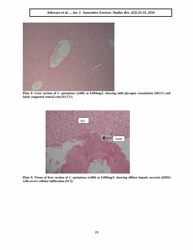
Plate 8: Liver section of C. gariepinus (x400) at 0.004mg/L showing mild glycogen vacuolation (MGV) and
fairly congested central vein (FCCV).
Plate 9: Tissue of liver section of C. gariepinus (x400) at 0.008mg/L showing diffuse hepatic necrosis (DHN)
with severe cellular infiltration (SCI).
DHN
SCI
Adewoye et al…. Int. J. Innovative Environ. Studies Res. 4(3):24-33, 2016
Ok

30
Plate 10: Liver section of C. gariepinus (x400) at 0.012m/L showing severe infiltration of leukocytes (arrow)
and lipid vacuolation (L).
Plate 11: Tissue of liver section of C. gariepinus (x400) at 0.016mg/L showing more pronounced hepatic
necrosis.
DISCUSSION
The exposure of fish to chemical contaminants is likely to induce a number of lesions in different organs
(Bucke et al., 1996). Gills (Poleksic et al., 1994), kidney (Bucher and Hofer, 1993), and liver (ICES,
1997) are suitable organs for histological examination in order to determine the effect of pollution. The
exposure of aquatic organisms to very low levels or sublethal concentration of pollutants in their
environment may result in various biochemical, physiological, and histological alterations in vital tissues.
In this study histological investigation of the liver tissues of C. gariepinus not exposed to sublethal
concentrations of cassava effluent showed a typical structural organization of the parenchymatous cell
appearance of the hepatocytes in the untreated fishes. However, the fishes exposed to the cassava effluent
L
L
L
Adewoye et al…. Int. J. Innovative Environ. Studies Res. 4(3):24-33, 2016
Ok

31
showed major histological abnormalities. The abnormalities observed include cellular infiltration,
congestion of central vein and cellular necrosis, which showed a progressive architectural distortion at
varied concentrations, this observation is in agreement with the submission of Strivastava, (1994).
In this study, the most generally encountered type of degenerative changes was congestion, vacuolization
of hepatocyte, cellular infiltration and necrosis. The liver of the exposed organisms revealed slight
vacuolated cells which is an indication of fatty degeneration of hepatocytes. Cellular necrosis as observed
in this study might have resulted from excessive work required by the fish liver to get rid of the toxicants
from its body during the process of detoxification. High accumulation of several components of the
cassava effluent in the liver is a pointer to the fact that, liver plays a major role in the accumulation and
detoxification.
Necrosis became evident as the concentration increases and this could be attributed to the inability of
fishes to regenerate new liver cells. It was also observed that the histopathological changes in the liver
caused metabolic problems; this lesion is characterized by the remains of the bile in the form of droplets
in the cytoplasm of the hepatocyte and this convincingly supported the submission of Pacheco and
Santos, (2002) that stated that bile is not being released from the liver which is also an indication of
possible damage to the hepatic metabolic functions of the liver. An increased in the degree of damages
done to the liver tissue of the fishes (Clarias gariepinus) held in 0.004 mg/L, 0.008 mg/L, 0.012mg/L,
0.016mg/L and 0.02mg/L cassava effluent, is generally related to important hepatic lesions such as
degenerative and necrotic processes, this observation was in line with the submission of Chang et al.,
(1998).
Additionally, the presence of bile stagnation or accumulation and melanomacarophages in great quantity
in the liver of exposed C. gariepinus is strong evident in that this organ suffered structural and metabolic
damage due to the exposure to the cassava effluent. This is in a way signalling the fact that this
environment where the effluent is discharged is grossly polluted and impaired. The histological alterations
identified within the hepatocytes in this study may have been the results of various biochemical lesions.
Anomalies such as irregular shaped central vein, cellular vacuolation and infiltration may be attributed to
the accumulation of lipids and glycogen due to liver dysfunction as a result of exposure to the effects of
cassava effluent, this is in conformity with the submission of Fanta et al., (2003).
Pacheco and Santo (2003) also described increased level of vacuolation of the hepatocytes as a signal to
the degenerating process that suggest metabolic damage, possibly related to exposure to contaminated
water. The liver parenchyma cells of all exposed fish showed signs of degeneration and this has been
reported by many authors to be associated with the exposure of fishes to certain toxicants (Chang et al.,
1998).
Evidently the damage done to the tissues of the liver and the gills seemed to increase with increase in
concentration as shown by the plates 1-11. This result is similar to the work of Wade et al. (2002) who
worked on the toxicity of cassava (Manihot esculenta) effluent on the Nile tilapia Oreochromis niloticus,
histopathological examination of gill, kidney, and liver indicated damage ranging from oedema and
talengiectasis of the gill lamella and gill hyperplasia to vacuolation of the liver cells and necrosis. The
Present findings are strongly supported by the works of Cengiz (2006) who observed and reported the
histopathological effects of δ-methrin on the gill of the common carp; Cyprinus Carpio (Linn.), the
changes at all doses of the test compound were desquamation and necrosis besides aneurism in secondary
lamellae, lifting of lamellar epithelium, edema, hyperplasia and fusion of the secondary lamellae were
reported.
The present findings are in conformation with the work of Dhanapakiam et al. (1998) who studied
histopathological changes in the gills of Channa punctatus (Bloch.) exposed to industrial effluents for 45
days, revealing deformities such as; secondary lamellae of primary filaments showing hyperplasia as
compared to the gills of controlled fish indicating that the industrial effluents of river Cauvery induced
considerable chemical stress on fish populations. Adhikari (1996) also studied the histopathological
changes in the gills of a cat fish; Heteropneustes fossilis on exposure to malathion at different
concentrations and different exposure times finding degenerative changes like complete secondary
lamellae fusion, edema, hypertrophy and hyperplasia of lamellar cells.
Adewoye et al…. Int. J. Innovative Environ. Studies Res. 4(3):24-33, 2016
Ok

32
Similarly, the present finding are in agreement with Tilak et al. (2001), Velmurugan et al. (2007) who
also observed the same histopathological changes in the gills of various fishes on exposure to different
toxins resulting in necrosis, vacuolar degeneration, dystrophy, desquamation, epithelial lifting, edema,
shorting of secondary lamellae and lamellar fusion. All these histopathological research studies on the
gills of fishes proved to be supporting the results of the present investigation. The observations in the
present investigation are also supported by the work of Nagarajan and Kumar (2006) who reported the
same histopathological changes in the gills of Labeo rohita (Ham.), exposed to sago effluent, which
included worn out gill filaments of primary and secondary gill lamellae.
The works of Pandey et al. (1997) provided confirmation to the present investigation who worked on
histopathological changes due to the effect of heavy metals on the gills of fishes. Fernandes et al. (2006)
observed the histopathological changes in gills epithelium of Oreochromis niloticus exposed to copper;
they reported edema, lifting of lamellae epithelia, lamellar fusion, thus also providing support for the
present investigation.
CONCLUSION This study has been able to establish the fact that; exposure of C. gariepinus to even low concentration of
cassava effluent can induce various toxicological effects and histological degradation which depends on
the period of exposure and concentration of the pollutant. In the view of the toxicity effect of this effluent,
it can be inferred that, indiscriminate discharge of cassava effluent can induce damage to the tissue and
organs which might make all the living entities in the polluted environments vulnerable to diseases and
eventually resulted to death.
REFERENCES
Adeyemo, O.K (2005), Haematological and Histopathological effects of Cassava
Adhikari S. 1996. Histopathological changes in the gills of a catfish; Heteropneustes fossilis (Bloch.) after
exposure to sub-lethal doses of malathion - A light microscope study. Journal of Freshwater
Biology 8(2): 99-104.
Akinsanya Bamidele (2007) Histopathological study on the parasitised visceral organs of some fishes of
Lekki Lagon, Lagos, Nigeria
AOAC 1987: Standard Method for Examination of Wastewater and Water, 17th edition, Washington
D.C., 8910 pp.
Bucher F, Hofer R(1993) The effects of treated domestic sewage on three organs (gills, kidney, liver) of
brown trout (Salmo trutta) Water Res 27:255–261.
Bucke D, Vethaak D, Lang T, Mellergaard S(1996) International Council for the Exploration of the Sea
Techniques in Marine Environmental Sciences (Copenhagen), Common Diseases and Parasites
of Fish in the North Atlantic: Training Guide for Identification.
Cengiz E I. 2006. Histopathological of gill and kidney in the fresh water; Cyprinus carpio (Linn.) after
acute exposure to δ-methrin. Environmental Toxicology Pharma 2: 13-16.
Daksh R. K and A Capoor (2011) Toxic Effects of Tannery Chemicals on the Histopathology of
FreshWater Teleost, Catla catla (Ham.)Research Journal of Agricultural Sciences 2(2): 351-353.
Dhanapakiam P, Ramasamy V K and Poorani S. 1998. A study on the histopathological changes in gills
of Channa punctatus (Bloch.) in Cauvery river water. Journal of Environmental Biology 19(3):
265-269.
Fernades A F, Cardoso J V F, Sontos S G and Carrola S M J. 2006. Histopathological changes in the liver
and gill epithelium of Oreochromis niloticus, exposed to copper. Bulletin of Environmental
Contamination Toxicology 76(2): 249-255.
ICES (International Council for the Exploration of the Sea)(1997) International Council for the
Exploration of the Sea (Copenhagen), Special Meeting on the Use of Liver Pathology of Flatfish
for Monitoring Biological Effects of Contaminants.
Adewoye et al…. Int. J. Innovative Environ. Studies Res. 4(3):24-33, 2016
Ok

33
JohnsonLL, StehrCM, OlsonOP, MyersMS, PierceSM, WigrenCA, McCainBB, VaranasiU(1993)
Chemical contaminants and hepatic lesions in winter flounder (Pleuronectes americanus) from
the Northeast Coast of the United States. Environ Sci Technol 27:2759–71.
Mercy, T. V. A., B. Madhusoodana, J. R. Nair (1996): Pesticide induced histological changes in juveniles
of Channa marulius. Fourth Ind. Fisheries Forum. pp. 81.
mill effluent in Clarias gariepinus. Dir. J.Biomed. Res. 8: 179-183.
Nagarajan K and Kumar R S. 2006. Observations on histopathological changes in the gills, liver and
intestine of an Indian fresh water major carp; Labeo rohita (Ham.) exposed to treated and
untreated sago effluent. Journal ofExperimental Zoology 9(1): 181-188.
Pandey A K, George K C and Mohamed M P. 1997. Histopathological alterations in the gill and kidney of
Liza parsia (Ham.) on exposure to lead. India Journal of Fisheries 44(2): 171-180.
Parrish, P.R. 1985. Acute toxicity test. In: G.M. Rand and S.R. Petrocelli (Eds.), Fundamentals of Aquatic
Toxicity. Hemissphere Publishing Corporation, Washington D.C.: 31-57.
Poleksic V, Mitrovic-Tutundzic V(1994) in Sublethal and Chronic Effects of Pollutants on Freshwater
Fish, Fish gills as a monitor of sublethal and chronic effects of pollution, eds Müller R, Lloyd R
(FAO, Fishing News Books, Oxford, UK), pp 339–352.
Rana K S and Raizada S. 2000. Acute toxicity of tannery and textile dye effluents on a common
teleost; Labeorohita (Ham.), histology alteration in liver. Journal of Environmental Biology
20(1): 33-36.
Schwaiger J, Fent K, Stecher H, Ferling H, Negele RD (1996) Effects of sublethal concentrations of
triphenyltinacetate on raibow trout (Oncorhynchus mykiss) Arch Environ Contam Toxicol
30:327–34.
Tilak K S, Veeraiah K and Yacobu K. 2001. Studies on histopathological changes in the gills liver and
kidney of Ctenopharyngodon indellus (Valen.) exposed to technical fenvalerate and EC 20%.
Pollution Research 20(3): 387-393.
Velmurugan B, Selvanayagam M, Cengiz E I and Unlu E. 2007. Histopathology of ג-cyhalothrin on
tissues such as the gills, kidney, liver and intestine of Cirrhinus mrigala (Ham.). Environmental
Toxicology and Pharmacology 24: 286-291.
Wade, J.W., Omoregie, E. and Ezenwaka, I. 2002. Toxicity of cassava (Manihot esculenta Crants)
effluent on the Nile tilapia Oreochromis niloticus (L) under laboratory condition. J. Aqua. Sci.,
17(2): 89-94.
Adewoye et al…. Int. J. Innovative Environ. Studies Res. 4(3):24-33, 2016
Ok