Student understanding of fluorescent microscopy as a research tool in biomedical science Dr Kirsten...
23
Student understanding of fluorescent microscopy as a research tool in biomedical science Dr Kirsten Zimbardi Educational Research Unit School of Biomedical Sciences The University of Queensland [email protected]
-
Upload
eleanore-cook -
Category
Documents
-
view
215 -
download
0
Transcript of Student understanding of fluorescent microscopy as a research tool in biomedical science Dr Kirsten...
Student understanding of fluorescent microscopy as a research tool in biomedical science
Dr Kirsten Zimbardi
Educational Research UnitSchool of Biomedical SciencesThe University of Queensland
The problem...
“All of them [the practicals] were awesome!... cause this is the first time I have done experiments in biology that do not involve them boring microscopes.”
How can we help students see microscopy as an exciting and powerful research tool?

My experiencePhD in developmental endocrinology

Science education for the 21st century• Call for reforms -> emphasise cognitive skills
– The Boyer Commission (1998) Reinventing undergraduate education: a blueprint for America’s research universities.
– National Research Council (2003) BIO2010: Transforming undergraduate education for future research biologists
– Lim (2010) Nature; Lin et al (2006) Science; Hanauer et al (2006) Science
• With exponential increases in knowledge availability, students need cognitive skills to deal with novel, complex, semi-structured problems
• Graduates need to be able to “think like a scientist”
“Scientific thinking” involves…identifying, understanding and analysing large pieces of scientifically valid and reliable information from multiple sources in a purposeful, reasoned and reflective manner to form a relevant and testable hypothesis. Carrying out high quality experiments to make well-informed and well-evidenced decisions or conclusions regarding a situation, issue or problem, while also recognising that these conclusions need to be tentative and restricted depending on the limitations of the available evidence.

Scientific method1. Evidence from scientific literature used to build hypothesis2. Hypothesis is testable and falsifiable
• Includes specific causes, measurable outcome and context
3. Methods provide valid test of hypothesis4. Results analysed and presented in relation to hypothesis5. Findings interpreted in relation to scientific literature
•Expected and unexpected findings

Educational context• Institutional context
– University of Queensland: large, research-intensive university
• Degree context– Majority of students enrolled in a Bachelor of Science (3 year degree)
• Course context– 5 vertically integrated courses for major in biomedical science
(specialisation in physiology)

Vertical integration across five courses with inquiry-based practical curricula
Modelled on “Research Skills Framework” Willison and O’Regan (2007)
Course timing and names Scientific method Scientific communication (Assessment)
Vertical progression Increased specialisation Increase in complexity More advanced aspects
Course 1 Cells to Organisms Year 1, Semester 2
Hypothesis construction and experimental design Written reports Strategic questions scaffold entire report
writing process.
Course 2 Integrative Cell & Tissue Biology
Year 2, Semester 1
Detailing the methods, statistical analysis, interpretation of results
Written proposals and reports
Emphasis on genre conventions for methods and results
Course 3 Systems Physiology
Year 2, Semester 2
Large scales clinical experiments, integration with primary literature
Oral proposals, written reports
Emphasis on integration of primary literature in introduction, methods and
discussion.
Course 4 Molecular & Cellular Physiology
Year 3, Semester 1
Critiquing methodological approaches, advanced techniques
Oral literature reviews, written report
Emphasis on range of advanced research methods
Course 5 Integrative Physiology & Pathophysiology
Year 3, Semester 2
Science as a social profession, advanced techniques
Conference abstract,professional proposal letter Emphasis on professional collegiality

Microscopy practical module• Integrative Physiology & Pathophysiology– Final semester of three year degree
• 4 week practical module on reproductive pathophysiology– 2 weeks of cell culture with microscopy– 2 weeks of PCR

Students are introduced to the research context
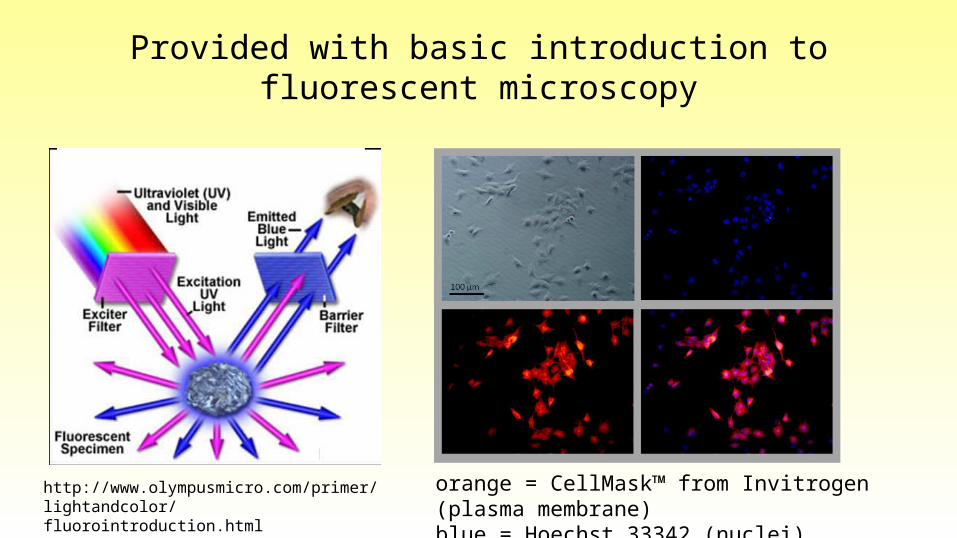
Provided with basic introduction to fluorescent microscopy
http://www.olympusmicro.com/primer/lightandcolor/fluorointroduction.html
orange = CellMask™ from Invitrogen (plasma membrane)blue = Hoechst 33342 (nuclei)

Experimental results
orange = CellMask™ from Invitrogen (plasma membrane); blue = Hoechst 33342 (nuclei)
Arrow: Trophoblast giant cell Arrow: Syncytiotrophoblast

Research questions1. How do science students begin to understand
the role of microscopy in research?
2. How can this understanding may be facilitated by virtual microscopy?

Determining student conceptions of microscopy in research
• Letter of proposal to researcher (assessment)– Analysed random sample of 60 proposals (50% cohort)
• Proportion of students who proposed experiments involving microscopy, or PCR or other outcome measure
• Analysed level of sophistication of conceptions about microscopy as a research tool– Type of staining, microscopy and outcome measures

Students’ proposed methods• 85% of students proposed using methods from class
(microscopy &/or PCR)– 47% also included method not from the class
• ~30% included in vivo experiments• Microarray, ELISA, flow cytometry
• 58% proposed an experiment using microscopy– 50% proposed microscopy with another outcome measure– 25% extended microscopy beyond class procedures
• E.g. IHC, FISH or GFP transfection

Microscopy proposals• 90% provided ‘accurate’ proposals• Large variation in level of detail provided– 44% provided no detail eg:
“To do so, immunocytochemistry would be used to visualise expression in the cultures, allowing for a qualitative assessment of the differences in gene expression (if present) between the observed cells.”

32% minimal detail (staining / microscopy)
“Morphological changes will then be assessed using bright field and fluorescent microscopy techniques.”
“Following nuclear and cell membrane staining, the cell morphology of differentiated cells will be assessed using light and fluorescent microscopy.”

24% provided detailed methods(1 correctly alluded to wavelengths)
• “…the cultures will be treated with ‘Hoechst 33342’ and ‘CellMask™’ to stain the cell membranes and nuclei respectively. They will also be treated with a Syncytin-A monoclonal antibody, linked to a fluorophore, effectively tagging the SynA gene product. The cultures can then be viewed under fluorescent microscopy, at appropriate wavelengths for the three fluorescent substances.”

… and 1 student proposed:“Following cell permeation by proteinase-K and pre-hybridization, RNA probes bound by digoxigenin uridine-5’-triphosphate would be utilized to hybridize synA mRNA at 70°C. Gene expression can be viewed immunohistochemically following the addition of an anti-digoxigenin antibody conjugated to alkaline phosphatase in the presence of specific substrates generating coloured products. Microscopy images can be taken, with colour intensity assessed and compared between treatment groups through software. Positive and negative controls for RNA-ISH would be probes against 18S (housekeeping gene) and probes against non-existent genes (e.g. bacterial exclusive genes) respectively.”

Research questions1. How do science students begin to understand
the role of microscopy in research?
2. How can this understanding may be facilitated by virtual microscopy?
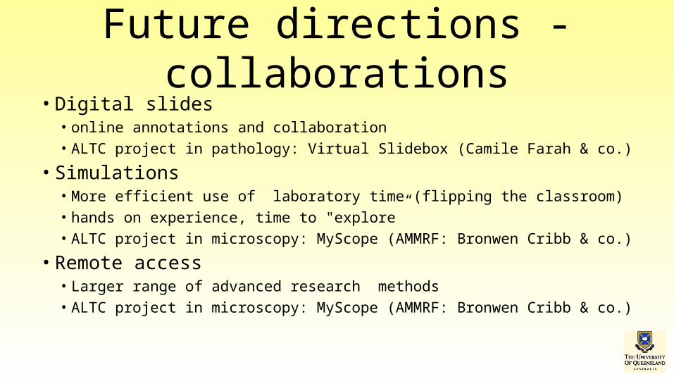
Future directions - collaborations• Digital slides
• online annotations and collaboration• ALTC project in pathology: Virtual Slidebox (Camile Farah & co.)
• Simulations• More efficient use of laboratory time (flipping the classroom)• hands on experience, time to "explore” • ALTC project in microscopy: MyScope (AMMRF: Bronwen Cribb & co.)
• Remote access• Larger range of advanced research methods • ALTC project in microscopy: MyScope (AMMRF: Bronwen Cribb & co.)

Expected outcomes• Engagement with experimental outcomes
– Deeper understanding of experimental model• Engagement with fundamental concepts of microscopy
– Detailed understanding of parameters demonstrated as details in proposed methods
• Experience in broad range of experimental techniques• Involvement in collaborative research

School of Biomedical Science, University of Queensland
Georgia KaferJane MooneyTamara Paravicini
Olympus Australia
ACMM22 / APMC 10 / ICONN 2012
Acknowledgements