STS General Thoracic Surgery Database Version 2.2 Version 2.2
105
GENERAL THORACIC SURGERY DATABASE v.2.3 effective January 1, 2015 TRAINING MANUAL 12/18/2014 1 The training manual is intended to clarify data definitions, provide examples and answer FAQs. It will be updated with new FAQs. Seq. #: 10 Long Name: Operations Table Record Identifier Short Name: RecordID Definition: An arbitrary, unique value generated by the software that permanently identifies each record in the participant's database (note that unlike the PatID value, this does not identify the individual patient). The value of the identifier is a combination of a code assigned to the software developer by the STS, and a value generated by the software to create a unique value. Once assigned to a record, this value can never be changed or reused. The data warehouse will use this value to communicate issues about individual records with the participant. It may also be used by the data warehouse to link this record to other clinical data. Intent/Clarification: A record should be initiated for inpatient and outpatient thoracic procedures on every visit to the operating room (includes the Endoscopy Suite or Outpatient Surgical Center) whether planned or unplanned. Seq. #: 20 Long Name: Procedures Table Record Identifier Short Name: RecordID Definition: This field is the foreign key that links this record with the associated records in the "Operations" table. Seq. #: 30 Long Name: Software Vendor's Identification Short Name: VendorID Definition: Software vendor's identification assigned by the STS. Seq. #: 40 Long Name: Vendor's Software Version Number Short Name: SoftVrsn Definition: Vendor's software product version number identifying the software which created this record. Vendor controls the value in this field. Version passing certification/harvest testing will be noted at the data warehouse. Seq. #: 50 Long Name: Version Of STS Data Specification Short Name: DataVrsn Definition: Version number of the STS Data Specifications/Dictionary, to which the record conforms. The value will identify which fields should have data, and what are the valid data values for those fields. It must be the version implemented in the software at the time the record was created. The value must be entered into the record automatically by the software. Seq. #: 60 Long Name: Participant ID Short Name: ParticID Definition: Participant ID is a unique number assigned to each database participant by the STS. A database participant is defined as one entity that signs a Participation Agreement with the STS, submits one data file to the harvest, and gets back one report on their data. The participant ID must be entered into each record.
Transcript of STS General Thoracic Surgery Database Version 2.2 Version 2.2
GENERAL THORACIC SURGERY DATABASE v.2.3 effective January 1, 2015 TRAINING MANUAL
12/18/2014 1
The training manual is intended to clarify data definitions, provide examples and answer FAQs. It will be updated with new FAQs. Seq. #: 10 Long Name: Operations Table Record Identifier Short Name: RecordID Definition: An arbitrary, unique value generated by the software that permanently identifies each record in the participant's database (note that unlike the PatID value, this does not identify the individual patient). The value of the identifier is a combination of a code assigned to the software developer by the STS, and a value generated by the software to create a unique value. Once assigned to a record, this value can never be changed or reused. The data warehouse will use this value to communicate issues about individual records with the participant. It may also be used by the data warehouse to link this record to other clinical data. Intent/Clarification: A record should be initiated for inpatient and outpatient thoracic procedures on every visit to the operating room (includes the Endoscopy Suite or Outpatient Surgical Center) whether planned or unplanned.
Seq. #: 20 Long Name: Procedures Table Record Identifier Short Name: RecordID Definition: This field is the foreign key that links this record with the associated records in the "Operations" table.
Seq. #: 30 Long Name: Software Vendor's Identification Short Name: VendorID Definition: Software vendor's identification assigned by the STS.
Seq. #: 40 Long Name: Vendor's Software Version Number Short Name: SoftVrsn Definition: Vendor's software product version number identifying the software which created this record. Vendor controls the value in this field. Version passing certification/harvest testing will be noted at the data warehouse.
Seq. #: 50 Long Name: Version Of STS Data Specification Short Name: DataVrsn Definition: Version number of the STS Data Specifications/Dictionary, to which the record conforms. The value will identify which fields should have data, and what are the valid data values for those fields. It must be the version implemented in the software at the time the record was created. The value must be entered into the record automatically by the software.
Seq. #: 60 Long Name: Participant ID Short Name: ParticID Definition: Participant ID is a unique number assigned to each database participant by the STS. A database participant is defined as one entity that signs a Participation Agreement with the STS, submits one data file to the harvest, and gets back one report on their data. The participant ID must be entered into each record.
GENERAL THORACIC SURGERY DATABASE v.2.3 effective January 1, 2015 TRAINING MANUAL
12/18/2014 2
Intent/Clarification: Each participant's data, if submitted to harvest, must be in one data file. If one participant keeps data in more than one file (e.g. at two sites), the participant must combine them back into one file for harvest submission. If two or more participants share single purchased software and enter cases into one database, the data must be extracted into two different files, one for each participant ID, with each record having the correct participant ID number.
Seq. #: 70 Long Name: Operations Table Patient Identifier Short Name: PatID Definition: The foreign key that links this record with the associated records in the "Demographics" table. Intent/Clarification: Once assigned to a patient, this number can never be changed or reused.
Seq. #: 80 Long Name: Demographics Table Patient Identifier Short Name: PatID Definition: An arbitrary value that uniquely and permanently identifies each patient. The value of the identifier is a combination of a code assigned to the software developer by the STS, and a value generated by the software to create a unique value. The value in this field cannot be a value that would identify the patient outside of the database (such as Medical Record Number or Social Security Number). Once a value has been assigned to a patient, it can never be changed or reused. This field is the primary key that links this record with the associated records in the "Operations" table.
Seq. #: 90 Long Name: Demographics Table Data Version Short Name: DemogDataVrsn Definition: Version number of the STS Data Specifications/Dictionary, to which the Demographics record conforms. The value will identify which fields should have data, and what are the valid data for those fields. It must be the version implemented in the software at the time the record was created. The value must be entered into the record automatically by the software. Note that the data version of the demographics record does not necessarily need to match the data version of all of the associated operation records for that patient. This is because new data versions might be implemented in the software and used for the creation of operation records after a demographics record has been created for a patient.
Seq. #: 100 Long Name: Medical Record # Short Name: MedRecN Definition: Indicate the patient's medical record number at the hospital where surgery occurred. This field should be collected in compliance with state/local privacy laws. Intent/Clarification: This field is not required for record inclusion
GENERAL THORACIC SURGERY DATABASE v.2.3 effective January 1, 2015 TRAINING MANUAL
12/18/2014 3
Seq. #: 110 Long Name: Patient's First Name Short Name: PatFName Definition: Indicate the patient's medical record number at the hospital where surgery occurred. This field should be collected in compliance with state/local privacy laws. Intent/Clarification: This field is not required for record inclusion
Seq. #: 121 Long Name: Patient's Middle Name Short Name: PatMName Definition: Indicate the patient's middle name as documented in the medical record. Leave "blank" if no middle name. This field should be collected in compliance with state/local privacy laws. Intent/Clarification: Leave “blank” if no middle initial. This field is not required for record inclusion
Seq. #: 130 Long Name: Patient's Last Name Short Name: PatLName Definition: Indicate the patient's medical record number at the hospital where surgery occurred. This field should be collected in compliance with state/local privacy laws. Intent/Clarification: This field is not required for record inclusion
Seq. #: 140 Long Name: Social Security Number Short Name: SSN Definition: Indicate the patient’s Social Security Number (SSN). Although this is the Social Security Number in the USA, other countries may have a different National Patient Identifier Number. For example in Canada, this would be the Social Insurance Number. This field should be collected in compliance with state/local privacy laws. Intent/Clarification: This field is not required for record inclusion
Seq. #: 151 Long Name: Patient Participating In STS‐Related Clinical Trial Short Name: ClinTrial Definition: Indicate which, if any, STS‐related clinical trial in which the patient is participating. The STS will assign a code to each clinical trial as they begin collecting data.
GENERAL THORACIC SURGERY DATABASE v.2.3 effective January 1, 2015 TRAINING MANUAL
12/18/2014 4
Intent/Clarification: This applies only to STS trials. The instructions will be posted here when trials are available. There are currently no trials underway.
Seq. #: 152 Long Name: Patient Participating In STS‐Related Clinical Trial ‐ Patient ID Short Name: ClinTrialPatID Definition: Indicate the patient identifier used to identify the patient in the clinical trial.
Seq. #: 160 Long Name: Date Of Birth Short Name: DOB Definition: Indicate the patient's date of birth using 4‐digit format for year. This field should be collected in compliance with state/local privacy laws. Intent/Clarification: This field is not required for record inclusion
Seq. #: 170 Long Name: Age At Time Of Surgery Short Name: Age Definition: Indicate the patient's age in years, at time of surgery. This should be calculated from the date of birth and the date of surgery, according to the convention used in the USA (the number of birth date anniversaries reached by the date of surgery). If patient is less than one year old, enter the value 1. Intent/Clarification: Age is needed for risk models
Seq. #: 180 Long Name: Postal Code Short Name: PostalCode Definition: Indicate the ZIP Code of the patient's residence. Outside the USA, this data may be known by other names such as Postal Code (needing 6 characters). Software should allow sites to collect at least up to 10 characters to allow for Zip+4 values. This field should be collected in compliance with state/local privacy laws. Intent/Clarification: This field is not required for record inclusion
Seq. #: 190 Long Name: Gender Short Name: Gender Definition: Indicate the patient's gender at birth as either male or female.
GENERAL THORACIC SURGERY DATABASE v.2.3 effective January 1, 2015 TRAINING MANUAL
12/18/2014 5
Intent/Clarification: Patients who have undergone gender reassignment surgery maintain the risk associated with their chromosomal gender. This field is included in risk models
Seq. #: 191 Long Name: Race Documented Short Name: RaceDocumented Definition: Indicate whether race is documented. Intent/Clarification: ‐ Yes ‐ No ‐ Patient declined to disclose Race should be self‐reported by the patient or family.
Seq. #: 200 Long Name: Race ‐ Caucasian Short Name: RaceCaucasian Definition: Indicate whether the patient's race, as determined by the patient or family, includes Caucasian. This includes a person having origins in any of the original peoples of Europe, the Middle East, or North Africa. Definition source: Standards for Maintaining, Collecting, and Presenting Federal Data on Race and Ethnicity: The minimum categories for data on race and ethnicity for Federal statistics, program administrative reporting, and civil rights compliance reporting. (www.whitehouse.gov/omb/fedreg/1997standards.html) Intent/Clarification: The Census Bureau collects race data in accordance with guidelines provided by the U.S. Office of Management and Budget and these data are based on self‐identification. The racial categories included in the census form generally reflect a social definition of race recognized in this country, and are not an attempt to define race biologically, anthropologically or genetically. In addition, it is recognized that the categories of the race item include racial and national origin or socio‐cultural groups. People may choose to report more than one race to indicate their racial mixture, Such as “American Indian and White.” People who identify their origin (ETHNICITY) as Hispanic, Latino or Spanish may be of any race. In addition, it is recognized that the categories of the race item include both racial and national origin and socio‐cultural groups. You may choose more than one race category.
Seq. #: 210 Long Name: Race ‐ Black / African American Short Name: RaceBlack Definition: Indicate whether the patient's race, as determined by the patient or family, includes Black / African American. This includes a person having origins in any of the black racial groups of Africa. Terms such as "Haitian" or "Negro" can be used in addition to "Black or African American."
GENERAL THORACIC SURGERY DATABASE v.2.3 effective January 1, 2015 TRAINING MANUAL
12/18/2014 6
Definition source: Standards for Maintaining, Collecting, and Presenting Federal Data on Race and Ethnicity : The minimum categories for data on race and ethnicity for Federal statistics, program administrative reporting, and civil rights compliance reporting. (www.whitehouse.gov/omb/fedreg/1997standards.html) Intent/Clarification: This includes a person having origins in any of the black racial groups of Africa. Terms such as "Haitian" or "Negro" can be used in addition to "Black or African American." Reference: www.whitehouse.gov/omb/fedreg/1997standards.html.
Seq. #: 220 Long Name: Race ‐ Asian Short Name: RaceAsian Definition: Indicate whether the patient's race, as determined by the patient or family, includes Asian. This includes a person having origins in any of the original peoples of the Far East, Southeast Asia, or the Indian subcontinent including, for example, Cambodia, China, India, Japan, Korea, Malaysia, Pakistan, the Philippine Islands, Thailand, and Vietnam. Definition source: Standards for Maintaining, Collecting, and Presenting Federal Data on Race and Ethnicity: The minimum categories for data on race and ethnicity for Federal statistics, program administrative reporting, and civil rights compliance reporting. (www.whitehouse.gov/omb/fedreg/1997standards.html)
Seq. #: 230 Long Name: Race ‐ American Indian / Alaskan Native Short Name: RaceNativeAm Definition: Indicate whether the patient's race, as determined by the patient or family, includes American Indian / Alaskan Native. This includes a person having origins in any of the original peoples of North and South America (including Central America), and who maintains tribal affiliation or community attachment. Definition source: Standards for Maintaining, Collecting, and Presenting Federal Data on Race and Ethnicity: The minimum categories for data on race and ethnicity for Federal statistics, program administrative reporting, and civil rights compliance reporting. (www.whitehouse.gov/omb/fedreg/1997standards.html) Intent/Clarification: American Indian or Alaska Native" refers to a person having origins in any of the original peoples of North and South America (including Central America) and who maintains tribal affiliation or community attachment. This category includes people who indicated their race(s) as "American Indian or Alaska Native" or reported their enrolled or principal tribe, such as Navajo, Blackfeet, Inupiat, Yup’ik, or Central American Indian groups or South American Indian groups. This includes all in North American native peoples such as American Indian/Alaskan Native, Inuit. [The 2010 Census Redistricting Data (Public Law 94‐171) Summary File]
Seq. #: 240 Long Name: Race ‐ Native Hawaiian / Pacific Islander Short Name: RacNativePacific
GENERAL THORACIC SURGERY DATABASE v.2.3 effective January 1, 2015 TRAINING MANUAL
12/18/2014 7
Definition: Indicate whether the patient's race, as determined by the patient or family, includes Native Hawaiian / Pacific Islander. This includes a person having origins in any of the original peoples of Hawaii, Guam, Samoa, or other Pacific Islands. Definition source: Standards for Maintaining, Collecting, and Presenting Federal Data on Race and Ethnicity: The minimum categories for data on race and ethnicity for Federal statistics, program administrative reporting, and civil rights compliance reporting. (www.whitehouse.gov/omb/fedreg/1997standards.html) Intent/Clarification: "Native Hawaiian or Other Pacific Islander" refers to a person having origins in any of the original peoples of Hawaii, Guam, Samoa, or other Pacific Islands. It includes people who indicated their race(s) as "Pacific Islander" or reported entries such as "Native Hawaiian", "Guamanian or Chamorro", "Samoan", and "Other Pacific Islander" or provided other detailed Pacific Islander responses. [The 2010 Census Redistricting Data (Public Law 94‐171) Summary File]
Seq. #: 250 Long Name: Race Other Short Name: RaceOther Definition: Indicate whether the patient's race, as determined by the patient or family, includes some other race or mixture of races not otherwise indicated. Definition source: Standards for Maintaining, Collecting, and Presenting Federal Data on Race and Ethnicity: The minimum categories for data on race and ethnicity for Federal statistics, program administrative reporting, and civil rights compliance reporting. (www.whitehouse.gov/omb/fedreg/1997standards.html) Intent/Clarification: "Some Other Race" includes all other responses not included in the White, Black or African American, American Indian or Alaska Native, Asian, and Native Hawaiian or Other Pacific Islander race categories described above. [The 2010 Census Redistricting Data (Public Law 94‐171) Summary File]
Seq. #: 270 Long Name: Hispanic Or Latino Ethnicity Short Name: Ethnicity Definition: Indicate if the patient is of Hispanic or Latino ethnicity as determined by the patient / family. Hispanic or Latino ethnicity includes patient report of Cuban, Mexican, Puerto Rican, South or Central American, or other Spanish culture or origin, regardless of race. Intent/Clarification: ‐ Yes ‐ No ‐ Not documented People who identify their origin as Hispanic, Latino or Spanish may be of any race. [The 2010 Census Redistricting Data (Public Law 94‐171) Summary File]
GENERAL THORACIC SURGERY DATABASE v.2.3 effective January 1, 2015 TRAINING MANUAL
12/18/2014 8
Seq. #: 271 Long Name: Date of Last Follow‐Up Short Name: LFUDate Definition: Indicate the date on which the last follow‐up was made. If patient dies in the hospital, this value will be the same as the date of death. If no follow‐up is made after patient is discharged, this value will be the same as the discharge date. Intent/Clarification: This field is for those patients diagnosed and surgically treated for Lung CA and Esophageal CA. Need to track patients for five (5) years from the date of the original surgery. Work with your cancer registry people for assistance with this information.
Seq. #: 272 Long Name: Mortality Status At Last Follow‐Up Short Name: LFUMortStat Definition: Indicate the mortality status of the patient at the time of the last follow‐up. If no follow‐up is made after patient is discharged, this value will be the same as the Mortality Status At Hospital Discharge. Intent/Clarification: This field was added to facilitate long term follow‐up for lung and esophageal cancer resection patients.
Seq. #: 273 Long Name: Mortality Date Short Name: MortDate Definition: Indicate the patient's date of death (even if after discharge).
Seq. #: 280 Long Name: Admission Status Short Name: AdmissionStat Definition: Indicate whether the procedure was an Inpatient or Outpatient / Observation procedure. Intent/Clarification: This field is required for Record Inclusion. If missing data, the entire record will be excluded from the analysis.
Outpatient/Observation should be selected if the operation was performed as an ambulatory procedure or if it included
a period of overnight observation.
‐ Inpatient
‐ Outpatient / Observation
Seq. #: 290 Long Name: Admission Date Short Name: AdmitDt
GENERAL THORACIC SURGERY DATABASE v.2.3 effective January 1, 2015 TRAINING MANUAL
12/18/2014 9
Definition: Indicate the date of admission. For those patients who originally enter the hospital in an out‐patient capacity, the admit date is the date the patient's status changes to in‐patient. Intent/Clarification: For purposes of this data definition, Outpatient and Observation status are the same. Enter INPATIENT admit date. This is a child field of admission status so if patient was never admitted as an inpatient you will not be asked to provide a date.
Seq. #: 411 Long Name: Primary Payor Short Name: PayorPrim Definition: Indicate the primary insurance used for this admission Intent/Clarification: ‐ None / self ‐ Medicare ‐ Medicaid ‐ Military Health ‐ Indian Health Service ‐ Correctional Facility ‐ State Specific Plan ‐ Other Government Insurance ‐ Commercial Health Insurance ‐ Health Maintenance Organization ‐ Non‐U.S. Plan Government insurance refers to patients who are covered by government‐reimbursed care. This includes Medicare, Medicaid, Military Health Care (e.g. TriCare), State‐Specific Plan, and Indian Health Service. CHIP (Children’s Health Insurance Plan), High Risk Pools Local Government Health Insurance Plan (LGHIP), state or federal prisoners. Blue Cross Federal Government is coded as Commercial insurance. If a pt is in a HMO, choose only HMO, you do not need to also choose commercial
Seq. #: 412 Long Name: Primary Payor Medicare Fee For Service Short Name: PrimMCareFFS Definition: Indicate whether the patient is covered by Medicare fee for service (Part B) Intent/Clarification: The Social Security Website at www.socialsecurity.gov has a list explaining what the letters behind the Medicare claim # stand for. Those letters do not tell you whether they have Part B/Fee for service. It is the relationship of the cardholder to the Medicare/SSN #. For example, B stands for "Aged wife, 62 or older". The A would stand for "Primary claimant=the wage earner". D1 is for an "Aged widower, age 60 or over".
GENERAL THORACIC SURGERY DATABASE v.2.3 effective January 1, 2015 TRAINING MANUAL
12/18/2014 10
This is used for PQRS Check with your hospital billing department if you are unsure whether the patient is considered Medicare Part B. Even if not using the registry for PQRS, CMS will be tracking outcomes for value based purchasing.
Seq. #: 413 Long Name: Secondary (Supplemental) Payor Short Name: PayorSecond Definition: indicate which, if any, secondary insurance was used for this admission Intent/Clarification: ‐ None / self ‐ Medicare ‐ Medicaid ‐ Military Health ‐ Indian Health Service ‐ Correctional Facility ‐ State Specific Plan ‐ Other Government Insurance ‐ Commercial Health Insurance ‐ Health Maintenance Organization ‐ Non‐U.S. Plan Government insurance refers to patients who are covered by government‐reimbursed care. This includes Medicare, Medicaid, Military Health Care (e.g. TriCare), State‐Specific Plan, and Indian Health Service. CHIP (Children’s Health Insurance Plan), High Risk Pools Local Government Health Insurance Plan (LGHIP), state or federal prisoners. Blue Cross Federal Government is coded as Commercial insurance. If a pt is in a HMO, choose only HMO, you do not need to also choose commercial
Seq. #: 414 Long Name: Secondary Payor Medicare Fee For Service Short Name: SecondMCareFFS Definition: Intent/Clarification: Indicate whether patient is covered by Medicare fee for service (part B) The Social Security Website at www.socialsecurity.gov has a list explaining what the letters behind the Medicare claim # stand for. Those letters do not tell you whether they have Part B/Fee for service. It is the relationship of the cardholder to the Medicare/SSN #. For example, B stands for "Aged wife, 62 or older". The A would stand for "Primary claimant=the wage earner". D1 is for an "Aged widower, age 60 or over". This is used for PQRS Check with your hospital billing department if you are unsure whether the patient is considered Medicare Part B. Even if not using the registry for PQRS, CMS will be tracking outcomes for value based purchasing.
GENERAL THORACIC SURGERY DATABASE v.2.3 effective January 1, 2015 TRAINING MANUAL
12/18/2014 11
Seq. #: 420 Long Name: Surgeon's Name Short Name: Surgeon Definition: Indicate the name of the surgeon responsible for the patient's care. Intent/Clarification: If two surgeons participate in the procedure and both surgeons are participating in the Database, the surgeon of record for the database is the physician under whom the patient is admitted or the physician responsible for the care of the patient. If this is not evident from the operative dictation, communication with the involved physicians is necessary.
Seq. #: 430 Long Name: Surgeon's National Provider Identifier Short Name: SurgNPI Definition: Indicate the individual‐level National Provider Identifier of the surgeon performing the procedure. For Non‐US surgeons a unique identifier will be assigned by STS. Intent/Clarification: The NPI is a unique identification number for health care providers. Health care providers will use the NPIs in the administrative and financial transactions adopted under HIPAA. The NPI is a 10 ‐ position, intelligence ‐ free numeric identifier (10 ‐ digit number) Meaning that the numbers do not carry other information about healthcare providers, such as the state in which they live or their medical specialty. NPI look up link: https://nppes.cms.hhs.gov/NPPES/NPIRegistryHome.do
Seq. #: 440 Long Name: Taxpayer Identification Number Short Name: TIN Definition: Indicate the Taxpayer Identification Number for the Taxpayer holder of record for the Surgeon's National Provider Identifier that performed the procedure. This may be an individual TIN or a group TIN depending on billing. This information is vital for PQRS reporting. This field will be blank for Non‐US participants Intent/Clarification: If the physician is part of a medical group practice, use the name and taxpayer identification number of the medical group.
Seq. #: 450 Long Name: Hospital Name Short Name: HospName Definition: Indicate the full name of the facility where the procedure was performed. Values should be full, official hospital names with no abbreviations or variations in spelling for a single hospital. Values should also be in mixed‐case. Intent/Clarification:
Seq. #: 460 Long Name: Hospital Postal Code
GENERAL THORACIC SURGERY DATABASE v.2.3 effective January 1, 2015 TRAINING MANUAL
12/18/2014 12
Short Name: HospZIP Definition: Indicate the ZIP Code of the hospital. Outside the USA, this data may be known by other names such as "Postal Code". Software should allow sites to collect up to 10 characters to allow for Zip+4 values. This field should be collected in compliance with state/local privacy laws. Intent/Clarification: This field is intended to allow analysis of geographical disparities in care
Seq. #: 470 Long Name: Hospital Region Short Name: HospStat Definition: Indicate the region of the country (i.e., state or province) in which the hospital is located. Intent/Clarification: This enables regional comparisions
Seq. #: 480 Long Name: Hospital National Provider Identifier Short Name: HospNPI Definition: Indicate the hospital's National Provider Identifier (NPI). This number, assigned by the Center for Medicare and Medicaid Services (CMS), is used to uniquely identify facilities for Medicare billing purposes. Non‐US participants will have a unique hospital ID number assigned by STS. Intent/Clarification: This is different from the surgeon NPI. This field will be used for hospital level analysis and eventually public reporting. Hospitals may have more than one NPI for inpatient services, lab, etc. Use the acute care hospital NPI. If the hospital ownership changes, this number may change. Notify STS. Lookup: https://nppes.cms.hhs.gov/NPPESRegistry/NPIRegistrySearch.do
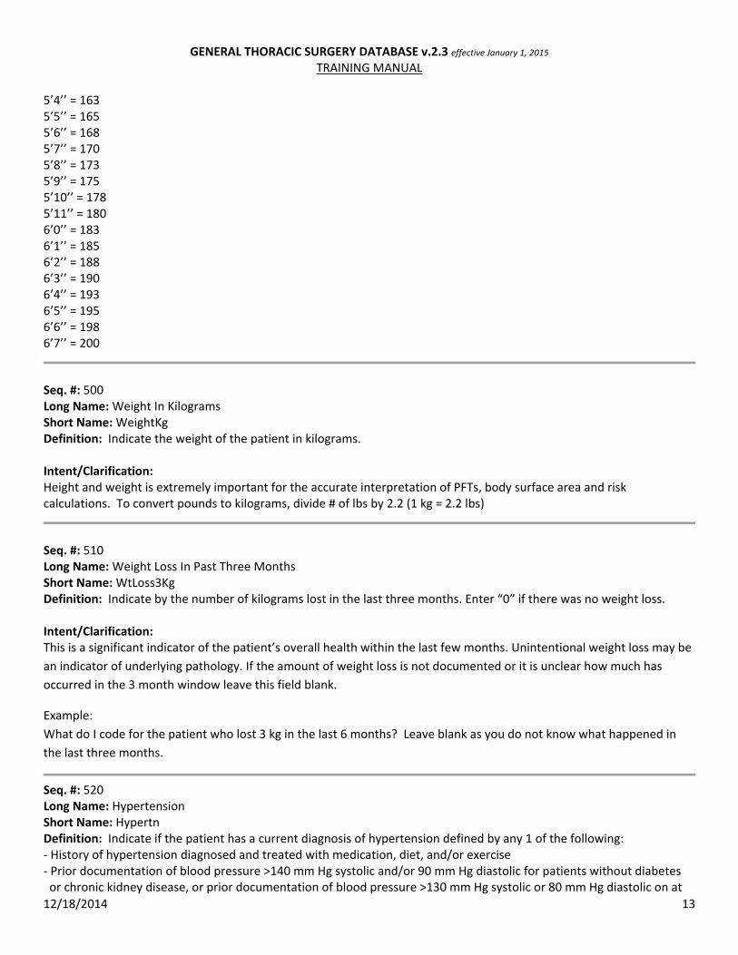
Seq. #: 490 Long Name: Height In Centimeters Short Name: HeightCm Definition: Indicate the height of the patient in centimeters. Intent/Clarification: Height and weight is extremely important for the accurate interpretation of PFTs, body surface area and risk calculations. Ft‐in = cm 4’10’’ = 147 4’11’’ = 149 5’0’’ = 152 5’1’’ = 155 5’2’’ = 157 5’3’’ = 160
GENERAL THORACIC SURGERY DATABASE v.2.3 effective January 1, 2015 TRAINING MANUAL
12/18/2014 13
5’4’’ = 163 5’5’’ = 165 5’6’’ = 168 5’7’’ = 170 5’8’’ = 173 5’9’’ = 175 5’10’’ = 178 5’11’’ = 180 6’0’’ = 183 6’1’’ = 185 6’2’’ = 188 6’3’’ = 190 6’4’’ = 193 6’5’’ = 195 6’6’’ = 198 6’7’’ = 200
Seq. #: 500 Long Name: Weight In Kilograms Short Name: WeightKg Definition: Indicate the weight of the patient in kilograms. Intent/Clarification: Height and weight is extremely important for the accurate interpretation of PFTs, body surface area and risk calculations. To convert pounds to kilograms, divide # of lbs by 2.2 (1 kg = 2.2 lbs)
Seq. #: 510 Long Name: Weight Loss In Past Three Months Short Name: WtLoss3Kg Definition: Indicate by the number of kilograms lost in the last three months. Enter “0” if there was no weight loss. Intent/Clarification: This is a significant indicator of the patient’s overall health within the last few months. Unintentional weight loss may be
an indicator of underlying pathology. If the amount of weight loss is not documented or it is unclear how much has
occurred in the 3 month window leave this field blank.
Example:
What do I code for the patient who lost 3 kg in the last 6 months? Leave blank as you do not know what happened in
the last three months.
Seq. #: 520 Long Name: Hypertension Short Name: Hypertn Definition: Indicate if the patient has a current diagnosis of hypertension defined by any 1 of the following: ‐ History of hypertension diagnosed and treated with medication, diet, and/or exercise ‐ Prior documentation of blood pressure >140 mm Hg systolic and/or 90 mm Hg diastolic for patients without diabetes or chronic kidney disease, or prior documentation of blood pressure >130 mm Hg systolic or 80 mm Hg diastolic on at
GENERAL THORACIC SURGERY DATABASE v.2.3 effective January 1, 2015 TRAINING MANUAL
12/18/2014 14
least 2 occasions for patients with diabetes or chronic kidney disease ‐ Currently undergoing pharmacological therapy for treatment of hypertension 2013 ACCF/AHA Data Standards Cannon et al. JACC Vol. 61, No. 9, 2013 Intent/Clarification: The History & Physical form will list the patient’s past medical history and also will list the current medications. Code ‘ye’s for patients who report a history of high blood pressure and are currently normotensive on antihypertensive medication.
Seq. #: 530 Long Name: Steroids Short Name: Steroids Definition: Indicate whether the patient was taking oral or IV steroids within 24 hours of surgery. This does not include a one‐time dose related to prophylaxis therapy (i.e., IV dye exposure for cath procedure or surgery pre‐induction), or non‐systemic medications (i.e., nasal sprays, inhalers, topical creams). Intent/Clarification: Systemic delivery only Non‐systemic delivery is not included in this data element. Non‐systemic delivery includes topical creams, nasal sprays, inhalers or ophthalmic or otic drops. Do not include one‐time dose as part of clinical pathway guideline or procedure/surgical preparatory order.
Yes‐Capture those who are prescribed to take medications on a regular schedule and are presumed to be at a therapeutic level, within 24 hours preceding surgery (entry into the OR) ‐ Do Not Include a one‐time dose
No–Patient did not receive a Steroid medication within 24 hours preceding surgery Examples of oral and intravenous steroid medications include prednisone, hydrocortisone, dexamethasone, and methylprednisolone.
Seq. #: 540 Long Name: Congestive Heart Failure
Short Name: CHF Definition: Indicate if there is physician documentation or report that the patient has been in a state of heart failure within the past 2 weeks. Heart failure is defined as physician documentation or report of any of the following clinical symptoms of heart failure described as unusual dyspnea on light exertion, recurrent dyspnea occurring in the supine position, fluid retention; or the description of rales, jugular venous distension, pulmonary edema on physical exam, or pulmonary edema on chest x‐ray presumed to be cardiac dysfunction. A low ejection fraction alone, without clinical evidence of heart failure does not qualify as heart failure. An elevated BNP without other supporting documentation should not be coded as CHF. Intent/Clarification: Congestive heart failure occurs when the heart is unable to pump blood effectively throughout the body. The term congestive is used because lung congestion causes some of the main symptoms of heart failure.
GENERAL THORACIC SURGERY DATABASE v.2.3 effective January 1, 2015 TRAINING MANUAL
12/18/2014 15
The intent is to capture the patient's actual status in the two weeks before surgery, the new diagnosis or exacerbation of an existing heart failure condition. DO NOT code stable or asymptomatic compensated failure or patients whose symptoms improved after medical therapy.
Seq. #: 550 Long Name: Coronary Artery Disease
Short Name: CAD Definition: Indicate whether the patient has a history of coronary artery disease (CAD) as evidenced by one of the following: 1. Currently receiving medical treatment for CAD 2. History of Myocardial Infarction 3. Prior CV intervention including, but not limited to, CABG and/or PCI Intent/Clarification: Coronary artery disease is a type of atherosclerosis in which plaque builds up inside the arteries that carry blood to the heart. As the artery walls thicken, the passageway for blood narrows. Sometimes platelets gather at the narrowing, forming a clot that decreases or prevents blood flow to the region of the heart supplied by the artery. Documented blockage ≥ 50% of one or more coronary arteries or documentation of CAD in H&P. Documentation of angina, myocardial infarction (MI), CABG, PCI*, or sudden cardiac death with no known cause may be included. *Percutaneous Coronary Intervention (PCI) includes angioplasty, coronary atherectomy and coronary artery stenting.
Seq. #: 560 Long Name: Peripheral Vascular Disease
Short Name: PVD Definition: Indicate whether the patient has Peripheral Arterial Vascular Disease, as indicated by: ‐ claudication either with exertion or rest; ‐ amputation for arterial insufficiency; ‐ aorto‐iliac occlusive disease reconstruction; ‐ peripheral vascular bypass surgery, angioplasty, or stent; ‐ documented AAA, AAA repair, or stent; ‐ non‐invasive/invasive carotid test with greater than 79% occlusion; ‐ previous carotid artery surgery/intervention for carotid artery stenosis. Intent/Clarification: This refers to diseases of blood vessels outside the heart and brain. It is often a narrowing of vessels that carry blood to the legs, arms, stomach or kidneys.
Seq. #: 570 Long Name: Prior Cardiothoracic Surgery
Short Name: PriorCTS
GENERAL THORACIC SURGERY DATABASE v.2.3 effective January 1, 2015 TRAINING MANUAL
12/18/2014 16
Definition: Indicate whether the patient has undergone any prior cardiac and/or general thoracic surgical procedure that required a general anesthetic and an incision into the chest or mediastinum. A thoracotomy, median sternotomy, anterior mediastinotomy or thoracoscopy would be included here. A cervical mediastinoscopy or tube thoracostomy would not be included. Intent/Clarification: Prior cardiothoracic surgery causes scar tissue to form and may increase difficulty and or risk in subsequent procedures. Do not include transcatheter procedures if no chest incision was performed.
Seq. #: 580 Long Name: Preoperative Chemo ‐ Current Malignancy
Short Name: PreopChemoCur Definition: Indicate whether the patient received preoperative chemotherapy for the current thoracic malignancy. Do not report treatment for prior cancers. Intent/Clarification: Do not include methotrexate given for arthritis.
Seq. #: 590 Long Name: Preoperative Chemo ‐ Current Malignancy ‐ When
Short Name: PreopChemoCurWhen Definition: Indicate when the patient received preoperative chemotherapy for the current thoracic malignancy.
Intent/Clarification: ‐ <= 6 Months ‐ > 6 Months
Seq. #: 600 Long Name: Preoperative Thoracic Radiation Therapy
Short Name: PreopXRT
Definition: Indicate if the patient has received preoperative radiation therapy to the chest for any reason prior to this
operation. May be included as a component of a chemotherapy radiation induction therapy. This item should also be
selected if the radiation oncologist gave the patient radiation therapy prior to sending the patient for any surgical
evaluation, if the intent of the radiation oncologist was to "shrink the tumor" prior to surgical intervention.
Intent/Clarification: Radiation therapy causes changes to the tissues which may increase difficulty and or risk in subsequent surgeries.
Seq. #: 610 Long Name: Preoperative Thoracic Radiation Therapy ‐ Disease And When Treated
Short Name: PreopXRTDisWhen
Definition: Indicate when the patient received preoperative thoracic radiation therapy and for what disease.
GENERAL THORACIC SURGERY DATABASE v.2.3 effective January 1, 2015 TRAINING MANUAL
12/18/2014 17
Intent/Clarification: If patient did not receive preoperative radiation therapy as indicated by a “Yes” in PreopXRT, there should be no option to answer. ‐ Same disease, <= 6 months ‐ Same disease, > 6 months ‐ Unrelated disease, <= 6 months ‐ Unrelated disease, > 6 months
Seq. #: 620 Long Name: Cerebrovascular History
Short Name: CerebroHx
Definition: Indicate if the patient has a history of cerebrovascular disease, documented by any one of the following:
• Cerebrovascular Accident (CVA): Patient has a history of stroke, i.e., loss of neurological function with residual
symptoms at least 24 hours after onset, presumed to be from vascular etiology.
• Transient Ischemic Attack (TIA): Patient has a history of loss of neurological function that was abrupt in onset but with
complete return of function within 24 hours, presumed to be due to vascular etiology
• Non‐invasive/invasive carotid test with greater than 79% occlusion.
• Previous carotid artery surgery/ intervention for carotid artery stenosis.
This does not include neurological disease processes such as metabolic and/or anoxic ischemic encephalopathy.
Intent/Clarification: If a history of previous cerebrovascular disease exists, it should be noted whether the patient’s symptoms were or reversible (i.e. transient ischemic attack) or whether the deficit is permanent (i.e. stroke). Example: What if a transient neuro event lasts more than 24 hours but resolves? Is this coded as reversible or irreversible? Use the 24 hour timeframe ‐ if symptoms resolve within 24 hours, code as reversible. If symptoms persist for more than 24 hours, code as irreversible. Do not code asymptomatic findings on neuro scans as stroke. ‐ No CVD history ‐ Transient Ischemic Attack – TIA ‐ reversible ‐ Cerebrovascular Accident – CVA – irreversible
Seq. #: 630 Long Name: Pulmonary Hypertension
Short Name: PulmHypertn
Definition: Indicate whether there is physician documentation of Pulmonary Hypertension as documented by:
‐ Right heart catheterization: mean pulmonary arterial pressure (PAP) > 25 mmHg at rest
or
‐ Echocardiographic diagnosis: PA systolic pressure >50 mmHg
Intent/Clarification:
GENERAL THORACIC SURGERY DATABASE v.2.3 effective January 1, 2015 TRAINING MANUAL
12/18/2014 18
High blood pressure in the arteries that supply the lungs is called pulmonary hypertension (PHT). The blood vessels that supply the lungs constrict and their walls thicken, so they cannot carry as much blood. This information may be found on a preoperative cardiac catheterization or echocardiogram. If the value is not known or documented, the data sheet should be marked accordingly. RV systolic pressure may be used if no PA pressure is available, provided there is no pulmonary stenosis. It is preferable to use pressures measured pre‐op, prior to induction of anesthesia.
Seq. #: 640 Long Name: Diabetes
Short Name: Diabetes
Definition: History of diabetes diagnosed and/or treated by a healthcare provider. The American Diabetes Association
criteria include documentation of the following:
1. Hemoglobin A1c >=6.5%; or
2. Fasting plasma glucose >=126 mg/dL (7.0 mmol/L); or
3. 2‐h Plasma glucose >=200 mg/dL (11.1 mmol/L) during an oral glucose tolerance test; or
4. In a patient with classic symptoms of hyperglycemia or hyperglycemic crisis, a random plasma glucose >=200 mg/dL
(11.1 mmol/L)
This does not include gestational diabetes.
2013 ACCF/AHA Data Standards
Cannon et al. JACC Vol. 61, No. 9, 2013
Intent/Clarification: Indicate if the patient has a history of diabetes mellitus regardless of duration of disease or need for anti‐diabetic agents. Exclusions are steroid induced hyperglycemia and gestational (transient), without elevated HbA1c and/or treatment, code ”no”. Not all patients receiving diabetic medications are considered diabetic. It is important to remember, some medications used to treat diabetes may be used to treat other conditions. A hemoglobin A1c value of >= 6.5%, collected within 3 months prior to surgery, is acceptable to use for documentation of diabetes = "yes".
Seq. #: 650 Long Name: Diabetes Therapy
Short Name: DiabCtrl
Definition: Indicate the diabetes therapy method. Patients placed on a preoperative diabetic pathway of insulin drip,
then were controlled with “none”, diet or oral methods, are not coded as insulin dependent.
Intent/Clarification:
Indicate the patient’s diabetes control method as presented on admission. Patients placed on a preprocedure diabetic pathway of insulin drip at admission but whose diabetes was controlled by diet or oral methods are not coded as being treated with insulin. Look for the long term management therapy that was used, if any.
GENERAL THORACIC SURGERY DATABASE v.2.3 effective January 1, 2015 TRAINING MANUAL
12/18/2014 19
*Oral treatments may include: Sulfonylureas ‐ Diabinese, glipizide (Glucotrol, Glucotrol XL), glyburide (Micronase, DiaBeta, Glynase), and glimepiride (Amaryl). Meglitinides ‐ Repaglinide (Prandin) and nateglinide (Starlix). Biguanides ‐ metformin (Glucophage). Thiazolidinediones ‐ rosiglitazone (Avandia) and pioglitazone (Actos). Alpha‐glucosidase inhibitors ‐ acarbose (Precose) and meglitol (Glyset). DPP‐4 inhibitor ‐ sitagliptin (Januvia). Choose the most aggressive therapy from the order below ‐ None = No treatment for diabetes. ‐ Diet only = Treatment with diet only ‐ Oral = Treatment with oral agent (includes oral agent with or without diet treatment) *see above list* ‐ Insulin = Insulin treatment (includes any combination with insulin) ‐ Other subcutaneous medication = Other subcutaneous medications (such as GLP‐1 agonists; Byetta, Bydureon, Victoza, Symlin) ‐ Other = Other adjunctive treatment, non‐oral/insulin/diet ‐ Unknown – choose unknown if the patient or family is unable to provide the information
Seq. #: 660 Long Name: Currently On Dialysis
Short Name: Dialysis
Definition: Indicate whether the patient is currently undergoing dialysis. This includes hemodialysis, peritoneal dialysis
or CRRT. Does not include ultra‐filtration.
Intent/Clarification: Includes any form of peritoneal or hemodialysis the patient is receiving prior to surgery. Also, may include Continuous Veno‐Venous Hemofiltration (CVVH, CVVH‐D), and Continuous Renal Replacement Therapy (CRRT) as dialysis. Code ‘”No” for renal dialysis if ultrafiltration is the only documentation found in the record since this is for volume management. Capture lab values if available. Not all patients will have (or need) all of the following labs drawn. This does not imply that the labs listed below are required or should be added to routine preop screening. Most hospitals have a policy on how far back preop labs can be drawn. Obviously as close to surgery as possible is preferred. STS recommends within 30 days of surgery except where stated otherwise. This includes POC (Point of Care) testing results.
Seq. #: 670 Long Name: Creatinine Level Measured
Short Name: CreatMeasured
Definition: Indicate whether the creatinine level was measured within one month prior to the surgical procedure and
prior to anesthetic management (induction area or operating room).
Intent/Clarification:
GENERAL THORACIC SURGERY DATABASE v.2.3 effective January 1, 2015 TRAINING MANUAL
12/18/2014 20
Creatinine, urea and urate all increase as the ability of the kidneys to filter fluid within the body declines. Creatinine is a marker for kidney function.
Seq. #: 680 Long Name: Last Creatinine Level
Short Name: CreatLst
Definition: Indicate the creatinine level closest to the date and time prior to surgery.
Intent/Clarification: Prior to anesthetic management (induction area or operating room). A creatinine level should be collected on all patients, even if they have no prior history of renal disease. A creatinine value is a high predictor of a patient's outcome and is used in the predicted risk models. Creatinine (Cr) is a chemical waste molecule that is generated from muscle metabolism. If the kidneys become impaired for any reason, the creatinine level in the blood will rise due to poor clearance by the kidneys. Abnormally high levels of creatinine thus warn of possible malfunction or failure of the kidneys. Anesthetic management begins when a member of the anesthesiology team initiates care. The administration of IV fluids in the holding area can cause dilution of blood. Do not capture labs drawn after the patient receives fluids in the holding area or O.R.
Seq. #: 690 Long Name: Hemoglobin Level Measured
Short Name: HemoglobinMeasured
Definition: Indicate whether the patient's hemoglobin level was measured within one month prior to this surgical
procedure.
Intent/Clarification: Hemoglobin is the protein molecule in red blood cells that carries oxygen from the lungs to the body's tissues and
returns carbon dioxide from the tissues to the lungs. The iron contained in hemoglobin is responsible for the red color of
blood.
Seq. #: 700 Long Name: Last Hemoglobin Level
Short Name: HemoglobinLst
Definition: Indicate the hemoglobin level closest to the date and time prior to surgery and prior to anesthetic
management (induction area or operating room).
Intent/Clarification: The hemoglobin (Hgb) test may be used to screen for, diagnose, or monitor a number of conditions and diseases that affect red blood cells (RBCs) and/or the amount of hemoglobin in blood. The hospital laboratory report should be accessed first when coding this variable. If this is unavailable, then additional source documents may be referenced for lab results.
GENERAL THORACIC SURGERY DATABASE v.2.3 effective January 1, 2015 TRAINING MANUAL
12/18/2014 21
Capture only measured hemoglobin levels, not calculated values. Anesthetic management begins when a member of the anesthesiology team initiates care. The administration of IV fluids in the holding area can cause dilution of blood. Do not capture labs drawn after the patient receives fluids in the holding area or O.R. The value used should be the most recent one prior to entering the operating room.
Seq. #: 710 Long Name: COPD
Short Name: COPD
Definition: Indicate whether the patient has a history of chronic obstructive pulmonary disease (COPD) as evidenced by
previous diagnosis, treatment, and/or spirometric evidence.
Intent/Clarification: Chronic Obstructive Pulmonary Disease (COPD) is a preventable and treatable lung disease with some significant extrapulmonary effects. It is characterized by airflow limitation that is not fully reversible, usually progressive and associated with an abnormal inflammatory response in lung tissue. Diagnosis is confirmed and severity is graded using pulmonary function testing (PFT). Bronchitis and emphysema are considered COPD, asthma is not. GOLD is short for the Global Initiative for Chronic Obstructive Lung Disease, collaboration between the National Institutes of Health and the World Health Organization. Spirometric evidence per GOLD criteria follows: No: FEV1/FVC >= 0.7 Yes**: Mild: FEV1/FVC <0.7 and FEV1>= 80% Moderate: FEV1/FVC <0.7 and FEV1 between 50‐80% Severe: FEV1/FVC < 0.7 and FEV1 <50%
Seq. #: 720 Long Name: Interstitial Fibrosis
Short Name: InterstitialFib
Definition: Indicate whether the patient has a diagnosis of interstitial fibrosis based on clinical and radiological or
pathological evidences.
Intent/Clarification: Interstitial lung disease (ILD), also known as diffuse parenchymal lung disease (DPLD), refers to a group of lung diseases
affecting the interstitium (the tissue and space around the air sacs of the lungs). [2] It concerns alveolar epithelium,
pulmonary capillary endothelium, basement membrane, perivascular and perilymphatic tissues. The term ILD is used to
distinguish these diseases from obstructive airways diseases; (ex. ILD, DPLD, Cystic Fibrosis)
Seq. #: 730 Long Name: Cigarette Smoking
Short Name: CigSmoking
GENERAL THORACIC SURGERY DATABASE v.2.3 effective January 1, 2015 TRAINING MANUAL
12/18/2014 22
Definition: Indicate the patient's history of smoking cigarettes.
Intent/Clarification: This field is Required for Record Inclusion. If missing data, the entire record will be excluded from the analysis. ‐ Never smoked ‐ Past smoker (anyone who has not smoked within 30 days prior to admission) ‐ Current smoker (within 30 days prior to admission) ‐ Unknown (patient and/or family unable to provide history, cannot determine from the medical record documentation) Electronic cigarettes (Ecig) = "No"
Example: How do you code smoking status if there is conflicting documentation in the chart? Code yes to smoking if any provider
documents it in the record and capture the highest number of pack years documented.
Example: Patient who smoked prior to admission, has been in the hospital > 2 weeks prior to surgery, and did not smoke
while in the hospital is captured as “Yes”. The patient smoked within the 30 day window.
Seq. #: 740 Long Name: Pack Years Known or can be estimated
Short Name: PackYearKnown
Definition: Indicate whether the number of pack years is known or can be estimated.
Intent/Clarification:
Seq. #: 750 Long Name: Pack‐Years Of Cigarette Use
Short Name: PackYear
Definition: Indicate the number or estimate of pack‐years by multiplying the average number of packs of cigarettes
smoked per day by the number of years of smoking. For example if the patient smoked 1 ppd for 10 years and 3 ppd for
the next 10 years, the average ppd would be 2 ppd x 20 years = 40 pack‐years of smoking.
Intent/Clarification: Code the highest # of pack years if you have a range, ex. 20‐30 years, code 30.
Seq. #: 760 Long Name: Pulmonary Function Tests Performed
Short Name: PFT
GENERAL THORACIC SURGERY DATABASE v.2.3 effective January 1, 2015 TRAINING MANUAL
12/18/2014 23
Definition: Indicate whether pulmonary function tests (PFT's) were performed prior to this operation. PFT's done more
than 12 months prior to the primary surgical procedure should not be included here.
Intent/Clarification: Pulmonary function testing is a valuable tool for evaluating the respiratory system, representing an important adjunct to the patient history, various lung imaging studies, and invasive testing such as bronchoscopy and open‐lung biopsy. Insight into underlying pathophysiology can often be gained by comparing the measured values for pulmonary function tests obtained on a patient at any particular point with normative values derived from population studies. The percentage of predicted normal is used to grade the severity of the abnormality. Pulmonary function testing is used in clinical medicine for evaluating respiratory symptoms such as dyspnea and cough, for stratifying preoperative risk, and for diagnosing common diseases such as asthma and chronic obstructive pulmonary disease. PFT = "yes" if only FEV1 is done. Use bedside PFTs if that's the only available test.
Seq. #: 770 Long Name: PFT Not Performed Reason
Short Name: PFTNotPerReas
Definition: Indicate the reason why pulmonary function testing was not done.
Intent/Clarification: There are acceptable reasons not to perform PFTs. These will be included in the NQF exclusions: ‐ Not Major Lung Resection ‐ Never smoked, no lung disease ‐ Patient unable to perform ‐ Tracheostomy or ventilator dependent ‐ Urgent or emergent status Example: The PFT field 770 should be answered “Not a major lung resection” for cases that are highlighted as “non‐analyzed” cases. Lung resections that are listed as “major” on the DCF should have PFTs. A therapeutic wedge is a major procedure, even though not a major anatomic resection, and PFTs are expected.
Seq. #: 780 Long Name: Forced Expiratory Volume Test Performed
Short Name: FEV
Definition: Indicate whether a Forced Expiratory Volume at 1 second (FEV1) test was performed. FEV1 test should be
performed for a major lung resection (e.g., wedge resection, segmentectomy, lobectomy, sleeve lobectomy,
bilobectomy, or pneumonectomy). Select "Not applicable" ONLY if none of these procedures was performed.
Intent/Clarification: This field is Required for Record Inclusion. If missing data, the entire record will be excluded from the analysis.
GENERAL THORACIC SURGERY DATABASE v.2.3 effective January 1, 2015 TRAINING MANUAL
12/18/2014 24
Seq. #: 790 Long Name: FEV1 Predicted
Short Name: FEVPred
Definition: Indicate the % predicted FEV1 obtained for the patient.
Intent/Clarification: This field is Required for Record Inclusion. If missing data, the entire record will be excluded from the analysis. Indicate the FEV1 % predicted from the most recent pulmonary function test prior to procedure. Do not use values obtained more than 12 months prior to surgery. Choose the highest value reported for % predicted, whether or not a bronchodilator was used. FEV1 is the maximal amount of air forcefully exhaled in one second. It is then converted to a percentage of normal. For example, the FEV1 may be 80% of predicted based on height, weight, and race. FEV1 is a marker for the degree of obstruction. In normal persons, the FEV1 accounts for the greatest part of the exhaled volume from a spirometric maneuver and reflects mechanical properties of the large and the medium‐sized airways. If there are multiple PFTs in the record, choose the study which best reflects the patient’s status just prior to surgery.
Seq. #: 800 Long Name: DLCO Test Performed
Short Name: DLCO
Definition: Indicate whether a lung diffusion test (DLCO) was performed. DLCO test should be collected for a major lung
resection (e.g., wedge resection, segmentectomy, lobectomy, sleeve lobectomy, bilobectomy, or pneumonectomy).
Select "Not applicable" ONLY if none of these procedures was collected.
Intent/Clarification: The diffusing capacity (DLCO) is a test of the integrity of the alveolar‐capillary surface area for gas transfer. Do not use values obtained more than 12 months prior to surgery.
Seq. #: 810 Long Name: DLCO Predicted
Short Name: DLCOPred
Definition: Indicate the % predicted DLCO value obtained for the patient.
Intent/Clarification: The diffusing capacity (DLCO) may be reduced, <80% predicted, in disorders such as emphysema, pulmonary fibrosis, obstructive lung disease, pulmonary embolism, pulmonary hypertension and anemia. DLCO>120% of predicted may be seen in normal lungs, asthma, pulmonary hemorrhage, polycythemia, and left to right intracardiac shunt. Choose the value that represents the highest % predicted unadjusted/uncorrected DLCO. DO NOT USE the DLCO/VA (adjusted/corrected).
GENERAL THORACIC SURGERY DATABASE v.2.3 effective January 1, 2015 TRAINING MANUAL
12/18/2014 25
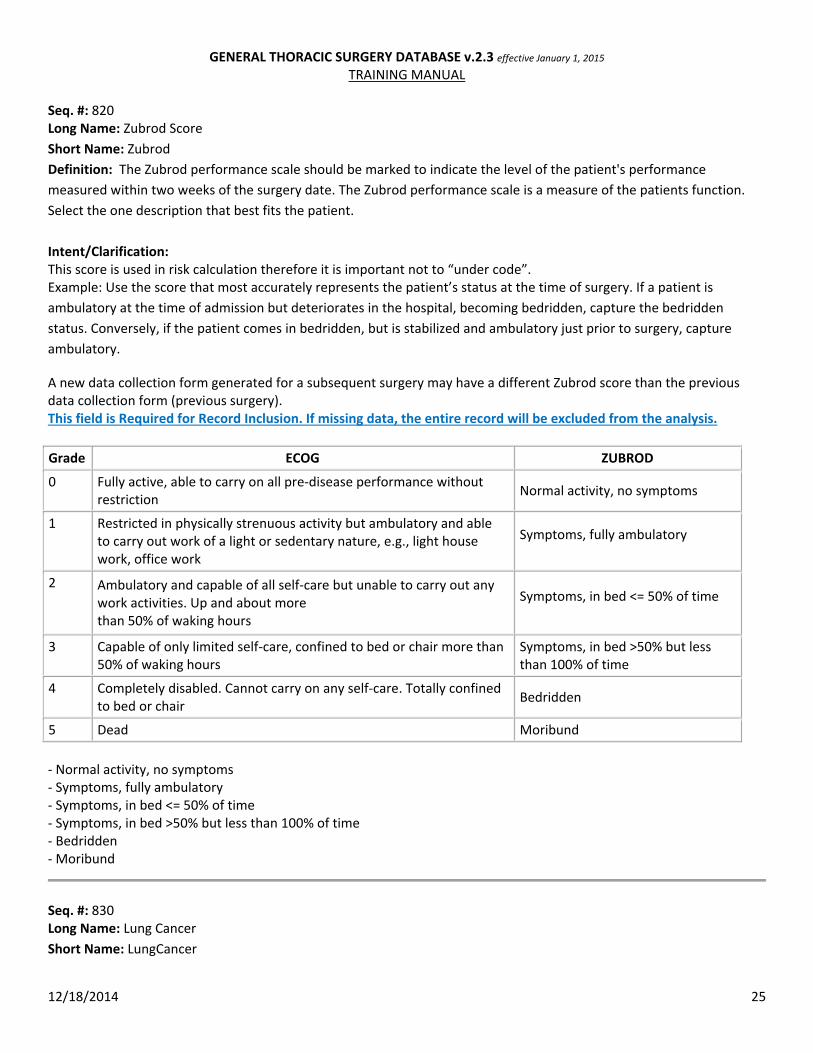
Seq. #: 820 Long Name: Zubrod Score
Short Name: Zubrod
Definition: The Zubrod performance scale should be marked to indicate the level of the patient's performance
measured within two weeks of the surgery date. The Zubrod performance scale is a measure of the patients function.
Select the one description that best fits the patient.
Intent/Clarification: This score is used in risk calculation therefore it is important not to “under code”. Example: Use the score that most accurately represents the patient’s status at the time of surgery. If a patient is
ambulatory at the time of admission but deteriorates in the hospital, becoming bedridden, capture the bedridden
status. Conversely, if the patient comes in bedridden, but is stabilized and ambulatory just prior to surgery, capture
ambulatory.
A new data collection form generated for a subsequent surgery may have a different Zubrod score than the previous data collection form (previous surgery). This field is Required for Record Inclusion. If missing data, the entire record will be excluded from the analysis.
Grade ECOG ZUBROD
0 Fully active, able to carry on all pre‐disease performance without restriction
Normal activity, no symptoms
1 Restricted in physically strenuous activity but ambulatory and able to carry out work of a light or sedentary nature, e.g., light house work, office work
Symptoms, fully ambulatory
2 Ambulatory and capable of all self‐care but unable to carry out any work activities. Up and about more than 50% of waking hours
Symptoms, in bed <= 50% of time
3 Capable of only limited self‐care, confined to bed or chair more than 50% of waking hours
Symptoms, in bed >50% but less than 100% of time
4 Completely disabled. Cannot carry on any self‐care. Totally confined to bed or chair
Bedridden
5 Dead Moribund
‐ Normal activity, no symptoms ‐ Symptoms, fully ambulatory ‐ Symptoms, in bed <= 50% of time ‐ Symptoms, in bed >50% but less than 100% of time ‐ Bedridden ‐ Moribund
Seq. #: 830 Long Name: Lung Cancer
Short Name: LungCancer
GENERAL THORACIC SURGERY DATABASE v.2.3 effective January 1, 2015 TRAINING MANUAL
12/18/2014 26
Definition: Indicate whether a major lung resection was performed for lung cancer (e.g. wedge, segment, lobe,
pneumonectomy), open or VATS.
If yes complete clinical and pathological staging.
Intent/Clarification: If Lung Cancer documented, and resection performed, complete both: Clinical Staging (ClinStageLungT, ClinStageLungN, and ClinStageLungM) AND Pathological Staging (PathStageLungT, PathStageLungN, and PathStageLungM) This field is Required for Record Inclusion. If missing data, the entire record will be excluded from the analysis. Seq. #: 840 Long Name: Clinical Staging Performed For Lung Cancer
Short Name: ClinStagDoneLung
Definition: Indicate whether clinical staging was performed on this patient related to this procedure.
Intent/Clarification: Clinical staging is based on evidence gathered before primary treatment. Diagnostic and/or radiologic tests are performed to determine the type and extent of the cancer and used to guide treatment decisions. Example: Patient had a VATS wedge for lung cancer in October last year. Now with recurrence and is having a completion
lobectomy. Can I use the Clinical Staging Methods that were done prior to the first surgery, or must they be after the
first surgery and up to the present procedure? Ideally, staging should be repeated. Bottom line, if the surgery is for
curative (therapeutic) intent, then staging needs to be done. If it is just to document a metastasis (diagnostic), then
would not provide clinical staging.
Seq. #: 841 Long Name: Preoperative Positive Tissue Diagnosis Obtained
Short Name: PreopPosTisOb
Definition: Indicate whether a positive tissue diagnosis was obtained prior to this operation.
Intent/Clarification:
Seq. #: 850 Long Name: Clinical Staging Method ‐ Lung ‐ Bronchoscopy
Short Name: ClinStagLungBronc
Definition: Was bronchoscopy used for clinical staging?
Intent/Clarification: Bronchoscopy is a procedure in which a cylindrical fiberoptic scope is inserted into the airways. This scope allows the visual examination of the lower airways. During a bronchoscopy, a physician can visually examine the lower airways,
GENERAL THORACIC SURGERY DATABASE v.2.3 effective January 1, 2015 TRAINING MANUAL
12/18/2014 27
including the larynx, trachea, bronchi, and bronchioles. The procedure is used to examine the mucosal surface of the airways for abnormalities that might be associated with a variety of lung diseases. Its use includes the visualization of airway obstructions such as a tumor, or the collection of specimens for the diagnosis of cancer originating in the bronchi of the lungs (bronchogenic cancer). It can also be used to collect specimens for culture to diagnose infectious diseases such as tuberculosis. The type of specimens collected can include sputum (composed of saliva and discharges from the respiratory passages), tissue samples from the bronchi or bronchioles, or cells collected from washing the lining of the bronchi or bronchioles. The instrument used in bronchoscopy, a bronchoscope, is a slender cylindrical instrument containing a light and an eyepiece or, more commonly, a direct video attachment. There are two types of bronchoscopes, a rigid bronchoscope is a metal tube that is use to visualize the airway. It has a larger lumen and larger instruments can be passed through it in addition to being able to ventilate the patient. A flexible bronchoscope is generally a smaller, flexible, fiber optic tube that has a smaller working port but is also easier to place into the airway.
Seq. #: 860 Long Name: Clinical Staging Method ‐ Lung ‐ EBUS
Short Name: ClinStagLungEBUS
Definition: Was Endobronchial Ultrasound used for clinical staging?
Intent/Clarification: EBUS is an invasive procedure in which physicians use ultrasound devices on the end of a special bronchoscope or placed
through a bronchoscope to examine the airways and the lung for exploration of the structures of airway walls, the
surrounding mediastinum, and the lungs. It is commonly used to biopsy lymph nodes outside the airway wall.
Seq. #: 870 Long Name: Clinical Staging Method ‐ Lung ‐ EUS
Short Name: ClinStagLungEUS
Definition: Was Endoscopic Ultrasound used for clinical staging?
Intent/Clarification: EUS is a procedure that combines endoscopy and ultrasound to obtain images and information about the digestive tract and the surrounding tissue and organs. In EUS a small ultrasound transducer is installed on the tip of the endoscope placed into the esophagus (not the airway) allowing the transducer to get closer to internal organs. This generally permits more accurate and detailed images of those organs than ones obtained by traditional ultrasound done from the surface of the body.
Seq. #: 880 Long Name: Clinical Staging Method ‐ Lung ‐ Mediastinoscopy/Chamberlain
Short Name: ClinStagLungMedia
Definition: Was Mediastinoscopy or Chamberlain procedure used for clinical staging?
Intent/Clarification: Mediastinoscopy is a procedure that enables visualization of the contents of the mediastinum, usually for the purpose of
obtaining a biopsy. Mediastinoscopy is often used for staging of lymph nodes of lung cancer or for diagnosing other
conditions affecting structures in the mediastinum such as sarcoidosis or lymphoma. Mediastinoscopy involves making
an incision approximately 1 cm above the suprasternal notch of the sternum, or breast bone. Dissection is carried out
GENERAL THORACIC SURGERY DATABASE v.2.3 effective January 1, 2015 TRAINING MANUAL
12/18/2014 28
down to the pretracheal space and down to the carina. A scope (mediastinoscope) is then advanced into the created
tunnel which provides a view of the mediastinum. The scope may provide direct visualization or may be attached to a
video monitor. The Chamberlain procedure is used to biopsy lymph nodes in the center of the chest, or to biopsy a mass
in the center of the chest. The Chamberlain procedure differs from a cervical mediastinoscopy by the location of the
incision, and the location of the lymph nodes or mass to be biopsied. The Chamberlain procedure is used to biopsy
lymph nodes or masses in the aorto‐pulmonary window on the left side of the chest, or nodes in the hilar areas of the
lung. (In contrast, the cervical mediastinoscopy procedure is used to biopsy nodes or masses to the front or side of
thetrachea, or windpipe.) The aorto‐pulmonary window is the area in the center of the chest bound by the aorta
superiorly, and the pulmonary artery inferiorly. This area contains lymph nodes that filter lymph coming from the left
lung, especially the left upper lobe. If a lung cancer is present in the left lung, the Chamberlain procedure is useful for
staging the cancer (determining the extent of spread.) The hilar areas of the lung (the hilum) are the areas of the lung
where the pulmonary artery and vein (the blood supply) join the lung.
Seq. #: 890 Long Name: Clinical Staging Method ‐ Lung ‐ PET or PET/CT
Short Name: ClinStagLungPET
Definition: Was PET scan or PET/CT used for clinical staging?
Intent/Clarification:
Positron emission tomography, also called PET imaging or a PET scan, is a type of nuclear medicine imaging. Nuclear medicine or radionuclide imaging procedures are noninvasive and, with the exception of intravenous injections, are usually painless medical tests that help diagnose medical conditions. These imaging scans use radioactive materials called radiopharmaceuticals or radiotracers.
Seq. #: 900 Long Name: Clinical Staging Method ‐ Lung ‐ CT
Short Name: ClinStagLungCT
Definition: Was CT scan used for clinical staging?
Intent/Clarification:
Computed tomography (CT) scan, also called computerized axial tomography (CAT) scan, is used to create cross‐sectional images of structures in the body. In this procedure, x‐rays are taken from many different angles and processed through a computer to produce a three‐dimensional (3‐D) image called a tomogram.
Seq. #: 910 Long Name: Clinical Staging Method ‐ Lung ‐ VATS
Short Name: ClinStagLungVATS
Definition: Was a Video Assisted Thoracoscopic procedure used for clinical staging?
Intent/Clarification:
Video‐assisted thoracoscopic surgery (VATS) is a minimally invasive surgical technique used to diagnose and treat
problems in the chest. During this surgery, a tiny camera (thoracoscope) and surgical instruments are inserted in the
GENERAL THORACIC SURGERY DATABASE v.2.3 effective January 1, 2015 TRAINING MANUAL
12/18/2014 29
chest through small incisions. The thoracoscope transmits images of the inside of the chest onto a video monitor,
guiding the surgeon performing the procedure. Video‐assisted thoracoscopic surgery (VATS) can be used for many
purposes, ranging from a biopsy to removal of tumors or entire lobes from the lung.
Seq. #: 920 Long Name: Clinical Staging Method ‐ Lung ‐ Laparoscopy
Short Name: ClinStagLungLap
Definition: Was a laparoscopy used for clinical staging?
Intent/Clarification:
Laparoscopy is a minimally invasive procedure used as a diagnostic tool and surgical procedure that is performed to examine the abdominal and pelvic organs. Tissue samples can also be collected for biopsy using laparoscopy and malignancies treated when it is combined with other therapies.
Seq. #: 921 Long Name: Clinical Staging Method ‐ Lung – Brain MRI
Short Name: ClinStagLungMRI
Definition: Was a brain MRI used for clinical staging?
Intent/Clarification:
Seq. #: 922 Long Name: Clinical Staging Method ‐ Lung – Brain Scan
Short Name: ClinStagLungBrain
Definition: Was a brain scan used for clinical staging?
Intent/Clarification:
CT scan of the brain with contrast and MRI of the brain are acceptable means of staging the brain. A CT scan of the head without contrast is not useful for staging the brain.
Seq. #: 923 Long Name: Clinical Staging Method ‐ Lung – Needle Biopsy
Short Name: ClinStagLungNeedle
Definition: Was a needle biopsy done for clinical staging?
Intent/Clarification:
FINA – fine need aspiration
Seq. #: 929 Long Name: Clinical Staging Method ‐ Lung – Other
GENERAL THORACIC SURGERY DATABASE v.2.3 effective January 1, 2015 TRAINING MANUAL
12/18/2014 30
Short Name: ClinStagLungOth
Definition: Indicate if method/technology other than those listed was used for clinical staging.
Intent/Clarification:
Indicate if any other method/technology was used for clinical staging.
Seq. #: 930 Long Name: Lung CA Tumor size – T
Short Name: ClinStagLungT
Definition: Choose the largest dimension of a solitary tumor. If more than one tumor is present, choose the from below.
Intent/Clarification:
Question:
How are small nodules reported on lung CT addressed for staging? If there is no biopsy, the PET CT is negative, nodules
are < 5 mm and the surgeon/oncologist chooses not to address these, do not consider them when staging. 40% of
people over the age of 50 have small lung nodules which are not cancerous.
‐For solitary masses: Tumor <= 2cm is T1a ‐ Tumor >2cm, <= 3cm is T1b ‐ Tumor > 3cm, <= 5 cm is T2a ‐ Tumor > 5 cm, <=7 cm is T2b ‐ Tumor > 7 cm is T3 ‐ Unknown Tx ‐ primary tumor cannot be assessed For multiple tumors: Separate tumor nodule in the same lobe is T3 Separate tumor nodule in a different lobe on the same side T4 Separate tumor nodule on the opposite side M1a Scenarios: A 5.5 cm tumor in the right lower lobe with a separate 1 cm tumor in the same lobe is what T stage? T3 A solitary 5.5 cm tumor in the right lower lobe is what T stage? T2b A 5.5 cm tumor in the right lower lobe with a separate 1 cm tumor in the right middle lobe is what T stage? T4 A 5.5 cm tumor in the right lower lobe with a separate 1 cm tumor in the left upper lobe is what T stage? T2b (This patient would be a T2bNxM1a which would make them a stage IV)
Seq. #: 940 Long Name: Lung Cancer‐ Invasion of Adjacent Structures
Short Name: LCInvAdjStr
Definition: Does clinical evaluation (imaging, endoscopy, etc . . .) indicate that the tumor invades adjacent structure(s)?
Intent/Clarification:
Based on preop testing, indicate whether the tumor appears to invade adjacent structures.
GENERAL THORACIC SURGERY DATABASE v.2.3 effective January 1, 2015 TRAINING MANUAL
12/18/2014 31
Seq. #: 950 Long Name: Clinical Staging Lung Tumor Invasive Pleura
Short Name: ClinStageLungTInvPl
Definition: Does imaging indicate tumor invasion of the pleura?
Intent/Clarification:
This refers to visceral pleura only. If the tumor invades the parietal pleura, code as invading the chest wall (next field). This is very difficult to diagnose prior to surgery.
Seq. #: 960 Long Name: Clinical Staging Lung Tumor Invasive Chest Wall
Short Name: ClinStageLungTInvCW
Definition: Does imaging or physical exam indicate tumor invasion of the chest wall?
Intent/Clarification:
Code tumors that invade the parietal pleura as invading the chest wall.
Seq. #: 970 Long Name: Clinical Staging Lung Tumor Invasive Diaphragm
Short Name: ClinStageLungTInvDia
Definition: Does imaging indicate tumor invasion of the diaphragm?
Intent/Clarification:
Seq. #: 980 Long Name: Clinical Staging Lung Tumor Invasive Phrenic Nerve
Short Name: ClinStageLungTInvPN
Definition: Does imaging indicate tumor invasion of the phrenic nerve?
Intent/Clarification:
Phrenic nerve invasion can be determined by a paralyzed diaphragm which appears elevated on an imaging study.
This may be documented with a fluoroscopy study (Sniff test) demonstrating lack of diaphragm movement when a
person is breathing.
Seq. #: 990 Long Name: Clinical Staging Lung Tumor Invasive Pericardium
Short Name: ClinStageLungTInvPer
Definition: Does imaging indicate tumor invasion of the pericardium?
GENERAL THORACIC SURGERY DATABASE v.2.3 effective January 1, 2015 TRAINING MANUAL
12/18/2014 32
Intent/Clarification:
Seq. #: 1000 Long Name: Clinical Staging Lung Tumor Invasive Main Bronchus
Short Name: ClinStageLungTInvMB
Definition: Does imaging or bronchoscopy indicate tumor invasion of the main bronchus?
Intent/Clarification:
Seq. #: 1010 Long Name: Clinical Staging Lung Tumor Obstructive
Short Name: ClinStageLungTInvOb
Definition: Does imaging indicate that the tumor is associated with atelectasis or obstructive pneumonitis of the entire
lung?
Intent/Clarification:
Seq. #: 1020 Long Name: Clinical Staging Lung Tumor Invasive Nodule(s)
Short Name: ClinStageLungTInvNod
Definition: Does imaging indicate separate tumor nodule(s) in the same lobe?
Intent/Clarification:
Seq. #: 1030 Long Name: Clinical Staging Lung Tumor Invasive Invasive Mediastinum
Short Name: ClinStageLungTInvMed
Definition: Does imaging indicate lung tumor invasion in mediastinum?
Intent/Clarification:
Seq. #: 1040 Long Name: Clinical Staging Lung Tumor Invasive Heart
Short Name: ClinStageLungTInvHt
Definition: Does imaging indicate lung tumor invasion into heart?
GENERAL THORACIC SURGERY DATABASE v.2.3 effective January 1, 2015 TRAINING MANUAL
12/18/2014 33
Intent/Clarification:
Seq. #: 1050 Long Name: Clinical Staging Lung Tumor Invasion Great Vessels
Short Name: ClinStageLungTInvGrVes
Definition: Does imaging indicate lung tumor invasion into the great vessels?
Intent/Clarification:
Seq. #: 1060 Long Name: Clinical Staging Lung Tumor Invasion Trachea
Short Name: ClinStageLungTInvTr
Definition: Does imaging or bronchoscopy indicate lung tumor invasion into the trachea?
Intent/Clarification:
Seq. #: 1070 Long Name: Clinical Staging Lung Tumor Invasive Recurrent Laryngeal Nerve
Short Name: ClinStageLungTInvRLN
Definition: Does imaging or clinical assessment indicate lung tumor invasion into the recurrent laryngeal nerve?
Intent/Clarification: Recurrent laryngeal nerve invasion leads to paralysis of a vocal cord and therefore hoarseness.
The diagnosis is generally made by direct visualization of cord function (laryngoscopy), often performed by an ENT.
Seq. #: 1080 Long Name: Clinical Staging Lung Tumor Invasive Esophagus
Short Name: ClinStageLungTInvEo
Definition: Does imaging or endoscopy indicate lung tumor invasion into the esophagus?
Intent/Clarification:
Seq. #: 1090 Long Name: Clinical Staging Lung Tumor Invasive Vertebral Body
Short Name: ClinStageLungTInvVB
Definition: Does imaging indicate lung tumor invasion into a vertebral body?
GENERAL THORACIC SURGERY DATABASE v.2.3 effective January 1, 2015 TRAINING MANUAL
12/18/2014 34
Intent/Clarification:
Seq. #: 1100 Long Name: Clinical Staging Lung Tumor Invasive Carina
Short Name: ClinStageLungTInvC
Definition: Does imaging or bronchoscopy indicate lung tumor invasion into the carina?
Intent/Clarification:
Seq. #: 1110 Long Name: Clinical Staging Lung Tumor Invasive Nodule(s) Diff Lobe
Short Name: ClinStageLungTInvNDL
Definition: Does imaging indicate lung tumor nodule(s) in a different ipsilateral lobe?
Intent/Clarification:
Seq. #: 1120 Long Name: Lung Cancer Nodes ‐ N
Short Name: ClinStageLungN
Definition: Indicate the appropriate descriptor for the lung cancer nodal metastases. All nodes > 1cm on CT or PET/CT
are considered positive. All PET positive nodes are considered positive. Results of previous invasive staging (EBUS,
Mediastinoscopy) should be included here.
Clinical staging is based on the PRE‐TREATMENT ESTIMATED staging workup which may include CT scan, PET scan,
endoscopic ultrasound, etc.
Intent/Clarification:
Code nodal involvement (if any.) Ipsilateral = same side as tumor, contralateral= opposite side ‐ N0 = No regional lymph node metastasis ‐ N1 = Metastasis in ipsilateral peribronchial or hilar and intrapulmonary nodes. Includes direct extension. ‐ N2 = Metastasis in ipsilateral mediastinal and/or subcarinal lymph nodes ‐ N3 = Metastasis in contralateral mediastinal or contralateral hilar nodes, ipsilateral or contralateral scalene or supraclavicular nodes (Lymph nodes may be reported by station #. Generally speaking ipsilateral (same side as tumor) lymph node with double digit numbers are N1 lymph nodes, ipsilateral lymph nodes with single digits are N2 lymph nodes, contralateral (opposite side as tumor) lymph nodes of any number are considered N3)
GENERAL THORACIC SURGERY DATABASE v.2.3 effective January 1, 2015 TRAINING MANUAL
12/18/2014 35
Seq. #: 1130 Long Name: Lung Cancer Metastasis ‐ M
Short Name: ClinStageLungM
Definition: Indicate the appropriate descriptor for the lung cancer distant metastases.
Clinical staging is based on the PRE‐TREATMENT ESTIMATED staging workup which may include CT scan, PET scan,
endoscopic ultrasound, etc.
Intent/Clarification:
Metastasis or metastatic disease (sometimes abbreviated mets), is the spread of cancer from one organ to another non‐adjacent organ or part.
‐ M0 = No distant metastasis ‐ M1 = Distant Metastasis
Seq. #: 1140 Long Name: Esophageal Cancer
Short Name: EsophCancer
Definition: Indicate whether an esophagectomy was performed for esophageal cancer.
If yes complete clinical and pathological staging.
Intent/Clarification:
If Esophageal Cancer documented, “Yes”, and resection performed, complete both Clinical Staging (ClinStageEsophT, ClinStageEsophN, ClinStageEsophM, ClinStageEsophH, and ClinStageEsophG) AND Pathological Staging (PathStageEsophT, PathStageEsophN, PathStageEsophM, PathStageEsophH, and PathStageEsophG). Guidelines (measurements) for coding esophageal tumor location ‐ Use these measurements in cm from incisors: Upper
third = 17‐25 cm
Middle third = 26‐34 cm Lower third = 35‐42 cm This field is Required for Record Inclusion. If missing data, the entire record will be excluded from the analysis.
Seq. #: 1150 Long Name: Clinical Staging Performed For Esophageal Cancer
Short Name: ClinStagDoneEsoph
Definition: Indicate whether clinical staging was performed on this patient related to this procedure.
If yes complete clinical and pathological staging.
GENERAL THORACIC SURGERY DATABASE v.2.3 effective January 1, 2015 TRAINING MANUAL
12/18/2014 36
Intent/Clarification:
Clinical staging is the Pre‐Treatment estimate of cancer. Indicate whether clinical staging was performed and if so choose the method(s).
Seq. #: 1160 Long Name: Clinical Staging Method ‐ Esophageal ‐ Bronchoscopy
Short Name: ClinStagEsophBronc
Definition: Was bronchoscopy used for clinical staging?
Intent/Clarification: Bronchoscopy is a procedure in which a cylindrical fiberoptic scope is inserted into the airways. This scope contains
allows the visual examination of the lower airways. During a bronchoscopy, a physician can visually examine the lower
airways, including the larynx, trachea, bronchi, and bronchioles. The procedure is used to examine the mucosal surface
of the airways for abnormalities that might be associated with a variety of lung diseases. Its use includes the
visualization of airway obstructions such as a tumor, or the collection of specimens for the diagnosis of cancer
originating in the bronchi of the lungs (bronchogenic cancer). It can also be used to collect specimens for culture to
diagnose infectious diseases such as tuberculosis. The type of specimens collected can include sputum (composed of
saliva and discharges from the respiratory passages), tissue samples from the bronchi or bronchioles, or cells collected
from washing the lining of the bronchi or bronchioles. The instrument used in bronchoscopy, a bronchoscope, is a
slender cylindrical instrument containing a light and an eyepiece. There are two types of bronchoscopes, a rigid tube
that is sometimes referred to as an open‐tube or ventilating bronchoscope, and a more flexible fiber optic tube. This
tube contains four smaller passages—two for light to pass through, one for seeing through and one that can
accommodate medical instruments that may be used for biopsy or suctioning, or that medication can be passed
through.
Seq. #: 1170 Long Name: Clinical Staging Method ‐ Esophageal ‐ EBUS
Short Name: ClinStagEsophEBUS
Definition: Was Endobronchial Ultrasound used for clinical staging?
Intent/Clarification: EBUS is an invasive procedure in which physicians use ultrasound devices inside the airways and the lung for exploration of the structures of airway walls, the surrounding mediastinum, and the lungs.
Seq. #: 1180 Long Name: Clinical Staging Method ‐ Esophageal ‐ EUS
Short Name: ClinStagEsophEUS
Definition: Was Endoscopic Ultrasound used for clinical staging?
Intent/Clarification: A procedure that combines endoscopy and ultrasound to obtain images and information about the digestive tract and the surrounding tissue and organs. In EUS a small ultrasound transducer is installed on the tip of the endoscope
GENERAL THORACIC SURGERY DATABASE v.2.3 effective January 1, 2015 TRAINING MANUAL
12/18/2014 37
introduced into the esophagus to permitting the transducer to get closer to the organs inside the body so the resultant ultrasound images are often more accurate and detailed than ones obtained by traditional ultrasound.
Seq. #: 1190 Long Name: Clinical Staging Method ‐ Esophageal ‐ Mediastinoscopy/Chamberlain
Short Name: ClinStagEsophMedia
Definition: Was Mediastinoscopy or Chamberlain procedure used for clinical staging?
Intent/Clarification: Mediastinoscopy is a procedure that enables visualization of the contents of the mediastinum, usually for the purpose of obtaining a biopsy. Mediastinoscopy is often used for staging of lymph nodes or for diagnosing other conditions affecting structures in the mediastinum such as sarcoidosis or lymphoma. Mediastinoscopy involves making an incision approximately 1 cm above the suprasternal notch of the sternum, or breast bone. Dissection is carried out down to the pretracheal space and down to the carina. A scope (mediastinoscope) is then advanced into the created tunnel which provides a view of the mediastinum. The scope may provide direct visualization or may be attached to a video monitor. The Chamberlain procedure is used to biopsy lymph nodes in the center of the chest, or to biopsy a mass in the center of
the chest. The Chamberlain procedure differs from a cervical mediastinoscopy by the location of the incision, and the
location of the lymph nodes or mass to be biopsied. The Chamberlain procedure is used to biopsy lymph nodes or
masses in the aorto‐pulmonary window on the left side of the chest, or nodes in the hilar areas of the lung. (In contrast,
the cervical mediastinoscopy procedure is used to biopsy nodes or masses to the front or side of the trachea, or
windpipe.) The aorto‐pulmonary window is the area in the center of the chest bound by the aorta superiorly, and the
pulmonary artery inferiorly.
Seq. #: 1200 Long Name: Clinical Staging Method ‐ Esophageal ‐ PET or PET/CT
Short Name: ClinStagEsophPET
Definition: Was PET scan or PET/CT used for clinical staging?
Intent/Clarification:
Positron emission tomography, also called PET imaging or a PET scan, is a type of nuclear medicine imaging. Nuclear medicine or radionuclide imaging procedures are noninvasive and, with the exception of intravenous injections, are usually painless medical tests that help diagnose medical conditions. These imaging scans use radioactive materials called radiopharmaceuticals or radiotracers.
Seq. #: 1210 Long Name: Clinical Staging Method ‐ Esophageal ‐ CT
Short Name: ClinStagEsophCT
Definition: Was CT scan used for clinical staging?
Intent/Clarification:
GENERAL THORACIC SURGERY DATABASE v.2.3 effective January 1, 2015 TRAINING MANUAL
12/18/2014 38
Computed tomography (CT) scan, also called computerized axial tomography (CAT) scan, is used to create cross‐sectional images of structures in the body. In this procedure, x‐rays are taken from many different angles and processed through a computer to produce a three‐dimensional (3‐D) image called a tomogram.
Seq. #: 1220 Long Name: Clinical Staging Method ‐ Esophageal ‐ VATS
Short Name: ClinStagEsophVATS
Definition: Was a Video Assisted Thoracoscopic procedure used for clinical staging?
Intent/Clarification:
Video‐assisted thoracoscopic surgery (VATS) is a minimally invasive surgical technique used to diagnose and treat
problems in the chest. During this surgery, a tiny camera (thoracoscope) and surgical instruments are inserted in the
chest through small incisions. The thoracoscope transmits images of the inside of the chest onto a video monitor,
guiding the surgeon performing the procedure. Video‐assisted thoracoscopic surgery (VATS) can be used for many
purposes, ranging from a biopsy to removal of tumors.
Seq. #: 1230 Long Name: Clinical Staging Method ‐ Esophageal ‐ EGD
Short Name: ClinStagEsophEGD
Definition: Was Esophagogastroduodenoscopy used for clinical staging?
Intent/Clarification:
Esophagogastroduodenoscopy (EGD) is an examination of the lining of the esophagus, stomach, and upper duodenum with a small camera (flexible endoscope) which is inserted down the throat.
Seq. #: 1240 Long Name: Clinical Staging Method ‐ Lung ‐ Laparoscopy
Short Name: ClinStagEsophLap
Definition: Was a laparoscopy used for clinical staging?
Intent/Clarification:
Laparoscopy is a minimally invasive procedure used as a diagnostic tool and surgical procedure that is performed to examine the abdominal and pelvic organs. Tissue samples can also be collected for biopsy using laparoscopy and malignancies treated when it is combined with other therapies.
Seq. #: 1241 Long Name: Clinical Staging Method ‐ Endoscopic Mucosal Resection
Short Name: ClinStagEsophEMR
Definition: Was an endoscopic mucosal resection used for clinical staging?
Intent/Clarification:
GENERAL THORACIC SURGERY DATABASE v.2.3 effective January 1, 2015 TRAINING MANUAL
12/18/2014 39
Seq. #: 1245 Long Name: Clinical Staging Method ‐ Esophageal – Other
Short Name: ClinStagEsophOth
Definition: Indicate if method/technology other than those listed was used for clinical staging.
Intent/Clarification:
Indicate if any other method/technology was used for clinical staging.
Seq. #: 1250 Long Name: Esophageal Cancer Tumor ‐ T
Short Name: ClinStagEsophT
Definition: Record T status based on EUS report. If EUS not done, estimate T based on CT or PET/CT. No esophageal
thickening = T1. If esophageal thickening is present, use T2. If CT or PET/CT indicated invasion of adjacent structures, use
T4.
Intent/Clarification:
Record T based on EUS if done, if not done estimate T based on CT or PET/CT. No esophageal thickening = T1. Choose T2 if esophageal thickening is present. If thickening noted on CT scan, code as T2. If stricture is noted on endoscopy or barium swallow or the patient is experiencing dysphagia, code as T3.
‐ T0 = No evidence of primary tumor ‐ Tis = High grade dysplasia ‐ T1 = Tumor invades lamina propria, mucosa or submucosa ‐ T2 = Tumor invades muscularis propria ‐ T3 = Tumor invades adventitia ‐ T4 = Tumor invades adjacent structures
Seq. #: 1251 Long Name: Clinical Diagnosis of Nodal Involvement
Short Name: ClinStageEsophNode
Definition: Indicate whether there was a clinical diagnosis of N1, N2 or N3 nodal involvement.
Intent/Clarification:
Indicate nodal status. Nodes > 1cm on CT or PET/CT or EUS are considered positive. All positive PET nodes are considered positive. Count biopsy positive nodes. Choose Nx if nodes cannot be assessed.
Seq. #: 1270 Long Name: Esophageal Cancer Metastasis ‐ M
Short Name: ClinStageEsophM
GENERAL THORACIC SURGERY DATABASE v.2.3 effective January 1, 2015 TRAINING MANUAL
12/18/2014 40
Definition: Indicate the appropriate descriptor for the esophageal cancer distant metastasis.
Clinical staging is based on the PRE‐TREATMENT ESTIMATED staging workup which may include CT scan, PET scan,
endoscopic ultrasound, etc.
Intent/Clarification:
Metastasis or metastatic disease (sometimes abbreviated mets), is the spread of cancer from one organ to another non‐adjacent organ or part. ‐ M0 = No Distant Metastasis ‐ M1 = Distant Metastasis
Seq. #: 1300 Long Name: Category Of Disease ‐ Primary
Short Name: CategoryPrim
Definition: Indicate the PRIMARY diagnosis (category of disease) for which the procedure was performed.
For the majority of cases, there will be only one condition treated (i.e., lung cancer treated by lobectomy and lymph
node dissection). Rarely, there will be cases where two unrelated conditions are treated at one time (i.e., a thymoma
and a lung cancer). In these rare cases, indicate the primary or most important diagnosis in this "Category of Disease ‐
Primary" field, followed by the secondary or lesser diagnosis treated in the "Category of Disease ‐ Secondary". For
example, in the case of lung cancer with incidental thymoma, the primary category of disease = lung cancer, and the
secondary category of disease = thymoma.
Intent/Clarification:
Choose the primary diagnosis or reason for the procedure. Input should be based upon the final pathology report. If you
entered a Category of Disease before final path, then you need to change it based on the final pathology.
Example:
If you start with a diagnosis of “abnormal radiological finding”, a wedge resection is done and cancer is found, the
diagnosis should be changed to cancer based upon the pathology report.
This field is Required for Record Inclusion. If missing data, the entire record will be excluded from the analysis.
Trachea
Dysphagia, unspecified (787.2, R13.10): Dysphagia is difficulty swallowing. It may be caused by esophageal disorders, neurologic pathology or neuromuscular disorders. Tracheomalacia‐congenital (748.3, Q32.0): Refers to a condition in newborns whose tracheal cartilage lacks its usual rigid structure. This leads to airway obstruction during expiration and infants will present with difficulty breathing and inability to clear secretions. Tracheomalacia‐acquired (519.1, J39.8): Condition in which the normal rigid tracheal cartilage becomes soft and flaccid. This may be due to changes in the airway as a result prolonged endotracheal intubation.
GENERAL THORACIC SURGERY DATABASE v.2.3 effective January 1, 2015 TRAINING MANUAL
12/18/2014 41
Tracheostenosis‐congenital (748.3, Q32.1): A process present in newborns in which the normal tracheal, and sometimes bronchial, airway diameter is significantly compromised. The amount of airway involvement can vary from case to case. Newborns or infants can present with stridor or difficulty in breathing or feeding. Tracheostenosis‐acquired postintubation (519.1, J39.8): Refers to narrowing of the normal tracheal diameter often by scar tissue formed from prolonged endotracheal intubation. Afflicted patients typically present with shortness of breath and stridor. Tracheostomy‐hemorrhage (519.09, J95.01): Describes excessive bleeding as a result of a tracheostomy tube. This may be due to granulation tissue within the airway or may represent the presence of a communication between the trachea and innominate artery or tracheoinnominate fistula. Tracheostomy related stenosis (519.02, J95.03): Refers to the process when the trachea is narrowed at the location of a healed tracheostomy stoma. Tracheal tumor, malignant (162.0, C33): Describes conditions where either primary cancer develops within the trachea or where cancers from the lung or thyroid gland grow into the trachea. Primary malignant tracheal tumors are often either squamous cell cancers or adenoid cystic carcinomas. Other malignant tumors of the trachea include sarcomas and mucoepidermoid carcinomas. Tracheal tumor, benign (212.2, D14.2): These are lesions that originate from the trachea itself and are not considered cancers. Chondromas, leiomyomas, and adenomas are some of the types of benign tracheal tumors. Tracheal tumor, metastatic (197.3, C78.30): A process when cancers of distant sites can occasionally spread to the trachea and lead to airway obstruction or bleeding. Renal cell carcinomas, breast cancers, and melanomas can metastasize to the airway.
Larynx
Subglottic stenosis‐congenital (748.3, Q31.1): This refers to a condition of narrowing of the subglottic larynx in the absence of an identifiable cause such as prior endotracheal intubation. Subglottic stenosis‐acquire (post intubation) (478.74, J38.6): Patients who have been intubated with either an oral endotracheal tube or a tracheostomy appliance can develop narrowing of their subglottic larynx due to airway irritation and scarring. Airway narrowing may lead to stridor and cause patients to suffer from shortness of breath. Vocal cord paralysis unspecified (478.3, J38.00): This refers to a complication of thoracic surgery where the patient’s vocal mechanism is impaired due to trauma to the nerve supply to the larynx. Paralysis of a vocal cord may lead to voice changes and may predispose a patient to experience aspiration events. Vocal cord paralysis, unilateral (478.31, J38.01): One of the two vocal cords is immobile or has extremely limited movement. This often impacts speech and swallowing. Vocal cord paralysis, bilateral (478.33, J38.02): Both vocal cords are immobile, often stuck partially open. This impacts speech and can lead to difficulty swallowing and aspiration.
Lung

GENERAL THORACIC SURGERY DATABASE v.2.3 effective January 1, 2015 TRAINING MANUAL
12/18/2014 42
Lung tumor, metastatic (197.0, C78.00): This condition includes all cancers of the body that spread to the lungs. A primary lung cancer may metastasize to a different lobe of the lung and be considered a metastatic lung tumor. Lung tumor, benign (212.3, D14.30): These are masses within lung tissue that are not malignant. They can grow, but rarely cause symptoms. Benign lung tumors include hamartomas, chondromas, and fibromas. Lung cancer, main bronchus, carina (162.2, C34.00): This is a condition where a centrally‐located lung cancer becomes locally advanced and involves either the right/left main bronchus or carina. Surgical resection involves removing involved airway and lung and may require removing a portion of the central airway as well. Lung cancer, upper lobe (162.3, C34.10): This refers to a primary lung cancer, usually non‐small cell or small cell carcinoma, that is located within either the right or left upper lobes. Lung cancer, middle lobe (162.4, C34.2): This refers to a primary lung cancer located within the right middle lobe of the lung. Lung cancer, lower lobe (162.5, C34.30): This refers to a primary lung cancer located within either the right or left lower lobe of the lung. Lung cancer, location unspecified (162.9, C34.90): This code should be used when the exact origin of the primary lung cancer cannot be exactly determined due to large size or when the location was not specifically documented by the surgeon. Lung abscess (513.0, J85.2): Represents an infectious condition of the lung when a collection of infected material develops within the substance of the lung. Pneumothorax (512.8, J93.1): This is a process that occurs when the lining of the lung parenchyma is disrupted and air leaks into the pleural space (the space between the lung and rib cage). This leads to varying degrees of lung collapse and subsequent symptomatology. In its most severe form, this can lead to acute respiratory failure. Bronchiectasis (494.0, J47.9): Refers to a localized, irreversible dilation of the bronchial tree. Patients can present to their physicians with recurrent respiratory infections and significant airway bleeding as a result. Empyema with fistula (510.0, J86.0): This describes an infectious process within the pleural space with evidence of a communication between the bronchial tree within the lung and the pleural space. Treatment involves appropriate antibiotics with drainage of the pleural infection and correction of the bronchopleural fistula. Empyema without fistula (510.9, J86.9): This describes an infectious process within the pleural space without evidence of a communication between the bronchial tree within the lung and the pleural space. Pleural infection is usually due to pneumonia within the lung tissue. Treatment involves appropriate antibiotics with drainage of the pleural infection. Emphysema (492.8, J43.8): Is a form of chronic obstructive pulmonary disease (COPD) characterized by loss of elasticity of the lung tissue. This results in air‐trapping and over distended lung tissue leading to shortness of breath and impaired gas exchange. Emphysematous bleb (492.0, J43.9): This refers to a collection of air within the lung tissue due to rupture of the alveolar space. These can be either single or multiple and can enlarge to the point of significantly compressing normal lung tissue resulting in shortness of breath.
GENERAL THORACIC SURGERY DATABASE v.2.3 effective January 1, 2015 TRAINING MANUAL
12/18/2014 43
Interstitial lung disease/fibrosis (516.3, J84.1): Refers to a number of conditions that lead to the progressive scarring of
lung tissue. This scarring results in significant respiratory dysfunction and in its most severe form can lead to respiratory
failure. In general, the scarring is irreversible.
Pneumonia (486, J18.9): Is a condition in which a portion of the lung is involved with an active infection. These can be due to bacterial, viral, or fungal organisms. Treatment is aimed at identifying the causative etiology and initiating appropriate antimicrobial therapy. Pulmonary insufficiency following surgery/trauma (ARDS) (518.5, J95.82): This refers to a diffuse inflammatory process that typically involves all lung tissue. This condition can lead to severe impairment of gas exchange within the lung despite mechanical ventilation. Hemothorax (511.8, J94.2): Is defined as the presence of blood within the pleural space. This may be due to a traumatic event with damage to the chest wall or lung. Treatment may require drainage with a chest tube or surgical intervention to address the bleeding source. Lung nodule, benign (not a tumor, e.g., granuloma, subpleural lymph node, pulmonary infarct) (518.89): This is used to classify lesions within the substance of the lung that do not contain malignant cells. Included in this category are hamartomas, non‐necrotizing granulomatous processes such as sarcoidosis and atypical infections that result in necrotizing granulomatous lesions. Acute respiratory failure (518.81, J96.00): Acute respiratory failure is a new onset of pulmonary dysfunction resulting in inadequate ventilation and gas exchange. Causes may include airway obstruction, damaged lung tissue, decreased respiratory drive or failure of the muscles that control breathing. Aspergillosis (117.3, B44.9): This is a fungal infection caused by aspergillus, a common mold. It can be seen in persons with compromised immune function. Cystic fibrosis with pulmonary manifestations (277.02, E84.0): CF is a life threatening genetic disease leading to production of thick, tenacious mucous resulting in frequent pulmonary congestion and infections. It also impacts digestive enzymes and function. Carcinoid tumor of the bronchus and lung; malignant, atypical (209.21, C7A.090): "Atypical carcinoid" ‐ angrier looking cells with more mitoses, more cellular irregularity, much higher incidence of lymph node metastases and higher incidence of M1 involvement with a lower 5 year survival than typical carcinoid. Carcinoid tumor of bronchus and lung; benign, typical (209.61., D34.090): "Typical carcinoid" ‐ regular appearing cells, low number of mitoses, slow growing, rarely involves lymph nodes and only rarely metastasizing with an 80‐90% cure rate following excision but still officially "malignant" Gangrene and necrosis of lung (513.0, J85.0): Death of lung tissue due to loss of blood supply. Primary causes include: pneumonia, pulmonary embolism, neoplasm (tumor). Secondary causes include: trauma, surgery disrupting blood supply, lobar torsion, septic emboli, systemic infection, lung toxicity of chemotherapeutic agents, radiation effect, and foreign body aspiration. Treatment and prognosis depend on the etiology and extent of lung damage. Solitary pulmonary nodule (not a tumor, e.g., granuloma, subpleural lymph node, pulmonary infarct) (793.11, R91.1): A solitary pulmonary nodule is defined as a discrete, well‐marginated, rounded opacity less than or equal to 3 cm in diameter that is completely surrounded by lung parenchyma, does not touch the hilum or mediastinum, and is not
GENERAL THORACIC SURGERY DATABASE v.2.3 effective January 1, 2015 TRAINING MANUAL
12/18/2014 44
associated with adenopathy, atelectasis, or pleural effusion. Lesions larger than 3 cm are considered masses and are have a higher risk of malignancy. Malignant neoplasm other parts of bronchus or lung (162.8, C34.8): Malignant (cancerous) tumor in a location not otherwise listed. Neoplasm of uncertain behavior of trachea, bronchus and lung (235.7, DM38.1): Lesion in trachea, bronchus or lung without a definitive diagnosis. Personal history of malignant neoplasm of bronchus and lung (V10.11, Z85.118): History of cancer of the bronchus and lung. Post inflammatory pulmonary fibrosis (515, J84.89): Post inflammatory pulmonary fibrosis is a condition in which the tissues in the lungs thicken or become scarred. The lung tissues also become rigid, which makes breathing difficult. As post inflammatory pulmonary fibrosis advances, lung tissue becomes more damaged and shortness of breath worsens. Post inflammatory pulmonary fibrosis typically occurs after some sort of infection that causes serious damage to the lung tissues. There is no cure for post inflammatory pulmonary fibrosis, but medications like corticosteroid drugs may be helpful in managing inflammation and swelling. Damage to the lungs caused by post inflammatory pulmonary fibrosis is permanent, and those with significant damage may need a lung transplant. Primary pulmonary hypertension (416.0, I 27.0): Primary pulmonary hypertension (PPH) is a rare disease characterized by elevated pulmonary artery pressure with no apparent cause. PPH is also termed precapillary pulmonary hypertension or, as is currently preferred, idiopathic pulmonary arterial hypertension (IPAH). Untreated IPAH leads to right‐sided heart failure and death. Pulmonary sequestration (748.5, Q33.2): Pulmonary sequestration (also called accessory lung) refers to aberrant formation of segmental lung tissue that has no connection with the bronchial tree or pulmonary arteries. It is a bronchopulmonary foregut malformation (BPFM). Transplanted lung complication(s) (996.84, T86.8XX): Some complications are related to the operation itself, others are a result of immunosuppressive medication, which is needed to prevent rejection. Complications may include bleeding, rejection, bronchiolitis obliterans syndrome, post‐transplantation lymphoproliferative disorder, infection, side effects of long term use of immunosuppressants.
Mediastinum
Mediastinitis (519.2, J98.5): Refers to either acute or chronic inflammation of the mediastinum. Acute mediastinitis is usually due to a bacterial infection from a perforation of the esophagus or due to sternal wound infections after cardiac surgery procedures. Treatment often requires antibiotics and surgical drainage. Chronic mediastinitis represents a fibrosis of the mediastinum and can be a result of radiation therapy or due to previous infection with histoplasmosis or tuberculosis. Mediastinal nodes, metastatic (196.1, C77.1): Refers to a process where cancers within the chest, or from other locations, spread to the lymph nodes within the mediastinum. These lymph nodes can be biopsied at mediastinoscopy.
GENERAL THORACIC SURGERY DATABASE v.2.3 effective January 1, 2015 TRAINING MANUAL
12/18/2014 45
Mediastinal nodes, benign (229.0, D36.0): Describes a condition where mediastinal lymph nodes demonstrate a benign or non‐malignant process such as sarcoidosis or anthrocosis. These conditions may result in the enlargement of the involved lymph nodes. Anterior mediastinal tumor, primary (germ cell cancer, seminoma) (164.2, C38.1): Refers to tumors of the mediastinum which are classified as either seminomas or nonseminomatous germ cell tumors of the mediastinum. These tumors often cause symptoms due to their size and resulting compression of heart, lung, or airway. Treatment often involves a combination of chemotherapy, surgery, and radiation therapy. Anterior mediastinal tumor‐metastatic (197.1, C78.1): Cancers from other locations can occasionally spread to the anterior mediastinum. These can originate from the lung, esophagus, breast, or other location and spread to the mediastinum via the lymphatic system. Anterior mediastinal tumor‐benign (e.g., teratoma) (212.5, D15.2): A teratoma is often a benign tumor which can be located within the anterior mediastinum. This tumor consists of normal types of cells, but in an abnormal configuration and location. They can produce symptoms from their large size and are treated with surgical resection. Anterior mediastinal tumor‐thymus tumor (thymoma, thymic carcinoma) (164.0, C37): The thymus gland is located
within the anterior mediastinum and serves a role in the development of the immune system. The thymus usually
disappears during childhood, but can be the source of particular tumors. These can range from less aggressive
thymomas to very malignant thymic carcinomas. Surgical resection is the procedure of choice for these abnormalities if
feasible.
Lymphoma, intrathoracic (202.82, C85.92): Lymphomas are a type of cancer that arises from cells of the immune system or lymphocytes. Thoracic surgeons are often involved in obtaining tissue via mediastinoscopy to assist medical oncologists in making the diagnosis of lymphoma. The treatment of these conditions centers on the use of chemotherapy. Posterior mediastinal malignant tumor‐primary (164.3, C38.2): These are malignant tumors located in the posterior third of the mediastinum between the posterior pericardium and spine. Malignant tumors in this location are rare and predominantly malignant neurogenic tumors. Posterior mediastinal tumor‐metastatic (197.1, C78.1): These are unusual occurrences where cancers from other locations can metastasize to the posterior mediastinum. Posterior mediastinal tumor‐benign (i.e., neurogenic tumor) (212.5, D15.2): These are masses that arise from peripheral nerves or the sympathetic ganglia. These typically are slow‐growing lesions that are asymptomatic. Schwannomas and neurofibromas are the usual tumor types. Myasthenia gravis (358.0, G70.00): This is a neuromuscular disease caused by antibodies generated in one’s own body. These antibodies lead to muscle weakness, fatigue, and occasionally respiratory failure. This condition is associated with thymomas and patients may gain significant symptom improvement with resection of a thymoma or even a normal thymus gland. Mediastinal cyst, bronchogenic (519.3, J98.5): Is the most common mediastinal cyst. These are thin walled cavities lined with respiratory epithelium and can cause symptoms due to their size or become infected. Surgical resection may involve removal of the cyst alone or may require concomitant lung resection.
GENERAL THORACIC SURGERY DATABASE v.2.3 effective January 1, 2015 TRAINING MANUAL
12/18/2014 46
Mediastinal cyst, foregut duplication (519.3, J98.5): These are benign cyst originating from and attached to the intrathoracic esophagus. These may be asymptomatic or associated with dysphagia due to compression of the adjacent esophagus. Removal requires simple resection of the cyst. Mediastinal cyst, pericardial (519.3, J98.5): These are unusual cysts arising from the pericardium. Treatment, when necessary, may involve CT‐guided needle aspiration and recurrences are treated with simple cyst excision. Mediastinal cyst, thymic (519.3, J98.5): This describes cystic lesions within the thymus gland. They can be associated with thymomas and rarely cause symptoms. Benign neoplasm of thymus (212.6, D15.0): Benign tumors of the thymic gland are relatively rare. Although most of these lesions are asymptomatic in nature, they may result in respiratory distress. Mediastinal abscess (513.1, J85.3): An infection manifested by a collection of pus in the mediastinal space. Neoplasm of uncertain behavior of pleura, thymus, mediastinum (235.8, D38.2‐D38.4): Growth of the pleura, thymus or mediastinum without a definitive diagnosis. Unspecified disease of thymus gland (254.9, E32.9): Disease of the thymus gland not otherwise listed.
Thyroid
Goiter, nodular (241.9, E04.9): This describes a condition of an enlarged thyroid gland which may be due to dietary deficiencies in iodine or autoimmune inflammation. Symptoms may occur due to excessive thyroid enlargement which can result in tracheal and esophageal compression. Thyroid neoplasm, malignant (193.0, C73): This condition refers to cancers that arise within the thyroid gland. Occasionally these cancers can enlarge and invade the underlying trachea which can result in airway obstruction or bleeding. Thyroid neoplasm, benign (226.0, D34): An overwhelming majority of nodules that arise within the thyroid gland are benign tumors. Fine needle aspiration of thyroid nodules can often distinguish whether they are benign or malignant.
Pleura
Pleural effusion, sterile (511.9, J90): This is a condition where fluid accumulates in the space between the lung and chest wall. This type of fluid is not due to cancer in the pleura nor is it infected. Pleural effusion, infected (empyema) (511.1, J86.9): Empyema describes a situation where infected fluid is present in the pleural space. This condition usually chest tube or surgical drainage for successful treatment. Pleural effusion, malignant (197.2, C78.2): Cancers from the chest or from elsewhere can spread to the pleural lining of the chest wall. This often, in turn, results in the production of excessive fluid within the pleural space. Patients may present complaining of chest pain and difficulty breathing. Treatment may involve sclerosis of the pleural space.
GENERAL THORACIC SURGERY DATABASE v.2.3 effective January 1, 2015 TRAINING MANUAL
12/18/2014 47
Pleural tumor, malignant (e.g., mesothelioma) (163.9, C38.4): Malignant mesothelioma is an aggressive type of cancer that originates from cells that line the pleural space. Asbestos exposure is a known risk factor for the development of this malignancy. Chemotherapy, surgery, and radiation therapy are often employed in the treatment of this disease. Pleural tumor, metastatic (197.2, C78.2): Cancers of the lung, breast, ovary, and kidney can spread to the pleura lining the chest wall and present as a pleural nodule or tumor. Pleural tumor, benign (212.4, D19.0): Rarely, a benign tumor of the pleura can develop. These are typically classified as benign fibrous tumors of the pleura and have no known association with asbestos exposure. They are usually discovered as incidental lesions on a chest x‐ray or CT scan. Treatment involves simple surgical excision. Pleural thickening (511.0, J94.9): This describes a nonspecific finding on a chest x‐ray or CT scan. Pleural thickening may be due to pleural plaques or calcified lesions which are frequently seen in patients with asbestos exposure. Pleural effusion, other specified, except TB (511.89, J90): Pleural effusion is excess fluid that accumulates in the pleural cavity, the fluid‐filled space that surrounds the lungs. Code effusions other than infection, malignant, sterile or those caused by tuberculosis here. These may include those caused by autoimmune diseases or medications. Malignant neoplasm other specified sites of pleura (163.8, C38.4): Malignant neoplasm (cancerous tumor) of contiguous or overlapping sites of pleura whose point of origin cannot be determined
Chest Wall
Pectus excavatum (754.81, Q67.6): Represents the most common congenital abnormality of the chest wall. Atypical rib and cartilage growth leads to the caved‐in or concave appearance of the anterior chest. Some degree of cardiopulmonary impairment may be present in severe cases. Pectus carinatum (754.82, Q67.7): Another congenital chest wall abnormality in which abnormal rib and cartilage growth leads to protrusion abnormalities of the anterior chest. No certain cardiopulmonary abnormalities are known to be caused by this deformity. Heart valve abnormalities have been found to be associated with this condition. Sternal tumor, malignant (170.3, C41.3): A variety of primary malignant tumors of the sternum have been described. A
majority of these are of the soft tissue sarcoma origin and many are thought to be related to previous external bean
radiation therapy. Treatment often consists of radical resection of the sternum with complex reconstructive
requirements.
Sternal tumor, metastatic (198.5, C79.51): This refers to the development of cancers within the sternum that are tumors that have originated from other locations in the body. Surgical resection for metastatic disease to the sternum is rare, but can be considered in well‐selected instances. Sternal tumor, benign (213.3, D16.7): Benign tumors of the sternum are quite unusual. Osteochondromas are the most common type of benign sternal tumor. Rib tumor, malignant (e.g., osteosarcoma, chondrosarcoma) (170.3, C41.3): Primary cancers of the chest wall can originate from the ribs. Chondrosarcoma is the most common primary malignant tumor of the chest wall. These cancers typically require extensive chest wall resection with complex reconstructive techniques. Malignant rib tumors can spread to other sites within the body.
GENERAL THORACIC SURGERY DATABASE v.2.3 effective January 1, 2015 TRAINING MANUAL
12/18/2014 48
Rib tumor, metastatic (198.5, C79.51): Cancers from distant sites can spread to bone and the ribs are a frequent site of bony metastases. Occasionally, rib resection is performed to determine the nature of a metastatic rib tumor. When symptomatic, metastatic rib tumors frequently cause pain at their location. Rib tumor, benign (e.g., fibrous dysplasia) (213.3, D16.7): It is often difficult to distinguish benign from malignant rib tumors without removing the mass and examining its cellular characteristics. Several benign rib tumors exist and include chondromas, osteomas, and fibrous dysplasia to name a few. Thoracic outlet syndrome (353.0, G54.0): This refers to a constellation of physical signs and symptoms related to compression of the brachial plexus and subclavian artery and vein. This can be caused by abnormalities of the first rib, clavicle, and musculature surrounding the brachial plexus and subclavian vessels as they travel out from the chest to supply the arm. Surgical intervention may be necessary to relieve the anatomic compression and improve symptoms.
Diaphragm
Diaphragmatic paralysis (519.4, J98.6): Each diaphragm is innervated by its respective phrenic nerve. Diaphragmatic paralysis can occur when there is injury to a phrenic nerve during a surgical procedure or may be related to a viral illness. Patients that suffer from high spinal cord injuries may be ventilator dependent as the innervation of both phrenic nerves becomes compromised by their spinal injury. Diaphragm tumor, malignant (171.4, C49.3): Primary malignant tumors of the diaphragm are quite rare. Occasionally, liver cancers can invade the diaphragm and require partial resection of the diaphragm during liver surgery. Diaphragm tumor, metastatic (198.89, C79.89): Cancers from other sites can spread to the chest and involve the pleura
as described. When this occurs, diaphragmatic involvement is usually encountered.
Diaphragm tumor, benign (215.4, D21.3): These are extremely rare tumors, but can include the same types of benign tumors seen elsewhere in the body. One type of benign diaphragmatic tumor is a lipoma. Diaphragmatic hernia with obstruction, without gangrene (552.3, K44.0): A diaphragmatic hernia is a defect or hole in the diaphragm that allows the abdominal contents to move into the chest cavity, in this case leading to gastrointestinal obstruction without development of gangrene. Diaphragmatic hernia with gangrene (551.3, K44.1): A diaphragmatic hernia is a defect or hole in the diaphragm that allows the abdominal contents to move into the chest cavity, in this case leading to ischemia of tissue and development of gangrene. Diaphragmatic hernia without obstruction or gangrene (553.3, K44.9): A diaphragmatic hernia is a defect or hole in the diaphragm that allows the abdominal contents to move into the chest cavity, in this case without gastrointestinal obstruction or development of gangrene.
Esophagus
Esophageal cancer, lower third (150.5, C15.5): This is the most common location of esophageal cancers in the United States and its incidence is steadily increasing. Lesions here are typically adenocarcinomas and are often treated by a combination of surgery, chemotherapy, and radiation therapy.
GENERAL THORACIC SURGERY DATABASE v.2.3 effective January 1, 2015 TRAINING MANUAL
12/18/2014 49
Esophagus cancer, middle third (150.4, C15.4): Refers to carcinomas arising in the mid‐thoracic esophagus. These are usually squamous cell carcinomas. Esophagus cancer, upper third (150.3, C15.3): These carcinomas are located within the esophagus located within the lower neck and upper chest. Esophageal cancer, esophagogastric junction (cardia) (151.0, C16.0): Describes cancers that are located with the junction between the esophagus and stomach and involve a portion of the cardia or upper part of the stomach. Esophageal tumor, benign (i.e., leiomyoma) (211.0, D13.0): This includes a variety of tumors that can exist within the esophagus, but no not spread to adjacent lymph nodes or other parts of the body. Patients can present with difficulty in swallowing. Surgical resection of the tumor alone often results in significant symptomatic improvement. Esophageal stricture (530.3, K22.2): Refers to a process in which the lumen of the esophagus is narrowed by a non‐malignant condition. This may result from a caustic substance that was ingested or from a hiatal hernia. Endoscopic dilation may improve symptoms of obstruction, but surgery is sometimes necessary. Barrett’s esophagus (530.85, K22.70): Is a condition where the normal lining of esophagus is altered due to the presence of reflux of acid from the stomach. Barrett’s esophagitis increases the risk of developing esophageal adenocarcinoma. Various medical, endoscopic, and surgical approaches have been utilized to treat this condition. Achalasia of the esophagus (530.0, K22.0): Describes a motility disorder of the esophagus that results in progressive difficulty in swallowing. The exact cause of achalasia is not known in most cases. Surgery aimed at dividing the outer muscular layer of the esophagus is usually very effective in addressing this problem. Esophageal perforation (530.4, K22.3): Refers to a violation in the wall of the esophagus. This disruption leads to contamination of the mediastinum and often pleural space and can be fatal if not addressed properly. Perforation may be due to an esophageal cancer or secondary to an endoscopic procedure. Zenker’s diverticulum (530.6, K22.5): Describes an out pouching of the esophagus within the neck that occurs as a result of an abnormally functioning upper esophageal sphincter. This out pouching can entrap ingested food and lead to difficulty swallowing and aspiration. Treatment is directed at correction of the overactive muscle. Epiphrenic diverticulum (530.6, K22.5): This refers to an esophageal out pouching that develops in a location just above the level of the diaphragm. This usually occurs due to an overactive lower esophageal sphincter. Patients can experience difficulty in swallowing and the regurgitation of undigested food. Gastroesophageal reflux (GERD) (530.81, K21.9): Is defined by the presence of symptoms or changes within the lining of the esophagus due to abnormal reflux of stomach contents. Symptoms include heartburn, chest pain, and difficulty swallowing. Tracheoesophageal fistula (530.84, J86.0): Refers to an abnormal communication between the esophagus and airway. This can be a congenital lesion that is diagnosed shortly after birth. In adults, this abnormality is frequently due to esophageal cancer that locally invades the trachea. Lung contamination from the esophageal contents results in infectious complications. Gastric outlet obstruction, pyloric stenosis, acquired (537.0, K31.1): This condition describes an abnormality within the outlet of the stomach to the small bowel. The cause of this condition is unknown. Obstruction of the stomach can result
GENERAL THORACIC SURGERY DATABASE v.2.3 effective January 1, 2015 TRAINING MANUAL
12/18/2014 50
in excessive emesis and malnutrition. Pyloric obstruction can be seen after esophagectomy due to interruption of neural input to the stomach and pylorus. Endoscopic dilatation of the pylorus is often effective in dealing with this problem. Acquired absence of esophagus (post esophagectomy) (V45.79, Z90.89): There are instances in which a patient will undergo an emergent esophagectomy without immediate reconstruction. Patients who are extremely ill due to esophageal perforation with prolonged thoracic contamination may need to return to the operating room at a later date to have continuity of their gastrointestinal tract restored. This diagnostic code describes such a patient. Barrett’s esophagus with High Grade Dysplasia (530.85, K22.711): High grade dysplasia (HGD) refers to precancerous changes in the cells of the esophagus. Gastroesophageal reflux disease (GERD) can be complicated by Barrett’s esophagus (BE), a change in the normal esophageal cells to intestinal‐like cells. BE cells can become abnormal or dysplastic. HGD significantly increases a person’s risk for esophageal adenocarcinoma and in most cases will progress to cancer without any treatment. When someone is diagnosed with HGD, an intervention is advised including endoscopic resection, ablation or in some cases, esophagectomy is recommended for treatment. Dyskinesia/spasm of esophagus (530.5, K22.4): This is a hypermotility disorder of the esophagus that is characterized by spastic non‐peristaltic responses to swallowing; chest pain; and dysphagia. It may include disorders affecting the motor function of the upper esophageal sphincter; lower esophageal sphincter; the esophagus body, or a combination of these parts. The failure of the sphincters to maintain a tonic pressure may result in gastric reflux of food and acid into the esophagus (gastroesophageal reflux). Other disorders include hypermotility (spastic disorders) and markedly increased amplitude in contraction (nutcracker esophagus). Esophagitis (530.1, K20.9): Esophagitis is a term used to describe inflammation, irritation or swelling of the esophagus. There are several types of esophagitis depending on the cause. Esophagitis can be caused by infection, irritation of the esophagus, or inflammation of the lining of the esophagus. Foreign body esophagus (935.1, T18.108a): An esophageal foreign body is any object that does not belong in the esophagus that becomes lodged. Malignant neoplasm stomach unspecified (151.9, C16.9): Cancerous tumor of the stomach, location and type not specified Malignant neoplasm of the esophagus, unspecified (150.9, C15.9): Cancerous tumor of the esophagus, location and type not specified Malignant other part esophagus, specified (150.8, C15.8): Cancer in part(s) of the esophagus not otherwise listed Mallory Weiss tear (530.7, K22.6): Mallory‐Weiss syndrome is characterized by upper gastrointestinal bleeding secondary to longitudinal mucosal lacerations (known as Mallory‐Weiss tears) at the gastroesophageal junction or gastric cardia. This may result from persistent retching and vomiting or after any event that provokes a sudden rise in intragastric pressure or gastric prolapse into the esophagus. Reflux esophagitis (530.11, K21.0): Reflux esophagitis is an esophageal mucosal inflammation that occurs secondary to retrograde flux of gastric contents into the esophagus. Clinically, this is referred to as gastroesophageal reflux disease (GERD). Typically, the reflux disease involves the distal 8‐10 cm of the esophagus and the gastroesophageal junction. The disease is patchy in distribution. Stricture and stenosis of esophagus (530.3, K22.2): Esophageal stricture or stenosis is narrowing or tightening of the internal diameter of the esophagus resulting in swallowing difficulties.
GENERAL THORACIC SURGERY DATABASE v.2.3 effective January 1, 2015 TRAINING MANUAL
12/18/2014 51
Ulcer esophagus with bleeding (530.21, K22.11): An esophageal ulcer is a defect in the lining of the esophagus. Esophageal ulcers can be caused by: GERD (gastroesophageal reflux disease), infection of the esophagus, irritants that damage the esophagus, excessive vomiting, chemotherapy or radiation. Bleeding may be acute or chronic. Ulcer esophagus without bleeding (530.2, K22.10): An esophageal ulcer is an open sore in the lining of the esophagus. Esophageal ulcers can be caused by: GERD (gastroesophageal reflux disease), infection of the esophagus, irritants that damage the esophagus, excessive vomiting, chemotherapy or radiation. Other digestive system complication (997.49, K91.XX): Any adverse event involving the digestive system not otherwise listed. Other disease of the esophagus (530.89, K22.8): Other disease or condition of the esophagus not listed.
Trauma
Rib fracture (807.0, S22.39xa): Injury to the chest wall may result in rib fractures. Alone, these injuries are usually self‐limited. However, rib fractures can cause a pneumothorax or hemothorax. Sternal fracture (807.2, S22.20xa): These can be caused by blunt trauma to the chest and may herald more serious injuries. If significantly displaced, surgical fixation may be necessary. Flail chest (807.4, S22.5xxa): Describes a condition when a segment of ribs becomes separated from the rest of the chest wall as a result of multiple rib fractures. Patients often experience respiratory compromise as a result of impaired breathing mechanics. Tracheal injury (807.5, S12.8xxa): This life‐threatening injury may be due to blunt or penetrating trauma to the neck or chest. Airway obstruction can result as a consequence. Surgical intervention is often required to address the airway injury. Traumatic pneumothorax (860.0, S27.0xxa): Collapse of a lung may occur as a result of either blunt or penetrating trauma to the chest. Chest tube placement is frequently needed to drain the pleural space. Rib fractures, multiple (807.0, S22.49xa): Fractures involving more than one rib, typically caused by trauma
Cardiovascular
Pericarditis with effusion (420.9, I30.9): Inflammation of the pericardium may lead to accumulation of fluid within the pericardial sac. This fluid may cause cardiac dysfunction and require a percutaneous drainage procedure or creation of a pericardial window. Pericardial effusion, malignant (198.89, C79.89): This occurs when malignant cancers spread to the lining of the pericardium and result in the buildup of fluid with the pericardial sac. SVC syndrome (459.2, I87.1): The superior vena cava (SVC) can be compressed by tumors of the mediastinum, lung cancers, or mediastinal lymphadenopathy. Obstruction of the venous drainage of the arms, upper chest, and head often
GENERAL THORACIC SURGERY DATABASE v.2.3 effective January 1, 2015 TRAINING MANUAL
12/18/2014 52
leads to severe swelling and engorged superficial veins. Therapy is aimed at restoring blood flow through this obstruction. Abdominal aneurysm without rupture (441.4, I171.4): Dilatation, expansion or bulging of the abdominal aorta without leakage of blood into a false lumen or outside the vessel wall Cardiac tamponade (423.3, I31.4): Collection of blood or fluid in the pericardial space which compresses the chamber walls of the heart preventing normal filling. This impairs cardiac output and requires immediate intervention. Pericarditis, constrictive (432.2, I31.1): Constrictive pericarditis is long‐term (chronic) inflammation of the sac‐like covering of the heart (the pericardium) with thickening, scarring, and muscle tightening (contracture) leading to disruption of cardiac function. Unspecified disease of the pericardium (423.9, I31.9): Pericardial condition or disease not otherwise listed.
Miscellaneous
Hyperhidrosis, focal, axilla (705.21, L74.510): Hyperhidrosis is a condition characterized by excessive sweat production. It may involve the hands, axillae, or feet. Disruption of the sympathetic chain via thoracoscopic techniques has proven to be a potentially therapeutic solution to this problem. Hyperhidrosis, focal, face (705.21, L74.511): Hyperhidrosis is a condition characterized by excessive sweat production. It may involve the hands, axillae, or feet. Disruption of the sympathetic chain via thoracoscopic techniques has proven to be a potentially therapeutic solution to this problem. Hyperhidrosis, focal, palms (705.21, L74.512): Hyperhidrosis is a condition characterized by excessive sweat production. It may involve the hands, axillae, or feet. Disruption of the sympathetic chain via thoracoscopic techniques has proven to be a potentially therapeutic solution to this problem. Lymphadenopathy (785.6, R59.9): This refers to enlargement of a lymph node or group of lymph nodes and may be due to benign processes or the presence of metastatic cancer. Abnormal radiologic finding (793.1, R91): This is a generalized explanation to describe atypical imaging results reported by a radiologist. Abnormal radiologic findings may initiate diagnostic procedures to determine the exact nature of the lesion identified. Chronic airway obstruction not elsewhere classified (496, J44.9): Includes COPD and chronic nonspecific lung disease Chylothorax (457.8, 189.8): Chylothorax refers to the presence of lymphatic fluid in the pleural space secondary to leakage from the thoracic duct or one of its main tributaries. Disruption of internal operation, surgical wound (998.31, T81.32XA): Disruption or dehiscence of closure of: fascia, superficial or muscular, internal organ, muscle or muscle flap, ribs or rib cage, sternum or sternotomy, deep disruption or dehiscence of operation. Do not assign this code when the surgeon purposely leaves the wound open. Hemorrhage complicating a procedure (998.11, multiple codes): Bleeding related to the surgical procedure. Do not assign hemorrhage as a complication of a procedure when the blood loss is from the disease itself, such as bleeding esophageal varices or angiodysplasia.
GENERAL THORACIC SURGERY DATABASE v.2.3 effective January 1, 2015 TRAINING MANUAL
12/18/2014 53
Hematoma complicating a procedure (998.12, multiple codes): A hematoma is a localized collection of blood outside the blood vessels, usually in liquid form within the tissue in this case resulting from a surgical procedure. The lay term is a bruise. Hemoptysis unspecified (786.3, R04.2): Hemoptysis is the coughing up of blood or bloody sputum from the lungs or airway. It may be either self‐limiting or recurrent. Hemoptysis can be caused by a range of disorders: infections (pneumonia; tuberculosis; aspergillosis; and parasitic diseases), tumors that erode blood vessel walls, cocaine abuse, trauma, vascular disorders, bronchitis, foreign bodies in airway, coagulopathies, or as a result of invasive procedures. Other non‐infectious disorders of lymphatic channels (457.8, I89.8): Condition of lymphatic system not related to infection or otherwise listed Malignant neoplasm of connective tissue and other soft tissue of the thorax (171.4, C49.3): Cancerous tumor of connective tissue, cartilage, fascia, fat, muscle of the thorax, excluding breast neoplasms Malignant poorly differentiated neuroendocrine carcinoma, any site (209.3, C74.1): Neuroendocrine tumors are a heterogeneous group of solid tumors that originate from neuroendocrine cells found throughout the body. The World Health Organization (WHO) classifies neuroendocrine tumors according to the malignant potential of these tumors into well‐differentiated neuroendocrine tumors (also referred to as grade 1 and 2), and poorly‐differentiated neuroendocrine tumor carcinoma. Poorly differentiated (grade 3) neuroendocrine tumors are extremely aggressive with poor prognosis. Non‐healing surgical wound (998.83, T81.89XA): A non‐healing or chronic wound is defined as a wound that does not improve after four weeks or does not heal in eight weeks. Other post‐ op infection (998.59, T81.4XXA): Infection acquired following surgery not otherwise listed Persistent post‐op fistula not otherwise classified (998.6, T81.83XA): A fistula is an abnormal connection between two epithelialized surfaces. Fistulas are usually caused by injury or surgery, but they can also result from an infection or inflammation Post‐operative air leak (512.2, J95.812): A post‐operative air leak may follow lung surgery and involves air escaping into the pleural space. This usually resolves with chest tube therapy. A prolonged air leak is an air leak that lasts beyond postoperative day 5. Secondary malignant neoplasm of other specified sites (198.89, C79.89): A cancerous tumor in a site or organ separate from the primary tumor, does not include lymph node metastasis Shortness of breath (786.05, R06.02): Shortness of breath (dyspnea) is a subjective experience of breathing discomfort that consists of qualitatively distinct sensations that vary in intensity. Distinct sensations include effort/work, chest tightness, and air hunger (the feeling of not enough oxygen). Dyspnea is a normal symptom of heavy exertion but becomes pathological if it occurs in unexpected situations. It may result from asthma, pneumonia, cardiac ischemia, interstitial lung disease, congestive heart failure, chronic obstructive pulmonary disease, or psychogenic causes such as panic disorder and anxiety. Swelling, mass or lump in chest (786.6, R22.2): Abnormal lesion which may or may not be cancerous in the chest, does not include breast masses Other unlisted category of disease: Diagnosis not in any of the listed categories
GENERAL THORACIC SURGERY DATABASE v.2.3 effective January 1, 2015 TRAINING MANUAL
12/18/2014 54
Seq. #: 1310 Long Name: Category Of Disease ‐ Primary ‐ Other Specify
Short Name: CategoryPrimOth
Definition: Indicate the PRIMARY diagnosis (category of disease) for which the procedure was performed.
Choose from the list when possible, if the category of disease is not listed, enter free text.
Intent/Clarification:
Capture unlisted primary diagnosis here after carefully reviewing choices above.
Seq. #: 1311 Long Name: Category Of Disease ‐ Primary ‐ Other ICD
Short Name: CategoryPrimOthICD
Definition: Enter ICD‐9 or ICD‐10 code, if known, of other primary diagnosis (category of disease) not listed.
Intent/Clarification:
The intent is to track category of disease codes for possible inclusion in next version and /or for internal analysis.
Seq. #: 1320 Long Name: Category Of Disease ‐ Secondary
Short Name: CategorySecond
Definition: Indicate the SECONDARY diagnosis (category of disease) for which the procedure was performed.
Intent/Clarification:
The Secondary diagnosis can be left blank. As long as a primary diagnosis is selected, the record will be accepted as complete without having a secondary indicated.
Seq. #: 1330 Long Name: Category Of Disease ‐ Secondary ‐ Other Specify
Short Name: CategorySecondOth
Definition: Indicate the SECONDARY diagnosis (category of disease) for which the procedure was performed if not
listed.
Intent/Clarification:
Capture unlisted secondary diagnosis here after carefully reviewing choices above.
Seq. #: 1331 Long Name: Category Of Disease ‐ Secondary ‐ Other ICD
Short Name: CategorySecondOthICD
Definition: Enter ICD‐9 or ICD‐10 code, if known, of secondary diagnosis (category of disease) not listed.
GENERAL THORACIC SURGERY DATABASE v.2.3 effective January 1, 2015 TRAINING MANUAL
12/18/2014 55
Intent/Clarification:
The intent is to track category of disease codes for possible inclusion in next version and /or for internal analysis.
Seq. #: 1340 Long Name: Date Of Surgery
Short Name: SurgDt
Definition: Indicate the date of surgery, which equals the date the patient enters the operating room.
Intent/Clarification:
This field is Required for Record Inclusion. If missing data, the entire record will be excluded from the analysis.
Seq. #: 1350 Long Name: OR Entry Time
Short Name: OREntryT
Definition: Indicate to the nearest minute (using 24 hour clock) the time the patient enters the operating room.
Intent/Clarification:
This field is Required for Record Inclusion. If missing data, the entire record will be excluded from the analysis.
Seq. #: 1360 Long Name: OR Exit Time
Short Name: ORExitT
Definition: Indicate to the nearest minute (using 24 hour clock) the time the patient exits the operating room.
Intent/Clarification:
Even if the thoracic surgeon was present for only part of the case, code the entire time.
This field is Required for Record Inclusion. If missing data, the entire record will be excluded from the analysis.
Seq. #: 1370 Long Name: Anesthesia Start Time
Short Name: AnesthStartT
Definition: Indicate the time of anesthesia induction.
Intent/Clarification:
This is the start of anesthetic management, placing lines, induction of anesthesia. This time should be recorded on the anesthesia record.
Seq. #: 1380 Long Name: Anesthesia End Time
Short Name: AnesthEndT
GENERAL THORACIC SURGERY DATABASE v.2.3 effective January 1, 2015 TRAINING MANUAL
12/18/2014 56
Definition: Indicate the anesthesia end time documented in the medical record. The definition of anesthesia end
time is when the anesthesiologist is no longer in personal attendance, that is, when the patient is
safely placed under post‐anesthesia supervision.
Intent/Clarification:
If the patient is extubated in the OR, indicate time of extubation otherwise use OR exit time as anesthesia end time.
Seq. #: 1390 Long Name: Procedure Start Time
Short Name: ProcStartT
Definition: Indicate the time the procedure started.
Intent/Clarification:
This field is Required for Record Inclusion. If missing data, the entire record will be excluded from the analysis.
Seq. #: 1400 Long Name: Procedure End Time
Short Name: ProcEndT
Definition: Indicate the time the procedure ended.
Intent/Clarification:
This field is Required for Record Inclusion. If missing data, the entire record will be excluded from the analysis.
Seq. #: 1410 Long Name: Multi‐Day Operation
Short Name: MultiDay
Definition: Indicate whether the operation continued through midnight from one day to the next.
Intent/Clarification:
These are cases that continue through midnight.
Seq. #: 1420 Long Name: Status
Short Name: Status
Definition: Indicate the status that best describes the clinical status of the patient at the time of the primary surgical
procedure.
1. Emergent: The surgical procedure must be performed within 24 hours of presentation.
2. Urgent: All of the following conditions are met:
a. Not elective status
b. Not emergent status.
c. Procedure required during same hospitalization in order to minimize chance of further clinical deterioration.
GENERAL THORACIC SURGERY DATABASE v.2.3 effective January 1, 2015 TRAINING MANUAL
12/18/2014 57
3. Elective: The patient has been stable in the days or weeks prior to the operation.
4. Palliative: procedure intended to provide comfort or relief of symptoms but not improve prognosis.
Intent/Clarification:
‐ Emergent status is coded for cases that require immediate intervention to prevent life threatening deterioration or death such as (but not limited to) esophageal perforation, severe hemorrhage or massive hemoptysis.
‐ Urgent status is coded for cases in which the operation must be performed before the patient can be discharged. Examples of urgent cases would include bronchopleural fistula, pneumothorax or decortication for empyema.
‐ Elective status is coded for cases that are performed during the same hospitalization for convenience would not be considered urgent. A medical patient with an incidental CXR finding who undergoes a diagnostic bronchoscopy or mediastinoscopy prior to discharge would have the procedure status coded as elective.
‐ Palliative – Treatment of malignant pleural effusions is often palliative and may include pleurodesis or placement of a chronic indwelling pleural drain (e.g. Pleurx catheter).
Seq. #: 1430 Long Name: Reoperation
Short Name: Reop
Definition: Indicate whether the patient ever had a previous surgical procedure in the same cavity.
Intent/Clarification:
If a patient is returned to the operating room from the post anesthesia care unit after the initial surgery, before they are
sent to a patient disposition location (ICU, Regular Floor Bed, etc.), is the second surgery considered a new operation,
thus requiring a separate STS data collection forms? For this purpose, PACU would = intermediate care. Yes, fill out a
2nd form. A patient who had a CABG 5 years ago and presents for lung resection is a redo surgery.
Seq. #: 1440 Long Name: Robotic Technology Assisted
Short Name: Robotic
Definition: Indicate whether the thoracic surgery was assisted by robotic technology.
Intent/Clarification:
Was robotic technology used for any part of the procedure?
Seq. #: 1441 Long Name: Unanticipated Surgical Approach Conversion
Short Name: UnanticConv
Definition: Indicate whether or not there was an unanticipated conversion of the surgical approach.
Intent/Clarification:
‐ VATS to open ‐ Robotic to VATS ‐ Robotic to open
GENERAL THORACIC SURGERY DATABASE v.2.3 effective January 1, 2015 TRAINING MANUAL
12/18/2014 58
‐ No Remember to capture both the original and converted procedure codes. Conversion in a procedure does not mean something always went wrong or was not appreciated preoperatively – many times it is done for better visibility, etc. This should not be viewed as a punitive data element.
Seq. #: 1442 Long Name: Unanticipated Surgical Approach Conversion Type
Short Name: UnanticConvTy
Definition: Indicate the type of surgical approach conversion.
Intent/Clarification:
‐ Elective ‐ Emergent
Seq. #: 1443 Long Name: Unanticipated Surgical Approach Conversion Reason
Short Name: UnanticConvRsn
Definition: Indicate the reason for the surgical approach conversion.
Intent/Clarification:
‐ Vascular‐ examples: pulmonary artery or vein injury, intercostal or other vascular injury ‐ Anatomy‐ examples: adhesions, visualization issues, tumor size or location ‐ Lymph nodes‐ examples: bulky, sticky or calcified lymph nodes ‐ Technical‐ examples: staple misfire, equipment malfunction
Seq. #: 1450 Long Name: Intraoperative Packed Red Blood Cells
Short Name: IntraopPRBC
Definition: Indicate whether the patient received packed Red Blood Cells intraoperatively.
Intent/Clarification:
Intraoperatively is defined as any blood started inside of the OR. For these Intraop Blood Product data fields the intent is to ONLY collect blood products that were transfused any time intraoperatively during THIS SURGERY.
Seq. #: 1460 Long Name: Intraoperative Packed Red Blood Cells ‐ Number
Short Name: IntraopPRBCNum
Definition: Indicate the number of units of packed Red Blood Cells the patient received intraoperatively.
Intent/Clarification:
GENERAL THORACIC SURGERY DATABASE v.2.3 effective January 1, 2015 TRAINING MANUAL
12/18/2014 59
Do not include autologous, cell‐saver, pump‐residual or chest tube recirculated blood.
Seq. #: 1470 Long Name: ASA Classification
Short Name: ASA
Definition: Indicate the patient’s American Society of Anesthesiologists Risk Scale for this surgical procedure. This
information can be found in the operating room Anesthesia Record.
Intent/Clarification:
ASA Classification is determined by the anesthesiologist of the procedure based on the patient’s condition. This is a standard risk scale for patients undergoing anesthesia.
‐ I = A normal healthy patient ‐ II = A patient with mild systemic disease ‐ III = A patient with severe systemic disease ‐ IV = A patient with severe systemic disease that is a constant threat to life ‐ V = A moribund patient who is not expected to survive without the operation ‐ VI = A declared brain‐dead patient whose organs are being removed for donor purposes This field is Required for Record Inclusion. If missing data, the entire record will be excluded from the analysis.
Seq. #: 1480 Long Name: Procedure
Short Name: Proc
Definition: Indicate the general thoracic procedures being performed during this operating room visit. Please note: A
separate data collection form should be completed for each general thoracic operating room or endoscopy suite visit
that involves a "major" procedure.
Intent/Clarification:
Check ALL the procedures that were performed. Complete Primary to indicate Primary procedure. The General Thoracic Surgery Database requires a separate data collection form for every OR / procedural area visit for major general thoracic procedure(s). Note: Not all procedures will have an assigned procedure code. Placeholders were not assigned in this version to avoid confusion when/if codes become available prior to the next upgrade. Remember that billing codes do not always accurately capture the clinical procedure. Search by key word and check with the surgeon if clarification is necessary. *** When trying to determine thoracotomy vs thoracoscopy, remember that if a rib spreader is used, the case is considered an open case (thoracotomy) regardless of the incision size. Minor (non‐analyzed) procedures are highlighted on the DCF, all others are considered major. This field is Required for Record Inclusion. If missing data, the entire record will be excluded from the analysis.
GENERAL THORACIC SURGERY DATABASE v.2.3 effective January 1, 2015 TRAINING MANUAL
12/18/2014 60
Trachea, Bronchi, Larynx
Tracheoplasty, cervical (31750): A rarely performed operation for a deformed trachea to restore its normal shape. Tracheoplasty is usually done for tracheomalacia limited to the cervical region. Tracheoplasty, intrathoracic (31760): An operation performed for a deformed and softened trachea via a right thoracotomy. The posterior membranous wall of the trachea is plicated and fixed to a piece of mesh to restore the normal “C” shaped trachea. Carinal reconstruction (31766): A complex airway reconstruction for a disease process that involves the carina (the bifurcation of the trachea into the two main bronchi). Usually done for tracheal tumors but (rarely )can be done for benign diagnoses as well. The carina is resected and then the three airway ends (the trachea and the two main bronchi) are reconstructed. This operation can be performed via a right thoracotomy, a sternotomy or a clamshell incision. Institution of cardiopulmonary bypass may be necessary during this operation. Bronchoplasty, excision stenosis and anastomosis (31775): An operation for a localized stenosis (stricture) of one of the major bronchi. Usually done for a benign process such as histoplasmosis or as a result of a stricture after a sleeve lobectomy. Usually done via a thoracotomy. The stenotic bronchus is resected and the two bronchial ends are then anastomosed together. Excision tracheal stenosis and anastomosis, cervical (31780): The operation performed for both benign obstructive lesions of the cervical tracheal. The involved trachea is resected and the two normal ends of the trachea are anastomosed together. This code would be used for those procedures conducted via a neck incision. Excision tracheal stenosis and anastomosis, cervicothoracic (31781): Another approach to address benign tracheal pathology where, due to disease location, a partial or complete sternotomy is performed in addition to the neck incision. Excision of tracheal tumor or carcinoma, cervical (31785): Resection of a tracheal tumor via a cervical approach. Involves resecting the section of trachea with the tumor and anastomosing the two divided ends of the trachea together. Excision of tracheal tumor or carcinoma, thoracic (31786): Resection of an intrathoracic tracheal tumor. Usually done via a complete sternotomy or a right thoracotomy. May include a limited cervical incision as well. Involves resecting the section of trachea with the tumor and anastomosing the two divided ends of the trachea together. Suture of tracheal wound or injury, cervical (31800): Partial disruption of the tracheal wall often requires direct surgical repair. When this injury is corrected in the neck, this code should be used. Suture of tracheal wound or injury, intrathoracic (31805): Describes direct surgical repair of the intrathoracic trachea, usually performed via a right thoracotomy. Tracheostomy, planned (31600): A planned surgical procedure to create a tracheostomy, an opening through the neck into the trachea (windpipe), a tube is usually placed through this opening to provide an airway and to remove secretions from the lungs. Tracheostomy replacement (tube change) prior to est. of fistula tract (31502): Trach placement involves a fistula tract from the skin of the anterior neck to the trachea. If the trach tube must be changed before the tract is fully established (usually after about seven days), report 31502.
GENERAL THORACIC SURGERY DATABASE v.2.3 effective January 1, 2015 TRAINING MANUAL
12/18/2014 61
Tracheostomy revision simple, without flap (31613): Surgical procedure to revise an existing tracheostoma, often enlargement Bronchogenic cyst removal: Bronchogenic cysts are abnormal growths of tissue that are congenital (present from birth). They typically have thin walls and are filled with fluid or mucous. Most bronchogenic cysts are found in the mediastinum. Thoracotomy, VATs or robotic approaches may be used for removal. Bronchial laceration suture: Surgical repair of laceration of the bronchus using suture Bronchial sleeve resection: A lung resection in which a section of the proximal bronchus is removed along with diseased lung tissue after which the proximal and distal ends of the bronchus are anastomosed Bronchoplasty, graft repair (31770): Surgical repair of a defect in the bronchus using tissue or synthetic graft material Bronchopleural fistula closure (32906): Bronchopleural fistula (BPF) is a communication in the form of a sinus tract between the pleural space and the bronchial tree. BPF carries a high morbidity and mortality and is associated with prolonged hospital stay and thus high resource consumption. Surgical closure may be attempted, although cavernostomy/Eloesser flap may be required. Partial laryngectomy (31370): Removal of part of the larynx, usually done in conjunction with a tracheal resection and reconstruction Rigid stent removal: Stents in the trachea or bronchus are often considered permanent but can be removed surgically or via bronchoscopy. Tracheostomy revision complex, with flap (31614): Revision of the tracheostoma using a tissue flap or pedicle Tracheostomy mediastinal: An anterior mediastinal tracheostomy involves the construction of a tracheostomy stoma on the anterior chest wall using the intrathoracic trachea when there is insufficient length to reanastomose the remaining trachea or to bring the trachea out of the superior mediastinum for a standard suprasternal stoma. The procedure involves laryngectomy (if not done previously) and resection of the upper sternum, the medial third of the clavicles, and the first and usually second ribs. The primary indications for this operation are mostly limited to advanced cervicothoracic neoplasms in the superior mediastinum, although it is done occasionally for benign disease.
Bronchoscopy
Tracheobronchoscopy through established tracheostomy incision (31615): Airway evaluation with a bronchoscope that is performed through a previously placed tracheostomy tube. Endobronchial ultrasound (EBUS) during bronchoscopic diagnostic or therapeutic intervention(s) (31620): Describes usage of an endoscopic ultrasound probe to evaluate structures outside of the tracheobronchial tree. Bronchoscopy, diagnostic, with or without cell washing (31622): Describes endoscopic evaluation of the tracheobronchial tree with or without washing the airway for cytological or microbiologic evaluation. Performed as a matter of routine during a majority of thoracic surgery.
GENERAL THORACIC SURGERY DATABASE v.2.3 effective January 1, 2015 TRAINING MANUAL
12/18/2014 62
Bronchoscopy, with brushing or protected brushings (31623): Describes endoscopic evaluation of the tracheobronchial tree with the use of a cytological brush to determine the etiology of an endobronchial abnormality. Bronchoscopy, with bronchial alveolar lavage (BAL) (31624): Describes endoscopic evaluation of the tracheobronchial tree with a thorough lavage of a bronchial tree. Bronchoscopy, with bronchial or endobronchial biopsy(s), single or multiple sites (31625): Describes endoscopic evaluation of the tracheobronchial tree with forceps biopsy of a directly visualized abnormality. This is done through the working channel of the bronchoscope. Bronchoscopy, with placement of fiducial markers (31626): Fiducial markers are metallic markers that are implanted in and/or around a soft tissue tumor, or within the bony spine, to act as a radiologic landmark, to define the target lesion's position with millimeter precision. These are placed during bronchoscopy in preparation for radiation therapy. Bronchoscopy, navigational (31627): Navigational bronchoscopy is used to reach tumors located in the periphery of the lungs, where smaller bronchi are not wide enough to allow passage of a traditional bronchoscope. Navigational bronchoscopy can be used to find lung tumors, take biopsies and administer treatment. Bronchoscopy, with transbronchial lung biopsy(s), single lobe (31628): Describes endoscopic evaluation of the tracheobronchial tree with forceps biopsy of a lesion outside of the bronchial tree. Often performed with x‐ray guidance during the procedure. Bronchoscopy, with transbronchial needle aspiration biopsy(s) (31629): Describes endoscopic evaluation of the tracheobronchial tree with a needle biopsy of a lesion outside of the bronchial tree. Often performed with x‐ray guidance during the procedure. Bronchoscopy, with tracheal/bronchial dilation or closed reduction of fracture (31630): Describes endoscopic evaluation of the tracheobronchial tree with dilatation of an airway stenosis. Bronchoscopy, with placement of tracheal stent(s) (includes tracheal/bronchial dilation as required) (31631): Describes endoscopic evaluation of the tracheobronchial tree with dilatation of a stenotic tracheal lesion with placement of a tracheal stent. Bronchoscopy, with transbronchial lung biopsy(s), each additional lobe (31632): Code use for each additional lobe in
which a transbronchial biopsy is performed.
Bronchoscopy, with transbronchial needle aspiration biopsy(s), each additional lobe (31633): Code use for each additional lobe in which a transbronchial needle aspiration biopsy is performed. Bronchoscopy, with removal of foreign body (31635): Describes endoscopic evaluation of the tracheobronchial tree with removal of a foreign body within the airway. Bronchoscopy, with placement of bronchial stent(s) (includes tracheal/bronchial dilation as required), initial bronchus (31636): Describes endoscopic evaluation of the tracheobronchial tree with dilatation of a stenotic bronchial lesion with placement of a bronchial stent. Bronchoscopy, each additional major bronchus stented (31637): Code use for each additional major bronchus in which a stent is placed.
GENERAL THORACIC SURGERY DATABASE v.2.3 effective January 1, 2015 TRAINING MANUAL
12/18/2014 63
Bronchoscopy, with revision of tracheal or bronchial stent inserted at previous session (31638): Describes endoscopic evaluation of the tracheobronchial tree with revision of a previously placed airway stent. Bronchoscopy, with excision of tumor (31640): Describes endoscopic evaluation of the tracheobronchial tree with destruction of an airway tumor by direct excision either by forceps or with rigid bronchoscopic techniques. Bronchoscopy, with destruction of tumor or relief of stenosis by any method other than excision (e.g., laser therapy) (31641): Describes endoscopic evaluation of the tracheobronchial tree with laser or photodynamic therapy treatment of an airway obstruction. Bronchoscopy, with placement of catheter(s) for intracavitary radioelement application (31643): Describes endoscopic evaluation of the tracheobronchial tree with placement of a catheter to deliver endobronchial radiation therapy (brachytherapy). Bronchoscopy, with therapeutic aspiration of tracheobronchial tree, initial (e.g., drainage of lung abscess) (31645): Describes endoscopic evaluation of the tracheobronchial tree with the establishment of drainage of a lung abscess within the bronchia tree. Bronchoscopy, with therapeutic aspiration of tracheobronchial tree, subsequent (31646): Describes endoscopic evaluation of the tracheobronchial tree for any other repeat lung abscess drainage procedures on the same patient.
Pleural Space & Lung
Thoracostomy; with rib resection for empyema (32035): This refers to opening the chest and removal of one or more ribs to drain an infected, intrapleural infection. It may be performed either when the lung is fixed to the chest wall or over a chest tube that is left in until pleural space stabilization has occurred. The goal is progressive obliteration of the space over time with granulation tissue formation. Thoracostomy; with open flap drainage for empyema (32036): This describes the classic Eloesser flap, an open drainage of intrapleural infection with removal of several ribs and sewing of the skin and subcutaneous tissue to the endothoracic fascia in order to maintain long‐term patency of the defect. This is typically performed in the setting of any large infected space, particularly following pneumonectomy. Thoracotomy biopsy of pleura (i.e., open lung biopsy) (32098): Synonymous with open lung biopsy, this is usually performed via a small anterior incision with the patient in the prone position. A small representative portion of lung is removed by wedge resection. Thoracotomy, with exploration (32100): Opening of the chest with rib spreading for the purposes of performing biopsies of either the lung or pleura. This is usually performed in anticipation of more extensive resection. Thoracotomy, major; with control of traumatic hemorrhage and/or repair of lung tear (32110): Refers to opening the chest with rib spreading following traumatic injury in order to ascertain any sites of vascular injury for repair either by primary repair or resection. Concomitant parenchymal lung injury may also be sutured or resected either by wedge or larger anatomic resection. Thoracotomy, major; for postoperative complications (32120): Describes opening the chest in order to address complications from a previous surgical procedure. It can be performed any time after the initial procedure depending on the nature of the complication (hemorrhage, infection, fistula, chyle leak, etc.)
GENERAL THORACIC SURGERY DATABASE v.2.3 effective January 1, 2015 TRAINING MANUAL
12/18/2014 64
Thoracotomy, major; with cyst(s) removal, with or without a pleural procedure (32140): Open removal of a congenital cyst, either bronchogenic, esophageal or pericardial with or without pleural flap reinforcement. Thoracotomy, major; with excision‐plication of bullae, with or without any pleural procedure (32141): Open removal of bullae, air spaces whose walls are made up of destroyed lung, in order to re‐establish ventilation and perfusion of the adjacent, normal, compressed lung. The bulla is opened, and the fibrous area resected using the walls to reinforce the staple line. Thoracotomy, major; with removal of intrapleural foreign body or hematoma (32150): Refers to opening the chest for evacuation of a large hematoma or removal of a retained foreign body, either traumatic or iatrogenic. Thoracotomy with cardiac massage (32160): This is a left‐sided, anterolateral, rib‐spreading incision usually performed in the setting of a traumatic arrest. The pericardial is opened for manual cardiac massage and placement of a large‐bore right atrial catheter for rapid infusion. The descending aorta may also be clamped from the left chest incision. Pleural scarification for repeat pneumothorax (32215): This describes mechanical abrasion of the parietal pleura in order to induce pleurodesis (adhesion formation and obliteration of the pleural space). It is most commonly performed for recurrent, spontaneous pneumothorax, but may be done for other indications, such as recurrent pleural effusion or for treatment of chylothorax. It may be done via video‐assisted thoracic surgery (VATS) or thoracotomy. Decortication, pulmonary‐total (32220): Refers to removal of fibrous scar tissue from the entire surface of the lung, typically in the setting of a chronic empyema and trapped lung. The goal is to expand the entire lung. This is typically performed through a thoracotomy. Decortication, pulmonary, partial (32225): Removal of fibrous scar tissue from a localized portion of the lung. This is usually done in the setting of less extensive empyema, chronic pleural effusion or organized hemothorax. This may be done via VATS or thoracotomy. Pleurectomy, parietal (32310): Describes removal of the parietal pleura, usually through a thoracotomy. It is most commonly performed for malignant pleural mesothelioma, although it is still occasionally performed as prophylaxis for malignant pleural effusion in the setting of incidental metastatic pleural disease. Decortication and parietal pleurectomy (32320): This refers to removal of the entire parietal and visceral pleural surfaces most commonly for malignant pleural mesothelioma. It is performed via thoracotomy. Removal of lung, total pneumonectomy (32440): Resection of the entire lung most commonly for primary lung cancer, although there are other indications such as metastatic or inflammatory disease. Intrapericardial pneumonectomy describes when the major blood vessels are isolated and divided within the pericardial sac. The procedures may be performed by VATS, thoracotomy or sternotomy. Removal of lung, sleeve (carinal) pneumonectomy (32442): Pneumonectomy with removal of both main stem bronchi with reconstruction of the remaining bronchus to the trachea by sutured anastomosis. This is usually done for primary airway tumors, such as adenoid cystic or mucoepidermoid carcinomas. Right‐sided resection is performed through a right thoracotomy, and left‐sided resection requires bilateral thoracotomies. Less commonly, a sternotomy may give access for either side. Removal of lung, total pneumonectomy; extrapleural (32445): This describes pneumonectomy coupled with resection of the visceral and parietal pleura. It is typically done for malignant pleural mesothelioma and occasionally for other cancers with isolated pleural metastases (lung, thymoma). If performed for neoplastic disease, it may involve diaphragm
GENERAL THORACIC SURGERY DATABASE v.2.3 effective January 1, 2015 TRAINING MANUAL
12/18/2014 65
and/or pericardial resection and reconstruction using prosthetic material. The procedure is usually performed via thoracotomy or sternotomy. Removal of lung, single lobe (lobectomy) (32480): Resection of a lobe of the lung most commonly for primary lung cancer. It can be performed by VATS, thoracotomy or sternotomy. Removal of lung, two lobes (bilobectomy) (32482): Removal of either the right upper and middle or the middle and lower lobes of the lung typically for lung cancer involving both adjacent lobes. It may be performed by VATS, thoracotomy or sternotomy. Removal of lung, single segment (segmentectomy) (32484): Describes resection of an anatomic segment within a lobe. It is performed for lesions occupying a segment as defined by a separate pulmonary artery, bronchus and segmental venous drainage that follows the fissures between segments. The indications also include benign tumors, metastatic and primary lung cancers. It can be performed by VATS, thoracotomy or sternotomy. Removal of lung, sleeve lobectomy (32486): Defined as a lobectomy with removal of additional airway supplying a neighboring segment or lobe of the lung or the entire lung and reconstruction of the airway by direct suturing. It is usually performed when a tumor or disease process is involving only a portion of the adjacent airway while sparing the lung parenchyma, as in squamous cell lung cancer and primary airway tumors such as carcinoids or mucoepidermoid carcinoma. This is typically performed via thoracotomy. Removal of lung, completion pneumonectomy (32488): Resection of the entire lung in a re‐operative setting following a previous lung resection, usually a lobectomy. It is performed most commonly for primary lung cancer, although there are other indications such as metastatic or inflammatory disease. Removal of lung, excision‐plication of emphysematous lung(s) for lung volume reduction (LVRS) (32491): Resection of the most severely emphysematous lung in patients with heterogenous disease distribution and evidence of severe airflow obstruction and hyperinflation of the lungs despite optimal medical management. This is usually performed bilaterally by VATS or sternotomy for upper lobe predominant disease. Resection and repair of portion of bronchus (bronchoplasty) when performed at time of lobectomy or segmentectomy (32501): This refers to removal of a portion of the airway beyond the anatomic confines of either a lobe or segment during anatomic resection followed by primary repair of the airway in order to preserve lung tissue unaffected by the disease process. Bronchoplasty is typically performed through a thoracotomy. Resection of apical lung tumor (e.g. Pancoast tumor), including chest wall resection, without chest wall reconstruction (32503): Describes resection of a primary lung tumor, usually NSCLC, located in the superior sulcus (anterior or posterior) with simultaneous removal of the involved ribs without prosthetic reconstruction. The lung resection is usually a lobectomy, but may also be a segmentectomy or wedge resection depending on the size of the lesion and respiratory capacity of the patient. Resection of apical lung tumor (e.g. Pancoast tumor), including chest wall resection, with chest wall reconstruction (32504): Describes resection of a primary lung tumor, usually NSCLC, located in the superior sulcus (anterior or posterior) with simultaneous removal of the involved ribs with prosthetic reconstruction. The lung resection is usually a lobectomy, but may also be a segmentectomy or wedge resection depending on the size of the lesion and respiratory capacity of the patient. Thoracoscopy, diagnostic lungs and pleural space, without biopsy (32601): Examination of pleural space and/or lungs with a thoracoscope through a small incision between the ribs. No biopsy specimens are obtained.
GENERAL THORACIC SURGERY DATABASE v.2.3 effective January 1, 2015 TRAINING MANUAL
12/18/2014 66
Thoracoscopy, surgical; with pleurodesis (e.g., mechanical or chemical) (32650): This is a therapeutic procedure to promote the sealing (desis) of the lungs and chest wall (pleurodesis). It is performed through small incisions using a thoracoscope and an abrasive or irritating agent. Common abrasives are Bovie scratch pads or gauze pads. Common irritants are sterile talc or doxycycline. Bleomycin could be used but would be rare for a surgical procedure. A chest tube is left to evacuate any residual air or fluid. This is usually done under a general anesthetic. It is done for either air or fluid problems within the pleural space. Thoracoscopy, surgical; with partial pulmonary decortication (32651): This is therapeutic procedure to re‐expand a part
of one lung done via small incisions (approximately 1 to 3 cm.) with a scope and other instruments to remove a fibrous
peel from the surface of the lung. This peel initially restricts the expansion of lung. Its removal allows the lung to re‐
expand and fill the pleural space. One or more chest tubes are placed at the end of the procedure to drain fluid and air.
Common indications for this procedure are chronic pleural effusions, parapneumonic effusions and malignant effusions.
Thoracoscopy, surgical; with total pulmonary decortication (32652): This is therapeutic procedure to re‐expand a complete lung on one side done via small incisions (approximately 1 to 3 cm.) with a scope and other instruments to remove a fibrous peel from the surface of the lung. This peel initially restricts the expansion of lung. Its removal allows the lung to re‐expand and fill the pleural space. One or more chest tubes are placed at the end of the procedure to drain fluid and air. Common reasons to do this procedure are chronic pleural effusions, parapneumonic effusions and malignant effusions. The complete lung needs to be freed. Thoracoscopy, surgical; with removal of intrapleural foreign body or fibrin deposit (32653): This is therapeutic procedure to re‐expand the lung done via small incisions (approximately 1 to 3 cm.) with a scope and other instruments to remove a gelatinous or fibrinous deposit from within the pleural space. The surface of the lung is not or only slightly involved and can spontaneously expand once the deposit is removed from the pleural space. This deposit initially restricts the expansion of lung. Its removal allows the lung to re‐expand and fill the pleural space. One or more chest tubes are placed at the end of the procedure to drain fluid and air. Common reasons to do this procedure are chronic pleural effusions, parapneumonic effusions and malignant effusions. Thoracoscopy, surgical; with control of traumatic hemorrhage (32654): This is therapeutic procedure done via small incisions (approximately 1 to 3 cm.) with a scope and other instruments to control bleeding from within the thoracic cavity. This typically involves clipping, suturing, ligating or cauterizing the lung or chest wall. Thoracoscopy, surgical; with excision‐plication of bullae, including any pleural procedure (32655): This is therapeutic procedure to remove a bullae or blister from the surface of the lung done via small incisions (approximately 1 to 3 cm.) with a scope and other instruments to remove a portion of the lung containing the bullae. Often times at the completion of this procedure, a technique to affect pleurodesis via mechanical abrasion, talc insufflation, or installation of doxycycline is commonly done. Thoracoscopy, surgical; with parietal pleurectomy (32656): This is therapeutic procedure to remove the pleural lining from the surface of the chest wall done via small incisions (approximately 1 to 3 cm.) with a scope and other instruments. The goal of this technique is to have the lung form adhesions to the chest wall to prevent further collapse of the lung, pneumothorax or pleural effusion. Thoracoscopy, surgical; with lobectomy, total or segmental (32663): This is therapeutic procedure to remove an anatomic lobe or segment of the lung requiring vascular and bronchial dissection done via small incisions (approximately 1 to 3 cm.) with a scope and other instruments. A rib spreader is not used.
GENERAL THORACIC SURGERY DATABASE v.2.3 effective January 1, 2015 TRAINING MANUAL
12/18/2014 67
Insertion of indwelling pleural catheter (32550): Usually done for malignant pleural effusions under local anesthesia. Using a seldinger technique (a needle and a guide wire placed thru the needle) a small plastic tube is inserted into the pleural space and is anchored with a cuff in the subcutaneous tissue. It is then connected to a vacuum drainage bottle to collect the pleural fluid. Often left in for weeks to months. The most common trade name of the catheter used is the Pleurx catheter. Repair lung hernia through chest wall (32800): An uncommon operation usually done after trauma and more rarely after a previous thoracotomy. The procedure addresses lung tissue which protrudes between missing or separated ribs. An incision is made over the defect which is then repaired. It usually involves reconstructing the missing ribs with mesh material. Closure of chest wall following open flap drainage for empyema (Claggett type procedure) (32810): This procedure is
performed for patients with a preexisting open window thoracostomy (a surgically created defect in the chest wall to
allow open drainage of an empyema) that is ready for closure. The soft tissues around the site are mobilized, the cavity
is washed out and filled with antibiotic solution, and then the wound is closed in layers.
Total lung lavage (for alveolar proteinosis) (32997): An uncommon procedure for a rare medical condition (alveolar proteinosis) in which a large amount of abnormal protein is deposited in the alveoli of the lung impairing lung function. Using general anesthesia and a double lumen endotracheal tube, the lungs are washed until no more protein comes out of the lungs. Usually 2‐5 liters of saline are used for each lung. Can be performed on one or both lungs. Radiofrequency ablation (RFA) lung tumor (32998): This procedure can be done by either radiologists or thoracic surgeons. Usually done under local anesthesia using CT scan guidance. Using image guidance a long needle is placed in a lung tumor (either lung cancer or a lung metastasis) and then energy is transmitted to the tip of the needle which makes the tip hot. The transmitted heat kills the tumor. Can also be done via VATS or open thoracotomy. Thoracoscopy, diagnostic; with biopsy(s) of lung infiltrate(s) (eg wedge), unilateral (32607): Minimally invasive retrieval of lung tissue sample from one side for diagnostic evaluation of a lung infiltrate. Thoracoscopy, sometimes abbreviated as ‘VATS’ (video assisted thoracoscopy) is performed through several small openings rather than a large chest wall incision. Thoracoscopy, diagnostic; with biopsy(s) of lung nodule(s) or mass(es) (eg incisional), unilateral (32608): Minimally invasive retrieval of lung mass or nodule tissue sample from one side for diagnostic purposes. Thoracoscopy, sometimes abbreviated as ‘VATS’ (video assisted thoracoscopy) is performed through several small openings rather than a large chest wall incision. Thoracoscopy, diagnostic; with biopsy(s) of pleura (32609): Minimally invasive retrieval of a pleural tissue sample from one side for diagnostic purposes. Thoracoscopy, sometimes abbreviated as ‘VATS’ (video assisted thoracoscopy) is performed through several small openings rather than a large chest wall incision Thoracotomy with biopsy(s) lung infiltrate(s) (e.g. wedge), unilateral (32096): Retrieval of lung tissue for diagnostic assessment of a lung infiltrate via surgical incision, unilateral= one side Thoracotomy with biopsy(s) lung nodule(s) or masses (e.g. incisional), unilateral (32097): Retrieval of lung mass or nodule for diagnostic purposes via surgical incision, unilateral= one side Thoracoscopy with therapeutic wedge resection (e.g. mass or nodule, initial, unilateral (32666): Minimally invasive removal of a section of diseased (typically cancerous) lung tissue. Thoracoscopy, sometimes abbreviated as ‘VATS’ (video assisted thoracoscopy) is performed through several small openings rather than a large chest wall incision
GENERAL THORACIC SURGERY DATABASE v.2.3 effective January 1, 2015 TRAINING MANUAL
12/18/2014 68
Thoracoscopy with therapeutic wedge resection (e.g. mass or nodule) each additional resection, ipsilateral (32667) List separately in addition to primary procedure code: Minimally invasive removal of additional lung tissue wedges on the same side as the initial wedge resection Thoracoscopy with diagnostic wedge resection followed by anatomic lung resection (32668), List separately in addition to primary procedure code: Minimally invasive removal of a lung tissue sample for biopsy/diagnosis prior to therapeutic resection (do not code this as primary procedure) Thoracoscopy with removal of a single lung segment (segmentectomy) (32669): Minimally invasive removal of a segment of lung tissue, larger than a wedge but smaller than a lobe, with segmental bronchus and pulmonary artery division Thoracoscopy with removal of two lobes (bilobectomy) (32670): Minimally invasive excision of two lobes of the right lung, either right upper and middle or right lower and middle lobes Thoracoscopy with removal of lung, pneumonectomy (32671): Minimally invasive excision of one lung Thoracoscopy with resection‐plication for emphysematous lung (bullous or non‐bullous) for lung volume reduction‐LVRS, unilateral including any pleural procedure (32672): In lung volume reduction surgery (LVRS), a large area of damaged lung is removed to allow the remaining lung tissue to expand. This surgery is done only for people with severe chronic obstructive pulmonary disease (COPD) or with certain types of emphysema. Unilateral = one side Thoracotomy with therapeutic wedge resection (e.g. mass nodule) initial (32505): Removal of a wedge of lung tissue with pathology (typically cancer) using an open surgical approach. These patients generally do not subsequently undergo lobectomy. Thoracotomy with therapeutic wedge resection (e.g. mass nodule) each additional resection, ipsilateral (+32506) List separately in addition to primary procedure code: Removal of multiple wedges of lung tissue with pathology (typically cancer) using an open surgical approach Ipsilateral = same side as primary resection. Do not code this as a primary procedure. Thoracotomy with diagnostic wedge resection followed by anatomic lung resection (+32507), List separately in addition to primary procedure code: Open surgical removal of a lung tissue sample for biopsy/diagnosis prior to therapeutic resection (do not code this as primary procedure) Thoracotomy with open intrapleural pneumolysis (32124): Open surgical lysis of adhesions in the pleural space. Surgical separation of the lung and costal pleura from the endothoracic fascia; formerly used in collapse therapy for tuberculosis. Unlisted procedure lung (32999): Use for novel operations that do not fit in other lung codes.
Lung, other
Open closure of major bronchial fistula (32815): Usually performed for a postoperative bronchopleural fistula (BPF) after a pulmonary resection but it can also be done for rare cases of cancer or infections causing a BPF. The BPF must involve a major bronchus (i.e.; the main bronchus after pneumonectomy or the right lower lobe bronchus after lower lobectomy). This code should not be used to close a lung parenchymal air leak after a previous pulmonary resection (not
GENERAL THORACIC SURGERY DATABASE v.2.3 effective January 1, 2015 TRAINING MANUAL
12/18/2014 69
a major bronchus). The bronchus can be sutured or stapled. A muscle or omental flap may be used to buttress the repair (code that as a secondary procedure). Thoracoplasty with closure of bronchopleural fistula (32906): Refers to a major resection of a large number of ribs in order to reduce the amount of existing pleural space. Additionally, closure of a communication between a bronchus or lung tissue and the pleura is performed during this procedure. Single lung transplant (32851): Involves excision of poorly functioning lung and implantation of a new donor lung (do not code for the pneumonectomy). Usually done for emphysema or interstitial lung disease. Single lung transplant with CPB (32852): A single lung transplant done with the aid of cardiopulmonary bypass (do not code for the pneumonectomy). Double lung transplant (32853): Excision of both lungs and replacement with two new donor lungs (do not code for the bilateral pneumonectomies). Usually done for cystic fibrosis, emphysema, bronchiectasis, interstitial lung disease. Double lung transplant with CPB (32854): Excision of both lungs and replacement with two new donor lungs (do not code for the bilateral pneumonectomies) with the aid of cardiopulmonary bypass. Usually done for cystic fibrosis, emphysema, bronchiectasis, interstitial lung disease.
Mediastinum and Diaphragm
Thoracoscopy, surgical; with excision of mediastinal cyst, tumor, or mass (32662): This is a procedure to remove a cyst, tumor or mass from the mediastinum done via small incisions (approximately 1 to 3 cm.) with a scope and other instruments. Thoracoscopy, diagnostic; mediastinal space, with biopsy (32606): Examination of the mediastinum, the space between the lungs/pleural space containing lymph nodes, adipose tissue, thymus, great vessels, heart from the pleural space. Access is via small incisions between the ribs. Specifically this is not a midline or subxiphoid approach. Specimens of lymph nodes, adipose tissue and/or thymus are obtained. Thoracic lymphadenectomy, regional, including mediastinal and peritracheal nodes (38746): This is an add‐on procedure that must be accompanied by a lung resection (usually lobectomy/pneumonectomy) for cancer. It denotes a systematic mediastinal lymph node dissection that is in addition to the lung resection and removal of hilar nodes with the lung specimen. Use this code to report systemic sampling of or subtotal resection of thoracic lymph nodes when done in conjunction with thoracic procedure. Do not use this code for excision of a single lymph node. (Do not use this code for VATS‐ use 32674) Mediastinotomy with exploration or biopsy; cervical approach (39000): A rarely used procedure to approach the superior mediastinum either for lymph nodes or anterior mass that was not diagnosed. If a resection such as thymectomy or substernal thyroid goiter is performed than this code should not be used. Mediastinotomy with exploration or biopsy; transthoracic approach (39010): Often this is referred to as a Chamberlain
Procedure or anterior mediastinotomy. It is usually performed through the 2nd
or 3rd
interspace just lateral to the sternum. It is used to approach anterior mediastinal masses or aortopulmonary window adenopathy on the left side. It typically involved use of a mediastinoscope to biopsy through the lighted channel. Many surgeons perform VATS or thoracoscopy for this type of biopsy because of the superior visualization offered with thoracoscopy.
GENERAL THORACIC SURGERY DATABASE v.2.3 effective January 1, 2015 TRAINING MANUAL
12/18/2014 70
Excision of mediastinal cyst (39200): These cysts can originate from the thymus, pericardium, bronchogenic or esophageal duplication cysts. All of these are mediastinal and the common element of a cyst is it is fluid filled and lined with an epithelial wall (almost always benign). These also are frequently removed using VATS. Excision of mediastinal tumor (39220): most commonly these refer to Schwannomas, teratomas, or other types of malignancies (thymectomy for Thymoma or thymic carcinoma has separate codes). These are almost always solid in nature and may require VATS or open technique for complete resection. Mediastinoscopy, with or without biopsy (39400): This refers to a commonly performed cervical mediastinoscopy (video‐assisted also being performed). This procedure is used to sample/biopsy mediastinal lymph nodes most frequently to stage lung cancer but also to diagnose conditions with enlarged mediastinal lymph nodes both benign (histoplasmosis / sarcoidosis ) and malignant (Lymphoma / Metastatic cancer from other sites than lung). Unlisted procedure, mediastinum (39499): Any mediastinal procedure not fitting into a described category). Repair, laceration of diaphragm, any approach (39501): A procedure usually performed in the setting of trauma, can be performed through the chest (thoracotomy/thoracoscopic) or the abdomen (laparotomy/laparoscopy. This refers to an acute injury that is amenable to primary suture repair. If a prosthetic patch is necessary, refer to 39540 (repair of diaphragmatic hernia – traumatic. Repair, diaphragmatic hernia (other than neonatal), traumatic; acute (39540): Almost always associated with blunt trauma and may be approached through the abdomen or chest. Can be a simple repair with sutures or with a patch as needed. Repair, diaphragmatic hernia (other than neonatal), traumatic; chronic (39541): Same as above except that the traumatic incident occurred in the past. A patch is more frequently required. Imbrication (i.e., plication) of diaphragm (39545): This is a procedure that is performed for diaphragmatic paralysis that can result in an elevated diaphragm that may impair lung function. The procedure can be performed via Thoracotomy or VATS or laparoscopy. The principle is to reef or plicate the flaccid diaphragmatic muscle stretching it flat to lower it and allow the lung to expand and ventilate better Resection, diaphragm; with simple repair (e.g., primary suture) (39560): Usually performed for cancer or malignant involvement. Primary tumors of the diaphragm are very rare. More frequently lung cancer surgery is being performed and the diaphragm must be removed for a complete enbloc resection. As a side note – removal of the diaphragm and reconstruction during an extrapleural pneumonectomy (as for mesothelioma) is not considered a separate procedure but part of the extrapleural pneumonectomy. Resection, diaphragm; with complex repair (e.g., prosthetic material, local muscle flap) (39561): Same as 39560 but requiring a reconstruction with a patch instead of just primary repair with sutures. Unlisted procedure, diaphragm (39599): Diaphragmatic procedures in and of themselves are rare. This should be used
for any surgeries involving the diaphragm not covered above.
Thymectomy, transcervical approach (60520): This approach uses a collar incision and a retracting arm to gain access to the anterior mediastinum dissecting the thymus up and removing through this neck incision. It is more frequently used for “normal” thymus glands and not for thymomas or tumors.
GENERAL THORACIC SURGERY DATABASE v.2.3 effective January 1, 2015 TRAINING MANUAL
12/18/2014 71
Thymectomy, transthoracic approach (60521): Almost always refers to a sternotomy and approach similar to a heart surgery with removal of the thymus via this wide exposure. Most frequent approach for larger tumors. Thymectomy, transthoracic approach, with radical mediastinal dissection (60522): Same as 60521 but with additional resection of pericardium, innominate vein, phrenic nerve and lymph nodes. Thoracoscopy with mediastinal and regional lymphadenectomy (+32674) List separately in addition to primary procedure code: Removal of lymph nodes using a minimally invasive approach from the mediastinum. Lymphadenectomy or lymph node dissection is the surgical removal of one or more groups of lymph nodes. Do not code for removal of one lymph node. It is almost always performed as part of the surgical management of cancer. Do not code as primary procedure.
Do not use for thoracotomy (use 38746) Thymus, resection via Thoracoscopy unilateral or bilateral (32673): Minimally invasive approach to resection of the thymus gland (one or both sides)
Esophagoscopy
Esophagoscopy (43200): Use of a flexible or rigid esophagoscope to examine the internal lumen of the esophagus. Esophagoscopy with biopsy (43202): Use of a flexible or rigid esophagoscope to obtain a biopsy of the esophageal mucosa or of an esophageal lesion. Esophagoscopy with removal of foreign body (43215): Use of a flexible or rigid esophagoscope to remove a foreign body from the internal lumen of the esophagus. Esophagoscopy with insertion of stent (43219): Use of a flexible or rigid esophagoscope to place a stent to allow the passage of oral intake through a benign or malignant esophageal stenosis or obstruction. Esophagoscopy with balloon dilation (43220): Use of a flexible or rigid esophagoscope with a balloon dilator to address a benign or malignant stenosis or obstruction. Esophagoscopy with insertion of guide wire followed by dilation over guide wire (43226): Use of a flexible or rigid esophagoscope with guide wire placement which enables progressive esophageal dilatation with the use of enlarging rubber dilating instruments. Esophagoscopy with ablation of tumor (43228): Use of a flexible or rigid esophagoscope and a device to locally destroy an esophageal malignancy. Types include: photodynamic therapy (PDT), Nd‐Yag laser, and radiofrequency ablation. Esophagoscopy with endoscopic ultrasound examination (EUS) (43231): Use of a flexible or rigid esophagoscope with an endoscopic ultrasound probe. This is used to determine the depth of tumor invasion and to assess the presence of paraesophageal lymph nodes with both enable the proper staging of esophageal cancer. Esophagoscopy with transendoscopic ultrasound‐guided fine needle aspiration (43232): Real‐time fine‐needle aspiration (FNA) may be performed with ultrasound guidance to prove the presence or absence of cancer within paraesophageal lymph nodes.
GENERAL THORACIC SURGERY DATABASE v.2.3 effective January 1, 2015 TRAINING MANUAL
12/18/2014 72
Upper gastrointestinal endoscopy, diagnostic (43235): Use of a flexible endoscope to examine the esophagus, stomach, pylorus and proximal duodenum. This differs from Esophagoscopy (43200) which involves examination of the esophagus alone. Upper gastrointestinal endoscopy with endoscopic ultrasound examination limited to the esophagus (43237): Same as esophagoscopy with EUS, except entire upper GI tract is evaluated with endoscope. Upper gastrointestinal endoscopy with transendoscopic ultrasound‐guided FNA (43238): same as Esophagoscopy with transendoscopic ultrasound‐guided fine needle aspiration, except entire upper GI tract is evaluated with endoscope. Upper gastrointestinal endoscopy with biopsy (43239): Same as Esophagoscopy with biopsy, except entire upper GI tract is evaluated with endoscope. Upper gastrointestinal endoscopy with dilation of gastric outlet for obstruction (43245): Use of a flexible endoscope to examine the esophagus, stomach, pylorus and proximal duodenum with pyloric dilatation for obstruction of the stomach. May be performed after esophagectomy in patients with gastric emptying problems. Upper gastrointestinal endoscopy with directed placement of percutaneous gastrostomy tube (43246): Use of a flexible endoscope to examine the esophagus, stomach, pylorus and proximal duodenum and then to place a percutaneous feeding tube into the stomach with endoscopic guidance. Upper gastrointestinal endoscopy with removal of foreign body (43247): Same as Esophagoscopy with removal of foreign body, except entire upper GI tract is evaluated with endoscope. Upper gastrointestinal endoscopy with insertion of guide wire followed by dilation of esophagus (43248): Same as Esophagoscopy with insertion of guide wire followed by dilation over guide wire, except entire upper GI tract is evaluated with endoscope. Upper gastrointestinal endoscopy with balloon dilation of esophagus (43249): Same as Esophagoscopy with balloon dilation, except entire upper GI tract is evaluated with endoscope. Upper gastrointestinal endoscopy with transendoscopic stent placement (43256): Same as Esophagoscopy with insertion of stent, except entire upper GI tract is evaluated with endoscope. Upper gastrointestinal endoscopy with ablation of tumor (43258): Same as Esophagoscopy with ablation of tumor, except entire upper GI tract is evaluated with endoscope.
Esophagus Resection
Transhiatal ‐ total Esophagectomy, without thoracotomy with cervical esophagogastrostomy (43107): Removal of the esophagus through an upper midline laparotomy and a neck incision. Intestinal continuity is restored by the formation of a gastric tube with an anastomosis between the gastric tube and remaining cervical esophagus. Three hole ‐ total Esophagectomy with thoracotomy; with cervical esophagogastrostomy (43112): Removal of the esophagus through an upper midline laparotomy, a right thoracotomy and a neck incision. Intestinal continuity is restored by the formation of a gastric tube with an anastomosis between the gastric tube and remaining cervical esophagus.
GENERAL THORACIC SURGERY DATABASE v.2.3 effective January 1, 2015 TRAINING MANUAL
12/18/2014 73
Ivor‐Lewis ‐ partial esophagectomy, distal two thirds, with thoracotomy and separate abdominal incision (43117): Removal of the distal two thirds of the esophagus through an upper midline laparotomy and a right thoracotomy. Intestinal continuity is restored by the formation of a gastric tube with an anastomosis between the gastric tube and remaining esophagus within the right chest. Thoracoabdominal‐partial esophagectomy, thoracoabdominal approach (43122): Removal of the distal esophagus through a left thoracoabdominal approach with anastomosis of the stomach to the distal esophagus in the left chest. Minimally invasive esophagectomy: Removal of the esophagus via minimally invasive technique. Minimally invasive esophagectomy, Ivor Lewis approach: Removal of the distal two thirds of the esophagus by laparoscopy and a right thoracoscopy. Intestinal continuity is restored by the formation of a gastric tube with an anastomosis between the gastric tube and remaining esophagus within the right chest. Minimally invasive esophagectomy, abdominal and neck approach: Removal of the entire esophagus laparoscopy and a left neck incision. Intestinal continuity is restored by the formation of a gastric tube with an anastomosis between the gastric tube and remaining cervical esophagus within the neck. Total esophagectomy without thoracotomy; with colonic interposition or small intestine reconstruction (43108): Removal of the esophagus through an upper midline laparotomy and a neck incision. Intestinal continuity is restored by the formation of a colonic or small bowel conduit with an anastomosis between the conduit and the remaining cervical esophagus. Total esophagectomy with thoracotomy; with colonic interposition or small intestine reconstruction (43113): Removal of the esophagus through an upper midline laparotomy, a right thoracotomy and a neck incision. Intestinal continuity is restored by the formation of a colonic or small intestine tube with an anastomosis between the gastric tube and remaining cervical esophagus. Partial esophagectomy, cervical with free intestinal graft, including microvascular anastomosis (43116): Removal of a short segment of cervical esophagus through a neck incision with or without sternal extension. Intestinal continuity is restored by the free transfer of small bowel requiring anastomosis between the conduit and the remaining proximal and distal esophagus. Blood flow must also be established to the small bowel segment by arterial and venous micro‐anastomoses. Partial esophagectomy, with thoracotomy and separate abdominal incision with colon interposition or small intestine (43118): Removal of the distal two thirds of the esophagus through an upper midline laparotomy and a thoracotomy. Intestinal continuity is restored by the formation of a colon or small intestine conduit with anastomosis between the conduit and remaining esophagus within the chest. Partial esophagectomy, distal two thirds, with thoracotomy only (43121): Removal of the distal esophagus through a left thoracotomy approach with anastomosis of the stomach to the distal esophagus in the left chest. Partial Esophagectomy, thoracoabdominal with colon interposition or small intestine (43123): Removal of the distal esophagus through a left thoracoabdominal approach. Intestinal continuity is restored by the formation of a colonic or small intestine tube with an anastomosis between the conduit and remaining esophagus within the left chest. Total or partial esophagectomy, without reconstruction with cervical esophagostomy (43124): Removal of the esophagus without re‐establishment of intestinal continuity. An end cervical esophagostomy or “spit fistulae” is created.
GENERAL THORACIC SURGERY DATABASE v.2.3 effective January 1, 2015 TRAINING MANUAL
12/18/2014 74
Minimally invasive three hole esophagectomy: The three hole technique consists of thoracic mobilization of the esophagus, laparoscopic construction of a gastric conduit and a cervical esophagogastrostomy via minimally invasive approach. Conduit revision s/p esophagectomy: Reoperation on a patient with a previous esophagectomy to revise the conduit
Esophagus‐other procedures
Thoracoscopy, surgical; with esophagomyotomy (Heller type) (32665): This is therapeutic procedure to dissect and split the muscle of the distal esophagus to treat achalasia done via small incisions (approximately 1 to 3 cm.) with a scope and other instruments. This is done between the ribs. Cricopharyngeal myotomy (43030): Surgical division of the cricopharyngeal muscle which is also referred to as the “upper esophageal sphincter.” Diverticulectomy of hypopharynx or esophagus with or without myotomy; cervical approach (43130): Removal of a diverticulum through a neck incision. The procedure most commonly includes a cricopharyngeal myotomy and is usually performed for a Zenker’s diverticulum of the esophagus. Diverticulectomy of hypopharynx or esophagus with or without myotomy; thoracic approach (43135): Removal of an esophageal diverticulum through a chest incision. Laparoscopy, surgical, esophagogastric fundoplasty (e.g., Nissen, Toupet procedures) (43280): Use of laparoscopy to create a full or partial wrap of stomach around the distal esophagus. The procedure is usually performed for reflux. Laparoscopic esophageal myotomy (43279): Use of laparoscopy to perform an esophageal myotomy (longitudinal
division of the esophageal wall muscle while leaving the underlying esophageal mucosa intact). The procedure is done
for esophageal motility disorders including achalasia.
Esophagomyotomy (Heller type); thoracic approach (43331): Longitudinal division of the esophageal wall muscle while preserving the underlying esophageal mucosa performed thru a thoracotomy. Esophagostomy, fistulization of esophagus, external, cervical approach (43352): This refers to the creation of a “spit fistula”, where either the end or side of the esophagus is brought out to exit on the skin of the neck. A drainage bag is often placed to drain saliva that is swallowed and exits onto the skin. Gastrointestinal reconstruction for previous esophagectomy with stomach (43360): In patients who undergo esophagectomy, delayed restoration of gastrointestinal continuity may be performed. Reasons for not undergoing immediate reconstruction include mediastinal contamination from a perforation and hemodynamic instability. This code should be used when the stomach is utilized as the conduit for reconstruction. Gastrointestinal reconstruction for previous esophagectomy with colon interposition or small intestine (43361): In patients who undergo esophagectomy, delayed restoration of gastrointestinal continuity may be performed. Reasons for not undergoing immediate reconstruction include mediastinal contamination from a perforation and hemodynamic
GENERAL THORACIC SURGERY DATABASE v.2.3 effective January 1, 2015 TRAINING MANUAL
12/18/2014 75
instability. This code should be used when either the colon or small intestine is utilized as the conduit for reconstruction. Here, the blood vessels supplying either the colon or small bowel are left attached in their normal location within the abdomen. Ligation or stapling at gastroesophageal junction for pre‐existing esophageal perforation (43405): This procedure describes the division of the esophagus at the gastroesophageal junction to address an esophageal perforation. The esophagus is typically resected and a cervical esophagostomy is created. Often, tubes are placed within the stomach and small bowel to drain and enable enteral nutrition, respectively. Suture of esophageal wound or injury, cervical approach (43410): Traumatic injuries to the esophagus may be addressed through direct suture repair. This code should be used when the esophageal injury is located within the neck. Suture of esophageal wound or injury, transthoracic or transabdominal approach (43415): Traumatic injuries to the
esophagus may be addressed through direct suture repair. This code should be used when the esophageal injury is
located within the chest or abdomen.
Closure of esophagostomy or fistula, cervical approach (43420): This describes a local closure of a previously placed loop cervical esophagostomy which was created to divert oral secretions onto the neck and away from the distal esophagus. Free jejunum transfer with microvascular anastomosis (43496): This refers to utilizing a piece of small bowel as a “free flap” to restore gastrointestinal continuity after esophagectomy. This code should be used when the vascular supply of the small bowel conduit is divided in the abdomen and then recreated utilizing blood vessels within the neck or chest. Total gastrectomy with esophagoenterostomy (43620): Refers to total resection of the stomach with gastrointestinal continuity restored with the remaining small bowel in an end‐to‐end fashion. Total gastrectomy with Roux‐en‐Y reconstruction (43621): Refers to total resection of the stomach with reconstruction performed different that 43620 (above). In this operation, a distal portion of small bowel is used for the anastomosis with the esophagus. This prevents more proximal small bowel contents, which contain significant quantities of digestive enzymes and bile, from refluxing up to the esophagoenteric anastomosis. Excision esophageal lesion with primary repair, cervical approach (43100): Removal of a proximal esophageal lesion via cervical (neck) approach as opposed to a thoracic approach Transoral fundoplication: Transoral incisionless fundoplication (TIF) is an endoscopic approach to reflux performed through the esophagus. TIF creates a wrap of stomach around the end of the esophagus creating a 240 degree partial wrap from the inside of the stomach. Per oral endoscopic myotomy (POEM): Endoscopic technique to treat achalasia, using a submucosal tunnel to perform myotomy on circular muscle bundles in the esophagus Laparoscopy, surgical with repair of paraesophageal hernia (fundoplasty) without mesh (43281): Minimally invasive abdominal approach to move the organs that have herniated into the chest back into the abdomen. The diaphragm is repaired using sutures, and part of the stomach is wrapped partially or completely around the esophagus in order to prevent further reflux symptoms. Laparoscopy, surgical with repair of paraesophageal hernia (fundoplasty) with mesh (43282): Minimally invasive abdominal approach to move the organs that have herniated into the chest back into the abdomen. The diaphragm is
GENERAL THORACIC SURGERY DATABASE v.2.3 effective January 1, 2015 TRAINING MANUAL
12/18/2014 76
repaired using mesh, and part of the stomach is wrapped partially or completely around the esophagus in order to prevent further reflux symptoms. Laparoscopy, surgical, esophageal lengthening procedure (Collis) (43283): Secondary Procedure code: Collis gastroplasty is a technique for lengthening a "shortened" esophagus, a condition that often results from gastroesophageal reflux disease (GERD). The stomach acid that flows back into the esophagus in GERD causes tissue changes, inflammation and scarring that can sometimes shorten the esophageal size. It is typically done in conjunction with a fundoplication procedure to prevent reflux. Laparoscopy is a minimally invasive abdominal approach. Nissen fundoplasty‐ laparotomy (includes partial fundoplication/wrap) (43327): Nissen fundoplication is a surgical procedure to treat gastroesophageal reflux disease (GERD). In GERD it is usually performed when medical therapy has failed. With a paraesophageal hernia, it is often used as component of the repair to prevent reflux. Laparotomy = open abdominal approach Transthoracic Fundoplication‐ open thoracotomy (includes Belsey/Nissen) (43328): Open surgical approach to treat reflux where part of the stomach is wrapped partially or completely around the esophagus in order to prevent further reflux. Repair, paraesophageal hiatal hernia via laparotomy without mesh (43332): Open surgical abdominal approach to move the organs that have herniated into the chest back into the abdomen. The diaphragm is repaired using sutures, and part of the stomach is wrapped partially or completely around the esophagus in order to prevent further reflux symptoms Repair, paraesophageal hiatal hernia via laparotomy with mesh (43333): Open surgical abdominal approach to move the organs that have herniated into the chest back into the abdomen. The diaphragm is repaired using mesh either instead of sutures or to augment a suture repair, and part of the stomach is wrapped partially or completely around the esophagus in order to prevent further reflux symptoms Repair, paraesophageal hiatal hernia via thoracotomy without mesh (43334): Open surgical thoracic approach to move the organs that have herniated into the chest back into the abdomen. The diaphragm is repaired using sutures, and part of the stomach is wrapped partially or completely around the esophagus in order to prevent further reflux symptoms Repair, paraesophageal hiatal hernia via thoracotomy with mesh (43335): Open surgical thoracic approach to move the organs that have herniated into the chest back into the abdomen. The diaphragm is repaired using mesh, and part of the stomach is wrapped partially or completely around the esophagus in order to prevent further reflux symptoms Repair, paraesophageal hiatal hernia via thoracoabdominal approach without mesh (43336): Open surgical thoracoabdominal approach to move the organs that have herniated into the chest back into the abdomen. The diaphragm is repaired using sutures, and part of the stomach is wrapped partially or completely around the esophagus in order to prevent further reflux symptoms Repair, paraesophageal hiatal hernia via thoracoabdominal approach with mesh (43337): Open surgical abdominal approach to move the organs that have herniated into the chest back into the abdomen. The diaphragm is repaired using mesh, and part of the stomach is wrapped partially or completely around the esophagus in order to prevent further reflux symptoms Esophageal lengthening procedure ‐ open (Collis) Secondary Procedure code (43338): Collis gastroplasty is a technique for lengthening a "shortened" esophagus, a condition that often results from gastroesophageal reflux disease (GERD). The stomach acid that flows back into the esophagus in GERD causes tissue changes, inflammation and scarring that can sometimes shorten the esophageal size. It is typically done in conjunction with a fundoplication procedure to prevent
GENERAL THORACIC SURGERY DATABASE v.2.3 effective January 1, 2015 TRAINING MANUAL
12/18/2014 77
reflux. Code the fundoplasty/fundoplication as primary. ”Open” refers to a traditional surgical incision on the abdomen rather than a minimally invasive approach. Excision Esophageal lesion with primary repair, thoracic approach (eg: leiomyoma) (43101): Removal of an esophageal lesion and repair of the esophagus using a thoracic (chest) approach Esophagoplasty with repair of TEF, cervical approach (43305): Esophageal reconstruction/repair as part of repair of a tracheoesophageal fistula via cervical (neck) approach Esophagoplasty with repair TEF, thoracic approach (43312): Esophageal reconstruction/repair as part of a repair of a tracheoesophageal fistula via thoracic (chest) approach Unlisted laparoscopy, esophagus (43289): Minimally invasive abdominal procedure of the esophagus, not covered above Unlisted procedure, esophagus (43499): Any surgery involving the esophagus not covered above.
Chest Wall & Neck
Major resection of chest wall (posttraumatic) (32820): An operation conducted for the reconstruction of a large (greater than two ribs) posttraumatic defect in the chest wall. The ribs are usually replaced with mesh or PTFE, although metallic rib struts or fasteners can be used as well. Muscle flap, neck (15732): Surgeon rotates a neck muscle flap as an adjunct to surgery, typically used to buttress or augment a suture line, anastomosis or fill a space. Commonly used neck muscles are strap muscles, sternocleidomastoid muscle, levator scapulae. Muscle flap, trunk (i.e., intercostal, pectoralis or serratus muscle) (15734): Used where a surgeon rotates a neck muscle flap as an adjunct to surgery, typically used to buttress or augment a suture line, anastomosis or fill the pleural space. Commonly used trunk muscles are the intercostal, serratus, pectoralis, or latissimus dorsi. Excision of chest wall tumor including ribs (19260): Excision of ribs and attached muscles for a benign or malignant tumor of the chest wall. When three or less ribs are taken or if the defect is covered by the scapula, reconstruction may not be necessary. Excision of chest wall tumor including ribs, with reconstruction (19271): Resection of the chest wall tumor with reconstruction of the defect, usually with plastic mesh (marlex, prolene), methylmethracralate/mesh sandwich or a muscle flap. Usually used for larger resections. Excision of tumor, soft tissue of neck or thorax, subcutaneous (21555): Excision of a tumor in the skin/fat of the chest wall‐typically a lipoma. Excision of a tumor, soft tissue of neck or thorax, deep, subfascial, intramuscular (21556): Excision of a deep chest wall tumor that involves the muscles but not the ribs. These would usually be benign tumors such as a fibroma or a deep lipoma. Radical resection of a tumor (e.g., malignant neoplasm), soft tissue of neck or thorax (21557): En‐bloc, radical excision of
a cancer of the chest wall muscles, involving the skin, fat and muscles. Typically it would be a desmoid tumor or a
sarcoma (MFH‐malignant fibrous histiocytoma, rhabdomyosarcoma).
GENERAL THORACIC SURGERY DATABASE v.2.3 effective January 1, 2015 TRAINING MANUAL
12/18/2014 78
Excision of rib, partial (21600): Removal of a part of a rib (but not the first for thoracic outlet syndrome), usually for a small tumor. Excision of first and/or cervical rib (21615): Removal of the first rib or a cervical rib for TOS (Thoracic Outlet Syndrome) Excision of first and/or cervical rib, with sympathectomy (21616): Rarely done now. Usually for Thoracic Outlet Syndrome with chronic arm pain from RSD (Reflex Sympathetic Dystrophy). Radical resection of sternum (21630): Involves radical removal of the sternum for either a tumor or severe sternal infection. Radical resection of sternum, with mediastinal lymphadenectomy (21632): Involves resection of the sternum and mediastinal lymph node dissection. Hyoid myotomy and suspension (21685): Typically done as a suprahyoid laryngeal release to reduce tension on a cervical tracheal resection anastomosis. The hyoid bone is cut laterally on both sides to allow it to drop down and thus lower the larynx and trachea. Division of scalene anticus, without resection of a cervical rib (21700): Usually done for a Thoracic Outlet Syndrome (TOS) variant where the muscle or a band from it impinges on the brachial plexus. Division of scalene anticus, with resection of a cervical rib (21705): Usually done for a TOS variant where the muscle or a band from it impinges on the brachial plexus along with resection of the abnormal cervical rib. Reconstructive repair of pectus excavatum or carinatum, open (21740): Repair of either of these two congenital chest wall deformities. Usually involves resecting several costal cartilages, a partial osteotomy of the sternum, and often placement of a temporary bar for stabilization (also known as a Ravitch repair.) Reconstructive repair of pectus, minimally invasive approach (Nuss procedure), without thoracoscopy (21742): Placement of a Nuss transverse chest wall bar to push the sternum forward to repair a pectus excavatum. Reconstructive repair of pectus, minimally invasive approach (Nuss procedure), with thoracoscopy (21743): Placement of a Nuss transverse chest wall bar to push the sternum forward to repair a pectus excavatum with the visual aid of thoracoscopy Open treatment of sternum fracture with or without skeletal fixation (21825): Repair of a sternal fracture with sutures, wires, plates or bars. Removal of sternal wire: Sternotomy incisions are typically closed with a series of wires to support the bone during healing. These are left in place unless the patient experiences irritation or infection Unlisted procedure, neck or chest wall (21899): Unlisted procedure not described above.
Miscellaneous
Thoracoscopy, surgical; with removal of clot or foreign body from pericardial sac (32658): This is a therapeutic procedure to remove clot or a foreign object (such as a bullet) from the pericardium done via small incisions (approximately 1 to 3 cm.) with a scope and other instruments. A drain is commonly left.
GENERAL THORACIC SURGERY DATABASE v.2.3 effective January 1, 2015 TRAINING MANUAL
12/18/2014 79
Thoracoscopy, surgical; with creation of pericardial window or partial resection of pericardial sac for drainage (32659):
This is therapeutic procedure to drain fluid from the pericardium and remove a segment of the pericardium done via
small incisions (approximately 1 to 3 cm.) with a scope and other instruments. A drain is commonly left.
Thoracoscopy, diagnostic pericardial sac, with biopsy (32604): Minimally invasive approach to remove a sample of
pericardial tissue for diagnostic purposes
Thoracoscopy, surgical; with total pericardiectomy (32660): This is an uncommon therapeutic procedure to remove the entire pericardium done via small incisions (approximately 1 to 3 cm.) with a scope and other instruments. Thoracoscopy, surgical; with excision of pericardial cyst, tumor, or mass (32661): This is a procedure to remove a cyst, tumor or mass from the pericardium done via small incisions (approximately 1 to 3 cm.) with a scope and other instruments. The important distinction is the complete removal of abnormal tissue. Thoracoscopy, surgical; with thoracic sympathectomy (32664): This is therapeutic procedure to divide or interrupt the sympathetic chain in the chest. It is commonly done to treat hyperhidrosis. The technique involves using small incisions (approximately 1 to 3 cm.) with a scope and other instruments. Ligation thoracic duct (38381): Tying off or clipping the main lymph channel in the chest. Usually performed at a level just above the diaphragm on the right side and is commonly done for a chyle leak (chylothorax); can be approached by VATS or open methods. Also includes obliterating or ligating the cisterna chyli. Intraoperative jejunostomy (44015): Placement of a tube in the jejunum during the course of another operation, usually an esophagectomy, gastrectomy or repair of a gastrointestinal perforation. Used for drainage, decompression or instillation of tube feedings. Omental flap (49904): Omentum (usually the greater omentum) is brought through a subcutaneous tunnel or the diaphragm to a cover soft tissue defect, bronchial stump or other structure to stimulate granulation and promote healing. Transthoracic thyroidectomy (60270): Removing part or all of the thyroid gland via a thoracic incision. Adding an upper sternal split to facilitate resection of a substernal goiter would not be in this definition (see below). Removing part or all of the thyroid gland by VATS would also be a transthoracic thyroidectomy. Removal substernal thyroid, cervical approach (60271): Removal of part or all of the thyroid gland via a cervical incision. The use of an upper sternal split to facilitate a thyroidectomy which is partially substernal would still be considered a cervical approach, since this is the dominant incision. Tube pericardiostomy (33015): This involves opening the pericardium and placing a tube into the pericardial space for drainage ‐ may be placed percutaneously via needle and guide wire, via thoracoscopy or thoracotomy or subxiphoid. If no tube placed in the pericardial space, see: Thoracoscopy (VATS), surgical; with creation of pericardial window or partial resection of pericardial sac for drainage. Pericardial window (33025): Opening a draining the pericardial space by making a small (usually 1 to 4 cm in diameter) hole in the pericardium. Done via thoracotomy or subxiphoid approach; if VATS used see: Thoracoscopy (VATS), surgical; with creation of pericardial window or partial resection of pericardial sac for drainage. If a tube is placed see: Tube pericardiostomy above.
GENERAL THORACIC SURGERY DATABASE v.2.3 effective January 1, 2015 TRAINING MANUAL
12/18/2014 80
SVC resection and reconstruction (34502): Removal of part or all of the superior vena cava with or without reconstruction. Application of wound vac (97605, 97606): Negative‐pressure wound therapy (NPWT) is a therapeutic technique using a vacuum dressing to promote healing in acute or chronic wounds. The therapy involves the controlled application of sub‐atmospheric pressure to the local wound environment, using a sealed wound dressing connected to a vacuum pump. The continued vacuum draws out fluid from the wound and increases blood flow to the area. The vacuum may be applied continuously or intermittently, depending on the type of wound being treated and the clinical objectives. Stereotactic radiosurgery (SRS) and stereotactic body radiotherapy (SBRT), surgeon participation (32701): Stereotactic radiosurgery (SRS) is a highly precise form of radiation therapy initially developed to treat small brain tumors and functional abnormalities of the brain. The principles of cranial SRS, namely high precision radiation where delivery is accurate to within one to two millimeters, are now being applied to the treatment of body tumors with a procedure known as stereotactic body radiotherapy (SBRT). Despite its name, SRS is a non‐surgical procedure that delivers precisely‐targeted radiation at much higher doses, in only a single or few treatments, as compared to traditional radiation therapy. This treatment is only possible due to the development of highly advanced radiation technologies that permit maximum dose delivery within the target while minimizing dose to the surrounding healthy tissue. The goal is to deliver doses that will destroy the tumor and achieve permanent local control. Other Minor Procedure: Unlisted minor procedure Other: Any procedure not covered by any of the above descriptions.
Seq. #: 1490 Long Name: Procedure Unlisted ‐ Specify
Short Name: ProcOth
Definition: Indicate the general thoracic procedure(s) not listed being performed during this operating room visit, free
text up to 150 characters.
Intent/Clarification:
Please check the list carefully.
Seq. #: 1491 Long Name: Procedure Unlisted ‐ CPT
Short Name: ProcOthCPT
Definition: Indicate 5 digit CPT code(s) of unlisted procedure(s).
Intent/Clarification:
Seq. #: 1500 Long Name: Primary Procedure
Short Name: Primary
Definition: Indicate whether this is the primary surgical procedure.
GENERAL THORACIC SURGERY DATABASE v.2.3 effective January 1, 2015 TRAINING MANUAL
12/18/2014 81
Intent/Clarification:
Seq. #: 1510 Long Name: Lung Resection Performed
Short Name: LungResect
Definition: Indicate whether a lung resection procedure was performed during this procedure.
Intent/Clarification:
For lung resections, including wedge resections that are listed as “major” procedure, code yes.
Seq. #: 1520 Long Name: Laterality
Short Name: Laterality
Definition: For lung resections only, indicate the laterality of the primary surgical procedure.
Intent/Clarification:
‐ Right ‐ Left ‐ Bilateral
Seq. #: 1521 Long Name: Bronchus Covered With Tissue
Short Name: BronCovTis
Definition: Indicate whether the bronchus was covered with vascularized tissue.
Intent/Clarification:
Vascularized tissue could include: muscle flap, pericardial fat, azygous vein, pleura, pericardium or other vascularized tissue. Code no for wedge resections since the bronchus is not involved.
Seq. #: 1522 Long Name: Esophagectomy Performed
Short Name: Esophag
Definition: Indicate whether an esophagectomy was performed.
Intent/Clarification:
Seq. #: 1523
GENERAL THORACIC SURGERY DATABASE v.2.3 effective January 1, 2015 TRAINING MANUAL
12/18/2014 82
Long Name: Gastric Emptying Intervention
Short Name: GasEmpty
Definition: Indicate which, if any, gastric emptying intervention was performed.
Intent/Clarification:
‐ Pyloroplasty ‐ Pyloromyotomy ‐ Botox injection ‐ Other ‐ None
Seq. #: 1530 Long Name: Patient Disposition
Short Name: PatDisp
Definition: Indicate the location to where the patient was transferred after leaving the OR and/or PACU for routine
recovery.
Intent/Clarification:
ICU level of care counts as ICU day ‐ ex. PACU used for ICU overflow. Do not include PACU stay unless patient was kept beyond the recovery phase as described above. If kept in PACU beyond recovery for extended care (not ICU overflow) choose intermediate care. ‐ ICU ‐ Intermediate Care Unit ‐ Regular floor bed ‐ Not applicable (expired in OR) This field is Required for Record Inclusion. If missing data, the entire record will be excluded from the analysis.
Seq. #: 1532 Long Name: Initial Visit To ICU
Short Name: ICUVisitInit
Definition: Indicate whether the patient was taken to the ICU at any time during this admission.
Intent/Clarification:
All ICU days should be included on first procedure.
Seq. #: 1533 Long Name: Initial ICU Visit Days
Short Name: ICUVisitInitDays
Definition: Indicate the number of days the patient spent in their initial visit to the ICU.
Intent/Clarification:
GENERAL THORACIC SURGERY DATABASE v.2.3 effective January 1, 2015 TRAINING MANUAL
12/18/2014 83
Seq. #: 1534 Long Name: Additional Visit To ICU
Short Name: ICUVisitAdd
Definition: Indicate whether the patient was readmitted to the ICU following the initial ICU stay and prior to any
subsequent procedures during this admission.
Intent/Clarification:
Seq. #: 1535 Long Name: Additional Visit To ICU Days
Short Name: ICUVisitAddDays
Definition: Indicate the total number of additional days the patient spent in the ICU.
Intent/Clarification:
Seq. #: 1540 Long Name: Pathologic Staging ‐ Lung Cancer ‐ T
Short Name: PathStageLungT
Definition: Indicate the appropriate descriptor for the lung cancer primary tumor based on final pathology report.
Intent/Clarification:
‐ TX = Tumor cannot be assessed ‐ TO = No evidence of primary tumor ‐ Tis = Carcinoma in situ ‐ T1a = Tumor <= 2cm ‐ T1b = Tumor >2cm but <= 3cm ‐ T2a = Tumor >3cm but <= 5 cm ‐ T2b = Tumor >5 cm but <=7 cm ‐ T3 = Tumor > 7 cm or one that invades pleura, chest wall, diaphragm, phrenic nerve, pericardium, main bronchus (not involving carina), causes obstructive atelectasis or pneumonitis or a separate tumor in the same lobe. ‐ T4 = Tumor of any size that invades mediastinum, heart, great vessels, trachea, recurrent laryngeal nerve, esophagus, vertebral body, carina or separate tumor in a different ipsilateral lobe.
Seq. #: 1550 Long Name: Pathologic Staging ‐ Lung Cancer ‐ N
Short Name: PathStageLungN
Definition: Indicate the appropriate descriptor for the lung cancer regional nodes based on final pathology report.
Intent/Clarification:
GENERAL THORACIC SURGERY DATABASE v.2.3 effective January 1, 2015 TRAINING MANUAL
12/18/2014 84
‐ NX = Regional lymph nodes cannot be assessed ‐ N0 = No regional lymph node metastasis ‐ N1 = Metastasis in ipsilateral peribronchial and/or ipsilateral hilar nodes, intrapulmonary nodes, includes direct extension ‐ N2 = Metastasis in ipsilateral mediastinal and/or subcarinal lymph node(s) ‐ N3 =Metastasis in contralateral mediastinal, contralateral hilar, ipsilateral or contralateral scalene or supraclavicular lymph node(s)
Seq. #: 1560 Long Name: Pathologic Staging ‐ Lung Cancer ‐M
Short Name: PathStageLungM
Definition: Indicate the appropriate descriptor for the lung cancer metastases based on final pathology report.
Intent/Clarification:
‐ M0 = No distant metastasis ‐ M1 = Distant metastasis
Seq. #: 1561 Long Name: Lung Cancer Histology
Short Name: LungCAHist
Definition: Indicate the appropriate descriptor for the lung cancer histology based on final pathology report.
Intent/Clarification:
‐ Carcinoma in situ ‐ Bronchioalveolar (BAC) ‐ Adenocarcinoma ‐ Squamous cell ‐ Large cell ‐ Small cell ‐ Neuroendocrine ‐ Mixed
Seq. #: 1562 Long Name: Lung Cancer Histology Grade
Short Name: LungCAHistGrade
Definition: Indicate the grade of the neuroendocrine lung cancer histology based on final pathology report.
Intent/Clarification:
‐ Low grade ‐ Intermediate grade ‐ High grade
Seq. #: 1570 Long Name: Lung Cancer ‐ Number of Nodes
GENERAL THORACIC SURGERY DATABASE v.2.3 effective January 1, 2015 TRAINING MANUAL
12/18/2014 85
Short Name: LungCANodes
Definition: Indicate the total number of nodes sampled/harvested.
Intent/Clarification:
Range is 0‐50, usual is 5‐15
Seq. #: 1571 Long Name: Total Number of Nodal Stations
Short Name: LungCANodStat
Definition: Indicate the total number of nodal stations sampled / harvested.
Intent/Clarification:
Remember that the number of nodes sampled and the number of nodal stations will not necessarily match. Only count the number of nodes that were actually harvested. If nodes examined but not harvested or not found, do not count.
Seq. #: 1580 Long Name: Lung Cancer ‐ Pathology Margins
Short Name: LungCAPathMarg
Definition: Indicate whether pathology report indicated positive surgical margins.
Intent/Clarification:
This indicates the presence of cancerous cells at the edges of resected tissue, potentially signaling incomplete resection
Seq. #: 1590 Long Name: Pathologic Staging ‐ Esophageal Cancer ‐ T
Short Name: PathStageEsophT
Definition: Indicate the appropriate descriptor for the esophageal cancer primary tumor based on final pathology
report.
Intent/Clarification:
‐ TX = Tumor cannot be assessed ‐ T0 = No evidence of primary tumor ‐ Tis = High Grade Dysplasia ‐ T1a = Tumor invades lamina propria or muscularis mucosa ‐ T1b = Tumor invades submucosa ‐ T2 = Tumor invades muscularis propria ‐ T3 = Tumor invades adventitia ‐ T4a= Resectable tumor invades pleura, pericardium or diaphragm ‐ T4b = Unresectable tumor invading other adjacent structures such as aorta, vertebral body, trachea, etc.
GENERAL THORACIC SURGERY DATABASE v.2.3 effective January 1, 2015 TRAINING MANUAL
12/18/2014 86
Seq. #: 1600 Long Name: Pathologic Staging ‐ Esophageal Cancer ‐ N
Short Name: PathStageEsophN
Definition: Indicate the appropriate descriptor for the esophageal cancer regional lymph nodes based on final
pathology report.
Intent/Clarification:
‐ NX = Regional lymph nodes cannot be assessed ‐ N0 = No regional lymph node metastasis ‐ N1 = Metastasis in 1‐2 regional nodes ‐ N2 = Metastasis in 3‐6 regional lymph nodes ‐ N3 = Metastasis in 7 or more regional lymph nodes
Seq. #: 1610 Long Name: Pathologic Staging ‐ Esophageal Cancer ‐ M
Short Name: PathStageEspohM
Definition: Indicate the appropriate descriptor for the esophageal cancer distant metastases based on final pathology
report.
Intent/Clarification:
‐ M0 = No distant metastasis ‐ M1 = Distant metastasis
Seq. #: 1620 Long Name: Pathologic Staging ‐ Esophageal Cancer ‐ H
Short Name: PathStageEsophH
Definition: Indicate the appropriate descriptor for the esophageal cancer histopathologic type based on final pathology
report.
Intent/Clarification:
‐ H1 = Squamous Carcinoma ‐ H2 = Adenocarcinoma ‐ Other = Other
Seq. #: 1630 Long Name: Pathologic Staging ‐ Esophageal Cancer ‐ G
Short Name: PathStageEsophG
Definition: Indicate the appropriate descriptor for the esophageal cancer histologic grade based on final pathology
report. If a range of differentiation is reported, choose the worst differentiation.
Intent/Clarification:
‐ GX = Grade cannot be assessed ‐ G1 = Well differentiated
GENERAL THORACIC SURGERY DATABASE v.2.3 effective January 1, 2015 TRAINING MANUAL
12/18/2014 87
‐ G2 = Moderately differentiated ‐ G3 = Poorly differentiated ‐ G4 = Undifferentiated
Seq. #: 1640 Long Name: Esophageal Cancer ‐ Number of Nodes
Short Name: EsophCANodes
Definition: Indicate the total number of nodes sampled/harvested.
Intent/Clarification:
Limits are 0‐80, usual range is 5‐15
Seq. #: 1650 Long Name: Esophageal Cancer Pathology Margins
Short Name: EsophCAPathMarg
Definition: Indicate whether pathology report indicated positive surgical margins?
Intent/Clarification:
Margins, also known as "margins of resection," refer to the distance between a tumor and the edge of the surrounding tissue that's removed along with it. “Positive margins” indicate cancer cells extend to the edge of resected tissue.
Seq. #: 1710 Long Name: Postoperative Events Occurred
Short Name: POEvents
Definition: Indicate whether the patient experienced a postoperative event at any time during this hospital visit
regardless of length of stay, and/or events that occur within 30 days of surgery if discharged from the hospital.
Intent/Clarification:
This field is meant to capture any instance of postoperative complications listed below that the patient developed due to the operation for which you are recording a Data Collection Sheet. These need to have occurred anytime during the patient’s entire hospital stay or until 30 days post‐op if they were discharged. This does not include events that occur during the operation or were present preoperatively, such as atrial fibrillation.
Seq. #: 1720 Long Name: Unexpected Return To The OR
Short Name: ReturnOR
Definition: Indicate whether the patient was unexpectedly returned to the OR during this hospital visit.
Intent/Clarification:
Do not capture planned (scheduled) or staged reoperations. This includes OR visits for surveillance bronchs or additional OR trips to assess the original surgery.
GENERAL THORACIC SURGERY DATABASE v.2.3 effective January 1, 2015 TRAINING MANUAL
12/18/2014 88
A second DCF should be completed for major procedures or if your surgeon(s) collect non‐analyzed procedures.
Seq. #: 1730 Long Name: Reason for Unexpected Return to the OR
Short Name: ReturnORRsn
Definition: Indicate the primary reason the patient returned to the OR.
Intent/Clarification:
‐ Bleeding ‐ Anastomotic Leak following esophageal surgery ‐ Bronchopleural Fistula ‐ Empyema ‐ Chylothorax, reoperation requiring surgical ligation of thoracic duct ‐ Conduit necrosis/failure following esophageal surgery ‐ Other
Seq. #: 1750 Long Name: Air Leak Greater Than Five Days
Short Name: AirLeak5
Definition: Indicate whether the patient experienced a postoperative air leak from the lung for more than five days.
Intent/Clarification:
Seq. #: 1760 Long Name: Atelectasis Requiring Bronchoscopy
Short Name: Atelectasis
Definition: Indicate whether the patient experienced atelectasis requiring a bronchoscopy in the postoperative period.
Intent/Clarification:
Atelectasis is collapse of lung tissue that is often diagnosed on chest x‐ray.
Seq. #: 1770 Long Name: Post‐op‐Pleural Effusion Requiring Drainage
Short Name: CPlEff
Definition: Indicate whether a postoperative pleural effusion required drainage via thoracentesis or chest tube
insertion.
Intent/Clarification:
Include only effusions requiring drainage with thoracentesis or chest tube. Do not code medically managed effusions.
GENERAL THORACIC SURGERY DATABASE v.2.3 effective January 1, 2015 TRAINING MANUAL
12/18/2014 89
Seq. #: 1780 Long Name: Pneumonia
Short Name: Pneumonia
Definition: Indicate if the patient experienced pneumonia in the postoperative period. Pneumonia is defined as meeting
three of five characteristics: fever, leukocytosis, CXR with infiltrate, positive culture from sputum, or treatment with
antibiotics.
Intent/Clarification:
Note: atelectasis and effusions do not necessarily indicate pneumonia, and neither does a single positive sputum culture without the other criteria/clinical findings documented. Code yes if three of the criteria are met.
Seq. #: 1790 Long Name: Adult Respiratory Distress Syndrome
Short Name: ARDS
Definition: Indicate whether the patient has evidence of ARDS (Adult respiratory distress syndrome). According to the
American‐European consensus conference, a diagnosis of ARDS is assigned if all of the following criteria are present:
1. Acute onset
2. Arterial hypoxemia with PaO2/FIO2 lower than 200 (regardless of PEEP level)
3. Bilateral infiltrates seen on chest radiograph or CT scan
4. Pulmonary artery occlusive pressure lower than 18 mm Hg or no clinical evidence of left atrial hypertension
5. Compatible risk factors
Intent/Clarification:
Code yes if ARDS is documented in the record or if the above criteria are met.
Seq. #: 1800 Long Name: Respiratory Failure
Short Name: RespFail
Definition: Indicate whether the patient experienced respiratory failure in the postoperative period requiring
mechanical ventilation and/or reintubation.
Intent/Clarification:
Inadequate gas exchange resulting in hypoxia and or hypercarbia.
***REMEMBER to collect reintubation here*** Do not count BiPAP as reintubation
Seq. #: 1810 Long Name: Bronchopleural Fistula
Short Name: Bronchopleural
GENERAL THORACIC SURGERY DATABASE v.2.3 effective January 1, 2015 TRAINING MANUAL
12/18/2014 90
Definition: Indicate if the patient experienced a documented bronchopleural fistula in the postoperative period.
Bronchopleural fistula is defined as a major bronchial air leak requiring intervention such as a chest tube, operation, or
other procedure.
Intent/Clarification:
There may be a complete or partial dehiscence of the bronchial stump in the postoperative period.
Seq. #: 1820 Long Name: Pulmonary Embolus
Short Name: PE
Definition: Indicate whether the patient experienced a Pulmonary Embolus in the postoperative period as experienced
by a V/Q scan, angiogram or spiral CT.
Intent/Clarification:
Seq. #: 1830 Long Name: Pneumothorax
Short Name: Pneumo
Definition: Indicate whether the patient experienced a postoperative pneumothorax requiring chest tube reinsertion.
Intent/Clarification:
Only code a pneumothorax that required reinsertion of a chest tube. Do not code pneumothorax mentioned on CXR but not treated. Example: Patient went home with Heimlich Valve due to “small apical pneumothorax.” The definition for Post‐op events states: "pneumothorax requiring chest tube reinsertion." This patient never had his removed, they just left it in. Does this count as a post op event or not? Code this as air leak >5 days, not pneumothorax.
Seq. #: 1840 Long Name: Initial Vent Support >48 Hours
Short Name: Vent
Definition: Indicate if the patient initially was ventilated greater than 48 hours in the postoperative period.
If the patient is reintubated, select the postoperative event "Respiratory failure" and do not select this element even if
the reintubation ventilator support is > 48 hours. Ventilator support ends with the initial removal of the endotracheal
tube or if the patient has a tracheostomy tube, until no longer ventilator dependent. Unanticipated extubation or tube
dislodgement with reintubation should be considered ongoing ventilator support and not reintubation.
Intent/Clarification:
The length of initial ventilatory support should be noted once the patient has the endotracheal tube removed after the
operative procedure. For patients that are re‐intubated in the operating room at the conclusion of the operation, this
GENERAL THORACIC SURGERY DATABASE v.2.3 effective January 1, 2015 TRAINING MANUAL
12/18/2014 91
should still be considered initial ventilator support and not re‐intubation.
Seq. #: 1860 Long Name: Tracheostomy
Short Name: Trach
Definition: Indicate whether the patient required a tracheostomy in the postoperative period whether performed in the
ICU or the OR.
Intent/Clarification:
Do not include changing out a tracheostomy tube that was present preoperatively or tracheostomy done
intraoperatively, during the initial operation.
Prophylactic mini‐tracheostomy performed during surgery should not be considered a complication.
Seq. #: 1861 Long Name: Tracheobronchial Injury
Short Name: TrachbronchInj
Definition: Indicate whether a tracheobronchial injury occurred.
Intent/Clarification:
Seq. #: 1870 Long Name: Other Pulmonary Event
Short Name: OtherPul
Definition: Indicate whether another pulmonary event occurred in the postoperative period.
Intent/Clarification:
Pulmonary events not listed that extend the length of stay or impact the patient’s outcome. Example: BiPap
Seq. #: 1880 Long Name: Atrial Arrhythmia Requiring Treatment
Short Name: AtrialArryth
Definition: Indicate whether the patient had a new onset of atrial fibrillation/flutter (AF) requiring treatment. Does not
include recurrence of AF which had been present preoperatively.
Intent/Clarification:
GENERAL THORACIC SURGERY DATABASE v.2.3 effective January 1, 2015 TRAINING MANUAL
12/18/2014 92
This field is intended to capture new onset of atrial arrhythmias that requires treatment. Treatment may include medications to slow the heart rate, increase the blood pressure, or any anti‐coagulation administered for embolic prophylaxis. This does not include those patients with a preoperative history of atrial arrhythmias.
Seq. #: 1890 Long Name: Ventricular Arrhythmia Requiring Treatment
Short Name: VentArryth
Definition: Indicate whether the patient, in the postoperative period, experienced sustained ventricular tachycardia
and/or ventricular fibrillation that has been clinically documented and treated with any of the following treatment
modalities:
1. ablation therapy
2. AICD
3. Permanent pacemaker
4. Pharmacologic treatment
5. Cardioversion
Intent/Clarification:
Atrial fibrillation with rapid ventricular response (RVR) is not a ventricular arrhythmia.
Seq. #: 1900 Long Name: Myocardial Infarct
Short Name: MI
Definition: Indicate if the patient experienced a MI postoperatively as evidenced by:
1. Transmural infarction: Defined by the appearance of a new Q wave in two or more contiguous leads on ECG, or
2. Subendocardial infarction: (non‐Q wave) Infarction, which is considered present in a patient having clinical,
angiographic, electrocardiographic, and/or
3. Laboratory biomarker (CPK, Troponin) evidence of myocardial necrosis with an ECG showing no new Q waves
Intent/Clarification:
Seq. #: 1910 Long Name: DVT Requiring Treatment
Short Name: DVT
Definition: Indicate whether the patient has experienced a deep venous thrombosis (DVT) confirmed by doppler study,
contrast study, or other study that required treatment.
Intent/Clarification:
Patients who have a “follow up” for a DVT, confirmed in the postoperative phase as “chronic” or dictation states “no significant interval change” should not be counted, even if the patient requires anticoagulation.
GENERAL THORACIC SURGERY DATABASE v.2.3 effective January 1, 2015 TRAINING MANUAL
12/18/2014 93
Seq. #: 1920 Long Name: Other Cardiovascular Event
Short Name: OtherCV
Definition: Indicate whether any other CV event occurred including distal arterial embolism in the postoperative period.
Intent/Clarification:
Cardiovascular events not listed that extend the length of stay or affected the patient’s outcome. Example: Pericardial effusion, pericarditis, etc.
Seq. #: 1940 Long Name: Ileus
Short Name: Ileus
Definition: Indicate whether the patient experienced an ileus lasting > 3 days as defined by limited GI motility requiring
treatment (e.g., nasogastric tube insertion for decompression, etc.) in the postoperative period.
Intent/Clarification:
Seq. #: 1950 Long Name: Anastomosis Requiring Medical Treatment Only
Short Name: AnastoMed
Definition: Indicate whether the patient experienced an esophageal anastomosis leak that required medical
management only (i.e., interventional radiation (IR) drainage, NPO, antibiotics, etc.) If a leak occurs on Barium Swallow
only and does not require surgical intervention/drainage, (i.e., treated with NPO and delay in oral intake), then code this
element as “Yes”.
Intent/Clarification:
Placement of a drain under image guidance (CT scan or ultrasound) is considered medical treatment of an anastomotic leak.
Seq. #: 1970 Long Name: Dilation Of The Esophagus
Short Name: DilationEsoph
Definition: Indicate whether the patient required dilation of the esophagus within the postoperative period.
Intent/Clarification:
This includes the entire 30‐day post‐op period.
Seq. #: 1971 Long Name: Conduit Necrosis Requiring Surgery
GENERAL THORACIC SURGERY DATABASE v.2.3 effective January 1, 2015 TRAINING MANUAL
12/18/2014 94
Short Name: CondNecSurg
Definition: Indicate whether a conduit necrosis/failure occurred requiring surgery.
Intent/Clarification:
Seq. #: 1972 Long Name: Delayed Conduit Emptying Requiring Intervention
Short Name: DelayCondEmp
Definition: Indicate whether delayed conduit emptying required intervention such as pyloric dilation, botox injection,
and/or maintenance of NG drainage for more than seven days.
Intent/Clarification:
Seq. #: 1973 Long Name: Clostridium Difficile Infection
Short Name: CDiff
Definition: Indicate whether a clostridium difficile infection developed in the postoperative period.
Intent/Clarification:
Seq. #: 1980 Long Name: Any Other GI Event
Short Name: OtherGI
Definition: Indicate if the patient experienced any other GI events in the postoperative period.
Intent/Clarification:
Gastrointestinal events not listed that extended the length of stay or affected the patient’s outcome.
Seq. #: 1990 Long Name: Postoperative Packed Red Blood Cells
Short Name: PostopPRBC
Definition: Indicate whether the patient received packed Red Blood Cells (RBC) postoperatively.
Intent/Clarification:
Do not count packed cells given or started in the OR during the initial operation.
GENERAL THORACIC SURGERY DATABASE v.2.3 effective January 1, 2015 TRAINING MANUAL
12/18/2014 95
Seq. #: 2000 Long Name: Postoperative Packed Red Blood Cells ‐ Units
Short Name: PostopPRBCUnits
Definition: Indicate the number of packed RBC units the patient received postoperatively prior to discharge.
Intent/Clarification:
Seq. #: 2010 Long Name: Urinary Tract Infection
Short Name: UTI
Definition: Indicate if the patient experienced a urinary tract infection (with positive urine cultures postoperatively)
requiring treatment.
Intent/Clarification:
Positive urine culture and treatment required. Do not code based on urinalysis results only.
Seq. #: 2020 Long Name: Urinary Retention
Short Name: UrinRetent
Definition: Indicate whether the patient experienced urinary retention requiring catheterization.
Intent/Clarification:
Patient’s requiring a straight cath count as a catheterization and should be captured unless this condition existed prior to surgery.
Seq. #: 2030 Long Name: Discharged With Foley Catheter
Short Name: DischFoley
Definition: Indicate whether the patient was discharged with a Foley catheter in place.
Intent/Clarification:
Seq. #: 2040 Long Name: Empyema Requiring Treatment
Short Name: Empyema
Definition: Indicate whether the patient experienced an empyema requiring treatment in the postoperative period (i.e.,
chest tube drainage by interventional radiology, etc.).
Intent/Clarification:
GENERAL THORACIC SURGERY DATABASE v.2.3 effective January 1, 2015 TRAINING MANUAL
12/18/2014 96
Empyema refers to an infected pleural space requiring additional antibiotic coverage or placement of additional chest
tubes/drains.
Diagnosis of empyema should be confirmed by thoracentesis: frank pus or cloudy fluid may be aspirated from the
pleural space. The fluid typically has leukocytosis, low pH (<7.2), low glucose (<60 mg/dl) hihh LDH, elevated protein and
may contain infectious organisms.
Seq. #: 2060 Long Name: Surgical Site Infection
Short Name: SurgSiteInfect
Definition: Indicate the extent of surgical site infection if one was present within 30 days of surgery.
Intent/Clarification:
Refer to the most current CDC definition for SSI which can be found in the training manual. ‐ None No evidence of surgical site infection ‐ Superficial Must meet the following criteria: Infection occurs within 30 days and involves only skin and subcutaneous tissue of the incision and patient has at least one of the following: a. purulent drainage from the superficial incision. b. organisms isolated from an aseptically‐obtained culture of fluid or tissue from the superficial incision. c. superficial incision that is deliberately opened by a surgeon, attending physician or other designee and is culture positive or not cultured and patient has at least one of the following signs or symptoms: pain or tenderness; localized swelling; redness; or heat. A culture negative finding does not meet this criterion. d. diagnosis of a superficial incisional SSI by the surgeon or attending physician or other designee. There are two specific types of superficial incisional SSIs: 1. Superficial Incisional Primary (SIP) – a superficial incisional SSI that is identified in the primary incision in a patient
that has had an operation with one or more incisions 2. Superficial Incisional Secondary (SIS) – a superficial incisional SSI that is identified in the secondary incision in a
Patient that has had an operation with more than one incision Do not include: A stitch abscess alone (minimal inflammation and discharge confined to the points of suture penetration) A localized stab wound or pin site infection. Diagnosis of “cellulitis” by itself ‐ Deep incisional SSI Must meet the following criteria: Infection occurs within 30 days after the operative procedure and involves deep soft tissues of the incision (e.g., fascial and muscle layers) and patient has at least one of the following: a. purulent drainage from the deep incision. b. a deep incision that spontaneously dehisces or is deliberately opened by a surgeon, attending physician or other designee and is culture‐positive or not cultured and patient has at least one of the following signs or symptoms: fever (>38°C); localized pain or tenderness. A culture negative finding does not meet this criterion. c. an abscess or other evidence of infection involving the deep incision that is detected on direct examination, during invasive procedure, or by histopathologic examination or imaging test. There are two specific types of deep incisional SSIs: 1. Deep Incisional Primary (DIP) – a deep incisional SSI that is identified in a primary incision in a patient that has had
GENERAL THORACIC SURGERY DATABASE v.2.3 effective January 1, 2015 TRAINING MANUAL
12/18/2014 97
an operation with one or more incisions 2. Deep Incisional Secondary (DIS) – a deep incisional SSI that is identified in the secondary incision in a patient that
has had an operation with more than one incision ‐ Organ/Space SSI Must meet the following criteria: Infection occurs within 30 days after the operative procedure and infection involves any part of the body, excluding the skin incision, fascia, or muscle layers, that is opened or manipulated during the operative procedure and patient has at least one of the following: a. purulent drainage from a drain that is placed into the organ/space (intra‐abdominal abscess) b. organisms isolated from an aseptically‐obtained culture of fluid or tissue in the organ/space c. an abscess or other evidence of infection involving the organ/space that is detected on direct examination, during invasive procedure, or by histopathologic examination or imaging test and meets at least one criterion for a specific organ/space infection of mediastinitis below MED‐Mediastinitis Mediastinitis must meet at least 1 of the following criteria: 1. Patient has organisms cultured from mediastinal tissue or fluid obtained during an invasive procedure. 2. Patient has evidence of mediastinitis seen during an invasive procedure or histopathologic examination. 3. Patient has at least 1 of the following signs or symptoms: fever (>38°C), chest pain*, or sternal instability* and at least 1 of the following: a. purulent discharge from mediastinal area b. organisms cultured from blood or discharge from mediastinal area c. mediastinal widening on imaging test. * With no other recognized cause Report mediastinitis following cardiac surgery that is accompanied by osteomyelitis as SSI‐MED rather than SSI‐BONE
Seq. #: 2070 Long Name: Sepsis
Short Name: Sepsis
Definition: Indicate whether the patient experienced sepsis (septicemia) requiring positive blood cultures in the
postoperative period.
Intent/Clarification:
Sepsis is defined as evidence of serious infection accompanied by a deleterious systemic response. In the time period of the first 48 postoperative or post procedural hours, the diagnosis of sepsis requires the presence of a Systemic Inflammatory Response Syndrome (SIRS) resulting from a proven infection (such as bacteremia, fungemia or urinary tract infection). In the time period after the first 48 postoperative or post procedural hours, sepsis may be diagnosed by the presence of a SIRS resulting from suspected or proven infection. During the first 48 hours, a SIRS may result from the stress associated with surgery and/or cardiopulmonary bypass. Thus, the clinical criteria for sepsis during this time period should be more stringent. A systemic inflammatory response syndrome (SIRS) is present when at least two of the following criteria are present: hypo‐ or hyperthermia (>38.5 or <36.0), tachycardia or bradycardia, tachypnea, leukocytosis or leukopenia, or thrombocytopenia. Indicate whether sepsis was diagnosed within 30 days of surgery.
Seq. #: 2080
GENERAL THORACIC SURGERY DATABASE v.2.3 effective January 1, 2015 TRAINING MANUAL
12/18/2014 98
Long Name: Other Infection Requiring IV Antibiotics
Short Name: OtherInfect
Definition: Indicate whether the patient experienced any other infection requiring IV antibiotics.
Intent/Clarification:
Seq. #: 2090 Long Name: New Central Neurological Event
Short Name: CentNeuroEvt
Definition: Indicate whether the patient experienced any of the following neurological events in the postoperative
period that was not present preoperatively:
1. A central neurologic deficit persisting postoperatively for > 72 hours.
2. A postoperatively transient neurologic deficit (TIA recovery within 24 hours; reversible ischemic neurologic deficit
with recovery within 72 hours).
3. New postoperative coma that persists for at least 24 hours secondary to anoxic/ischemic and/or metabolic
encephalopathy, thromboembolic event or cerebral bleed.
Intent/Clarification:
Stroke Occurs when the blood supply to part of the brain is suddenly interrupted or when a blood vessel in the brain bursts, spilling blood into the spaces surrounding brain cells or blood flow is otherwise obstructed. Brain cells die when they no longer receive oxygen and nutrients from the blood or there is sudden bleeding into or around the brain. The symptoms of a stroke persist for 24 hours or more and may include sudden numbness or weakness, especially on one side of the body; sudden confusion or trouble speaking or understanding speech; sudden trouble seeing in one or both eyes; sudden trouble with walking, dizziness, or loss of balance or coordination; or sudden severe headache with no known cause. There are two forms of stroke: ischemic ‐ blockage of a blood vessel supplying the brain, and hemorrhagic ‐ bleeding into or around the brain. Central events are caused by embolic or hemorrhagic events. Neurological deficits such as confusion, delirium and/or encephalopatic (anoxic or metabolic) events are not to be coded in this field. Transient Ischemic Attack (TIA) A TIA is a transient neurologic event that lasts less than 24 hours, sometimes only for a few minutes. It occurs when the blood supply to part of the brain is briefly interrupted. TIA symptoms, which usually occur suddenly, are similar to those of stroke but do not last as long. Most symptoms of a TIA disappear within an hour, although they may persist for up to 24 hours. Symptoms can include: numbness or weakness in the face, arm, or leg, especially on one side of the body; confusion or difficulty in talking or understanding speech; trouble seeing in one or both eyes; and difficulty with walking, dizziness, or loss of balance and coordination. Patients who have suffered a TIA have an increased risk of peripheral and coronary artery atherosclerosis, and an increased risk of subsequent heart attack and stroke. Coma Sometimes also called persistent vegetative state, is a profound or deep state of unconsciousness. Persistent vegetative state is not brain‐death. An individual in a state of coma is alive but unable to move or respond to his or her environment. Encephalopathy is a term for any diffuse disease of the brain that alters brain function or structure. Encephalopathy may be caused by infectious agent (bacteria, virus, or prion), metabolic or mitochondrial dysfunction, brain tumor or increased pressure in the skull, prolonged exposure to toxic elements (including solvents, drugs, radiation, paints, industrial chemicals, and certain metals), chronic progressive trauma, poor nutrition, or lack of oxygen or blood flow to
GENERAL THORACIC SURGERY DATABASE v.2.3 effective January 1, 2015 TRAINING MANUAL
12/18/2014 99
the brain. The hallmark of encephalopathy is an altered mental state. Depending on the type and severity of encephalopathy, common neurological symptoms are progressive loss of memory and cognitive ability, subtle personality changes, inability to concentrate, lethargy, and progressive loss of consciousness. Other neurological symptoms may include myoclonus (involuntary twitching of a muscle or group of muscles), nystagmus (rapid, involuntary eye movement), tremor, muscle atrophy and weakness, dementia, seizures, and loss of ability to swallow or speak. Blood tests, spinal fluid examination, imaging studies, electroencephalograms, and similar diagnostic studies may be used to differentiate the various causes of encephalopathy. Reference: http://www.ninds.nih.gov/disorders/stroke/stroke.htm
Seq. #: 2100 Long Name: Recurrent Laryngeal Nerve Paresis
Short Name: RecLarynParesis
Definition: Indicate whether the patient experienced in the postoperative period, paresis or paralysis of the recurrent
laryngeal nerve that was not identified during the preoperative evaluation.
Intent/Clarification:
The recurrent laryngeal nerve (RLN) is a branch of the vagus nerve (cranial nerve X) that supplies all the intrinsic muscles
of the larynx, with the exception of the cricothyroid muscles. There are two recurrent laryngeal nerves, right and left, in
the human body. The nerves emerge from the vagus nerve at the level of the arch of aorta, and then travel up the side
of the trachea to the larynx. The recurrent laryngeal nerves may be injured as a result of trauma, during surgery, as a
result of tumor spread, or due to other means. Injury to the recurrent laryngeal nerves can result in a weakened voice
(hoarseness) or loss of voice (aphonia) , aspiration or other problems in the respiratory tract.
Seq. #: 2110 Long Name: Delirium
Short Name: Delirium
Definition: Indicate whether the patient experienced delirium in the postoperative period marked by illusions,
confusion, cerebral excitement, and having a comparatively short course.
Intent/Clarification:
Seq. #: 2120 Long Name: Other Neurological Event
Short Name: OtherNeuro
Definition: Indicate whether the patient experienced any other neurologic event in the postoperative period.
Intent/Clarification:
Seq. #: 2140
GENERAL THORACIC SURGERY DATABASE v.2.3 effective January 1, 2015 TRAINING MANUAL
12/18/2014 100
Long Name: Renal Failure ‐ RIFLE Criteria
Short Name: RenFailRIFLE
Definition: Indicate whether the patient had acute renal failure or worsening renal function resulting in ONE OR BOTH
of the following:
1. Increase in serum creatinine level 3.0 x greater than baseline, or serum creatinine level >=4 mg/dL. Acute rise must be
at least 0.5 mg/dl
2. A new requirement for dialysis postoperatively.
Intent/Clarification:
The Acute Dialysis Quality Initiative, a multidisciplinary collaboration, defined a range of acute renal dysfunction called the RIFLE classification system. It is used to define grades of severity based on objective measurements. STS will use the underlined values to analyze post op renal function.
Classifications of Loss and End‐stage disease are beyond the current scope of follow‐up. Code yes if the patient meets the highlighted RIFLE Failure criteria or if dialysis was newly required post op. Risk (R) ‐ Increase in serum creatinine level X 1.5 or decrease in GFR by 25%, or UO <0.5 mL/kg/h for 6 hours Injury (I) ‐ Increase in serum creatinine level X 2.0 or decrease in GFR by 50%, or UO <0.5 mL/kg/h for 12 hours Failure (F) ‐ Increase in serum creatinine level X 3.0, or serum creatinine level ≥4 mg/dL with at least a 0.5 mg/dl rise, or decrease in GFR by 75%; UO <0.3 mL/kg/h for 24 hours, or anuria for 12 hours Loss (L) ‐ Persistent ARF, complete loss of kidney function > 4 weeks End‐stage kidney disease (E) ‐ Loss of kidney function >3 months
Seq. #: 2150 Long Name: Chylothorax Requiring Drainage/Medical Treatment Only
Short Name: ChyloMed
Definition: Indicate whether the patient experienced a chylothorax in the postoperative period that required drainage
and medical intervention only (i.e., NPO, TPN, etc.).
Intent/Clarification:
Chylothorax is identified by the milky appearance of pleural fluid, which, if analyzed would likely have triglyceride levels >110 mg/dl
Seq. #: 2170 Long Name: Other events requiring OR with general anesthesia
Short Name: OtherSurg
Definition: Indicate whether the patient experienced any other surgical events in the post‐operative period requiring a
procedure with general anesthesia.
Intent/Clarification:
Seq. #: 2180 Long Name: Unexpected Admission To ICU
GENERAL THORACIC SURGERY DATABASE v.2.3 effective January 1, 2015 TRAINING MANUAL
12/18/2014 101
Short Name: UnexpectAdmitICU
Definition: Indicate whether there was an unplanned transfer of the patient to the ICU due to deterioration in the
condition of the patient.
Intent/Clarification:
Seq. #: 2190 Long Name: Discharge Date
Short Name: DischDt
Definition: Indicate the date the patient was discharged from the hospital (acute care). If the patient expired in the
hospital, the discharge date is the date of death.
Intent/Clarification:
Do not include transfers to other services, such as renal care unit. If the patient is discharged (given a new account number) to hospice care but remains in the same bed/unit, the discharge date is that date. If the patient is discharged (given a new account number) to a psychiatric or rehab unit, even if located in the same building, the discharge date is that date.
Seq. #: 2200 Long Name: Discharge Status
Short Name: MtDCStat
Definition: Indicate whether the patient was alive or dead at discharge from the hospitalization in which the primary
surgery procedure occurred.
Intent/Clarification:
Indicate if the patient was “alive” or “dead” at the time of discharge. The intent is to capture all patient deaths occurring within the acute care hospitalization following surgery. This includes patients transferred to another acute care facility. Do not capture patients discharged to hospice, rehab, SNF, psych or long term care. Example: A patient undergoes a wedge resection at hospital A and five days later is transferred to hospital B for a lobectomy. The patient dies 40 days later. Code “dead” since this patient died during the acute care hospitalization. ‐ Alive
‐ Dead
Seq. #: 2210 Long Name: Discharge Location
Short Name: DisLoctn
Definition: Indicate the location to where the patient was discharged.
Intent/Clarification:
‐ Home ‐ Extended Care/Transitional Care Unit/Rehab
GENERAL THORACIC SURGERY DATABASE v.2.3 effective January 1, 2015 TRAINING MANUAL
12/18/2014 102
‐ Other Hospital ‐ Nursing Home ‐ Hospice ‐ Other If the patient resided in a nursing home before surgery and is discharged to a nursing home, code as “Nursing Home” even though it is considered the patient’s “home”.
‘Other’ can include a Guest House (for transplant patients who live too far from the transplant hospital) or a Correctional Facility. An “assisted living facility” that was the patient’s baseline prior to admission is captured as home.
Seq. #: 2220 Long Name: Discharge With Chest Tube
Short Name: CTubeDis
Definition: Indicate whether the patient was discharged with a chest tube for persistent air leak or to drain a
postoperative effusion.
Intent/Clarification:
Include all types of tubes.
Seq. #: 2230 Long Name: Readmission within 30 days of Discharge
Short Name: Readm30Dis
Definition: Indicate whether patient was readmitted to any hospital within 30 days of discharge.
Intent/Clarification:
Code yes for inpatient admissions to an acute care facility. Include ‘all cause’ readmissions, planned or unplanned.
Do not capture ED or outpatient visits (see below) or admission to a skilled facility or nursing home.
‐ It is understood that some readmissions are planned; these are still counted as readmissions. ‐ Readmission does not need to be at same institution as surgical procedure. ‐ Obtain information as close to 30 days from date of discharge as possible. ‐ Do not include Emergency Dept. visits or observation (no matter how long) unless the ED visits lead to a hospital
admission. The intent is to capture inpatient readmissions to acute care and primary care institutions only. If a patient is readmitted to an inpatient rehabilitation hospital, code “No”. On occasion a patient is readmitted twice within the 30 day time frame from the date of the procedure. This is a Yes/No question, and does not ask how many times readmitted. Any time the patient is readmitted to a hospital ≤ 30 days from the date of discharge regardless if the readmission was planned or unplanned, related or unrelated. You code the first readmission only. Example # 1: A patient is re‐admitted to the hospital after a lobectomy for reasons that were planned (ex, colon resection or cholecystectomy). Code these readmissions “Yes”.
GENERAL THORACIC SURGERY DATABASE v.2.3 effective January 1, 2015 TRAINING MANUAL
12/18/2014 103
Example # 2: A patient is readmitted as an observation patient, (not an inpatient) and was in the hospital for 3 days and had an insertion of a Pleurx catheter: Code this “NO” as a readmission. Example # 3: A patient is transferred to your facility from a hospital that does not do thoracic surgery. Surgery is performed and once stabilized the patient is transferred back to the original hospital for the conclusion of a six‐week course of IV antibiotics: Code “No” for a readmission, this is an extension of the acute care hospital stay.
Seq. #: 2231 Long Name: Readmission Related To Operative Procedure
Short Name: Readm30DisRel
Definition: Indicate whether the readmission was related to this operation.
Intent/Clarification:
The intent is to differentiate between readmissions related to the operation and unrelated readmissions.
Seq. #: 2240 Long Name: Status 30 Days After Surgery
Short Name: Mt30Stat
Definition: Indicate whether the patient was alive or dead at 30 days post‐surgery (whether in the hospital or not).
Intent/Clarification:
Use the 30th calendar date after the Date of Surgery to determine mortality status. This is your 30‐day post‐surgery death, regardless of location. ‐ Alive ‐ Dead ‐ Unknown
Seq. #: 2290 Long Name: IV Antibiotics Ordered Within One Hour
Short Name: IVAntibioOrdered
Definition: Indicate whether an order for IV antibiotics to be given within one hour of the skin incision was given.
Intent/Clarification:
Indicate whether prophylactic antibiotics were ordered to be given within one hour of surgical incision or start of procedure if no incision required.
Seq. #: 2300 Long Name: IV Antibiotics Given Within One Hour
Short Name: IVAntibioGiven
Definition: Indicate whether IV antibiotics were given within one hour of the skin incision.
Intent/Clarification:
GENERAL THORACIC SURGERY DATABASE v.2.3 effective January 1, 2015 TRAINING MANUAL
12/18/2014 104
Indicate whether prophylactic antibiotics were administered within one hour of surgical incision or start of procedure if no incision required (two hours if receiving Vancomycin or fluoroquinolone). The surgical incision time is the time of the first incision, regardless of location. Example #1: Is it considered an antibiotic timing complication if a 30 minute antibiotic infusion is hung 1 hour and 14 minutes prior to procedure start time? More than half the antibiotics will be running after the 1 hour pre‐ procedure mark. – The antibiotic start time must be with 1 hour of the incision. The measure is not met in this case. The goal is to have blood and tissues levels of antibiotics maximized at the time of incision.
Seq. #: 2310 Long Name: Cephalosporin Antibiotic Ordered
Short Name: CephalAntiOrdered
Definition: Indicate whether an order for first or second‐generation cephalosporin antibiotic or appropriate therapeutic
substitute (in case of allergy) for prophylaxis was given.
Intent/Clarification:
Examples of other abx may include Vancomycin, Clindamycin
Seq. #: 2320 Long Name: Prophylactic Antibiotics Discontinuation Ordered
Short Name: AntibioticDiscOrdered
Definition: Indicate whether an order to discontinue prophylactic antibiotics within 24 hours of the procedure was
given.
Intent/Clarification:
Determining the timeframe (within 24 hours) begins at the "surgical end time” – the time the patient leaves the operating room. Example #1: How do you code antibiotic discontinue time when the patient returns to the OR in the acute phase (within 24 hours)? The 24 hour interval begins after the last OR exit time Example #2: The patient is allergic to penicillin and is given vancomycin appropriately before and after surgery. Standing orders are followed to dc the vancomycin but the surgeon restarts it to treat endocarditis. Do I code yes for discontinued? ‐ Yes, the prophylactic antibiotic was discontinued. If it was continued without stopping you would mark ‘no, due to documented infection’.
Seq. #: 2330 Long Name: DVT Prophylaxis Measures
Short Name: DVTProphylaxis
Definition: Indicate whether prophylactic measures (TED stockings, pneumatic compression devices and/or
subcutaneous heparin or low molecular weight heparin) were taken to prevent DVT. Select "Not applicable" if not
indicated, or due to documented DVT or contraindications to all methods of prophylaxis.
GENERAL THORACIC SURGERY DATABASE v.2.3 effective January 1, 2015 TRAINING MANUAL
12/18/2014 105
Intent/Clarification:
Deep vein thrombosis (DVT) is the formation of a blood clot in the deep veins within the body, such as in the leg or pelvis. This kind of thrombosis can occur after surgery and may cause redness, pain and swelling. DVT prophylactic measures should be taken in the pre‐operative setting and/or in the operative suite prior to incision.
Seq. #: 2340 Long Name: Smoking Cessation Counseling
Short Name: SmokCoun
Definition: Indicate whether the patient received cigarette smoking cessation counseling (must include oral counseling,
written material offered to patient and offer of referral to smoking cessation program).
Intent/Clarification:
Indicate whether, prior to discharge from the acute care facility, the patient received smoking cessation counseling. Please select "Not Applicable" for those patients with no prior history of smoking or remote (more than 1 year) history. This is a Joint Commission endpoint and it must be documented that either literature and/or counseling was offered and provided to the patient.